Cardiovascular System - Coarctation of the Aorta: Difference between revisions
mNo edit summary |
mNo edit summary |
||
| Line 67: | Line 67: | ||
| colspan=2|[[File:Aorta coarctation echocardiogram.jpg|alt=Aorta coarctation echocardiogram]] | | colspan=2|[[File:Aorta coarctation echocardiogram.jpg|alt=Aorta coarctation echocardiogram]] | ||
|- | |- | ||
| '''A''' Two-dimensional transthoracic echocardiogram image obtained from the suprasternal notch in an 11-day-old infant demonstrating discrete coarctation (arrow) | | width=300px|'''A''' Two-dimensional transthoracic echocardiogram image obtained from the suprasternal notch in an 11-day-old infant demonstrating discrete coarctation (arrow). | ||
| '''B''' Colour Doppler of the same image with aliasing of flow at the site of coarctation (arrow). | | width=300px|'''B''' Colour Doppler of the same image with aliasing of flow at the site of coarctation (arrow). | ||
|} | |} | ||
Revision as of 14:48, 8 March 2019
| Embryology - 19 Apr 2024 |
|---|
| Google Translate - select your language from the list shown below (this will open a new external page) |
|
العربية | català | 中文 | 中國傳統的 | français | Deutsche | עִברִית | हिंदी | bahasa Indonesia | italiano | 日本語 | 한국어 | မြန်မာ | Pilipino | Polskie | português | ਪੰਜਾਬੀ ਦੇ | Română | русский | Español | Swahili | Svensk | ไทย | Türkçe | اردو | ייִדיש | Tiếng Việt These external translations are automated and may not be accurate. (More? About Translations) |
| ICD-11 |
|---|
LA8B.21 Coarctation of aorta
|
| ICD-11 Structural developmental anomalies of the circulatory system (draft) |
|---|
| ICD-11 Beta Draft - NOT FINAL, updated on a daily basis, It is not approved by WHO, NOT TO BE USED for CODING except for agreed FIELD TRIALS.
20 Developmental Anomalies - Structural Developmental Anomalies Beta coding and tree structure for "structural developmental anomalies" within this section are shown in the table below. |
| Structural developmental anomalies of the circulatory system |
|
| CD-11 Beta Draft - NOT FINAL, updated on a daily basis, It is not approved by WHO, NOT TO BE USED for CODING except for agreed FIELD TRIALS.
|
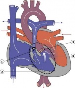
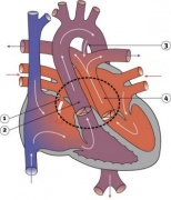
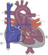
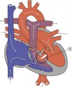
Introduction

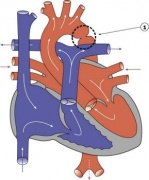
- 5-8% of Congenital Heart Disease
- Aortic constriction.
- Treatment aims at maintaining the ductus arteriosus via prostaglandins and by surgical intervention.
- Recent review[1] suggests it may:
- affect the aortic arch in a highly variable manner
- be associated with a host of other left sided heart lesions
- represent a wider vasculopathy within the pre-coarctation arterial tree
- Links: Search PubMed | PMID 21947983 | 2011 Review PDF
Some Recent Findings
|
| More recent papers |
|---|
 This table allows an automated computer search of the external PubMed database using the listed "Search term" text link.
More? References | Discussion Page | Journal Searches | 2019 References | 2020 References Search term: Coarctation of the Aorta |
| Older papers |
|---|
| These papers originally appeared in the Some Recent Findings table, but as that list grew in length have now been shuffled down to this collapsible table.
See also the Discussion Page for other references listed by year and References on this current page. |
Sex Ratios
| Male preponderance | Female preponderance |
|---|---|
|
|
| Cardiac defects | |
|
|
| Table data[3] Links: abnormal development | cardiovascular abnormalities | USA | Male | Female | cleft lip and palate | |
Ultrasound
Echocardiography uses standard two-dimensional, three-dimensional, and Doppler ultrasound to create images of the heart.
| Echocardiogram Coarctation of the Aorta[4] | |
|---|---|

| |
| A Two-dimensional transthoracic echocardiogram image obtained from the suprasternal notch in an 11-day-old infant demonstrating discrete coarctation (arrow). | B Colour Doppler of the same image with aliasing of flow at the site of coarctation (arrow). |
References
- ↑ Kenny D & Hijazi ZM. (2011). Coarctation of the aorta: from fetal life to adulthood. Cardiol J , 18, 487-95. PMID: 21947983
- ↑ Erben Y, Oderich GS, Verhagen HJM, Witsenburg M, van den Hoven AT, Debus ES, Kölbel T, Arko FR, Torsello GB, Torsello GF, Lawrence PF, Harlander-Locke MP, Bacharach JM, Jordan WD, Eskandari MK & Hagler DJ. (2019). Multicenter experience with endovascular treatment of aortic coarctation in adults. J. Vasc. Surg. , 69, 671-679.e1. PMID: 30528403 DOI.
- ↑ Michalski AM, Richardson SD, Browne ML, Carmichael SL, Canfield MA, VanZutphen AR, Anderka MT, Marshall EG & Druschel CM. (2015). Sex ratios among infants with birth defects, National Birth Defects Prevention Study, 1997-2009. Am. J. Med. Genet. A , 167A, 1071-81. PMID: 25711982 DOI.
- ↑ Torok RD, Campbell MJ, Fleming GA & Hill KD. (2015). Coarctation of the aorta: Management from infancy to adulthood. World J Cardiol , 7, 765-75. PMID: 26635924 DOI.
Reviews
Articles
Search Pubmed
Search Pubmed: Coarctation of the Aorta
Search OMIM: Coarctation of the Aorta
External Links
External Links Notice - The dynamic nature of the internet may mean that some of these listed links may no longer function. If the link no longer works search the web with the link text or name. Links to any external commercial sites are provided for information purposes only and should never be considered an endorsement. UNSW Embryology is provided as an educational resource with no clinical information or commercial affiliation.
- MedlinePlus Patent Ductus Arteriosus
- OMIM 607411 Patent Ductus Arteriosus
Glossary Links
- Glossary: A | B | C | D | E | F | G | H | I | J | K | L | M | N | O | P | Q | R | S | T | U | V | W | X | Y | Z | Numbers | Symbols | Term Link
Cite this page: Hill, M.A. (2024, April 19) Embryology Cardiovascular System - Coarctation of the Aorta. Retrieved from https://embryology.med.unsw.edu.au/embryology/index.php/Cardiovascular_System_-_Coarctation_of_the_Aorta
- © Dr Mark Hill 2024, UNSW Embryology ISBN: 978 0 7334 2609 4 - UNSW CRICOS Provider Code No. 00098G