Cardiovascular System - Blood Vessel Development: Difference between revisions
mNo edit summary |
mNo edit summary |
||
| Line 19: | Line 19: | ||
|-bgcolor="F5FAFF" | |-bgcolor="F5FAFF" | ||
| | | | ||
* '''Review - The Molecular Regulation of Arteriovenous Specification and Maintenance'''<ref name="PMID25641373"><pubmed>25641373</pubmed></ref> "The formation of a hierarchical vascular network, composed of arteries, veins and capillaries, is essential for embryogenesis and is required for the production of new functional vasculature in the adult. Elucidating the molecular mechanisms that orchestrate the differentiation of vascular endothelial cells into arterial and venous cell fates is requisite for regenerative medicine, as the directed formation of perfused vessels is desirable in a myriad of pathological settings, such as in diabetes and following myocardial infarction. Additionally, this knowledge will enhance our understanding and treatment of vascular anomalies, such as arteriovenous malformations (AVMs). From studies in vertebrate model organisms, such as mouse, zebrafish and chick, a number of key signaling pathways have been elucidated that are required for the establishment and maintenance of arterial and venous fates. These include the Hedgehog, Vascular Endothelial Growth Factor (VEGF), Transforming Growth Factor-β (TGF-β), Wnt and Notch signaling pathways. In addition, a variety of transcription factor families acting downstream of-or in concert with-these signaling networks play vital roles in arteriovenous (AV) specification. These include Notch and Notch-regulated transcription factors (e.g. HEY and HES), SOX factors, Forkhead factors, β-Catenin, ETS factors and COUP-TFII. It is becoming apparent that AV specification is a highly coordinated process that involves the intersection and carefully orchestrated activity of multiple signaling cascades and transcriptional networks. This review will summarize the molecular mechanisms that are involved in the acquisition and maintenance of AV fate, and will highlight some of the limitations in our current knowledge of the molecular machinery that directs AV morphogenesis. | |||
* '''Specification of arterial, venous, and lymphatic endothelial cells during embryonic development''' <ref name="PMID20238301"><pubmed>20238301</pubmed>| [http://www.ncbi.nlm.nih.gov/pmc/articles/PMC2899674 PMC2899674]</ref> "The groundbreaking discovery about arterial and venous expression of ephrinB2 and EphB4, respectively, in early embryonic development has led to a new paradigm for vascular research, providing compelling evidence that arterial and venous endothelial cells are established by genetic mechanisms before circulation begins. For arterial specification, vascular endothelial growth factor (VEGF) induces expression of Notch signaling genes, including Notch1 and its ligand, Delta-like 4 (Dll4), and Foxc1 and Foxc2 transcription factors directly regulate Dll4 expression. Upon activation of Notch signaling, the Notch downstream genes, Hey1/2 in mice or gridlock in zebrafish, further promote arterial differentiation. On the other hand, the orphan nuclear receptor COUP-TFII is a determinant factor for venous specification by inhibiting expression of arterial specific genes, including Nrp1 and Notch. After arterial and venous endothelial cells differentiate, a subpopulation of venous endothelial cells is thought to become competent to acquire lymphatic endothelial cell fate by progressively expressing the transcription factors Sox18 and Prox1 to differentiate into lymphatic endothelial cells." | * '''Specification of arterial, venous, and lymphatic endothelial cells during embryonic development''' <ref name="PMID20238301"><pubmed>20238301</pubmed>| [http://www.ncbi.nlm.nih.gov/pmc/articles/PMC2899674 PMC2899674]</ref> "The groundbreaking discovery about arterial and venous expression of ephrinB2 and EphB4, respectively, in early embryonic development has led to a new paradigm for vascular research, providing compelling evidence that arterial and venous endothelial cells are established by genetic mechanisms before circulation begins. For arterial specification, vascular endothelial growth factor (VEGF) induces expression of Notch signaling genes, including Notch1 and its ligand, Delta-like 4 (Dll4), and Foxc1 and Foxc2 transcription factors directly regulate Dll4 expression. Upon activation of Notch signaling, the Notch downstream genes, Hey1/2 in mice or gridlock in zebrafish, further promote arterial differentiation. On the other hand, the orphan nuclear receptor COUP-TFII is a determinant factor for venous specification by inhibiting expression of arterial specific genes, including Nrp1 and Notch. After arterial and venous endothelial cells differentiate, a subpopulation of venous endothelial cells is thought to become competent to acquire lymphatic endothelial cell fate by progressively expressing the transcription factors Sox18 and Prox1 to differentiate into lymphatic endothelial cells." | ||
* '''Developmental origin of smooth muscle cells in the descending aorta in mice'''<ref><pubmed>18417617</pubmed></ref> "Aortic smooth muscle cells (SMCs) have been proposed to derive from lateral plate mesoderm. ....(these results) suggested that all SMCs in the adult descending aorta derive from the somites, whereas no contribution was recorded from lateral plate mesoderm." | * '''Developmental origin of smooth muscle cells in the descending aorta in mice'''<ref><pubmed>18417617</pubmed></ref> "Aortic smooth muscle cells (SMCs) have been proposed to derive from lateral plate mesoderm. ....(these results) suggested that all SMCs in the adult descending aorta derive from the somites, whereas no contribution was recorded from lateral plate mesoderm." | ||
| Line 236: | Line 237: | ||
<references/> | <references/> | ||
===Reviews=== | |||
<pubmed></pubmed> | |||
<pubmed></pubmed> | |||
<pubmed>25641373</pubmed> | |||
===Articles=== | |||
<pubmed></pubmed> | |||
<pubmed></pubmed> | |||
<pubmed>10948449</pubmed> | <pubmed>10948449</pubmed> | ||
<pubmed>12406884</pubmed> | <pubmed>12406884</pubmed> | ||
Revision as of 17:55, 4 February 2015
| Embryology - 19 Apr 2024 |
|---|
| Google Translate - select your language from the list shown below (this will open a new external page) |
|
العربية | català | 中文 | 中國傳統的 | français | Deutsche | עִברִית | हिंदी | bahasa Indonesia | italiano | 日本語 | 한국어 | မြန်မာ | Pilipino | Polskie | português | ਪੰਜਾਬੀ ਦੇ | Română | русский | Español | Swahili | Svensk | ไทย | Türkçe | اردو | ייִדיש | Tiếng Việt These external translations are automated and may not be accurate. (More? About Translations) |
Introduction

Blood develops initially within the core of "blood islands" in the mesoderm. During development, there follows a series of "relocations" of the stem cells to different organs within the embryo. In the adult, these stem cells are located in the bone marrow. At the time when blood first forms, there are no bones!
Angioblasts initially form small cell clusters (blood islands) within the embryonic and extraembryonic mesoderm. These blood islands extend and fuse together making a primordial vascular network. Within these islands the peripheral cells form endothelial cells while the core cells form blood cells (haemocytoblasts).
Recent work has shown that the formation of the initial endothelial tube is by a process of coalescence of cellular vacuoles within the developing endothelial cells, which fuse together without cytoplasmic mixing to form the blood vessel lumen.
See also the related pages Placental Villi Blood Vessels and Coronary Circulation Development.
Some Recent Findings

|
| More recent papers |
|---|
 This table allows an automated computer search of the external PubMed database using the listed "Search term" text link.
More? References | Discussion Page | Journal Searches | 2019 References | 2020 References Search term: Blood Vessel Embryology <pubmed limit=5>Blood Vessel Embryology</pubmed> |
Endothelial Progenitors
Recent work has shown that the formation of the initial endothelial tube is by a process of coalescence of cellular vacuoles within the developing endothelial cells, which fuse together without cytoplasmic mixing to form the blood vessel lumen. [5]
Endothelial Tube Formation

Vessel Specification

The following data is from a recent review.[2]
Arterial Specification
| Factor | Function |
| Shh | Loss of Shh results in lack of arterial identity in zebrafish. Shh acts upstream of VEGF. |
| VEGF | VEGF acts downstream of Shh signaling to activate Notch via the PLCγ/ERK pathway in zebrafish. Mutant mice expressing only VEGF188 lack arterial differentiation. |
| Nrp1 | Null mice display impaired arterial differentiation. Nrp1 is involved in a positive feedback loop of VEGF signaling. |
| Notch | Notch acts downstream of Shh and VEGF signaling in zebrafish. Notch1; Notch4 mutant mice have abnormal vascular development. |
| Dll4 | Null mice lack arterial specification. |
| Dll1 | Null mice fail to maintain arterial identity. |
| Hey1/2 (Grl) | Null mice lack arterial specification. Lack of grl in zebrafish results in loss of arterial specification. |
| Foxc1/c2 | Foxc1; Foxc2 mutant mice lack arterial specification. Foxc1 and Foxc2 directly regulate Dll4 and Hey2 expression. Foxc1 and Foxc2 are also involved in lymphatic vessel development. |
| Sox7/18 | Lack of Sox7/18 results in loss of arterial identity in zebrafish. |
| Snrk-1 | Snrk-1 acts downstream or parallel to Notch signaling in zebrafish. |
| Dep1 | Dep1 acts upstream of PI3K in arterial specification in zebrafish. |
| Crlr | Shh regulates VEGF activity by controlling crlr expression in zebrafish. |
| EphrinB2 | Null mice lack boundaries between arteries and veins. EphrinB2 is involved in lymphatic vascular remodeling and maturation. |
Venous Specification
| Factor | Function |
| COUP-TFII | COUP-TFII suppresses arterial cell fate by inhibiting Nrp1 and Notch. COUP-TFII also interacts with Prox1 to regulate lymphatic gene expression. |
| EphB4 | Null mice lack boundaries between arteries and veins. |
Lymphatic Specification
| Factor | Function |
| Sox18 | Null mice fail to specify lymphatic endothelial cells. Sox18 induces Prox1 expression. |
| Prox1 | Prox1 induces lymphatic markers and maintains lymphatic cell identity. |
Vascular Endothelial Growth Factor
Growing blood vessels follow a gradient generated by tagret tissues/regions of Vascular Endothelial Growth Factor (VEGF) to establish a vascular bed. Recent findings suggest that Notch signaling acts as an inhibitor for this system, preventing sprouting of blood vessels.
Notch is a transmembrane receptor protein involved in regulating cell differentiation in many developing systems.
Links: OMIM - VEGFA | OMIM - Notch
Regulators of Growth
The following data is from a review article on ovary vascular development.[6]
Stimulators of Angiogenisis
|
Inhibitors of Angiogenisis
|
Histology
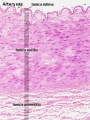
Vein Light Microscopy

The entire developing and adult cardiovascular system (blood vessels and heart) is lined by a simple squamous epithelium. (Stain - Haematoxylin Eosin)
Capillaries
Electron Micrographs

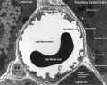
Capillary 1 large labeled
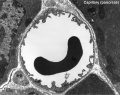
Capillary 1 large unlabelled

Capillary 1 small labeled

Capillary 1 small unlabelled
![endothelium detail[7]](/embryology/images/thumb/6/6a/Blood_capillary_EM_01.jpg/120px-Blood_capillary_EM_01.jpg)
endothelium detail[7]
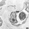
Containing white blood cell


![endothelium detail[7]](/embryology/index.php?title=File:Blood_capillary_EM_01.jpg)

Arteries
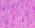
Artery overview
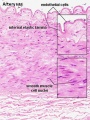
Artery detail
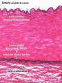
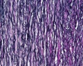
Artery elastin
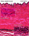
Artery elastin detail
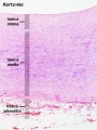
Aorta overview
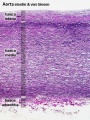
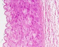
Aorta elastin
Artery overview

Artery elastin

Artery tunica media elastin

Artery elastin detail
Aorta overview
Aorta elastin


Cardiac Blood Vessels
Earliest vessels in the heart wall develop subepicardially (beneath the outside surface of the heart) near the apex at Carnegie stage 15, which then extends centripetally and at stage 17 coronary arterial stems communicate with the aortic lumen.[8]
References
- ↑ <pubmed>25641373</pubmed>
- ↑ 2.0 2.1 <pubmed>20238301</pubmed>| PMC2899674
- ↑ <pubmed>18417617</pubmed>
- ↑ <pubmed>18245384</pubmed>
- ↑ <pubmed>11827993</pubmed>
- ↑ <pubmed>11141338</pubmed>
- ↑ <pubmed>21702933</pubmed>| PMC3141733 | BMC Cell Biol.
- ↑ <pubmed>8915616</pubmed>
Reviews
<pubmed></pubmed> <pubmed></pubmed> <pubmed>25641373</pubmed>
Articles
<pubmed></pubmed> <pubmed></pubmed> <pubmed>10948449</pubmed> <pubmed>12406884</pubmed>
Search Pubmed
Click on the listed keywords below (used to search the external database) the most current references on Medline will be displayed.
Search Pubmed: Blood Vessel Development | Blood Vessel embryology | Blood Vessel smooth muscle Development | Blood Vessel smooth muscle Development
Terms
- angioblasts stem cells in blood islands generating endothelial cells
- angiogenesis the formation of blood vessels also called vasculogenesis in the embryo
- blood islands earliest sites of blood vessel and blood cell formation, seen mainly on yolk sac chorion
- vascular endothelial growth factor (VEGF) protein growth factor family that stimulates blood vessel growth, a similar factor can be found in the placenta (PIGF).
External Links
External Links Notice - The dynamic nature of the internet may mean that some of these listed links may no longer function. If the link no longer works search the web with the link text or name. Links to any external commercial sites are provided for information purposes only and should never be considered an endorsement. UNSW Embryology is provided as an educational resource with no clinical information or commercial affiliation.
Glossary Links
- Glossary: A | B | C | D | E | F | G | H | I | J | K | L | M | N | O | P | Q | R | S | T | U | V | W | X | Y | Z | Numbers | Symbols | Term Link
Cite this page: Hill, M.A. (2024, April 19) Embryology Cardiovascular System - Blood Vessel Development. Retrieved from https://embryology.med.unsw.edu.au/embryology/index.php/Cardiovascular_System_-_Blood_Vessel_Development
- © Dr Mark Hill 2024, UNSW Embryology ISBN: 978 0 7334 2609 4 - UNSW CRICOS Provider Code No. 00098G