Cardiovascular System - Abnormalities: Difference between revisions
mNo edit summary |
mNo edit summary |
||
| Line 21: | Line 21: | ||
* '''Spontaneous Closure of Muscular Trabecular Ventricular Septal Defect: Comparison of Defect Positions''' <ref><pubmed>21517965</pubmed></ref> "We performed a historical cohort study for which 150 patients <3 months of age (median age, 9 days) diagnosed as having a muscular trabecular VSD were selected. ...We infer that midventricular muscular trabecular VSD tends to close spontaneously earlier and more frequently than either anterior or apical muscular trabecular VSD." | * '''Spontaneous Closure of Muscular Trabecular Ventricular Septal Defect: Comparison of Defect Positions''' <ref><pubmed>21517965</pubmed></ref> "We performed a historical cohort study for which 150 patients <3 months of age (median age, 9 days) diagnosed as having a muscular trabecular VSD were selected. ...We infer that midventricular muscular trabecular VSD tends to close spontaneously earlier and more frequently than either anterior or apical muscular trabecular VSD." | ||
|} | |} | ||
{| | |||
|-bgcolor="F5FAFF" | |||
| | |||
* <ref name=PMID><pubmed></pubmed> | |||
|} | |||
{| class="wikitable mw-collapsible mw-collapsed" | |||
! More recent papers | |||
|- | |||
| [[File:Mark_Hill.jpg|90px|left]] {{Most_Recent_Refs}} | |||
Search term: [http://www.ncbi.nlm.nih.gov/pubmed/?term=Cardiovascular+Abnormality ''Cardiovascular Abnormality''] | |||
<pubmed limit=5>Cardiovascular Abnormality</pubmed> | |||
|} | |||
==Heart Abnormalities== | ==Heart Abnormalities== | ||
===Ventricular Septal Defect=== | ===Ventricular Septal Defect=== | ||
Revision as of 06:40, 6 April 2016
| Embryology - 19 Apr 2024 |
|---|
| Google Translate - select your language from the list shown below (this will open a new external page) |
|
العربية | català | 中文 | 中國傳統的 | français | Deutsche | עִברִית | हिंदी | bahasa Indonesia | italiano | 日本語 | 한국어 | မြန်မာ | Pilipino | Polskie | português | ਪੰਜਾਬੀ ਦੇ | Română | русский | Español | Swahili | Svensk | ไทย | Türkçe | اردو | ייִדיש | Tiếng Việt These external translations are automated and may not be accurate. (More? About Translations) |
Introduction
Heart defects and preterm birth are the most common causes of neonatal and infant death. The long-term development of the heart combined with extensive remodelling and post-natal changes in circulation lead to an abundance of abnormalities associated with this system.
A UK study literature showed that preterm infants have more than twice as many cardiovascular malformations (5.1 / 1000 term infants and 12.5 / 1000 preterm infants) as do infants born at term and that 16% of all infants with cardiovascular malformations are preterm. (0.4% of live births occur at greater than 28 weeks of gestation, 0.9% at 28 to 31 weeks, and 6% at 32 to 36 weeks. Overall, 7.3% of live-born infants are preterm)[1]
"Baltimore-Washington Infant Study data on live-born cases and controls (1981-1989) was reanalyzed for potential environmental and genetic risk-factor associations in complete atrioventricular septal defects AVSD (n = 213), with separate comparisons to the atrial (n = 75) and the ventricular (n = 32) forms of partial AVSD. ...Maternal diabetes constituted a potentially preventable risk factor for the most severe, complete form of AVSD." [2]
In addition, there are in several congenital abnormalities that exist in adults (bicuspid aortic valve, mitral valve prolapse, and partial anomalous pulmonary venous connection) which may not be clinically recognized.
| Heart Abnormal: Tutorial Abnormalities | atrial septal defects | double outlet right ventricle | hypoplastic left heart | patent ductus arteriosus | transposition of the great vessels | Tetralogy of Fallot | ventricular septal defects | coarctation of the aorta | Category ASD | Category PDA | Category ToF | Category VSD | ICD10 - Cardiovascular | ICD11 |
Some Recent Findings
|
|
Patent Ductus Arteriosus
|
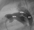
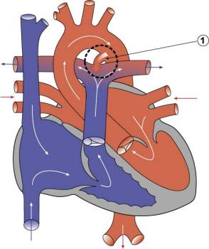
Patent ductus arteriosus (PDA), or Patent arterial duct (PAD), or common truncus, occurs commonly in preterm infants, and at approximately 1 in 2000 full term infants and more common in females (to male ratio is 2:1). Can also be associated with specific genetic defects, trisomy 21 and trisomy 18, and the Rubinstein-Taybi and CHARGE syndromes. The opening is asymptomatic when the duct is small and can close spontaneously (by day three in 60% of normal term neonates), the remainder are ligated simply and with little risk, with transcatheter closure of the duct generally indicated in older children. The operation is always recommended even in the absence of cardiac failure and can often be deferred until early childhood.
ICD-10 Q25.0 Patent ductus arteriosus Patent ductus Botallo Persistent ductus arteriosus
|
Tetralogy of Fallot
|
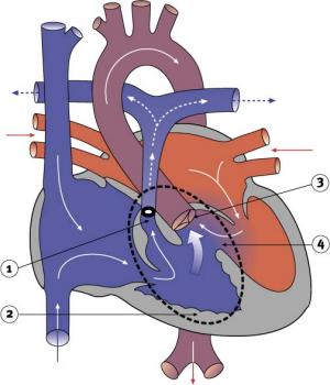
Named after Etienne-Louis Arthur Fallot (1888) who described it as "la maladie blue" and is a common developmental cardiac defect. The syndrome consists of a number of a number of cardiac defects possibly stemming from abnormal neural crest migration.
ICD-10 Q21.3 Tetralogy of Fallot Ventricular septal defect with pulmonary stenosis or atresia, dextroposition of aorta and hypertrophy of right ventricle.
|
Hypoplastic Left Heart
|
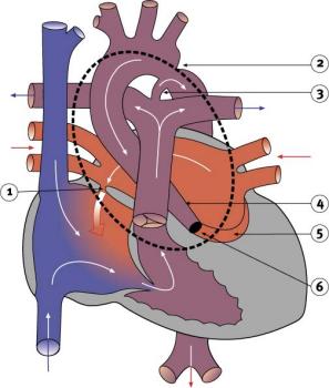
Characterized by hypoplasia (underdevelopment or absence) of the left ventricle obstructive valvular and vascular lesion of the left side of the heart.
ICD-10 Q23.4 Hypoplastic left heart syndrome Atresia, or marked hypoplasia of aortic orifice or valve, with hypoplasia of ascending aorta and defective develop-ment of left ventricle (with mitral valve stenosis or atresia).
|
Double Outlet Right Ventricle
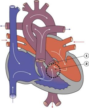
|
De-oxygenated blood enters the aorta from the right ventricle and is returned to the body. ICD-10 Q20.1 Double outlet right ventricle Taussig-Bing syndrome
|
Tricuspid Atresia
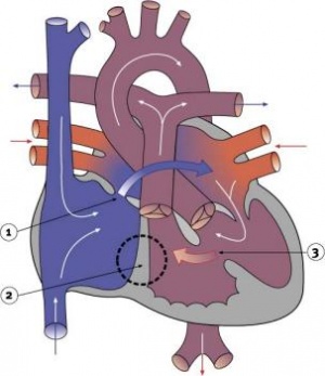
|
Blood is shunted through an atrial septal defect to the left atrium and through the ventricular septal defect to the pulmonary artery. The shaded arrows indicate mixing of the blood.
ICD-10 Q22.4 Congenital tricuspid stenosis Tricuspid atresia Fontan Procedure: a surgical procedure developed by Fontan and Baudet (1971) to restore a circulation in patients with tricuspid atresia.
|
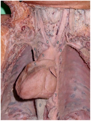
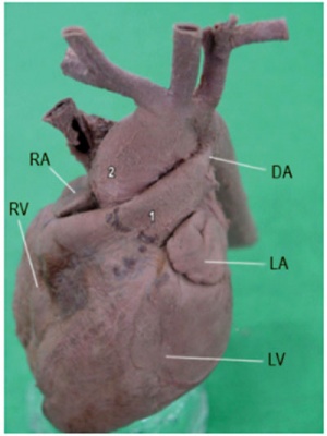
Dextrocardia
|
|
| Dextrocardia anatomical heart position[4] | Dextrocardia (postnatal 1 year old)[4] |
Initial malrotation of the heart tube bending left instead of right. Results in heart and greater vessels reversed. Can also occur with situs invertus, where viscera are transposed LR.
Anatomical left-right normal asymmetry is called situs solitus. The alternative heterotaxy can be either randomization (situs ambiguus) or a complete reversal (situs inversus) of normal organ position.
Abnormalities of Conducting System
Also variously called the cardiac conduction system (CCS), cardiac pacemaking and conduction system (CPCS), or atrioventricular conduction system (AVCS). Recently animal models (CCS-lacZ transgenic mouse) have helped identify key processes in the development of this specialized conduction system.
"Known arrhythmogenic areas including Bachmann's bundle, the pulmonary veins, and sinus venosus derived internodal structures, demonstrate lacZ expression." (Jongbloed et al, 2004)
Long QT Syndrome
Congenital long QT syndrome (LQTS) is a group of rare genetic disorders with prolonged ventricular repolarization and a risk of ventricular tachyarrhythmias. Cause is mutations in genes encoding either cardiac ion channels or channel interacting proteins.
Search NCBI Bookshelf: Congenital long-QT syndrome
- Links: Search PubMed
Heart Vessel Abnormalities
Transposition of the Great Vessels
|
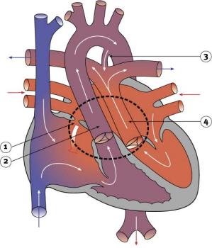
Characterized by aorta arising from right ventricle and pulmonary artery from the left ventricle and often associated with other cardiac abnormalities (e.g. ventricular septal defect).
|
Coarctation of the Aorta
|
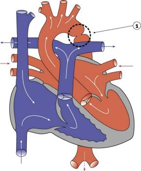
|
Interrupted Aortic Arch
- LInks: Search PubMed
Pulmonary Atresia
|
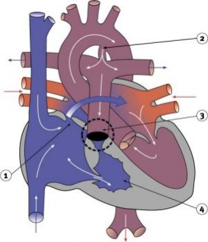
|
Total Anomalous Pulmonary Venous Connection
|
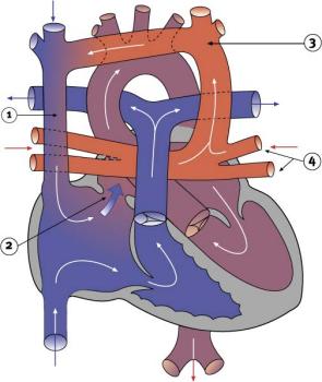
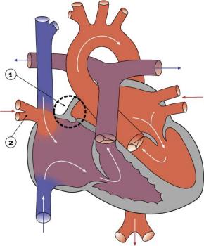
Total Anomalous Pulmonary Venous Connection (TAPVC) or Total Anomalous Pulmonary Venous Return (TAPVR) occurs when pulmonary veins connect to the right atrium (RA) and not the left atrium (LA). This abnormal connection returns oxygenated pulmonary blood from the lungs back to the right atrium or a vein flowing into the right atrium.
|
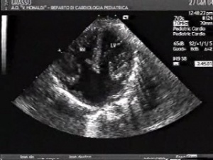
Complete Atrioventricular Canal
|
|
Partial Anomalous Pulmonary Venous Drainage
|
|
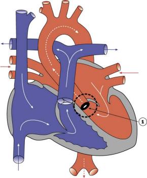
Aortic Stenosis
|
|
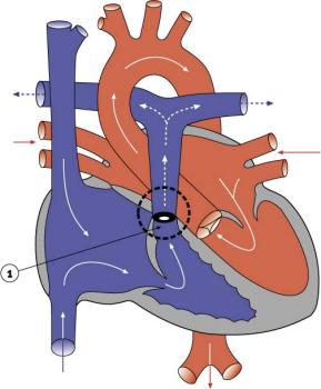
Pulmonary Stenosis
|
ICD-10 Q25.6 Stenosis of pulmonary artery Supravalvular pulmonary stenosis
|
Blood Disorders
Sickle Cell Anemia
| People who have this form of sickle cell disease inherit two sickle cell genes (“S”), one from each parent. This is commonly called “sickle cell anemia”, and is usually the most severe form of the disease. The name comes from the "sickle" shape of the RBC compared to the normal "donut" shape.
|
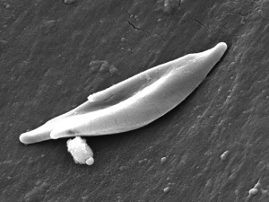
Sickle cell RBC (Image CDC) |
Thalassemia
Thalassemia is a group of inherited (genetic) blood disorders most frequently in people of Italian, Greek, Middle Eastern, Southern Asian and African Ancestry. The most severe form of alpha thalassemia, affecting mainly people of Southeast Asian, Chinese and Filipino ancestry, results in fetal or newborn death.
The two main types of thalassemia are called "alpha" and "beta," depending on which part of an oxygen-carrying protein in the red blood cells is lacking. Both types of thalassemia are inherited in the same manner. A child who inherits one mutated gene is a carrier, which is sometimes called "thalassemia trait." Most carriers lead completely normal, healthy lives.
International Classification of Diseases
The International Classification of Diseases (ICD) World Health Organization's classification used worldwide as the standard diagnostic tool for epidemiology, health management and clinical purposes. This includes the analysis of the general health situation of population groups. It is used to monitor the incidence and prevalence of diseases and other health problems. Within this classification "congenital malformations, deformations and chromosomal abnormalities" are (Q00-Q99) but excludes "inborn errors of metabolism" (E70-E90).
Congenital malformations of the circulatory system (Q20-Q28)
Q20 Congenital malformations of cardiac chambers and connections
Excl.: dextrocardia with situs inversus (Q89.3) mirror-image atrial arrangement with situs inversus (Q89.3)
- Q20.0 Common arterial trunk Persistent truncus arteriosus
- Q20.1 Double outlet right ventricle Taussig-Bing syndrome
- Q20.2 Double outlet left ventricle
- Q20.3 Discordant ventriculoarterial connection Dextrotransposition of aorta Transposition of great vessels (complete)
- Q20.4 Double inlet ventricle Common ventricle Cor triloculare biatriatum Single ventricle
- Q20.5 Discordant atrioventricular connection Corrected transposition Laevotransposition Ventricular inversion
- Q20.6 Isomerism of atrial appendages Isomerism of atrial appendages with asplenia or polysplenia
- Q20.8 Other congenital malformations of cardiac chambers and connections
- Q20.9 Congenital malformation of cardiac chambers and connections, unspecified
Q21 Congenital malformations of cardiac septa
Excl.: acquired cardiac septal defect (I51.0)
- Q21.0 Ventricular septal defect
- Q21.1 Atrial septal defect Coronary sinus defect Patent or persistent: foramen ovale ostium secundum defect (type II) Sinus venosus defect
- Q21.2 Atrioventricular septal defect Common atrioventricular canal Endocardial cushion defect Ostium primum atrial septal defect (type I)
- Q21.3 Tetralogy of Fallot Ventricular septal defect with pulmonary stenosis or atresia, dextroposition of aorta and hypertrophy of right ventricle.
- Q21.4 Aortopulmonary septal defect Aortic septal defect Aortopulmonary window
- Q21.8 Other congenital malformations of cardiac septa Eisenmenger's defect Pentalogy of Fallot Excl.: Eisenmenger's complex (I27.8) syndrome (I27.8)
- Q21.9 Congenital malformation of cardiac septum, unspecified Septal (heart) defect NOS
Q22 Congenital malformations of pulmonary and tricuspid valves
- Q22.0 Pulmonary valve atresia
- Q22.1 Congenital pulmonary valve stenosis
- Q22.2 Congenital pulmonary valve insufficiency Congenital pulmonary valve regurgitation
- Q22.3 Other congenital malformations of pulmonary valve Congenital malformation of pulmonary valve NOS
- Q22.4 Congenital tricuspid stenosis Tricuspid atresia
- Q22.5 Ebstein's anomaly
- Q22.6 Hypoplastic right heart syndrome
- Q22.8 Other congenital malformations of tricuspid valve
- Q22.9 Congenital malformation of tricuspid valve, unspecified
Q23 Congenital malformations of aortic and mitral valves
- Q23.0 Congenital stenosis of aortic valve Congenital aortic: atresia stenosis Excl.: congenital subaortic stenosis (Q24.4) that in hypoplastic left heart syndrome (Q23.4)
- Q23.1 Congenital insufficiency of aortic valve Bicuspid aortic valve Congenital aortic insufficiency
- Q23.2 Congenital mitral stenosis Congenital mitral atresia
- Q23.3 Congenital mitral insufficiency
- Q23.4 Hypoplastic left heart syndrome Atresia, or marked hypoplasia of aortic orifice or valve, with hypoplasia of ascending aorta and defective develop-ment of left ventricle (with mitral valve stenosis or atresia).
- Q23.8 Other congenital malformations of aortic and mitral valves
- Q23.9 Congenital malformation of aortic and mitral valves, unspecified
Q24 Other congenital malformations of heart
Excl.: endocardial fibroelastosis (I42.4)
- Q24.0 Dextrocardia Excl.: dextrocardia with situs inversus (Q89.3) isomerism of atrial appendages (with asplenia or polysplenia) (Q20.6) mirror-image atrial arrangement with situs inversus (Q89.3)
- Q24.1 Laevocardia Location of heart in left hemithorax with apex pointing to the left, but with situs inversus of other viscera and defects of the heart, or corrected transposition of great vessels.
- Q24.2 Cor triatriatum
- Q24.3 Pulmonary infundibular stenosis
- Q24.4 Congenital subaortic stenosis
- Q24.5 Malformation of coronary vessels Congenital coronary (artery) aneurysm
- Q24.6 Congenital heart block
- Q24.8 Other specified congenital malformations of heart Congenital: diverticulum of left ventricle malformation of: myocardium pericardium Malposition of heart Uhl's disease
- Q24.9 Congenital malformation of heart, unspecified Congenital: anomaly disease NOS of heart
Q25 Congenital malformations of great arteries
- Q25.0 Patent ductus arteriosus Patent ductus Botallo Persistent ductus arteriosus
- Q25.1 Coarctation of aorta Coarctation of aorta (preductal)(postductal)
- Q25.2 Atresia of aorta
- Q25.3 Stenosis of aorta Supravalvular aortic stenosis Excl.: congenital aortic stenosis (Q23.0)
- Q25.4 Other congenital malformations of aorta Absence Aplasia Congenital: aneurysm dilatation of aorta Aneurysm of sinus of Valsalva (ruptured) Double aortic arch [vascular ring of aorta] Hypoplasia of aorta Persistent: convolutions of aortic arch right aortic arch Excl.: hypoplasia of aorta in hypoplastic left heart syndrome (Q23.4)
- Q25.5 Atresia of pulmonary artery
- Q25.6 Stenosis of pulmonary artery Supravalvular pulmonary stenosis
- Q25.7 Other congenital malformations of pulmonary artery Aberrant pulmonary artery Agenesis Aneurysm, congenital Anomaly Hypoplasia of pulmonary artery Pulmonary arteriovenous aneurysm
- Q25.8 Other congenital malformations of great arteries
- Q25.9 Congenital malformation of great arteries, unspecified
Q26 Congenital malformations of great veins
- Q26.0 Congenital stenosis of vena cava Congenital stenosis of vena cava (inferior)(superior)
- Q26.1 Persistent left superior vena cava
- Q26.2 Total anomalous pulmonary venous connection
- Q26.3 Partial anomalous pulmonary venous connection
- Q26.4 Anomalous pulmonary venous connection, unspecified
- Q26.5 Anomalous portal venous connection
- Q26.6 Portal vein-hepatic artery fistula
- Q26.8 Other congenital malformations of great veins Absence of vena cava (inferior)(superior) Azygos continuation of inferior vena cava Persistent left posterior cardinal vein Scimitar syndrome
- Q26.9 Congenital malformation of great vein, unspecified Anomaly of vena cava (inferior)(superior) NOS
Q27 Other congenital malformations of peripheral vascular system
Excl.: anomalies of: cerebral and precerebral vessels (Q28.0-Q28.3) coronary vessels (Q24.5) pulmonary artery (Q25.5-Q25.7) congenital retinal aneurysm (Q14.1) haemangioma and lymphangioma (D18.-)
- Q27.0 Congenital absence and hypoplasia of umbilical artery Single umbilical artery
- Q27.1 Congenital renal artery stenosis
- Q27.2 Other congenital malformations of renal artery Congenital malformation of renal artery NOS Multiple renal arteries
- Q27.3 Peripheral arteriovenous malformation Arteriovenous aneurysm Excl.: ac
- Quired arteriovenous aneurysm (I77.0)
- Q27.4 Congenital phlebectasia
- Q27.8 Other specified congenital malformations of peripheral vascular system Aberrant subclavian artery Absence Atresia of artery or vein NEC Congenital: aneurysm (peripheral) stricture, artery varix
- Q27.9 Congenital malformation of peripheral vascular system, unspecified Anomaly of artery or vein NOS
Q28 Other congenital malformations of circulatory system
Excl.: congenital aneurysm: NOS (Q27.8) coronary (Q24.5) peripheral (Q27.8) pulmonary (Q25.7) retinal (Q14.1) ruptured: cerebral arteriovenous malformation (I60.8) malformation of precerebral vessels (I72.-)
- Q28.0 Arteriovenous malformation of precerebral vessels Congenital arteriovenous precerebral aneurysm (nonruptured)
- Q28.1 Other malformations of precerebral vessels Congenital: malformation of precerebral vessels NOS precerebral aneurysm (nonruptured)
- Q28.2 Arteriovenous malformation of cerebral vessels Arteriovenous malformation of brain NOS Congenital arteriovenous cerebral aneurysm (nonruptured)
- Q28.3 Other malformations of cerebral vessels Congenital: cerebral aneurysm (nonruptured) malformation of cerebral vessels NOS
- Q28.8 Other specified congenital malformations of circulatory system Congenital aneurysm, specified site NEC
- Q28.9 Congenital malformation of circulatory system, unspecified
Chapter XVI Certain conditions originating in the perinatal period (P00-P96)
P29 Cardiovascular disorders originating in the perinatal period
Excl.: congenital malformations of the circulatory system (Q20-Q28)
- P29.0 Neonatal cardiac failure
- P29.1 Neonatal cardiac dysrhythmia
- P29.2 Neonatal hypertension
- P29.3 Persistent fetal circulation Delayed closure of ductus arteriosus Pulmonary hypertension of newborn (persistent)
- P29.4 Transient myocardial ischaemia of newborn
- P29.8 Other cardiovascular disorders originating in the perinatal period
- P29.9 Cardiovascular disorder originating in the perinatal period, unspecified
Statistics
Australia
Data shown as a percentage of all major abnormalities based upon published statistics using the same groupings as Congenital Malformations Australia 1981-1992 P. Lancaster and E. Pedisich ISSN 1321-8352.
Belgium
Congenital heart disease in 111 225 births in Belgium 2008 study[6]:
- 921 children with congenital heart disease (birth prevalence of 8.3 per 1000).
- 33% ventricular septal defects most frequently occurring condition
- 18% ostium secundum atrial septal defects
- 10% pulmonary valve abnormalities
- 39% had either cardiosurgical operation or catheter intervention.
- 4% of the children died (higher in univentricular physiology, pulmonary atresia with VSD, left ventricle outflow obstruction and tetralogy of Fallot).
South America
Paediatric and congenital heart disease in South America: an overview.[7]
- Ventricular septal defects - Surgical mortality with repair in infancy, mainly in the muscular septum, probably slightly higher in South America than in North America and Europe.
- Secundum atrial septal defects - trans-catheter closure is the preferred method of treating most patients.
- Pulmonary valve stenosis and atresia with intact ventricular septum - balloon pulmonary valvuloplasty for PVS.
- Aortic stenosis - balloon valvuloplasty.
- Coarctation of the aorta - children balloon angioplasty, adolescents and adults a trend towards the use of bare and covered stents.
- Tetralogy of Fallot - patients with severe hypoxaemia have a modified Blalock–Taussig shunt.
USA
"Baltimore-Washington Infant Study data on live-born cases and controls (1981-1989) was reanalyzed for potential environmental and genetic risk-factor associations in complete atrioventricular septal defects AVSD (n = 213), with separate comparisons to the atrial (n = 75) and the ventricular (n = 32) forms of partial AVSD. ...Maternal diabetes constituted a potentially preventable risk factor for the most severe, complete form of AVSD." [2]
References
Articles
<pubmed>17967198</pubmed>
Search Pubmed
Search Pubmed: Cardiovascular System Abnormalities
5 Most Recent
Note - This sub-heading shows an automated computer PubMed search using the listed sub-heading term. References appear in this list based upon the date of the actual page viewing. Therefore the list of references do not reflect any editorial selection of material based on content or relevance. In comparison, references listed on the content page and discussion page (under the publication year sub-headings) do include editorial selection based upon relevance and availability. (More? Pubmed Most Recent)
Ventricular Septal Defect
<pubmed limit=5>Ventricular+Septal+Defect</pubmed>
Atrial Septal Defect
<pubmed limit=5>Atrial+Septal+Defect</pubmed>
Patent Ductus Arteriosus
<pubmed limit=5>Patent+Ductus+Arteriosus</pubmed>
Tetralogy of Fallot
<pubmed limit=5>Tetralogy+of+Fallot</pubmed>
Glossary Links
- Glossary: A | B | C | D | E | F | G | H | I | J | K | L | M | N | O | P | Q | R | S | T | U | V | W | X | Y | Z | Numbers | Symbols | Term Link
Cite this page: Hill, M.A. (2024, April 19) Embryology Cardiovascular System - Abnormalities. Retrieved from https://embryology.med.unsw.edu.au/embryology/index.php/Cardiovascular_System_-_Abnormalities
- © Dr Mark Hill 2024, UNSW Embryology ISBN: 978 0 7334 2609 4 - UNSW CRICOS Provider Code No. 00098G


