Book - Uterus And Embryo - Plates (1889)
| Embryology - 20 Apr 2024 |
|---|
| Google Translate - select your language from the list shown below (this will open a new external page) |
|
العربية | català | 中文 | 中國傳統的 | français | Deutsche | עִברִית | हिंदी | bahasa Indonesia | italiano | 日本語 | 한국어 | မြန်မာ | Pilipino | Polskie | português | ਪੰਜਾਬੀ ਦੇ | Română | русский | Español | Swahili | Svensk | ไทย | Türkçe | اردو | ייִדיש | Tiếng Việt These external translations are automated and may not be accurate. (More? About Translations) |
Minot CS. Uterus And Embryo - I. Rabbit II. Man. (1889) J Morphol. 2:
| Historic Disclaimer - information about historic embryology pages |
|---|
| Pages where the terms "Historic" (textbooks, papers, people, recommendations) appear on this site, and sections within pages where this disclaimer appears, indicate that the content and scientific understanding are specific to the time of publication. This means that while some scientific descriptions are still accurate, the terminology and interpretation of the developmental mechanisms reflect the understanding at the time of original publication and those of the preceding periods, these terms, interpretations and recommendations may not reflect our current scientific understanding. (More? Embryology History | Historic Embryology Papers) |
Explanation of Plates
Nearly all the figures were drawn by Mr. E. Stanley Abbot under my supervision. The outlines were drawn with the camera lucida, and the details added free-hand. The drawings are all accurate representations of the preparations, and though of course not photographically exact, are not diagrammatic, except in the case of a few figures expressly specified below. I owe much to Mr. Abbot's patient skill.
Reference Letters
| all, allantois.
mes, mesoderm. Cim, circular muscles. mo.cl, monster cells. conn, connective tissue. |
msth, mesothelium.
ecto, foetal ectoderm. muc, mucosa. emb, embryo. muse, muscularis. |
en, entoderm.
ob.pl, ob-placenta. endo, endothelium. o.z, outer zone of placenta ep, epithelium. |
P, periplacenta.
f, placental fissure. per.v, perivascular cells. f.v, foetal blood-vessel. sp.pl, sub-placenta. |
gl, gland ; glandular layer.
Sgl.z, subglandular zone. h.ep, hyaline epithelium. V, blood-vessel. l, leucocytes. |
vac, vacuole.
Im, longitudinal muscles. |
Plate 26
EXPLANATION OF PLATE XXVI.
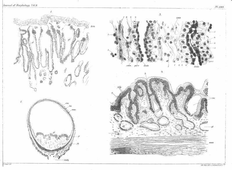
Fig. I. Placenta of rabbit at eight days, with dilated glands,^/, and superjacent foetal ectoderm, ecto (X 125 diams.).
Fig. 2. Rabbit's uterus at nine days, transverse section of a swelling (X 7 diams.).
Fig. 3. Portion of the placenta of Fig. 2 (X 445 diams.), to show the connective tissue, conn, the perivascular cells, per.v, and the thickened endothelium, endo, of the blood capillaries.
Fig. 4. Portion of the periplacenta of Fig. 2 (X 175 diams.), to show the degeneration of the epithelium, h.ep.
Plate 27
EXPLANATION OF PLATE XXVII.
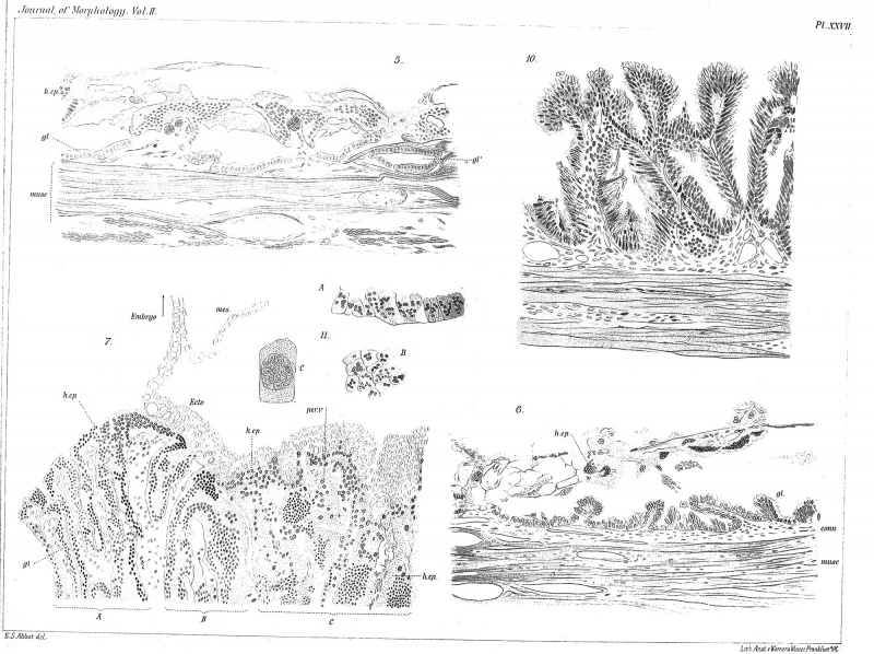
Fig. 5. Portion of the ob-placenta of Fig. 2 (x 175 diams.), to show the degenerated epithelium, h.ep, and the saucer-shaped glands, gl, gV.
Fig. 6. Rabbit's uterus of eleven days; portion of the ob-placenta to show the degenerated epithelium, h.ep, and the regenerated glands,^/ ( X 175 diams.).
Fig. 7. Portion of the placenta of Fig. 2 ( X 175 diams.), to show the degeneration of the uterine tissue and the relations of the foetal ectoderm to the placental surface.
Fig. 10. Rabbit's uterus of thirteen days; portion of the ob-placenta (X 175 diams.), to show the regenerated glands.
Fig. II. Portions of the epithelium of the periplacenta at thirteen days. A, vertical section (X 175 diams.). B, surface view (X 175 diams.). C, single cell ( X 445 diams.).
Plate 28
EXPLANATION OF PLATE XXVIII.
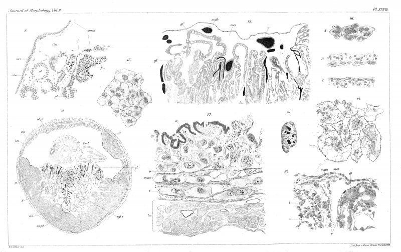
Fig. 8. Portion of a vertical section of the placenta at eleven days of a rabbit, to show the relations of the mesothelium, msth, to the top, and of the ectoderm, edo, to the side of the placenta (X 175 diams.).
Fig. 9. Complete transverse section of a rabbit's uterus at thirteen days, with the embryo, emb, in place ( X 7 diams.) ; the details are only approximately accurate ; X, mass of perivascular decidual cells, developed in the region of the ob-placenta.
Fig. 12. Rabbit's uterus at fifteen days; portion of a section through the placenta (X 90 diams.), to show the degenerated glands,^/, and the mesoderm, vies, and mesothelium, msth, covering the surface of the placenta ; the blood-vessels are drawn dark.
Fig. 13. Portion of upper part of a rabbit's placenta at fifteen days ( X 340 diams.), to show the histological structure of the glandular layer of the placenta.
Fig. 14. Multinucleate decidual cells from the subglandular zone of a rabbit's placenta at fifteen days ( X 540 diams.).
Fig. 15. Uninucleate perivascular decidual cells from the outer zone of a rabbit's placenta at fifteen days ( X 540 diams.) .
Fig. 16. Endothehum from the blood-vessels of the periplacenta of a rabbit at fifteen days ( X 240 diams.). A, surface view; B, C, in section.
Fig. 17. Ob-placenta of a rabbit at fifteen days (X 125 diams.), to show the monster cells, a, b, c, and the uterine epithelium, ep.
Fig. 18. Nucleus of a monster cell from the ob-placenta of a rabbit at fifteen days ( X 445 diams.).
Plate 29
EXPLANATION OF PLATE XXIX.
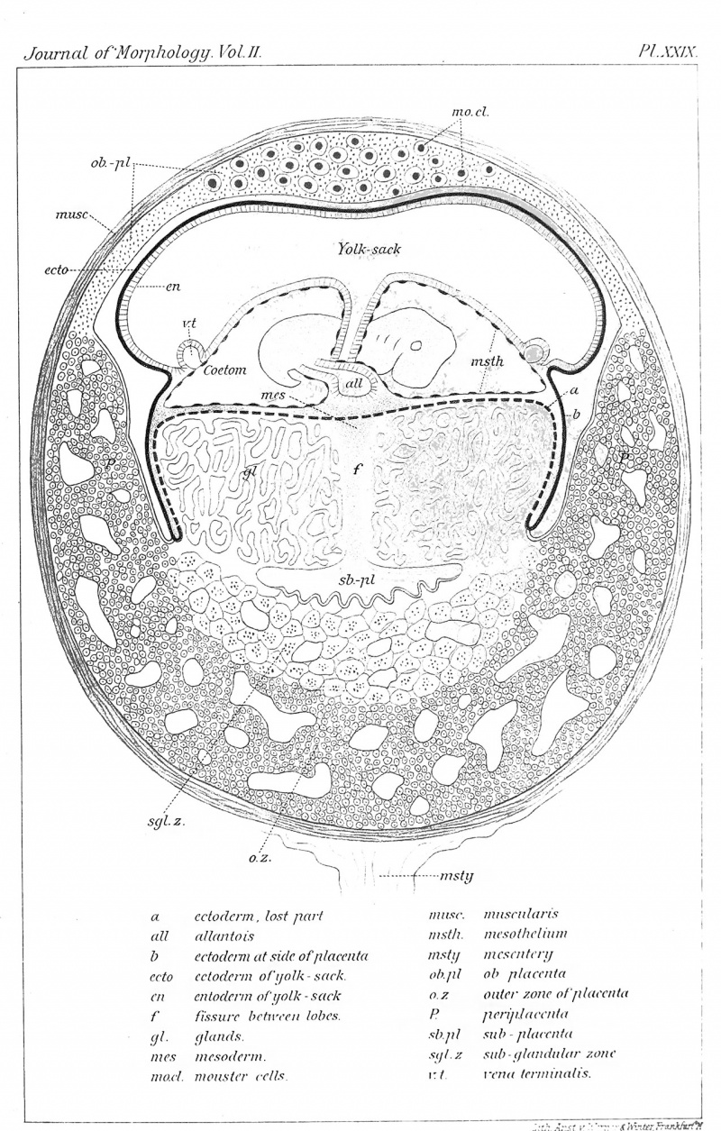
Diagram to show the relations of the embryo and uterus in the rabbit from the eleventh to the thirteenth day of gestation.
Minot, C.S. Uterus And Embryo (1889) - I. Rabbit II. Man - Plates
| Historic Disclaimer - information about historic embryology pages |
|---|
| Pages where the terms "Historic" (textbooks, papers, people, recommendations) appear on this site, and sections within pages where this disclaimer appears, indicate that the content and scientific understanding are specific to the time of publication. This means that while some scientific descriptions are still accurate, the terminology and interpretation of the developmental mechanisms reflect the understanding at the time of original publication and those of the preceding periods, these terms, interpretations and recommendations may not reflect our current scientific understanding. (More? Embryology History | Historic Embryology Papers) |
Cite this page: Hill, M.A. (2024, April 20) Embryology Book - Uterus And Embryo - Plates (1889). Retrieved from https://embryology.med.unsw.edu.au/embryology/index.php/Book_-_Uterus_And_Embryo_-_Plates_(1889)
- © Dr Mark Hill 2024, UNSW Embryology ISBN: 978 0 7334 2609 4 - UNSW CRICOS Provider Code No. 00098G


