Book - The Frog Its Reproduction and Development 5: Difference between revisions
mNo edit summary |
|||
| Line 80: | Line 80: | ||
the embryo, provided the distortion is not too great. | the embryo, provided the distortion is not too great. | ||
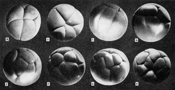
[[File:Rugh_060.jpg|thumb|center|600px|The third, fourth, and fifth cleavages from the animal pole and lateral views.]] | |||
The third cleavage begins about 30 minutes after the second is completed, or 4 hours after fertilization. The cleavage rate is accelerated | The third cleavage begins about 30 minutes after the second is completed, or 4 hours after fertilization. The cleavage rate is accelerated | ||
| Line 113: | Line 114: | ||
this point on is a rarity, and yet the embryo will develop perfectly | this point on is a rarity, and yet the embryo will develop perfectly | ||
normally. | normally. | ||
Latest revision as of 11:08, 12 April 2013
| Embryology - 18 Apr 2024 |
|---|
| Google Translate - select your language from the list shown below (this will open a new external page) |
|
العربية | català | 中文 | 中國傳統的 | français | Deutsche | עִברִית | हिंदी | bahasa Indonesia | italiano | 日本語 | 한국어 | မြန်မာ | Pilipino | Polskie | português | ਪੰਜਾਬੀ ਦੇ | Română | русский | Español | Swahili | Svensk | ไทย | Türkçe | اردو | ייִדיש | Tiếng Việt These external translations are automated and may not be accurate. (More? About Translations) |
Rugh R. Book - The Frog Its Reproduction and Development. (1951) The Blakiston Company.
| Historic Disclaimer - information about historic embryology pages |
|---|
| Pages where the terms "Historic" (textbooks, papers, people, recommendations) appear on this site, and sections within pages where this disclaimer appears, indicate that the content and scientific understanding are specific to the time of publication. This means that while some scientific descriptions are still accurate, the terminology and interpretation of the developmental mechanisms reflect the understanding at the time of original publication and those of the preceding periods, these terms, interpretations and recommendations may not reflect our current scientific understanding. (More? Embryology History | Historic Embryology Papers) |
Chapter 5 - Cleavage
Determination of Planes
The plane of the first cleavage is determined in a very different manner. The cytoplasm within which the cleavage is initiated is somewhat flattened beneath the pigmented cortex of the animal hemisphere. The egg nucleus lies within this cytoplasm, and, as the sperm nucleus approaches it, preceded by its divided centrosome, an amphiaster is set up to divide the chromatic material of the two syngamic nuclei. The cleavage plane therefore is determined by the direction of approach of the sperm nucleus, or rather, the copulation path. It can be assumed that in the undisturbed egg the cytoplasm also has a slight axial distribution, paralleling that of the yolk, with its maximum in the opposite direction to the yolk. The cleavage furrow will form at right angles to the amphiaster, and this coincides exactly with the sperm copulation path. One may conclude, therefore, that under undisturbed conditions the first cleavage furrow includes (or is determined by) the copulation path.
It should be made clear at this point, however, that the cleavage plane need not necessarily represent the median longitudinal axis (sagittal plane) of the embryo, cutting it into right and left halves. Should this happen it would mean that the penetration and the copulation paths were in exactly the same plane and the first cleavage would represent a median sagittal plane of the future embryo. Further, the mere pressure from other eggs in the same clutch can so alter the distribution of cytoplasm that the cleavage spindle may be shifted from its expected position. The median plane of the bilaterally symmetrical embryo may, but does not necessarily, coincide with the plane of egg symmetry.
To summarize, the penetration point combined with any two points on the egg axis establishes a plane which represents the median sagittal plane of the future embryo. The copulation (syngamic) path of the sperm nucleus to the egg nucleus determines the first cleavage plane. Should the penetration and copulation paths be in a direct line to the egg nucleus, the first cleavage would bisect the future embryo ( and the gray crescent) into right and left halves. There is a natural tendency for the coinciding of the gravitational plane, the sperm entrance point, the sperm penetration path, the median plane of the egg, the median plane of the embryo, and the plane of the first cleavage. But this is not essential to the production of a normal embryo.
Cleavage Laws
There are four major cleavage laws, known by the names of those who first emphasized their importance.
- Pfluger's Law, The spindle elongates in the direction of least resistance.
- Balfour's Law. The rate of cleavage tends to be governed by the inverse ratio of the amount of yolk present, in holoblastic cleavage. The yolk tends to impede division of both the nucleus and the cytoplasm.
- Sack's Law. Cells tend to divide into equal parts and each new plane of division tends to bisect the previous plane at right angles.
- Hertwig's Law. The nucleus and its spindle generally are found in the center of the active protoplasm and the axis of any division spindle lies in the longest axis of the protoplasmic mass. Divisions tend to cut the protoplasmic masses at right angles to their axes.
Steps in the Cleavage Process
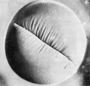
The frog's egg is telolecithal, meaning that there is a large amount of yolk concentrated at one pole, opposite to the concentration of cytoplasm and the location of the nucleus. The cleavages are holoblastic (i.e., total), and, after the second cleavage, they are unequal. They eventually cut through the entire egg from the surface inward. About 2Vi hours after the egg is fertilized there appears a very short inverted fold near the center of the pigmented and slightly depressed animal hemisphere cortex. This depression is extended in both directions and in the pigmented surface coat there appear lines radiating outwardly from the deepening first cleavage furrow. It appears as though some internal force is drawing this cortical region (surface) of the egg toward its center, and the apparent tension lines may be the result of these inner pulling forces. The facts that the radiating "tension" lines are not always developed, that they vary in different eggs, and that they can be chemically obliterated without interfering with the cleavage indicate that they are not the forces involved in cleavage but that they result from such forces. As the furrow is deepened and becomes fully formed, these lines disappear, and the surface of the egg again becomes smooth. Similar though less pronounced lines generally are seen in association with the second and subsequent cleavages.
The furrow is at first very shallow (superficial), extending from the
center of the animal hemisphere around the egg toward the vegetal
hemisphere. Its greatest depth is where it is first seen, in the animal
hemisphere. Within about 3 hours after fertilization the entire egg is
ringed by a vertical furrow. This is always slightly deeper at the animal than at the vegetal hemisphere, and is clearly marked by the
concentration of surface pigment. Such grading of the furrow may be
due to the mechanical factor of the more resistant yolk at the vegetal
pole. In most, but not in all, cases the first cleavage cuts through the gray crescent. After the first cleavage and up to the late blastula stage,
each cell of the embryo is known as a blastomere.
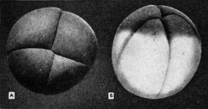
Before the first cleavage furrow has completely encircled the egg, a second furrow begins at the center of the animal hemisphere and at right angles to the first furrow. This cleavage is also vertical and will progress down and around the egg to become deeper and deeper, but always deepest at the animal hemisphere. This second cleavage first begins to appear at about SVi hours after fertilization, or 1 hour after the completion of the first cleavage. Again the so-called tension lines appear, and, since the lines of the first furrow have by this time flattened out, the presence of these new lines plus the relative shallowness of the furrow will help in its identification as the second furrow. As in the case of the first, this second furrow progresses superficially around the egg until the two ends meet at the vegetal pole. In the meantime the first cleavage furrow has cut more deeply into the egg so that in transverse (horizontal) sections of such an egg one can identify the two cleavage furrows easily by their relative depth.
We can picture the egg at this stage as a core of yolk, concentrated more toward the vegetal pole, and with the cytoplasm, nuclei, and deepest portions of the cleavage furrows located at the pigmented animal hemisphere. However, all of the animal hemisphere structures will spread progressively toward the vegetal pole. With each division of the cytoplasm, cells are formed which contain yolk. By the time the third cleavage makes its appearance, the first cleavage has all but cut the entire egg mass into two equal parts. Each of these first two blastomeres (and the subsequent four blastomeres) is identical with all of the others with respect to the cytoplasm, pigment, and yolk gradients. In the 4-cell stage they are not qualitatively identical, for only two of the four cells contain any of the gray crescent material.
Often in closely packed eggs these first two cleavages may appear
not quite in the center of the animal hemisphere, or may be not at
right angles to each other. This would be the exception, however, and
generally is due to the physical compression of the closely packed eggs
whereby (as pointed out by Sachs and Hertwig) the cytoplasmic axis
is shifted. The description given here applies to the isolated and unrestricted egg and should be considered as the more nearly normal.
However, it must be pointed out that this minor compression and
shifting of cleavage planes does not affect the normal development of
the embryo, provided the distortion is not too great.
The third cleavage begins about 30 minutes after the second is completed, or 4 hours after fertilization. The cleavage rate is accelerated with each division, possibly because the resulting blastomeres are progressively smaller. The third cleavage plane is horizontal (latitudinal ) and at right angles to both the first and the second cleavages. It is slightly above the level of the equator, about 60° to 70° from the center of the animal hemisphere. This shifting of the cleavage plane toward the animal hemisphere from the equator of the otherwise equatorial cleavage is due, no doubt, to the presence of more cytoplasm in that region and to relatively less yolk. Yolk is generally resistant to cleavage forces. When the cleavages are completed, the uppermost and smaller cells are sometimes designated as micromeres (smaller blastomeres) and the lowermost and larger cells are then designated as macromeres (larger blastomeres). This third cleavage furrow is seen as a horizontal groove varying from 20° to 30° above the equatorial line, toward the animal hemisphere. It provides four clear-cut blastomeres in the animal hemisphere, each equally pigmented. Since the second cleavage may not have been completed at the vegetal pole, there is a brief transitional period in which from some aspects there appear to be the surface markings of 6 rather than 8 cells.
The fourth cleavage is usually double and occurs about 20 minutes
after the third. It tends to be vertical, but is seldom meridional. The
furrows begin near the center of the animal pole and progress vegetally,
dividing each of the original four blastomeres of the animal hemisphere. Instead of intersecting at the center of the animal hemisphere
the fourth cleavage furrows may run into the first or the second
cleavage furrows. This cleavage shows the first digression from a regular pattern in the undisturbed egg, and may appear even as a welldefined furrow before the third (horizontal) cleavage has reached its
maximum depth. The cleavage rate is accelerated with each of the
early divisions, and, since the blastomeres are of unequal size and
have varying amounts of cytoplasm and yolk, synchronous cleavage is
lost and there is an obvious overlapping of the divisions. The uppermost cells (micromeres) divide the most rapidly so that for a period
there may be a 12-cell stage, made up of 8 upper and 4 lower blastomeres. Perfect symmetry in cleavage and in the blastomeres from
this point on is a rarity, and yet the embryo will develop perfectly
normally.
The fifth cleavage (also double), which should provide a 32-cell
embryo, is more apt to be out of line, and the symmetry and regularity
of the earlier cleavages is lost. The gradient of cleavage rate from the
rapidly dividing animal hemisphere blastomeres to the slowly dividing
vegetal hemisphere cells now becomes more apparent. These new
furrows tend to be horizontal (latitudinal), as was the third cleavage.
One cuts the animal hemisphere blastomeres and the other the vegetal
hemisphere blastomeres horizontally, so that tiers of blastomeres are
formed. The smallest cells, which contain the most pigment, are now
found at the animal pole, and the largest blastomeres with no pigment
and the most yolk are at the vegetal pole. There is evident, however,
some movement of the surface (cortical) pigment in the direction of
the vegetal pole accompanying each division of the blastomeres. This
process is known as epiboly, to be described later in this book.
| Historic Disclaimer - information about historic embryology pages |
|---|
| Pages where the terms "Historic" (textbooks, papers, people, recommendations) appear on this site, and sections within pages where this disclaimer appears, indicate that the content and scientific understanding are specific to the time of publication. This means that while some scientific descriptions are still accurate, the terminology and interpretation of the developmental mechanisms reflect the understanding at the time of original publication and those of the preceding periods, these terms, interpretations and recommendations may not reflect our current scientific understanding. (More? Embryology History | Historic Embryology Papers) |
Reference
Rugh R. Book - The Frog Its Reproduction and Development. (1951) The Blakiston Company.
Cite this page: Hill, M.A. (2024, April 18) Embryology Book - The Frog Its Reproduction and Development 5. Retrieved from https://embryology.med.unsw.edu.au/embryology/index.php/Book_-_The_Frog_Its_Reproduction_and_Development_5
- © Dr Mark Hill 2024, UNSW Embryology ISBN: 978 0 7334 2609 4 - UNSW CRICOS Provider Code No. 00098G


