Book - The Frog Its Reproduction and Development 17: Difference between revisions
m (→Chapter 1 -) |
|||
| (44 intermediate revisions by the same user not shown) | |||
| Line 5: | Line 5: | ||
==Chapter 1 - Introduction== | ==Chapter 1 - Introduction== | ||
<gallery> | <gallery> | ||
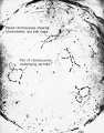
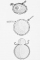
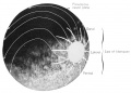
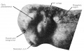
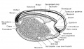
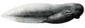
File:Rugh 001.jpg| | File:Rugh 001.jpg|Frog's egg and swollen jelly shortly after fertilization. | ||
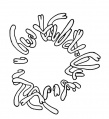
File:Rugh 002.jpg| | File:Rugh 002.jpg|Spermist's conception of the human figure in miniature (1769-1832). | ||
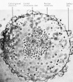
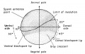
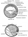
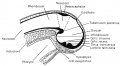
File:Rugh 003.jpg| | File:Rugh 003.jpg|Planes in which the embryo may be cut or sectioned. | ||
</gallery> | </gallery> | ||
==Chapter 2 - General Introduction to the Embryology of the Leopard Frog ''Rana pipiens''== | ==Chapter 2 - General Introduction to the Embryology of the Leopard Frog ''Rana pipiens''== | ||
<gallery> | <gallery> | ||
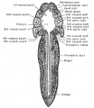
File:Rugh 004.jpg| | File:Rugh 004.jpg|The leopard frog, Rana pipiens. | ||
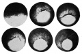
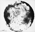
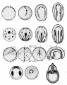
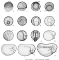
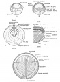
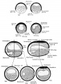
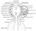
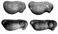
File:Rugh 005.jpg| | File:Rugh 005.jpg|Early development of the frog's egg. | ||
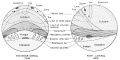
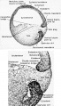
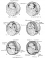
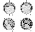
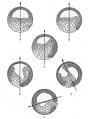
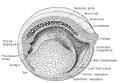
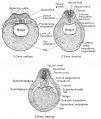
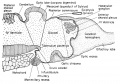
File:Rugh 006.jpg| | File:Rugh 006.jpg|Gastrulation in the frog. | ||
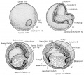
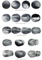
File:Rugh 007.jpg| | File:Rugh 007.jpg|Early development of the frog embryo. | ||
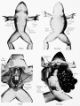
File:Rugh 008.jpg| | File:Rugh 008.jpg|Early development of the frog embryo. Ventral view of the 11 mm larva. | ||
File:Rugh 009.jpg| | File:Rugh 009.jpg|Development and absorption of the external gills of the frog larva. | ||
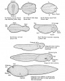
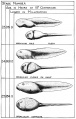
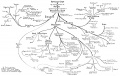
File:Rugh 010.jpg| | File:Rugh 010.jpg|Metamorphosis of the frog, Rana catesbiana. | ||
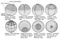
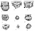
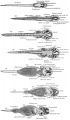
File:Rugh 011.jpg| | File:Rugh 011.jpg|Stages in the Normal Development of Rana pipiens. | ||
File:Rugh 012.jpg| | File:Rugh 012.jpg|Stages in the Normal Development of Rana pipiens. | ||
File:Rugh 013.jpg| | File:Rugh 013.jpg|Stages in the Normal Development of Rana pipiens. | ||
</gallery> | </gallery> | ||
==Chapter 3 - Reproductive System of the Adult Frog Rana pipiens== | ==Chapter 3 - Reproductive System of the Adult Frog Rana pipiens== | ||
<gallery> | <gallery> | ||
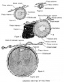
File:Rugh 014.jpg| | File:Rugh 014.jpg|The oocyte and spermatozoa maturation process. Schematized drawings. | ||
File:Rugh 015.jpg| | File:Rugh 015.jpg|Prophases of the heterotype division in the male Axolotl. | ||
File:Rugh 016.jpg| | File:Rugh 016.jpg|Normal cyclic changes in the primary and secondary sexual characters of the frog, Rana pipiens. | ||
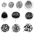
File:Rugh 017.jpg| | File:Rugh 017.jpg|Spermatogenesis in the frog, Rana pipiens. | ||
File:Rugh 018.jpg| | File:Rugh 018.jpg|Spermatogenetic stages in the seminiterous tubule of the frog testis. | ||
File:Rugh 019.jpg| | File:Rugh 019.jpg|Frog spermatozoon. | ||
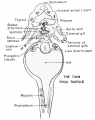
File:Rugh 020.jpg| | File:Rugh 020.jpg|Kidney of the male frog during amplexus. | ||
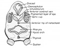
File:Rugh 021.jpg| | File:Rugh 021.jpg|Brain regions for the mediation of sexual behaviour. | ||
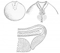
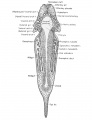
File:Rugh 022.jpg| | File:Rugh 022.jpg|Urogenital system of the male frog. | ||
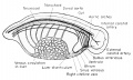
File:Rugh 023.jpg| | File:Rugh 023.jpg|Late development of the frog ovary. | ||
File:Rugh 024.jpg|Small ovarian egg of the frog surrounded by its follicle | File:Rugh 024.jpg|Small ovarian egg of the frog surrounded by its follicle. | ||
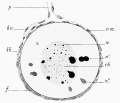
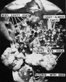
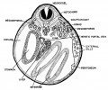
File:Rugh 025.jpg|Section of a mature ovarian egg to show the area of ultimate follicular rupture and the surrounding membranes of the egg | |||
File:Rugh 026.jpg|Growing Oocyte of the Frog | |||
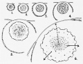
File:Rugh 027.jpg|Reactions of the frog ovary to stimulation by the frog anterior pituitary hormone | |||
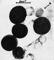
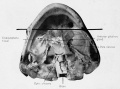
File:Rugh 028.jpg|Photograph of Rana pipiens female body cavity at the height of ovulation | |||
File:Rugh 029.jpg|Deposition of jelly on the frog's egg | |||
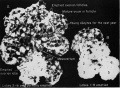
File:Rugh 030.jpg|Recently emptied ovarian follicle of the frog | |||
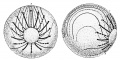
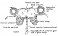
File:Rugh 031.jpg|Follicular rupture and ovulation in the frog | |||
File:Rugh 032.jpg|Photograph of the open body cavity of an actively ovulating female frog showing the entrance of an egg into the ostium | |||
File:Rugh 033.jpg|Distribution of coelomic cilia within the body cavity of the female frog | |||
File:Rugh 034.jpg|Passage of eggs through the oviduct | |||
File:Rugh 035.jpg|Oviducts of the frog under various states of sexual activity | |||
File:Rugh 036.jpg|Prophases of the heterotypic division in the female (ovary of tadpole) | |||
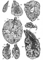
File:Rugh 037.jpg|Normal nuclear growth cycle of the ovum of Rana pipiens | |||
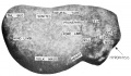
File:Rugh 038.jpg|Ovarial wall of Rami temporaria | |||
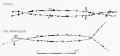
File:Rugh 039.jpg|The entire set of chromosomes of Rana lemporaria | |||
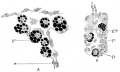
File:Rugh_040.jpg|Similarity of frog and of salamander chromosome structure | |||
File:Rugh_041.jpg|Lateral loops of the amphibian chromosome | |||
File:Rugh_042.jpg|Chromosomes in the amphibian nucleus | |||
File:Rugh_043.jpg|Diploid metaphase chromosomes from the tail fin of a 15day-old Rana pipiens tadpole | |||
File:Rugh_044.jpg|Isolated germinal vesicle (nucleus) of Rami pipiens | |||
File:Rugh_045.jpg|Stage 5 germinal vesicle of Rami catesbiana | |||
File:Rugh_046.jpg|Production of ova and the process of ovulation | |||
File:Rugh_047.jpg|The maturation divisions in the female (Axolotl) | |||
File:Rugh_048.jpg|The position of the anterior pituitary of Rana pipiens | |||
</gallery> | |||
==Chapter 4 - Fertilization of the Frog's Egg== | |||
<gallery> | |||
File:Rugh_049.jpg|Amplexus in the toad Bufo fowleri | |||
File:Rugh_050.jpg|Oviposition and fertilization in Rana | |||
File:Rugh_051.jpg|The egg of the frog 35 minutes after fertilization | |||
File:Rugh_052.jpg|Polar body emergence Rana pipiens | |||
File:Rugh_053.jpg|The four stages in polar body emergence in Rana pipiens | |||
File:Rugh_054.jpg|Invasion of the frog's egg by a spermatozoon | |||
File:Rugh_055.jpg|Fertilization of the Frog's Egg | |||
File:Rugh_056.jpg|Surface changes of the Frog's Egg at the time of Fertilization | |||
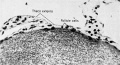
File:Rugh_057.jpg|Formation ol the gray crescent | |||
</gallery> | |||
==Chapter 5 - Cleavage== | |||
<gallery> | |||
File:Rugh_058.jpg|The first cleavage | |||
File:Rugh_059.jpg|The 4-cell stage | |||
File:Rugh_060.jpg|The third, fourth, and fifth cleavages from the animal pole and lateral views | |||
</gallery> | |||
==Chapter 6 - Blastulation== | |||
<gallery> | |||
File:Rugh_061.jpg|Progressive Stages in Blastulation | |||
File:Rugh_062.jpg|Blastulation in the Frog | |||
</gallery> | |||
==Chapter 7 - Gastrulation== | |||
<gallery> | |||
File:Rugh_063.jpg|Derivatives of the Primary Germ Layers | |||
File:Rugh_064.jpg|Morphogenetic Movements during Gastrulation and Neurulation | |||
File:Rugh_065.jpg|Fate map showing presumptive regions of the anuran blastula | |||
File:Rugh_066.jpg|Gastrulation in the frog, Rana pipiens | |||
File:Rugh_067.jpg|Blastula to tail bud stages | |||
File:Rugh_068.jpg|Gastrula | |||
File:Rugh_069.jpg|Blastula to Gastrula Directions of Movement | |||
File:Rugh_070.jpg|Involutional movements during gastrulation outlined on the living gastrula | |||
File:Rugh_071.jpg|The Process of Gastrulation in the Frog | |||
File:Rugh_072.jpg|Surface Changes during Gastrulation in the Frog | |||
File:Rugh_073.jpg|Gastrulation and mesoderm formation | |||
File:Rugh_074.jpg|Gastrulation and mesoderm formation | |||
File:Rugh_075.jpg|Blastula to gastrula stages in the frog | |||
File:Rugh_076.jpg|Blastula to gastrula stages in the frog | |||
File:Rugh_077.jpg|Rotation of the amphibian egg in the gravitational field during gastrulation | |||
File:Rugh_078.jpg|Rotation of the amphibian egg during gastrulation | |||
</gallery> | |||
==Chapter 8 - Neurulation and Early Organogeny== | |||
<gallery> | |||
File:Rugh_079.jpg|Formation of the Neural Tube | |||
File:Rugh_080.jpg|Gastrulation and early neurulation in the frog | |||
File:Rugh_081.jpg|Development of the frog (Rana pipiens) from the yolk plug stage to the neurula. | |||
File:Rugh_082.jpg|Early development of caudal structures | |||
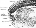
File:Rugh_083.jpg|Organ fields or anlagen of the closed neural tube stage | |||
File:Rugh_084.jpg|Photograph of the neurenteric canal of the late neurula stage in the frog | |||
File:Rugh_085.jpg|A face view of the 5 mm frog tadpole | |||
File:Rugh_086.jpg|Anlagen of the head region 5 mm frog tadpole | |||
File:Rugh_087.jpg|The 7 mm frog tadpole frontal section | |||
File:Rugh_088.jpg|Development of the respiratory systems of the frog larvae | |||
File:Rugh_089.jpg|The 3 mm frog tadpole sagittal section | |||
File:Rugh_090.jpg|Formation of the neural groove and neural tube from the neural (medullary) plate | |||
File:Rugh_091.jpg|Formation of the tail bud | |||
File:Rugh_092.jpg|Formation of the neural groove and neural tube from the neural (medullary) plate | |||
</gallery> | |||
==Chapter 9 - A Survey of the Major Developmental Changes in the Early Embryo== | |||
<gallery> | |||
File:Rugh_093.jpg|Development of the Eye of the Frog | |||
File:Rugh_094.jpg|Photograph of endocrine anlagen at the 5 mm stage of the frog tadpole | |||
File:Rugh_095.jpg|Reconstruction of the 5 mm tadpole in sagittal section | |||
File:Rugh_096.jpg|Three-dimensional representation of the late neurula stage of the frog Rana pipiens | |||
File:Rugh_097.jpg|Three-dimensional representation of the tail bud stage of the frog embryo Rana pipiens | |||
File:Rugh_098.jpg|Frontal ( horizontal) section of the 7 mm frog larva at the level of the developing heart | |||
File:Rugh_099.jpg|The 7 mm frog tadpole frontal section | |||
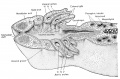
File:Rugh_100.jpg|The 7 mm frog tadpole transverse sections through the mid-body level | |||
File:Rugh_101.jpg|The 5 mm frog tadpole at mid-body level photograph of cross section | |||
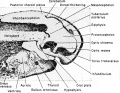
File:Rugh_102.jpg|Frontal (horizontal) section of the 7 mm frog larva at the level of the pharynx | |||
File:Rugh_103.jpg|The earliest complete closed blood vascular system of the frog embryo (found at the 4 mm stage) | |||
File:Rugh_104.jpg|Frontal section through the level of the heart of the 7 mm tadpole | |||
File:Rugh_105.jpg|The 7 mm frog tadpole transverse section through the level of the thyroid gland | |||
File:Rugh_106.jpg|The 7 mm frog tadpole transverse section through the level of the heart | |||
File:Rugh_107.jpg|Development of the Heart of the Frog Embryo | |||
File:Rugh_108.jpg|Representative transverse sections of an 8 mm frog larva | |||
</gallery> | |||
==Chapter 10 - A Survey or the Later Embryo or Larva== | |||
<gallery> | |||
File:Rugh_109.jpg|Development of the external gills of Rana pipiens | |||
File:Rugh_110.jpg|Tadpole with external gills | |||
File:Rugh_111.jpg|The 7 mm frog larva in serial frontal sections | |||
</gallery> | |||
==Chapter 11 - The Germ Layer Derivatives - The Ectoderm and Its Derivatives== | |||
<gallery> | |||
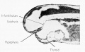
File:Rugh_112.jpg|Early organogeny of the frog tadpole, showing the primary brain vesicles in sagittal section | |||
File:Rugh_113.jpg|Pre-metamorphic stage | |||
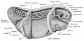
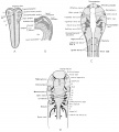
File:Rugh_114.jpg|Adult brain | |||
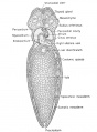
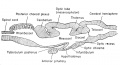
File:Rugh_115.jpg|Reconstruction of the 7 mm frog larva showing the major organ systems from the right side | |||
File:Rugh_116.jpg|Development of the pituitary gland of the frog | |||
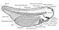
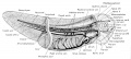
File:Rugh_117.jpg|Median sagittal section of the 7 mm frog tadpole | |||
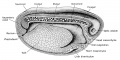
File:Rugh_118.jpg|Median sagittal section of the 11 mm frog tadpole | |||
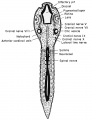
File:Rugh_119.jpg|Development of the spinal cord of the frog Spinal cord of the 7 mm larva | |||
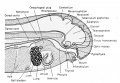
File:Rugh_120.jpg|Schematic diagram of the developing eye parts of the frog | |||
File:Rugh_122.jpg|View into the larval eye Rana pipiens, photograph | |||
File:Rugh_122.jpg|Development of the optic cup and lens in Siredon pisciformis | |||
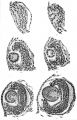
File:Rugh_123.jpg|Development of the amphibian eye | |||
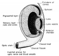
File:Rugh_124.jpg|Schematic Diagram through Frog's Eye | |||
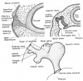
File:Rugh_125.jpg|Auditory apparatus of an 11 mm frog tadpole | |||
File:Rugh_126.jpg|Development of the auditory apparatus of the frog | |||
File:Rugh_127.jpg|External and internal nares of the 11 mm frog tadpole | |||
File:Rugh_128.jpg|Development of the olfactory organ of the frog | |||
File:Rugh_129.jpg|Origin of the lateral line sense organ system in the frog larva | |||
File:Rugh_130.jpg|Origin and derivatives of the cranial ganglia | |||
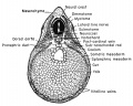
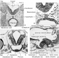
File:Rugh_131.jpg|Early organogeny the 5 mm frog tadpole at mid-body level | |||
File:Rugh_132.jpg|Development of the spinal cord and sympathetic nerves | |||
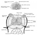
File:Rugh_133.jpg|Relation of the spinal and the sympathetic nervous systems. | |||
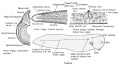
File:Rugh_134.jpg|Brain of the adult frog. | |||
File:Rugh_135.jpg|Sagittal section through the anterior end of the 8 mm frog larva. | |||
</gallery> | |||
==Chapter 12 - The Endodermal Derivatives== | |||
<gallery> | |||
File:Rugh_137.jpg|Frontal (horizontal) reconstruction of the external gill stage of the frog larva | |||
File:Rugh_138.jpg|Relation of the pharynx to the internal and external gills of the frog transverse section | |||
File:Rugh_139.jpg|Early development of the thyroid gland of the frog | |||
File:Rugh_140.jpg|Early development of the thyroid gland of the frog | |||
File:Rugh_141.jpg|Thyroid gland at the time of metamorphosis | |||
File:Rugh_142.jpg| | |||
File:Rugh_143.jpg| | |||
</gallery> | |||
==Chapter 13 - The Mesodermal Derivatives== | |||
<gallery> | |||
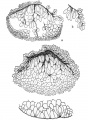
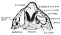
File:Rugh_143.jpg|Rana pipiens embryonic chondrocranium. | |||
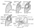
File:Rugh_144.jpg|Skull of Rana during metamorphosis | |||
File:Rugh_145.jpg|Skull of Rana during metamorphosis | |||
File:Rugh_146.jpg|Development of the thyroid gland and related hyoid cartilages | |||
File:Rugh_147.jpg|Development of the pronephric tubule | |||
File:Rugh_148.jpg|Persistent nephrostomes of the frog | |||
File:Rugh_149.jpg|Urogenital system of the male frog during amplexus | |||
File:Rugh_150.jpg|Adrenal gland of the recently metamorphosed frog | |||
File:Rugh_151.jpg|Adrenal cortical anlage at the 10 mm. stage of the frog tadpole | |||
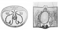
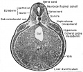
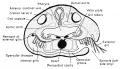
File:Rugh_152.jpg|Schematized diagram through the level of the gonad primordium of the 11 mm frog tadpole | |||
File:Rugh_153.jpg|Gonad primordia of the 11 mm frog tadpole | |||
File:Rugh_154.jpg|Primordial germ cells (g.c.) in the tadpole of the common frog {Rana temporaria) | |||
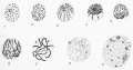
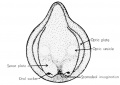
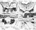
File:Rugh_155.jpg|Sections through the gonads of Anurans before and after differentiation | |||
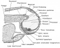
File:Rugh_156.jpg|The coelom and its derivatives in the frog | |||
File:Rugh_157.jpg|Development of the amphibian heart | |||
File:Rugh_158.jpg|Larval respiration in the frog. Development of the external gills | |||
File:Rugh_159.jpg|Larval respiration in the frog. Changes from internal to external gill circulation | |||
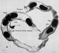
File:Rugh_160.jpg|Blood vascular system of the developing frog embryo Early embryo, from the right side. | |||
File:Rugh_161.jpg|Blood vascular system of the developing frog embryo Late frog embryo, from the right side. | |||
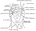
File:Rugh_162.jpg|Fate of the aortic arches of the frog embryo | |||
File:Rugh_163.jpg|Blood vascular system of the frog tadpole Venous system ventral view | |||
File:Rugh_164.jpg|Blood vascular system of the frog tadpole Arterial system ventral view | |||
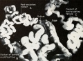
File:Rugh_165.jpg|Serial transverse sections of the 11 mm frog larva | |||
File:Rugh_166.jpg|Serial transverse sections of the 11 mm frog larva | |||
</gallery> | |||
==Appendix: Chronological Summary of Organ Anlagen Appearance of Rana pipiens== | |||
<gallery> | |||
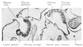
File:Rugh_167.jpg|Parts of the middle and inner ear of the frog schematized drawing | |||
File:Rugh_168.jpg|Olfactory organs of Rana pipiens tadpoles at 20 mm total body length | |||
File:Rugh_169.jpg|Stages in the metamorphosis of Rana pipiens | |||
File:Rugh_170.jpg|Stages in the metamorphosis of Rana pipiens | |||
File:Rugh_171.jpg|Stages in the metamorphosis of Rana pipiens | |||
File:Rugh_172.jpg|Stages in the metamorphosis of Rana pipiens | |||
File:Rugh_173.jpg|Stages in the metamorphosis of Rana pipiens | |||
File:Rugh_174.jpg|Adult Frog | |||
</gallery> | </gallery> | ||
{{Rugh1951 footer}} | {{Rugh1951 footer}} | ||
Latest revision as of 22:52, 26 April 2013
| Embryology - 19 Apr 2024 |
|---|
| Google Translate - select your language from the list shown below (this will open a new external page) |
|
العربية | català | 中文 | 中國傳統的 | français | Deutsche | עִברִית | हिंदी | bahasa Indonesia | italiano | 日本語 | 한국어 | မြန်မာ | Pilipino | Polskie | português | ਪੰਜਾਬੀ ਦੇ | Română | русский | Español | Swahili | Svensk | ไทย | Türkçe | اردو | ייִדיש | Tiếng Việt These external translations are automated and may not be accurate. (More? About Translations) |
Rugh R. Book - The Frog Its Reproduction and Development. (1951) The Blakiston Company.
| Historic Disclaimer - information about historic embryology pages |
|---|
 |
Figures
This gallery of images was not a section in the original book.
Chapter 1 - Introduction
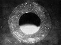
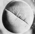
Frog's egg and swollen jelly shortly after fertilization.
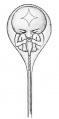
Spermist's conception of the human figure in miniature (1769-1832).
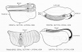
Planes in which the embryo may be cut or sectioned.


Chapter 2 - General Introduction to the Embryology of the Leopard Frog Rana pipiens
The leopard frog, Rana pipiens.
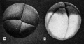
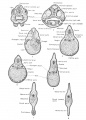
Early development of the frog's egg.
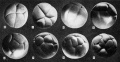
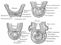
Gastrulation in the frog.
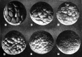
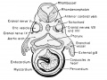
Early development of the frog embryo.
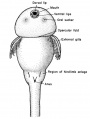
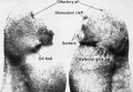
Early development of the frog embryo. Ventral view of the 11 mm larva.
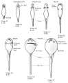
Development and absorption of the external gills of the frog larva.
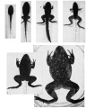
Metamorphosis of the frog, Rana catesbiana.
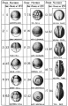
Stages in the Normal Development of Rana pipiens.
Stages in the Normal Development of Rana pipiens.
Stages in the Normal Development of Rana pipiens.


Chapter 3 - Reproductive System of the Adult Frog Rana pipiens
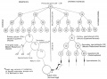
The oocyte and spermatozoa maturation process. Schematized drawings.
Prophases of the heterotype division in the male Axolotl.
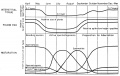
Normal cyclic changes in the primary and secondary sexual characters of the frog, Rana pipiens.
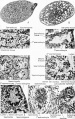
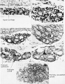
Spermatogenesis in the frog, Rana pipiens.
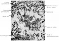
Spermatogenetic stages in the seminiterous tubule of the frog testis.
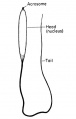
Frog spermatozoon.
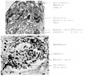
Kidney of the male frog during amplexus.
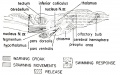
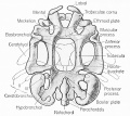
Brain regions for the mediation of sexual behaviour.
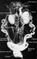
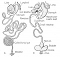
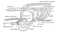
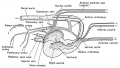
Urogenital system of the male frog.
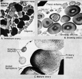
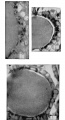
Late development of the frog ovary.
Small ovarian egg of the frog surrounded by its follicle.
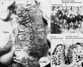
Section of a mature ovarian egg to show the area of ultimate follicular rupture and the surrounding membranes of the egg
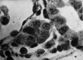
Growing Oocyte of the Frog
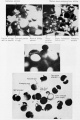
Reactions of the frog ovary to stimulation by the frog anterior pituitary hormone
Photograph of Rana pipiens female body cavity at the height of ovulation
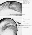
Deposition of jelly on the frog's egg
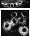
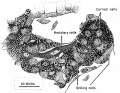
Recently emptied ovarian follicle of the frog
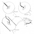
Follicular rupture and ovulation in the frog
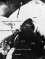
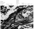
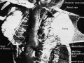
Photograph of the open body cavity of an actively ovulating female frog showing the entrance of an egg into the ostium
Distribution of coelomic cilia within the body cavity of the female frog
Passage of eggs through the oviduct
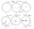
Oviducts of the frog under various states of sexual activity
Prophases of the heterotypic division in the female (ovary of tadpole)
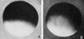
Normal nuclear growth cycle of the ovum of Rana pipiens
Ovarial wall of Rami temporaria

The entire set of chromosomes of Rana lemporaria
Similarity of frog and of salamander chromosome structure
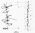
Lateral loops of the amphibian chromosome
Chromosomes in the amphibian nucleus
Diploid metaphase chromosomes from the tail fin of a 15day-old Rana pipiens tadpole
Isolated germinal vesicle (nucleus) of Rami pipiens
Stage 5 germinal vesicle of Rami catesbiana
Production of ova and the process of ovulation
The maturation divisions in the female (Axolotl)
The position of the anterior pituitary of Rana pipiens


Chapter 4 - Fertilization of the Frog's Egg
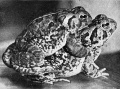
Amplexus in the toad Bufo fowleri
Oviposition and fertilization in Rana
The egg of the frog 35 minutes after fertilization
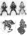
Polar body emergence Rana pipiens
The four stages in polar body emergence in Rana pipiens
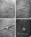
Invasion of the frog's egg by a spermatozoon
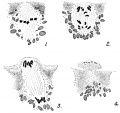
Fertilization of the Frog's Egg
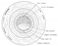
Surface changes of the Frog's Egg at the time of Fertilization
Formation ol the gray crescent


Chapter 5 - Cleavage
The first cleavage
The 4-cell stage
The third, fourth, and fifth cleavages from the animal pole and lateral views


Chapter 6 - Blastulation
Progressive Stages in Blastulation
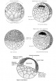
Blastulation in the Frog


Chapter 7 - Gastrulation
Derivatives of the Primary Germ Layers
Morphogenetic Movements during Gastrulation and Neurulation
Fate map showing presumptive regions of the anuran blastula
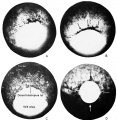
Gastrulation in the frog, Rana pipiens
Blastula to tail bud stages
Gastrula
Blastula to Gastrula Directions of Movement
Involutional movements during gastrulation outlined on the living gastrula
The Process of Gastrulation in the Frog
Surface Changes during Gastrulation in the Frog
Gastrulation and mesoderm formation
Gastrulation and mesoderm formation
Blastula to gastrula stages in the frog
Blastula to gastrula stages in the frog
Rotation of the amphibian egg in the gravitational field during gastrulation
Rotation of the amphibian egg during gastrulation


Chapter 8 - Neurulation and Early Organogeny
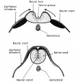
Formation of the Neural Tube
Gastrulation and early neurulation in the frog
Development of the frog (Rana pipiens) from the yolk plug stage to the neurula.
Early development of caudal structures
Organ fields or anlagen of the closed neural tube stage
Photograph of the neurenteric canal of the late neurula stage in the frog
A face view of the 5 mm frog tadpole
Anlagen of the head region 5 mm frog tadpole
The 7 mm frog tadpole frontal section
Development of the respiratory systems of the frog larvae
The 3 mm frog tadpole sagittal section
Formation of the neural groove and neural tube from the neural (medullary) plate
Formation of the tail bud
Formation of the neural groove and neural tube from the neural (medullary) plate


Chapter 9 - A Survey of the Major Developmental Changes in the Early Embryo
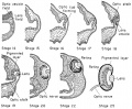
Development of the Eye of the Frog
Photograph of endocrine anlagen at the 5 mm stage of the frog tadpole
Reconstruction of the 5 mm tadpole in sagittal section
Three-dimensional representation of the late neurula stage of the frog Rana pipiens
Three-dimensional representation of the tail bud stage of the frog embryo Rana pipiens
Frontal ( horizontal) section of the 7 mm frog larva at the level of the developing heart
The 7 mm frog tadpole frontal section
The 7 mm frog tadpole transverse sections through the mid-body level
The 5 mm frog tadpole at mid-body level photograph of cross section
Frontal (horizontal) section of the 7 mm frog larva at the level of the pharynx
The earliest complete closed blood vascular system of the frog embryo (found at the 4 mm stage)
Frontal section through the level of the heart of the 7 mm tadpole
The 7 mm frog tadpole transverse section through the level of the thyroid gland
The 7 mm frog tadpole transverse section through the level of the heart
Development of the Heart of the Frog Embryo
Representative transverse sections of an 8 mm frog larva


Chapter 10 - A Survey or the Later Embryo or Larva
Development of the external gills of Rana pipiens
Tadpole with external gills
The 7 mm frog larva in serial frontal sections


Chapter 11 - The Germ Layer Derivatives - The Ectoderm and Its Derivatives
Early organogeny of the frog tadpole, showing the primary brain vesicles in sagittal section
Pre-metamorphic stage
Adult brain
Reconstruction of the 7 mm frog larva showing the major organ systems from the right side
Development of the pituitary gland of the frog
Median sagittal section of the 7 mm frog tadpole
Median sagittal section of the 11 mm frog tadpole
Development of the spinal cord of the frog Spinal cord of the 7 mm larva
Schematic diagram of the developing eye parts of the frog
View into the larval eye Rana pipiens, photograph
Development of the optic cup and lens in Siredon pisciformis
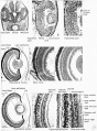
Development of the amphibian eye
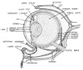
Schematic Diagram through Frog's Eye
Auditory apparatus of an 11 mm frog tadpole
Development of the auditory apparatus of the frog
External and internal nares of the 11 mm frog tadpole
Development of the olfactory organ of the frog
Origin of the lateral line sense organ system in the frog larva
Origin and derivatives of the cranial ganglia
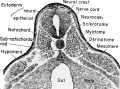
Early organogeny the 5 mm frog tadpole at mid-body level
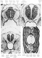
Development of the spinal cord and sympathetic nerves
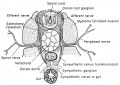
Relation of the spinal and the sympathetic nervous systems.
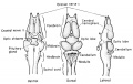
Brain of the adult frog.
Sagittal section through the anterior end of the 8 mm frog larva.


Chapter 12 - The Endodermal Derivatives
Frontal (horizontal) reconstruction of the external gill stage of the frog larva
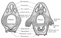
Relation of the pharynx to the internal and external gills of the frog transverse section
Early development of the thyroid gland of the frog
Early development of the thyroid gland of the frog
Thyroid gland at the time of metamorphosis


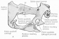
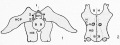
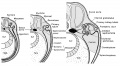
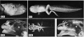
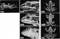
Chapter 13 - The Mesodermal Derivatives
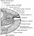
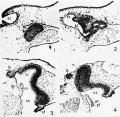
Rana pipiens embryonic chondrocranium.
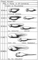
Skull of Rana during metamorphosis
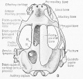
Skull of Rana during metamorphosis
Development of the thyroid gland and related hyoid cartilages
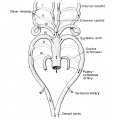
Development of the pronephric tubule
Persistent nephrostomes of the frog
Urogenital system of the male frog during amplexus
Adrenal gland of the recently metamorphosed frog
Adrenal cortical anlage at the 10 mm. stage of the frog tadpole
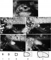
Schematized diagram through the level of the gonad primordium of the 11 mm frog tadpole
Gonad primordia of the 11 mm frog tadpole
Primordial germ cells (g.c.) in the tadpole of the common frog {Rana temporaria)
Sections through the gonads of Anurans before and after differentiation
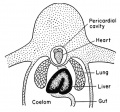
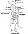
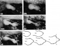
The coelom and its derivatives in the frog
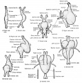
Development of the amphibian heart
Larval respiration in the frog. Development of the external gills
Larval respiration in the frog. Changes from internal to external gill circulation
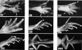
Blood vascular system of the developing frog embryo Early embryo, from the right side.
Blood vascular system of the developing frog embryo Late frog embryo, from the right side.
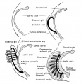
Fate of the aortic arches of the frog embryo
Blood vascular system of the frog tadpole Venous system ventral view
Blood vascular system of the frog tadpole Arterial system ventral view
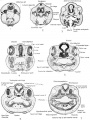
Serial transverse sections of the 11 mm frog larva
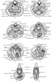
Serial transverse sections of the 11 mm frog larva


Appendix: Chronological Summary of Organ Anlagen Appearance of Rana pipiens
Parts of the middle and inner ear of the frog schematized drawing
Olfactory organs of Rana pipiens tadpoles at 20 mm total body length
Stages in the metamorphosis of Rana pipiens
Stages in the metamorphosis of Rana pipiens
Stages in the metamorphosis of Rana pipiens
Stages in the metamorphosis of Rana pipiens
Stages in the metamorphosis of Rana pipiens
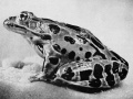
Adult Frog


| Historic Disclaimer - information about historic embryology pages |
|---|
|
Reference
Rugh R. Book - The Frog Its Reproduction and Development. (1951) The Blakiston Company.
Cite this page: Hill, M.A. (2024, April 19) Embryology Book - The Frog Its Reproduction and Development 17. Retrieved from https://embryology.med.unsw.edu.au/embryology/index.php/Book_-_The_Frog_Its_Reproduction_and_Development_17
- © Dr Mark Hill 2024, UNSW Embryology ISBN: 978 0 7334 2609 4 - UNSW CRICOS Provider Code No. 00098G