Book - The Elements of Embryology - Mammalian 4
| Embryology - 25 Apr 2024 |
|---|
| Google Translate - select your language from the list shown below (this will open a new external page) |
|
العربية | català | 中文 | 中國傳統的 | français | Deutsche | עִברִית | हिंदी | bahasa Indonesia | italiano | 日本語 | 한국어 | မြန်မာ | Pilipino | Polskie | português | ਪੰਜਾਬੀ ਦੇ | Română | русский | Español | Swahili | Svensk | ไทย | Türkçe | اردو | ייִדיש | Tiếng Việt These external translations are automated and may not be accurate. (More? About Translations) |
Foster M. Balfour FM. Sedgwick A. and Heape W. The Elements of Embryology (1883) Vol. 1. (2nd ed.). London: Macmillan and Co.
| Historic Disclaimer - information about historic embryology pages |
|---|
| Pages where the terms "Historic" (textbooks, papers, people, recommendations) appear on this site, and sections within pages where this disclaimer appears, indicate that the content and scientific understanding are specific to the time of publication. This means that while some scientific descriptions are still accurate, the terminology and interpretation of the developmental mechanisms reflect the understanding at the time of original publication and those of the preceding periods, these terms, interpretations and recommendations may not reflect our current scientific understanding. (More? Embryology History | Historic Embryology Papers) |
The Organs Derived from Mesoblast
The vertebral column
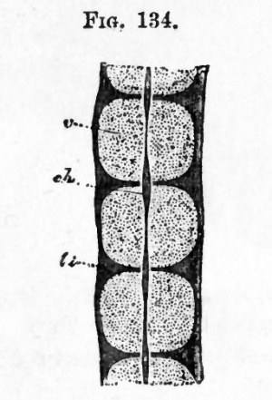
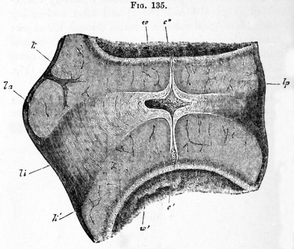
The early development of the perichordal cartilaginous tube and rudimentary neural arches is almost the same in Mammals as in Birds. The differentiation into vertebral and intervertebral regions is the same in both groups; but instead of becoming divided as in Birds into two segments attached to two adjoining vertebrae, the intervertebral regions become in Mammals wholly converted into the intervertebral ligaments (Fig. 135 li). There are three centres of ossification for each vertebra, two in the arch and one in the centrum.
The fate of the notochord is in important respects different from that in Birds. It is first constricted in the centres of the vertebrae (Fig. 134) and disappears there shortly after the beginning of ossification ; while in the intervertebral regions it remains relatively unconstricted (Figs. 134 and 135 c) and after undergoing certain histological changes remains through life as part of the nucleus pulposus in the axis of the intervertebral ligaments. There is also a slight swelling of the notochord near the two extremities of each vertebra (Fig. 135 c and c"}.
In the persistent vertebral constriction of the notochord Mammals retain a more primitive and piscine mode of formation of the vertebral column thai} tfye majority either of the Reptilia or Amphibia.
The Skull
Excepting in the absence of the interorbital plate, the early development of the Mammalian cranium resembles in all essential points that of Aves, to our account of which on p. 235 et seq. we refer the reader.
Fig. 135. Longitudinal section through the intervertebral ligament and adjacent parts of two vertebra from the thoracic region of an advanced embryo of a sheep. (From Kolliker.)
- la. .ligamentum longitudinale anterius ; Ip. ligamentum long, posterius ; li. ligamentum intervertebrale ; , k'. epiphysis of vertebra ; w. and w f . anterior and posterior vertebrae ; c. intervertebral dilatation of notochord ; c.' and c". vertebral dilatation of notochord..
The early changes in the development of the visceral arches and clefts have already been described, but the later changes undergone by the skeletal elements of the first two visceral arches are sufficiently striking to need a special description.
The skeletal bars of both the hyoid and mandibular arches develop at first more completely than in any of the other types above Fishes ; they are articulated to each other above, while the pterygo-palatine bar is quite distinct.
The main features of the subsequent development are undisputed, with the exception of that of the upper end of the hyoid, which is still controverted. The following is Parker's account for the Pig.
The mandibular and hyoid arches are at first very similar, their dorsal ends being somewhat incurved, and articulating together.
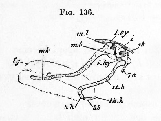
In a somewhat later stage (Fig. 136) the upper end of the mandibular bar (mb), without becoming segmented from the ventral part, becomes distinctly swollen, and clearly corresponds to the quadrate region of other types. The ventral part of the bar constitutes Meckel's cartilage (mk).
Fig. 136. Embryo Pra, An inch and a third long ; side view of mandibular and hyoid arches. the main hyoid arch is seen as displaced backwards after segmentation from the incus. (From Parker.)
- tg. tongue ; mJc. Meckelian cartilage ; ml. body of malleus ; ml). inanubrium or handle of the malleus ; tjy. tegmen tympani ; ?'. incus ; st. stapes ; i.hy. interhyal ligament ; st.h. stylohyal cartilage ; h.h. hypohyal ; b.h. basibranchial ; th.h. rudiment of first branchial arch ; la. facial nerve.
The hyoid arch has in the meantime become segmented into two parts, an upper part (i), which eventually becomes one of the small bones of the ear the incusand a lower part which remains as the anterior cornu of the hyoid (st.h). The two parts continue to be connected by a ligament.
The incus is articulated with the quadrate end of the mandibular arch, and its rounded head comes in contact with the stapes (Fig. 136, sf) which is segmented from the fenestra ovalis.
According to some authors the stapes is independently formed from mesoblast cells surrounding a branch of the internal carotid artery.
The main arch of the hyoid becomes divided into a hypohyal (hJi) below and a stylohyal (st.h) above, and also becomes articulated with the basal element of the arch behind (bh).
In the course of further development the Meckelian part of the mandibular arch becomes enveloped in a superficial ossification forming the dentary. Its upper end, adjoining the quadrate region, becomes calcified and then absorbed, and its lower, with the exception of the extreme point, is ossified and subsequently incorporated in the dentary.
The quadrate region remains relatively stationary in growth as compared with the adjacent parts of the skull, and finally ossifies to form the malleiis. The processus
gracilis of the malleus is the primitive continuation into Meckel's cartilage.
The malleus and incus are at first embedded in the connective tissue adjoining the tympanic cavity, which with the Eustachian tube is the persistent remains of the hyomandibular cleft ; and externally to them a bone known as the tympanic bone becomes developed so that they become placed between the tympanic bone and the periotic capsule. In late foetal life they become transported completely within the tympanic cavity, though covered by a reflection of the tympanic mucous membrane.
The dorsal end of the part of the hyoid separated from the incus becomes ossified as the tympano-hyal, and is anchylosed with the adjacent parts of the periotic capsule. The middle part of the bar just outside the skull forms the stylo-hyal (styloid process in man) which is attached by ligament to the anterior cornu of the hyoid (cerato-hyal). The tympanic membrane and external auditory meatus develop as in the chick (p. 166).
The ribs and sternum appear to develop in Mammals as in Birds (p. 234).
The pectoral girdle, as in Birds (p. 234), arises as a continuous plate of cartilage, the coracoid element of which is however much reduced.
The clavicle in Man is provided with a central axis of cartilage, and its mode of ossification is intermediate between that of a true cartilage bone and a membrane bone.
The pelvic girdle is formed in cartilage as in Birds, but in Man at any rate the pubic part of the cartilage is formed independently of the remainder. There are the usual three centres of ossification, which unite eventually into a single bone the innominate bone. The pubis and ischium of each side unite ventrally, so as completely to enclose the obturator foramen.
The skeleton of the limbs develops so far as is known as in Birds, from a continuous mesoblastic blastema, within which the corresponding cartilaginous elements of the limbs become differentiated.
The body cavity
The development of the body cavity and its subsequent division into pericardia! pleural and peritoneal cavities is precisely the same in Mammalia as in Aves (p. 264 et seq.). But in Mammalia a further change takes place, in that by the formation of a vertical partition across the body cavity, known as the diaphragm, the pleural cavities, containing the lungs, become isolated from the remainder of the body or peritoneal cavity. As shewn by their development the so-called pleurae or pleural sacs are simply the peritoneal linings of the anterior divisions of the body cavity, shut off from the remainder of the body cavity by the diaphragm.
The vascular system
The heart. The two tubes out of which the heart is formed appear at the sides of the cephalic plates, opposite the region of the mid- and hind-brain (Fig. 107). They arise at a time when the lateral folds which form the ventral wall of the throat are only just becoming visible. Each half of the heart originates in the same way as in the chick ; and the layer of the splanchnic mesoblast, which forms the muscular wall for each part (ahh). has at first the form of a half tube open below to the hypoblast.
On the formation of the lateral folds of the splanchnic walls, the two halves of the heart become carried inwards and downwards, and eventually meet on the ventral side of the throat. For a short time they here remain distinct, but soon coalesce into a single tube.
In Birds, it will be remembered, the heart at first has the form of two tubes, which however are in contact in front. It arises at a time when the formation of the throat is very much more advanced than in Mammalia ; when in fact the ventral wall of the throat is established as far back as the front end of the heart.
In the lower types the heart does not appear till the ventral wall of the throat is completely established, and it has from the first the form of a single tub".
It is therefore probable that the formation of the heart as two cavities is a secondary mode of development, which has been brought about by variations in the period of the closing in of the wall of the throat.
The later development of the heart is in the main similar to that of the chick (p. 256 et seq.).
The arterial system. The early stages of the arterial system of Mammalia are similar to those in Birds. Five arterial arches are formed, the three posterior of which wholly or in part persist in the adult.
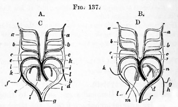
The bulbus arteriosus is divided into two (fig. 137 B), but the left fourth arch (e), instead of, as in Birds, the right, is that continuous with the dorsal aorta, and the right fourth arch (i) is only continued into the right vertebral and right subclavian arteries.
The fifth pair of arches which is continuous with one of the divisions of the bulbus arteriosus gives origin to the two pulmonary arteries. Both these however are derived from the arch on one side, viz. the left (fig. 137 B); whereas in Birds, one pulmonary artery comes from the left and the other from the right fifth arch (fig. 137 A).
The ductus Botalli of the fifth arch (known in Man as the ductus arteriosus) of the side on which the pulmonary arteries are formed, may remain (e.g. in Man) as a solid cord connecting the common stem of the pulmonary aorta with the systemic aorta.
The diagram, Fig. 137, copied from Rathke, shews at a glance the character of the metamorphosis the arterial arches undergo in Birds and Mammals.
Fig. 137. Diagrams illustrating the metamorphosis of the arterial arches in a bird A. and a mammal B. (From Mivart after Kathke.)
- A. a. internal carotid ; b. external carotid ; c. common carotid ; d. systemic aorta ; e. fourth arch of right side (root of dorsal aorta) / right subclavian ; g. dorsal aorta ; h. left subclavian (fourth arch of left side) ; i. pulmonary artery ; Jc. and I. right and left ductus Botalli of pulmonary arteries,
- B; a.' internal carotid ; b. external carotid ; c. common carotid ; d.' systemic aorta ; e. fourth arch of left side (root of dorsal aorta) ; /. dorsal aorta ; g. left vertebral artery ; h. left subclavian artery ; i. right subclavian (fourth arch of right side) ; Jc. right vertebral ; I. continuation of right subclavian ; m. pulmonary artery ; n. ductus Botalli of pulmonary artery.
In some Mammals both subclavians spring from a trunk common to them and the carotids (arteria anonyma) ; or as in Man and some other Mammals, the left one arises from the systemic aorta just beyond the carotids. Various further modifications in the origin of the subclavians are found in Mammalia, but they need not be specified in detail. The vertebral arteries arise in close connection with the subclavians, whereas in Birds they arise from the common carotids.
The venous system
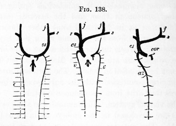
In Mammals the same venous trunks are developed in the embryo as in Birds (Fig. 138 A). The anterior cardinals or external jugulars form the primitive veins of the anterior part of the body, and the internal jugulars and anterior vertebrals are subsequently formed. The subclavians (Fig. 138 A, s), developed on the formation of the anterior limbs, also pour their blood into these primitive trunks. In the lower Mammalia (Monotremata, Marsupialia, Insectivora, some Rodentia, etc.) the two ductus Cuvieri remain as the two superior venae cavse, but more usually an anastomosis arises between the right and left innominate veins, and eventually the whole of the blood of the left superior cava is carried to the right side, and there is left only a single superior cava (Fig. 138 B and C). A small rudiment of the Jeft superior cava remains however as the sinus coronarius and receives the coronary vein from the heart (Figs. 138 C, cor and 139 cs).
The posterior cardinal veins form at first the only veins receiving the blood from the posterior part of the trunk and kidneys ; and on the development of the hind limbs receive the blood from them also.
An unpaired vena cava inferior becomes eventually developed, and gradually carries off a larger and larger portion of the blood originally returned by the posterior cardinals. It unites with the common stem of the allantoic and vitelline veins in front of the liver.
Fig. 138. Diagram of the development of the paired venous system of mammals (man). (From Gegenbaur.)
- j. jugular vein ; cs. vena cava superior ; s. subclavian veins ; c. posterior cardinal vein ; v. vertebral vein ; az. azygos vein ; cor. coronary vein.
- A. Stage in which the cardinal veins have already disappeared. Their position is indicated by dotted lines.
- B. Later stage when the blood from the left jugular vein is carried into the right to form the single vena cava superior ; a remnant of the left superior cava being however still left.
- C. Stage after the left vertebral vein has disappeared ; the right vertebral remaining as the azygos vein. The coronary vein remains as the last remnant of the left superior vena cava.
At a later period a pair of trunks is established bringing the blood from the posterior part of the cardinal veins and the crural veins directly into the vena cava inferior (Fig. 139, il). These vessels, whose development has not been adequately investigated, form the common iliac veins, while the posterior ends of the cardinal veins which join them become the hypogastric veins (Fig. 139 hy).

Posterior vertebral veins, similar to those of Birds, are established in connection with the intercostal and lumbar veins, and unite anteriorly with the front part of the posterior cardinal veins (Fig. 138 A).
Upon the formation of the posterior vertebral veins, and upon the inferior vena cava becoming more important, the middle part of the posterior cardinals becomes completely aborted (Fig. 139 c), the anterior and posterior parts still persisting, the former as the continuations of the posterior vertebrals into the anterior vena cava (az\ the latter as the hypogastric veins (%).
Though in a few Mammalia both the posterior vertebrals persist, a transverse connection is usually established between them, and the one (the right), becoming the more important, constitutes the azygos vein (Fig. 139 az\ the persisting part of the left forming the hemiazygos vein (ha).
The remainder of the venous system is formed in the embryo by the vitelline and allantoic veins, the former being eventually joined by the mesenteric vein so as to constitute the portal vein.
The vitelline vein is the first part of this system established, and divides near the heart into two veins bringing back the blood from the yolk-sac (umbilical vesicle). The right vein soon however aborts.
The allantoic (anterior abdominal) veins are originally paired. They are developed very early, and at first course along the still widely open somatic walls of the body, and fall into the single vitelline trunk in front. The right allantoic vein disappears before long, and the common trunk formed by the junction of the vitelline and allantoic veins becomes considerably elongated. This trunk is soon envelop'ed by the liver, and later in its passage through, gives off branches to, and also receives branches from this organ near its anterior exit. The main trunk is however never completely aborted, as in the embryos of other types, but remains as the ductus venosus Arantii.
With the development of the placenta the allantoic vein becomes the main source of the ductus venosus, and the vitelline or portal vein, as it may perhaps be now conveniently called, ceases to join it directly, but falls into one of its branches in the liver.
The vena cava inferior joins the continuation of the ductus venosus in front of the liver, and, as it becomes more important, it receives directly the hepatic veins which originally brought back blood into the ductus venosus. The ductus venosus becomes moreover merely a small branch of the vena cava.
At the close of foetal life the allantoic vein becomes obliterated up to its place of entrance into the liver; the ductus venosus becomes a solid cord the so-called round ligament and the whole of the venous blood is brought to the liver by the portal vein.
Owing to the allantoic (anterior abdominal) vein having merely a foetal existence an anastomosis between the iliac veins and the portal system by means of the anterior abdominal vein is not established.
The supra-renal bodies
These are paired bodies lying anterior to the kidneys and are formed of two parts, (1) a cortical and (2) a medullary portion. They first appear in the Rabbit on the 12th or 13th day of gestation, and arise as masses of mesoblast cells lying between the aorta and the mesentery and to one side of the former. On the 14th day they are well marked, and lying dorsal to them is another mass of cells which is found to be continuous with the sympathetic nervous system.
On the 16th day processes from the sympathetic mass enter the mesoblastic tissue and become transformed into the medullary portion of the adult suprarenal ; while the mesoblastic tissue gives rise to the cortical layer,
The urinogenital organs
The history of these organs in Mammalia, excepting so far as concerns the lower parts of the urinogenital ducts, is the same as in the Chick.
The Wolffian body and duct first appear, and are followed by the Miillerian duct and the kidney. The exact method of development of the latter structures has not been followed so completely as in the Chick; and it is not known whether the peculiar structures found. at the anterior end of the commencing Miillerian duct in Aves occur in Mammalia.
The history of the generative glands is essentially the same as in the Chick.
Outgrowths from a certain number of Malpighian bodies in the Wolffian body are developed along the base of the testis, and enter into connection with the seminiferous stroma. It is not certain to what parts of the testicular tubuli they give rise, but they probably form at any rate the vasa recta and rete vasculosum. Similarly intrusions from the Malpighian bodies make their way into the ovary of the female, and give rise to cords of tissue which may persist throughout life.
The vasa efferentia (coni vasculosi) appear to be derived from the glandular tubes of part of the Wolffian body. The Wolffian duct itself becomes in the male the vas deferens and the convoluted canal of the epididymis ; the latter structure except the head being entirely derived from the Wolffian duct.
The functionless remains of the embryonic organs described for the chick (p. 224) are found also in mammals.
The Miillerian ducts persist in the female as the Fallopian tubes and uterus.
The lower parts of the urinogenital ducts are somewhat further modified in the Mammalia than the Chick.
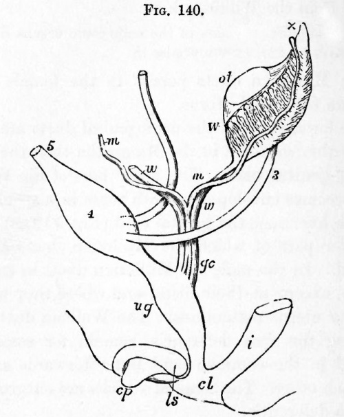
The genital cord. The lower part of the Wolffian ducts becomes enveloped in both sexes in a special cord of tissue, known as the genital cord (Fig. 1 40 gc), within the lower part of which the Mulleriaii ducts are also enclosed. In the male the Miillerian ducts in this cord atrophy, except at their .distal end where they unite to form the uterus masculinus. The Wolffian ducts, after becoming the vasa deferentia, remain for some time enclosed in the common cord but afterwards separate from each other. The seminal vesicles are outgrowths of the vasa deferentia.
In the female the Wolffian ducts within the genital cord atrophy, though rudiments of them are for a long time visible or even permanently persistent. The lower parts of the Miillerian ducts unite to form the vagina and body of the uterus while the upper become the horns of the uterus and the Fallopian tubes. The junction commences in the middle and extends forwards and backwards ; the stage with a median junction being retained permanently in Marsupials.
The urinogenital sinus and external generative organs. The dorsal part of the cloaca with the alimentary tract becomes partially constricted off from the ventral, which then forms a urinogenital sinus (Fig. 140 ug).
Fig. 140. Diagram of the urinogenital organs of a mammal at an early stage. (After Allen Thomson ; from Quain's Anatomy.)
- The parts are seen chiefly in profile, but the Mlillerian and Wolffian ducts are seen from the front.
- 3. ureter; 4. urinary bladder; 5. urachus ; ot. genital ridge (ovary or testis) ; W. left Wolffian body ; x. part at apex from which coni vasculosi are afterwards developed ; w. Wolffian duct ; m. Miillerian duct ; gc. genital cord consisting of Wolffian and Miillerian ducts bound up in a common sheath ; i. rectum ; ug. urinogenital sinus ; cp. elevation which becomes the clitoris or penis ; Is. ridge from which the labia majora or scrotum are developed.
In the course of development the urinogenital sinus becomes, in all Mammalia but the Ornithodelphia, completely separated from the intestinal cloaca, and the two parts obtain separate external openings. The ureters (Fig. 140, 3) open higher up than the other ducts into the stalk of the allantois which here dilates to form the bladder. That part of the stalk which connects the bladder with the ventral wall of the body constitutes the urachus, and loses its lumen before the close of embryonic life. The part of the stalk of the allantois below the openings of the ureters narrows to form the urethra, which opens together with the Wolffi an and Mullerian ducts into the urogenital cloaca.
In front of the urogenital cloaca there is formed a genital prominence (Fig. 140 cp) with a groove continued from the urinogenital opening, and on each side a genital fold (Is). In the male the sides of the groove on the prominence coalesce together, embracing between them the opening of the urinogenital cloaca, and the prominence itself gives rise to the penis, along which the common urinogenital passage is continued. The two genital folds unite from behind forwards to form the scrotum.
In the female the groove on the genital prominence gradually disappears, and the prominence remains as the clitoris, which is therefore the homologue of the penis : the two genital folds form the labia majora. The urethra and vagina open independently into the common urogenital sinus.
The Elements of Embryology - Volume 2 (1883)
The History of the Mammalian Embryo: General Development | Embryonic Membranes and Yolk-Sac | Organs from Epiblast | Organs from Mesoblast | Alimentary Canal | Appendix | Figures
| Historic Disclaimer - information about historic embryology pages |
|---|
| Pages where the terms "Historic" (textbooks, papers, people, recommendations) appear on this site, and sections within pages where this disclaimer appears, indicate that the content and scientific understanding are specific to the time of publication. This means that while some scientific descriptions are still accurate, the terminology and interpretation of the developmental mechanisms reflect the understanding at the time of original publication and those of the preceding periods, these terms, interpretations and recommendations may not reflect our current scientific understanding. (More? Embryology History | Historic Embryology Papers) |
Glossary Links
- Glossary: A | B | C | D | E | F | G | H | I | J | K | L | M | N | O | P | Q | R | S | T | U | V | W | X | Y | Z | Numbers | Symbols | Term Link
Cite this page: Hill, M.A. (2024, April 25) Embryology Book - The Elements of Embryology - Mammalian 4. Retrieved from https://embryology.med.unsw.edu.au/embryology/index.php/Book_-_The_Elements_of_Embryology_-_Mammalian_4
- © Dr Mark Hill 2024, UNSW Embryology ISBN: 978 0 7334 2609 4 - UNSW CRICOS Provider Code No. 00098G

