Book - The Early Embryology of the Chick 3
| Embryology - 19 Apr 2024 |
|---|
| Google Translate - select your language from the list shown below (this will open a new external page) |
|
العربية | català | 中文 | 中國傳統的 | français | Deutsche | עִברִית | हिंदी | bahasa Indonesia | italiano | 日本語 | 한국어 | မြန်မာ | Pilipino | Polskie | português | ਪੰਜਾਬੀ ਦੇ | Română | русский | Español | Swahili | Svensk | ไทย | Türkçe | اردو | ייִדיש | Tiếng Việt These external translations are automated and may not be accurate. (More? About Translations) |
Patten BM. The Early Embryology of the Chick. (1920) Philadelphia: P. Blakiston's Son and Co.
| Online Editor |
|---|
| This historic 1920 paper by Bradley Patten described the understanding of chicken development. If like me you are interested in development, then these historic embryology textbooks are fascinating in the detail and interpretation of embryology at that given point in time. As with all historic texts, terminology and developmental descriptions may differ from our current understanding. There may also be errors in transcription or interpretation from the original text. Currently only the text has been made available online, figures will be added at a later date. My thanks to the Internet Archive for making the original scanned book available.
By the same author: Patten BM. Developmental defects at the foramen ovale. (1938) Am J Pathol. 14(2):135-162. PMID 19970381 Those interested in historic chicken development should also see the earlier text The Elements of Embryology (1883). Foster M. Balfour FM. Sedgwick A. and Heape W. The Elements of Embryology (1883) Vol. 1. (2nd ed.). London: Macmillan and Co.
Modern Notes |
| Historic Disclaimer - information about historic embryology pages |
|---|
| Pages where the terms "Historic" (textbooks, papers, people, recommendations) appear on this site, and sections within pages where this disclaimer appears, indicate that the content and scientific understanding are specific to the time of publication. This means that while some scientific descriptions are still accurate, the terminology and interpretation of the developmental mechanisms reflect the understanding at the time of original publication and those of the preceding periods, these terms, interpretations and recommendations may not reflect our current scientific understanding. (More? Embryology History | Historic Embryology Papers) |
The Process of Segmentation
The Effect of Yolk on Segmentation
Immediately after its fertilization the ovum enters upon a series of mitotic divisions which occur in close succession. This series of divisions constitutes the process of segmentation or cleavage. In birds segmentation takes place before the egg is laid, during the time it is traversing the oviduct.
A mitotic division, whether it be a cleavage division of the ovum or the division of some other cell, is carried out by the active protoplasm of the cell. The food material stored in an egg cell as deutoplasm is non-living and inert. The deutoplasm has no part in mitosis except as its mass mechanically influences the activities of the protoplasm of the cell. It is obvious that any considerable amount of yolk will retard the division, or prevent the complete division, of the fertilized ovum. The amount and distribution of the yolk will therefore determine the type of segmentation.
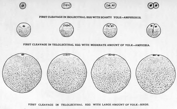
Figure 4 shows diagrammatically the manner in which the first cleavage division is carried out in three types of eggs having different relative amounts and different distributions of yolk and protoplasm. In the egg of Amphioxus the yolk is relatively meager in amount and fairly uniformly distributed throughout the cell. An ovum with such a yolk distribution is termed isolecithal (homolecithal). An isolecithal egg undergoes a type of cleavage which is essentially an unmodified mitosis. The yolk is not sufficient in amount, nor sufficiently localized to alter the usual mode of cell division.
Fig. 4. Schematic diagrams to indicate the effect of yolk on the first cleavage division.
In Amphibia the ovum contains a considerable amount of yolk and the accumulation of the yolk at one pole has crowded the nucleus and active cytoplasm of the ovum toward the opposite pole. An egg in which the yolk is thus concentrated at one pole is termed telolecithal. Cleavage in such an egg is initiated at the animal pole where the nucleus and most of the active cytoplasm are located. The division of the nucleus is a typical mitotic division. The division of the cytoplasm is effected rapidly at the animal pole of the egg where the active cytoplasm is aggregated. When, however, the yolk mass is encountered, the process is greatly retarded. So slowly, in fact, is the division of the yolk accomplished, tlrat succeeding cell divisions begin at the animal pole of the egg before the first cleavage is completed at the vegetative pole.
The eggs of birds are also telolecithal, but the amount of yolk which they contain is both relatively and actually much greater than that in Amphibian eggs. Cleavage in bird's eggs begins as it does in the eggs of Amphibia, but the mass of the inert yolk material in them is so great that the yolk is not divided. The process of segmentation is limited to the small disc of protoplasm lying on the surface of the yolk at the animal pole, and is for this reason referred to as discoidal cleavage (Fig. 5). The fact that the whole egg is not divided is indicated by designating the process as partial (meroblastic) cleavage in distinction to the complete cleavage (holoblastic) seen in eggs containing less yolk. The cells formed in the process of segmentation are known as blastomeres whether they are completely separated as results in holobastic cleavage or only partially separated as results in meroblastic cleavage.
The Unsegmented Blastodisc
In the egg of a bird which is about to undergo cleavage, the disc of active protoplasm at the animal pole (blastodisc) is a whitish, circular area about three millimeters in diameter. The central portion of the blastodisc is surrounded by a somewhat darker appearing marginal area known as the periblast. The protoplasm of the blastodisc, especially in the periblast region, blends into the underlying white yolk 50 that it is difficult to make out any line of demarcation between the two. It is in the central area of the blastodisc that cleavage furrows first appear. Neither the nuclei resulting from the early cleavages nor the cleavage furrows invade the marginal periblast until very late in the process of segmentation.
The Sequence and Orientation of the Cleavage Divisions in Birds
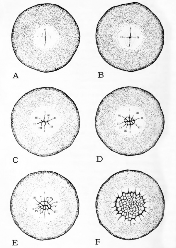
The nature of the series of divisions in the meroblastic, discoidal cleavage characteristic of the eggs of birds is largely determined by the amount and distribution of the yolk. Another determining factor is the tendency of mitotic spindles to develop so that the long axis of the spindle lies at right angles to the axis of least dimension of the mass of unmodified cytoplasm. The cleavage furrow always arises at right angles to the long axis of the mitotic spindle. Figure 5 shows the succession of the cleavage divisions in the egg of the pigeon. The diagrams represent surface views of the blastodisc and an area of the surrounding yolk, the shell and albumen having been removed. The observer is looking directly at the animal pole. Figure 5, A, should be compared with Figure 4. The diagrams of Figure 4 are of sections cut in a plane which passes vertically through the blastodisc and which is at right angles to the plane of the first cleavage (Fig. 5, 1-1)- The first cleavage furrow cuts into the egg in a plane coinciding with the imaginary axis passing through the animal pole and the vegetative pole. The two daughter cells or blastomeres resulting from the first cleavage are not completely walled off but each remains unseparated from the underlying yolk (Fig. 4).
In each of the two blastomeres resulting from the first cleavrage division, mitotic spindles initiating the second cleavage arise at right angles to the position which was occupied by the first cleavage spindle. This determines that the two second cleavage furrows will be at right angles to the first. Since these two second cleavage furrows lie in the same plane and are apparently continuous they are usually considered together. They mark the position of the second cleavage plane which cuts the egg in the animal-vegetative axis but which lies at right angles to the first cleavage plane (Fig. 5, B, 11-11). A very good way of getting a clear conception of the orientation of the cleavage planes is to cut them in an apple. Let the core of the apple represent the animal- vegetative axis of the egg. The first cleavage furrow can be represented by notching the apple lengthwise, that is as one ordinarily starts to split an apple into halves. The second cleavage furrow can be represented by cutting into the apple again in a plane passing through the axis of the core, but at right angles to the first cut, as one would start to quarter the apple.
The third cleavage furrows are variable in number and in position. In the most typical cases each of the four blastomeres established by the first two cleavages divides again so that eight blastomeres are formed (Fig. 5, C). Frequently, however, the third cleavage appears at first in only two of the blastomeres, so that six cells result instead of eight.
The fourth series of cleavages takes place in such a manner that the central (apical) ends of the eight cells established by the third cleavage are cut off from their peripheral portions.* The combined contour of the fourth cleavage furrows forms a small irregularly circular furrow the center of which is the point at which the first two cleavage planes intersect (Fig. 5, D). The central cells now appear completely separated in a surface view of the blastoderm, but sections show them still unseparated from the underlying yolk.
Fig. 5. Surface aspect of blastoderm at various stages of cleavage. (Based on Blount's photomicrographs of the pigeon's egg.)
- The blastoderm and the immediately surrounding yolk are viewed directly from the animal pole, the shell and albumen having been removed. The order in which vthe cleavage furrows have appeared is indicated on the diagrams by Roman numerals.
- A, first cleavage; B, second cleavage; C, third cleavage; D, fourth cleavage; E, fifth cleavage; F, early morula.
After the fourth, the succession of cleavages becomes irregular. In surface view it is possible to make out cleavage furrows that divide off additional apical cells, and other, radial furrows that further divide the peripheral cells. Figure 5, E andF, show the increase in number of cells and their extension out over the surface of the yolk, resulting from the succession of cleavages. When the process of segmentation has progressed to the stage in which the succession of cleavages is irregular and the number of cells considerable, the term blastoderm is applied to the entire group of blastomeres formed by the cleavage of the blastodisc.
In addition to the cleavages which are indicated on the surface, at about the 32-cell stage sections show cleavage planes of# an entirely different character. These cleavages appear below the surface and parallel to it. They establish a superficial layer of cells which are completely delimited. These superficial cells rest upon a layer of cells which are continuous on their deep faces with the yolk. Continued divisions of the same type eventually establish several strata of superficial cells. This process appears first in the central portion of the blastoderm. It progresses centrifugally as the blastoderm increases in size but does not extend to its extreme margin. The peripheral margin of the blastoderm remains a single cell in thickness and the cells there lie unseparated from the yolk.
While but a single spermatozoon takes part in fertilization other spermatoza become lodged in the cytoplasm of the blastodisc. The nuclei of these spermatozoa migrate to the peripheral part of the blastoderm where they are recognizable for some time as the so-called accessory sperm nuclei. Some of them appear to undergo divisions which are accompanied by slight indications of division in the adjacent cytoplasm. The short superficial grooves thus formed are termed accessory cleavage furrows. No cells are formed by the accessory "cleavages." The sperm nuclei soon degenerate, the superficial furrows fade out, and usually as early as the 32-cell stage all traces of the process have disappeared without, as far as is known, affecting in any way the development of the embryo.
- Next: Entoderm
The Early Embryology of the Chick: Introduction | Gametes and Fertilization | Segmentation | Entoderm | Primitive Streak and Mesoderm | Primitive Streak to Somites | 24 Hours | 24 to 33 Hours | 33 to 39 Hours | 40 to 50 Hours | Extra-embryonic Membranes | 50 to 55 Hours | Day 3 to 4 | References | Figures | Site links: Embryology History | Chicken Development
| Historic Disclaimer - information about historic embryology pages |
|---|
| Pages where the terms "Historic" (textbooks, papers, people, recommendations) appear on this site, and sections within pages where this disclaimer appears, indicate that the content and scientific understanding are specific to the time of publication. This means that while some scientific descriptions are still accurate, the terminology and interpretation of the developmental mechanisms reflect the understanding at the time of original publication and those of the preceding periods, these terms, interpretations and recommendations may not reflect our current scientific understanding. (More? Embryology History | Historic Embryology Papers) |
Glossary Links
- Glossary: A | B | C | D | E | F | G | H | I | J | K | L | M | N | O | P | Q | R | S | T | U | V | W | X | Y | Z | Numbers | Symbols | Term Link
Cite this page: Hill, M.A. (2024, April 19) Embryology Book - The Early Embryology of the Chick 3. Retrieved from https://embryology.med.unsw.edu.au/embryology/index.php/Book_-_The_Early_Embryology_of_the_Chick_3
- © Dr Mark Hill 2024, UNSW Embryology ISBN: 978 0 7334 2609 4 - UNSW CRICOS Provider Code No. 00098G


