Book - The Cell - outlines of general anatomy and physiology (1895): Difference between revisions
mNo edit summary |
mNo edit summary |
||
| (4 intermediate revisions by the same user not shown) | |||
| Line 3: | Line 3: | ||
{{Historic Disclaimer}} | {{Historic Disclaimer}} | ||
=The Cell - Outlines of General Anatomy and Physiology= | =The Cell - Outlines of General Anatomy and Physiology= | ||
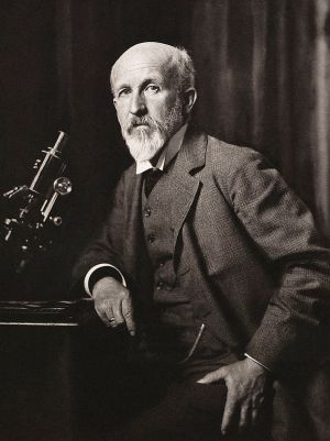
[[File:Oscar Hertwig.jpg|thumb|link=Embryology History - Oscar Hertwig|Oscar Hertwig (1849-1922)]] | |||
Dr. {{Oscar Hertwig}} | Dr. {{Oscar Hertwig}} | ||
Translated By M. Campbell, | Translated By M. Campbell, and Edited by | ||
Henry Johnstone Campbell, M.D | Henry Johnstone Campbell, M.D | ||
Assistant Physician to the City of | Assistant Physician to the City of London | ||
| Line 159: | Line 160: | ||
==Editor's Preface== | ==Editor's Preface== | ||
The translation of Professor Hertwig's book bas been no easy | |||
task. The extreme complexity of much of the matter treated, in | |||
The translation of Professor Hertwig | |||
task. | |||
addition to the large number of sabjects referred to, has often | addition to the large number of sabjects referred to, has often | ||
rendered it extremely difficult to express the author's meaning in | rendered it extremely difficult to express the author's meaning in | ||
| Line 171: | Line 170: | ||
account of the Anatomy and Physiology of the Cell, as the one | account of the Anatomy and Physiology of the Cell, as the one | ||
contained in Professor Hertwig's book. | contained in Professor Hertwig's book. | ||
In many cases it has been extremely difficult to find equivalents | In many cases it has been extremely difficult to find equivalents | ||
for terms used in the German. Amongst these the word | for terms used in the German. Amongst these the word "Anlage" may be specially mentioned. Various terms have been | ||
used by different translators to express the meaning of this word, | used by different translators to express the meaning of this word, | ||
but none of them seems to be applicable to all cases. Professor | but none of them seems to be applicable to all cases. Professor | ||
Mark has introduced the word | Mark has introduced the word "fundament," and Mr. Mitchell has | ||
suggested the term | suggested the term "blast," but neither of these appears to express | ||
the meaning of the German word sufficiently accurately to justify | the meaning of the German word sufficiently accurately to justify | ||
the use of either of them exclusively. Hence, we thought it best | the use of either of them exclusively. Hence, we thought it best | ||
in some cases to employ the somewhat cumbrous expression, | in some cases to employ the somewhat cumbrous expression, | ||
" elemental germ," although it is undoubtedly open to objection ; | "elemental germ," although it is undoubtedly open to objection ; | ||
however, it frequently seemed to ns to convey the author's idea | however, it frequently seemed to ns to convey the author's idea | ||
most correctly. On other occasions we have thought better to | most correctly. On other occasions we have thought better to | ||
make use of a paraphrase. | make use of a paraphrase. | ||
that the English student might wish to consult. The frequent | Several additions have been made to the Bibliography of papers that the English student might wish to consult. The frequent | ||
quotations from English authors have in most cases been | quotations from English authors have in most cases been verified by reference to the originals ; but in some cases, despite careful search, we have been unable to find the passages in question. | ||
search, we have been unable to find the passages in question. | |||
H. Johnstone Campbell. | H. Johnstone Campbell. | ||
64, Welheck Street, London, W, | |||
==Contents== | ==Contents== | ||
CHAPTER I. | CHAPTER I. Introduction | ||
Introduction | |||
The Higtory of the Cell Theory | The Higtory of the Cell Theory | ||
| Line 210: | Line 204: | ||
Literature | Literature | ||
CHAPTER II. | CHAPTER II. The ChEMICO-PHYSICAL AMD MOBPHOLOOICAL Properties OF THB Cell | ||
I. The Chemioo-physioal and Morphological Properties of the Protoplasm | I. The Chemioo-physioal and Morphological Properties of the Protoplasm | ||
| Line 226: | Line 218: | ||
[e) Uniformity of Protoplasm. Diversity of the Cell | [e) Uniformity of Protoplasm. Diversity of the Cell | ||
( | (f) Various examples of the Structure of the Cell-body | ||
1. Cells consisting almost entirely of Protoplasm | 1. Cells consisting almost entirely of Protoplasm | ||
| Line 232: | Line 224: | ||
2. Cells which contain several different substances in their Protoplasm | 2. Cells which contain several different substances in their Protoplasm | ||
CHAPTER II The Chemico-physioal and Morphological Properties of the Nucleus | |||
(a) The form, size and number of Nuclei | (a) The form, size and number of Nuclei | ||
| Line 242: | Line 235: | ||
III. Are there Elementary Organisms existing without Nuclei? | III. Are there Elementary Organisms existing without Nuclei? | ||
IV. | IV. The Central or Pole Corpuscles of the Cell | ||
V. Upon the Molecular Structure of Organised Bodies | V. Upon the Molecular Structure of Organised Bodies | ||
| Line 248: | Line 241: | ||
Literature | Literature | ||
CHAPTER III. | CHAPTER III. The Vital Pbopbbtixs of the Cell | ||
The Phenomena of Movement | The Phenomena of Movement | ||
| Line 256: | Line 247: | ||
I. Protoplasmic Movements | I. Protoplasmic Movements | ||
(a) The Movements of naked Protoplasm | (a) The Movements of naked Protoplasm (by the Movements of Protoplasm inside the Cell-Membrane | ||
(by | |||
naeerning Protoplasmic Movements | naeerning Protoplasmic Movements | ||
| Line 275: | Line 264: | ||
Literature | Literature | ||
CHAPTER IV. The Vital Properties of the Cell | |||
Phenomena of Stimulation | Phenomena of Stimulation | ||
| Line 303: | Line 291: | ||
Literature | Literature | ||
CHAPTER V. | CHAPTER V. The Vital Properties of the Cell | ||
The Vital Properties of the Cell | |||
Metabolism and Formative Activity | Metabolism and Formative Activity | ||
| Line 329: | Line 315: | ||
Literature | Literature | ||
CHAPTER VI. | CHAPTER VI. The Vital Phenomena of the Cell | ||
The Vital Phenomena of the Cell | |||
Reproduction of the Cell by division | Reproduction of the Cell by division | ||
| Line 338: | Line 322: | ||
II. Nuclear Division | II. Nuclear Division | ||
1. Naclear Segmentation. Mitosis (Flemming) ; Karyokinesit (Schleicher) | 1. Naclear Segmentation. Mitosis (Flemming) ; Karyokinesit (Schleicher) | ||
| Line 373: | Line 354: | ||
la. Eqaal Segmentation | la. Eqaal Segmentation | ||
1b. Unequal Segmentation | |||
Ic. Cell-Budding | Ic. Cell-Budding | ||
| Line 388: | Line 369: | ||
CHAPTER VII. The Vital Properties or the Cell | |||
CHAPTER VII. | |||
The Phenomena and Methods of Fertilisation | The Phenomena and Methods of Fertilisation | ||
| Line 407: | Line 385: | ||
8. The Fertilisation of Infusoria | 8. The Fertilisation of Infusoria | ||
4. The various forms of Sexual Cells ; equivalence of | 4. The various forms of Sexual Cells ; equivalence of participating Substances during the Act of Fertilisation ; Conception of Male and Female Sexual Cells | ||
5. Primitive and Fundamental modes of Sexual Generation and the first appearance of Sexual Differences | 5. Primitive and Fundamental modes of Sexual Generation and the first appearance of Sexual Differences | ||
| Line 437: | Line 414: | ||
CHAPTER VIII. | CHAPTER VIII. Metabolic Changes occurring between Protoplasm, Nucleus and Cell Products | ||
Metabolic Changes | |||
I. Observations on the Position of the Nucleus, as an indication of its participation in Formative and Nutritive Processes | I. Observations on the Position of the Nucleus, as an indication of its participation in Formative and Nutritive Processes | ||
| Line 449: | Line 424: | ||
CHAPTER IX. | CHAPTER IX. The Cell as the Element Germ of an Organism. Theories of Heredity | ||
The Cell | |||
I. History of the older Theories of Development | I. History of the older Theories of Development | ||
| Line 457: | Line 430: | ||
IL More Recent Theories of Reproduction and Development | IL More Recent Theories of Reproduction and Development | ||
III. The Nucleus as the Transmitter of Hereditary Elemental Germs | |||
1. The Equivalence of the Male and Female Hereditary Masses | 1. The Equivalence of the Male and Female Hereditary Masses | ||
| Line 467: | Line 440: | ||
4. Isotropy of Protoplasm | 4. Isotropy of Protoplasm | ||
IV. Development of the Elemental | IV. Development of the Elemental Germs | ||
Literature | Literature | ||
Latest revision as of 07:24, 5 April 2019
| Embryology - 19 Apr 2024 |
|---|
| Google Translate - select your language from the list shown below (this will open a new external page) |
|
العربية | català | 中文 | 中國傳統的 | français | Deutsche | עִברִית | हिंदी | bahasa Indonesia | italiano | 日本語 | 한국어 | မြန်မာ | Pilipino | Polskie | português | ਪੰਜਾਬੀ ਦੇ | Română | русский | Español | Swahili | Svensk | ไทย | Türkçe | اردو | ייִדיש | Tiếng Việt These external translations are automated and may not be accurate. (More? About Translations) |
Hertwig O. The Cell - Outlines of general anatomy and physiology (1895)
| Historic Disclaimer - information about historic embryology pages |
|---|
 |
The Cell - Outlines of General Anatomy and Physiology
Dr. Oscar Hertwig
Translated By M. Campbell, and Edited by
Henry Johnstone Campbell, M.D
Assistant Physician to the City of London
Illustrations
London
SWAN SONNENSCHEIN U CO
NEW VORK: MACMILLAN & CO
1S95
Butler & Tanner, The Selwood Printing Works, Frome, ako London.
To His Friend and Colleague
W. Waldeyer
Author's Preface
- Each living being most he considered a microcosm, a small universe, which is formed from a eoUeotion of organisms, which reproduce themselves, which are extremely small, and which are as numerous as the stars in heaven. - Darwin
A glance at the namerous text-books on histology shows as that
many questions of great interest in scientific investigation are
scarcely mentioned in them, whilst many branches of knowledge
which are closely connected with histology are more or less
excluded. The student is taught the microscopic appearances
which are presented by the cell and the tissaes, after these have
been prepared according to the different methods which are most
suitable to each, but he is taught very little of the vital properties
of the cell, or of the marvelloas forces which may be said to
al amber in the small cell-organism, and which are revealed to us
by the phenomena of protoplasmic movements, of irritability, of
metabolism, and of reproduction. With regard to the different
subjects which he studies, if he wish to be in touch with the
progress of science, and to understand the nature and attributes
of the cell-organism, he must read the works of specialists.
It is not difficult to discover the reason for this ; it is chiefly
due to the division of what was previously one subject into two,
namely, into anatomy and physiology. This sub-division has
been extended to the cell, and, it seems to me, with rather unfortunate results ; for the separation which, in spite of the many
disadvantages which are naturally attached to it, is in many
respects a necessity in the investigation of the human body as a
whole, is not practicable in the study of cells, and has in reality
only brought about the result, that the physiology of the cell has
been dogmatically treated as a part of descriptive anatomy, rather
than as a science, and that in consequence much that the diligence
of scientists has brought to light is barren of results. In this book
I have avoided the beaten track, and in order to emphasise this
fact, I have added io the principal title of the whole work, " The
Cell and the Tissues,*' the secondary title " Oatlines of General
Anatomy and Physiology." Farther, I am able to say, as I said
of my Text-hook of Embryology: Man and Mammals^ that it has
been produced in close connection with my academical labours.
The contents of the first part, in which I have endeavoured to
sketch a comprehensive picture of the structure and life of the
cells, were the subject of two lectures which I delivered at the
University of Berlin four years ago, under the titles of '* The Cell
and its Life," and " The Theory of Generation and Heredity."
Besides wishing to communicate to a larger circle of readers
the views which I had often expressed verbally, I had the further
desire of giving a comprehensive review of results obtained by
private research, some of which were recorded in various Journals,
whilst others appeared in the six papei*8 on '* The Morphologj and
Physiology of the Cell," which I wrote in conjunction with my
brother.
Finally, a third reason which induced me to write this book
was, that it should supplement my Text-hook of Embryology : Man
and Mammals, In it I have endeavoured to state the laws which
underlie animal formation, according to which cells, formed from
the fertilised egg-cell by repeated division, split up, as a result
of unequal growth, the complicated layers and outgrowths into
germinal folds, and finally into individual organs.
In addition to the distribution of cell-masses and to the arrangement of cells, that is to say, in addition to the morphological differentiation, a second series of processes, which may be grouped together under the term histological differentiation, takes place during development. By means of histological differentiation, the morphologically separated cell material is capable of performing the different functions into which the vital processes of the developed collective organism may be divided.
In my Text-hook of Embryology ^ it was impossible to deal exhaustively with the second or more physiological side of the process of development. The Anatomy and Physiology of the Cell^ forms a necessary complement to it, as I mentioned above. This will be especially noticed by the student in the first part of the book, which deals with the cell alone. For not only is there, in the seventh chapter, a detailed description of the anatomy and physiology of reproduction, which is ultimately a cell phenognaenon, but at the end of the book, in the ninth chapter, there is a section entitled " The Cell as the Elemental Germ of an Organism," in which both the older and more recent theories of heredity are dealt with.
The second part of the complete work, which is to deal with the tissues, will be of abont the same length, and will form to a greater extent a supplement to the Text-book of Embryology, For in addition to a description of the tissues, especial emphasis will be laid upon their origin of histogenesis and upon the physiological causes which underlie the formation ; the other side of the process of development, that is to say, histological differentiation, will also be discussed.
In the account, which I have endeavoured to make as intelligible as possible, scientific views have primarily guided me. What I have striven to do to the best of my ability is, to fix the scientific stand-point occupied at present by the doctrines of cell and tissue formation. Further, I have tried to delineate the historical course of the development of the more important theories. With regard to disputed points I have frequently compared various opinions. If, as is natural, I have placed my own views in the foreground, and, moreover, if I have occasionally differed from the views and explanations of prominent and highly-esteemed scientists whose opinions I value extremely, it is only due to them to say that I do not on that account consider the conceptions preferred by me to be unconditionally correct, still less do I wish to belittle the views from which I differ. Antagonistic opinions are necessary to the life and development of science; and, as I have observed in studying the history of the subject, science progresses most rapidly and successfully in proportion to the diversity of the opinions held by different authorities. As is only human, almost all observations and the conclusions deduced from them are onesided, and hence continually need correction. How necessary then must this be in the subject of the present inquiry, that is to say, in the cell, which is a marvellously complicated organism, a small universe, into the construction of which we can only laboriously penetrate by means of microscopical, chemico-physical and experimental methods of inquiry.
Oscar Hewtwig.
Berlin, October, 1892.
Editor's Preface
The translation of Professor Hertwig's book bas been no easy task. The extreme complexity of much of the matter treated, in addition to the large number of sabjects referred to, has often rendered it extremely difficult to express the author's meaning in readable English. Of one thing there can be no doabt, and that is, that the subject matter is of very great importance; moreover, it cannot but prove most useful to the student who does not read German fluently, to possess in English so comprehensive an account of the Anatomy and Physiology of the Cell, as the one contained in Professor Hertwig's book.
In many cases it has been extremely difficult to find equivalents
for terms used in the German. Amongst these the word "Anlage" may be specially mentioned. Various terms have been
used by different translators to express the meaning of this word,
but none of them seems to be applicable to all cases. Professor
Mark has introduced the word "fundament," and Mr. Mitchell has
suggested the term "blast," but neither of these appears to express
the meaning of the German word sufficiently accurately to justify
the use of either of them exclusively. Hence, we thought it best
in some cases to employ the somewhat cumbrous expression,
"elemental germ," although it is undoubtedly open to objection ;
however, it frequently seemed to ns to convey the author's idea
most correctly. On other occasions we have thought better to
make use of a paraphrase.
Several additions have been made to the Bibliography of papers that the English student might wish to consult. The frequent
quotations from English authors have in most cases been verified by reference to the originals ; but in some cases, despite careful search, we have been unable to find the passages in question.
H. Johnstone Campbell.
64, Welheck Street, London, W,
Contents
CHAPTER I. Introduction
The Higtory of the Cell Theory
The History of the Protoplasmic Theory
Literature
CHAPTER II. The ChEMICO-PHYSICAL AMD MOBPHOLOOICAL Properties OF THB Cell
I. The Chemioo-physioal and Morphological Properties of the Protoplasm
(a) Justification of the Use of the Term Protoplasm
(b) General Characteristics of Protoplasm
(e) Chemical Composition of Protoplasm
{d) The more minute Structure of Protoplasm
[e) Uniformity of Protoplasm. Diversity of the Cell
(f) Various examples of the Structure of the Cell-body
1. Cells consisting almost entirely of Protoplasm
2. Cells which contain several different substances in their Protoplasm
CHAPTER II The Chemico-physioal and Morphological Properties of the Nucleus
(a) The form, size and number of Nuclei
(b) Nuclear Substance
(c) The Structure of the Nucleus. Examples of its various Properties
III. Are there Elementary Organisms existing without Nuclei?
IV. The Central or Pole Corpuscles of the Cell
V. Upon the Molecular Structure of Organised Bodies
Literature
CHAPTER III. The Vital Pbopbbtixs of the Cell
The Phenomena of Movement
I. Protoplasmic Movements
(a) The Movements of naked Protoplasm (by the Movements of Protoplasm inside the Cell-Membrane
naeerning Protoplasmic Movements
II. Movements of Flagella and Cilia
(a) Cells with Flagella
[b) Cells with nnmerous Cilia
III. The Contractile Vacaoles, or Vesicles, of Unicellular Organisms
IV. Changes in the Cell daring passive movement
Literature
CHAPTER IV. The Vital Properties of the Cell
Phenomena of Stimulation
I. Thermal Stimuli
II. Light Stimuli
III. Electrical Stimu
Phenomena produced by Q^lvanotropism
IV. Mechanical Stimuli
V. Chemical Stimuli
(a) Chemical Stimuli which affect the whole body
(6) Chemical Stimuli which come into contact with the Cell body at one spot only
1. Gases
2. Liquids
Literature
CHAPTER V. The Vital Properties of the Cell
Metabolism and Formative Activity
I. Absorption and Excretion
1. The Absorption and Excretion of Gaseous Material
2. The Absorption and Excretion of Fluid Substances
8. The Absorption of Solid Bodies
II. The Assimilative and Formative Activity of the Cell
1. The Chemistry of Assimilation
2. The Morphology of Metabolism
(a) Internal Plasmic Products
{b) External Plasmic Products
Literature
CHAPTER VI. The Vital Phenomena of the Cell
Reproduction of the Cell by division
I. History of Cell-formation
II. Nuclear Division
1. Naclear Segmentation. Mitosis (Flemming) ; Karyokinesit (Schleicher)
(a) Cell division as it occurs in Salamandra maculata
First Stage. Preparation of the Naolens for Division
Second Stage of Division
Third Stage of Division
Foorih Stage of Division
{b) Division of the Egg-cells of Atcarit megaloeephala and Toxopnewttet Uoidus
(e) Division of Plant Cells
(d) Historical remarks and nnsolved problems concerning Nuclear Segmentation
3. Direct Naclear Division. Fragmentation. Amitosi
8. Endogenous Naclear Multiplication, or the Formation of Multiple Nuclei
III. Various methods of Cell Multiplication
1. General Laws
3. Review of the Various Modes of Cell Division
la. Eqaal Segmentation
1b. Unequal Segmentation
Ic. Cell-Budding
2. Partial or Meroblastic Segmentation
8. So-called Free Cell Formation
4. Division with Beduction
IV. Influence of the Environment upon Cell Division. Degeneration
Literature
CHAPTER VII. The Vital Properties or the Cell
The Phenomena and Methods of Fertilisation
I. The Morphology of the Process of Fertilisation
1. The FertUisation of the Animal Egg
(a) Echinoderm Eggs
(5) Eggs of Atcarit mfga^x>€ephala
2. The Fertilisation of Phanerogamia
8. The Fertilisation of Infusoria
4. The various forms of Sexual Cells ; equivalence of participating Substances during the Act of Fertilisation ; Conception of Male and Female Sexual Cells
5. Primitive and Fundamental modes of Sexual Generation and the first appearance of Sexual Differences
II. The PhjsioloRj of the Process of Fertilisation
1. The Need of Reproduction of Cells
(a) Parthenogenesis
(b) Apogamy
2. Sexual Affinity
(a) Sexual Affinity in general
{b) More minute discussion of Sexual Affinity, and its different gradations
a. Self-fertilisation
b. Bastard Formation, or Hybridisation
c. The Influence of Environment upon Sexual Affinity
d. Becapitnlation and Attempted Explanations
Literature
CHAPTER VIII. Metabolic Changes occurring between Protoplasm, Nucleus and Cell Products
I. Observations on the Position of the Nucleus, as an indication of its participation in Formative and Nutritive Processes
II. Experiments proving Reciprocal Action of Nucleus and Protoplasm
Literature
CHAPTER IX. The Cell as the Element Germ of an Organism. Theories of Heredity
I. History of the older Theories of Development
IL More Recent Theories of Reproduction and Development
III. The Nucleus as the Transmitter of Hereditary Elemental Germs
1. The Equivalence of the Male and Female Hereditary Masses
2. The equal Distribution of the Multiplying Hereditary Mass
8. The Prevention of the Summation of the Hereditary Mass
4. Isotropy of Protoplasm
IV. Development of the Elemental Germs
Literature
Chapter I Introduction
Both plants and animals, althoagh thej differ so widely in their external appearance, are fundamentally similar in their anatomical structure; for both are built up of similar elementary units, which, as a rule, are only to be seen with the microscope. These units, in consequence of a hypothesis which was once believed in, but is now discarded, are called cells ; and the view that plants and animals are built up in a similar manner of these extremely minute particles is called the cell-theory. The cell-theory is rightly considered to be one of the most important and fundamental theories of the whole science of modem biology. In the study of the cell, the botanist, the zoologist, the physiologist, and the pathologist go hand in hand, if they wish to search into the vital phenomena which take place during health and disease. For it is in the cells, to which the anatomist reduces both plant and animal organisms, that the vital functions are executed; they, as Virchow has expressed it, are the vital elementary units.
Regarded from this point of view, all the vital processes of a complex organism appear to be nothing but the highly-developed result of the individual vital processes of its innumerable variously functioning cells. The study of the processes of digestion, of the changes in muscle and nerve cells, leads 6nally to the examination of the functions of gland, mnscle, ganglion, and brain. And just as physiology has been found to be based upon the cell- theory, so has the study of disease been transformed into a cellular pathology.
Hence, in many respects, the cell-theory is the centre around which the biological research of the present time revolves.
Further, it forms the basis of the study of minute anatomy, now more commonly called histology, which consists in the examination of the composition and minute structure of the organism.
The conception or idea connected with the word " cell," used
scientifically, has been considerably altered during the last fifty
years. The history of the varioas changes in this conception, or
the history of the eell-theory, is of great interest, and nothing
con Id be more suitable than to give a short account of this history
in order to introduce the beginner to the series of conceptions
connected with the word ** cell " ; this, indeed, may prove useful
in other directions. For whilst, on the one hand, we see how
the conception of the cell, which is at present accepted, has
developed gradually out of older and less complete conceptions,
we realise, on the other hand, that we cannot regard it as final or
perfect ; but, on the contrary, we have every ground to hope that
better and more delicate methods of investigation, due partly to
improved optical instruments, may greatly add to our present
knowledge, and may perhaps enrich it with a quite new series of
conceptions.
The History of the Cell-Theory. The theory, that organisms are composed of cells, was first suggested by the study of plant- structure. At the end of the seventeenth century the Italian, Marcellus Malpighi (I. 15), and the Englishman, Grew (I. 9), gained the first insight into the more delicate structure of plants ; by means of low magnifying powers they discovered, in the first place, small room-like spaces, provided with firm walls, and filled with fluid, the cells ; and in the second, various kinds of long tubes, which, in most parts of plants, are embedded in the ground tissue, and which, from their appearance, are now called spiral ducts or vessels.
Much greater importance, however, was attached to these facts after the investigations, which were carried on in a more philosophical spirit by Bahn towards the end of the eighteenth century, were published.
Caspar Friedrich Wolff (I. 34, 13), Oken (I. 21), and others, raised the question of the development of plants, and endeavoured to show that the ducts and vessels originated in cells. Above all, Treviranus (I. 32) rendered important service by proving in his treatise, entitled Vom inwendigen Bau der Oewiichse, published in 1808, that vessels develop from cells ; he discovered that young cells arrange themselves in rows, and become transformed, by the breaking down of their partition walls, into elongated tubes ; this discovery was confirmed and established as a scientific fact by the subsequent researches of Mohl in 1830.
THE HISTORY OF THE CKLL-THBORT 3
The study of the lowest plants has alscf proved of the greatest importance in establishing the cell-theory. Small algsB were observed, which daring their whole lifetime remain either single cells, or consist of simple rows of cells, easily to be separated from one another. Finally, the stady of the metabolism of plants led investigators to believe that, in the economy of the plant, it is the cell which absorbs the nutrient substances, elaborates them, and gives them up in an altered form (Turpin, Raspail).
Thus, at the beginning of our century, the cell was recognised by many investigators as the morphological and physiological elementary unit of the plant. This view is especially clearly expressed in the following sentences, quoted from the Text-hook of Botany (1. 16), published by Meyen in 1830 : *' Plant-cells appear either singly, so that each one forms a single individual, as in the case of some algsB and fungi, or they are united together in greater or smaller masses, to constitute a more highly-organized plant. Even in this case each cell forms an independent, isolated whole ; it nourishes itself, it builds itself up, and elaborates the raw nutrient materials, which it takes up, into very different substances and structures." In consequence, Meyen describes the single cells as '* little plants inside larger ones.*'
These views, however, only obtained general acceptance after the year 1838, when M. Schleiden (I. 28), who is so frequently cited as the founder of the cell-theory, published in Miiller's Archiveg his famous paper '*Beitrttge zur Phytogenesis.** In this paper Schleiden endeavoured to explain the mystery of cell -formation. He thought he had found the key to the difficulty, in the discovery of the English botanist, R. Brown (I. 5), who, in the year 1833, whilst making investigations upon oi*chids, discovered nuclei. Schleiden made further discoveries in this direction ; he showed that nuclei are present in many plants, and as they are invariably found in young cells, the idea occurred to him, that the nucleus must have a near connection with the mysterious beginning of the cell, and in consequence must be of great importance in its lifehistory.
The way in which Schleiden made use of this idea, which was based upon en-oneons observations, to build up a theory of phytogenesis, must now be regarded as a mistake (I. 27) ; on the other hand, it must not be forgotten that his perception of the general importance of the nucleus was correct up to a certain point, and that this one idea has in itself exerted an influence far beyond the narrow limits of the science of botany, for it is owing to this that the cell-theorj was firat applied to animal tissues. For it is jnst in animal cells that the nuclei stand out most distinctly from amongst all the other cell-contents, thus showing most evidently the similainty between the histological elements of plants and animals. Thus this little treatise of Schleiden*s, in 1838, marks an important historical turning-point, and since this time the most important work, in the building up of the cell-theory, has been done upon animal tissues.
Attempts to represent the animal body as consisting of a large number of extremely minute elements had been made before Schleiden*s time, as is shown by the hypotheses of Oken (I. 21), Heusinger, Raspail, and many other writers. However, it was impossible to develop these theories farther, since they were based upon so many incorrect observations and false deductions, that the good in them was outweighed by their errors.
It was not until after some improvements had been made in optical instruments, during the years from 1830-1840, that work justifying the application of the cell-theory to animal tissues was accomplished.
Pnrkinje (I. 22) and Valentin, Job. Miiller (I. 20) and Henle (I. 11), compared certain animal tissues with plant tissues, and recognized that the tissue of the chorda dorsal is, of cartilage, of epithelium and of glands, is composed of cells, and in so far is similar in its construction to that of plants. Schwann (1.31), however, was the first to attempt to frame a really comprehensive cell-theory, which should refer to all kinds of animal tissues. This was suggested to him by Schleiden*s " Phytogenesis," and was carried out by him in an ingenious manner.
During the year 1838 Schwann, in the course of a conversation with Schleiden, was informed of the new theory of cell-formation, and of the importance which was attached to the nucleus in plantcells. It immediately struck him, as he himself relates, that there are a great many points of resemblance between animal and vegetable cells. He therefore, with most praiseworthy energy, set on foot a comprehensive series of experiments, the results of which he published in 1839, under the. title, Mikroscopische untersuchungen iiher die UehereinsHmmung in der Structur und dem Wachsthum der TJiiere und Pfianzen. This book of Schwann's is of the greatest importance, and may be considered to mark an epoch, for by its means the knowledge of the microscopical
THE HISTORY OF THE CELL-THEORY 5
anatomy of animals was, in spite of the greater difficult j of observation, immediately placed upon the same plane* as that of plants.
Two circamstances contributed to the rapid and brilliant result of Schwann's observations. In the first place Schwann made the greatest use of the presence of the nucleus in demonstrating the animal cell, whilst emphasizing the statement that it is the most characteristic and least variable of its constituents. As befoi*e mentioned, this idea was suggested to him by Schleiden. The second, no less important circumstance, is the accurate method which Schwann employed in carrying out and recording his observations. As the botanists by studying undeveloped parts of plants traced the development of the vessels, for instance, from primitive cells, so he, by devoting especial attention to the history of the development of the tissues, discovered that the embryo, at its earliest stage, consists of a number of quite similar cells ; he then traced the metamorphoses or transformations, which the cells undergo, until they develop into the fully-formed tissues of the adult animal. He showed that whilst a portion of the cells retain their original spherical shape, others become cylindrical in form, whilst yet others develop into long threads or star-shaped bodies, which send out numerous radiating processes from various parts of their surface. He showed how in bones, cartilage, teeth, and other tissues, cells become surrounded by firm walls of varying thickness ; and, finally, he explained the appearance of a number of the most atypical tissues by the consideration that groups of cells become, so to speak, fused together ; this again is analogous to the development of the vessels in plants.
Thus Schwann originated a theory which, although imperfect in many respects, yet is applicable both to plants and animals, and which, further, is easily understood, and in the main correct. According to this theory, every part of the animal body is either built up of elements, corresponding to the plant cells, massed together, or is derived from such elements which have undergone certain metamorphoses. This theory has formed a satisfactory foundation upon which many further investigations have been based.
However, as has been already mentioned, the conception which Schleiden and Schicann formed of the plant and animal element was incorrect in m^ny respects. They both deBned the cell as a small vesicle, with a firm membrane eTiclosing fluid contents, that is to say.
as a small chamber, or cellula, in the true sense of the word. Thej considered the membi^ne to be the most important and essential part of the vesicle, for they thought that in consequence of its chemico- physical properties it regulated the metabolism of the cell. According to Schwann, the cell is an organic crystal, which is formed by a kind of crystallisation process from an organic mothersubstance (cytoblastema) .
The series of conceptions, which we now associate with the word " cell," are, thanks to the great progress made during the last fifty years, essentially different from the above. Schleiden and Schwann*s cell-theory has undergone a radical reform, having been superseded by the Protoplasmic theory, which is especially associated with the name of Max Schultze.
The History of the Protoplasmic theory is also of supreme interest. Even Schleiden observed in the plant cell, in addition to the cell sap, a delicate transparent substance containing small granules ; this substance he called plant slime. In the year 1846 Mohl (I. 18) called it Protoplasm, a name which has since become so significant, and which before had been used by Purkinje (I. 24) for the substance of which the youngest animal embryos are formed. Further, he presented a new picture of the living appearances of plant protoplasm ; he discovered that it completely filled up the interior of young plant cells, and that in larger and older cells it absorbed fluid, which collected into droplets or vacuoles. Finally, Mohl established the fact that protoplasm, as had been already stated by Schleiden about the plant slime, shows strikingly peculiar movements ; these were first discovered in the year 1772 by Bonaventura Corti, and later in 1807 by C. L. Trevirapus, and were described as " the circulatory movements of the cell-sap.*'
By degrees further discoveries were made, which added to the importance attached to these protoplasmic contents of the cell. In the lowest algsB, as was observed by Cohn (I. 7) and others, the protoplasm draws itself away from the cell membrane at the time of reproduction, and forms a naked oval body, the swarmspore, which lies freely in the cell cavity ; this swarm-spore soon breaks down the membrane at one spot, after which it creeps out through the opening, and swims about in the water by means of its cilia, like an independent organism ; but it has no cell membrane.
Similar facts were discovered through the study of the animal cell, which could not be reconciled with the old conception of the cell. A few years after the enunciation of Schwann's theory, various investigators, KoUiker (I. 14), Bischoff (I. 4), observed many animal cells, in which no distinct membrane could be discovei'ed, and in consequence a lengthy dispute arose as to whether these bodies wei*e really without membranes, and hence not cells, or whether they were true cells. Further, movements similar to those seen in plant protoplasm were discovered in the granular ground substance of certain animal cells, such as the lymph corpuscles (Siebold, Kolliker, Bemak, Lieberkiihn, etc.). In consequence Bemak (I. 25, •26) applied the term protoplasm, which Mohl had already made use of for plant cells, to the groand substance of animal cells.
Important insight into the nature of protoplasm was afforded by the study of the lowest organisms, Rhizopoda (Amoeba^), Myxomycetes, etc. Dojardin had called the slimy, granular, contractile substance of which they are composed Sarcode. Subsequently, Max Schultze (I. 29) and de Bary (I. 2) proved, after most careful investigation, that the protoplasm of plants and animals and the sarcode of the lowest organisms are identical.
In consequence of these discoveries, investigators, such as N&geli, Alexander Braun, Leydig, Kolliker, Cohn, de Bary, etc., considered the cell membrane to be of but minor importance in comparison to its contents ; however^ the credit is due to Max Schultze, above all others, of having made use of these later discoveries in subjecting the cell theory of Schleiden and Schwann to a searching critical examination, and of founding a protoplasmic theory. He attacked the former articles of belief, which it was necessary to. renounce, in four excellent though short papers, the first of which was published in the year 1860. He based his theory that the cell- membrane is not an essential part of the elementary organisms of plants and animals on the following three facts : first, that a certain substance, the protoplasm of plants and animals, and the sarcode of the simplest forms, which may be ' I'ecognised by its peculiar phenomena of movement, is found in all organisms ; secondly, that although as a rale the protoplasm of plants is surrounded by a special firm membrane, yet under certain conditions it is able to become divested of this membrane, and to swim about in water as in the case of naked swarm-spores ; and finally, that animal cells and the lowest unicellular organisms very frequently possess no cell-membrane, but appear as naked protoplasm and naked sarcode. It is tme that he retains the term *' cell,'* which was introdaced into anatomical language by Schleiden and Schwann ; but he defines it (I. 30) as : a small mass of protoplasm endowed with the attrUmtes of life.
Historical accuracy requires that it should be mentioned that in this definition Max Schultze reverted to the older opinions held by Purkinje (I. 22-24) and Ai*nold (I. 1), who endeavoured to build up a theory of granules and masses of protoplasm, but without much result, for the cell theory of Schwann was both more carefally worked out, and more adapted to the state of knowledge of the time.
The term, a small mass of protoplasm, was not intended by Max Schultze and other investigators even then to mean so simple a matter as appears at first. The physiologist, Briicke (I. 6), especially came to the correct conclusion, gathered with justice from the complexity of the functions of life, which are inherent in protoplasm, that the protoplasm itself must be of a complex construction, that is must possess an extremely intricate structure,'* into which, as yet, no satisfactory insight has been gained owing to the imperfections of our means of observation. Hence Briicke very pertinently designated the "ultimate particle" of animals and plants, that is the mass of protoplasm, an elementary organism.
Hence it is evident that the term " cell *' is incorrect. That it, nevertheless, has been retained, may be partly ascribed to a kind of loyalty to the vigorous combatants, who, as Bi*iicke expresses it, conquered the whole field of histology under the banner of the cell-theory, and partly to the circumstance, that the discoveries which brought about the new reform were only made by degrees, and were only generally accepted at a time when, in consequence of its having been used for several decades of years, the word cell had taken firm root in the literature of the subject.
Since the time of Briicke and Max Schultze, our knowledge of the true nature of the cell has increased considerably. Great insight has been gained into the structure and the vital pi*operties of the protoplasm, and in especial, onr knowledge of the nucleus, and of the part it plays in cell- multiplication, and in sexnal reproduction, has recently made great advances. The earlier definition, " the cell is a little mass of protoplasm,*' must now be replaced by the following : " the cell is a little mass of protoplasm^ which contains in its interior a specially formed portion^ the nucleus^
The history of these more recent discoveries will be entered into later, being onlj incidentally mentioned here and there in
the following accoant of onr present knowledge of the nature of the elementary organism.
The enormous amount of knowledge which has been acquired through a century of investigation will be best systematically arranged in the following manner : —
In the first section the chemioo-physical and morphological properties of the cell will be described.
The second section will treat of the vital properties of the cell. These are, (1) its contractility, (2) its irritability, (3) the phenomena of metabolism, (4) its power of reproduction.
Further, in order to complete and amplify our account of the nature of the cell, two sections more speculative in character will be added, one treating of the i^elationship between the protoplasm, the nucleus, and the cell products, and the other of the cell considered as the germ of an organism.
Literature I.
1. Fb. Armold. Lehrlueh der Phytiologie det Mentchen, 2 Theil. ZUrieh.
1842. Handbueh der Anatonde de$ Meruehen, 1845.
2. DK Baby. Myxomyeeten, Zeitaehrift f, wiuenschaftl, Zool, 1859.
8. Lionel S. Bbalk. On the Structure of the Simple Ti$*uet of the Human Body. 1861.
4. BiBCBOFF. Entwieklung$-ge$ehiehte dee Kanineheneie$. 1842.
5. B. Bbown. Obeervations on the Organt and Mode of Fecundation in Orehidete
and Aiclepiade€e, Tramactiona of the Linnean Soc, London, 1883.
6. BBtcKB. Die Elementarorganiamen, Wiener Sitzungiher, Jahrg, 1861.
XLIV, 2. Abth, Clelamd. On Cell Theories, Quar. Jour, Mierose, Se, XIIL^ p. 255.
7. GoHN. Nachtr&ge t, Naturgeechiehte dee Protococcue pluviatilit. Nova acta.
Vol, XXII,, pp, 607-764.
8. BoNAVBNTUBA CoBTi. Obatrvaziont microic. tulla Tremella e tulla circola xione delfluido in una pianta acquaiola, 1774. Dallinoeb and Dbtsdale. Beuarehft on the Life Hiitory of the Monade, Month, Mie, Journ, Vole. X,-X1II,
9. Gbew. The Anatomy of Plants,
10. Habckbl. Die Badiolarien, 1862. Die Muneren,
11. Hbnle. Symbola ad anatomiam villarum intestinalium, 1887.
12. OscAB Hbbtwio. Die Geschichte der Zellentheorie, Deutsche Rundschau, 18. HuxLET. On the Cell Theory, Monthly Journal, 1853.
11. KOllikbb. Die Lehre von der thierischen Zelle, Schleiden u, NUgeli nUsensehaftl. Botanik, Heft 2, 1815. K5LLIKXB. Manual of Human Histology , trans, Sydenham Society, 1858.
15. Malpiohi. Anatome plantarum.
16. Metbn. Phytotomie. Berlin. 1830.
17. H. V. MoHL. Ueber die Vermehrung der PflanzenzeUen dureh Theilang.
Dissert. TUhingen, 1835. Flora, 1837.
18. H. y. MoHL. Ueber die Saftbewegung im Innern der Zellen. Botunische
Zeitung, 1846.
19. H. v. MoHL. Grundzilge der Anatomie und Phytiologie der vegetabiliichen
ZeUe, Wagnere HatidwSrterbueh der Phytiologie, 1851.
20. J. MtLLER. Vergleichende Anatomie der Myxinoiden,
21. Okbn. Lehrbuch der Naturphilotophie. 1809.
22. PuRKiNJK. Berieht ilber die Versammlung dentscher Natur/ortcher und
Aertzte in Prag im September, 1837. Prag, 1838, pp. 174, 175.
23. Pdbkinjk. Uebersicht der Arbeiten und Verdnderungen der ecklesischen
GeteUechaft filr vaterl8ndi$ehe Cultur im Jahre, 1839. Bretlau, 1840. 21. PuBKiMjE. JahrbUeher fiir tn8ten$chaftliche Kritik. 1840. Hr 5, pp. 33-38.
25. Bbmas. Ueber extraceUuldre Entstehung thieriteher Zellen und iiber Ver mehrung dertelben durch Theilung. MUllert Archiv. 1852.
26. Bbmak. On the Embryological Boiii of the Cell Theory {translated).
Q. J. m. S. IL, p. 277.
27. Sachs. Ge$ehichte der Botanik. 1875.
28. Matthus Schlbidbn. Beitrdge zur Phytogenesis. Miillere Archiv. 1838.
Principles of Scientljie Botany, trafislated by Lankester. 1849.
29. Max Schulzb. D(u Protoplasma der Rhizopoden und der Pflanzenielle.
30. Max Schulzb. Ueber M uskelkiirperchen und was man cine ZeUe zunennen
babe. Archiv fiir Anatomie und Physiologic. 1861.
31. Th. Schwann. Mikroscnpisehe Untersuchungen ilber die Uebereinstimmung
in der Structur und dem Wachsthum der Thiere und Pflanzen. 18i9. Schwann and Schleioen. Microscopical Researches, trans, Sydenham Soe. 1837.
82. C. L. ThEViRANUS. Vom inwendigen Ban der Gewdchse, 1806.
83. R. Vibchow. Cellular Pathology as based upon Physiological and Patho logical Histology, trans, by Chance. 1860. 34. Carp. Fbiedb. Wolff. Theorie von der Generation. 1761.
Chapter II The Chemicophysical And Morphological Properties Of The Cell
The cell is an organum^ and by no means a simple one, being built np of many different parts. To ascertain with accuracy the true nature of all these constituents, which, for the greater part, elude our observation at present, will remain a problem for biological research for a long time. Oar position, with regard to the cell, is similar to that of investigators towards the whole animal or vegetable body a hundred years ago, before the discovery of the cell theory. Tn order to penetrate more deeply into the secrets of the cell, optical instruments, and, above all, methods of chemical examination, must be brought to a much higher degree of perfection than they have attained at present. It seems best to me to lay stress on these points to start with, in order that the student may have them always before his mind s eye in reading the following account.
In each cell there is invariably to be seen one specially welldefined portion, the nucleus, which throughout the whole of the animal and vegetable kingdom is very uniform in appearance; evidently the naclens and the remaining portion of the cell have different functions to perform in the elementary organism. Hence the examination of the chemico-physical and morphological properties of the cell becomes naturally divided into two sections, the examination of the protoplasm and of the nucleus.
To these, three short sections are added. The first deals with the question, Are there cells which possess no nuclei P The second treats of the pole or central corpuscles, which are at times found as special cell-structures in addition to the nucleus ; and in the third a short account is given of Nageli's theory of the molecular structure of organic bodies.
I. The Chemico-physical and Morphological Properties
of the Protoplasm. Some animal and plant-cells appear to
differ so much from one another as to their form and contents that, at first sight, they seem to have nothing in common, and that hence it is impossible to compare them. For instance, if a cell at the growing-point of a plant be taken and compared with one filled with starch grannies from the tnber of a potato, or if the contents of an embryo cell from a germinal disc be compared -with those of a fat cell, or of one from the egg of an Amphibian filled with yolk grannies, the inexpenenced observer sees nothing bat contrasts. Nevertheless, all these exceedingly different cells are seen on closer examination to be similar in one i*espect, i.e. in the possession of a very important, peculiar mixture of substances, which is sometimes present in lai-ge quantities, and sometimes only in traces, but which is never wholly absent in any elementary organism. In this mixture of substances the wonderful vital phenomena, which are dealt with later, may very frequently be observed (contractility, irritability, etc.) ; and, moreover, since in young cells, in lower organisms, and in the cells of growing-points and germinal areas, it is in the cell-substance alone (the nucleus of course being excepted) that these properties have been observed, this substance has been recognised as the chief supporter of the vital functions. It is the protoplasm or forming matter'* of the English histologist, Beale (I. 3).
a. Justification of the Use of the Term Protoplasm. In order to know what protoplasm is, it is advisable to examine it in those cells in which it is present in large quantities, and in which it is as free as possible from admixture with other bodies ; and amongst such the most suitable are those organisms ivom. the study of which the founders of the protoplasmic theory formed their conception of the nature of this substance. Such organisms are, young plant-cells, Amoebad, and the lymph corpuscles of vertebrates. After the student has learnt to recognise the characteristic properties of protoplasm in such bodies, he will be able to discover it in others, in which it is only present in small quantities and is more or less concealed by other substances.
It has been proposed (11. 10) to give up altogether the use of the term protoplasm, since it has been associated ivith such mistaken views; for the word has now come to be used in so indefinite and vague a manner, that it may be questioned whether it is not at present more misleading than useful.
However, this pi'oposition cannot be considered to be advisable or even justifiable in the present condition of affairs, for, although it must be admitted that the word is frequently used incorrectly ; and that farther, it is impossible in a short phrase to give an adeqnate definition of its moaning; and finally, that frequently it is difficult to determine what part of the cell really consists of protoplasm, and what does not; yet, in spite of all this, the necessity of the conception remains. Similar objections conld be raised against a number of other words which we use for certain definit-e compounds present in organic bodies. For instance, to designate a certain portion of the nucleus we ase the term nuclein or chromatin, which is considered fairly adequate by many people. And yet the mioroscopist is boand to admit that it is impossible to state exactly which part of a resting nucleus consists of linin, and which of nuclein, or to determine in any special case whether too much or too little has been stained.
Now the term protoplasm is quite as necessary in speaking about the constituent parts of a cell. Only it must be stipulated that the word protoplasm must not be understood to designate a substance of definite chemical composition.
The ward protoplasm is a morphological term (the same is true in a greater or less degree of the word nuclein, and of many others) ; it is an expression for a complex substance, which exhibits a variety of physical, chemical, and biological properties. Such expressions are absolutely necessary in the present state of our knowledge. Any one who is acquainted with the history of the cell knows what a number of observations and how much logical thought were necessary before this conception was arrived at, and further is quite aware that with the creation of this expression the whole theory of cells and tissues gained in depth and significance. How much wordy warfare was necessary before it was established that the cell contents, and not the cell membrane, constitute the essential portion of the cell, and further that amongst these cell contents a peculiar substance is invariably present, which takes part in the vital processes in quite a different way from the cell sap, the starch granules, and the fat globules.
Thus we see that the use of the word protoplasm is not only justifiable from an historical point of view, but also from a scientific one, and we will now proceed to endeavour to explain what is meant by the term.
h. General Characteristics of Protoplasm. The protoplasm of unicellular organisms, and of plant and animal cells (Figs. 1 and 2), appears as a viscid substance, which is almost always ooloorless, which will not mix with water, and which, in conseqaence of a certain resemblance to slimy sDbstancea, was called by Schleiden the alime of the cell. Its refractive power ia greater than that of water, so that the most delicate threnda of protoplasm, although coloarless, may be diatingniahed in this mediam. Minnte granules, the microsomes, which look only like dots, are always C J5 present in greater
p or lees nambers
in all protoplasm, and may be seen with a low power of the microscope to bo embedded in a homogeneooa groand .nbstance. According to whether there are few or many of theae microsomes in the protoplasm, it is more transparent (hyaline) or darker and more granular in appearance.
The distribution of theae granules in the body of the cell is rarely regular. Generally a more or less thin outer zone remains free from grannies. Now aa this layer appears to be somewhat firmer in consistence than the more watery gran u la
F e 1 — PBreDcbTDik M 1* From Oit cortliwl lajsr of ttaa
root of JFM Unm m]»rulH longi ad n>l Hctlon* (>c (HI),
kfurBKlia(lSI Pig 7S ^ tdt; ;<nins call*, u ysC wlthoM
oell^ap. tram doH to tba apu of tbe root ; B celli ot Cba Mma
daacription, ftboallmin kboia tfao Bpei or Lbs roat>— tbeoeU■ap (>) tbrma in ths protoplum (p) wparue dnpi, bntman
wbloh are the partition nllaotCbaproloplum! C sallaortbs
■ama dcachptloJi, about 7-6 mm, above t^aapexj.tha two lowar
oelU on 'ba rlgbbhand aide an aaan In a front viaw, tba lariie
call on tbe lafl lida U aaan In optioal Hotlon. (be nppgr rightluuid call ia opened b; Lbe Hotion) the naclaoi (■)[) bat ft
pacnllar appearance* baiag dUlanded with w^tar whlDb it has
abwTbod I k nnoleni i tt sotltdiu g k manOiniiw.
mass, it has been thonght advisable to distingniRh two kinds of protoplasm, the ectoplasm or hyalopUism, and the endopUum or granularplasm (Fig. 2, ek, en).
Many investigators, such as PfefPer, de Vries, etc., are inclined to consider that this peripheral layer is a specially differenticUed organ of the cell and is endowed toith special functions. The following experiment which I have made seems to bear oat this view.
Some ripe e^fgs of Bana temporaria^ which had entered the ovidact and were sarronnded with a gelatinous coating, were carefully pierced with the exceedingly fine point of a glass needle. The puncture thus made was not visible external ly after the operation, nor was any yolk seen io exude through the holes. However, some time after fertilisation of the eggs had taken place, a fair quantity of yolk began to make its way out of all the punctured eggs, and to form a more or less large ridge (exti*aovat, Roux) between the membrane of the egg and the yolk. This welling out of the yolk substance was indaced by the act of fertilisation, for the entrance of the spermatozoon stimulates the surface layer to contract energetically, as may be easily demonstrated under suitable conditions. Hence the puncture must have caused a wound in the peripheral layer, which had not time to heal before fertilisation took place, and through which the yolk was only pressed out after the contraction caused by the fertilisation had taken place. Now since between the piercing of the eggs and their fertilisation a fairly long interval, which however I did not accui*ately measure, had elapsed, this experiment seems to show that the peripheral layer possesses a structure differing somewhat from that of the rest of the cell contents, and also that it has properties peculiar to itself.
c. Chemical Composition of Protoplasm. Our knowledge of the chemical nature of protoplasm is most unsatisfactory. It has sometimes been described as an albuminous body, or as '* living albumen.*' Such expressions may give rise to utterly incorrect conceptions of the nature of protoplasm. On this account I will recapitulate what I said in section a : Protoplasm is not a chemical, but a morphological conception; it is not a single chemical substance, however complex in composition, but is composed of a large number of different chemical substances, which we have to picture to ourselves as most minute particles united together to form a wondei'fully complex structure.
Chemical substances exhibit similar properties under different circatDBtances (aa, for instance, heemo^lobin, whether present as a constituent of the blood corpancles, or dissolved in water, or in the form of crystals). Protoplasm, on the other hand, cannot be placed nnder different conditions without ceasing' to bo protoplasm, far its essential properties, in which its life manifests itself, depend npoti a fixed organisation. For ae the principal attribates of a marble statae consiBt in the form which the scalptor's hand has given to the marble, and as li statue ceases to be a statne if broken np into small pieces of marble (Nageli II. 28), so a body of protoplasm is no longer protoplasm after the organisation, which constitntes its life, has been destroyed ; we only examine the considerably altered mins of the protoplasm when we treat the dead cells with chemical reIt is possible that after a time onr knowledge of chemistry may have advanced sufficiently to enable ns to produce albnminoaa bodies artificially by synthesis. On the other hand, the attempt to make a protoplasmic body would be like Wagner's endeavour to crystallise out a homuncnlus in a flask. For, as far as we know at present, protoplofmie bodies are only repTodtuxd froTn exUting protopUum, and in no other way ; hence the present organisation of protoplasm is the resalt of an exceedingly Umg process of development. It is very difficult to determine the chemical nature of the snbstances which are pecaliar to living protoplasm. For setting aside the fact that the bodies are so nnstable that the least interference with them essentially alters their constitntion, the difficulty in analysing them is considerably increased by the presence in each cell of various waste products of metabolism, which it is not easy to separate from the rest of the cell contents. Amongst these complex substances the protoids, as the trne snstainers of the vital processes, are of especial importance ; these proteids are the most complex of all known organic substances, bnt ap till now very little has been determined as to their chemical strnctare. This complex structare depends, in the first place, upon the very remarkable chemical properties of carbon (Haeckel II. 15). In proteids carbon occurs combined with four other elements, hydrogen, oxygen, nitrogen, and sulphur, in proportions which, it has been endeavoured to express by the following formula : C^^ H^^N^^SO^ (composition of a molecule of egg-alhumen) .
Amongst the various kinds of proteid bodies (albumins, globulins, fibrins, plastins, nucleins, etc.) plastin alone seems t'O be peculiar to protoplasm (Reinke II. 32; Schwarz II. 87; Zacharias II. 44) ; plastin is insoluble in water, in 10 per cent, salt solution, and in 10 per cent, solution of sulphate of magnesia ; it is precipitated by weak acetic acid, whilst concentrated acetic acid causes it to swell up; it is precipitated in concentrated salt solution ; it resists both pepsin and tiypsin digestion. It is hardly, or not at all, stained by basic aniline dyes, but is stained by acid ones (eosin and acid fuchsine).
In addition, globulins and albumins are present in smaller quantities ; these are also found in solution in the cell-sap of plants.
Protoplasm is x^ery rich in watery which, as Sachs (II. 38) states, is built up infco the structui*e of its molecule, in the same sense as, for example, the water of crystallisation is a necessary constituent of many crystals, which lose their characteristic form if the water of crystallisation is withdrawn. Reinke (II. 32) found 71*6 per cent, of water and 28'4 per cent, of solid substances in fresh sporangia of the ^Ethaltum septicum (66 per cent, of this water could be squeezed out).
Further, a number of various salts are present in protoplasm ; these remain as ash when the protoplasm is burnt ; in the case of the JEthalium septicum, the ash contains the following elements : chlorine, sulphur, phosphorus, potassium, sodium, magnesium, calcium, and iron.
Living protoplasm is distinctly alkaline in reaction ; red litniDs paper is turned blue by it, as is also a red colouring matter, which is obtained from a species of cabbage, and which has been used by Schwarz. This is also the case with plants, although the cell-sap, as a rule, has an acid reaction. According to the investigations of Schwarz (II. 37) on plants, this alkaline reaction is due to the presence of an alkali, which is united with the proteid bodies in living protoplasm. Reinke (II. 32) states that the ^thalium septicum gives oft ammonia after it has been dried.
Moreover, the most different metabolic products are always to be demonstrated in protoplasm ; these are prod need either by progressive or retrogressive metamorphosis. There is a great similarity shown between the substances occurring in plant and in animal cells. For example, the following substances are found in both, — pepsin, diastase, myosin, sarcin, glycogen, sugar, inosit, dextrin, cholesterin and lecithin, fat, lactic acid, formic acid, acetic acid, butyric acid, etc.
As an example of the quantitative composition of a cell including its nucleus, Kossel (II. So) quotes in his text-book, the analysis of pus-corpuscles which was made by Hoppe-Seyler. According to this statement, 100 parts by weight of organic substance contain :
Varioas albuminoas substances 18*762
Nuolein 34-267
Insoluble substances . 20*566
Lecithin and fat 14*383
Cholesterin 7'400
Cerebrin 5*199
Extractives 4*433
Phosphorus, sodium, iron, magnesium, calcium, phosphoric acid and chlorine were found in the ash.
As regards the physical properties of protoplasm, streaming protoplasmic threads are sometimes noticed in which double refraction is seen, the movements being for the most part in a direction such that their optical axes coincide (Engelmann).
d. The more minute Structure of Protoplasm. Protoplasm was defined above as a combination of substances, the most minute particles of which we must picture to ourselves as united i together) to form a complex structure. Investigators have endeavoured to discover more about this marvellous structure, partly by speculation, and partly by microscopical observation.
As to the first, Nageli has made some important suggestions, a more detailed account of which is given later in the section entitled ** The Molecular Structure of Organised Bodies."
As to the second, numerous investigators, amongst whom Frommann, Flemming, Biitschli and Altmann are conspicuous, have recently been working at the subject. Living protoplasm, as well as that which has been killed by special reagents, has been examioed ; in the latter, its most minnte atrnctnre has been rendered visible, by means of various otaining reaf^nta ; thus we have already a considerable amount of literature on the subject of the stmctnre of protoplasm.
Starting with the a8§nmption that protoplasm consists of a mistnre of a small quantity of solid substances with a large quantity of fluid, to which circumstance it pwes its peculiar viscid property as a whole, the qnestioQ might be raised as to whether it be possiblp, by using the strongest tenses, to distinguish optically the solid particles from the fluid which contains them, and to reoog^iae their arrangement into special structures. A priori, it does not seem to be necessary to distinguish them from one another, since the solid particles are so very small, and since they differ so little from the fluid in their refractive power. Thus, according to tfageli's micellar theory, which will be described in detail later on, they are supposed to be arranged as a framework, which, hoiuever, in eomequence of Ike minute »ize of the hypothetical mieellx, eteapet our oh$eriiatiim. In a word, it is possible that protoplasm may have a very complicated structure, although it appears to us to be a homogeneous body. Hence the expression homogeneous protoplasm does not necessarily imply that protoplasm does not possess a definite structure or organisation.
Recent observations, for which powerful oil immersion lenses have been successfully used, point more and more to the conclusion that protoplasm possesses a structure which may be optically demonstrated ; however, individual microscopists differ so essentially in their views upon the nature of this structure, that it is impossible to come to any definite decision upon the subject.
At the present time, at least four conflicting theoriea hold the field ; these may be described as the framework theory, the foam or honeycomb theory, the filament theory, and the grannia theory.
The /rameuwri theory has been advocated by Fitimmann (If. 14), Heitzmann (II. 17), Klein (II. 21), Leydig (II. 26), Schmits (II. 3ti), and by others. According to this theory, protoplasm consists of a very fine network of fibrillie or threads, in the interstices of which the fluid is held. Thus, roughly speaking, it is like a sponge, or, shortly expressed, its structure is spongioso. The microsomes, which are seen in the endoplasm (granular plasma), are nothing but the points where the fibrillte intersect.
A glance over the literature on this subject shows the reader that very different appearances are sometimes described under the title, " The spongiose stractare of protoplasm." Sometimes the description refers to coarser frameworks, which, being due to the deposition in the protoplasm of varioas kinds of substances, should not be considered as pertaining to protoplasm, nor should they be included in its description. This holds true, for example, of the description of the goblet cells of List (II. 48) (see p. 36, fig. 17). This subject is more fully discussed later on.
Sometimes net-like structures are described and depicted, which, as they are evidently caused by coagulation (due to some precipitation process), must be considered as artificial products. For instance, artificial framework structures may be easily produced, if a solution of albumen or gelatine be caused to coagulate by the addition of chromic acid, picric acid, or alcohol. Thus Heitzmann (II. 17) demonstrates, in a somewhat diagrammatic manner, the presence of networks in the most vanous cells of the animal body, which does not cori^spond to actual fact. Butschli also remarks in his abstract of the literature on the subject (II. 7b, p. 113): "Above all, it is frequently very difficult to determine whether the net-like appearances described by earlier observeris are really delicate protoplasmic structures, or whether they are caused by coarser vacuolisation. Since the same appearance is produced in either case, it is only possible to form a fairly correct opinion by considering their relative sizes." Butschli foand that in all cases the spaces in the meshes of the protoplasm measured barely 1 fi.
Thus, although no doubt many statements may be legitimately questioned, yet it is undeniable that many investigators (Frommann, Schmitz, Leydig) have really based their descriptions upon the more delicate structures of the cell.
In the explanation of these so-called net-work appearances, Butschli takes up a position which is different from that of the other observers who have been mentioned, and which has caused him to advance a foam or honeycomb theory of protoplasm (11. 7a, 7b).
He succeeded in producing a \erj delicate emulsion by mixing inspissated olive oil with KjCOg, common salt, or cane-sugar.
This emulsion consists of a groundwork of oil, containing an exceedingly large number of spaces, which are completely closed in and filled with watery liquid ; if the emulsion is too fine to be seen except under the microscope, the diameter of the spaces is generally less than '001 mm. In appearance they are very like the cella of a honeycomb, being in the form of very varying polyhedra ; they are separated from one another by the most delicate lamellie of oil, which refract the light Romewhat more strongly than the watery liquid does. Aa a rennlt of physical laws, only three lamellm can touch at one edge. Hence it appears in opiieal teetion, that only three linei meet in any one point- If before the formation of the emolsion fine par- F.o. 3,-OpUoal -ciioo of the edf . rf J- 1 uTj » drop ot .n emoWon mull wllh ohre
tides of lamp-black are distributed oil v,d nit; the nivaoiu i^Ter (ntt.) u
throughout the oil, these collect at "t '>'»"n«- ""• """i!? >>"p- ("
,v . . ,. , .- r.- I. l»0! after Bouclili, PI, III.. Fi«.V)
the point of intersection. Finally,
the 8nper6cia1 layer is composed of a delicate froth, the framework of which is arranged in a peculiar fashion, the partition walls of oil, which touch the surface, being porpendicnlar to it, and thus appearing parallel to one another in optical section. Btitschli describes this as the olteolaT layer (Fig. 3 alv.).
Batschli conxiders that the protoplasm of all plant and animal cells (Figs, i, b) possesses a stmoture which is similar to this.
Fio. *. Fio. t.
Fio. L—TwD llTJns iBuuli of plMnu from a bklr-cell at a Katlgit. [x about 3,(100:
Fib. t.— Wab-Uka •iMiulon, TS17 dlitlnot in UrnMon. bom tha pMndopodio nat t>[ > KaiaU tronUh. (k BbaalS,<)00i allerBatHhIl, Pl.U.,Flg.S.)
His opinion is based npon his experiments on living objects, ip^hich he treated with various reagents. In his opinion there is a framework of plasma corresponding to the lamellad of oil, which, in the artificial emulsion, separate the droplets of fluid from one another. Similarly here also granules (microsomes) are collected together at the points of intersection. Further the protoplasmic body is frequently differentiated externally to form an alveolar layer. The appearance, described by other observers as a thread or net-like structure with spaces which communicate and contain fluid, Btltschli considers to be due to the presence of a froth or honeycomb structure, in which the cavities are closed in on all sides ; he himself, however, remarks that, in consequence of the minuteness of the structures in question, it is impossible to decide finally, simply by the appearance under the microscope, whether a net-like or honeycomb structure really exists (II. 7b, p. 140), since " in either case the appearance under the microscope is the same."
Now it seems hardly justifiable, that this similarity to an artificially prepared froth, although it has caused BUtschli finally to make up his mind, should be allowed to settle the question.
Two objections to this theory of Biitschli's must be mentioned. The first is that it does not apply to nuclear substance, which without doubt is similar in its organisation to protoplasm. For during the process of nuclear division threadlike arrangements in the form of spindle- threads and nuclein-threads are so distinctly to be seen, that their existence certainly cannot be questioned by any one.
The second objection is more theoretical in nature. The oil lamellae are composed of a fluid which does not mix with water. Now if the comparison between the structure of this emulsion and that of protoplasm is to depend upon something r^fek^e than a mere superficial similarity, the plasma lamellse, correspdtiding to the oil lamell89, must be composed of a solution of albumen or of liquid albumen. Now this cannot be the case, for a solution of albumen is capable of mixing with water, and hence would of necessity mix with the contents of the spaces ; hence the albuminous froth would have to be prepared with air. In order to get over this difficulty, Biltschli assumes that the chemical basis of the framework substance is a fluid, composed of molecules of albumen combined with those of a fatty acid (II. 7b, p. 199) ; this supposition, and especially the theory that the framework substance is a fluid, is nnt likely to meet with mach snpport. For on many accoante it seems to be trae that the stractDral elements of protoplasm, whether they form the threads of a net, or the lamellffi of a honeycomb, or grannies, or what not, mast be solid in their natore. Protoplasm doee not consist of two non-miscible fluids, snch as water and oil, bntof a combination of solid organic particles with a large quantity of water. Hence quite different physical conditions are necessarily present. (Compare section on molecnlar strnctnre, p. 58.)
The third of the above-mentioned views, or the, filantenC theory, is connected with the name of Flemming (II. 10).
Whilst examining a large nQmberof liviog cells (cartilage, liver, connective tissue, and ganglion cells, etc.), Flemming observed in the protoplasm (Fig. 6) the presence of extremely delicate threads which have somewhat greater refractive power than the intervening gronnd substance. These threads vary iu length, being longer in some cells than in others ; sometimes larger nuiDbers are present than at others. It seemed impossible to determine with certainty whether they &re separated from one another all along their length, or whether they join together to form a net; if they do form a net, then its meshes mRst be very uneven in size. Hence Flemmiug considers that two different Bubstances occur in proto- FiD.8.-LiTingcuiiia«ecBii oi
' It Sblamnndtr lu-n. much mag plasm, a thread tuoslance and an inter- niReO. wiih cievly marktd flUttilial sabtlaace, or a Jilamentotis and an m™!*"* sobM*nM -. utor fimd mlnfl (Irom Hauchak Fig. jy.
interfilamtmtout subilane-e (mitome and
paramitome) ; upon the chemical nature of these substances and upon their geniral condition Flemming does not enlarge. How mnch importance should he attached to this structure, aboat which at present nothing further can be stated, it remains for the future to reveal.
In this section, "On tbeStractareof Protoplaam," the raj<1ike arrangement of the protoplasm which ia obeeTred at eertain itages of the diriaioQ of the naolens, or the striated appearance which is exhibited bj the protoplasm of aeoietorj oell^migbt be more tall; deseribed. Sinoe, however, such itmetures only oooni under special conditiont, it ha* been considered moie adfisable to defer their oonsicleiatioa to a later period.
Foorthlj, and finally, come the attempts of Altmann (II. 1) to
2J} THE CELL
demonstrate a still more minute strnctare of pi*otoplasm (granula theory). By means of a special method of treatment, this investigator has sacceeded in rendering minute particles visible in the body of the cell ; these he calls granula. He preserves the organ in a mixture of 5 per cent, solution of potassium bichromate with 2 per cent, solution of perosmic acid; he then prepares thin sections of the organ and stains them with acid fuchsine, finally treating them with alcoholic solution of picric acid, by means of which the differentiation is rendered more distinct. The result of these staining reactions is to render visible a large number of very minute dark-red granules. Sometimes they are seen to be isolat-ed, sometimes more densely packed; sometimes they are near together, sometimes further apart ; or they may be united in rows to form threads.
In consequence of these observations, Altmann has propounded a very important and far-reaching hypothesis. He considers these granules to be still more minute elementary organisms, of which the cell itself is composed ; he calls them biohlasts, attributes to them the structure of organised crystals, and looks upon them as equivalent to the micro-organisms which, as individuals, arrange themselves in masses to form a zooglea, or in rows to form threads. *"* As in a zooglea the single individuals are connected together by means of a gelatinous substance secreted by themselves, and at the same time are separated from one another by it, so in the cell the same might occur with the granula ; in this case also we must not consider that there is merely water and salt solation surrounding the granula, but similarly that a more gelatinous substance (intergranu la substance) is present; this is sometimes liquid, and sometimes fairly viscid in consistency. The great mobility, peculiar to most protoplasm, renders the former probable. If this intergranula substance becomes collected without granula at any point in the cell, a true hyaloplasm may be formed, which, being free from living elements, does not really deserve the name of protoplasm."
Thus Altmann defines protoplasm as " a colony of bioblasts, the individual elements of which are grouped together either in a zooglea condition or in the form of threads, and which are connected by an indifferent substance." ^' Hence the bioblast is the mach-soaght-after, morphological unit of all organic substances, with which all biological investigation must finally deal.*' However, the bioblast is not able to live alone, but dies with the cell
ITS CHEMICO-PHTSICAL AND MOBPHOLOOTCAL PROPERTIES 25
in which, according to AltmaDn, it mnlti plies bj fission (omne granulum e granule).
Many objections may be raised to this hypothesis of Altmann*s, in so far as it refers to the interpretation of recorded observations. Firstly, the most minute micro-organisms of a zoogloaare connected by means of a great number of forms, which are intermediate as to size, with the larger fission and yeast fnngi ; and since these are not to be distinguished from cells in their construction, they also must, according to Altmann, be colonies of bioblasts. Further, Btitschli has shown that the larger micro-organisms are most probably divided into nucleus and protoplasm, and hence are similar in structure to other cells. The flagella, also, which have been demonstrated in many micro-organisms, must be considered to be cell organs. Secondly, we have not been sufficiently enlightened upon the nature and function of the granula in the cell, excepting that for some reason or other we are to conclude that they are its true vital elements. According to AItmann*s hypothesis, the relative importance which has been attached to cell -substances is completely reversed. The substance which he calla intei*granula substance, and which in its physiological importance he considers to correspond to the gelatinous substance of the zooglea, is to all intents and purposes the protoplasm of the genei*ally accepted cell theory, that is to say, the substance which is considered to form the most important generator of the vital processes ; on the other hand, the granula belong to the category of protoplasmic contents, and as such have had a much less important role ascribed to them. Thus Altmann designates the melanin granules of a pigment cell as the bioblasts, and the connecting protoplasm as the intergranula substance. Similarly he completely reverses the physiological importance of the substances in the naclens, as will be shown later on, in that he considers that his granula are contained in the nuclear sap, whilst his intergpranula sabstance corresponds to the nuclear network, containing the chromatin.
Under the term granula, Altmann has, according to our opinion, classed together substances of very different morphological importance, some of which should be considered as products of the protoplasm. However, he has rendered important service by facilitating the investigation of protoplasm by means of new methods, although his bioblastic theory, which is based upon these experiments, is not likely to attract many supporters. (See the concluflioii of the ninth chapter.)
e. Uniformity of Protoplasm. Diversity of the CelL A great uniformity of appearance is manifested by protx)plasm in all organisms. With oar present means of investigation we are unable to discover any fundamental difference between the protoplasm present in animal cells and that in plant cells, or unicellalar organisms. This uniformity is of necessity only apparent, being due to the inadequacy of our methods of investigation. For since the vit>al processes occar in each organism in a manner peculiar to itself, and since the protoplasm, if the nucleus be excepted, is the chief site of the individual vital processes, these differences must be dne to differences in the fundamental substance, that is to say, in the protoplasm. We must therefore accept, as a theory, that the protoplasm of different organisms varies in its material, composition and structure. Apparently, however, these important differences are due to variations in molecular arrangement.
In spite of the uniform appearance of the protoplasm, the individaal cell, of which after all the protoplasm forms only a more or less important part, when taken as a whole, may vary very much in appearance ; this is due partly to variations in external form, but chiefly to the fact, that sometimes one, and sometimes another substance is stored up in the protoplasm, in such a manner as to be distinguishable from it. Sometimes this occnrs to so great a degree that the whole cell appears to be composed almost entirely of substances which under other circumstances are not present in protoplasm at all. If we imagine that these substances have been eliminated, a number of larger and smaller gaps would be naturally produced in the cell, between which the protoplasmic groundwork of the cell would be seen as partition walls and frameworks, which are sometimes extremely delicate. This arrangement of the protoplasm, as has been already mentioned (p. 19), must not be confused with the network structure, which, according to the opinion of many investigators, is inherent to protoplasm itself, and which was more fully described in the chapter on the structure of protoplasm.
The names deutoplasm (van Beneden) and paraplasm (Kupffer, II. 24) have been proposed for these adventitious substances. Since, however, the idea of an albuminous substance is always connected with the word plasm — and these substances may consist of fat, carbohydrates, sap, and of many other bodies — the use of the above terms does not seem desirable, and it is better either to class them generally as intraplasmic products and adventitious cell contents
ITS CHEMICO- PHYSICAL AND MOBPHOLOQICAL PROPEBTIKS 27
or, according to their sigDificance, as reserve matenal-BJid secretions^ or indeed to specify them, as yolk grannies, fat globules, starch granules, pigment grannies, etc.
The difference between the protoplasm and these substances, which may be classed together as cell contents, is the same as that between the materials of which the organs of our body are composed and those substances which in the first place are taken up as food by our bodies, and which later on are circulated in a liquid form as a nutrient fluid through all the organs ; the former, which are less dependent upon the condition of nourishment of the body for the time being, and hence are less subject to variations, are called in physiological language tissue substances, the latter circulating substances. The same distinction may be applied to the substances which compose the cell. Protoplasm is the tissue material, whilst the adventitious bodies are circulating substances.
/. Various examples of the structure of the cell body. In connection with the chemico-physical and morphological pix>perties of the cell, a few especially pertinent examples may be of nse in order to explain the general statements. For this purpose we will compare .various lower unicellular organisms, both plant and animal, choosing first, cases in which the body consists almost entirely of protoplasm, and secondly, those in which the cells also contain considerable quantities of various adventitious substances, and hence are very much altered in appearance.
Unicellular organisms, which live in water or on damp earth, such as AmoebsB, Mycetozoa, and Reticularia, form very useful subjects for examination in studying the cell ; in addition, lymph corpuscles, the white blood corpuscles of vertebrates, and young plant cells are most suitable objects for investigation.
1. Cells consisting almost entirely of Protoplasm. An Amoeba (Fig. 7) is a small mass of protoplasm, from the surface of which, as a rule, a few short irregtilar processes (pseudopodia) or foot-like organs are extended. The body is quite naked, that is to say, it is not separated from the surrounding medium by any special thin coating or membrane ; the only differentiation being that the superficial layer of the protoplasm (ectoplasm), ek, is free from granules, and hence is transparent, like glass; this ectoplasm is most marked in the pseudopodia; below the ectoplasm lies the darker and more liquid endoplasm (en), in which the yesicolar nnclens (n) is embedded.
Very similar in appearance to the Amceba, bat much smaller in size, are the Khite blood corputr.Ug and ike lymph corpuiclei of the vertebrates (Fig. 8). If they are enamined joat after they have been taken from the body of the living animal, they are seen to be more or leas globnlar masses of protoplasm, each one consisting of a scarcely visible hyaline layer, enclosing a granular internal portion in which the nnclena is aitnated. Hotvever, whilst the specimen is fresh, this nncleus can hardly be distinguished, and sometimes even is qnite invisible. Aftor a time, the little body begins to pnsh ont from its snrface, pi'ocesses similar to the psendopodia of the Amceba.
Fio. 7,— imala preint {ttUr Lald^: from R. Hartwj)[, Flfj. IB): n nuciBOi i ci conlAfltUa TkcnoLe i 4 food tuooIm ; n mdaplAun ; tk ecioplBim.
Fio. 8.— a lencoCTU of Uio ISvf, cnntunins k Baitiri^m whiota ia nndtrgoing tha prooMl of dtgeWloo ; (hs BwUrium hM bMn anioed with vaaaTtna. Tha wrn Ogartm rapnHDt two HiccaHlTs obaugM of Bhapa in tlio lune oelL (A.tMr ketaohnlkoD, Fi«. H.)
Hyxomycetes and Reticnlaria, which also consist of naked protoplasm, are very different in appearance. The Myxomycet«, which is best known to ns, is the ^thtdium lepticum, which forms the so-called ,^ou«rj of tan and grows over large portions of the surface of tan-pits, during its vegetative condition, like a thin coherent skin of protoplasm (plasmodinm).
Ghondrioderma is another slime fnngns which is nearly allied to the above. A small piece of its edge is represented in Fig. 9.
ITS CBEHICO-PBTSICAL INU HORPHOLOOICAL
29
TowardH its edge the plaamodiam becomea broken op into a namber of threads of protoplMm, which are sometimes exceedingly thin, and sonietimen somewhat thicker, and which anite together to form a fine network. In the thicker threads it is possible to dixtingaieh both a thin layer of homogeneoos ectoplasm, and also the endoplasm which it encloses; these cannot, however, be made oat in the thinner ones. Throaghoat the whole mass of protoplasm, which is sometimes very eztensire, a large namber of minnte nuclei are seen to be dijitriboted.
Amongst the Reticnlaria, of wbiah many different kinds occnr in fi'Bsh and salt water, Qromia ooiformU (Fig. 10) is especially well known, in conseqaence of the experiments which have been made npon it by Max Schnltze (I. 29). Part of the granular protoplasm, which contains a few small nuclei, lies within the oval shell, in which there is a wide opening at one pole, whilst the remainder protrndes throngh this open, ing, covering the snrface of the shell with a thin layer. If the oi^anism has not been disturbed, very delicate threads of protoplasm (psendopodia) stretch ottt from this layer into the water in every direction ; sometimes these psendopodia are exceedingly long, many become forked, others break np into nnmeroas .minnte threads, whilst yet others send off side branches, which unite with neighbonr> ing psendopodia.
Dajardia gave the nftma of mreodt to the peoolikT aabalance ol vthioh the bodies ot the lower orsBDiuns, deeoiibed above, are com- mcUiavmimolcaiDiiTbe (sen.) poaed, beniiBe, liiu the muRol«-«ibitftnae ot
the higher uiimiili, it is aspftble of eihibiting movemeDle. InSuenced b7 Schleiden and 8eli*ftnii'B call theory, inreBtig«tor» utteropted to prove that utoode was oomposed o( a nnrabor of minute oelU, ao tbat the sareoda OTgaDiami might be incladed in the cell hvp >thesiB. However, the solntion to the diffloalt; waa tound to be in quite another direotion. InvestigatorE like Cohn (I. 7) and linger were the first to compare sarcode with the protoplasniia eontenti o( a plant-oell, in ooniaqnenoe of the eimilarity of the vital phenomenn. Finally, Max SobnltM (L V" - (L 3), and Eaeokel (1. 10) esUblished
the innatormitl
wblcli are cooin
togsthgr to form
Ti«, prodncsd by
. plumodlnoi.
bejond a doubt the identitj ol uroode with the protoplasm of plant and Einimal cells ; Bud this disooveiy vu most helpful to Mai Sohultze in wotkiug oat fais mU theory, and iu establiBhing his tbeor; of protoplasm (p. 6).
lo Amceba, lymph cells, Mycetozoa, and "Reticalaria, we have learnt to recognise naked cells; those of plants on the contrary are almost invariably enclosed by a welldefined layer, which is BometimeB very thick and firm ; this is also very frequently the case with animal, cells (membrane, intercellular Bobstance), and thns in BQch cases a little chamber, or cell, in the true sense of the word is formed. Young cells from the neighbourhood of the growing point of a plant, and cartilage cells from a Salamander larva, are very good examples of this.
The cells at the growing point of a plant (Fig. 12 A), where they multiply very rapidly, are very small, and are very similar to animal cells. They are only separated from one another by very thin
Fto. 10.— Qmuainifi
ITS CHKHIC0-PHY9ICAL AND MORPHOLOGIC A I. PBOPERTIES 31
cellulose walla. The small cell spaces are completely filled up with the cell-sobstance, which, with the exception of the nnclena and chlorophyll, consistB solely of finely grnnnlar protoplasm.
Flamming recommends cartila^ cells from yoang Salamander larrn as affording the beat and most reliable material for the study of the structure of living pi-otoplaam (Fig. II). The cell-subatance, which dnring life, as in the yonng plant-cells, completely fills the spaces in the cartilaginous grODnd-snbstance, ifl traversed by wavy threads of fairly high refractive power ; these are less than 1 /I in diameter, and are generally most nnmerons, and at the same time ^'*- "■-i-i'init ™rtii«)ro cell
. ■ ,• -i_L LJt" Balinandtr larva, inocb
most wavy, in the neighbourhood of n.»Knin«d,wiiiiai.iiMHrniftrk».i the nucleus; sometimes the periphery Hiro«li. (AftmFlMiiniinKi from of the cell ia iiearly, if not entirely, • . s- J
free from threada, bnt sometimes they are present in great numbers here also.
2. Cell* wMch contaiiL uveral differsnt labttanceB in their pTOtoplaitn. In plants, and in unicellular organiama, the protoplasm frequently contains drops of flnid, in which salt, sugar, and albuminates are dissolved (circulating substancea). The farther we go (Fig. 12 A) from the growing-point of a plant, whvre i^^^^the minute elementary particlea of pure protoplasm aa described abovearegronped, thelarger do the individnal cells (c) appear, until they are frequently seen to be more than a hundred times as large as they were originally, whilst, in addition, their cellaloBe wall has become considerably thicker. However, this growth depends only to a very small estent upon any marked increase of the pi-otoplasmic substance. The cavity of such a large plant cell is never seen to be completely filled with granular protoplasmic substance. The increase in the size of the cell ia dae much more to the way in which the amall amoant of protoplaamic substance, which was originally present at the growing point, takes up Snid, which in the form of cell-sap aepatatea out into small spaces in the interior, called vacuolei. By thia means a frothy appearance ia produced (Fig. ]'2 B, i).
More or less thick protoplasmic strands stretch out from tbo mass of protoplaam in whicb the naalens is embedded. These strands serve to separate tll» 19 Taonoles from one
32 THE CELL
iMiother, and in addition they nnite tofcether on tLe snrface to form R GontiiiDons layer (primordial utricle), which adheres closely to the inner surface of the enlarged and thickened cellulose membrane.
Two different conditions which are found in the fnlly grown plant cell are the resalt of this arrangement. Through the farther increase of the cell-sap, the vacuoles are enlarged, and the partition wall attenuated. Finally the latter partially breaks down, ao that the separate spaces are connected by openings, and thus form one continaous vacuole Consequently part of the protoplasmic substance becomes transformed into a fairly thin layer lying close to the cellulose membrane, and the rest into more or
Fie. II.— Parenchj'mii cclla Irom Cbe ci>rclca1 lajer of ih«
root of frifiUcria iTHpnialii {longltndlDkl •ecElom, x UO: (ftrr
Saoha II. S3, Fig. Ji): A very joiiD([ csUi, IB jel without
ccll-Hp, tromcloie to the apex ottberooti S cells of tbg ume
deacripUon. kbouc I nun. mbofe Ibe &p«i of the root i the cell.
Nip (d) torma in the protoplum {p> wpeinile dmpi beifrern
which kre partition wmlU of protoplum i C Mile of the ume
datcripUon, ftboat 7-8 mm. above (he apei ; tbe tiro loner
cell* oa the right hand ride ue Hen in m front ilsw ; ihe
large celt on the left bend ifde In seen In optini wcdon : tbe
npper right, hand sell is opened bj tbe secUon; tbe nac1eiu{zv)
hui ■ peculiar appearance, in conaeqnence of ita being dia.
■ended, onlng to the abaarpiion of water; t uncleua; U nn
lesi
ands
threads traversing tbe large contin nons vacuole which is filled with fluid (Fig. 12, right side, and
ITS CHEMICO- PHYSICAL AND HOBPHOLOOICAL FROPGEtTIES 3:1
Fig. 13). Finally, in other caaen, even these strandi) of protoplasm in the interior of the cell may disappear. Then the protoplasmic substance is represented solely by a thin skin, which lines the interior oE the little chamber, to use an espreHsioD of Sachs (II. 33), as the paper covers the walls of a room, and which contains one single Urge sap vacuole (Fig. 12 C, left lower cell, and Fig. 59). In very targe cells this coating is sometimes so thin that, except for the nncleas, the presence of protoplasm can hardly be demonstrated at alt in the cell, even when a high power of the microscope is nsed, so that special methods of investigation are necessary in order to render it visible.
Fio. 13.— A oill from k
Inn HXwporM (iltMr Bucbij rrom R. Hsrtwlg'i ZnotoqU, Pig. 110): A ■ portion al ibg tiircnil of Iba tiff, wltti the call oontenU juM «Mmp.
r (port nndergolas serniinBiian.
It was by the study of sach cells, that the earlier investigators, each as Treviranns, Schleiden, and Schwann, arrived at their conception of the cell. Hence it is not surprising that they considered that the cell membrane and the nucleus constituted the essential portions of the cell, and quite overlooked the importance of the protoplasm. That this latter is the true living body in the pbnt-oell too, and that it is able to exist independently of the
84 THE CELL
membrane, has been proved bejond a doubt by the following observation, which has played such an important part- in the history of the cell theory (I. 7). In many algea (CEdogonium, Fig. 14), at the time of reproduction, the protoplasmic substance becomes detached fi-om the cellulose cell-wall, and, whilst parting with some of its fluid contents, contracts up into a smaller volume, so that it no longer quite fills up the cavity ; it thus forms a naked swarmspore, which is either globular or oval in shape (-4). After a time this swarmspore breaks down the original cell- wall, and, escaping through the opening it has made, reaches the exterior. It then develops cilia (C) upon its surface, by means of which it moves about pretty quickly in the water, until after a time it comes to rest (D), when it differentiates a delicate new membrane upon its surface. Thus Nature herself has afforded us the best evidence that the protoplasmic body is the true living elementary organism.
A similarly great formation of vacuoles and separation of sap, as is found in plant-cells, is also seen in the naked protoplasm of the lower unicellular organisms, especially in certain Reticularia and Radiolarians ; thus the Actinosphcerium, which is depicted in Fig. 15, presents quite a frothy appearance, resembling the fine froth which is produced when albumen or soap-suds are beaten up. An immense number of larger and smaller vacuoles, filled with fluid, are distributed throughout the whole body. These are only separated from one another by delicate partition walls of protoplasm, which are sometimes too thin to be measured. The protoplasm consists of a homogeneous ground substance, in which granules are embedded.
The result of this formation of vacuoles is that the protoplasmic substance becomes broken up, so that surfaces of it become exposed to the nutrient solutions in the vacuoles, in consequence of which diffusion can take place between them. Evidently the whole arrangement adds considerably to the facility with which materials are taken up and given out. This internal increase of surface may be compared with the external increase of surface, which is shown in the formation of many-branched pseudopodia (Fig. 10), and indeed it answers the same purpose.
In animal-cells, on the contrary, the formation of vacuoles and the secretion of sap only take place extremely rarely, for instance, in notochordal cells ; on the other hand, adventitious substances, such as glycogen, mucin, fat globules, albuminous substances, etc.,
ITS CBKMIGO-I
35
are more freqaently found ; these either distend the coll or render it somewh&t solid. When there has been a cMtneidemble development of snch sdbstauces, the protoplasm may again assame a frothy appearance, as in Actinotphterium (Fig. 15), or it ma,j become tranaformed into a network stractare, as in a Tradescanlia cell (Fig. It)), the only difference being that the interstices are filled with snbstancea denser than sap.
The most, pemect examples are often seen in animal egg-cells
The exceedingly large size, which is attained by many of these, is
not so much caased by an increase of protoplasm, as by the storing
npof reserve materials, which vary very mnuh as to their chemical
composition, being sometimes formed and sometimes unformed
sabatances, and which are intended for fatnre nse in the economy
of the cell. Very often the egg-cell appears to be almost entirely
omnpoaed of Bach substances. The protoplasm only fills np the
MmU tpMea between them, like the mortar between the stones of
a. piece of masoDry (Fig'. 16) ; if & section be made of an egg, t-he protoplasm is seen to be prefient in the form of a delicate net< work, in the larger and smaller meshee of which these reserve substances are deposited. The only place where it is collected together iuUi a thick, cohesive lajer is oa the anrface of the egg, and in the neigh boarhood of the nnclens.
Another good example of a protoplasmic fmmework stmctnre, caused by the deposition of varioae Biibstances, is afforded ns by the macons cells of vertebrates (Fig. 17) and invertebrates. The section varies according as to whether it is taken from the epithelial surface, or from the base of the goblet. In the former case it is wider, and is seen to consist chiefly of homogeneous shining secretion, the mnoilaginous substance, which is evacuated
Fia. la.— An egg of Auarii iiufal«*)ihalii, Fia.
whidi hu JTat been [eitiliHd {nfMr Vui B«n«- bladder epIllieUum ot SfiulMa tvi den; rrom O. Hanwig. Pig. a): ik ipemuto- garit, burdened [n Uailer>i Said.
UKm, iiilti iw nncleua which bu JnU entered ; (Alur LJM, PUt« I.. Pig. 8.) /glistening fatty material of epannmcoioon ^ lA female pronoclena.
from time to time by the cell, ihroagh a small opening at its free end, and transformed into mnoin. The protoplasm traverses the mass of secretion in the form of fine threads, which join together to make a wide meshed network, only forming a compact body at the lower extremity of the cell, in which also the nncleas is situated.
II. The Chemico-physical and Morphological Properties of the Nucleus. The nncleaa is quite as important as the protoplasm in the economy of the cell. It was first discovered, in 1833, by Robert Bi^)wn (I. 5), in plant-cells ; soon afterwards Schleiden (I. 28) and Schwann (I, 31) made it the foundation stone of their theory of cell formation ; after that the study of the nucleus remained for some time in the background, as tlie
ITS CHIHICO-PUraiC&L IND MORPHOLOOICAb PBOPEBTIES 37
interestiDg vital phenomena of the protoplasm became more falljr known. Daritig the last thirty jears, however, one discoverj after another has been made about the nacleua, the rasnlt of which is that this ne)^Iected body has been shown to be of as mttch importance to the elementary organism as the protoplasmic sabatance.
It is of interest that the htatory of tlie naclenit is analt^ons in aome reapects to that of the cell. The nncleaa was also considered at Brat to consist of a vesicle i indeed, it was even held to be a smaller cell inside the larger one. Bnt jast as it came to be recognised that the protoplasm is the vital substance of the cell, BO by degrees it came to be seen that the form of the nacleos is of minor importance, and that its vitality depends far more upon the preaence in it of certain anbstances, the arrangement of which may vary very considetably according as to whether the nacleos is in an active or a passive condition.
Richard Hertnig (II. 18) waa the first to ennnciate this clearly in a short paper entitled, "Beitrage za einer einheitlichen Anffassnng der Terschiedenen Kemformen," in the following words : " It is necessary to state at the commencement of my observations, as the most important point to be considered in claaaifying the variona nnclear forms, that they all possess a certain uniformity in composition. Whether the naclei of animals, plants, or Protista be nnder examination, it is invariably seen that they are composed of a larger or smaller qaantity of a material which, like the earlier writers, I shall call nnclear snhfltance (nnclein). We mast commence with the properties of this substance in the same way as he who wishes to describe the important characteristics of the cell mnat begin with the cell substance, i.e. protoplasm."
Hence the nncteaa is now defined, not, according to Schleiden and Schwann's idea, as a reside in the cell, but as a portion of a tpecial aubttanee which it dixtincl from the protoplaiin, and to a certain Aitenl teparata from, it, and tchich may vary eontiderably, at la form, both in the resting awl in the aeticely iividing condition.
We will now consider the form, the size, and the number of nuclei in a cell, and then the substances contained in the nucleas, and their various modes of arrangement ^the structure of the nnclon.i).
a. The form, size and number of Nuclei. As a rale the nDclens in plant and animal-cells appears as a round or oval body (Figi. 1, 8. *- '"^ utnated in the middle of the cell. Since it is
frequently richer in water than protoplaHm is, it may bo distingnished from the latter even in the living cell, appearing as a bright spot with indistinct ontlines, or a? a vesicle or vacnole. Bnt this is not always the case. In many objects, snch as lymph corpnacles, corneal cells, and the epithelial cells of gills of Salamander larves, no naclei can be distingniahed during life, although they immediately become visible when coagulation, induced either by the death of the cell, or by the addition of distilled water or weak acids, occnrs.
In many kinds of cells, and in the lower organisms, the naclena may assnmo very various shapes. Sometimes it is in the shape of a horse-shoe (many Infnsoria), sometimee of a more or less twisted
Fio. IS.— (ARer Pul UufSr, trom KorscbeU. Ff
dagsof ayonngPhroniiM, « mm. in length (xM). .
biiir-grawn PhrPHinrilii <■ BO). C Agranpot
strand (Yorticella), and sometimes it is very mach branched, stretching into the protoplasm in every direction (Pig. 18 B, 0). This latter form chiefly appears in the large gland-cells of many insects (in the Malptghian tubes, in the spinning and salivary glands, etc.), and similarly in the gland-cells of the crnstaoean Pkronima.
The size to which the Rnclens attains is generally proportional to the size of the mass of pi^toplasm sarronnding it ; the larger this is, the larger is the nactens. Thus, in the great ganglionic
ITS CHEMICO- PHYSICAL AND HOB PHO LOGICAL PROPERTIES 39
c«Il8 of the spinal cord, extremely larf^e Teaicalar nnclei are seen. Similarly, enormonal; large nuclei occur in immature e^g-cells, which themselves are of a great size. Sometimes the nuclei of immatnre eggs of Fishes, Amphibians, and Reptiles are perceptible to the. naked eye as small spots; nnder these circnmstances the^ can be easily extracted with needles and ixolated. Yet there are exceptions to this role; for even these same eggs which, when immatnre, have sncb immense nactei, when they are matnre and fertilised contain anch niinnte nnclei, that they can only be demonstrated with the greatest difficalty.
The lowest organisms, when of a considerable size, freqaently possess one single large nnclenn. It is sometimes enormonsly large in the central capsnles of many Badiolanans.
Ah regards the nnmber present, as a general rnle there is only one uaclens in each cell in plants and animals. To this rnle, however, there are some exceptions ; there are frequently two nnclei in liver cells, whilst a hundred or more have been observed in the giant cells of bone marrow. Osteoclasts and the cells of many tumours, the cells of several Fungi, and of many of the lower plants, such as Cladophora (Fig. 19) and Siphoneie (Botrt/dium, Vaucheria, Caulerpa, etc), are remarkable for this plurality of nuclei, as has been described by Schmitz.
Similarly, a large number of the lowest organisras, such as Myiomycetes, many Mono- and Foly-thalamia, Badiolarians, and Infnsoria (^Opalina ranorum), possess many nuclei in each cell. Frequently in these cases the nnclei are so minute, and are distributed in snch numbers throughout the protoplasm, '^'"'" that they have only been demonstrated quite recently by the most improved methods of staining (Myiomycetes).
ilUr BlrMburgBr, Pratt.
I, rii- IS): n nuolcu> ;
oTDaloptaorM i p kmylold
(PTnnolda) j a aUrcb
40 THE CELL
h. Nuclear Substance. As regards its composition, the nacleus is a fairly fixed body. Two chemically distinct proteid substances, which can be distingaished from one another with the microscope, are always present ; very often there are more. The two constant ones are naclein or chromatin, and parannclein, or pyrenin ; in addition, linin, nnclear sap, and amphipyrenin are generally to be found.
Of these, nuclein, or chromatin, is the most characteristic proteid of the nucleus, and it generally preponderates as regards quantity. When fresh it resembles non-granular protoplasm (hyaloplasm), but it can be easily distinguished from this substance by its behaviour towards certain staining solutions. After it has been caused to coagulate by means of reagents, it takes up the colouring matter from suitably prepared staining solutions (solutions of carmine, hsamatoxylin, aniline dyes), as has been discovered by Gerlach. This occurs to a more considerable extent during the stages preceding division, and during division itself, than when the nucleus is in a resting condition. Whether this is due to chemical or to physical causes has not yet been worked out. The art of staining is now so fully understood that it is quite easy to make the nuclein of the nucleus stand out clearly from the rest of the nucleus and the protoplasm, which are either quite colourless or are only slightly stained. In this manner even small particles of nuclein, only about as large as Bacteria, may be rendered visible in comparatively speaking large masses of protoplasm, as, for example, the minute heads of spermatozoa, or the chromosomes of the direction spindles in the centres of large egg-cells.
The following fact, which is emphasised by Fol (II. 13), may at some future period prove to be of far-reaching importance : " that the staining of the nucleus with neutral staining solutions always produces the same shade of colour as the dye in question assumes when a small quantity of a substance of basic reaction is added to it. For example, red alum carmine becomes lilac when the solution is rendei*ed slightly alkaline, Bohmer*s violet hsBmatoxylin becomes blue, red ribesia (blackcurrant juice) bluish-green, whilst the red dye made from red cabbage turns green. Now, it has been observed that nuclei of tissue-cells, stained with neutral solutions of these substances, exhibit a corresponding colouration ; that is to say, they become lilac in alum carmine, blue in haBmatoxylin, light blue in ribesia, green in the colouring matter of red cabbage. ThcU part of the nucleus which can be stained (the nuclein') ^
ITS CHEMICO- PHYSICAL AND MORPHOLOGICAL PROPERTIES
41
CL8 a rule, towards the staining substance united to it, like a weakly alkaline body *' (Fol).
Farther, naclein ^hibits characteristic chemical reactions, which mnst not be forgotten in preparing nnclear structures for preservation (Schwarz II. 37, Zacharias II. 43, 45). It swells up in distilled water, in very dilute alkaline solutions, and in 2 or more per cent, solution of common salt, of sulphate of magnesia, or of monopotassium phosphate and of lime-water. If solutions of from 10 per cent, to 20 per cent, of the above-named salts are used, the nuclein, whilst swelling gradually, becomes quite dissolved. Similarly, it dissolves completely in a mixture of ferrocyanideof potassium and acetic acid, or in concentrated hydrochloric acid, or if it is subjected to pancreatic digestion. It becomes precipitated in a fairly unaltered form if treated with acetic acid from 1 to 50 per cent, in strength, when it can be very clearly distinguished from the protoplasm by its gi*eater refractive power, and by a glistening appearance which is peculiar to it.
BCD
Fxo. SO.— il rcstixtg nacleaa of a •permAto-genetio cell of A*caH» megaXocejlhaUi btvolfiw. B Naoleos of a •penn-mnther-€«ll from the oommeocoment of the firrowth-sone of Awcarit m«0aloe«pHaIa bivol^ns. C Betting naoleuaof a ipenn'motbei'-cell of the growth lone of A»cari» tMg9Xoc€jflKa\a Mval«ii«. D 61«dder-like nacleas of • sperm •mother-cell of jMarit tM0<iIoo«phala hivaXnu, from the commencement of the dividing xone, short) j before divieion.
In the nuclear vesicle (Fig. 20), the nuclein sometimes appears as isolated granules {A), or as delicate network {B, 0), or as threads (JD).
Miescher (11. 49) has attempted to obtain pure nuclein from pus corpuscles and from spermatozoa, in the heads of which it is present. An important ingredient in its composition is phosphoric acid, of which at least 3 per cent, is always present. Sevei*al facts seem to indicate that the nuclein of the nucleus *' consists of a combination of an albuminous body with a complex organic compound containing phosphoric acid (Kossel II. 35). This latter has been called nucleic acid, and Miescher has calculated its formula
to be CfgH^yN^PsOsa.
- If subjected for a long time to the action of weak acids or
rihiliflli or even if kept in a damp condition, nuclein becomes de
42 THE CELL
composed, albnmen and nitrogenons bases being formed, whilst in addition phosphoric acid separates oat. The two latter decomposition products are also formed from nticleic acid. The bases are : adenin, hjpoxanthin, gnanin, and xanthin."
Paranuclein, or pyrenin, is a proteid substance, which is always present in the nucleus ; however, the paH it plays in the vital functions of the latter has not yet been worked out, much less being known about it than about nuclein. It occurs in the nucleus in the form of small granules, which are described as true nucleoli or nuclear corpuscles (Fig. 20).
These paranuclein bodies resist the action of all the media (distilled water, very dilute alkaline solutions, solutions of salt, sulphate of magnesia, potassium phosphate, lime-water) in which nuclein substances swell up. Whilst the latter disappear fix>m view in the nuclear cavity, which has become homogeneous in appearance, the former often stand out with greater clearness. They are invariably more easily seen after death than during life.
This explains the fact that these nuclear corpuscles were well known long ago to the older histologists, Schleiden and Schwann, who always examined their tissues in water.
Osmic acid is a very useful reagent for rendering these corpuscles visible, for it very much increases their refractive power, whilst rendering the nuclein structures paler.
Paranuclein and nuclein behave quite differently towards acetic acid (1 to 50 per cent.). Whilst the latter coagulates, and increases in refractive power, the nuclear corpuscles swell up more or less, and may become quite transparent ; however, they do not become dissolved, for if the acetic acid is washed away, they shrink up, and become visible again.
In addition, it must be pointed out that paranuclein, in contradistinction to nuclein, is insoluble in 20 per cent, solution of common salt, in a saturated solution of sulphate of magnesia, in 1 per cent, and 5 per cent, solutions of potassium phosphate, of ferrocyanide of potassium plus acetic acid, and of copper sulphate ; finally, it is very resistant to the action of the pancreatic juice. ^
Further distinct differences are shown in their behaviour towards staining solutions. As Zacharias has observed, and as I can corroborate as a general rule from my own experience, nuclein bodies become especially clearly and intensely coloured in acid staining solutions (aceto-carmine, methyl green, and acetic acid).
ITS CHEMICO-PHYSICAL AND MORPHOLOGICAL PROPERTIES 43
whilst panmnolem bodies remain almost unaffected ; on the other hand, the latter become better stained in ammoniacal staining solutions, snch as ammonia, carmine, etc. Many sabstances, such as eosin, acid fnchsine, etc., have a greater affinity for paranuclein. Hence it is possible, by nsing two staining solutions at the same time, to stain the nuclein bodies a different colour from the paranuclein ones, thus bringing about a so-called contrast staining (fnchsine and solid green, haematoxylin and eosin, Biondi*s stain) ; howerer, since the nature of staining processes is as yet very imperfectly understood by us, it is not possible at present to lay down general rules concerning the staining properties of these two nuclear substances.
I consider that nuclein and paranucleia are the essential constituents of the nucleus, and that its physiological action depends in the first instance upon their presence. They seem to me to be correlated in some way or other. Flemming (II. 10) has suggested, that the nucleoli may consist of nuclein in a special condition of development and density, thus representing a preliminary chemical phase of it. The material that we have at present for examination is not sufficient to enable us to decide these questions.
The three other substances which may be distinguished in the nucleus, linin, nuclear sap, and amphipyrenin, appear to me to be of much less importance ; it is possible also that they are not always present.
The name linin has been applied by Schwai*z (II. 37) to the material of which the threads, which frequently form a network or framework in the nuclear cavity, consist ; these threads are not affected by the ordinary staining reagents used for the nucleus, and can by this means, as well as by their different chemical reactions, be easily distinguished from the nuclein, which is deposited upon them in the form of small particles and granules (Fig. 20 ii, C). In many respects it resembles the plastin of protoplasm, and indeed Zacharias has called it by that name.
NucLEAK BAP may be present in larger or smaller quantities ; it fills up the interstices left in the structures composed of nuclein, linin, and paranuclein. It may be compared to the cell-.sap which is contained in the vacuoles of the protoplasm, and no doubt functions in a similar manner, by nourishing the nuclear substances, just as the cell-sap nourishes the protoplasm. By the action of several reagents, such as absolute alcohol, chromic acid, etc., finely granular preeipitates are caused to make their appearance in the nuclear
44 THE CELL
sap ; these, being artificial prodncts, mnst not be confused with the normal structures. Hence cell-sap must contain varioas substances in sol at ion, amongst which albuminates are probably present; Zacharias has grouped these together under the common name of paralinin, a t«rm which maj well be dispensed with.
The name amphipyrenin has been applied by Zacharias to the substance of the membrane which separates the nuclear space from the protoplasm, j ast as this latter is separated from the exterior by the cell membrane. In many cases it is as difficult to demonstrate the presence of this nuclear membrane, as to decide the vexed question whether a large number of cells are enclosed by membranes or no. It is most easily seen in the large germinal vesicles of many eggs, such as those of Amphibians, where it is at the same time somewhat dense in consistency. It is on this account that it is so easy to extract the nucleus quite intact from immature eggs with a needle. The nuclear membrane can be ruptured, as a result of which its contents flow out, and may be spread out in the liquid in which the examination is taking place. But it seems to me to be equally certain that, in other cases, a true nuclear membrane is absent, so that the nuclear substance and protoplasm come into direct contact. Thus Flemming (II. 10), in the blood cells of Amphibians, and I myself, in the sperm-mother-cells of Nematodes at a certain stage of their development (Fig. 20 B), have failed to discover a nuclear membrane.
Altmann has endeavoured, by means of a special staining process with cyanin, to demonstrate a granula structure in the nucleus as well as in the protoplasm. By means of this process he has succeeded in intensely staining the sap which fills up the interstices in the nuclear network, and in thus showing up granula, whilst the nuclear network remains uncolonred, and is designated intergranuta substance. In this manner Altmann has obtained a, so to speak, negative impression of the nuclear straoture, as it becomes revealed by staining the nuclear network with the usual nuclear staioing reagents. Since he considers that the granula form the most important part of the nucleus, his opinion of the relative importance of the nuclear substances differs from the one which is generally accepted, and according to which the nuclear sap is of less importance than the nuclein and par«nuclein.
\ c. The Structure of the Nucleus. Examples of its
various Properties. The above-mentioned substances, of which nuclein and paranuclein at any rate are never absent, occur in very different forms in the nuclei of various plant and animal cells ; this is especially true of nuclein, which may be present as
ITS CHEIIIC0-PHT8ICAL AND MORPHOLOGICAL PROPERTIES
45
P
tt
fine granales, as large masses, as fibrils, as a framework, or in the form of a honeycomb structnre. Further, one such stractnre may develop into another during the various vital phases of the ceH's life-history.
Hence in formulating a definition of the nucleus, its varying form must be qnite disregarded ; the difficulty consists in defining the active substances contained in it, similarly as, in defining the cell, the difficulty lies in describing protoplasm. The nucleus consists of a mass of substances, which are peculiar to it, and which, to a certain extent, differ from protoplasm, and may be distinguished from it. On this account, in all definitions of the nnclens, more importance should be attached to the properties of its structural components than is nsually the case.
The following selection of typical examples will serve to show what a multiplicity of forms may be assumed by the internal structure of the resting nucleus.
It is beyond dispute that the simplest structure — disregarding the molecular conditions
discussed later — is seen in the nuclei of mature
sperm-cells. When the sperm -cells, as is the
rule, assume a thread-like form, being the one
most suitable for boring their way into the
egg-cells, the nuclei constitute the anterior
ends or heads of the threads. In the Sola mandra macalcUa the head is like a sword,
terminating in a sharp point (Fig. 21 k); it
consists of dense nnclein which, even when
most highly magnified, is still homogeneous in
appearance. A short cylindrical body, the socalled middle portion (m), which also appears
homogeneous, is joined on to the head; this
portion reacts like paranuclein. Hence, apparently, it must be considered to form part
of the nuclear portion of the sperm-thread ;
this, however, can only be finally proved when
its further development has been observed.
Further, in sperm elements, where the form of the cell has been
Fio. 21. — Spennatosoon of Salamandra maculata ; k head ; m middle portion ; <>/ terminal portion ; »p apex ; u nodalatinff mem* brane.
46 THE CELL
retained, the nncleas appears as a compact globular mass of naclein ; this is the case in the sperm elements of Ascm-is megalocephala (Fig. 22), which, when immature, are shaped like
fairly large, round cells, and when mature
^^ assume the form of a thimble.
^B|-* / Having examined this simple condition
^^B» j^ of the nucleus, as it occurs in sperm-cells,
^ & and where it is composed almost entirely
Pig. ». — Sperm-cell of of active nuclear substances, being nearly
Van Beneden; from o. ^^ee from the admixture of other sub Hertwig's Bmhryoiogy, Fig. stances, we may now proceed to examine
21): k lucieaa; 6 ba*e of ^^^ nuclear forms. In these we see that
cone, by which it attache*
itself to the egg; /shining the chief cause for tJie variety in form, which substance resembling fat. ^^ ^^^ observed in plant and animal cells, is
the fact, that the active nuclear substances evince a great inclination to take up liquid, with the substances dissolved in it, and to store it up, generally to such an extent, that the whole nucleus acquires the appearance of a bladder enclosed in protoplasm.
Thus in the nucleus, a process takes place similar to that which occurs in protoplasm, where the cell-sap becomes collected in vacuoles or large sap-cavities. This circumstance bears the same significance in either case. These vacuoles are concerned in the metabolism both of the cell and of the nucleus, for they contain in solution nutrient materials, which can be easily taken up by the active substances, in consequence of the great superficial development of the vacuoles.
This process of sap absorption may be directly observed when, after fertilisation has taken place, the nucleus of the spermatozoon, in performance of its function, enters the egg-cell. Jn many cases it begins to swell up gradually, until it becomes ten to twenty times as large as it was originally ; this is not due to any increase of its active substances, which remain absolutely unaltered in quantity, but entirely to the absorption of fluid substances which were held in solution in the yolk. In such a nucleus, which has become transformed into a vesicular body, the nuclein is spread out in fine threads to form a net ; in addition, one or two globules of paranuclei n (nucleoli) are now to be seen. A similar process occurs each time a nucleus divides, when the daughter nuclei are being reconstructed.
According as to whether the nucleus has absorbed a greater or less quantity of nuclear sap, its solid constituents, which on account
ITS CHEHICO-PHYSICAL AND MOHPHOLOQICAL PROPERTIES
47
of their chemical properties have been distingniahed above aa linin and nactein, arrange tbemBelves in the form of a more or lets fine framework gtrtteture. Figs. 23-26 show na examples of the varions modifications which maj occni'.
Fig. 23 represents the nttcleue of a cilio-Jlagellate organiim. It consists, like the chief nncleng of the Inf nsoria, of a small-meshed framework of nnclein. Butschli (11. 5) considered that it is in the form of a small delicate honeycomb ; in his opinion the naclens ifl composed of extended faviform chambeni, with three or more sides, separated from one another by very delicate partition walla of nuclein, and enclosing the naclear sap, which is only slightly aSected by staining reagents. Similarly their npper sarfaces are aepai-ated from the protoplasm by means of a delicate layer of nuclein, there being no distinct true nuclear membrane. The points
Fib. M.— Naclau* c tluna Mil from (ba perlLoncnm oE k Salamandtr lano. *ltb ESDtnl oorpuH'ealjlnsiwaTlt. (Atwr FIsmmiDc,
n*. n. - KnclaiM of CiraNiim ln|v»,
plalDlj ihowu Iftrwr unuclili, PI. M. FlK. 14); A venlnl t1«<t i B launl rigir. Both lUBKrMloIis npniwnt optlskl Mctlont only .
where the partition walls meet are thickened like colnmns. Tlie appearance varies according to the point of view from which the nncletiB is seen, in consequence of the extended form of the faviform chambers, which lie parallel to one another ; a glance at Fig. 23 A, B, explains this. One or two nncleoli are to be seen in the cavity. Fig. 24 represents the nucUar framework of a contieclive tutue cell of a Salamander larva. It consists of a fairly close network composed of extremely delicate threads. A few denser swellings occnr here and there, usually where several threads crosa ; these swellings retain the stain with especial tenacity. They consist of collections of nnclein, and may look very like true nucleoli, which
consist of porannclein, and on this acconnt Flemming has called them nst-kiioU, in order to distingaish them from nucleoli.
The framework of the nuclei of the various animal tissue cells maj be fine or coarse. In the latter case it consists of only a few strands, so that " it hardly deserves the name of a net or framework." As a rale, the nnclei of jonng, embryonic and growing tissues possess, as Flemming has observed, networks coarser than those of similar tissues in the adalt.
For the m,o*t part the nuclear framework u compofed of tKO different nahttanceg., linin and nuclein; of thoBe the latter alone is capable of absorbing and retaining the ordinary staining reagents. The two snbstanoes are generally so arranged that the nnclein, in the form of coarser and finer granules, is evenly diatribnted npon and throaghont the colonrlesB linin. When the meshes of the framework are very fine (Fig. 24) it may be very difficnlt, or indeed impossible, to distinguish the two substances from one another. In a coarser network, snch as is represented in Fig. 25, it is much easier to do so ; here a reeling rmdeaa from the protoplagmic lining of the tnall of the embryo-foc of Fritillaria imperialis is portrayed. According to Strasbni^r's description, the delicate framework threads as a rule do not become stained ; hence Tia. 3S. — FriiiiiaiM ^^^7 niost consist of linin, Coloured nuclein jnpfnaiiL A retting granules of varying siie are seen to be dehimrBr Fig 191 A) posited upon them. In addition a number of
variously sized nucleoli are to be seen. If any one should wish to convince himself of the fact that a special framework of linin is present in the nucleus, he cannot do better than examine the nuclei of the sperm- mother-cells, of the round worm of the horse (Fig. 26). During the early stages of division, all the nuclein is gathered into eight bent hook-shaped rods, which collect together into two bnndles ; they are, as it were, ' suspended in the nuclear cavity, for colourless threads of linin connect them both to the nuclear membrane and to one another. It is impossible for these threads to be coagula in the nuclear eap, produced by the use of reagents, since they are invariably regularly arranged. Similarly their chemical reaction and their behaviour during the process of division show that they are composed of a substance which differs somewhat from nuolein and parajjnc}eia.
ITS CH EH ICO -PHYSICAL AND KORPHOLOOICAL PROPKBTIES
49
Moreover, the nnclein is not nlwaj^B Rprend oat npon a framework. For enarople, the lai^ veticular nuclei of Chironnmut larvie (Fi^. 27) enctoae, as Bftlbiani (IE. 2) has discovered, a ainf^le thick nnclear thread ; this ia varionsly twisted, and in stained preparation is eeea to be composed of re^lar altemaMl; stained and Dnstained layers. This has also been observed by Strasbnrger in some plants. The two ends of the thread t«rmiast« in nucleoli.
Further, in other cases the greater part of the nnclein is collected into a larf^ ronnd body, which looks like a nacleolas, but which is really very different from the ubove-dcscribed true nucleoli, which contain parannclein (p. 42). In oi'der to avoid confusion it is best to call such bodies nnclein corpuscles. As an example o( this class the nucIeiM of Spirogyra may be mentioned ; the nuclei of many of the lower organisms are very similar to it in structure. It consists
Fie.M.
Fia. 18.— Noclau, kbont to dIridB
»t«hi nucltar tegmanU ■mngea In
11. lgNTi>b.II.,Fla. 18.)
oellflotntheutirkr; lUnd afCklniiuniu. (After
BalblMDl, Znlog. Aiutifr, IMI, rig. I.)
of a vesicle which is separated from the protoplasm by a delicate membrane, and which contains a fine nuclear framework. Since this is incapable of retaining the dyes of staining solutions, it is evident that it consists chiefly of linin, upon which only a few nucleiu granules are deposited. One large nnclein body is present ill the framework; occasionally, however, it is divided into two smaller ones. That this body really consists oE nucleiu is proved partly by its behaviour towards staining solutions, but chiefly by the fact that during nuclear division its substance brt^aks up into granules, thns forming the nuclear segments.
Similar nude in bodies, which in literatoi-e generally go under the name of nacleoli, play a very important part in the tfruclnre of the germinal veiicla of animal egg-cdU. These germinal Tesicles
50 THE CELL
differ considerably in their stmctnt'e fi-om the nuclei met with I ordinary tiasaes, as may be seen from i'ips. 28, 2S>, :J0.
FJK. 28 ropi-eaentB the immatiire ej,'^ of a sea. nrchin ; it it in exftmined when alive, an enceedingly ciiiM-.se netvfdrk of rather thick isolated threads can be distin<^uished. These, as is shown by their tniei-o-chemical propevtieg, consist chiefly of linin. The stained raatet'ial is nearly all collected into » single large itmnd body, the "germimil gp't," which lies in a net-knot of the framework, where the greatest number of linin thi-eada intersect,
la the enoi'moiisly lai^e germinal vesicles, for which the large eggs o( Fishes, Amphibians, and Reptiles, which are so rich in yolk, lire remarkable, the nuraber of germinal spots inci'cases considerr
\i). HertiTlv, Bmbrvoto^v, Fig. ] )
Fio. W.-Oermtiml v«-iclo nf « ■mult iminnliin tgg trom tho Fi'ot net (Ul • VBfjf UrgB nnmlier of pruiDiDii] npuU, aioally prripUe™! (0. aeriwiB. fi.nl.T,.Iovir. Pig. »■)
ably daring the gi-owth of the cell, until finally they may nnmber some hundrads; whe^ther thismnltipHcation takes place liy division or in some other fushion la not yet known. The position of the germinal spots varies at different times; generally, however, they ara situated on the sarface of the vesicle, being distributed at even distances over the membi'ane, as is shown in Pig, 2D, whei-e the nucleus of a i-ather small immature egg of a frog is depicl«d.
The fhape of llie germinal tpolt also varies ; they may be round — this is esperiftlly the case wheu they are isolated — or oval; sometimes they are somewhat extended, at others they are coustHcced iu the middle; occasionally they ai-e irregular in outline, and w)ieit ,
ITS CHEMICO-PHYSICAL AND MORPHOLOGICAL PROPERTIES 51
they are very na mere us, they show considerable differences in their size. Very frequently a few small vacuoles filled with fluid are to be seen. The examination of living egg-cdlls shows that these vacuoles are not artificially produced. Additional vacuoles may be formed after the death of the egg, whilst those already present may increase in siee, as has been pointed out by Flemming (II. 10, p. 151).
These germinal spotg differ in their chemical properties from true nucleoli, which consist of paranuclein and do not become stained with the usual nuclear staining i^eagents. On the other hand, it has not yet been discovered whether their substance is quite identical with the nuclein of the framework. Up to the present this point has not yet been satisfactorily worked out, in spite of the numerous experiments which have been made upon the nucleus. One thing alone can be accepted as certain — that the more or less rounded bodies present in various plant and animal nuclei, which in scientific literature are classed together, for the most part incorrectly, under the name of nucleoli, show material differences amongst themselves. This has been proved beyond a doubt by the investigations made by Flemming (II. 10), Carnoy (II. 8), myself (II. I9a), Zacharias (II. 45), and others. Either such very different bodies should not be called by the same name, or if, merely on account of their similarity in form, the common name of nucleolus or nuclear body is retained for all round nuclear contents, at any rate in each case an accurate description of the chemical nature of the nucleolus in question sliould bo given. Above all, as has been already remarked, in all examinations of the nucleus, more attention should be paid to the chemical properties of it« individual constituents than to their form and arrangement, which are always of comparatively little importance. For the function of a framework in the nucleus composed of linin threads differs considerably from thut of one consisting of nuclein, or of a combination of the two substances, and similarly the function of the nucleolus varies according to the material of which it is composed.
I will conclude this discussion of nucleoli with the i*emark that germinal spirts exist which are most evidently built up of twtt different substances. This circumstance was first observed by Leydig in a lamellibranchiate Mollusc, and his statement has since been verified by Flemming (II. 10) from observations on the same animal, and by myself (II. 19) from those on other objects. I hei-e quote the doscription as it is given by Flemming.
52 THE CELL
In Cijdas cornea and in tlie Naiadeee a principal nncleolas, in addition to a few smaller Hecoudary nucleoli, is present in the germinal vesicle. " The fomner consists of two differently condtitated portions ; these may bo seen in Fig. 30 as a smaller, strongly stained more refractive part, and a larger, paler, less chromatic one, which swells np more in acids. In Anodon these two portions are closely cohei'ent; in Unio they very fi'eqnently only jast tonch each other, or, indeed, may lie apart. The smaller secondary nucleoli, which lie in the meshes of the framework, show the same power of refracting light, of swelling up, and of becoming stained, as the larger portion of the principal nacleolus. If water is added, this larger portion disappears, as well as the small nucleoli, amongst the strands of the fiumeworki the small, strongly chromatic portion of the principal nucleolns alone remains; this becomes more sharply defined, shrinking np somewhat, and developing a clearly marked outline. The addition of strong acetic acid (5 per cent, or more) causes the larger paler portion of the principal nucleolus to swell up rapidly and to disappear, whilst the smaller shining portion, though also swelling up somewhat, remains visible." "When nnclear staining reagents are used, both portions of the nncleolas, and also the secondary nucleoli, become coloured to a considerable extent; the most strongly refractive part of the former, however, is especially intensely stained." "Snch a differentiation of the i»nt«rone. ^ Du«r»miiiHuo reprwenu oa principal nncleolus into two parts occurs in the egg-cells of many animals.- In Dreimena polymorpha the strongly refractive chromatic portion covers the paler one like a hollow cap."
lII ponlon i>I ibe nnc
Tb« fniinaworl
kdd faded
, til
Hnalln
portion of
lbs
princip«l
Dclao
u> ft less
b Va
eolus or im
»gg
ot Ticlwic
Ufa p.
t^rpk
- th. pripoip.1
Sli>Ui>ins
portio
a leati 1
ke>»pupo
tL8
PHTSICAL AND UOSPItOUXIICAL PROPERTIES 5^
I have obsert'ed (IT. 19) that the f^erminal fipot in composed of two snbBtanceH in Helix, Tellina, ami Asleracanthion, as well as in Anodon. Attfracanlhvm (Fig. 31) ia of special interest, as the separation int« two Babstanceii {pn,nn') only becoraes dintinctly visible when the gevminal vesicle commencea to break np and to form the polai' spindle ont of its contents.
Finally, in the description of the stractnre of the resting nnclens, attention must be drawn to one other important point. Aeeordiny to the age or itage of development of a cell, the retting nucfctw may preient very eotuiderablevnriatioru in nil it* i-parate parti: at to the appearance of its franetoi/riCjandaitothe number, fise, and peculiarilietofitiinueleoli. Thns, as Flemminf^ (II. 10) reniarkn, "In young eggs fi'om the ovaries of Lam el I ! branch s, this twofold composition of the large nncleolas is not to be seen ; it only develops in the mature egg." Above all, the germinal vesicles of the eggs undergo daring their development important metamorphoses, which at present have been but little investigated, whilst their significance is still less nnderstood. The same ia trae of the nuclei of sperm mother cells. I have endeavoored to follow accurately
these changes of form in such Fia. si.— HecdonfrumanCKKnt jlnlrrlu
cells obtained from the testis of ^^'n.,',^";"* ThiNbl'^to^h^nk i,*"
Aiicari* megaloeephaln (II. 1!) b), wbilai ■ mva ot nutiiWd prmapliwin li)
which are very suitable for the '""" "* "*' '""o ■*" '"Wrior, br»kii>g purpose. (t/) „ rtiii vi.ii)i«. biic i« dividsd inb. two
As is shown in Fig. 32, form A gradually becomes transformed
(Vh). (O. HcrtwlR, Embrvolan. Fig. II.)
nto form 71, and this during the proceaa of development of the spermatozoon into form G ; the youngest sperm mother cells (B) have naked nnclei containing dense nuclear frameworks, and eaperlicially -placed nucleoli ; this form develops in older cells (C) into a vesicular nucleus with a distinctly marked membrane. In the vesicle a few linin threads are extended through the nuclear sap, the nnclein heaped up into one or two irregular masses, amongst which the more or less globular nucleolus is aitaated. In cells which are not yet matnrc, the nnclein is collected chiefly at one spot of the nuclear membrane in the form of a thick layer, whilst granules of varying size lie upon the surface of the tinio.
54
THE CELL
!|
•l
threads, a few of which are extended thronghont the nuclear space. A considerable time before division occurs, the nuclein
A
B
D
wyi
7/
Fig. 32.— il Resting nneleus of a primitive ppenn cell of Atcarit megaXoceithalti bivalent ; B nuclens of a sperm mother cell from the commencement of the growth zone ; C renting nucleus of a soerm mother cell from the growth zone ; D veAicnlar nucleus of a sperm mother cell from the commencement of the dividing zone just before division.
becomes arranged in definite threads (D). A nucleolus is always present in the meshes of the framework.
III. Are there Elementary Organisms existing without
Nuclei ? The important question, as to whether the nuclens is an indispensable portion of every cell, follows naturally on the description of the chemical and morphological properties of the nucleus. Are there elementary organisms without nuclei ? Formerly investigators were not at a loss to answer this question. For since, in consequence of the inadequacy of former methods of examination, no nuclei had been discovered in many of the lower organisms, the existence of two different kinds of elementary cells was assumed : more simple ones, consisting only of a mass of protoplasm, and more complex ones, which had developed in their interior a special organ, the nucleus. The former were called cytodes by Haeckel (I. 10; II. 15), to the simplest, solitary forms of which he gave the name of Monei^a; the latter he called cellulsB, or cytes. But since then the aspect of the question has become considerably changed. Thanks to the improvements in optical instruments, and in staining methods, the existence of organisms without nuclei is now much questioned.
In many of the lower plants, such as Algoe and Fungi, and in Protozoa, Vampyrella, Polythalamia, and Myxomycetes, all quoted formerly as examples of non-nucleated cells, nuclei may now be demonstrated without much trouble. Further, since the nucleus has been discovered in the mature ovum (Hertwig II. 19 a), we may safely say that, in the whole animal kingdom, there is not a single instance where the existence of a cell without a nucleus has been proved. I shall probably be confronted with the red corpuscles of Mammals. It is true that they contain no nuclei, but then neither do they contain any true proto
ITS CHEHICO-PHTSICAL AND MORPHOLOGICiL PROPEHTIES
55
plMm, nnd hence the theory, more fully described later, that the blood discs of MammaU are not tree cells, but only the pniductH of the metamorphosis, or of the development of former cells, may be defended for many reasonn.
The only remnining instance of cells in which, on acoonnt of their extreme minuteness, no differentiation into protoplasm and noclear aabstanceuan be demonstrated, is fuminhed by 6nct«ria and other allied forms. However, even here Butschli (II. 6) has endeavoured to prove the existence of a nnclear-Iilce body. Thus in Oscillaria and in others (Fig. US A, li), he has pointed ont bodies which are not digested by gastric juico, and which contain a few grannies, which stain intensely (probably nuclein grannies); these make np the greater part of
the ci^ll substance, the protoplasm B
being present only as a delicate />\
envelope. Biitschli's views nve for P^i]
the most part shared by Zochariaa r' ffil
(II. 47).
Kven if it is objected tliat the above citatement is at pi-esent anproven, it cannot be denied that the snpposition that Bacteria consist entirely, or principally, of naclear substance, sfems at any mt« as probable, if not more so, as the one that they are minut« masses of pure protoplasm. The extraordinary affinity of these organiBms for staining reagents is very much in favour of the first view.
IV. The Central or Pole Corpuscles of the Cell. Lnng ago an exceedingly minute object, which, on account of its function, is of the greatest importance, was observed in addition to the DQclens in the protoplasm of some cells; this is the central or pole eorpaicle {centroaome). This was first noticed during ceil division (which is described later on in Chapter IV.), and here it plHys a most important part, as it forms a central point for the peculiar radiated appearances, and above all functions as the centre of the cell, around which the various cell contents are, to a certain extent, arranged.
As to tiie, it is only just visible, and is frequently much smaller
m
Tia.as.—A OKitlaria^OpliulHcttnn or K rell Imm » itinHul. kiltnl irTtli ■loibol HDil •Mlna.l witli hicmMaiylin (■rWrnatMhli. FijT. Iln). B Hai'Irriunt l.xtola (C<ilin), in uplloliMiinn, kiilea
oTjIiD (arur BDucbll, Vig. 3a).
56 THE CELL
than the meat mtnnte micro-organism. As to its componfion. It appears to consist of the same aabstance as the so-called neck or middle portion of the seminal thread, to which, further, dnrinjf the procesH of fertilisation, genetic fnnctions have been ascribed {vide Chap. VII., 1). When the ordinary methods for staining the naclens are employed it does cot absorb any of the dye ; if, however, special reagents, especially acid aniline dyes, snch as acid fnchsine, safranin, and orange, are nsed, it becomes vividly coloured. This is the only way to distingnish the central corpuscle from the other granules in the cell (microsomes) anless it is enclosed by a special radiation sphere or envelope. If we disregard the processes of cell division and of fertilisation, which are treated of in later sections, the central corpascles have been, np till now, most frequently observed in lymph cells (Flemming II. 11, 12 b, and Heideuhain II. It>), in the pigment cells of the Pike (Solger II. 38), and in the flattened epithelial, endothelial, and connective tissue cells of Saiamander larva (Flemming II. 12 b).
As a rnle there is only one central corpnacle present in each lymph cell (Fig. 'H); this can be seen without having been stained, since the protoplasm in its immediate neighbourhood assumes a distinctly ray-like appearance forming the radiation, or attraction sphere, which later on will occupy su mnch of our attention. The central corpuscle is sometimes situated io an indentation of the nucleus, or, tf the latter has broken down into several pieces, a condition which is frequently seen in lymph cells, it lies between them and some portion or other of the protoplasmic body.
In pigment cells (Fig. 35), Solger (11. 38) was able to make out the radiation sphere as a bright spot between the pigment granules, and in consequence he concluded that the central corpuscle was present.
In the epithelium of the lung, and in the
endothelium and connective tissue cells of
the peritoneum of Salamander larvce (Fig.
36 A, JI), Flemming found, almost without
[, Fis- ^) exception, that instead of a single central
firan the perl
toneumuf k
Mr 1
lam. For
tLe uk<
- of .
clnmSH in
thg Sgni
paKle. .
inded b; IM
..pbi
]».hul«n
diMlDKd
br»briKW
^.rSHDt
ilchl,
■ nut really
ITS CHGUICO-PHTStCAL AND MORPBOLOUICAI. PROPERTIES
corpuRcle, two were present, lying flloM together, either in the im. mediate neighbourhood of the resting nncleus, or in an indeotation of it, directly in contact with the unclear membrane. As a rnle no radiation sphere was to be seen in these cases ; sometimes the two central corpuscles, instead of touching each other closely, were somewhat separated from one another, and under these circomstancos the first commencement of a spindle formation between them was visible.
u^-ys~;v"'i,.( >^
Ih iha polo eorpiw
Van Beneden (II. 52) first advanced the theory that the central crputele, like the aucletu, ii a corwlant orgun of each celt, and that, it mtist be present in the cell in some portion of the protoplasm near the nncleus. The property possessed by the central corpuscle of being able to mnltiply itseH by spontaneoui division (vide Chap. VI.) seems to be in support cf the first part of this view, as is also the roU it plays in the process of fertilisation (vide Chap. VII. 1) ; bat the second portion of tliis theory, although it is very generally Accepted, that the central corpnscle belongs to the protoplasm, appears to rae, on the contrary, less certainly true.
58 THK CELL
I have for some time held the opinion, which, for reasons that I will state later (vide Chap. VI.)» I still hold to be worthy of consideration, that the central corpuscles are generally constitaent parts of the resting nucleus, since after division has taken place they enter its interior, and whilst it is preparing for division come out again into the protoplasm. Only in rare cases do the central corpuscle or corpuscles remain in the protoplasm itself, whilst the nucleus is resting, and then to a certain extent they represent a subordinate nucleus in addition to the principal one. This theory would explain the fact that, even with the more recent methods and most improved optical instruments, the central corpuscles as a rule cannot he demonstrated near the resting nucleus in the protoplasm of the cell,
V. Upon the Molecular Structure of Organised Bodies.
In order to explain the chemico-physical properties of organised bodies, Niigeli (V. 17, 18 ; II. 27, 28"^ has advanced a micellar theory, which, although undoubtedly to a great extent hypothetical, is very useful in rendering many complicated conditions more easy of comprehension, and above all more easily pictured to the imagination. A short abstract of this micellar theory, which deserves attention, if only on account of the strictly logical manner in which it has been worked out, will not be out of place here.
One of the most remarkable properties of an organised body is its capacity of swelling up, that is to say, of absorbing into its interior a large, though not unlimited, quantity of water, with the substances dissolved in it. This may take place to such an extent that in an organised body only a small percentage of solid substances may be present.
The body increases in size in proportion to the amount of water absorbed, shrinking up again when the water is expelled. Hence the liquid is not stored up in a pre-existent cavity, which before was filled with air, as in a porous body, but becomes evenly distributed amongst the organised particles, which, as the body swells up, must become farther and farther pushed apart, being separated from one another by larger and larger envelopes of water. In spite of the absorption of so much water, none of the organised substance becomes dissolved. In this respect the phenomenon differs from that which takes place with a crystal of salt or sugar, which on the one hand does not possess the power of swelling up, and on the other becomes dissolved
ITS CH EM ICO- PHYSICAL AND MORPHOLOGICAL PROPERTIES 59
in the water, its raolecnles separating from one another, and distributing themselves evenly thronghont the water.
Its power of swelling np and its non-solability in water are the most important properties of an organised bodj, without which it is inconceivable that the vital processes could proceed.
Many organised bodies may be dissolved if treated according to special methods, as for example starch and gelatine-pix>ducing substances, which become dissolved when they are boiled in water. But even these starch and gelatine solutions differ very much in their chemical properties from solutions of salt or sugar. The latter diffuse easily through membranes, whilst the former either do not do so at all, or only to a very small extent, whilst their solutions are slimy or viscous. Graham distinguishes between the two groups of substances, which exhibit such different properties in solution, by calling them crystalloiils and colloids.
Now Nageli has attempted to explain all these phenomena as being due to differences in the molecular structure of the various bodies. As atoms combine together to form molecules, thus producing so great a variety of chemical substances, ho he considei*s that the molecules unite together in groups to form still more complex units, the micelUv^ and that in this manner the complex properties of organised bodies arise. In comparison with that of the molecule^ the size of the micella is considerable^ although too small to he seen with the microscope; it m>ny he built up, not only of hundreds, but even of many thousands of molecules.
Nageli ascribes a crystalline structuit) to these micellce, in consequence of their power of double refraction, which further is exhibited by many organised bodies, such as cellulose, starch, muscular substance, and even protoplasm itself in polarised light. In addition, great differences may be present in their outward appearance as well as in their size.
The micellfo have an affinity for water as well as for each other; hence their power of swelling up. In a dry organised body the micellae lie close together, being only separated by delicate envelopes of water; as more water becomes absorbed, these envelopes increase considerably in size, since at first the micellje have a stronger affinity for water than for each other. Thus they become pushed apart from each other by the peneti-ating water as with a wedge ; " however, organised bodies cannot Ix'come really dissolved, for the molecular attraction of the micellsB for the water diminishes with distance at a proportionally greater
60 THE CELL
rate than that of the micellsB for each other, and hence when the envelopes have reached a certain size a condition of equilibrium, the limit of the power of the body io swell up, is reached."
When, however, by means of special methods of treatment, the attraction of the micelloe for each other is quite overcome, a micellar solution is obtained. This solution is cloudy and opalescent, which is an indication that the light is unevenly refracted. N&geli compares this with the slimy opalescent masses produc< d when Schizomycetes are crowded together in large numbers.
N£lgeli explains the differences, which Graham has described as existing between crystalloids and colloids, by the statement that in the former isolated molecules are distributed amongst the particles of water, whilst in the latter crystalline groups of molecules or isolated micell89 are so distributed. Hence numbers of the one group form molecular solutions, and those of the other micellar solutions (such as egg-albumen, glue, gum, etc ). The raicellflB themselves have considerable power of preventing the substance from breaking down into molecules. Such a breaking down is generally accompanied by chemical transformation. Thus starch, after it has been converted into sugar, is capable of forming a molecular solution, as is also the case with proteids and gelatine^yielding substances after they have been converted into peptones.
In organised bodies the micellas unite together to form regularly arranged colonies, in which the individual micellee may consist of similar or different chemical substances, and may vary as to size and form ; further, they may unite in smaller or larger groups of micellae within the colony itself. The mictUie within these micellar coUyniea appear as a rule to hang together in chains^ which further unite together to form a frame or network structure with more or less wide meshes. In the gaps or micellar interstices the water is enclosed, ** Only in this manner is it possible to have a firm structure, composed of a large quantity of water and a small quantity of solid matter, such as is seen in a jelly.'*
The water, which is contained in organised bodies, may be found in three conditions, distinguished by Nageli under the names wafer of constitution or of crystallisatvtn, water of adhesion^ and ctipillary water. By the first are understood the molecules of water, which, as in a crystal, are united firmly to the molecules of the substance in a fixed proportion, thus entering into the structure of the micella.
ITS CHEMICO- PHYSICAL AND MORPHOLOGICAL PROPERTIES 61
The water of adhesion consists of molecules of water, which are held closely to the surface of the micella by molecular attraction.
- 'The concentric layers of water, which compose the spherical
envelope surrounding the micella, vary considerably as to their density and their immobility ; they ai'e naturally most dense and firmly attached when they are in direct contact with the surface of the micella " (PfefFer).
The capillary water finally is outside the sphere of attraction
of the individual micellas and fills up the gaps in the micellar network.
" These three kinds of water show considerable variation as to the degree of motility shown by their molecules. The molecales of capillary w»iter are as free in their movements, as those of free water; in the water of adhesion the progressive movements of the molecules are more or less diminished, whilst the molecules of the water of constitution are fixed and non-motile." Hence only the waters of capillarity and of adhesion can pass through a membrane by osmosis.
Just as water particles may be firmly held upon the surface of the micellsB by molecular atti*actiou, other substances (calcium and silicon salts, colouring matter, nitrogenous compounds, etc.), having been taken up in solution into the organised body, may be deposited upon them. The growth of organised matter by intussusception is explained by Niigeli, by the supposition that particles of material in solution make their way into the organised body, such as, for example, molecules of sugar into a cellulose membrane, where they may either become deposited upon the micelles which are already present, thus adding to their size, or to a certain extent they may crystallise out to form new micellae situated between the ones already present. As an example of this, the phenomenon of sugar molecules becoming converted into cellulose molecules may be quoted.
This micellar hypothesis of Nageli is frequently referred to in later chapters, as it often is of great use in forming a mental picture of the complex arrangement of matter in the elementary organism.
Literature II.
1. Altmann. Du Elementarorganitmen u. ihre Beziehungen zu den Zellen,
Leipzig. 1890.
2. Jul. Arnold. Ueher feinere Struetur der Zellen unter normalen und
pathologUchen Bedingungen. Virehow$ Arehiv, Bd, 77, 1879, p, 181.
62 THE CELL
8. Balbuni. Sur la structure du noyau des cellulrs salivaires cliez let larcet de Chironoimu. Zoologischer Anzeiger, 1881, p. 637.
4. VAN Benbden et Neyt. Houvelles recherches sur la fecundation et la
division mitoiique chez Va*carlde migalociphale. Leipzig. 1887.
5. BtJrscHLi. Einige liemerkungen iiber geioiise Organisatiotuverhaltnisse
der sogenannten Cilioftagellaten und der Noctiluca, Morph. Jahrbuch, Bd. X. 18S6.
6. BfirscHLi. Ueber den Bau der Bakterien und verwandter Organitnun,
Leipzig, 1890. 7a. B{}r8CHLi. Ueber die Structar de$ Protoplasman, Verhandlungen des
Naturhigt.-3Ied,-VereiM zu Heidelberg. N. F., Bd. IV., Heft 3. 1880.
Heft 4. 1890. (See Quar. Jour. Mic. Soc, 1890.) 7b. BOrscHU. Untersuchunjen Hber mikroskopitche Schdume u. das Proto pkuina. 1892. .
8. Cabnot. Several papers in La Cellule. Recueil de Cytologic et d'hiBto logic gtniraU. La cytodi^ihic chez les Arthropodes, T. L
La viiicule germinative et les ylob. polairet chez divers nematode*. See also Conference donn^c a la socifM beige de microAcopie, T. III. bee also A. B. Lre. On Carnoy's cell researches. Quar. Jour. Mic. Soc, Vol. A'A'K/., pp. 481-497.
9. Engelmann. Ueber den fasrigen Bau d, contractilen Su^tstanzen.
P/iugers Archiv. Bd. 2r». Physiology of Protoplasmic Mocer^nt, trans. Quar. Jour. Mic. Soc. Vol. XXIV. , p. 370.
10. Flemuino. Zellsubstanz^ Kern und Zelltlitilung. Leipzig. 1882.
11. Flemmino. Ueber Theilung u, Kernfonnen bei Leukoeyten und fiber deren
AttractiotisspMren. Archiv. f. mikroskop. Anat. Bd. 37, p. 249. 12a. Flemmino. Neuc Beitrdge zur Kenntuiss der Zelle. IL Ttieil. Archiv. f.
mikroskop. Anai. Bd. 37, p. 685. 12b. Flemmino. Attractionssphdren und Centralk'drper in Qewebszell n und
Wanderzellen. Anatomischer Anzeiger, Bd. VI. See also John E. S. Moobe. On the Relationships and Role of the
Archoplasin during mitosis in t/tc Larval Salamander, Quar. Jour. Mic.
Soc. Vol. XXXIV., p. IQi. 13. FoL. Lehrbuch der vergleichen mikroskop. Anatomie. Leipzig. 1884. 14a. Fbommann. Zur Lelire von der Structar der Zellen, Jenaische Zeit schriftf. Med. und Naturw. Bd. 9. 1875. 14b. Fbommann. ZtlU. Realencyklopddie der gtsammten Heilkunde. 2 Aufl.
1890.
15. Haeckel. Ginirale Morphologic.
16. Mabtin Hkidenhaim. Ueber Kern und Protoplasma. Festschrift fiir
KiMliker. 1892. See also W. D. Hallibobton. Gulstonian Lectures on the Chemical Physiology of the Anitiud Cell. Brit. Med. Jour. Vol. L 1833.
17. C. Heitzmann. Untersuch. fiber Protoplasma. Wiener Sitzungs*ier. math.
naturw. Classe. Bd. LXVII. 1873.
18. BicHABD Hebtwio. BeitrSgc zu ein r einheitlichen Auffassung der ver , schiedenen Kerfformen. Morphol. Jahrbuch. Bd. 2. 1876.
ITS CHEMICO-PHYSICAL AND MORPHOLOGICAL PROPERTIES 63
19a. Oscar Hbbtwio. Beitriige tut Kenntniss der Bildung, Bffnichtung und Theilung des Thurisehen EUs, Morphol, Jahrhuch. Bd. /., //., IV,
19b. Oscar Hrrtwio. VergUich der Ei- u, Samenbildung hei Nematoden, Arehiv. f. mikroskop, Anatomie. Bd. 86. 1890.
20. HoFMKisTBR. Vie Lehre von der Pftamenzelle. "Leipzig, 1867.
21. £. Klein. Observations on the Structure of CeUs and Nuclei, Quar, Jour^
lite. Soc. Vol, XV III,, 1878, p. 316.
22. KoLLiKBR. Ha'tdbuch der GewbAehre. 1889.
23. EossKL. Zur Chemie des ZeUkerns, Zeitschrift fiir physiolog. Chemie
von Hoppe Seyler, 1882. Bd. 7. Untersuehungen ilber die Nueleine und ihre Spaltungsprodukte, Strassburg,
1881. Eamthack and Hardt. Proceedings of the Royal Society, Vol. LIT,
24. C. EuPFFBR. Ueber Differenzirung der Protoplasma an den Zellen thier ischer Gewebe, Schriften des natunolssenschaftl. Vereins fUr SchleswigHoUUin. J5rf. /., p. 229. Heft^, 1876.
25. Lbtdio. Unterswihungen zur Anatomie u. Histologic der Thiere, Bonn,
1883.
26. LsTDia. Zelle uml Gewebe, Bonn. 1885.
27. Naoeli u. Schwendener. The Microscope. Theory and Practice, trans,
London.
28. C. NXoBLi. Meehaniseh'physiologische Tlieorie der Abstammungslehre,
Milnchen und Leipzig, 1884.
29. Pfitzner. Beitrdge zur Lehre vom Bau des ZeUkerns w. seinen The>l ungserscheinungen. Archiv. f. mikrosk. Anatumie. Bd. 22. 1883. J. Priestley. Recent Researches on the Nuclei of Animal and Vegetable CeUs. Quar. Jour. Mic. Soc, Vol. XVI., pp, 131-162.
80. ▼. Bath. Ueber eine eigenartige polycentrische Anordnung des Chromatins
Zoolog, Anzeiger. 1890.
81. Bauber. Neue Grundlegungen zur Kenntniss der Zelle. Morph, Jahrb,
VUL 1882.
82. Beinkb u. H. Bodbwald. Studien ilber das Protoplasma. Untersueh ungen aus dem, botanischen Institut der Universitiit. G'dttingen. Heft 2. 1881.
83. 8ach8. Textbook of Botany, Morphological and Physiological, trans, by
JS.H. Vines, 18^2.
84. Schafer and £. Bay Lankbster. Discussion on the Present Aspect of
the Cell Question. Nature. Vol. XXX VL 1887. See also 8cbAfer in Quain's Anatomy, Vol. L, pt, 2, 1891.
85. Schieferdeckbr u. Kosbel. Gewebelehre mit besondere Beriicksichtigung
des menschl, Kiirpers.
86. ScHMiTZ. Untersuehungen ilber die Struetur des Protoplasmas und der
ZeU kerne der Pflatuenzellen, Sitz.Ber. der Niedenh. Gesellsch. f, Natar u, Heilk, Bonn, 1880.
87. Frank Schwarz. Die morphologische und ehemische Zusamniensetzung des
Protoplasmas, Beitriige zur Biologic der Pflanzen, Bd.IV. Breslau. 1887.
88. SoLOER. Zur Kenntniss der PigmentzelUn, Anatomischen Anzeiger, Jahrg,
VL, p, 182.
64 THE CELL
ad. Strasboroer. Zellhildiing und Zelltheilung. 2 Aufl. Jena, 1876.
40. Strasburoer. Studien Uber das ProtopUuma, Jenaische ZeiUchrift. 1876.
Bd. X.
41. Strasburoer. Practical Botany j trans, by Hillhouse. London,
42. WiBSNRR. Elementarstructur und Wachsthum der lebenden Substam,
48. Zachabias. Ueber den Zellkern, Botanische Zeitung^ 1882, p. 639. ,
44. Zachariab. Ueber huceiss^ Nuclein und Plastin, Botanische Zeitung.
1883.
45. Zacharias. Ueber den Nucleolus, Botanische Zeitung, 1885.
46. Zachabias. BeitrHge zur Kenntniss des Zellkerns u, der SexualzelUn.
Botan. Zeitung, 1887. Bd, 45. ^
47. Zacharias. Ueber die Zellen der Cyanophyceen, Botan, Zeitung, 1890. See also Halliburton loc, cit,
43. List. Untersuch. Uber das Cloakenepitliel der Plagiostomen. Sitxungsber.
der kaiserl, Acad, der Wissensch, zu Wien, Bd. XCII, HL, Abth, , 1885.
49. MiEscHfSR. Verhandl. der natur/orsefienden Cesellschaft in Basel, 1874.
50. An£B\ch. Organologische Studien, Heft I, 1874.
Chapter III. The Vital Properties Op The Cell
I. The Phenomena of Movement. All the mysteries of life, which are exhibited by plants and animals, are present, as it were in a rudimentary form, in the simple cell. Each individual cell, like the whole complex organism, has an independent life of its own. If we wish to study more deeply the true nature of protoplasm, we must above all things investigate its most important properties, its so-called vital properties. However, life, even the life of the simplest elementary organism, is a most complex phenomenon, which it is most difficult to define ; it manifests itself, to use a wide generalisation, in this, that the cell in consequence of its own organisation, and under the influence of itSj environment, experiences continual changes and develops powers,/ by means of which its organic substance is being continually broken down and built up again. During the former process, energy is set free. The whole vital process, as Claude Beniard (lY. 1a) expresses it, depends upon the continual co-relation of this organic destruction and restoration.
It is most convenient to classify these most complex phenomena under four heads. Thus each living organism exhibits four different fundamental functions or properties, by means of which its life is made manifest : it can alter its form, and exhibit movements ; it reacts to certain external stimuli in various ways, that is to say, it is irritable ; it has the power of nourishing itself, it can by absorbing and transforming food material, and by giving np waste products, form substances, which it utilises for growth, for building up tissues, and for special vital functions ; finally, it can reproduce itself.
Hence we will discuss the vital properties of the cell in four chapters, which we will take in the following order :
1. Phenomena of movement.
2. Phenomena of irritability.
3. Metabolism and formative activity.
4. Reproduction.
In addition there will be a special chapter on the process of
fertilisation.
The cell may exhibit several kinds of movement, as is seen if an extensive comparative study is made. We will here distinguish between : (1) true protoplasmic movements ; (2) ciliary or flagellar movements ; (3) the movements of the pulsating vacuole ; (4) the passive movements and changes of shape exhibited by cells.
In addition to these four, there are a few special phenomena of motion, of which it will be best to treat in later chapters, for example, the formation of the receptive protubei^nce which appeal's in the egg-cell in consequence of fertilisation ; the radiation figures which are seen in the neighbourhood of the spermatozoon after it has penetrated into the ovum, and those which occur during the process of cell division, when the cell body splits up into two or more parts.
Protoplasmic Movements. Although it is probable that movements take place in all protoplasm, yet in most cases, with our present means of observation, they cannot be perceived on account of their great slowness ; hence in only a few objects in the plant and animal kingdoms can this phenomenon be studied and demonstrated. The movement manifests itself partly in changes in the external form of the cell, and partly in the arrangement of the structure enclosed in the protoplasm, the nucleus, the granules, and the vacuoles.
These movements differ somewhat according as t-o whether they are manifested in naked protoplasm, or in that which is enclosed by a firm membrane. •
o. The Movements of naked Protoplasm. Small unicellular organisms, white blood corpuscles, lymph corpuscles, connective tissue cells, etc., exhibit movements which,^in consequence of their similarity to those seen in the Amoeba, are termed amoeboid.
If a lymph corpuscle of a Frog (Fig. 37) is observed under suitable ^circumstances, it is seen to undergo continual changes of form. Small processes of protoplasm, the foot-like processes, or pseudopodia, are protruded from its surface ; at first as a rule they consist of hyaloplasm alone, but after a time granular protoplasm streams into them. By this means the pseudopodia are increased in size ; they become broader, and may in their turn extend new, more minute processes from their surface. Or the protoplasm may
THE VITAL PROPERTIBA OF THE CELL
Sow back again, thns caasing them to decrease in size, until finally they are completely withdrawn, whilst new processes are being protruded from another portion of the body. By meani of these alternate protrnsionB and retractions of their psendupodia, the small bodies of protoplasm ai-e enabled to move from place to place, crawling over the objects to whose surfaces they cling at a rate which can only be measured nnder the microscope. Amceba are able to trarerse a distance of J mm. in a minnte.
In this manner tbe white blood corpuscles daring inflammation are able to pass throngh the walls of the capillaries and of the smaller vessels, and the lymph corpnscles make their way as wandering cells into the connective tJHSne spaces, such as the interlamellar spaces of the cornea, where the resistance to be overcome is not great, or they force their way between epithelial
cells, and
the
MelMbnlkotr, Fig. G
Pto. 3S.— ^fluAa t>n>t«u (nfMr Ltldy i rrom B-Bartwif, rig. la): n nuelaiu; 0nol>; K fDodTkciiDlcai
face of an epithelial membrane.
This extension and retraction of psendopodia is most marked in a small Amaba (Fig, 38), which was descril)ed as far back as 1765 by Roesel von Rosenhof, who on account of its energetic changes of form called it the small ProSomewhat different movements take place in Myxomycetes, and in Thalamophora, Heliozoa, and Radiolaria.
The Plasmodia of some species of Myxomycetes, snch as the jEthalium septicum, often spread
themselves oat over the object; npon nbich the; rest, in large masses aboat the size of a iist. In order to make a saitabte preparation for obBorvation of snch ft plaHmodinm, it is best to hold a moistened slide aear to its ed^ in an obliqne position, and to cause a stream of water by means of a special contrivance to flow hIowIjt down the slide. The plasmodia of the ^thalium possess the property of moving in a direction opposite to that of the stream of water (rheotropism) ; hence they protrude innumerable psendopodia, and by this meann crawl up on to the moistened slide, where they spread themselven out, and, by uniting neighbonring pseadopodia together by means of transverse branches, tbey form a delicate transparent net— - rf/ * ysi work (Fig. 39). When this network is
D ^^i A V^ examined with a bigb power, it can be " .«.> n \o"a^r*s gg^Q (p exhibit two kinds of move At first the granular protoplasm which is present in the threads and strands, where it is surrounded by a thin peripheral layer of by aline "protoplasm, is seen to have a qnick, flowing movement, which is chiefly observable becanse of the movement of the small granules, and which resembles the circulation of the blood in the vessels of a living animal. There is no distinct boundary line between the motile endoplasm and the non-motile ectoplasm, for the grannies at the edge of the stream move much more slowly than those in the centre; indeed, sometimes they may keep quite still for a time, g^b*^.)""'" "^ "" ^'""" *o ^^ l*^"" o" ^*'° caught np by the stream and carried along with it. In the thinner threads there is always only one stream flowing longitndinally, but in the thicker branches there are often two flowing along side by side in opposite directions. "In the flat membranelike extensions " which are developed here and there in the network, " there are generally a large number of branched streams flowing either in the same or in different directions; not infrequently we find streams flowing along side by side in opposite
a. at. — Oatiriodtrmt Of• (KfMr BtrHbargn}. Fart
r*irly old pLsvQocUqn
Eogfther u totm > plumodinm.
THE VITAL PROPERTIES OF THE CELL 69
directions." Further, the rate of movement may vary in different places, or it may gradually alter ; it may be so great that under a powerful lens the granules appear to travel so fast that the eye can scarcely follow them; on the other hand, it may be so small that the granules scarcely appear to change their place.
The second kind of movement consists of a change of form in the individual threads and in the network as a whole. As in the Amfehuj processes are protruded and withdrawn fi*om various places, a mass of homogeneous protoplasm being first protruded, into which the granular protoplasm flows later on. Occasionally, when the streaming movements are very powerful, it appears as though the granular endoplasm is pressed forcibly into the newly formed processes. By this means the plasmodium can, like the ilm(E6a, crawl slowly along over a surface in a given direction; new processes are continually being protruded from the one edge, towards which the endoplasm chiefly streams, whilst others are withdrawn from the opposite one.
Oromia oviformis (Fig. 40) is a classical object amongst the Reticularia, for the study of protoplasmic movements (see p. 29). If the little organism has not been disturbed in any way, a large number of long fine threads may be distinguished stretching out from the protoplasm, which has made its way out of the capsule, and spreading themselves out radially in every direction into the water; here and there lateral branches are given off, and occasionally all the threads are united together into a network by such branches. Even the most delicate of these threads exhibit movements. As Max Schnltze (I. ^9) aptly describes it, " a gliding, a flowing of the granules which are imbedded in the thread substance," may be seen with a high power; ** they move along the thread, more or less quickly, either towards its periphery or in the other direction ; frequently streams flowing in both directions may be seen at the same time even in the finest threads. When granules are moving in opposite directions, they either simply pass by each other, or else move round one another for a time, until after a short pause they either both go on in their original directions, or one takes the other along with it. All the granules in a thread do not move along at the same rate ; hence sometimes one may overtake another, either passing it or being stopped by it." Many evidently pass along the outermost surface of the thread, beyond which they can be plainly seen to project. Frequently other larger masses of substance, such as spindle
^
sliaped swellings or lateral seen to move in a similar
na, W.— OrMMJa «*V«nN<k (kHtTX. Schultu.)
ilations in a thread, may be Iven foreign bodies which adhere to the thread substance, and have been taken in by it, are seen to join in this movement, the rate of which may attain to '02 mm. per second. 'Where several threads overlap each other ftrannles may be seen passing from one into the other. At such places broad flat snrfaces may be produced by the heaping np of the thread Bubotance.
A special kind of protoplasmic movement is described by ^ Engelmann (III. 5, 7) nnder the name of gliding movement (Glitschbewegong). It has been observed chiefly in Diatoms and Oscillaria. In the former the protoplasm is snrronnded by a siliceous shell, in the latter by a cellnlose membrane. However, ontside this covering there is an exceedingly delicate layer of hyaloplasm, qnite free from grannies, which cannot be seen in the living ob
THE VITAL PROPERTIES OF THE CELL 71
jeot, but which may sometimes be demonstrated by means of reagents. Hence, since this layer moves in a certain direction over the siliceons shell, or cellulose membrane, the small organisms can ** move in a gliding or creeping fashion over a solid surface ** (Engelmann).
b. The movements of Protoplasm inside the Cell Membrane. This kind of movement is chiefly seen in the vegetable kingdom, and as a rule is best observed in the cells of herbaceous plants rather than in those of shrubs and trees. According to de Vries (III. 25), these movements are never tiotally absent in any plant-cell, but frequently they are so slow as to escape direct observation. They are best seen in vascular tissues, and in those where materials have been stored up, and further at such times when considerable quantities of plastic substances are being transported in order to supply the material necessary for the continuation of growth, for local accumulations, and for special needs (de Vries). Hence this movement of the protoplasm appears to be directly of importance during the conveyance of materials from one part of the plant to another. More rarely it may be seen in the lower organisms, and in the animal kingdom, as in Noctiluca in the vesicular cells in the centre of the tentacles of Coslenterata, etc.
Two kinds of movements may be distinguished in plants, Rotation and Circulatvm,
These movements of rotation were first observed in 1774 by Bonaventura Corti (I. 8) ; after that they wei*e lost sight of for a time, but were i*e-discovered by Treviranus. The most suitable objects for studying them are afforded us by the Characese ; roothairs of the Hydrocharis morsus ranee, and of Trianea hogotensiSj leaves of VaUisneria spiralisj etc., are also very convenient for observations. In the large cells of the CharaceoB, the protoplasm, as has already been described on p. 33, is spread out as a thick cohesive layer upon the inner surface of the cellulose membrane, surrounding the large quantity of cell-sap like a closed sac. In this lining two distinct layers of protoplasm can always be distinguished : an outer one, touching the cellulose membrane, and an inner one, in contact with the cell-sap. The former is always motionless; in Hydrocharis it is very thin, in Characeae it is somewhat thicker, and it also contains a greater number of chlorophyll grains, which remain motionless. This immotile layer gradually passes over into the inner motile one, which in
72 THE CELL
Chara contains no chlorophyll corpuscles, but only nuclei and granules. The pi'otoplasm of the inner layer, which, compared to that of the outer lajrer, appears to be richer in water, exhibits rotatory streaming movements, which take place in the following manner. The current passes up along the longitudinal wall of an elongated cell, then, turning round past a transverse wall, flows down the opposite longitudinal side, until, curving round again at the second transverse wall, it reaches the starting point, when the cycle recommences. Between the upward and downward streams there is a more or less broad neutral strip where the protoplasm is at rest, and where as a rule it is reduced to a very thin layer. In Nitella there are no chlorophyll corpuscles along this neutral strip in the outer layer.
A connecting link between the rotatory movement and true circulation is afforded us by the so-called ^^fountain-like rotation (Klebs III. 14). This, which as a rule but rarely occurs, is found in young endosperm cells of Ceratophyllum^ in young wood vessels of the leaf -stem of Bicinus, etc., etc. Here the protoplasm, in addition to spreading itself out in a thick layer over the inner surface of the cellulose wall, stretches itself in the form of a thick central strand along the longitudinal sap-cavity of the cell. Under these circumstances a single stream flows along this central strand, spreading itself out in all directions like a fountain upon the transverse wall, upon which it impinges ; then streaming down the sides of the cell, it collects again at the opposite transverse wall, where it re-enters the main axial stream.
The motion which is described as circulation is observed in those plant and animal cells in which the protoplasm spreads itself out, both as a thin layer beneath the membrane, and also in the form of moi-e or less delicate threads, which traverse the sap-cavity and are united together to form a net-like structure.
The objects which have been most examined are the staminal hairs of the various kinds of Tradescantia, and young hairs of the stinging nettle, and of pumpkin shoots.
The phenomenon of circulation resembles that observed in the protK)plasmic nets of Myxomycetes, and of the delicate pseudopodia of the Rhizopoda. Circulation consists of two kinds of movements. In the first place attention must be drawn to the streaming movements of the granules. In the thinnest threads they move more or less quickly over the sui-face of the walls in one direction, whilst in broader bands several separate streams may
THE VITAL PROPEBTtES OF 1
73
', sometimes in the same, sometimes ii
circnlate quite close to^thei opposite directions. The nucleus, as well as the chlorophyll and starch grains, which He embedded in the protoplasm, are carried elowly along hy the current. Similarly in this case the most external hyaline layer of protoplasm, which is in contact with the cellulose membrane, is, comparatively speaking, at rest. In the second place, the whole body of protoplasm itself slowly moves along, in consequence of which it changes its form. Broad bands become narrowed, and may aft«r a time disappear, delicate threads increase in size, and new processes are formed, just as new pseud opodia are protruded to the exterior by Myxomycetes and Kbiitopoda. Large masses of protoplasm become heaped up here and there npbn the layer lining the cell-wall, whilst at other places the coating becomes thinner.
e. Theories concerning Protoplasmic Movements. Attempts have lately been made by various investigators, Quincke ([II. 17). Biitschii (II. 7b), Berthold (III. 2), and others, to compare these protoplasmic movements with those exhibited by a mixture of inorganic substances, and thus to explain tbem.
Qaincke has carefully investigated the movements which occur at the areas of contact of various flaids. He placed in a glass conttuning water a drop of a mixture of almond oil and chloroform.
74
THE CELL
the specific gravity of which is slightly greater than that of water, and then, by means of a fine capillary tube, he caused a drop of 2 per cent, solution of soda to approach the globule of oil. This latter then exhibited changes in shape, which are similar to those observed with the microscope in certain Amoebas, The explanation of this is that the soda solution gradually spreads itself out over the surface of the oil, forming a soap.
Quincke is of opinion that the protoplasmic movements ai*e analogous to these. In the plasmolysis of plant cells, the protoplasm frequently breaks up into two or more balls, which spread themselves out, and then either re-unite, or remain separated from one another by an even surface, just as two soap bubbles of equal size which are placed in contact may touch each other, without uniting. In consequence of these appearances he is of opinion that, considering the physical properties of delicate solid or fluid lamellae, the protoplasm must be surrounded by a very delicate fluid membrane, just as in the soap bubble the air is enclosed in a thin skin layer of soap solution. " The substance of the membrane snri*ounding the protoplasm," as Quincke proceeds to state, "must be a fluid which forms drops in water. Since of all the substances known in nature oil is the only one which possesses this property, the membrane must consist of an oil, that is to say of a fluid fat. The thickness of this layer may be most minute, less than •0001 mm., and hence it is not perceptible even with the microscope.'* Through the action of the albumen upon this oil, a substance is produced upon the areas of contact, which is soluble in water, and spreads itself out just like the soap produced by the combination of soda and oil. Hence it is called albuminous soap.
Thus Quincke considers the cause of the protoplasmic movements to be a periodic spreading out of albuminous soap upon the inner surface of the envelope of oil surrounding the protoplasm. This soap, in being continually re-formed on the area of contact as fast as it is dissolved and diffused throughout the surrounding fluid, remains constant in quantity; thus, since the presence of oxygen is necessary in this chemical process, the fact is explained, that, in its absence, the protoplasmic movements are arrested, and similarly their cessation at extreme temperatures may be ascribed to chemico-physical conditions.
Biitschli, being stimulated by these investigations of Quincke,
THE VITAL PROPERTIES OF THE CELL 75
has nndertaken some interesting experiments based on the assumption of his foam or emulsion theory of protoplasm, and these, as it appears to him, throw light upon the cause of the protoplasmic movements. He prepared froth j structures of oil in various ways. The most delicate and instructive masses were obtained by mixing a few drops of olive oil, which had been kept for some time in a warm chamber, with some finely powdered K2CO3, until a viscous mass was produced; a small drop of this mixture was then introduced into water. The emulsion which is produced in this manner is milky white in appearance, and consists of minute vacuoles, filled with the solution of soap, which is formed at the same time : it may be cleared by adding to it a few drops of dilute glycepine. By this means active streaming movements are produced, which, in a successful preparation, last for at least six days, and which are certainly surprisingly like the pi*otoplasmic movements of an Amceha, " From one place on the edge the currentflows through the axis of the drop ; it then streams away from the edge down both sides, in order to unite again, gradually to form the axial current again. Here and there a blunt process is pi*otruded and withdrawn, and so on ; indeed, individual drops may exhibit fairly active locomotive powers for a time.** Biitschli, in accordance with Quincke*s experiments, explains these phenomena of movement in the following manner : " On some place on the surface some of the delicate chambers of the froth structure burst, and thus the soap solution at this region is able to reach the surface of the drop, which is composed of a very thin lamella of oil. The necessary consequence of this is a diminution of surfacetension at this spot, and hence a slight bulging and out-streaming occur. Both of these induce a flow of foam -substance from the interior to this spot. A few more meshes may be broken down by this current, and so on, the result being that a streaming, once induced, is persistent unless considerable obstacles present themselves." Biitschli is quite convinced that the streaming movements seen in these saponified fat drops are identical in all essentials with amoeboid protoplasmic movements.
These experiments of Quincke and Biitschli are of the greatest interest, for they prove that very complex phenomena of movement may be induced by means of comparatively simple methods. On the other hand, various objections may be raised against their deduction, that in protoplasmic movements similar processes occur. Even the hypothesis, that the protoplasmic substance is
76 THE CELL
enveloped bjr a delicate lamella of oily substance, is exceedingly doubtful. For if we only take into account the single fact that protoplasm is composed of a great number of chemical substances, which, during the metabolic processes upon which its life depends, are continually undergoing chemico-physical changes, we cannot but think that conditions much more complex in their nature must be necessary for its movements, than those required for a moving drop of saponified oil, and, indeed, the complexity of these conditions must be proportionate to the immense difference in the complexity of the chemical composition and organisation of the two substances in question [cf. statements already made on this subject on p. 22 ; and Die Bewegung difr lebendigen Subatanz by Verworn (III. 24)]. Further, all the protoplasmic movements — the streaming movements, the radial arrangement round attraction centres, the movements of cilia and flagella, and muscular contraction — together constitute a single g^up of correlated phenomena which demand a common explanation. This, however, is not afforded us by the experiments of either Quincke or Biitschli. The movements, induced by them in a mixture of substances, bear the same relation to the movements of living bodies, as the structure of the artificial cell produced by Traube does to the structure of the living cell.
Fig. 42, which is taken from a paper by Verworn (III. 24), shows what very different appearances, closely resembling the various kinds of pseudopodic extensions, may be produced by the simple spreading out of a drop of oil upon a watery solution ; a-d is a drop of salad oil which has spread itself out upon soda solutions of different degrees of concentration ; in a it has assumed the form of Amoeba guttula, in b and c of Amosba proteus^ and in d of a Plasmodium of a Myxomycete. Figs, e and /, which represent drops of almond oil, resemble the formation of pseudopodia in Heliozoa and Radiolaria, whilst g is taken from Lehmann's Molecular Physics, and represents a drop of creasote in water, in which it has assumed a form resembling a typical Actinosphaerium (Verworn III. 24, p. 47).
Other attempts to explain the protoplasmic movements (Engelmann III. 6, Hofmeister II. 20, Sachs) lead us into the domain of theories upon the molecular structure of organised bodies, since the cause of the movements is supposed to lie in the changes of form of the most minute particles. A discussion of Verworn *s latest attempt (III. 24) would lead us too far in another direction.
THE VITAL |-R0rKRTIE8 OF THB CELL 77
Once for all, it must be admitted that none of the lij[>otheiiua
irhiah have, np till now, !>een pi-oponndwl, are able to fnmiali ns
(rith n satisfftctory conceptiua of the causes and meohanical eon Hitions of the plaHmic inovementa, and tliat, therefore, wo iiinst
! ourselves to a siiuplo description of observed uonditions.
■'Thifl, however, ia not to he wondcml at, when we i^nnsider whiit
k number of different opinions are held with regard t(i the ultimate
Itroctnre of protophmm itself (sec pp. 18-26], and this mast of
lOnrBe affect the explanations tendered of its movements.
II. Movements of Flagelta and Cilia. Unicellalar orgnn 11, bj means of their tlugella and L'ilia, ai-e able to move from
78 THE CELL
place to place much more rapidlj than can be effected by means of pseudopodia. Flagella and cilia are delicate bair-1 ike processes, which extend in greater or less numbers from the surface of the cell. Thej are composed of a homogeneous, non-granular substance, and in this respect resemble short, thin pseudopodia, when these consist of hyaloplasm alone. However, they differ from pseudopodia in two respects : firstly, they move in a different and more energetic fashion, and secondly, they are not transitory, but permanent organs, being neither protruded nor withdrawn. Fundamentally, however, the movements of flagella and pseudopodia are identical in kind, as is shown by the observations made by de Bary (I. 2) on swarmspores of Myxomycetes, and by Ha«ckel, Engelmann, R. Hertwig (III. 12b), and others on Rhizopoda.
Many of the lower organisms reproduce themselves by means of small spores, which resemble Amoibce in their appearance and in their mode of movement (Fig. 43). After a time such spores usually protrude two thread-like pseudopodia (Fig. 43 a), which exhibit slow oscillatory movements, and develop into flagella,
whilst the remainder of the body withdraws all its other pseudopodia, and so becomes spherical in shape. As the movements become stronger, the spore travels more and more rapidly, by means of its Fio. f-^^;>?^l'^ ';r'^; JLT^; two flagella, through the water
bold cell (tt) which haa been produced by p> » o
division, and haa wandered from the (Fig. 43 b) ; thus a " SWarm colony ; and which, having withdrawn all g ^^ ,. j^^ developed Out of the
its pseudopodia. with the exception of two, ^^ v • j 1 1
which have developed into flagella. be- little amceboid cell.
comes transformed into a swarmspore (b). J^ jjjj^y y^Q safely deduced from
(From Herhciy, PI. 6. dRnd«.) , .. . A^ l jj u
these discoveries that flagella are developed jrcym, delicate protoplasmic processes, which become especially contra^^tile, and in consequence differ somewhat in their .properties from the remaining protoplasm. Hence they may be considered as constituting a special plasmic product or ceH'Organ, composed of contractile substance.
Flagella and cilia always arise directly from the body of the cell. If the cell is enveloped by a membrane, they protrude through pores in it. At their bases they are always somewhat thickened, frequently starting from the surface of the protoplasm
THE VITAL PROPERTIES OF THE CELL
79 3 they
as Bmall bntton-like protoberances, whilst at their free c gradnally become reduced to fine poiats.
Ciliary ot^ans may occar in large or small nambers. In the latter case, when only from one to fonr are present, and nhen they are generally longer and more powerful, they atv called Jlagella ; in the former case, they cover the whole surface of the cell in lai^ nambers, thoasands being frequently present, they are then smaller and more delicate, and are called cilia. V^ a. Cells with Flagella. Flagella occur either at the anterior or posterior end of the body, producing a correspondingly different movement in the body. In the first case the flagella travel forwards, dr^ging the body along after them ; in the iiecond they propel it from behind. The former mode of locomotion has
ri«. *L— -I Biijttiin tiiidii (nfwr Sieln): n nDclcua ; c cnnlrMlilB TMUOle; cplgnieiil'
■poL B Huamilai injlnliu (kher Sisln). C diilomomu paramuTiiiiii (kRar DfiucbLi):
HCTtottomai • oonlrultlfl ruaoli ^ ■ ducIcui. (rmm HartirlK, Rgi. IM-IU.J
been chiefly observed in Flagellata and kindred organisms (Fig. 44 A, B, G), in many kinds of Bacteria (Fig. 33 b), in antherozoids (Mosses, Ferns, Equisitacete), and in ewarmspores, nnder which name the reproduction bodies of many Algte and Fungi are included ; the latter method of locomotion occurs in the spermatozoa of most animals (Fig. 45).
The ciliary organs of unicellular ot^anisms have a double
80 THE CELL
fanction to perform. Firstly, they have to keep the cell body afloat by means of their activity, since its specific gravity is somewhat greater than that of the surrounding medium. This is proved by the fact that dead swarmspores and spermatozoa sink to the bottom of the vessel. Secondly, they have to propel the body in a certain direction by means of their movements. Pifl.«.-Maturehumaii Nageli (III. 16) has made most careful
BpermatoEoon from tiro observations upon the mechanism of the movecomposed of k head; m 'inentn of the motile cells of plants. Accordmiddle portion; and < ing to this investigator, the oscillations of
the Hagella impart a two-fold movement of the body-^a forward, and at the same time a rotatory movement. Hence the resultant motion resembles that of a ball shot out of a rifle. Such motions may bo divided into three types : —
" Many motile cells travel forwards in a straight or somewhat curved line, the anterior and posterior ends of their axes remaining exactly in the same direction ; these swim steadily forward, without deviating from a fixed path. With others it may be distinctly seen that they describe a straight, or somewhat bent spiral, in which one revolution round the axis always corresponds to one turn of the spiral (a given side of the cell always facing outwards), whilst the axis of the cell is parallel to that of the spiral. Finally, there are other cells whose anterior ends describe spirals, whilst their posterior ends proceed in a straight line, or in a spiral of smaller diameter. The nature of the second and third of these movements can only be distinguished if they are very slow. If they are rather quicker, only a kind of wavering can be made out, which, especially in the third, is of a peculiar character."
The direction in which the motile cells rotate about their longitudinal axis generally remains constant for each kind, species, or family; many rotate from south to west {Ulothrix), othei-s from south to east (antherozoids of Ferns), others are somewhat uncertain in their rotations, turning now from south to east, and now from south to west (Qonium). If motile cells .strike against any object, they cease for a time their forward movements, but continue to rotate about their longitudinal axes ; then, " as a rule, they commence to retreat, their posterior ends being in advance,
THE VITAL PROPERTIES OF THE CELL 81
and to rotate themselves in an opposite direction.** This backward movement never lasts for long, and is always slower than the forward one ; however, the cell soon returns to its normal mode of progression, which usually takes place in a somewhat oblique direction.
In consequence of his investigations, NSlgeli is of opinion that if zocspores and spermatozoa be qaite regular in form, if their substance be evenly distributed throughout their mass, and farther, if the medium be quite homogeneous, they must travel in a perfectly straight line, and hence that all deviations from this straight line, both as regards rotation round the axis and forward progression, must be ascribed either to the circumstance that they are not symmetrical in form, and that their centres of gravity ai'e not in the centres of their bodies; or to the fact that the frictional opposition which they encounter is not equal in every direction.
By means of Hagella a far greater speed is attainable than by means of pseudopodia. According to Nageli, zoospores usually proceed at the rate of one foot per hour ; the quickest, howevei*, take only a quarter of an hoar to traverse the same space ; whilst a man, at ordinary speed, traverses a distance of rather more than half his length in a second, a swarmspore in the same time covei-s a distance of nearly three times its own diameter. However, although the rate at which they move appears, when they are seen under the microscope, to bo very gi*eat, we must take into account the fact that the distance is also magnified, and that in consequence they appear to move much more rapidly than they do in reality. As a matter of fact, their movements are exceedingly slow. "Without magnitication, even if the organisms could be plainly seen, no movement could be perceived on account of its slowness.*'
Spermatozoa (Fig. 45) may be distinguished from the zoospores of plants by their possessing one single thread-like flagellam, situated at the posterior end of the body. The spermatozoon, being pi*opelled by it, advances by means of snake-like movements, resembling those of many fishes. In some cases the stracture is more complicated, a delicate contractile or unduJatintj niemhranef which may be compared to the edge of a fish*s fin, being present. This is especially well developed on the posterior end of the large spermatozoa of the Salamander and the Triton (Pig. 46).
82
THE CELL
Ji
m
u
If this undulating membrane be examined with a very high power of the microscope, waves are seen to travel continually over its surface, passing fi-om the front to the back. "These," as Hensen explains, ** are caused by each successive transverse
portion passing one after the other from one extreme position (Fig. 47) to the other. For instance, if at the initial period a portion of the edge, which is seen from above, occupies position i to 7^ (Fig. 47), it is seen at the end of the first quarter of the period to have assumed position II to Ji^, or, which amounts to the same thing, position II ^ to 11^. At the end of the second quarter the portion 11^ to il * is in the position III to III ' or, which is the same. III ^ to III *. At the end of the third quarter III^ to J//* has passed into the position IV to IK^, whilst at the end of the whole period it has again taken up position I to J^ The movements follow after each other with a certain degree of force and speed ; it remains now to be seen how a forwai'd motion results from them. Any one point on the sui'face of the undulating border (Fig. 47) moves, as is indicated by the arrow, from S to y with the force a; = ay. This force can be resolved into its two components a^ and py. The force op is exerted in the direction of the border, compressing it, apd apparently producing no further effect. Force fiy may be again split up into yS and yc. yc exerts a direct backward pressure on the '•' water, and hence, in consequence of the reFi*. 46. — spemmto- sistance of the water, propels the body in a
forward direction. Foixie y8 would cause the body to rotate on its own axis ; but opposed to it is the opposite force, which is developed at all the places where the arrows point in an opposite direction (as for instance over D). Further the same force yc is present in Fig. D as in Fig. 0, only the shaded portions of Fig. A develop the forces which are opposed to yc. It is seen, however, that the size of the sui'faces in question, and hence
soon of Salamandra maeulaia: k head ; wi middle portion ; «/ tail ; »p anterior end ; u undalating membrane.
THE VITIL PROPERTtEiI OF THE CELL 83
of their force compnoents, is inrariabl/ of minor importance " (HenseD III. U).
rof ItaaHag^lum
III 'to III', lb« third) irutlV, tha fourth lUga ot the bratling ol iha bordar Id ■ lODil il nndalatlon. B Section of the threitd-tilca tall Bad nwmbrane. In iu two potlUoni iMar alongMiOQ. C *nd D raanlutlon of force*. Z UovanMnt at ma ordlnu'/ apar■■ of thU moTemaal.
b. Cells with numerous Cilia The Infneoria are chiefly to be diitinffaiBbed from other nnicellDiar oi^nisma by the large numlier of cilia ihtiypoBBesB on which acconnt they are called Ctltata (Fig 48) Cilia are much smaller than flaftella, beinfi, as a rale, abont 'I to '3 /i thick, and abont 15 /a loofif. They may number many thoasands. For example, it has been calonlated, that the Paramaiciwn aurelia possesses approximately 2,500. As for the Balantidium eUmgalvm, which is parasitic in the Frog, and which is very thickly ciliated, Butschli (HI. 3) is of opinion that it has nearly ten thousand cilia ; these are generally arranged in Heveral longitudinal rows, which either encircle the body in spirals, or ai-e confined to a certain portion of its surface.
In addition to the cilia, many Infusoria possess special large organs of locomotion, cirri, and nndulating membranes. The
84 THB CELL
former may be distingnished from cilia by their greater thickness and length, and by the fact that they are somewhat wide at the base, whilst they taper off to a tine point (Fig. 48). Farther, like other special contractile tissues (muscular fibres), they exhibit a fibrillar diffei*entiation, so that they may be split up into many delicate fibrils (Biitschli). These cirri occur with especial fi'equency in hypotrichous Infusoria, being situated chiefly ai-ound ihe mouth. The undulating membranes also terminate at the mouth cavity. They are locomotive organs which have been developed superficially; they may frequently be seen to be distinctly marked with delicate striae extending from the base to the free edge, and hence they, like the cirri, must possess a fibrillar structure.
Infusoria have various methods of locomotion. As a rule the body, when it moves freely through the water, revolves about its longitudinal axis. It has the power of changing the direction in which it travels; the rate at which the cilia move may suddenly be altered, being either slowed or quickened ; the body may even keep still for a short time, without any apparent external cause. Hence various kinds of movements take place, suggesting the idea of volition. In addition, it is remarkable that the cilia, often thousands in number, of one and the same individual, always act together in a strictly co-ordinate fashion. "They do not only always oscillate at the same rate, and with the same amplitude of beat (rhythm), but they always strike the water in the same direction, and in the same order " (Verworn). This co-ordination is carried out to such an extent, that two individuals which have been produced by the division of a parent cell always exhibit uniform and synchi*onous movements as long as they ai*e united by a bridge of protoplasm. Hence it follows, that although the cilia possess the power of spontaneous contraction, yet their working together is regulated by stimuli from the protoplasmic body itself.
The ectoplasm seems to play an especially important part in the transference of these stimuli, as is shown by an experiment made by Verworn (IV. 40). He made a slight incision with a lancet in ISpirostomum amhiguum (Fig. 49) and in Stentor cvsruleiis in the ectoplasm supporting the rows of cilia. ** Under these circumstances it could be plainly seen that the ciliary waves did not cross the area of the incision, but were confined to the one side, and could not be seen on the other." Occasionally
THE VITAL
I OF THE CELL
85
a position throagh which the cilia I time in one hnlf
I on the
/%
rIro he obnerv-ed that the i
oscillated WAS different for a
of the rowfl of cilia from that e
other aide. III. The Contractile Vacuoles, or
Vesicles, of Unicellular Organisms.
Contractile vacaoles occnr very frequently in
Amoibffi, Reticnlaria, Flagellata (Figs. 7, 4:1,
44), and Ciliata (Pig. 50 crj. In the last,
where they have been most acoamtely ex nmined, there is generally only one single
vacaole in the whole body ; occaaionHlly two
are present (Fig. 50), or rarely a few more ;
they are always sitnated just below the sai" face of the body, under the ectoplasm. They
may be easily distingaisbed from the other flnid vHcnnles, of which large numbers may be distribnted throaghout the body, by the f«ct, that at regular intervals they discharge their contents to the exterior, and then gradaally fill up again. Heni.'e they tern- niiieb b»n tbs pariporarily disappear (Fig. intl^uoioii \v sn in" 50 cv) to reappear again daion. (AncrVervrorn in a short time (cv')- t^"- ">••"«■ »' The evacuation takes place through one or more specinl pores, which can be observed on the sarface of the infusorJan immediately over the vacuole. " Each pore appears as a rule as a minute circle, the border of which
- is dark, but which is bright in the centre ;
this brightness of the centre is due to the refractive power of the pellicular and alreolnr layer, ijometimes each pore is connected t^t the vacuole by means of a fine excretory tnbe. In addition, it is not uncommon to find special conducting canals (I, '2, or more) regularly arranged in its neighbourhood. In
matlo (a. Her
ouatriHail* •
S6 THE CELL
Paramsecium aurelia and Paramsecium caudatum (Fig. 50), there is a system of conducting canals, which have been known for a long time, and have been worked at more than any others ; from each of the two dorsal vacuoles about eight to ten fairly straight tubes radiate ; their course may be traced almost all over the whole body. However, the two systems remain independent throughout their whole extent." They are thickest in the neighbourhood of the vacuoles, becoming gradually finer distally.
The Paramascium affords us an excellent subject for a closer study of the working of this peculiar apparatus. When both the contractile vacuoles have attained their greatest size, their whole contents are suddenly and energetically ejected to the exterior through their efferent canals and pores, so that for a time the vacuole cavities quite disappear. This condition, as with the heart, is termed the systole^ whilst the period during whicb the vacuoles become again filled with fluid, and hence distended and visible, is called the diastole.
They become filled in the following manner : Even before the systole has commenced, the above-described conducting canals have collected fluid from the endoplasm of the body of the infusoi*ian ; this fluid pi'obably is charged with carbonic acid and other decomposition products. According to Schwalbe (III. 21) the process occurs in consequence of " the condition of pressure of the fluid in the animal's body, this pressure being due to the ever-increasing amount of water which is continually being taken in by the mouth." The conducting canals can be easily seen, at this time being full of water. They become swollen in the neighbourhood of the contractile vacuole, which is now fully distended, so that they look like a circle of rosette-shaped vacuoles surrounding it ; these have been called formative vacuoles by Biitschli. In consequence of their being in this condition, the contractile vacuole cannot, during its systole, discharge its contents back through them, but only forwards to the exterior. As soon as the diastole again occurs, the distended formative vacuolefi empty themselves into the contractile vacuole, which in consequence becomres visible again ; it then gradually distends itself until it reaches its maximum size. Hence at the commencement of the diastole the emptied formative vacuoles disappear for a time ; however, they continue to collect fluid from the parenchyma of the body until the commencement of the next systole.
When several vacuoles are present they generally empty them
THE VITAL PROPERTIES OF THE CELL 87
selves in turn, with the result that the water is discharged as regularly as possible. The frequency with which these evacuations take place varies considerably in different species. According to the observation of Schwalbe (III. 21) the following law may be stated : that the smaller the vacuoles are, the more frequently are they emptied. For instance, in Chilodon cucullulua they contract about 13 to 14 times in two minutes, in Paratnsecium aurelia, only 10 or 11 times in the same period, whilst in Vorttcella 'microstoma^ only once or twice. In Stentor and Spirostomum the contractions occur less frequently still. Of all the above-mentioned animals, the two last have the largest contractile vacuoles, next comes Vorttcella, then Paramascium aurelia, and lastly Chilodon cucullulus, whose vacuoles are only half as large in diameter as those of Paramfee* iunty where the diameter is about '0127 mm. ; in Vorticella it is •0236 mm (Schwalbe).
The interval which elapses between the two evacuations is very regular at the same temperature ; it is, however, considerably affected if the temperature is i*aised or lowered (Rossbach III. 19, Maupas). For instance, with Euplotes charon^ the interval between the contractions is 61 seconds; at 30^ Celsius, it has diminished to 23 seconds (Rossbach); that is tosay, the frequency has become nearly trebled.
The amount of water which in this manner passes through the animal is extremely great. According to the computations of 3iaupas, ParamaBcium aurelia, for example, evacuates, in 46 minutes at 27^ Celsius, a volume of water equal to its own volume.
From the above-mentioned observations, it may be concluded that contractile vacuoles are not merely simple variable drops of water in the plasma, hut that they are permanent morphological differentia^ tions in the body of the Protozoon ; that is to say, true cell organs, which appear to perform, an important function in the carrying on of breathing and excretion. The energy with which the vacuole discharges its Contents, so that it completely disappears, indicates that its walls, which consist of hyaline substance resembling the flagellum substance, must be contractile to an exceptional degree, and by means of this property are to be distinguished from the endoplasm of the infusorian body. It must, however, be admitted that no special membrane, clearly defined from the remainder of the body mass, can be seen microscopically, just as with smooth muscle fibres the contractile substance and the protoplasm are not sharply
88 THE CELL
defined from one another, and further as flagella pass over imperceptibly at their base into the protoplasm of the cell.
Therefore I agree with Schwalbe (III.21) and with Engelmann, that the vacuoles possess contractile w^alls although they are not clearly defined from the rest of the protoplasm. In addition, it is well known that delicate membranes are often imperceptible with the microscope although they are undoubtedly present. In many plant cells it is impossible to see the so-called primordial utricle as long as it adheres closely to the cellulose membrane ; its existence, however, cannot be doubted, as its presence can be proved by plasmolysing it.
In this opinion, however, I find myself in opposition to Biitschli (III. 3). He considers that the contractile vesicle is simply a drop of water in the plasma. ** Each vacuole after evacuation ceases to exist as such. The one that takes its place is a new formation, a newly created drop, which in its turn only exists until it has discharged itself." In his opinion they are due to the flowing together of several formative vacuoles, which separate out as small drops in the plasma, where they increase in size until, by breaking down the partition walls, they coalesce. However, the existence of the conducting and afferent canals, described by Biitschli himself, the fact that the number of vacuoles present remains constant, and the circumstance that during the diastole the vacuole is seen to occupy the same position as during the systole, and moreover, that the frequency of contraction bears a fixed relation to changes of temperature, all appear to me to support the former view, and to be opposed to Biitschli^s theory. The fact that at the end of the systole the vacuole, having evacuated its contents, is for a moment invisible, does not seem to weigh much against the theory of its constancy, especially if one considers that even large lymph spaces and capillary blood vessels in vertebrates elude perception in an uninfected condition.
IV. Changes in the Cell during passive movement. In
order to complete the subject of the movements of protoplasm, it is necessary to consider finally the changes of form which, to a certain extent, the cell may experience in consequence of passive movements. Under these circumstances, the cell is in the same condition as a muscle which, being excited by an external stimulus, becomes extended and then contracted again.
In this manner the cells of an animal body may become considerably altered in form, in adapting themselves to all the
THE VITAL PROPERTIES OF THE CELL
89
Ji
chaDges of shape which an indlvidaal* organ experiences as a conseqaence of moscalar action or of distension through a collection of fluid or nutriment. Thread-like epithelial cells have to become cylindrical, and cylindrical ones to become flat, when the surface increases in size through the distension of an organ, whilst, on the other hand, the reverse takes place when the whole organ, including its surface, decreases in size.
How powerful and sudden may be the changes of form which the protoplasm of a cell, in consequence of passive movement, may experience without damage to its delicate structure, can be best seen in Coelenterata, in which extended portions of the body, like palpocils, may suddenly shorten by about a tenth or more of their length, in consequence of sudden energetic muscular contraction (III. 12 a). The form which an epithelial cell assumes varies very considerably, according as to whether it has been taken from a portion of a body which is moderately or strongly contracted, as may be seen by comparing Fig. 51 A, B. The former was taken from the tentacle of an Actinia, which was only moderately contracted, since by means of chemical reagents it had been rendered nonsensitive before it was killed ; the second was derived ivova the tentacle of another individual which had contracted strongly in death.
Fio. 61. — Mascular epithelial cell from the
endodermal iiuiface of tbe tentacle of an
Actinia (Sa^rartta para*ii\ea) (ttfierO. and R.
Hertwijr, PI. VI.. Fi^. U ; from Hatacbek.
Fiff. 108): A extended condition of tentacle;
B strongly contracted condition of same.
Literature III.
1. i>'R^kVi, Die Mycetozoen, Zeittchriftf, wissemchafU. Zoologie. IJd, 10.
1860.
2. G. Bebthold. Stitdien fiber rrotoplaamamechanik, Leipzig. 1886.
3. BOtschli. trotozoen, hint Volume of bronnU *» Classen und Ordnungen
d^t Thierreiehs:* 1889.
4. Albx. Eckeb. Zur Lehre rom Bau m. Lehen der eontractilen Suhstant der
nifdersten Thiere, Zeitschrift f, mssenschaftl. Zoologie. lid. I, 1849.
5. Enoelmann. Frotoplai'm and Ciliary Morewent^ trans, by Bourne ftom
Hermann's " Haudhnch der Physiologie" Bd. I. Quar. Jour. Mic. Soc. 1880.
90 THE CELL
C. Enoeucann. Contract ititdt nnd Doppelbrechung. Archiv.f. die fietammte
Phytiohgie, Bd, XL See also E. A. Schafkr. On the Structure of Amfehoid Protoplasm^ etc,
with a Suggeition a* to the Mechanism of Ciliary Action, Proc, Roy,
Soc. 1891. J. Clark. Protoplasmic Movements and their Relation to Oxygen Pressure,
Proc, Roy, Soc, 1859.
7. Enoelvann. Veber die Bewegungen der OsciUarien und Diatomen, PftUgers Archiv, Bd, XIX.
8. Enoelxann. Ueber die Flimmerbewegung, Jenaische Zeitschrift f. Medicin und Naturwinsenschaft. Bd. IV, 1868.
9. Fbommann. Beobachtungen iiber Structur und Bewegungserscheinungen des Protoplasnuu der Pflanzenzelle, Jena, 1880.
10. Froxmann. Ueber neuere Erklfirungsoersucfie d. Protoplasmatitr'dmungen u. ilber Srhaumstructuren BiitschU^s, Andtom, Anzeiger. 1890.
11. Hensen. Physiologie der Zeugung. Handbuch der Physiologic. Bd, IV. 1881.
12a. O. and R. Hbbtwio. Die Aetinien. Jena, 1879.
12b. Richard Hehtwio. Ueber Mikrogromia socialise eine Colonic bi'dende
Monothalamie des silssen Wowsers, Archiv. f. mikroskop, Aiiat, Bd, X,
1874.
13. J0BOENSEN. Ueber die in\ den ZeVen der Vallisneiia spiralis stattfindenden Beitegungnerscheinungtn, Studien des Physiol, Instituts zu Breslau, 1861. Heft I,
14. Elebs. Form vnd Wesen der Pflanzlichen Protoplasmabewegung. Biologisches Centralblatt, Bd. I,
15. EoLLXANK. Ueber thierisches Protaplasma. Biol, Centralblatt, Bd, It.
16. C. Naoeli. Die Bewegung im Pjianzenniche, Beitrage zur wissetischafttichen Botanik, Heft II, 1860.
Naoeli. Rechtjs und links, Ortsbewegungen dtr PflanzemeUen und ihre Theile,
17. G. Quincke. Ueber periodische Antbreituvg an Fliissigkeitsoberfldclien u. dadurch hervorgervfene Bew^gungnerscheinungen. Sitzungsber. der
' Akademie der Wissenschaften za Berlin. 1888.
18. PuBKiNJB u. Valentin. De phaenomeno^ geiurali et fundamentali motu» mbratorii continui. 1835.
19. RossBACH. Die rhythmischen Bewegungserscheinungen der einfa^hsten Orgunismen und ihr I'erhalten gegen physikalische Agentien u Arznei*
I mittel, Arbeiten a, dem. zool, zoot, Institut zu Wiirzhurg, 1874.
20. Sachs. Experimentalphysiologie der Pftanzen, Leipzig, 1865.
21. ScHWALBB. Ueber die contractilen Behditer der Infusorien, Archiv. fur mikroskopische Anatomic, Bd, II,
22. Vblten. Einwirkung str'dmender Elektrieitfit auf die Bewegung des Protoplasmas, etc, Sitzungsber, d. Wiener Akademie, 1876. Bd, 73.
j. 23. Verworn. Studien zur Physiologie der hUmmerbewegung, PflUgers
Archiv. Bd. 48. 1890.
24. Verworn. Lie Bewegung der lebendigen Substanz, Jena. 18;c 2.
25. DE Vries. Ueber die Bedeutung der Circulation und der Rotation des J^rotopUumasfUr den Stofftransport in der Pflanze. Botanische Zeitung. 1885*
Chapter IV. The Vital Pbopebties Of The Cell (continued)
Phenomena of Stimulation. The most remarkable property of protoplasm is its power of reacting to stimuli: — its Irritability.
By this is understood, as Sachs (IV. 32a) exprewies it, the power possessed bj living organisms alone of reacting to the most varioas external stimuli in one way or another.** It is chiefly through this irritabilitj that living objects can be distinguished from non-living ones, and in consequence the earlier natural philosophers considered that it was the expression of a special vital farce which was only to be seen in organised nature.
Modem science has discarded the theory of vitalism (vital ism us) ; instead of explaining irritability by means of a special vital force, it is considered to be a very complicated chemico-physical phenomenon, differing only in degree from other chemico-physical phenomena of inanimate nature. That is to say, the external stimuli come into contact with a substance very complex in structure, an organism, which is an exceedingly complicated material system, and in consequence they give rise to a series of very complex phenomena.
However, care must be taken in accepting this mechanical conception not to fall into the very common mistake of trying to explain vital processes as being due directly to mechanical rati«?if, in consequence of their analogy to many phenomena seen in
^ Clande Bernard (TV. la), in Lis lectares on vital pLeoomena, arriTeft at the ■ame ood^xuiod, hU opinion being based on a nutuber of considerations :
- Arrives mm tenne de dob etodes, nous vovons qa'elles nous impoEent nne
eondoskm tr^gen£ra]e, fruit de rezperienee, c*est, a savoir, qa entre les deoz eooles qni font des pLenontees vitauz qnelqne ebose d*abK>li2itient difctinet des phenoiiimes phjsioo-chimiqoes on qaelqae cboM de tout a fait identiqne k eaz il j a plaee pour one troivieme doctrine, eelle du vitalisijue phjaiqae, qni tient eomffte de ee qo*il j a de special dans les manifeBtatious de la vie et de ee qn*il 7 ade oonforme a Inaction des forces geoerales : lelement altinc da pbeoomene est pbjsiqoe ; rarrangement est vital ! "
02 THE CELL
inanimate objects. It mast never be forgotten that there is no substance in inanimate nature which remotely approaches the living cell for complexity of structure, and that hence the reactions of such a substance are of necessity correspondingly complex in character.
The field of the phenomena of irritability is exceedingly wide, since it embraces all the correlations which take place between the organism and the outer world. The stimuli which act upon us from without are innumerable. For the sake of clearness, we will consider them under five heads : (1) thermal stimuli, (2) light stimuli, (3) electrical stimuli, (4) mechanical stimuli, (5) the almost infinite variety of chemical stimuli.
The manner in which an organism responds to one of these stimuli is called its reaction. This may vary very considerably with different individuals even when they are exposed to the same stimulus. It depends entirely upon the structure of the organism, or upon its finer properties, although these may not be perceptible to us. Different organisms, to use a simile of Sachs (IV. 32a), may in this respect be compared with variously constructed machines, which, when set in motion by the same external force, heat, pi-oduce different useful effects according to their internal structures. Similarly^ the same stimulus may produce quite different effects in different organisms, ac-cording to their specific structure.
We shall see later on that many protoplasmic bodies are to a certain extent attracted, whilst others are repelled, by light ; a similar diffei^ence will be seen when the action of chemical reagents, etc., on protoplasm is studied. The terms positive and negative heliotropism, positive and negative chemotropism, galvanotro' pism, and geotropism are u^^ed to describe these varying effects.
Another phenomenon, in some respects the exact opposite of the ones described above, must also be explained by the varying specific structure of the stimulated substance ; the term specific energy has been used to describe this phenomenon. Whilst, as described above, we see that protoplasmic bodies, differing in structure, react in various ways to the same stimulus, wo find, on the other hand, that similar effects are produced upon the same protoplasmic body by very different stimuli, SQch as light, electricity, or mechanical injury.
A muscle cell responds to all kinds of stimuli by contracting, a gland cell by secreting ; an optic nerve can only experience the sensation of light, whether stimulated by light waves, electricity.
THE VITAL PROPERTIKS OF THE CELL 93
or pressure. Similarly, as Sachs has pointed oat, plant cells also are furnished with their specific energies. Tendrils and roots bend themselves in a manner pecaliar to themselves, whether stimulated by light, gravitation, pressai*e, or electricity. The effect of a sHmulus bears the specific stamp, so to speak, of the special structure of the stimulated substance, or, in other wordu, irritability is a fundamental pn per ty of living protoplasm, but it manifests itself ifi specijic actions OAXording to the specific structure of the protoplasm under the influence of the external world.
The same idea is expressed by Claude Bernard (IV. la) in the following words : " La sensibilite, consideree comme propriete du systeme nerveux, n*a rien d*essentiel ou de speciBquement distinct; c'est ririiiabilite speciale au nerf, comme la propriete de contraction est Tirritabilit^ speciale au muscle, comme la propriete de secretion est rirritabilite speciale a I'eleraent glandulaire. Ainsi, ces propriet^s snr lesquelles on fondait la distinction des plantes et animaux ne touchent pas k leur vie m^me, mais seulement aux m^canismes par lesquels cette vie s'exerce. Au fond tous ces mecanismes sont soumis h, une condition gen6rale et commune, rirritabilite.*'
In speaking generally of irritability, another peculiar phenomenon deserves especial attention, namely the transmission or con^ duction of stimuli. If a small portion of the surface of a protoplasmic body is stimulated, the effect produced is not limited to this point alone, but extends to far outlying ones. Hence the changes produced by the stimulus at the point of contact must be more or less quickly shared by the rest of the body. Stimuli, as a rule, are more quickly transmitted in animal than in vegetable bodies ; in human nerves, for example, the rate is 34 metres per second ; it is always slower in plant protoplasm.
We imagine that the substance which is capable of receiving stimnli forms a system of exceedingly elastic particles in a condition of unstable equilibrium. In such a system it is sufBcient for one of the particles to receive a slight shock, in order to set all the others in motion, since each transmits its movement to anothei*. This theory explains the phenomenon, that exceedingly great effects are often pi*oduced by very slight stimuh', just as a small spark, by setting on fire a single grain of powder, may cause a powder magazine to explode.
Finally, another pecnliarity of organic matter is its capacity of returning more or less completely to its original condition, after a
.94 THE CELL
period, varying in length, of rest or recuperation has elapsed since the cause of irritation was removed. I say advisedly more or less completely, for very often the organic substance is permanently altered in its structure and reacting powers by the application, for ^ considerable period, of a stimulus, or by the repeated action of the same stimulus. The phenomena thus produced are spoken of as the after-effects of stimulation.
As a rule, we ai'e not in a position to determine whether or no a body can be stimulated, that is to say, whether it reacts to changes in its environment, since most of ths effects due to stimulation are imperceptible to us. Sometimes the protoplasm responds by exhibiting movements, or by striking changes of form; but, as has been just remarked, such phenomena constitute only a small and limited portion of the result's produced, although naturally they are the most important to the investigator, since they are apparent to his perception. In consequence, in the following pages, we will chiefly consider the way in which protoplasm responds, by means of movements, to the stimuli, which have been grouped into the above five classes. I have therefore decided to commence my consideititions of the vital properties of the elementary organism with contractility.
I. Thermal Stimuli. One of the essential conditions for the vital activity of protoplasm is the temperature of its environment. This temperature can only vary between certain fixed limits; if it oversteps either of these, the protoplasm invariably dies immediately. These limits, it is true, are not the same for all protoplasmic bodies ; some are able to withstand extremes of temperature better than others.
The maximum temperature for plants and animals is generally about 40° C. Exposure for a few minutes to such a temperature suffices to cause the protoplasm to swell up and become coagulated, and thereby its irritable structure and its life are destroyed. If an Amosba is placed in wat.er at 40°, it dies immediately ; it di-aws in its pseudopodia and '* converts itself into a globular vesicle, whose sharply defined double contour encloses a large, turbid mass which, by transmitted light, looks brownish in colour" (Kiihne IV. 15). The same temperature causes death from heat '* in JSthulium septicuWy coagulation being induced. In Actinophrys, however, instantaneous death occurs at a temperature of 45°, whilst the cells of Tradescantia and Vallisneria are only killed by a temperature of 47-48° C. (Max Schultze I. 29).
THE VITAL PROPERTIES OF THE CELL 95
The protoplasm of organisms which live in hot springs is able to sustain much higher temperatares. Colin found specimens of Leptothrix and Oscillaria in the Karlsbad springs at 53^ C, whilst Ehrenberg observed Algm in the warm springs of Ischia.
Bat even in these cases we have not arrived at the extreme limit of heat which can be sustained for a time by living substance. For endogenous spores of Bacilli, which are furnished with unusually resistent envelopes, remain capable of germination after they have been heated for a short time in a liquid at a temperature of 100^. Many even can endure 105-130° (de Bary IV. 5fc, p. 4). It is only after a substance has been exposed to the action of dry heat of 140^ for a period of three hours that we can assume with certainty that all life has been complet-ely destroyed in it.
It is even more difficult to determine the lower limit at which
- death from cold " occurs. As a rule, temperatures below 0^ are
less injunous to protoplasm than high ones. It is true that if the eggs of Echinodermata, which a]*e about to divide, are placed in a freezing mixture at a temperature of from 2^ to 3° C, the process of division is temporarily arrested (IV. 12) ; but division recommences and proceeds in a normal fashion when the eggs are slowly warmed, even if they have been kept in the freezing mixture for a quarter of an hour. Indeed, the greater number of the eggs are found to be uninjured even if they have been kept at this temperature for two hours. Plant-cells may be frozen until ice crystals develop in the sap, and yet, after they have been thawed, they exhibit the streaming movements of protoplasm (IV. 15).
Sudden exposure to temperatures below zero produces striking changes of form in the protoplasm of plants ; however, it reverts to its normal condition on being thawed. When Kiihne (IV. 15) examined in water cells of Tradescantia, which had been kept for a little more than five minutes in a freezing mixture at 14° C, he found, in the place of the ordinary protoplasmic net, a large number of isolated, round drops and globules. These, after a few seconds, began to show active movements, and in a few minutes commenced to join themselves one to another, and thus to gradually become i*econstructed into a network, in which active streaming movements soon commenced to take place.
Kiihne describes in the following words another experiment: — " If a preparation of Tradescantia cells is kept for at least one hour in a space which is maintained by means of ice at a tempera
96 THE CELL
ture of 0°, the protoplasm is foand to exhibit an inclination to break up into separate drops. Even where the network still persists, it is composed of extremely fine threads, which are st added hei'e and there with large globules and drops ; several other globules float about freely in the cell fluid, in which, without moving much from place to place, they revolve about their own axes with active, jerking movements. After a few minutes, the free globules ai'C seen to unite themselves to the delicate threads, or to amalgamate themselves with some of the globules hanging on to the threads, until the appearance of the streaming protoplasmic network is quite restored."
In plants, as a rule, their power of resistance to cold is inversely in pi*oportion to the amount of water they contain ; seeds which have dried in the air, and winter-buds, the cells of which consist almost entirely of pure protoplasm, can withstand intense cold, whilst young leaves, with their sap-containing cells, are killed even by frosty nights. However, the power of resistance to cold varies according to the specific organisation of different plants, or rather of their cells, as is proved by daily experience (Sachs IV. 32b).
Micro-oi'ganisms are able to resist exceedingly low tempera* tures. Frisch has discovered that the spores, and indeed the vegetative cells of the Anthrax hacillHs do not lose their capacity of development by being cooled down in a liquid to a temperature of — 110° C, from which they wei^e extracted after it had been thawed.
Before reaching the above-mentioned extreme temperatures, at which death by heat or cold is produced, phenomena known as heat ri^or or heat tetanus, and cold rigor, occur ; when the protoplasm is in either of these conditions, all the attributes which show it to be alive, especially those of movement, are arrested so long as the tempei^ature in question is maintained ; but when this is either raised or lowered, as the case may be, after a period of rest, they asrain manifest themselves.
Cold rigor generally occurs at a temperature of about 0^ C, whilst heat rigor sets in at a temperature only a few degrees lower than that at which immediate death results; in both cases the protoplasmic movements become gi-adu ally slower and slower, until at last they quite cease. AmasbsBj Reticularia^ and white blood corpuscles draw in their pseudopodia and become converted into globular masses. Most plant cells assume the appearance described above by Ktlhne. If the tempejutui-e is either slowly raised or
THE VITAL PROPERTIES OF THE CELL 97
lowered, as the case may be, the vital appearances gradually become Dormal. It is trae that if the condition of rigor produced by cold is maintained for a considerable time, death may ensue, although cold is better withstood, and for a longer time, than heat. When the protoplasm dies it becomes coagulated and turbid, whilst commencing to swell np and to decompose. At the temperatures lying between these extremes, the vital processes are pei*formed in a manner which varies in intensity with the degree of temperature. This is especially true of the movements which take place at different speeds, increasing in rate up to a certain point, as the temperature rises, until they reach a certain fixed maximum speed. This occurs at the so-called Ofdimv/m temperature, which is always several degrees below that at which heat ngor is produced. As the tempei*atnre passes this limit, the protoplasmic movements are seen to slacken, until at last rigor sets in.
White blood corpuscles have been much used in studying the effects produced by heat ; for this purpose Max Schnitzels warm stage, or Sachs* warm cells, are most suitable. In a fresh drop of blood the corpuscles are seen to be motionless and globular in form. If the drop is warmed — the necessary precautions being of course observed — the corpuscles gradually commence to extend pseudopodia, and to move about. As the temperature approaches the optimum for the time being, these changes of shape become more rapid. In Myxoinycetes, lihizopoda^ and plant cells, the effect produced by an access of heat is exhibited by an increase of rapidity of the streaming movements of the granules. Thus, accoixling to the measni'ements of Max Schnltze (I. 29), the granules in the hair-cells of Urtica and Tradesrantia travel at ordinary temperatures at a rate of 'OO^-'OOS mm. per second, whilst if the temperature is raised to 85° C, their speed is increased to '003 mm. per second. In Vallisneria the rate of circulation may be increased to '015 mm., and in a species of Chara even to '04 mm. per second. The diffei*ence between the slow and accelerated movements may be so great that whilst with the former the length of a foot is traversed in fifty hours, with the latter the same distance may be covered in half an hour.
Nageli (III. 16) has expressed the acceleration produced by an accession of heat in the granular streaming movements in the cells of Nitella by the following figures : in order to traverse a distance of 'I mm. the granules i*equire 60 seconds at 1° C. ; 24 seconds at 5^ C. ; 8 seconds at 10^ C. ; 5 seconds at 15° C. ; 8*6 seconds at
98 THE CELL
20° C. ; 2-4 seconds at 26° C. ; 1 5 seconds at 31° C. ; and '65 seconds at 37° C. From these figures it is apparent that "each consecutive degree of temperature produces a corresponding slight acceleration " (Nageli, Velten).
Finally, it is necessary to mention the remarkable behaviour of protoplasm towards sudden great fluctuations of temperature, and also towards partial or uneven heating.
Fluctuations of femperature may be either positive or negative, that is to say, they may be caused by a raising or a lowering of temperature. The consequence of a violent thermal stimulation is a temporary cessation of all movements. However, after a time, the motion recommences at a rate corresponding to the temperature (Datrochet, Hofmeister, de Vries). The accuracy of these observations has been questioned by Velten (IV. 38). According to his experiments, fluctuations of temperature between the necessary limits produce neither a cessaticm nor a slackening of the protoplasmic movements, which, on the contrary, immediately proceed at a rate corresponding to the temperature which has been attained.
Stahl (IV. 35), in his experiments upon the plasmodia of Myxomycetes, has made some very interesting discoveries concerning the effects produced by partial heating. If a portion of such a Plasmodium, which has spread its network out over an even surface, be cooled, the protoplasm is seen to travel gradually from the cooler to the warmer part, so that the one portion of the network is seen to shrink up, whilst the other becomes swollen. The experiment may be condacted in the following manner : Two beakers, one filled with water at 7", and the other with water at 30°, are placed quite close to one another ; a wetted strip of paper over which a plasmodium has spread itself is then placed over their contingent edges, so that one of its ends dips into each beaker; the teroperatare of the water in the beakers is not allowed to vary. After a time the plasmodium, by stretching out and drawing in its protoplasmic thread, succeeds in creeping over to the medium which is best adapted to it.
No doubt most free-living protoplasmic bodies move somewhat in this fashion, for as a rule their movements are regulated by expediency, that is to say, they take place in order that the life of the organism may be maintained. For instance, flowers of tan sink down during the autumn to a depth of several feet into the warmer layers of the tan, in order to pass the winter there.
THE VITAL PROPERTIES OF THR CELL 99
Then daring the Rpring, as the tempei*ature rises, they move in an opposite direction, ascending to the warmer superficial layei-s.
II. Light Stimuli. In many cases light, like heat, acts as a stimalas to animal and plant protoplasm. It induces characteristic changes of form in individnal cells, and causes movements in fixed directions in free-living unicellnlar organisms. Botanists have obtained especially interesting resalts in this department.
The Plasmodia of JEthaliunt septicum only spread themselves out on the surface of the tan in the dark ; in the presence of light they sink down below the surface. If a small pencil of light is allowed to fall upon a plasmodium which has spread its network upon a glass slide, the protoplasm is immediately seen to stream away fi'om the illuminated portion, and to collect in the parts which are in shadow (Barenezki, Stahl IV. 35).
Pelomyxa paltistrisy an organism resembling the Amceba^ is actively motile in shadow, extending and protruding broad pseudopodia. If a fairly powerful ray of light impinges upon it, it suddenly draws in all its pseudopodia, and transforms itself into a globular body. Only after it has rested quietly in the shade for a time does it gi*adually recommence its amceboid movements.
- ' If, on the other hand, daylight is admitted gradually during a
period of rather less than a quarter of an hour, no effects of stimulation are to be perceived ; this is also the case when, after a prolonged illumination, the light is suddenly withdrawn" (Engelmai^n IV. 6 b).
The star-shaped pigment cells of many invertebrates and vertebrates, which have been described under the name of chromatjphores (IV. 3, 29, 30, 33), react very actively to light ; they are the cause of the changes of colour so often seen in many Fishes, A.mphibians, Reptiles, and Cephalopods. For example, the skin of a Frog assumes a lighter shade of colour when under the influence of light. This is due to the fact that the light causes the black pigment cells, which extend their numerous processes through the thick skin, to contract up into small black points. In addition, as they become less prominent, the green and yellow pigment cells, which do not contract, become more easily seen.
Further, the pigment cells of the retina become considerably altered in form under the influence of light, both in vertebrates (Boll) and in invertebrat.es, for instance in the eyes of Cephalopoda (Rawitz IV. 31).
It is a well-known fact that many unicellular organisms which
100 THE CELL
propel themselyes by means of cilia or flagella, such as Flagtllata, Ciliata^ the swarm-spores of Algce^ etc., prefer to collect either on that side of the cultivation dish which is nearest the window, or on the one which is away from it.
This may be easily proved by means of a simple experiment described by Nageli (III. 16). A piece of glass tabing three feet in length is filled with water containing green swarm-spores of Algce (tetras pores), and is placed perpendicularly. Then, if the upper part of the tube is covered with black paper, and light is allowed to fall upon the lower portion, it is seen after a few hours that all the spores have collected in this lower portion, leaving the upper part colourless. If now the upper portion is uncovered, and the paper is transferred to the lower part, all the swarmspores ascend the tube, and collect on the surface of the water.
EugJena viridis is exceedingly sensitive to light (Fig. 44 A, IV. 8). If a drop of water containing Euglencn is placed upon a slide, and only a small portion of it is illuminated, all the individuals collect in this area, which, to quote an expression of Engelmann's, acts like a trap. This organism is especially interesting, because the perception of light is restricted to a definite portion of the body. Each Eugletia consists of two portions, a large posterior one containing chlorophyll, and a colourless anterior, flagel la- bearing one, in which there is a red pigpnent spot. Now, it is only when this anterior portion comes into contact with light, or is placed in shadow, that the organism is seen to react by altering the direction of its movements (Engelmann). Hence, in this case, a certain part of the body functions to a certain extent as an eye.
Stahl (IV. 34) and Strasburger (IV. 37) have investigated most fully the action of light upon swarm-fspores. The former sums up his results in the following words : — *' Light effects an alteration in the direction of the movements of swarm-spores by causing them to make their longitudinal axes coincide approximately with the light. The colourless flagellated end may be directed either towards or away from the source of light. Either position may become exchanged for the other under otherwise unaltered external conditions, and, indeed, this occurs at very different degrees of light intensity. The intensity has the greatest influence over relative positions. When the light is very intense, the anterior end is directed away from the source; when it ifl lesB strong, the swarm-spores move towards the light.'*
THE VITAL PROPERTIES OF THE CELL 101
This sensitiyeness towards light varies considerably both in different species and in individual members of the same species ; indeed, even in the same individual, considerable differences may be seen under different external conditions. This varying power of reaction in swarm-spores has been called phototonus or lighttone by Strasbarger.
Swarm-spores of the Botrydiuni and TJlothrixy which react some-' what differently nnder the inflaence of light, are very suitable for experiments on this subject.
If some swarm-spores of Botrydium are placed in a drop of water upon a coverglass, and are kept in shadow, they spread themselves out evenly in the water. If a light is allowed to fall on them, they are seen to immediately direct their anterior ends towards the source of light, and to hurry in fairly parallel paths towards it. After a short time, at most from one and a half to two minutes, almost all of them have collected at the illuminated side of the drop, which, for the sake of brevity, Strasburger has named the positive edge, to distinguish it from the opposite or negative edge. Here they are seen to intermingle and to conjugate in large numbers. If the slide is now turned round through an angle of 180°, all the spores which are still capable of movement immediately forsake the edge of the drop, which is now turned away from the light, and hasten back towards the light. If the microscope is fitted with a rotating stage, it is possible by turning the latter to make the swarm-spores continually keep changing their course. They always travel in a straight line towards the light.
Ulothriz zoospores behave in a somewhat different manner. «'* These also travel quickly, and in approximately straight paths towards the positive edge of the drop ; however, as a rule, they do not all move in this manner; on the contrary, it is generally the case that a larger or smaller number of individuals in each preparation are seen to move rapidly in the opposite direction, that is to say, towards the negative edge. A most peculiar spectacle is thus produced, for the spores, since they go in opposite directions, appear to travel at double speed as they pass each other. If the preparation is turned through an angle of 180°, the spores which had collected on the side which was positive are seen to hasten to the other edge, whilst the others, which were collected on the iide which was negative, travel in the opposite direction, and •*ied at their destination, they commence to move about
102 THE CELL
amongst themselves, keeping more or less close to the edge of the drop, according to the condition of the preparation. Continually, individual spores are seen to suddenly forsake the side, either positive or negative, at which they were stationed, and to hurry through the drop to the opposite one. Such an exchange is continually taking place between the two sides. Indeed, it frequently occni*s that certain individuals, which have just left one side and arrived at the other, hasten back to the one from which they originally came. Others become arrested in the middle of their course, and then return to their starting-point, in oi*der eventually to oscillate backwards and forwards for a considerable time like a pendulum."
The following experiment, described by Strasburger, shows how sensitively and quickly the zoospores react to light : — " If a piece of paper is placed between the microscope and the source of light, just as the zoospores are on their way from one edge of the drop to the other, they immediately turn to one side, many rotating in a cii'cle ; this, however, only lasts for a moment, after which they continue to move in the same direction as before (interruption movements)." Strasburger (IV. 37) has named those zoospores which hasten towards the source of light light-seekinij (photophylic)f and those which travel from it light-avoiding {photophobic).
As has been already remarked, the way in which the spores collect at one or other side of the drop, thus indicatini; their special kind of phototonus, depends upon external circumstances, such as the intensity of the light, the temperature, the aeration of the water, and their condition of development.
It is possible to entice spores, which under intense illumination have collected on the negative side, to come over to the other side. The intensity of the light must be gradually diminished in proportion to their phototonus by introducing one, two, three or more screens of gi'ound glass between the preparation and the source of light. The same result may be more easily obtained by moving the microscope slowly away fi*om the window, and thus rendering the illumination less intense.
The temperature of the environment often has a considerable influence upon the degree of sensitiveness to light which is evinced by many zoospores. When the temperature is raised they become, so to speak, attuned to a greater degree of sensitiveness ; whilst, at the same time, their movements are rendered more active : the
THE VITAL PROPERTIES OF THE CELL 103
reverse is the case when the temperature is lowered. In the first case they also become more phot/ophylic (light-seeking), and in the latter more photophobic (light-avoiding).
- In addition, zoospores alter as regards their phototonus daring
the course of their development, for they appear to be able to withstand greater intensity when they are young than when they are old."
As is shown by the experiments of Cohn, Strasburger, and others, not all the rays of the spectrum are able to exert an influence upon the direction of the movements of the spores, it being only those which are strongly refracted (blue, indigo and violet) that produce stimulation.
If a vessel containing a deep-coloured solution of ammoniated copper oxide, which only transmits blue or violet rays, be placed between the source of light and the preparation, the spores are seen to i*eact just as if they came in contact with ordinary white light ; on the other hand, they do not react at all to light which has passed through bichromate of potassium solution, through the yellow vapour of a sodium flame, or through ruby-red glass.
Another very important and complex manifestation of the effects due to light is seen in the movements of the chlorophyll corpuscles in plant cells. The light acts as a stimulus to protoplasm, which contains chlorophyll, causing the latter to collect by means of slow movements in suitable places within the cellulose membrane.
The most suitable object for the study of these phenomena is the Alga, Mesocarpus, upon which Stahl (IV. 34) has made some most convincing observations.
In the cylindrical cells, which are nnit«d together to form long threads, a narrow band of chlorophyll is extended longitudinally along the middle of the vacuole, which is thus divided into two equal parts ; the ends of this band pass over into the protoplasmic lining of the wall. Now this chlorophyll band changes its position according to the direction of the impinging light. If it is exposed directly from above or below to weak daylight, it turns its surface towards the observer. If, however, on the contrary, it is arranged so that only such rays as are parallel to the stage of the microscope are allowed to reach the preparation from one side, the green plates are seen to turn about through an angle of 90°, so that they take up an exactly vertical position, assuming now an appearance of dark green longitudinal stripes, stretching them
1C4 THE CELL
selves throngh the otherwise transparent cell. The band is able to assume every possible intermediate position in its endeavonr to place its surface at right angles to the impinging light. On a warm summer's day this change of position is effected in a very few minutes, being brought about by the active movements which the protoplasm makes inside the cell membrane.
The effect produced varies in this case also, as with the zoospores, according to the intensity of the light. Whilst diffuse daylight has the effect described above, direct sunlight brings about a quite opposite result, for in this case the chlorophyll bands turn one of their edges to the sun. Hence we can educe the following: "Light exerts an influence upon the position of the chlorophyll bands of Metacarpus. If the light is fairly weak, the bands turn themselves at right angles to the path of the rays; if, however, it is intense, they place themselves in the same direction as the rays.'* Stahl calls the first arrangement surface position^ and the second, profile position.
If illuminated intensely for a considerable period, the whole band contracts to form a dark green vermiform body; it is, however, under favouitkble conditions capable of resuming its original form.
The purpose of all these various movements of the protoplasm under the influence of light is, on the one hand, to bring the chlorophyll bands into a favourable position for the exercise of their functions ; and, on the other, to protect them from the injurious action of a too powerful illumination.
Further, the plant-cells which contain chlorophyll granules^ and which are connected to form tissues, are also subjected to the influence of light, as is so plainly seen in Mesocarptu, Only in this case the phenomena are somewhat more complex (Fig. 52).
Sachs was the first to notice that the colour of leaves is lighter when they are exposed to direct sunlight, than when they are in shadow, or when the light is less intense. In consequence of this discovery, Sachs was able to produce light pictures upon leaves, by partially covering them with strips of paper, and exposing them to intense light (IV. 32a) ; after a ceHain time the stnps of paper were removed, and it was then seen that the poHions which they covered appeared as dark-green stripes upon a light-green background.
This phenomenon may be explained by the law which was laid down in the case of Mesocarpus ; this has been proved by the
THE VITAL PROPERTIES OF THE CELL
105
investigation of Stahl (IV. 34), which he conducted on the lines laid down hj Famintzin, Frank, and Borodin. When the illnmination is faint, or when the leaves are in shadow, the protoplasm moves so that the chlorophyll gunnies are arranged upon those external surfaces of the cells which are turned towards the light (Fig. 52ii), having completely forsaken the side-walls. On the other hand, the protoplasm, under the influence of direct sunlight, streams away towards the side-walls, until the external surface is quite free from chlorophyll granules, that is to say, in the first
B
Fig. 62.— Transverse section through the leaf of lemna iritulca (aft^r Btahl) : A surface position (position assumed in diffused sunlight) ; B arrangement of chlorophjll granules under the influence of intense light ; C position assumed by chlorophyll granules in the dark.
case, the whole chlorophyll-bearing substance, as in Mesocarpus^ assumes a surface position towards the impinging light, and in the second, a profile position ; hence the varying colour of the leaves.
In addition, the eklaropkyU granules themeelvet, when under thu infuenee of intense light, alter their shape, beccmCng smaller and more globular.
All these ocourrences serve to accomplish the same end : " Chlorophyll grannies protect themselves by turning on their axes (ifesoearpus), by migration, or by altering their shapes from intense illnminatinn." " If the illamination is weak, the latest surfaces are turned towards the light, in order that as moch of it may be received as possible. The bebavioar is exactly the opposite when the lijii^ht is strong, a sraaller sarface being then cxpoxed to the liftht."
III. Electrical Stimuli. As has been shown by the experiments of Max Schultze (I. 29), of Kiihne (IV. 15). of Engelmann, and of Verworn (IV. 39), electrical carrentB, both constiint and induced, act aa stimnli npon protoplasm, when they flow directly through it.
If some ttaminal hairs of Tradeacantia (Fig. £3) are placed between non-polarisablo electrodes which are close together, and are then stimulated by means of weak induction shocks, the gi-anular streaming movements can be seen to have been influenced in that portion of the protoplasmic net through which the current flowed. Irregular masses and globules develop upon the protoplasmic threads ; these separate oiT at the Fis. ss.~A, B cell of a (MmiDfti hair of Tro- thinnest places, and become d,,«»(«.«ni';n^. JN(™i«.i.dit[oBotproto. absorbed into neighbooHng pi«imi,itioou«eqneEceofMinn(i«iion,bMmMBeii threads. After a short ii«if<nHita*]i>,<.wU.»^iibtTH>.i>ver»<r^ior period of rest, the move t«o cfltli; f, d balls ot proloplaam. (After '
Sfikuei tromVerwom.Fig.is.) meuts recommence, the
VITAL PROPEKTIEH OV TUT. CELL
107
masses and globnleu being gmdaally taken np hy the neighbonrin^ streams of protoplasm, carried along by them, and finally ttplit np. If strong; shocks are repeatedly administered, so that the whtilt; cell is affected, a return to the normal canditiun is impossible, for the protoplasmic bo<1y, by becoming partially coagalated, has been transformed into turbid flakes and masses.
In Amcebw and while blood O/rputclei the streaming motions of the granules and the crawling movoments of the whole cell atti both arrested for a time by slight induction shocks ; after a while they are resumed and proceed in a normal fashion. If stronger induction shocks are administered, the result is that the pseudopodia are qnickly withdi'awn, and the body contracts up into a bull ; finally, very strong shocks cause the bursting and conscqnent destruction of the contracted spherical body.
If the induclitm current in applied for a cmuiderable lime to one. of the lower unicellular organitm*. it can be gradually dedroyed bit by bit, atid thtit diminUhed in •ise. In Actiniiephitrimn the pro<;ess in aa follows ; the pseudopodia, which are parallel to the current, soon exhibit varicosities; they are gradually completely withdrawn, whilst the pi-otoplasm becomes masned together to form little balls and spindles (Fig. 5i) ; then at this place the surface of the body becomes gradually destroyed by a to a certain extent a kind of melting down vacuoles, which arc contained in the protoplasm, burst. On the other hand, those pseudopodia which ai'C at right angles to the cnri-ent are unaffected. When the stimulus is removed, the body, which has thus been reduced to about a half or a third of its original size, gradually recovers, and reproduces the parts which have been destroyed.
The action of the constant current upon the Actitto$ph<Brium (Fig. o5), .4ciinophryt, Felomyxa, and Myxomycetei, is similar to this. When the circnit is closed, an excitation occurs at the positive pole or anode
us.— JetiAH|/hariiim KeUdrbi
, tiiii« ■TLer the dIobI
108 THI CELL
(in Fig. 55, + ) which is manifested b; the retraction of the psendopodia, and, if the stimnlas lasts long, by the destruction of the protoplasm tit the place where the current enters. When communication is broken, the destmctive process at the anode immediately ceases, whilst, on the other hand, a transitory contraction occurs at the surface which is turned towards the cathode.
Perhaps even more interesting and important than these processes are the phenomena produced by Galvanotropism, which have been observed by Verwom in a number of
Kt.noi.r d«..™ou,n of «.. p™topi«„ nnJceUulaT oiwiniams (IV. 39,
nthoils tba pHudapodin hare beoome 40).
normal again. (Altar Var-on., Tab. 1, J^(^ny orgauisniB, in conse quence of the influence of the constant current, are caused to move in certain fixed directions, just aa they move when stimnlated by a ray of light (heliotrapism). " If a drop, containing as many Paramaecia aurelia, as possible, is placed upon a slide between two non-polarisable electrodes, and the constant galvanic circuit is closed, it is seen that the Faranuecia immediately leave the anode in a mass, and hurry in a dense swarm to the cathode, where they collect in great numbers. After a few seconds the rest of the drop becomes completely free from Protista, whilst at the cathode there is a dense seething crowd of them. Here they remain as long as the current persists. When connection is broken, the whole swarm immediately foi'SHkes the cathode to swim back in the direction of the anode. However, they do not all collect at the anode, part of them remaining scattered aboat in the drop; at first they do not come near to the cathode, but after a time they gradually approach it, nntil finally all the Protista are again evenly distributed throughout the drop."
If pointed electrodes are employed, the Paratncecia swarm inwards to form a galvanic figure around the cathode (Fig. 56 A).
THE VITiL PKOPERTIEB Or THE CELL 1
An appear&nce Bimilar to that prodnced when iron filings ( attracted by a mapiet is aoen, " Under the circumstaiicen,"
In oouplKlng Uu eireolt ot tta« oonitant oDimit *I1 ths Par
D witblD ttaB oarrtt of tbd «lActrlc oiinvDC tovanlB Lbe nesati
iaty eollect on tha ottaar alda ot Iba pols (B). (Afisr Venri>r
.poleU),niiiil IV. «, Fia. ».)
Verworn remarka, " it may bo observed that after all the FaraTotecia have wandered over to the negative ^le, the largest coUectioa is formed behind, that is to say — reckoning from the positive pole — on the other side of the negative pole, and that only a few remain on this aide of the pole (Fig. 56 B). When the connection is broken the Protista swim back again, in the manner described above, towards the positive pole, keeping at first, jnst as before, well within the cnrve of the electric cnrrent, nntil gradually the movement, and with it the division into groups, becomes irregular again."
In the same manner, a nnmber of other Ciliata (such as Stentor, Colpoda, Halteria, Colep», JjTucentum) and FIugellHtn (such aa Trachelomonas, Peridinium) are galvanotropic.
AmcfbcB I'eact in a similar manner. At the first moment after the circuit of the constant current has been completed a cessation of the streaming movements of the grannies occurs; very soon, hiiwever, the hyaline psendopodia are suddenly protruded from the end which is turned towards the cathode, and, whilst the remainder of the body substance flows in the same direction, and keeps continually stretching out new pseudopodia, the Amvbtt creeps towards the cathode. When the current is reversed it is seen that the granular streaming movements are also ininiediately reversed, and the Amteba commences to ci-eep in the opposite direction.
The movement towards the cathode may be called negaiire galvanolropi$m. As there exist both negative and positive faeliotropism and thermotropism, so wg occasionally find Uolated
110 THE CELL
instances of 'positive galvanotropism. It has been observed by Verwom in Opalina ranarum^ and in a few Bacteria and Flagellata such as Cryptomcyiias and Chilomrmas. When the circuit is completed the above-named species travel towards the anode instead of towards the cathode, and collect there. If Ciliata and Flagellata ate present side by side in one drop, they are seen under the influence of the constant current to hasten in opposite directions, so that finally two distinct groups are to be seen, the Flagellata being at the anode, and the Ciliata at the cathode. If the current is now reversed they advance like two ho.stile armies upon one another, until they assemble again at the opposite poles. Each time the current was made it pi*oduced in a few seconds a dis.tinct sorting out of the crowd of Infusoria, which were otherwise in inextricable confusion.
IV. Mechanical Stimuli. Pressure, violent shaking, crushing, all these act as stimuli to protoplasm. Weak mechanical stimulations only pi-odnce an efEect upon the point of contact ; strong stimuli afPect a larger area and produce a more rapid and more powerful effect than weak ones. If a cell of a Trculescantla or Chara or the Plasmodium of an JEthalium be violently shaken, or pressed upon at one place, the granular movement is temporarily arrested, whilst swellings and knots may even appear on the protoplasmic threads, such as are produced by the electrical cun-ent. Hence it frequently occurs, that in pi-eparing the slide for observation all the protoplasmic movements may be bi*ought to a standstill, simply by putting on the coverglass. They gradually return after a period of rest.
AmoehoR and white blood corpuscles withdraw their pseudopodia and assume a globular form when they are violently shaken. Reticularia, which have extended their long processes, often withdraw them with so much energy that the ends which were attached to the slide are torn off (Verworn). A localised stimulus can be produced at a given point with a fine needle. If the stimulus is weak the efPect is confined to this point, a varicosity being formed and a shortening of the pseudopodium being produced. Strong and repeated stimuli cause neighbouring pseudopodia, which were not directly touched, to conti-act (Fig. 57 B),
If an Infusorian or other small animal comes in contact with an outstretched pseudopodium, it is firmly grasped by it, and becomes surrounded by the protoplasm. As the pseudopodiuia
THE VITAL PRUFERTIES OF THK CELL
Fia. ST.— OrMlstitn. A portlDD at the iiirtMS with lu pwndnpndla: A lEmllttiirbed i B
thB wbolt hu bHQ «c)mnUi«a lij r«pe«»d ■hmkinj. 'Atwr Verwnro 111. 11. Fib. T.) Thl»
ii or imporUinM to Rhliopo<l> in iibMirbinR food.
grulnally aKorten8 itaelf, a motion in which the neighbouring threads ev^entaallj participate, the Iitfuaoriftn ia gradually drawn into the centre of the protoplasmic mass, where it endergoes digf«tion.
V. Chemical Stimuli. A living cell is able to a certain extent to adapt itself to choraieal changes in its environment. For this, however, one thing is most important, namely that the changes nhonld be made gi-adnally, not saddonly.
jEthaliuTn plauTnodia flourisli in a 2 per cent, solntion of grapesugar, if the latter is added in gradnally increaning quantities to the water (IV. 35). If they were to be transferred straight from pare water into this chemieally different environment, the sndden change would result in theii- death ; this would also occur if they were to bo suddenly placed back into pure water from the 2 per cent, sugar solution. It is evident that the protoplasm needs time to adapt it-self to its altered condition, probably by increasing or diminishing the amonnt of water it contains.
Marine Amoebn and Reticularia remain alive after the wat«r which contains them, in consequence of being in an open vessel, has evaporated so much that it contains 10 per cent, of salt. Fresh wat«r Amoebs can gradually accustom themselves to a 4 per cent, solution of common salt, whereas, if they are suddenly immersed in a 1 per cent, solution, they contract into balls, and in time become broken np into glistening droplets. During the process of adaptation to a new chemical environment, the individual
112 THE CELL
cells may undergo greater or less changes in their structure and vital properties. When such changes are apparent to us, we speak of the effects of chemical stimulation. Thexe phenomerni, which are so exceedingly numerous^ may vary considerably, according as to whether the whoUy or only party of the cell-hody is affected by the stimulus.
a. First group of experiments. Chemical stimuli which affect the whole of the body. In order to throw light upon this first groap of phenomena, the behaviour of proloplasm, towards certain gasesy which are grouped under the common name of anaesthetics, must be investigated.
The protoplasmic movements of a plant cell soon become arrested, if, instead of being put into water, it is placed in a drop of olive oil, by which means the air is excluded (IV. 15). After the oil has been removed, the movements are seen to gradually recommence.
The streaming movements may in a similar manner be slackened and finally completely stopped, if the air is replaced by carbon dioxide or hydrogen. For these experiments special slides with gas chambei's have been constructed through which a current of carbon dioxide or hydrogen may be conducted. If the plant cell is kept from 45 minutes to an hour in a current of carbon dioxide, the movements are as a rule completely stopped ; when hydrogen is used, a longer time must be allowed (III. 5). This protoplasmic paralysis may, if it has not been allowed to last too long, be removed by the addition of oxygen. " Apparently living protoplasm unites chemically with the oxygen of its environment. The definite oxygenated compound thus produced, of which under ordinary conditions a considerable amount must be assumed to exist in every protoplasmic body, is continually broken down during the movements, whilst carbon dioxide is probably given off " (Engelmann III. 5). Hence the removal of oxygen has a paralysing effect upon the irritability, and indeed upon all the vital activities of the protoplasm.
Such anaesthetics, as chloroform, morphia, chloral-hydrate, etc., have a marked influence upon the vital activities of the cell. ■These substances do not affect the nervous system alone, as is frequently believed, but all the protoplasm of the body. The difference is only a matter of degi'ee ; the irritability of the nerve-cells is moi*e quickly lowered and finally destroyed than that of the protoplasm of other cells. Further, when narcotics
THK VITAL PROPERTIES OF THE CELL 113
are employed medicinally, the attempt is made to act upon the nervous system alone, for if all the elementary cells were affected, a cessation of the vital processes would result, and death might ensue. However, the following examples will prove clearly that the irritability of animal and vegetable protoplasm may be temporarily destroyed without permanent harm.
The sensitive plant, or Mxinosa pudica^ is very easily affected by mechanical stimulation. When a leaflet is shaken a little, it immediately closes itself up, and forsaking its upright position, droops downwards. In addition, it forms an example of the rapid manner in which a stimulus is conducted in plants, in which, since no nerves are pi'esent. it must be simply transmitted by each protoplasmic cell quickly conveying the impulse to its neighbour. In consequence of this, if the stimulus is sufficiently strong, not only do the leaves which were directly touched close up, but also those on the same branch, and eventually even the whole plant, are affected. In consequence of the stimulation, certain mechanical arrangements, not suitable for present discussion, come into play.
In order to study the effect of anaesthetics, a sensitive plant, in a condition of normal irritability, should be placed under a belljar, and when the leaves are fully extended, a sponge soaked with chloroform or ether should be inserted (Claude Bernard IV. 1). After about half an hour it is seen that the chloroform or ether vapour has caused the protoplasm to lose all its irritability. When the bell-jar is removed, the leaves, which are spread out as usual, may be touched, or even severely crushed or cut, without any reaction being produced ; the result is the same as that produced on one of the higher animals provided with nerves. And yet, if proper precautions have been taken, it is found that the protoplasm has not been killed, for after the sensitive plant has been for a short time in the fresh air, the narcosis gradually disappears; at flrst, individual leaves gradually close up when they are roughly handled, until finally complete irritability is restored.
Ova and spermatozoa may be subjected to the action of narcotics in a similar manner. When Richard Hertwig and myself (IV. 12a) placed the actively motile spermatozoa of a sea-urchin in a '5 per oent. solution of chloral-hydrate in sea water, we found that after five minutes, their motions were completely arrested; however, these soon recommenced, after the chloral solution had been diluted with pure sea water. Further, those spermatozoa which had been
114 THE CELL
temporarily paralysed in this manner united with ova when they were brought to them, almost as quickly as fresh spermatozoa. When they were kept for half an hour in the chloral solution, a more marked paralysis was produced, which persisted for a long time after the noxious agent had been removed. It was not until some few minutes had elapsed that certain individual isolated spermatozoa commenced to exhibit snake-like movements, which gradually became more active. Even when they were brought into the neighbourhood of ova, it was observed, that after ten minutes none of these were fertilised, although several spermatozoa had attached themselves to their surfaces, and had bored their way in. But even in this case fructification and the subsequent normal division of the eggs took place finally.
Similarly, egg-cells become affected, as regards their irritability, by a '2 to '6 per cent, solution of chloral hydrate or of some similar drug ; this may be recognised by the abnormal manner in which, after the seminal fluid has been added, the process of fertilisation takes place. For whilst under ordinary circumstances only one single spermatozoon penetrates into the ovum, with the result that a firm yolk membrane is immediately fonned, which prevents the entrance of other spei*matozoa, in chloraltsed eggs viultiple fertilisatioii takes place. It has been proved that, according to the intensity of the action of the chloral, that is to say, the stronger the solution, and the longer it is allowed to act, the greater is the number of spermatozoa which make their way into the ovum before the formation of the yolk and membiane. Evidently the effect of this chemical reagent is to lower the power of reaction of the egg plasma, so that the stimulus which is produced by the enti*ance of one spermatozoon is now no longer sufficient, but the ovum must be stimulated by the entrance of two, three, or even more spermatozoa, before it is sufficiently excited to form a membrane.
Finally, another example will show that the chemical processes ftf the cell may also he hindered by atuesthetics. As is well known, the yeast fungi {Saccharomyces cerevisue) produce alcoholic fermentation in a solution of sugar, and during this process bubbles of carbon dioxide rise through the fluid. When Claude Bernard (IV. 1) added chloroform or ether to the solution of sugar, before adding the yeast, no fermentAtion took place, although in other respects the circumstances were favourable. But when the yeast, after having been filtered out from the chloroform
THE VITAL PROPERTIES OF THE CELL 115
solution, and rinsed with clean water, was placed in pure sugar solution, he found that fermentation soon occurred; hence the yeast had recovered its power of converting sugar into alcohol and carbon dioxide, this power having, by the action of the chloroform and ether, been temporarily suspended.
In a similar manner the functions which the chlorophyll performs in plants, and the dependent process of giving o£F oxygen in the sunlight, may be an'csted by means of chloroform (Claude Bernard) .
b. Second Group of Experiments. Chemical Stimuli which come into contact with the cell-body at one spot only. Yery interesting and varying phenomena are produced when chemical substances, instead of coming into contact with the body all round, only impinge upon it, at a definite fixed point. Such stimuli may produce changes in form, and movements in a definite direction, which phenomena have been classed under the name of Chemotropism {Chemotdxis).
Chemotropic viovements may he directed towards the stimulating source^ or, on the contrary^ away from it. In the first case the chemical substance is said to attract, and in the second to repel, the protoplasmic body. This depends partly upon the chemical nature of the substance, partly upon the individual properties of the special kind of plasma, and, finally, upon the degree of condensation of the chemical substance. A substance, which when dilute may attract, may repel when the solution is strong. Hei-e, as with strong and weak light, special differences are present. Just as heliotropism may be positive or negative, so may chemotropism be positive or negative.
We will first examine the action of gases, and next that of solutions ; at the same time we will become acquainted with a very iugenioas method of investigation, for which we must especially thank the botanist Pfeffer (IV. 26).
1. Gases. Oxygen has gi^at attractive powers for freely moving cells, as has been shown by the experiments of Stahl, Engelmann, and Verworn.
Stahl has made experiments upon the plasmodia of ^thalium septicum (IV. 85). He half filled a glass cylinder with thoroughly boiled water, which, in order to exclude the air, he covered with a very thin layer of oil. Ho then took a strip of filter paper, over which a plasmodium had extended itself, and placed it along the side of the cylinder in such a manner that one half of it was
116 TBE CELL
iromeraed in the water. The Btrands of protoplasm, which were placed in the non-oxygenated water, were seen to grow gradually thinner, nntil after a time all the protoplasin had crept up above the Iftyer of oil, which, except in exclnding the air, had no deleterious effect upon it, to the npper poi'tion of the cylindei', where it could come into contact with the ozjgen of the air. Another method of performing the same expeiimeot is to place a Plasmodium in a cylinder which is quite full of thoroughly boiled water; to close the opening with a perforated cork, and then to place the cylinder npaido down in a plate of fresh water. Verysoon the Plasmodium is »eea to have wandered through the small hole in the cork into the mediam which contains oxygen.
Engelmann (IV. 7) has made some very interesting experiments npon the directing influence exerted by oxygen upon the movements of bacteria. He shows that many gpecies nf bacturia may be tupd OS o very delicate test fi/r minute quantities of oxygen. If into a fluid which contains certain bacteria, a small alga or diatom is introduced it is seen after a short time to be surrounded with a dense envelope of bacteria, which have been attracted by the oxygen set free by the action of its chlorophyll.
Verwom (IV. 40) saw a dia "- torn quite enclosed by a wall of
motionless Spiruehietm whilst
-' the rest of the preparation was
quite fi-ee from them (Fig. 5HJ.
Suddenly the diatom moved a
Mhort distance away, getting out
t of the crowd of Bacteria. The
X 1 ^ Spirochxtx, 80 suddenly left in
,'- »- the lurch by the producer of
~ - •, oxygen, remained quiet for a
'.-J, second, but soon conimenced to
move about quickly, and to
, ' swim after the diatom in dense
masses. After a minute or two
they had nearly all reassembled
1 {PtmiuUna) round about it, after which they
1 by s Uif DombBT of Spin, remained motionless as before.
' sluitd pUaatilu. (Aftw Tarvon IV. «. „, .
fly, 14J 1 his attractive power pos.
THE VITAL PROPERTIES OF THE CELL 117
sessed by oxygen explains the fact that in microscopic preparations almost all Bacteria, Flagellata, and Ciliata are found collected together round the edges, or round any air bubbles which may be present in the water.
Verworn describes a most instructive experiment (IV. 40). A large number of Paramrecia are placed in a test-tube, which is filled with water, poor in oxygen. The test-tube is then reversed and placed under mercury. Very soon the movements of the cilia commence to slacken, in consequence of the lack of oxygen. If now a bubble of pare oxygen is introduced through the mercury into the test-tube, it will be seen after a few seconds to be surrounded by a thick white envelope of Parammcia, ** which, driven by their thirst for oxygen, throw themselves energetically upon the bubble of this gas."
2. Liquids. Stahl and Pfeffer have made systematic experiments upon the stimulating action of fluid substances.
Stahl (IV. 35) has again made great use of flowers of tan. Upon this organism even pure water has a stimulating effect, a phenomenon described by Stahl as positive and negative hydrotroputm. If a plasmodium is evenly spread out over a strip of damp filter paper, it is seen, as soon as the paper commences to dry, that the plasmodium makes its way to the dampest parts. If, whilst the drying process is going on, 9^ slide covered with gelatine is held perpendicularly at about two mm. distance above the paper, a few branches are seen to extend themselves upwards towards the gelatine, attracted by the water vapour it gives off, until finally they reach it and spread themselves out upon it possibly, during the coarse of a few hours, the whole plasmodium may transfer itself to the damper surface. When Myxomycetes are about to fructify, negative instead of positive hydrotropism takes place. Under these conditions the plasmodia seek the driest portions of the environment, and withdraw themselves from any damp gelatine or moistened filter paper which may be brought into their neighbourhood.
These phenomena of hydrotropism are easily explained by the fact that protoplasm contains a certain quantity of imbibition water, which may fluctuate up to a certain extent, and may even increase or decrease during the development of the cell-body. The more saturated the protoplasm is with water, the more active as a rule are its movements. During the vegetative period the plasmodium of the ^thalium tends to increase its supply of water^
118 THE CELL
and bonce it moves towards the source of water ; when the reproductive period commences, it shuns moisture, because, at the time when spores are being formed, it diminishes its water supply.
Many chemical substances attract, whilst others repel plasmodia. If a net of ^thalium, which has spread itself out upon a moist substratum, is brought into contact with a ball of filter paper, which is saturated with an infusion of tan, individual strands of ])lasma immediately commence to creep towards the nutrient medium. After a few hours all the spaces in the paper ball are filled up with the slime fungus.
In order to study negative chemotropism, a crystal of common salt or of saltpetre, or a drop of glycerine, may be brought to the edge of the piece of damp filter paper upon which the slime fungus has spread itself out. It can then be seen how, as the concentrated solution of salt or of glycerine gradually creeps along the filter paper, the protoplasm shrinks away from the source of stimulation in ever-widening circles.
Hence the naked plasmodia, which are so easily destroyed, possess the marvellous property, on the one hand, of avoiding harmful substances, and, on the other, of searching all through the medium in which tl\ey are, for substances which are of value to them for purposes of nutrition, and of absorbing them. " For instance, if one of the numerous branches of a plasraodium, by chance comes across a place which is rich in nutriment, an influx of plasma immediately occurs to this favourable spot.'*
PfefFer has very accurately examined the chemotropism of small, freely motile cells, such as spermatozoa, Bacteria, Flagellata, and Ciliata, in some pioneering investigations that he has made, and by this means has discovered a yery simple and ingenious method of investigation.
He takes some fine glass capillary tubes from 4 to 12 mm. long ; one end of each tube is closed, whilst at the other there is an opening varying in inside diameter from '03 to '15 mm., according to the size of the organism to be examined. He fills these tubes for about a half or a third of their length with the stimulating substance, there being a space filled with air at the closed end.
In order to explain their use, we may quote the following experiment. PfefEer has discovered that malic acid has a strong affinity for the antherozoids of Ferns, and that probably it is on this account that it is secreted normally by the archegonia. A
THE VITAL PROPERTIES OF THE CELL 119
capillary tube is filled with '01 per cent, of malic ncid, and after its snrface has been most scrapalously cleansed, is reversed and carefally placed in a drop of water containing a large number of Fern antherozoids. With a ma^ifjing power of 100 to 200 diameters, it can be seen that some antherozoids immediately begin to make their way towards the opening of the tube, from which the malic acid commences to diffuse itself throughout the water. They soon force their way right into the tube itself, until after five or ten minutes several hundreds of them have collected there. After a short time there are only a few left outside of the tube.
If experiments are made with solutions of malic acid of varying strengths, a law similar to that of the effect produced by various degrees of heat upon protoplasmic streaming movements may be deduced. Beyond a certain minimum concentration (about '001 per cent.) tchich m,ay he considered to constitute the stim^ulative starting point, every increase in concentration produces a corresponding in* creased effect , until a certain fixed point is reached, when the optimum or maximum result is produced; if the concentration is increased above this point the attraction of the malic acid for the antherozoids deci*eases, until finally the positive chemotropism is converted into negative chemotropism.
Hence a very strong solution produces an exactly opposite effect to that produced by a weak one, the antherozoids being repelled instead of attracted. How small a quantity of malic acid is necessary to produce a result may be seen from the fact that in a capillary tube which contains a '001 per cent, solution only •0000000284 milligramme, or tjoo^uuots of a milligramme, of malic acid is present.
As has been already stated, if the chemical stimulus is to produce movements in a certain direction, it must only be strongly applied at one point, or at any rate from one side. This is the cafie in the above experiment, for as the malic acid becomes diffused through the opening in the surrounding water, the antherozoids, passing through the opening and making their way up the tube, come into contact with solutions gradually increasing in strength. The diffusion causes an unequal distribution of the stimulus about the bodies of the antherozoids : ** thus varying with its varying degrees of concentration, the malic acid exerts a stimulus which causes a movement in a fixed direction."
The antheix)zoids, as might be expected, are distributed evenly throughout a homogeneous solution, yet even under these condi
120 ' THE CELL
tions a specific Btimnlative effect is exerted upon them. This, however, can only be perceived indirectly, and can only be explained by the supposition that the attitude, so to speak, of the antherozoids towards malic acid has experienced some modification. Pfeffer is able in this case to demonstrate a relation similar to that expressed by the Weber- Fechner law for the mental perceptions of man: " Whilst thestiranlas increases in geometrical progression, the perception or reaction increases ih arithmetical progression."
This ratio, which in many respects is very important, can be observed in the behaviour of antherozoids towards malic acid.
To the finid, containing the fern antherozoids, some malic acid is added in such a quantity that when the two are well mixed together a solution of 'OOOS per cent, is produced. If now a capillary tube containing a solation of '001 per cent, is inserted, attractive influence, as was the case when the antherozoids were in pure water, can be perceived. The tube must now contain a '015 per cent, solution in order to produce an effect, and if the water, in which the antherozoids are, contains *05 per cent, of malic acid, the solution in the tube must be 1*5 per cent, in strength. Or more generally expressed, the solution in the tube must be thirty times as strong as that from which the antherozoids are to be attracted. The sensitiveness to stimuli, or the stim,ulation tone of the antherozoids, is affected, if they are present in a liquid which contains a certain propttrtional amount of the substance which is to act (w the stimulus. Thus it is possible in an artificial way to render them nonsensitive towards weak solutions of malic acid, which under ordinary circumstances constitute excellent stimuli, whilst on the other hand they may be made susceptible to attraction from strong concentrations of malic acid, which would repel antherozoids accustomed to living in pure water.
Individual cell bodies behave very variously towards chemical substances, just as they do towards light. Malic acid, which exerts such a powerful attraction upon fern antherozoids, does not affect those of Feather-moss at all. For these, however, a 1 per cent, solution of cane sugar acts as a stimulus, whilst on the other hand neither of these substances has any effect on Liverwort or. Chara^ceos.
A 1 per cent, solution of meat extract or of Asparagin exerts a strong attraction upon Bacterium, termo. Spirillum undula, and many other unicellular organisms.. Even after a short period,
THE VITAL PROPERTIES OF THE CELL 121
varying from two to five minutes, a distinct plug of bacteria is seen to have collected at the mouth of a capillary tube, which has been placed in a drop of water containing these micro-organisms.
On account of the different ways, in which various cell bodies react towards different chemical stimuli, the method, which Pfeffer has perfected and used with various reagents, may be employed, not only to attract one individual organism sensitive to one special reagent, but also to separate different species which are mixed together, as has also been done by means of galvanotropism or heliotropism. Glass tubes provided with suitable attractive material, and inserted in fluids, may be used as traps for Bacteria or Infusoria,
Further, it follows from the atove-mentioned experiments, that organisms which are specially sensitive towards a given chemical substance may be used as reagents to indicate the presence of this stimulating sobstance. Thus, according to Engelmanu (IV. 7), certain Schizomycetes form an excellent test for oxygen, of which such a minute portion as one trillionth of a milligramme is sufficient to attract them.
Not every substance which attracts an organism is useful to it as food, or is even innocuous to it ; many, such as sodium salicylate, saltpetre, strychnine, or morphia, even cause the immediate death of the organisms which they have enticed. However, as a role the substances which are hurtful to protoplasm generally repel it ; this is the case with most acid and alkaline solutions. Even 2 per cent, solutions of citric acid and sodium carbonate exert a distinctly repellent influence.
Hence, within the above-mentioned limitations, the general rule may be stated that organisms are, throogh positive chemotropism, enabled to seek suitable nutriment, whilst in consequence of negative chemotropism they avoid hurtful substances.
These phenomena of chemotropism are of the greatest importance in understanding many processes in the bodies of man and of other vertebrates. Here also there are cells which react to chemical stimuli by changes of shape, and movements in special directions. These cells are the white blood corpuscles and lymph cells (leucocytes or wandering cells).
The chemical irritability of leucocytes has been established as a fact by the experiments of Leber (IV. 17a, b) ; Massart and Bordet (IV 20, 21) ; Steinhaus (IV. 36) ; Gabritschevsky (IV. 10) ; and Buchner (IV. 2). If, in accordance with Pfeffer's
122 THE CELL
method, fine capillary tubes, filled with small quantities of some
- irritating substance," are introduced into the anterior chamber
of the eye or the lymph sac of a fro^, they become filled in a short time with leucocytes, whilst tubes filled with distilled water exert no attractive power upon the leucocytes. When introduced into the subcutaneous connective tissue the tubes cause the outwandering of the leucocytes from the neighbouring capillary vessels (diapedesis), and under certain conditions produce suppuration.
Amongst substances which will set up inflammation, many micro-organisms and their metabolic products are in the first rank. Thus, Leber found during his experiments that an extract of Staphylococcus pyogenes proved very effectual as an inflammatory agent. Hence the study of chemotropisra is of the greatest importance in the investigation of the diseases produced by the presence of pathogenetic micro-organisms. Accurate knowledge of the former will no doubt explain many apparently contradictory phenomena, which are met with in the study of infectious diseases.
It may be taken for granted at the outset, that if leucocytes can be stimulated by means of chemical substances produced by micro-organisuis, such stimulation can only occur in accordance with laws similar to those which have been established generally with regard to cells. Positive and negative chemotropism — excitation, and the variations which may occur in it owing to the even distiibution of the existing agent — the effects of stimulation — all these must be taken into account.
Hence the behaviour of the leucocytes towards the stimulating substance assumes the form of a complicated process, which may vary very considerably according to the special conditions. For the metabolic products excreted by micro-organisms may, according to their nature and state of concentration, exert an attractive or repellent influence. In addition, the effect produced may vary according as to whether these products are restricted to the region where they are produced, and from which they attack the leucocytes, or whether they are in addition evenly distributed throughout the blood. For in the latter case the presence of the bacterial products in the blood will modify the way in which the leucocytes react towards those which are collected in considerable quantities near the diseased spot; and as was the case with the antherozoids and malic acid (pp. 118-120), the result will depend upon the rela
THE VITAL PROPERTIES OF THE CELL 123
tive proportions of the stimulating substance which is present in each region.
The namerons possibilities may be grouped under two heads.
First group. — The metabolic products are evenly distributed or approximately so throughout the blood and the diseased tissues. Since under these conditions there can be no special point of stimulation, it stands to reason that the leucocytes cannot wander away from the diseased spot.
Second group, — The collections of products are unequal in concentration, and further, the difference in their concentration is sufficient to give rise to an effective stimulation. Two alternatives may occur. Either the higher degree of concentration is present at the seat of the disease, or in the blood-vessels. In the first case only will the leucocytes collect around the affected tissne.
The consideration of these relative conditions appears to me to explain many interesting phenomena, which have been observed by certain French investigators, Roger, Charrin, Bouchard (IV. lb), etc., during their various experiments with the catabolic products of the Bacillus pyocyaneas, of the Anthrax bacillus , etc. ; and by Koch in his observations upon the action of Tuherculin, I have endeavoured to explain such phenomena in a short popular paper : " Ueber die physiologische Grundlage der Tuberculin wirkung, eine Theorie der Wirknngsweise bacillarer Stoffwechselproducte" (IV. 13), to which I refer the reader for information with regard to physiological experiments and the explanation of the special phenomena of disease.
Literature IV.
1a. Claude Bernard. Le<;ons tur Ua pMnom^ne$ de la vie commune mix
animaux et avx vigitaux, 1b. Bouchard. Thiurie de Vinfection. Verhandl. dea X. intern, med. Con greaea ta Berlin. Bd. I. 1891.
2. BucHNBR. Die chemiache Reizbarkeit der Leukocyten und deren Beziehnng
zur EntzUndung und Eiterung. Berliner kliniache Woefieaehn. 1890.
3. BbGcke. Unterauchungen iiber den Farbenweehael dea afrikan. Chamaleona.
Denkachrift d.math. tiakurw., Claaae der Akad. d. Wisaenach. Bd. IV. 1854. T. Laudbb Brunton. Action of Druga on Protoplaam, Pharmacology Therapeutica and Materia Medica, London.
4. Bunoe. Vitaliamua und Mechaniamua,
5a. de Bart. Vorleaungen iiber Bacterien. 1885.
124 THE CELL
6b. Dbhneckk. Einige Beoliachtungen ilber den Einfliufs der Pr^nrations methods attfdie Jiewegungen des Protoplasmas der Pflanzenzellen. Flora
1881. 6a. Enoelmann. Beitrfige zur Physiologie des Protoplasmat-PJIugers Archiv.
Bd. II. 1869. 6b. Enoelmann. Ueber Beizung contractilen Protopla$mat durch plotzUche
Beleuchtung. PJlUgers Archiv, Bd. XIX.
7. Enoelmann. Neue Methode zur Untertuehung der Sauerstoffausachfidnng
pflanzHcher u. thierischer Organumen. PJlfigert Archiv. Bd. XXV.
8. Enoelmann. Ueber Licht u. Farbenperception niedenter Organismen.
PJliigers Archiv. Bd. XXIX. 1882. 0. Enoelmann. Bacterium photometricum. FAn Beitrfig zur vergleichenden Physiologic des Licht und Farbensinnes. PJlUgers Archiv. Bd. XXX.
10. Oabbitchetskt. Snr les proprietis chimiotactiques des leucocytes. Annales
de VInstitut Pasteur 1890.
11. BicHABD Hertwxo. Erythropsis agilis, cine neue Protozoe. Morph. Jahrh.
Bd.X. 12a. Oscar n. Richabd Hebtwio. Ueber den Befruchtungs und Theilungs vorgang des thierischen Eies unter dem EinJIuss iiusserer Agentien,
1887. 12b. Oscab n. Richabd Hebtwio. Experimentelle Studienam thieriscften Ei vor,
wiihrend und nach der Befruchtung. 1890.
13. Oscar Hebtwio. Ueber die physiologische Grundlage der Tuberculinicir hung. Kine Theoric der IVirkungsweise ba/nllHrer Stoffwechselprodacte. Jena. 1891.
14. Klebs. Beitrfige zur Physiologic der Pfianzenzelle. Untersuch avs dem
botanischen Institut zu Tilbingen. Bd. II. p. 489.
15. W. KChne. Untersuchungen iiber das Protoplasma und die Contractilitdt,
1864.
16. KOnstleb. Les yeux des infusoires flagcUiflres. Journ. Mic. Paris.
10th year. 17a. Lebeb. Ueber die Entstehung der Entziindung und die Wirkung der
entzUndungserregenden Schddlichkeiten. Fortschritte der Medici n^ 1888,
p. 460. 17b. Leber. Die Enstehung der Entziindung und die Wirkung der entziindung snregenden SchHdlichkeiten. Leipzig. 1891.
18. J. Lokb. Der HeUotropismus der Thiere und seine Ueber einstimmung mil
dem Heliotropismus der Pfianzen. Wiirzburg. 1890.
19. J. Loeb. Weitere Untersuchungen iiber den Heliotropismus der Thiere.
PJliigers Archiv. Bd. XLVII. 1890.
20. J. Massabt et Bobdbt. Recherches sur VirritabilitS des leucocytes et snr
Vintervention de cette irritabiliU dans la nutrition des cellules et dans Vinflammation. Journ. de la Soc. R. des Sciences medicates et natureiUs de BruxelUs. 1890.
21. J. Massabt et Bobdet. Annales de VInstitut Pasteur. 1891.
22. Mbtchnixoff. Lectures on the Comparative Pathology of Injlammation,
trans, by F. A. and E. H. Starling. 1893.
23. W. Pfeffeb. Handbuch der Pjlanzenphysiologie. Bd. I. 1881.
THE VITAL PROPERTIES OF THE CELL 125
2i, W. Pfbtfsr. LocomoUtrische Bichtunrf«h^w^gungen darch chemische Reize. Untersuch, au$ d, botan. Imtitut zu Tubingen, Bd, /.
25. W. PFK77BB. Zur Kenntni»» der Contacireize, Untenuch, au$ dem
botan, Imtitut zu Tflbingen. Bd, I,
26. W. Pfbffbb. Ueber chemotactinclte Bewegungen von Bakierien^ FlagrUaten
iind Voloocinten, Untenuch, au» d. botan. Itistitut zu I'Ubingen, Bd, II,
27. Gbobob Pouchbt. D'un ail veritable chez les Protozoaii en, C, R, »oc,
Biol, No. 86.
28. Gbobob Pouchbt. Du rUe des nerft dans les changements de coloration det
poiuons. Journ, de Vanat, et de la phyn, 1872.
29. Gbobob Pouchbt. Note *ur Vinfluence de Vablaiion de» yeux sur la colora tion de certainee esphcee animales, Journ, de Vanat, et de la phy», T, X, 1874.
80. F. A. Pouchbt. Sur la mutabilitS de la coloration de* reinettet et sur U
structure de leur peau, Compt, rend, T, 26. F. E. Bbi>dabd. Animal Colouration, London, Poultom.
81. Bawxtz. Zur Physiologic der Cephalopodenretina, Archiv f, Anat, u,
Physiologie, 1891. A. BurrBB. On the Phagocytes of the Alimentary Canal, Quar, Journ,
Mic. 8oc. 1891. A. BurrsB. ' Immunity against Microbes, Quar, Journ. Mic, S(h;, 1891. S2a. SikORB. Lectures on the Physiology of Plants^ trans, by Mamhall Ward,
Oxford, 1887. 82b. Sacbb. Handbueh de* Experimentalphy»iologie der Pflanzen, 1865. Sachs. Text-book of Botany, trans, by Bennet and Dyer, 1875.
83. Sbidutz. Beitrhge zur Descendenztheorie. Leipzig. 1876.
84. Stahl. Ueber den Einjluss von Bichtung u, StHrke der Beleuchtung auf
einige BeieegungesersehHuungen im Pftanzenreich, Botan, Zeitung, 1880. 35. Stahl. Zur Biologic der Myromyceten, Botan, Zeitung. 1881.
86. 8rBi!fHAU8. Die Aetiologie der acuten Eiterungen, Leipzig, 1889.
87. Stbasbobobb. Wirkung des Liehts und der Wdrme auf die Schwdrmsporen,
Jena, 1878. S. VocBB. Lectures on the Physiology of Plants. Cambridge. 1H86. 38. Ybltbb. Einwirkung der Temperatur auf die Protopla*mabewegungen,
FlorOf 1876. 89. VcBWOBX. Die polare Erregung der Protinten durch den galvanitchen
Strom. PflBgers Archiv, Bd. XLV. v. XLVI, 40. Tbbwobx. PsychO'physiologische Protisten-Studien, Jena, 1889.
Chapter V. The Vital Properties Of The Cell (continued)
Metabolism and Formative Activity.
General Characteristics. Each livinp^ cell exhibits the phenomena of metabolism ; it absorbs nutrient material, which it elaborates, retaining certain portions of it within its body, whilst it rejects others ; it resembles a small chemical laboratory, for the most varying chemical processes are almost continually taking place in it, by means of which substances of complex molecular structure are on the one hand being formed, and on the other ai'e being broken down again. The moi*e intense is the vitality of the cell, the more considerable are these processes of destruction and reconstruction, the latter keeping pace with the former. In the chemistry of the cell these two principal phenomena must be clearly kept apart, namely the phenomena of progressive and i-et regressive metabolism, or, as Claude Bernard (IV. la) expresses it, '^ les phenomenes de destruction et de creation organique, de decomposition et de composition."
During its destruction the living substance, as a result of its own decomposition, passes through a series of intermediate stages of more simple chemical combinations, the precise nature of which is at present unknown. Carbon dioxide and water are the simplest final products of this decomposition. Tension (potential energy) is converted into active vital force (kinetic energy). Intra- molecular 'heat becomes free, and represents the living force, which is the essential condition for the production of work in the cell body. The fact that the slightest shock often suffices to call forth great changes and to cause work to be done shows that vital Kubstances are exceedingly unstable in composition: as PflUger (V. 25, 26) remarks : '* Ave not the forces which act in a ray of light truly inconceivably small ? and yet they produce most marked effects upon the retina and the brain. How infinitesimal are the
foi*ces which serve to excite the nerves ; how extremely minute
I'M
THE VITAL PROPERTIES OF THE CELL 127
the amoant of certain poisons which suffices to kill a large living animal."
In the reconstruction of living substance, or in progressive metabolisni, new material is taken up from outside, to I'eplace that which has been used up ; these substances become incorporated and transformed into new chemical combinations. During the execution of this work, more or less heat is rendered latent, and is converted into potential energy ; this latent heat is derived partly from the intramolecular heat, which is released by the process of decomposition, partly, and in the case of plants chiefly, from the vivifying heat of the sun's rays, by means of which a large amount of kinetic enei'gy is conveyed to the organic world, and is converted in the protoplasmic body into potential energy. The substances taken up from outside, and the heat rays from the sun, supply in the last instance the material and energy required for the carrying on of the vital processes of alternate decomposition and reconstruction.
According to Pfluger's definition, — ** The vital force is the intramolecular heat. The highly unstable molecules of albumen, which are built up in the cell substance, and which become decomposed through a splitting up of the molecules— carbon dioxide, water, and nitrogenous bodies being chiefly formed — becoming continually regenerated and rearranged."
In spite of the great variety of metabolic processes which occur in a single individual, there is a series of fundamental processes, which are common to all organic bodies, and which take place in the lowest unicellular organisms, as well as in the bodies of plants and animals. Thus the unity of the entire organic kingdom is exhibited in these fundamental processes of metabolism, just as in the phenomena of movement and of reaction to stimuli.
Up to this point they may be included in the general anatomy and physiology of the cell. This uniformity is especially noteworthy in the following three points : —
1. Each cell, whether plant or animal, respires, that is to say, it is essential to it, to take up oxygen from its environment, by means of which it oxidises the carbo-hydrates and albuminous substances of its own body, and produces as end products carbon dioxide and water.
2. In both organic kingdoms to a large extent, corresponding substances make their appearance during metabolism, such as pepsin, diastase, myosin, xanthin, sarcin, sugar, inosit, dextrin, glycogen, lactic acid, formic acid, acetic acid, and butyric acid.
128 THE CELL
3. In both kingdoms a great many identical, or at any rat« very similar, processes occnr, bj means of which complex chemical combinations are produced. These, however, differ essentially from the synthetical methods employed by chemists for the production of different organic compounds. In the chemistry of the cell, whether plant or animal, ferments play an important part (diastase, pepsin, trypsin, etc.). By the term ferment is understood an organic substance, produced by the living cell, of which an exceedingly minute quantity is sufficient to bring about a considerable chemical effect, and which, without being itself, to any appreciable extent, consumed, is able to produce characteristic chemical changes both in carbo-hydrates and albuminous bodies.
" Le chimisme du laboratoire est execute a Taide d'agents et d*appareils que le chimistre a crees, et le chimisme de Teti^ vivant est execute a Taide d'agents et d'appareils que Torganisme a crees " (Claude Bernard IV. la).
In the following pages we will consider the individual phenomena of metabolism, chiefly from a morphological point of view, without entering more fully into the chemical processes, which for the most part are very complicated, and as yet to a great extent obscure. During the course of metabolism three stages may be recognised : the absorption of new material, the consequent transformation effected in the interior of the protoplasm, and the excretion of waste products. We will first consider together the first and third of these stages, and later on the second by itself.
I. Absorption and Excretion. All cells absorb gases, and also substances in a fluid or dissolved, and hence diffusible, condition ; finally many cells can make use of solid substances as food. These three series of phenomena must be considered apart.
1. The Absorption and Excretion of Gaseous Material. Protoplasm can absorb the most various kinds of substances in a gaseous condition (oxygen, nitrogen, hydix)gen, carbon dioxide, carbon monoxide, nitrous oxide, ammonia, chloroform, ether, and a large number of similar substances).
Amongst these substances, oxygen and carbon dioxide at*e the only ones of general importance in metabolism, and of these oxygen is the more important.
Without the absorption of oxygen, that is to say without respiration, life ' cannot continue. With very few exceptions
THE VITAL PROPERTIES OP THE CELL 129
(anaerobic Bacterta^ et<5.) the respiration of oxygen is a fundamental characteristic of the whole of organic nature, being absolutely necessary for the continuance of the metabolic processes upon which life depends, and through which by the oxidising of complex molecular compounds the vital forces must be produced. As a rule the lack of oxygen very quickly arrests the functions of the cell (its irritability, powers of movement, etc.) : and finally death of necessity ensues.
Some of the fermentation organiBms, the fission and pullolating fungi, appear to form an exception to this fundamental process of respiration. For they are able to grow'and multiply in a suitable nutrient fluid when completely shut off from oxygen. In this case, however, the oxygen necessary for the oxidation processes in the protoplasm is obtained through the decomposition of the fermenting substance. Similarly intestinal parasites are able to exist in an environment comparatively free from oxygen by splitting up of compounds of which a superfluity is supplied to them (Bange V. 2).
What is the part played by the oxygen after it has been taken up by the cell?
It was formerly believed that the oxygen directly oxidised the living material, so that, as it was figuratively expressed, a process of combustion was called forth, as the I'esult of which heat was givei) off. However, there seems to be little doubt bat that the forces which result in the combination of the oxygen originate in the vital substance itself. In this mixture of special albuminous bodies, and their derivatives, which goes under the name of protoplasm, and in which, moreover, fats and carbohydrates are stored up, important molecular re-arrangements and re-groupings of atoms, often the result of very minute exciting causes, take place ; amongst these, decomposition and dissociation occur. *' Under these circumstances many decomposition products continually develop an affinity for free oxygen (oxidative decomposition), and it is in this way that oxygen takes part in the process of metabolism ** (Pfliiger V. 25, 26). Hence in consequence of respiration, and at the cost of the organic substance, combinations rich in oxygen are produced ; and finally, through the repeated dissociation and oxidation of these substances, carbon dioxide and water, the most important final products of the destructive processes of living substance during respiration are produced.
This is true for every animal and every plant cell.
If plant cells (staminal hairs of Tradescantia, cells of Characese),
130 THE CELL
in which active streaming protoplasmic movements are taking place, are immersed in a drop of pure olive oil, the movements, in consequence of the exclusion of the oxjgen, soon commence to slacken, and finally quite cease. The same occurs when plantcells are introduced into an atmosphere consisting exclusively of carbon dioxide or of hydrogen, or of a mixture of the two. At first the functions of the protoplasm are only arrested, and if* the olive oil, carbon dioxide, or hydrogen, be soon removed, the irritability and movements return gradually after a period of rest. If however the cells are deprived of oxygen for a considerable time, their functions become paralysed, until finally death, accompanied by the turbidity, coagulation, and decomposition of the protoplasm, ensues.
In a similar manner each animal cell respires. If a hen*s egg, which has been incubated, and which, being in an early stage of development, consists simply of small cells, is placed in an atmosphere of carbon dioxide, or if its porous shell is so saturated with oil that no interchange of gases can take place between the embryo and the outer air, the egg dies in a few hours.
The oxygen which is absorbed by man through the lungs serves to satisfy the need of oxygen evinced by all the cells contained in the various tissues of our bodies. This last process is designated in animal physiology internal or tissue respiration, in contradistinction to the taking in of oxygen or lung respiration.
In the whole organic kingdom^ respiration is united with the excre^ tion of carbon dioxide and with the production of heat. The following is a simple chemical law : *^ A certain amount of heat is evolved during respiration, just as it is produced in every other case when carbon and hydrogen are oxidised into carbon dioxide and water" (Sachs IV. 32a). Plant cells expire carbon dioxide and evolve heat, just like animal cells.
The formation of heat is most easily demonstrated in portions of plants which are growing rapidly ; such as in germinating seeds. It can be especially well detected in the flowers of Aroidese. These become heated to as much as 15° C. above the temperature of their surroundings.
The living cell itself is able, by means of its respiration, to regulate the amount of oxygen which it consumes. This depends simply upon the degree of its functional activity, to which the decomposition of organic substance is proportionate. An unfertilised egg-cell and a resting plant seed breathe in very minute
THE VITAL PROPERTIES OF THE CELL 131
quantities of oxygen ; however, after the egg-cell has been fertilised, and division is proceeding rapidly, or when the plant seed germinates, the amount of oxygen which is absorl)ed increases. This absorption of oxygen is one of the functions of active living protoplasm (Sachs). Thus the following is easily explained, that the absorption of oxygen by the living cell " is, within certain wide limits, quite independent of the gaseous tension of the oxygen" (Pfliiger).
One important phenomenon must be described before closing this chapter on respiration. Even when oxygen is absent the cells are able to excrete carbon dioxide and evolve heat for a longer or shorte'r time. If germinating plants are introduced into a Torricellian vacuum, they continue to exhale a normal quantity of carbon dioxide for about an hour, after which the quantity gradually decreases.
According to Pflttger's experiments, Frogs can live for several hours in a bell-jar which is free from oxygen and filled with nitrogen, during which time they exhale a considerable quantity of carbon dioxide.
Both these experiments prove, that for a time, without direct access to oxygen, but simply through the decomposition of organic substances, carbon and oxygen atoms may unite together in the cell to form carbon dioxide.
This process is termed intramolecular respiration. As long as this persists, the cell lives, and remains irritable and capable of performing its functions, although with continually decreasing energy, by using up a portion of the oxygen contained in combination in its substance. However, when oxygen is withheld for a considerable time, death invariably ensues.
Upon these phenomena of intramolecular respiration the proposition already mentioned rests : '* that the first impulse to the chemical processes of respiration is not given by the oxygen which enters from without, but that first and primarily a decomposition of albumen molecules resulting in the formation of carbon dioxide takes place inside the protoplasm, and that hence the incoming oxygen effects a restitutio in integrum.**
In fermentation processes, daring which the ferments grow, multiply, and evolve carbon dioxide, without having access to oxygen, we see an instance which resembles intramolecular respiration ; to this Pfeffer (V. 22) has called especial attention.
132 THE CELL
Whilst the absorption of oxygen and the giving np of carbon dioxide indicate the beginning and end of a series of complicated processes which belong chiefly to retrogressive or destructive metabolism (catabolism), the absorption and elaboration of carbon dioxide in the cell afford us an insigl^t into the opposite process, progressive metabolism (anabolism), or the reproduction of organic substance. This process, in contradistinction to respiration, is termed assimilation.
Respiration of oxygen and assimilation of carbon dioxide are in every respect opposite processes. The former is a fundamental phenomenon common to nearly the whole organic kingdom, the latter is confined to the vegetable kingdom alone, and even here occurs only in such cells as contain chlorophyll or xanthophyll in their protoplasm. The respiration of oxygen conduces to oxidation decomposition processes, whilst on the contrary the assimilation of carbon dioxide causes the reduction of the latter, and the synthetic formation of complex molecular organic substances. These are carbo-hydrates, especially starches, which are found deposited in the form of small granules in the green portions of plants (chlorophyll corpuscles and chlorophyll bands).
The individual stages of the synthetic processes which take place in the plant-cell during the assimilation of carbon dioxide are as yet unknown. Only so much may be said : carbon dioxide and water form the initial material for the synthesis ; further, as a result of the reduction of the carbon dioxide and water, oxygen is evolved, and is given off largely in the form of a gas. This transformation can only take place in protoplasm when chlorophyll is present ; but it is possible that other chemical substances are also concerned in the process. Finally, carbon dioxide assimilation can only occur under the influence of light. Heat is necessary in order to liberate the oxygen from the molecules of carbon dioxide and water. In this point also carbon dioxide assimilation and oxygen respiration are opposed : in the latter heat is evolved through oxidation, which is a process of combustion, and vital force is set free ; in the former heat is used up in reducing the carbon dioxide, and as potential heat is rendered latent in the assimilation products. The heat required for this process is afforded by the sun's rays.
If an aquatic plant is introduced into water containing carbon dioxide, and is placed in the sunlight, innumerable small bubbles of gas are soon seen to rise ; if these are collected in a bell-jar, they can be shown by chemical analysis to consist chiefly of oxygen. The amount of oxygen exhaled is in proportion to the carbon dioxide which is simaltaneonsly absorbed oat of the water, and the carbon of which is elaborated into carbo-hydrates. It has already been mentioned in a previons chapter (p. 103), that the living protoplasm, which is sensitive to light, endeavours to bring the chlorophyll corpuscles into favourable positions for receiving the direct powerful rays of light.
The process of assimilation proceeds in such an energetic manner under the influence of sunlight that, in comparison to it, the respiration of oxygen and the exhalation of carbon dioxide, which are absolutely essential for the maintenance of the vital processes, are placed quite in the background, so much so, indeed, that in former times they were quite overlooked. But in plants which are placed in the dark, the expiration of oxygen and, to an equal degree, the absorption of carbon dioxide are immediately arrested, whilst respiration continues in precisely the same manner as when the plants were in the light. The gas now given o£E is seen to be carbon dioxide, the quantity of which, however, is much less than that of the oxygen in the preceding experiment.
Claude Bernard (IV. la) has drawn attention to a very interesting difference existing between the respiration of oxygen and the assimilation of carbon dioxide in plants. He narcotised waterplants by means of chloroform or ether, and then found that they no longer gave off oxygen in direct sunlight. Thus the function of the chlorophyll, the capacity of forming starch by synthesis from carbon dioxide and water, is absolutely suspended daring narcosis, just as the irritability and power of motion are arrested in the protoplasm. This capacity returns when the plants are transferred into pure water. . But it is still more remarkable that respiration, including the exhalation of carbon dioxide, is uninterrupted during narcosis. This difference may be probably traced back to the fact that respiration, and the decomposition in connection with it, stand in a much closer relationship to the whole vital economy, and hence can only be qaite extinguished with the life of the cell itself. But long before this occurs, the functions of the cell are paralysed during narcosis, and with them the chlorophyll function.
2. The Absorption and Excretion of Fluid Substances. Most of the substances concerned in metabolism are taken up by the organism in a fluid condition. Unicellular and aquatic plants extract
134 THE CELL
them from the fluid by Tvhieh they are surrounded, whilst terrestrial plants take them up with their roots from the soil, which is saturated with moisture. The cells of the higher animals nourish themselves by absorbing substances held in solution in fluid media, which must first, by means of complicated processes, be introduced by them into their bodies. These fluid media are the chyme of the intestinal canal, blood, chyle, and lymph. They play the same part in the economy of the animal cell as the water and moisture of the earth do in that of the lower organisms and of plants.
In opposition to the antiquated physiological view that the principal metabolic processes take place in the fluids of the body, too much stress cannot be laid upon the following proposition, — that the cells are the site of the absorption, excretion, and transformation of material ; tlie fluids only function in conveying the nutrient material in a fluid condition to the cells, and in carrying away the waste products.
Between the cell and its surrounding medium, there exist the most complicated physical and chemical conditions of interchange. Their investigation is a most difficult undertaking, and can only be entered into here to a very limited extent.
Each cell adapts itself most closely in its organisation to the surrounding medium, any considerable variation in the concenti-ation or composition of which causes its death. However, in many cases, great alterations may be permanently endured, provided that the consecutive stages are allowed to merge slowly and gradually into one another, so that the cell has time to adapt itself to its new conditions.
As has been already mentioned in the chapter on chemical stimuli (p. Ill), fresh-water Amoehas are able to accustom themselves to living in salt water, whilst marine animals can adapt themselves to the presence of a grater or less percentage of salt in the water surrounding them. Apparently they adapt themselves by adjusting the fluid they contain to the surrounding medium. It is on this account that when the changes are made suddenly, death immediately ensues, the pi*otoplasm either swelling up, or shrinking and coagulating.
Since in Vertebrates the cells which are bathed in the tissuefluids exist under such extremely complex conditions, it is difficult to keep small portions of tissue alive, even for a short time, when once they have been separated from the rest of the body ; for even the tissue-fluids become quickly altered as soon as they are sepa
THE VITAL PROPERTIES OF THE CELL 135
rated from the living body. Hence, in examining a tissue outside of the body, blood serum, aqueous humour, amniotic fluid, iodised serum, or artificially prepared mixtures resembling these fluids, only function, to a certain extent, as indifferent, supplementary fluids. As a matter of course, they cannot at all supply the natural conditions for the cell.
In endeavouring to understand the relationship which exists between the cell and the fluid which bathes it, care must be taken at the outset to avoid the idea that the former is simply saturated by the latter. Such a conception is wholly fallacious ; on the contrary, each cell is an independent unity which selects certain substances from the mixture of fluids surrounding it, and absorbs a varying quantity of them, whilst others it quite rejects. In all these respects different cells behave very differently : in a word, the cells, to a certain extent, make a selection from the substances offered them.
Such selective powers, often very different in character, may be easily demonstrated by the following : —
Amongst the lowest unicellular organisms there are some which possess silicious skeletons, whilst others construct theirs out of carbonate of lime. Hence they exhibit quite opposite powen* of selection towards these two substances, both of which occur in small quantities in solution in water, and by this means very important effects have been produced in the formation of chalk, and of the geological strata, consisting of silicious shells. Similarly, different plants, which thrive side by side under similar conditions and in the same water, take up from it very different salts, and these in very varying quantities. The relative proportions which occur may be easily computed by drying and burning the plants, and then reckoning out the proportion which the ash bears to the whole of the dried substance, and further the proportion the separate constituents of the ash bear to the pure ash.
The ashes of several kinds of Fucus which were collected on the west coast of Scotland were examined, and the results obtained were tabulated by Pfeffer (V. 23) in his Plant Physiology.
136
THE CELL
Facns
Facas
FncQS
Laniinaria
Tesicalosag.
nodonns.
Berratns.
di^ritata.
Pare as
h . per cent.
13-89
14-51
13-89
18-64
KjO. .
15 23
10-07
451
22-40
Na,0 .
24-54
26-59
31-87
2409
CaO .
9-78
1280
16-36
11-86
MgO .
7-16
10-93
11-66
7^44
FegOj .
•33
•29
-84
-62
P1O5 .
18«
1-62
4-40
2-56
SOa. .
2816
26-69
2106
13-26
SiO.. .
1-35
1-20
•43
1-56
CI .
15-24
12-24
11-89
1723
I. .
•31
•46
113
308
Marine plants show most clearly, in what very unequal proportions, thejr absorb from the multitude of salts offered them in seawater, the ones which are necessary to them. For instance, they only stoi'e up very small quantities of common salt, of which about 3 per cent, is present in the water, whilst, on the contrary, they take up relatively large amoants of potassium, mag^nesium, and calcium salts, of which there are only traces. And in a similar manner, the analysis of the ashes of different land-plants which have flourished side by side in the same earth yields very different results.
Investigation of the metabolism occurring in the animal body leads to the same conclusion. Only certain cells have the tendency to take possession of the lime-salts, which are present in almost inappreciable amounts in the fluids of the body, and to deposit them in the osseous tissues ; other groups of cells, such as those in the kidneys, take up the substances from the blood, and excrete them in the form of urine ; others store up fat, etc., etc.
The factors concerned in this absorption and non- absorption of matter are at present quite beyond our comprehension. It is curious that the need which is evinced by the economy of a cell for a certain substance does not always imply that this will be taken up. Cells may absorb materials which are either directly hurtful or completely useless to them. In this respect the very different ways in which living plant cells take up aniline dyes are very instructive (Pfeffer V. 22b).
Although solutions of methylene blue, methyl violet, cyamn, Bismark brown, fuchsine and safranin, are absorbed, those f nigrosin, aniline blue, methyl blue, eosin, and congo-red, are m
THE VITAL PROPERTIES OF THE CELL 137
As to whether a given snbstance will be absorbed or not can, according to Pfeffer, who has carefullj studied the subject, only be decided empirically.
The substances excreted by cells also vary. Just as with absorption, excretion depends upon the special individual properties of the living cell body. The red or blue-coloured petals of phanerogamic flowers do not allow the concentrated solution of colouring matter which they contain to become diffused into the surrounding water as long as they are alive. However, as soon as the cells die, the colouring matter commences to pass through the cell-wall.
In order to really understand all these complicated phenomena, it would be necessary to possess an exhaustive knowledge of the chemistry and physics of the cell. For the property, which I have designated above as the power of selection, must in the last instance be traced back to the chemical affinities of the very numerous substances which, being formed during the process of metabolism, are present for a time in the cell. The same thing, doubtless, occurs here as with the absorption of oxygen and carbon dioxide, which can only take place when, through metabolic processes, substances with chemical affinities for them are set free. It is on this account that no carbon dioxide is taken up by plants in the dark, although it is immediately absorbed, if, under the influence of direct sunlight, the chemical process for which it is necessary is started.
The same thing occurs when living cells absorb aniline dyes. Azolla, Spirogyra, the root-hail's of Lemna, et^., gradually draw up into themselves so much colouring matter out of a very weak solution of methylene blue, that they acquire a deep blue colouration, such as is seen in a 1 per cent, solution. The methylene blue does not stain the protoplasm itself, but simply passes through it, thus forming in the cell sap a solution of ever-increasing strength. Hence the death of the cell, which would inevitably occur if the poisonous methylene blue were to be collected in such quantities in the protoplasm itself, does not ensue. This storing up in the cell sap is caused by the presence in it of substances which, with the aniline dye, form compounds, which osmose with difficulty. Pfeffer oonsidfirs that the tannin which is so frequently found in plant cells is a ^ * this nature. This tannin, with the
aniline eokmi which are sometimes insoluble,
andhenoear 1 sap (methylene blue, methyl
138 THE CELL
violet), and sometimes are more or less soluble (fuchsine, methyl orange, tropeeolin).
Further, animals afEord us good examples of this storing np in living cells. Fertilised eggs qf Echinaidea acquire a more or less intense blue colouration, if they are placed for a short time in a very dilute solution of methylene blue (Hertwig IV. 12b). A small accumulation of colouring matter does not arrest the process of segmentation, which still continues, although somewhat slowly, in a normal fashion, and in some cases may go on even until the gastrula is formed. Here the colouring matter is chiefly deposited in the endoderm cells, which points to the conclusion that it is by the agency of the yolk material that the accumulation takes place. Living Frog and Triton larvae become of an intense blue colour if they are left for from five to eight days in a weak solution of methylene blue. In this case the colouring matter combines with the granules in the cells (Oscar Schultze V. 44). After remaining for days in pure water they commence to become colourless again. If indigo-carmine is injected directly into the blood of a mammal, it is soon taken up both by the liver-cells and by the epitheliiim of the convoluted tubules of the kidney, and then is excreted either into the biliary ducts, or into the kidney tubules (Heidenhain V. 42). If methylene blue is injected into the blood, it combines with the substance of the nerve fibres, imparting to them a dark blue colouration (Ehrlich V. 41). Alizarin is stored up in the ground substance of the bones.
Next to the chemical affinities, which exist between the particles of matter within the cell and those outside of it, the study of the physical processes of osmosis is of the greatest importance for the comprehension of the absorption and rejection of matter. We must here observe whether the membrane, when present, is more or less permeable. As a rule it is much more permeable to dissolved substances than is the protoplasmic substance itself. This latter is separated from the exterior by a peripheral layer (c/. p. 15), which, according to Pfeffer, plays a most important part in the process of osmosis. If some substance in solution is to be taken up into the protoplasm, it must first be imbibed by the peripheral layer ; that is to say, its molecules must become deposited between the plasmic particles, and from there be transferred to the interior. Further, a substance in solution can, even if it be not actually absorbed, produce an osmotic action by exerting an attraction upon the water contained in the cell, and by thus
THE VITAL PROPERTIES OF THE CELL
131)
inducing a flow of water towards the exterior. ** Essentially osmosis consists in this, that two fluids simultaneously pass through a membrane in opposit.e directions ; with regard to an endosmotic equivalent (a term expressing the proportionate interchange, upon which there is frequently too much stress laid), this cannot be spoken of in such cases where only water is diosmosed through a membrane ** (Pfeffer V. 23).
On account of their fragility and small size, experiments upon osmosis can only be made in animal cells with great difficulty. Hence the osmotic processes have been investigated chiefly by botanists in plant cells, which are much more suitable, and our
Fig. 60.— 1. A youDg, at most half-grown, cell from the cortical parenchyma of the flower
peduncle of C«phalarta leucantha, 2. The same cell immeraed in a 4 per cent, eolation. 3.
The same cell in a 6 per cent, solution. 4. The same cell in a 10 per cent, solution (Nos. 1
and 4 are taken from nature, Nos. 2 and 3 are diagrammatic ; all in optical longitudinal
section), h Peripheral la jer ; p protoplasmic coating of wall; le nucleus; c chlorophyll
granules ; • cell sap ; « salt solution which has penetrated into the interior. After do
Tries (V. 36).
knowledge has been especially advanced by the following experiments.
If plant cells containing a lai*ge sap space are placed in a 5 to 20 per cent, solution of a suitable salt, or of sugar or glucose (Fig. 59), they are seen to diminish somewhat in sizeirom having given up water from the interior to the exterior; in consequence, as this process of water abstraction proceeds, the protoplasmic coating becomes separated from the cellulose membrane, which, on account of its greater firmness, is unable to shrink any more (de Vries V. 36).
140 THE CELL
Thns the salt or sngar solution must make its way through the cellulose membrane, after which it continues to abstract more water from the protoplasm, which shrinks more and more according to the concentration of the solution, so as to occupy a smaller and smaller space. The sap which it encloses becomes correspondingly more concentrated. In spite of these changes, which are grouped together under the'^ame plasmolysis, the protoplasm may remain alive for weeks, and exhibit ite usual streaming movements ; it may even surround itself with a new peripheral layer, although it remains in its contracted condition.
Two conclusions may be deduced from the process of plasmolysis : (1) that the cellulose membrane is pervious to the salt solutions which were used ; (2) " that the amount of dissolved salt which diosmoses through the peripheral layer is not worth mentioning, for if a considerable quantity penetrated into the protoplasm, or into the cell sap, an increase in the quantity of the substances setting up osmosis would be produced within the protoplasmic membrane, and thus an increase in the volume of the protoplasmic body would result " (Pfeffer).
If the cells which have become ilaccid through plasmolysis are carefully removed and placed in pure water, the reverse process occurs. The sugar solution which was enclosed within the cellulose membrane becomes diffused into the water. In con'sequence, the peripheral protoplasm layer becomes distended, because its cell sap is now richer in osmotolytic substances than its environment, and so water is caused to flow in the opposite direction. This distension gradually increases, as the water becomes absorbed, until the peripheral layer of protoplasm comes into close contact with the cellulose membrane, and until finally the cell has dilated to its original size.
Other experiments have shown that the sap contained in the plant cell is under a considerable pressure, often of several atmospheres. This produces the natural turgescence of certain portions of plants. The cause is, — that powerfully osmotolytic substances are present in the cell sap, such as saltpetre, vegetable acids, and their potassium salts, which have a strong affinity for water (Pfeffer V. 23 ; de Vries V. 36).
Therefore under these conditions the protoplasmic coating containing the cell sap may be compared to a very elastic thin-walled bladder, which is filled with a concentrated salt solution. If such a bladder is put into pure water, the solution attracts the water,
THE VITAL PROPERTIES OF THE CELL 141
and 80 prodaces a curreut, the result being that the bladder swells up in consequence of the increased pressure of its contents, and its wall grows thinner and thinner. The distension of the bladder only ceases when the external and internal liquids are in osmotic equilibrium. Thus the protoplasmic coating of many plant-cells would be very much distended in consequence of the internal pressure (turgor) wei*e it not that a limit is set to its distension hy the less elastic cellulose membrane.
Equilibrium between the cell-sap and the surrounding fluid might be established, if the osmotic substances were *to become diifused into the water, so as to remove the cause of the internal pressure. However, this is prevented by the properties of the living plasmic membrane. As the plasmic membrane, if the expression may be allowed, decides whether a body may be admitted into the interior of the cell or no, similarly it has the important power of retaining in the cell-sap dissolved substances which otherwise would be washed out by the water bathing the cell ; of this property mention has already been made, and an instance cited (PfefPer V. 23).
That, in fact, the cell-sap exists under a pressure greater than that of its environment, for instance, that the pressure in aquatic plants is greater than that of the surrounding water, may be easily proved by some simple experiments, as has been shown by Nageli (V. 16). If a cell of Spirogyra be opened by an incision, so that part of its contents flows out, the transverse walls of the two neighbouring cells bulge out towards the cavity of the injured one. Hence the pressure in the uninjured cells must be greater than that in the injured one, the tension of which has sunk down to the level of that of the surrounding water.
3. Absorption of Solid Bodies. Cells, which either are not surrounded by a special membrane, or possess apertures in their membranes, are able to take solid bodies up into their protoplasm, and to digest them. Thus Rhizopoda capture other small unicellular organisms with which their widely outstretched pseudopodia come into contact (Figs. 10, 60). The pseudopodia which have seized the foreign body contract, and so gi*adually draw it into the mass of the protoplasm ; here the nutrient substances are extracted, whilst the indigestible remains, such as skeletal structures, are after a time ejected to the exterior. Even solid substances, which possess but small nutritive value, are taken np. If carmine or cinnabar grannies are introduced into the water.
142 THE CELL
the Rhizopoda eagerly seize upon them, so tliat after a short time their whole bodies are qnite filled with them.
Infusoria (Fig. 50) eat Plagellata, nnicellnlar Algte and Bacteria, conveying them into their endoplasm throagh an opening in their cuticle which functions as a month. Here a vacnole filled with flaid forma itself round each foreign body, which digestion.
In a similar manner to that shown by unicellular organisms,
many tissue cells of Metazoa devour solid substances offered to
them, and digest them.
Intracellular digestion, as it baa been termed by MetchnikoS {V, 12), occnrs very fi-eqaently in Invertebrates ; it may be best demonstrated by means of feeding experiments with easily recognisable snbstancefl, such as granules of colonring matter, globnlM of milk, spores of fungi, etc. In some Coelenterata the eotod«nii as well as the endoderm takes up foreign bodies. Tfae tentBOoIi ends of Actinia may load themselves with carmine granDlefl,wli
THE VITAL PROPERTIES OF THE CELL 143
may also be found distributed throughout the whole endoderra of Actinia larvoB after suitable feeding.
But white blood corpuscles, lymph cells and the migratory cells of the mesoblast, in both Vertebrates and Invertebrates, afford us the best material for observation, in consequence of their power of absorbing and digesting solid bodies. This important fact was first observed by Haeckel (V. 4a), who injected a mollusc (Tethys) with indigo, and found after a short time that indigo granules were present inside the blood corpuscles.
Metchnikoff (V. 12) has further investigated the phenomenon most thoroughly. He found that if powdered carmine were injected under the skin of another species of mollusc (the transparent Phyllirhoe), the smaller granules were eaten up by some of the migratory cells, while the larger ones attracted a number of other migratory cells around them, which surrounded them like an envelope, and fused themselves together to form a plasmodium or multinucleated giant cell.
That the same thing occurs in Vertebrates may be easily proved by injecting some carmine into the dorsal lymph sac of a Frog, and, after a short time has elapsed, removing some drops of lymph, and examining them with the microscope. Further, the eating process can be directly followed under the microscope if powdered carmine or a little milk be added to some fresh drops of lymph or of blood which have been carefully drawn off, certain precautions having been observed. If the blood has been taken from man or some other mammal, the preparation must be carefully heated on Max Schultze's warm stage until it has attained a temperature of 30-35° Celsius (V. 43). The white blood corpuscles now commence to show ama^boid movements ; they seize with their pseudopodia the carmine granules, or milk globules with which they come in contact, and draw them into their bodies. On this account Metchnikoff designates them as phagocytes, and the whole process as phagocytosis.
This capacity of the amceboid elements of the animal to take up solid substances is of great physiological importance ; for herein the organism possesses a means of ridding itself of foreign and nozioiui organic particles which are present in its tissues. There ihxm different conditions of the body, partly normal and hologioal, when the phagocytes exercise this function.
the process of development in many Inverte& Vertebrates, certain larval organs lose their
144 THE CELL
importance, and undergo fattjr degeneration. Thus, daring the metamorphosis of Echinoderm larvae and of Nemertines^ certain portions disappear; and, similarly, the yoang Frog daring its development loses its conspicuoas tail, which acted as a radder. In all these cases the cells of these degenerating organs undergo a fatty metamorphosis, die and disintegrate. In the meantime a large number of migratory cells or phagocytes have collected in their neighbourhood, and these commence to devour and digest the degenerated tissue, as can be plainly seen during life in transparent marine animals.
Secondly, just as during the normal processes of development, the phagocytes occupy themselves in reabsorbing particles, the death or disintegration of which has been brought about either by normal or pathological conditions. Red blood corpuscles become destroyed after they have circulated in the blood for a certain time. • In splenic blood their remains have been seen in the bodies of white corpuscles, which here again fulfil their function of getting rid of dead material. When in consequence of a woand an effusion of blood occurs in the tissue, and thousands of blood corpuscles and elementary particles are destroyed, the migratory cells again set to work, and produce reabsorption and healing.
Thirdly, and lastly, the phagocytes during infectious diseases constitute a body-guard to the organism, in opposing the spread of the micro-organisms in the blood and tissaes.
Metchnikoff has rendered great service in drawing attention to this circumstance (V. 13-15, IV. 22). He succeeded in showing that the Cocci of erysipelas, the Spirilla of relapsing fever, and the Bacilli of anthrax were eaten up by the wandering cells, and thns rendered harmless (Fig. 61). The micro-organisms, of which as many as from ten to twenty may be present in one cell, after a certain time show distinct signs of degeneration. If the microorganisms are present in the blood, they are destroyed, especially in the spleen, liver, and red bone marrow. If they succeed in settling down in some place in the tissue, the body endeavours to get rid of the intruders by collecting as the result of inflammatory processes a large number of migratory cells to the spot.
As Metchnikoff expresses it, between micro-organisms and phagocytes an active war is raging. This is settled in favour of one or other party, resulting, as the case may be, in the recovery or death of the affected animal.
The power possessed by migratory cells of destroying certain
THE VITAL PROPERTIEH Or TBIi: CELL
145
species of micro-organisms appears U> vary
conBiderablf in different animals, and to
depend largely upon the most varying
conditions ; for instance, chemical stimali
play an CRpecially important part, as has
been already mentioned on p. 121 (negative
and positive chemotropism ; Hertwig TV.
13). Apparently it is npon this that the
great«r or less immnnity of oi^anisms
from many infections diseases depends.
This discovery opens a wide vista in the
field of the comprehension and treatment
of infections diseases.
IT. The Assimilative and Formative Activity of the Cell. The gases, the fluids, and the solid sabstances, which are introduced into the protoplasm as food, and throngh respii-ation, compose the very varying raw materials which ai-e elaborated in the chemical workshop of the cell, and which are converted into an exceedingly l^fium^^™'t«inert' with large namber of substances. Amongst »e"i>"tne- The two fl«unM these the most important for both plants and th* aams cell, (uur and animals are : carbo-hydi'ates, fats, pi-o- Meicnuikoff, Fig. M.) teids, and their numei-oua componnds.
Similarly the ways in which they are utilised in the vital processes of the cell vary very considerably. They serve partly to replace the sabstances, which, dui-ing the vital process, become decomposed in the cell, sach as the substance which is oxidised during respiration, and which thus furnishes the vital energy necessary for the activity of the cell. They are also utilised for that growth and increase of the protoplasm which is absolutely indispensable for the function of reproduction. Further, some {>! the substances formed in the chemical laboratory are stored up Tor future nso in the cell-body in some form or other, thus constitnting reserve material. Finally tbey may be set aside to faliil some function inside or outside the cell.
Thus arise the different materials which, especially in the animal kingdom, are very numerous, and npon which the ferentiatioa of tissues depends : glandular secretions, v passed to the exterior, niembmues, and iutci-ccllulai
i dif
146 THE CELL
verj varying chemical composition, and muscle and nerve fibres, which, in consequence of their peculiar organisation, are endowed in a special manner with contractility and the power of conducting stimuli. In the last ca,se the chemical activity of the cell assumes a character which Max Schultze has designated as its formative activity. The protoplasm makes use of the raw material which is brought to it, and prepares from it often very wonderfully constructed substances, which answer special purposes. In this activity the cell appears, to a certain extent, like a builder, or, as Haeckel (V. 4b) has it, like a modeller or sculptor.
This formative activity of the cell, or, as it is better expressed, the power of the protoplasmic body to create different structures, is of extreme importance; for it is solely due to this power that there is so great a diversity of elementary particles, in consequence of which the animal body is able to attain to so high a degree of perfection. The division of labour, which is so successful amongfst cells, is based solely upon this foundation, and by its means the capacity for work of the cell community is rendered much greater.
Hence this subject of the assimilation of material must be examined from two points of view ; the first is a chemical one, in so far as it treats of the formation of innumerable substances by means of the protoplasm, whilst the second is more morphological, in so far as the various substances present in the protoplasm may be seen to differ from it, to occupy a definite position, to have a fixed form and structure, and to obey special laws of development.
One of the most important tasks for the biological chemist of the future is to render accessible to morphological investigation the various substances distributed throughout the cell body by means of differential staining mixtures.
1. Chemistry of Assimilation. The chemical processes of the cell, which are at present shi*ouded in mystery, can only be ti*eated here in so far as they are connected with fundamental problems, such as the synthesis of carbo-hydrates, fats, and proteids out of more simple elementary substances.
The chemical processes in the animal kingdom appear to differ considerably from those occurring in the vegetable kingdom. Only that protoplasm present in plant cells, which contains chlorophyll, is able to make high molecular ternary compounds out of carbon dioxide and water ; the protoplasm which does not contain chlorophyll, and which is present in animals and certain colourless portions of plants, is only able to undertake further synthesis
THE VITAL PROPERTIES OF THE CELL 147
with this original material, and thas to produce qnaternary compounds.
It is as jet impossible to say what chemical processes occur in the green protoplasm, when, under the influence of the sun's vital energy, carbon dioxide and water are taken up, and oxygen is given ofiF. The first product of assimilation, which can be definitely made out, is starch, or perhaps, as a preliminary stage, sugar. It is almost inconceivable that either of these could be formed by a direct synthesis of carbon and water ; apparently a number of intermediate substances ai*e formed during the course of a complicated process. '^ Indeed, it is not impossible,** as Sachs (IV. 32a) remarks, " that cei*tain closely-connected constituents of the green plasma themselves participate in the process ; that, for example, the molecules of the green protoplasm become split up, and that certain atoms are given up and others substituted for them. The theory has a certain degree of probability from the observation that in many, though not all cases, the mass of chlorophyll substance gradually decreases, and finally quite disappears, whilst the starch granules which it contains become larger and larger."
The carbo-hydrates (starch) which, by means of the chlorophyll function, have accumulated in the body of the plant, form the material which is converted in the protoplasm into the vegetable oils. The ternary non-nitrogenous, organic compounds supply further the basis for the synthesis of quaternary albuminous substances, and thus assist in the completion and increase of the protoplasm. However, for these processes, nitrates and sulphates are necessary, and these are obtained by the plants from the earth by means of their roots.
That proteid substances can be formed by the living cell out of such material has been experimentally proved by Pasteur. He cultivated low Schizomycetes, such as Mycoderma aceti, Yeast, etc., in artificially prepared nutrient solutions. Thus he showed that Mycoderma aceti can multiply actively in the dark, if only a iew cells are placed in a nutrient solution, composed of a salt of ammonia, phosphoric acid, potash, magnesia, water, and alcohol or acetic acid of suitable strength. Hence the fungi cells, if they have multiplied to a considerable extent, must have formed proteid materials by means of the decomposition of these substances, in addition to cellulose and fats.
Thus plants, which by means of their chlorophyll produce carbo
148 THE CELL
hydrates, and convert these again into fats and albnminons substances, supply to the animal organism the ternary and quaternary substances which are necessary for its nutriment, and which it is unable to elaborate, as the plants do, from such simple substances. In this manner the vegetable and animal kingdoms constitute a life cycle, in which they assume opposite positions and complement each other. This antithesis may be formulated as follows : —
In the green plant cell the organic substance is formed synthetically from carbon dioxide and water, whilst the vital force which is obtained from the sunlight becomes potential ; on the other hand, the animal cell uses as nutriment the ternary and quateraary compounds formed in the vegetable kingdom, for the most part oxidising them. By this means it reconverts the potential enei^gy stoi*ed up in the complex compounds into vital energy whilst performing work and evolving heat. The plant, whilst its chlorophyll is exercising its function, absorbs carbon dioxide, and gives off oxygen ; the animal breathes in oxygen, and breathes out carbon dioxide. In the chemical processes of the plant reduction and synthesis predominate, whilst in those of the animal oxidation, combustion and analysis are most important.
However, from this one example of antithesis occurring in the economy of nature between the animal and vegetable kingdoms, it must not be concluded that plant and animal cells are quite opposed in all their ordinary vital phenomena ; for this is not true. Close investigation shows that there is universal unity in the fundamental proces-ses of the whole organic world. The above-mentioned difference is only due to the fact that the plant cell has developed a special faculty which is lacking in animal cells, namely, the power of decomposing carbon dioxide by means of its chlorophyll. With the exception of this one function, exercised by chlorophyll, many of the metabolic pi-ocesses which are essential for the maintenance of life ai*e performed in the protoplasm in a perfectly similar manner in both plant and animal cells.
In both the protoplasm must breathe, take up oxygen, evolve heat, and give up carbon dioxide if the vital processes are to be carried on. In both plants and animals the decomposition and reoonstrQciion of protoplasm follow one another, and complicated processes of correlated chemical analysis and synthesis occur.
This similarity can be mure easily undei'stood when it is re
THE VITAL PROPERTIES OF THE CELL 149
merabered that a large proportion of plant cells, namely all those which do not contain chlorophyll, are in a position similar to that occapied by animal cells; these also, since they cannot assimilate directly, mast obtain from the green cells, the material necessary for the maintenance of their life, for their growth, and for their reproduction. Thus the same antithesis, which is present in the economy of natare between plants and animals, also exists in the plant itself between its colourless and its chlorophyll-containing cells.
Claude Bernard has shortly and in a striking way expressed the relationship in the following words :
" If, in the language of a mechanician, the vital phenomena, namely the construction and destruction of organic substance, may be compared to the rise and fall of a weight, then we may say that the rise and fall are accomplished in all cells both plant and animal, but with this difference, that the animal element finds its weight already raised up to a certain level (niveau), and that hence it has to be raised less than it subsequently falls. The reverse occurs in the green plant cells. In a word, * Des deux versants, celui de la descente est preponderant chez Tanimal ; celui de la montee, chez .le vegetal ' " (Claude Bernard, IV. la, vol. ii. p. 614).
Now, having placed the subject of the chlorophyll function in its true position, we will proceed to examine the important uniformity which exists in the chemistry of metabolism between plant and animal cells.
We must first lay stress upon the fact that a large number of the materials made use of in progressive and retrogressive metamorphosis are common to both plants and animals.
Further, the means by which certain important processes in plant and animal cells are carried out appear to be similar. Carbo-hydrates, fats and albuminous substances are not adapted in every condition for direct use in the laboratory of the cell and for conversion into other chemical compounds. It is necessary to prepare them by transforming them into a soluble and easily diffusible form. This occurs, for instance, when starch and glycogen are converted into grape sugar, dextrose and levulose; when fat is split up into glycerine and fatty acids, or when proteids are peptonised.
Sachs (IV. 32a) describes the above-mentioned modifications of carbo-hydrates, fats and proteids as their active condition, in dis
150 THE CELL
tinction to their passive condition, when they either remain accumulated in the cell as fixed reserve materials — starch, oil, fat, albumen crystals — or are taken up as nourishment by animals. It is only when they are in the active condition that the plastic materials in both plant and animal bodies can accomplish their migrations, by means of which they reach the places where they are either to be temporarily stored up or immediately used.
For instance, the starch, which is accumulated in seeds or in portions of plants which are underground, such as tubers, was not assimilated at these spots. It originated in the assimilating" green cells, from which it was ti'ansported, often through long distances, by means of intermediate cells to the tubers or seeds. Now, since starch gi-ains cannot pass through the cell-membrane, this migration can only occur when the substances are in a soluble form (sugar) ; when they reach the place where they are to be stored up, they are re-converted into the insoluble form (starch). If now the gerrii develops, either in the tuber or in the seed, the passive reserve materials assume the active form and make their way to the place where they are needed, namely, to the cells of the developing germ. Similarly the carbo-hydrates, fats and proteids which enter the body in the form of food, must be rendered soluble, so that they may be able to reach the place where they will be used, and the fats which are stored up in fatty tissues must be altered before they can be used in any part of the body.
In plant and animal cells this important transformation of carbo-hydrates, fats and proteids from a passive into an active condition is efl&ciently accomplished by means of very peculiar chemical substances called ferments. These are aUied to the albumens, and indeed are derived from them ; they are present in very minute quantities in the cell, but nevertheless produce powerful chemical effects, and induce chemical processes without being essentially altered themselves. This process of fermentation is very characteristic of the chemistry of the cell. There are special ferments for carbo-hydrates, others for proteids, and others for fats.
Whenever starch is rendered soluble in plants, the process is effected by means of a ferment, diastase, which can easily be obtained from germinating seeds. Its efficacy is so great, that one part by weight of diastase is sufficient to convert in a short time 2,000 parts of starch into sugar. Another ferment, invertin,
THE VITAL PROPERTIES OF THE CELL 151
which acts upon carbo-hydrates, is present in some fission fungi and moulds ; it splits cane sugar np into dextrose and levulose.
The salivary ferment in the animal, ptyalin, which converts starch into dextrin and maltose, corresponds to the diastase in the plant. Similarly the non-dilFusible glycogen, which in consequence of its properties has been called animal starch, must, if it is to be utilised further, be converted by means of a sugar-forming ferment, wherever it occurs, into sugar (liver, muscles).
Albuminous bodies are peptonised before they can be absorbed. In the animal body this takes place chiefly by means of a ferment, pepsine, which is secreted by the cells of the gastric glands. A small quantity of pepsine is able either in the stomach or in a test-tube to dissolve a considerable amount of coagulated albumen in the presence of free hydrochloric acid, thus converting it into such a form that it is able to diffuse through membranes.
Peptonising ferments have been also demonstrated in plant cells. For example, one has been extracted in the form of a digestive juice from those organs of carnivorous plants which are adapted for the capture of insects, such as the glandular hairs of the leaves of the Drosera; in this manner the small dead animals are partially dissolved and absorbed by the plant cells. A ferment resembling pepsine has also been demonstrated in germinating plants, where it serves to peptonise the proteid bodies which are stored up as reserve material in the seed. The peptonising ferment from the milky juice of the Carica papaya and of other species of Carica is well known on account of its energetic/ action. Finally, a similar ferment has been discovered in the body of the Myxomycetes by Kru ken berg.
In the animal body fats are split up into glycerine and fatty acids. This result is effected mainly by the pancreatic juice. Claude Bernard endeavoured to trace this back to a fat decomposing ferment secreted by the pancreas. Further, it is supposed that during the germination of fat-containing plant seeds the oils are split up into glycerine and fatty acids by means of ferments (Schiitzenberger) .
Thus even from these few data it may be seen that, although at present so little is known about the subject, there appears to exist a far-reaching uniformity throughout the whole organic kingdom as regards the elaboration of material in the cell.
One of the points which is least understood concerning the metabolism of the cell is the part played by the protoplasm.
152 THE CELL
This is especially trne of all the processes which are described above as belonging to the formative activity of the cell. What relationship does the protoplasm bear to its organised products, such as the cell membrane, the intercellular substance, etc.?
Two quite opposite views have been suggested upon this subject. According to the one, the organised substances are formed by the transformation of the pixjtoplasm itself, that is to say, through the chemical rearrangement or splitting up of the protoplasmic molecules ; according to the other, on the contrary, they are supposed to be formed of plastic materials, carbo-hydrates, fats, peptonised proteids, etc., which are taken up during metabolism by the pix)toplasm, conveyed to the place where they are required, and there brought into a suitable condition for secretion.
This difference may be best explained by an example, such as the formation of the cellulose membrane of the plant cell.
According to a hypothesis which has been strongly supported by Strasburger (V. 31-33) amongst others, the microsome containing protoplasm becomes directly transformed into cellulose lamellae ; that is to say, cellulose, as a firm organised substance, is formed directly out of the protoplasm.
Another theory is, that some non-nitrogenous plastic substance, such as glucose, dextrin, or some other soluble carbo-hydrate, forms the materials from which the cell membrane is constructed. These materials are conveyed by the protoplasm to the place where they are required, and are here converted into an insoluble modification, cellulose. Since this cellulose acquires a fixed structure from the beginning, the protoplasm must, in a manner at pi*esent unknown to us, assist in its construction ; this process is described by the expression " formative activity."
According to the first hypothesis, the cellulose membrane may be described shortly as a metabolic product of the protoplasm, and, according to the second, as a separation product of it.
The question of the formation of chitinous skin, of the gfround substance of cartilage and bone, of calcareous and gelatinous substances, may also be regarded from the same two points of view ; in fact, all conceptions of the metabolism of the cell pi*esent the same difficulty.
Claude Bernard (IV. la) described this relationship in the following words : " From a physiological standpoint it may be conceived that in the organism only one synthesis occurs, that of
THE VITAL PROPERTIES OF THE CELL 153
protoplasm, which grows and develops itself at the expense of the substances which it absorbs. Then, from the splitting up of this most complex of all organised bodies, all the complicated ternary and quaternary compounds must arise, the formation of these being ordinarily ascribed to a dii-ect synthesis. Hence Sachs was obliged to allow that it was possible, although he considered it improbable, that in the assimilation of starch decomposition and restitution occur in the molecules of the green protoplasm."
These remarks show how difficult the whole subject is in so far as it concerns the chemical processes in question.
If it is allowable to draw conclusions from analogous cases, I must certainly decide in favour of the second hypothesis, according to which the protoplasm participates more indirectly than in the first in the formation of the greater number of intercellular substances. For in the cases where organisms construct a silicious or calcareous membrane the nature of the substance itself distinctly shows that it could not proceed directly as a firm organised substance out of protoplasm. This latter in such a case, in consequence of its chemical composition, can only play the part of an intermediary, by selecting the substances from its environment, absorbing them, accumulating them at the places where they are required, and depositing them in a distinct form as firm compounds, which are invariably joined to an organic substratum.
Such a conception appears to me to be nearer the truth in the case of the formation of the cellulose membrane also, if the facility with which various carbo-hydrates become transformed into one another is taken into account, as well as the complicated process, which would be necessary if protoplasm were to be converted into cellulose. And even those intercellular substances which are chemically more nearly related to protoplasm, such as chondrin, gluten, etc., may be governed by the same laws of construction. For, apart from the organised proteid substances, protoplasm and nuclear substance, there ai*e always present in each cell a large number of unorganised proteids ; these serve as formative material, and occur in a condition of solution in the cell sap of plant cells, in the nuclear sap, and in the blood and lymph of animals. Instead of the protoplasm itself being directly seized upon and used up in the formation of nitrogenous intercellular substances, it is possible that the unorganised proteid materials
154 THE CELL
may be utilised by the formative activity of the cell, in the same way as has been suggested above, that other substances are used for the formation of the cellulose membrane.
In what way the protoplasm executes its above-mentioned function of adoption is quite beyond our comprehension at this present time, when the majority of the bio-chemical processes escape our observation. This function of the protoplasm, however, may consist in this, that certain particles of its substance may unite, through molecular addition, with particles of other substances present in the nutrient solutions, and thus become transformed into an organic product. Thus soluble silicious compounds may unite with molecules of organic substance to form a silicious skeleton ; thus particles of cellulose may be formed through the influence of particles of protoplasmic substance from soluble carbo-hydrates, forming with them a compound (probably permanent, but possibly only temporary), and becoming organised to form a cell-membrane. This conception is quite in accordance with the fact that in many objects freshly-formed layers of cellulose are found to pass imperceptibly into the neighbouring protoplasm.
2. The Morphology of Metabolism. The formative activity of the Cell. The substances which are formed dunng the metabolism of the cell may be included under the head of morphology, in so far as they can be optically distinguished from the protoplasm. They may be differentiated out in a formed or unformed condition, either in the interior of the protoplasm, or upon its surface ; according to their position they are distinguished as internal or external plasmic products. However, as is so often the case in biological classifications, a sharp line of distinction cannot be drawn between the two groups.
a. Internal Plasmic Products. Substances dissolved in water may separate out as larger or smaller drops in the protoplasm, and thus cause cavities or vacuoles. These play a most important part, especially in the morphology of plants. As has already been described in detail on p. 31, a plant cell (Fig. 62) is able by secreting sap to increase its size in a short time more than a hundred- fold. It is by means of the simultaneous action of a large number of such cells that in spring-time certain organs of plants are able to grow to such a considerable size. The solid substance contained by a plant very rich in water may be as little as 5 per cent., or even only 2 per cent.
THE VITAL PROPERTIES OF TBE CELL
The ce)l nap, bowever, is not pare water, bat a very complex nutrient Rolntion containiDg veget able acids and their ealta, nJt rates and phos p hates, itn);ar, and small quantities of dissolved pro leids, etc. Thus between the pro toplasm and the M>p material ia interchanged to K considerable ex tent, snbstances for use being ex ti-Hctad from the one, wbich in retom receives
Since tin presents
a con
■ 11 3S Fi
onKlUiilinil iwnioTi
(■entrated solu
tion of osmotic
substances, it ex
erts a powerfal
attraction upon
water, and also
an internal presanre, which is of- i>m • twcnltar
ten considerable, cicnim) b men
upon the envelope
sarronnding it, thus prodnci:
described on p. 141 as turgor.
Many botanists, especially do Vries (V. 35) and Went, consider tbe Tacuoles to be special cell organs, which are not of accidental
tense condition, which
15G THE CENT.
formation in the cell-body, but which can only be produced by divinion. Even in the young'eBt plant-cells, accofdiii^ to their opinion, minute vacuoles are present, which multiply continually by fission, and which are distributed amongst the daughter cells when cell division occurs. Here all the vacuoles of the whole plant would originate from those of the meriatem. This theory liowever is disputed by other investigatovs. Just as the protoplasm is bounded externally by a peripheral layer, the vaonoles, in de ViHes' opinion, possess a special wall (the tonoplast), which regulates the secretion and accunialation of the dissolved substances present in the cell sap.
The formation ot vacuoles also occnrs to a considerable extent
in the lower organisms. In Actinosphwrium, for example, the
protoplasmic body has quite a foamy appearance, in consequence
of the lar^ unmber of great and small vacuoles present in it.
A few vacnolos, the number of which is constant, acquire a
specially contractile peripheral layer; they are then described as
THK VITAL PROPERTIES OF THE CELL 1.57
contractile vacuoles or reservoirs (p. 85). This occurs with especial frequency in Ciliata.
Finally, it occasionally, although rarely, happens that the sap collects into special vacuoles; this may occur in various kinds of animal cells, and especially in structures which have a suppoHing function in the body. In the tentacles of many Coelente rates, in certain appendages of Annelids, and also in the chorda dorsalis of Vertebrates, there are comparatively largo vesicular cells, which are separated fi^om the exterior by a thick membrane, and which contain hardly anything but cell sap, only a very minute quantity of protoplasm being present. This is spread out in a very thin layer over the membrane, extending threads here and there acn)ss the sap space ; the nucleus is generally embedded in a somewhat denser collection of protoplasm, either in the peripheral layer, or in the network. Here also, as in plants, the firm cell-wall is tensely distended in consequence of the osmotic action of the substances in the sap. Although no experimental investigations have yet been made concerning the turgoscence of the organs in question, yet it can only be explained in this manner: that the notochord functions in the body of a Vertebrate as a supporting organ. The very numei-ous small turgescent notochord cells being built up into one organ, and also shut off from the exterior by means of a firm elastic sheath, their individual tensions are summed up, and through the internal pressure of the sheath the structure is kept rigid.
The absorption and secretion of sap occur in nuclear substance, just as in protoplasm. The sap serves the same purpose in both cases, namely to offer a large surface to the active substances, and to put them into direct communication with the nutrient fluid.
Although the formation of sap vacuoles occurs but rarely in animal cells, various substances, such as fat, glycogen, mucin, albuminates, etc., fi*equently separate out from the protoplasm.
The fat is seen to occur at first as small drops in the protoplasmic body, resembling the drops of cell sap in young plant cells. Just like such vacuoles, the droplets increase in size, and run together, producing, finally, one single large di*op, which fills the whole internal space of the cell, and which is surrounded by a delicate cell-membrane, and by a thin layer of protoplasm, which contains the nucleus.
Glycogen collects in separate particles in the liver cells; those
lo8
THE CELL
drops, when a solation of iodine in iodide of potassium is added to them, acquire a mahogany -brown coloration, by means of which they can be easily seen.
Macigenous substances often fill up the interior of the cells, by which they are secreted (Fig. 64) in such quantities that the cells swell up into vesicles, or assume the form of goblets. The greater part of the protoplasm is collected at the base of the cell, where the nucleus also is situated, whilst the remainder surrounds the mucigenons substance with a thin envelope, and extends into it a few threads which unite together to form a net. The mucigenous substances can be clearly distinguished from protoplasm when the cell is stained with one of several aniline dyes.
The internal plasmic products very frequently acquire greater solidity in egg-cells, which are loaded in the most various ways with reserve materials. These are grouped according to their form as yolk-globules (Fig. 65), yolk grannies, and yolk lamella;, and from a chemical point of view chiefly consist of a mixture of albuminates and fats. The more numerous, small, and closely packed these yolk-clemeuts are, the more the plasmic body assumes a foamy or net-like appearance.
Fio. W.— Gohle*.
cell from the blatl.
der epithelium of
Sqnatina vulgaris,
hardened in M&ller'ii fluid. (After
List, Plate I., Fig. 9.)
Fio. 66.- Tolk elements out of a Hen's egg (after Balfour) : A yelloir yolk spheres; B white yolk spheres.
Many plasmic prodncts are crystalline in character, such as the guauin crystals, to which the glistening silvery appearance in the skin and peritoneum of fishes is due, or as the pigment granules in the pigment cells.
Plasmic products, similar to those in animal cells, occur also in plant cells ; however, in this case they are generally present in a few special organs, which are utilised either for the storing np of I'eserve material, or, as with seeds, for purposes of reproduction.
THE VITAL FBOPEBTIES OF TUG CELL 159
Uoder socb oircamstances the cells are 6Iled with drops of oil (oilj seeda), with grannies of varioux albuminous snbatancett (ritellia, glaten, aleoron), with cryatalloidB of proteinaneoaa anbBtance, or with starch granoles, aboot which mare will be aaid later.
The above-mentioned internal plaamic prodncta beinf^ only temporarily accnmnlated during metabolism before being ntilised, vary considerably in compoaition, bnt there are others which attain a higher degree of oi^i^ieation, and which participate permanently in the fanctions of the cell. To auch belong the internal skeletal strncturea of the protoplasm, the various anbatancea in plant cella, described under the common name of trophoplaata, the cnidoblasts of Ccelenterata, and, finally, the sheaths of the muscle and uerve fibres, etc.
Internal akeletoos are found in the bodies of a large number of Protozoa, bnt especially in great variety and beanty in Radiolariana. They consint sometimes of regularly arranged spicnlea, aometimea of a fine, open trellis- work, and sometimes of a combination of the two kindaof atructnres(Fig. 66). In aome families of Badiolariana they rig, » are compoaed of an or- J^^," ganic substance which is soluble in acids and alkalies, but in most cases, on the contrary, they consist of silicioaa matei'ial which is nnited to an organic Bubstratum, just as, in the bones of Vertebrates, the phosphates are nuited with the ossein. In each species the skeleton has a constant and characteristic atructuit;, and follows certain fixed laws during the process of its developraeut (Richard Uertwig, 5.40).
Under the name tropboplasts, the highly organised differentiated products of vegetable protopla.tm ai-e included ; these occur
Via. SS.— Ifiil.Diiimd <r
n inMTiial vwlcle
160
THE CELL
as constantly as the nucleus, and possess great functional independence. They are of great importance in the nutrition of plants, for the whole pi*oces8 of assimilation and the formation of starch takes place in them (Meyer V. 9-11).
Trophoplasts are small bodies, which are generally either globular or oval in shape; they are composed of a substance veiy similar to and yet distinct from protoplasm. They are easily desti'oyed, whilst the preparation is being made, by either water or reagents, and are most successfully fixed by means of tincture of iodine, or concentrated picric acid. They acquire a steely blue coloration in nigrosin, and thus stand out clearly from the protoplasmic body. They often occur in great numbers in the cell, and may actively change their form. According to the investigations of Schmitz (V. 29), Schimper (V. 27, 28), and Meyer (V. 9-11), trophoplasts are not direct new formations in the protoplasm, but on the contrary reproduce themselves, like nuclei, fi-om time to time by division. According to this conception, all the trophoplasts in the generations of cells which spring from the original vegetable egg cell are derived from those trophoplasts which were originally present.
Various kinds of trophoplasts may occur, fulfilling various functions ; these are distinguished as starch-forming corpuscles, as chlorophyll corpuscles, and as pigment-gi*anules (amylo- or leuco plasts, chloi'oplasts, chromoplasts). Most starch-forming corpuscles (amyloplasts) (Fig. 67) occur in the non-assimilating cells of young plant organs, and in all underground portions, as also in stems and petioles. In the pseudo- tubers of Phajus grandifolius, which are especially suitable for investigation, they form, when viewed on the flat, ellipsoidal finely granular discs, whilst when viewed from the side they look like small rodlets ; these when treated with picro-nigrosin stain a steely blue colour, and so stand out clearly from the surrounding protoplasm. On one of the flat
p'
Fio. 67.— Phajtu grandi/oliu§t amylo>
plants from the tabOT(af ter Strasburger,
BotanUch99 Prakticum, Fig. 30) : A, C,
I>, Rnd £are seen from the side, B from
above, £ ia coloured green. ( x 540.)
THE TITAL PROPERTIES OF THE CELL
UU
Hides of the disc, » starch grrmnole is sitoated. When this is small, it is completeljT coTered with a thin coating of the substance of the amjloplast ; when it is somewhat larger, onlj the side turned to the amjloplast is so coated. Farther, a concentric stratitioation maj occnr ; nnder these conditions the hilani, which is snrroonded bj the concentric layers, is situated near the surface, which is turned awaj from the amjloplast. Hence the la vers on this sur» face are Terj thin, becoming^ gradnallj thicker and thicker as thej approach the starch- form in|^ corpuscle, which is onlj natural, since thej grow out of it, and are formed bj it. Frequent I j a rod-shaped crjstal of albumen raaj be seen embeddeil in the substance of the amjloplast, on the surface which is turned awaj from the starch granule.
Now since starch, as has been alreadj mentioneil, can onlj be produced sjntheticallj in the green portions of plants, the3e white amjloplasts cannot be regarded as its true places of origin. It is much more likelj to bo true that thej have obtained the starch, in a soluble form, probably as sugar (Sachs), fi-oni those places where assimilation occurs, so that their onlj function is to reconvert this soluble substance into a solid, organised boilj.
The chlorophjll granules (Fig. OS) must be closelj connected with the starch-forming corpuscles, since the latter raaj be converted directlj into them — this occurs when chlorophjll under the influence of sunlight develops in them. In such a case the amjloplasts turn green, increase in size, and part with their starch granules, which become dissolved. In addition, chlorophyll grannies ai^e formed from the colourless trophoplasts, which ai^e developed at the growing points in the form of undifferentiated corpuscles ; (inallj thoj mnltiplj bj division in the following manner (Fig. 68) : to start with, their substance increases in size, and thej elongate themselves; thej next become l)i80uit«sha{>cd, and finallj divide into two equal portions.
The chlorophjll gi*anules consist of two substances : a ground substance, which reacts like albumen, and a green colouring matter (chlorophjll), which saturates the stroma. Tliis may bo extracted bj means of alcohol, when it is seen to be distinctlj fluorescent, appearing green with transmitted, and bluish red with reflected, light.
M
a
Pio. ew.- airtrojihyii
9ranM{«« fn>m th« leaf of Funaria hygromttrica, both in a re»tinK con* dition and uiulorfioinff diviKion. (x 6U): after Straabcrgor, Vract. Bot,, FlK. 17.)
162 THE CELL
Several small starch grannies are generally enclosed in the chlorophyll corpuscles, being formed in them through assimilation. They are most easily seen, if, when the chlorophyll has been extracted by means of alcohol, tincture of iodine is added to the preparation.
As has been proved by Stahl's investigations, the chlorophyll granules, quite apart from the changes of position brought about by the streaming movements of the protoplasm (vide p. 104), are able to change their shape under the stimulating influence of the 8un*s rays, to a surprising extent. Whilst in diffused daylight they assume the shape of polygonal discs with their broad sides directed towards the source of light, in direct sunlight they contract up into little round balls or ellipsoidal bodies. By this means they effect a change which is necessary for the performance of the chlorophyll function, by "offering to direct sunlight a small surface, and to diffused daylight a larger one, for the absorption of the rays of light. In this, they offer us an insight into the high degree of the differentiation that they have attained which we could never have arrived at simply by the study of their chemical activity " (de Vries V. 46). As regards their mode of multiplication by division, their active motility, their functions in the processes of assimilation, etc., they appear, like nuclei, to be very highly specialised plasmic products.
Finally another variety of trophoplasts, the colour- granules, must be mentioned : the red and orange red coloration of many flowers is caused by their presence. They consist of a protoplasmic substratum which may assume very various fonns, occurring sometimes in the shape of a spindle and sometimes of a sickle, a triangle or a trapezium. In this substratum crystals of colouring matter are deposited. In this case also colourless trophoplasts may, in suitable objects, be seen to develop gradually into colour granules. Further Weiss has observed spontaneous movements and changes of form in these granules also.
We will conclude this review of the various kinds of trophoplasts by describing in more detail the structure of the starch grains, which have acquired considerable theoi'etical importance in consequence of Nageli*s (V. 17, 20) researches, and the conclusions which have been deduced from them.
The starch grains (Fig. 69) in a plant cell may vary considerably as to size. Sometimes they are so small that even with the Btrongest powers of the microscope they only appear as minute
THK TITAL PROPERTIES OF TKB CKLL 163
points, whilat at others thej may be as large as 2 mm. in circnm ference. Their reaction towards iodiue solntion is oharacteristii] j
the; become either dnrk or
light bine according to the
strength of the eolation.
In warm water they swell
np considerably, and if fnr^
ther heated tnrn into a
paste.
Their shape also yariee, being sometimes oval, sometimes ronndt and sometimes irregular. When strongly magnified they are seen to be distinctly stratified, and in an optical section bright broad bands are seen to alternate with more narrow dark ones. Nageli explains this appearance by the supposition that the starch grain is composed of lameliie of starch snbatance, which are alternately rich and poor in water, Straabnrger (V. 31), on the other hand, is of opinion, that "the darker lines represent the specially marked adhesion sQi-faces of consecutive lamellte, which," he conaiders, "are more or less identical with each other in composition."
The lamellfB (Fig. G'J) are arranged round a hilura, which is either sitaated in the centre of the whole grain {D, C) or, as is more frequently tho case, is eccentric in position (A). Further it is not rare to find starch grains, which consist of two (B, C) or three (D) systems of lamella?, united together; these are termed compound grains, in contradistinction to others which contain one single hilnm. When thehilam is in the centre, the strata of starch surrounding it are fairly nniform in thickness. On tho other hand when its position is eccentric, only tho inner layers surround it completely, whilst the peripheral layers are of greatest thickness on that side which is turned away from the hilnm, and grow thinner and thinner as they approach it., becoming finally ho narrow, that they either fuse with neighbouring lamella), or end freely.
In each starch grain the amount of water contained is greatest
t,a.
SB, — 8Uf0h irnln
. from » Fo
(.Itar
tgtr. Pnct.
Bot..ne.3):
gnim
B»nj
grUa 1 C M
poaxdsniui
e tha bllam
,. (-MO.)
164 THE CELL
at the centre, and diminishes as the sarface is approached. The hilum is richest in water, whilst the superBcial layer, bordering on the protoplasm, is most dense in composition. To this canse we can trace the fissures which occur in the hilum of the starch grain as it dries, and which extend outward from it towards the periphery (Nageli V. 17).
As has been already mentioned, the starch grains of plants do not, as a rule, arise directly in the protoplasm, but in certain special difEerentiation products of it, the starch-forming corpuscles (amyloplasts, and chlorophyll bodies). According to the investigations of Schimpfer (V. 27), the special variety of stratification which occurs in the grain depends upon whether it is situated in the interior or upon the surface of one of these corpuscles. In the firat case, the starch lamellsB arrange themselves evenly around the hilum since they receive equal accretions on every side from the starch-forming corpuscle. In the second case, that portion of the grain, which adjoins the fi*ee surface of the amyloplast, is under less favourable conditions for growth, for the surface of the grain, which is directed towards the centre of the starch-forming corpuscle, acquires the most substance, and in consequence the layers are thicker at this point, and grow gradually thinner as they approach the opposite side.
Hence the hilum, about which the layers are arranged, becomes pushed further and further beyond the sui*face of the amyloplast, assuming a more and more eccentric position in the stratification.
That the starch grains grow by the deposition of new layers upon the surface, that is by apposition, may be deduced from a statement of Schimpfer^s. He observed, that around the corroded centres of starch grains whose surfaces had been dissolved away new layers had been deposited.
Strasburger is of opinion that starch grains may be occasionally produced in the protoplasm itself, without the intervention of special starch-forming corpuscles. He found them in the cells of the medullary rays of Contferfe, during their early stages of development, as minute granules, embedded in the strands of the plasmic network. As they grew larger they were to be plainly seen situated in the plasmic cavities. These cavities have highly refracting walls, upon which microsomes are situated.
One of the most remarkable of the internal plasmic products is the nematocyst (Fig. 70), which functions in C(jel&i.teraia as a weapon of attack, in the cnidoblasts, which are distributed
THE VITAL FROPRRTIES 07 THE CELL
165
throajfhont the ectoderm. It consiatH of an oval capsnle (a and
b), which 18 formed of a f^listening subatance, and which has an
opening in that end which is directed towards the external surface. The internal snrface in lined with a delicate lamella which,
at the edge of the opening, merges with the sheath of the cap*
sale ; the atractnre of thin sheath is frequently very complicated
(</. Fig. 70 a, h). In the fignre, this sheath consists of a very
delicate filament and of a broad, conical,
proximal portion, which is sitnat«d in the
interior of the capsnle, and is provided with
ithorter and longer barhs. The filament
stretches from the end of the conical portion, and is wound spirally ronnd and
roand it several times; the free, internal
cavity is filled with an irritating secretion ;
the protoplasm, which borders on the nematocyst, is differentiated to form a contractile envelope, which also has an opening to the exterior (Schneider V. 45).
Near the opening of the capsnle a rigid, glistening, hair-like process, the cnidocil, stretches oat from the free surface of the cell. If this is touched by any foreign body, it communicates the stimulus to the protoplasm. In consequence, the cnidoblast, enclosing the nematocyst, contracts J ^ "^^ "" suddenly and forcibly, thereby compressing
it, and forcing out the thread which is in the interior, so that it is turned inside out, like the finger of a glove (Fig. 70 b). At first the conical proximal portion is protruded with the barbs extended outwards, next cornea the delicate, rolled-np thread. The irritating secretion is apparently poured out through an opening in the capsule.
Some light is thrown upon the formation of this extraordinary apparatus by the history of its development. Firat of all, an oval secretion cavity is formed in the cnidoblast ; this cavity is separated from the protoplasm by a dt^licate membrane, then a delicate prot<)p1asniiu process grows into the secretion cavity from the free end of the cell ; it gradually sssames the position and form of the internal thread apparatus, separating upon its surface the delicate enclosing membrane. Finally, the shining, tough, ex
IC6 THE CELL
ternal wall ol tfae cwpaale, with ita opening, becomes differentiated, knd around it the contractiie sheath develops.
b. Bxternal Plasmic Products. The external plasmic prodncts may be divided into three jtronps,— cell membranes, en tic ular formations, and intercellular substances.
Cell membranes are stmctnreg which separate ont, and envelop the whole fiarfaoe of the cell-body. In the vegetable kingdom they are very important, and caeily seen, whilst in the animal kingdom they are freqaently absent, or are bo slightly developed that they can hardly be made oat even with the strongest powers •of the microscope.
In plants, the cell niemhrane is oomposed of cellulose, a carbohydrate very nearly allied to starch. The presence of this substance may generally be easily demonstrated by a very characteristic reaction. If a section of a plant tissne, or- a single plant cell, is saturated Rrst with a dilate solution of iodine in potassic iodide, and then (after the excess of the iodine solntion has been removed) the preparation is immersed in sulphario acid (2 parts acid to 1 part water), the cell membranes assume a lighter or darker bine coloration. Another reaction for cellnJoBe is seen when chlorzinciodine solntion is used (Schulze's solution).
The membranes of plant cells often become thick and firm, and then they show, in section, a distinctly marked striation, being composed, like starch grains, of alternate bands of high and low
JavUrp'i proliftra at bI UfiTS from Cltmali
THE VITAL PROPERTIES OF THE CELL 167
refractive power (Figa. 71, 72 A and B). However, when the surface is examined, a still more delicate stractnre can frequently be seen. The cell membrane is faintly striated, looking as though it were composed of a large number of parallel layers ; these ai'e crossed by others running in an opposite direction. They run either longitudinally and transversely — that is to say, like rings round the cell — or are arranged diagonally to the longitudinal axis of the cell. Nageli and Strasburger hold different opinions concerning the relation of this delicate striatiou towai*ds the separate cellulose lamellaB.
Nageli (V. 19) considers that both systems of striation ai*e present in each lamella; further that, as in starch grains, the lamellae, as well as tlte intersecting bands, consist of substances alternately rich and poor in water, and hence are alternately dark and light in appearance. In consequence, a lamella is, as it were, divided into squares or rhomboids, Hke a parquetted floor. These may assume one of three appeai*aDces ; they may consist of substances of grreator, of less, or of medium density, according as to whether they occur at the point of intersection of two denser, of two less dense bands, or of one dense and one less dense band.** Hence Nageli is of opinion that the whole cell membrane is divided in three directions into lamellae, which consist of substances alternately rich and poor in water, and which intersect in a manner similar to that seen in the intersecting lamina) of a crystal. The lan^inae in one direction compose the layers, those in the others the two striated systems. These latter may intersect at almost any angle ; they both meet the lamellae of the layers^ apparently, in most cases at right angles.**
On the other hand, in opposition to Nageli, Strasburger (V. 31-33) and other botanists, whose statements are not to be disputed, consider tliat intersecting strife never belong to the sanve lamella; they think it much more likely that if one lamella is striated in a longitudinal direction, the next one is striated transversely, and so on alternately. Strasburger does not believe that the difference, either in the lamellae or the striae, is due to the varying amount of wat^r which they contain. The lamellae and the stnae^ in them are separated from one another by their surfaces of contact,, which, in consequence of being seen at different angles (cross section and surface view), appear as darker Lines. Thus the axrangement is similar, in the main, to that seen ia tlie cornea, which consists of laminae formed of
168 THE CELL
bundles of white fibres which cross one another at right angles in alternate lamince.
Not infrequently cellalose membranes show delicate sculpturings, especially upon the inner surface. Thus thickenings may originate in the interior ; these may run into each other to form a spiral, or may be arranged in large numbers transversely to the long axis of the cell, or finally, may be united together in an irregular fashion to form a network. On the other hand, the thickenings may be absent at various places, where neighbouring cells touch, and thus pits or perforations are produced (Fig. 72-4), by means of which neighbouring cells can interchange nutrient substances with greater ease.
Moreover, as regards its composition, the cell- wall can alter its character in various ways soon after its original formation ; this may be produced by the deposition of various substances upon it, or by its transformation into wood or cork.
Lime salts or siliceous substances are not infrequently deposited in the cellulose, thus producing greater solidity and hardness of the walls. When portions of such plants are burnt, the cellulose is destroyed and a more or less perfect skeleton of lime or silica i*emains in the place of the framework of the cell. Lime is deposited in Corallinece, in GharaceoBy and in Cucurhitacece ; and silica in DtatomacecPj EqutsitacecBj Grasses, etc.
Similarly the cell-wall obtains very great strength through the formation of wood. Here the cellulose becomes mingled with another substance, woody substance (lignin and vanillin), this may be dissolved away by means of potassic hydrate, or with a mixture of nitric acid and chlorate of potash, after which a framework, which gives the reaction, of cellulose remains.
In the formation of cork the cellulose becomes united in larger or smaller quantities with corky substance or suberin. In this case, also, the physical properties of the cell-wall are altered, it being no longer permeable to water. Thus cork cells are formed on the surface of many parts of plants in order to prevent evaporation.
Whilst it is evident, that in the deposition of lime and silica, the particles of these substances must be conveyed by the protoplasm to the place where they are required, and where they are deposited between the particles of cellulose, whereupon molecular combinations are again called into play, two explanations may be given concerning the formation of wood and cork» Either the wood and cork substances are constructed in a solaUe '
THE VITAL PROPERTIES OF THE CELL 169
means of the protoplasm, and, like the lime and silica particles, are deposited as an insoluble modification in the cellulose membrane, or both substances originate on the spot, through a chemical transformation of the cellulose. This is another problem which mast be decided by means of physiological chemistry rather than through morphological investigations (vide p. 153).
The question as to how the cell membrane grows is a very im-' portant problem, and has led to much discussion ; it is very difficult to come to any decision on the subject. Two methods of growth may be distinguished, a superficial and an interstitial method. The delicate cellulose coating, which at first is scarcely mcasureable, may by degrees attain a very considerable thickness, growing by the addition of numerous laminsB, the number of which varies with the thickness. It is most probable that layer after layer is deposited by the protoplasm of the outer layer which was at first differentiated off. This method of growth is termed " growth by apposition,** in contradistinction to " growth by intussusception,*' which, accoi*diug to Nageli, is the way in which the cell- wall grows, that is to say, by deposition of particles in the interstices between the particles already present.
The app€(sition theory is supported by the following three observations : (1) Before the ridge-like thickenings are formed upon the inner surface of a cell- wall, the protoplasm is seen to collect together at those places, where thickening of the wall is about to occur, in masses, which exhibit active streaming movements. (2) When, in consequence of plasmolysis, the protoplasmic body has receded from the cell-wall, a new cellulose membrane is seen to appear on its naked surface (Klebs IV. 14). If the plasmolysing agent be removed, and the cell-body bo made to increase in size by the absorption of water, so that its new cellulose membrane comes into close contact with the original cell- wall, they unite with one another. (3) When a plant cell divides, it may often be plainly seen that each daughter cell surrounds itself with a new wall of its own, so that the two newly-formed walls of the daughter-cells are enclosed by the old wall of the mother-cell.
It is more difficult to explain the growth in superficial area of the cell-wall. This may be effected by two different processes, working either singly or in unison. The membrane may become stretohed, like an elastic ball which is inflated with air; or it may grow by intaBSOSception, that is to say, by the deposition of new oeUuloM particles between the old ones.
170 THE CELL
That snch a stretching of the cellulose membrane does actually occur is proved by several phenomena. The turgescence already mentioned causes distension. When a cell is plasmolysed it at first contracts somewhat as a whole, in consequence of the loss of water, before the outer layer of the protoplasm becomes separated from the cell -wall. This indicates that it was subjected to internal pressare. It may be observed in many Algce, that the cellulose lamellse, which are first formed, are eventually ruptured by the stretching, and discarded (Rivularia, Glceocapffa, Schizochlamys gelalinosay etc ). Each distension and contraction must be connected with a change of position of the most minute particles, which become located either on the surface or in the deeper layers.
Thus the way in which a membrane increases in size when stretched offers many points of resemblance to growth by intussasception. The difference consists in this, that in the first case particles of cellulose already present are deposited in the surface, whilst in the second case particles in process of formation are so deposited.
However, I do not wish ta totally disregard growth through intussusception, as Strasburger formerly did (V. 31). On the contrary, I consider it to* form, in addition to apposition, a second important factor in the formation of the cell-wall, although it is certainly not the only factor,, as is dogmatically stated in Nageli*s theory.
Many phenomena in cell-growth nwiy be most easily explained by means of intussusception, as has been done by Nageli, whilst the apposition theory presents numerous difficulties.
It does not often occur that the cell -wall becomes ruptured by sti^tching, and yet the increase in size which occurs in nearly all cells from their initial! formation nntil their full growth, is quite out of proportion to the elasticity of the cell- wall, which, as it is composed of cellulose, cannot be assumed to be very great. Many plant cells grow until they are a humdred or even two hundred times as long as they were- originally (Chora).
The fact that many cells are very irregular in form would be very difficult to explain if the cell membrane were considered to increase superficially solely by stretching, like an indiantbber bladder. For example,. Caulerpa, Acetahularia, etc., are apparently differentiated, like multicellular plants, into root-like, stem -like, and leaf -like structures, although each plant consists ol only a single cell-cavity. The growth of each of these parts prot^
THE VITAL PROPERTIES OF THE CELL 171
according to a law of its own. Many plant cells grow only at one point : either at the apex or near the base, or they develop lateral outgrowths and branches. Othei-s undergo during gi*owth complicated changes of direction, as in the internodes of the Characece.
Finally, N&geli states, as a point in favour of the theory of growth by intussusception, that many membranes increase considerably both superficially and in thickness after they have become separated from the pi'otoplasmic body, in consequence of the formation of special membranes around the daughter-cells;
- ^ OloBocapsa and 0l<Mocy8t%» appear first as simple cells with a thick
gelatinous cell-wall. The cell divides into two, whereupon each develops for itself a similar enclosing celKwall,and in this manner the enveloping process proceeds." The outermost gelatinous cellwall must in consequence become larger and larger. According to Nageli*s computation, their volume during successive developmental stages may increase from 830 cubic micromillimctres to 2,442, to 5^615s and finally to 10,209 cubic micromillimetres.
In another species the gelatinous eel l< wall was seen to increase from 10 to 60 micromillimetres, that is to say, it became six times as thick. "In Apiocy^tis the pear^haped colonies, which consist of cells embedded in a very soft gelatinous matrix, are suri*ounded by a thicker membrane. In thia case, moreover, the membrane increases with age, not only in circumference but also in thickness ; for whilst in smaller colonies it is barely 3 micromillimetres thick, in larger ones it is 45 micromillimetres thick ; in the former it is 27,000. square micromillimetres in area, and in the latter 1,500^000 square micromillimetres. Thus the thickness of the sheath increases at a ratio of 1 to 15<, the superficial area of 1 to 56, and the cubic contents of 1 to 833. That apposition should take place U|)<)n the inner surface of this sheath is out of the question, for its smooth internal surface never comes into contact with the small spherical cells, or only does so in a few isolated spots."
In all these cases I am obliged to agree with Niigeli, who considers that we have to make too many improbable assumptions, if we attempt to explain the superficial growth of the cell membi^ane solely by the deposition of new layera, whereas the above-mentioned ^^ phenomena (variations in fonn ami directicyiiy uneven grmvth of vatious parts, torsions) may he explained in tJui simplrst and fa$hum by intussusception. Everything d^ends upon this,
17'2 THE CELL
that the new particles become deponted in definite positioni, in definite guantitiet, and in definite directions, between thoie already present.
Moreover, the process of intassosception is not to be disregarded in those cases where calcium and silicon salts are deposited in the cell-wall, for this mostlj occnrs at a later period, the salts beinp frequently only fouad in the superficial layers. It could only be proved that it is impossible for particles of cellulose to be deposited in a similar manner, if it coald be shown that cellulose is actually only produced by the direct metamorphosis of layers of protoplasm. However, up till now this is anything bat proved ; and, moreover, it seems that the study of plant anatomy, by means of roioroscopic observation alone, is insuJGcient to establish this theory, and that in addition a very mnch improved and advanced knowledge of micro- chemistry must be reached, as in the case mentioned on pp. 158, 151. Consideration of the statements made there shows especially, that under certain conditions in the formation of cellolose there is not the marked difference that is frequently considered to exist between growth by apposition and growth by intDSSDSception.
Cnticalar structures are the skin-like formations with which a cell covers its external surface — not all over, however, but only on one side. In the animal kingdom, those cells which are situated on the surface of the body, or which cover the internal surface of the alimentary canal, are frequently provided with a cuticle, which protects tlie underlying protoplasm from the hurtful inflnences of the Burronndiug media. The caticle usually consists of thin lamellffi, intersected by fine parallel pores, into which delicate processes stretch from the underlying protoplasm. As cnticnlar formations of a peculiar kind, which eihibit at the same time a very marked structure, the outer portions of the tods and cones in the retina may be cited.
Cuticniar membrane-like formations, consisting of cells united
iroutHi) (rrom R.lLaniHg|
THK VITAL PROrKRTIES OF TIIK VV.U.
\7:\
Tff'
together, form by their coalescence extensive struct ores (Fip. 7:i\ which, especially in Worms and Arthropods, serve iis a pi'r)tectiun to the whole surface of the body. This skin consists chiefly of chitin, a substance which is only soluble in boiling sulphuric acid. In its minute structure it very cl«>8ely resemhlos cellulose membranes, especially in its sti-atitication, which indicates that ^:rowth has taken place by the deposition of new lamellip upon the inner surface of those already formed.
Occasionally the old chitinous sheaths are ruptui*ed and discarded after they have developed beneath them a younjrer, moi-e delicate skin to take their place; this process is termed slou^)iin<^. Calcium salts may be deposit-i^d, by means of intussu!«ception, in the chitinous skin in order to strt>ngthen it.
Finally, intercellular substances are formed, when niini(*i*i>us cells si*crete from their entire surfaces solid substances, which, however, do not remain isolattul as in ceil membranes, but which coalesce to form a coherent masH, it b(>ing impossible to recognise fi*oni which cells the vanous portions of it originated ( Fig. 74). Thus, in tissues with intercellular substance, the individual cells cannot be separated from one another, as they can be in plant tissue. In the continuous groundsubstance, which may consist of very difTerent chemical substances (mucin, chondrin, glutin, os.sein, elastin, tunicin, chitin. etc.), and which further may be either htiniogeneous or tibivius, small spaces are pirsent, which contain the protoplasmic bodies. Now, since the area of inteivelluiar substance in the neighbourhood of the cell space is contnilled to a considemble extent by the protoplasmic bodies it contains, it has been called by Virchow (I. 'A'A) a cell territory. Sn<'h a <'»I1 territory, however, is of necessity not marked ofT fmm neii^lilMiuring ones.
Amongst the cell products, which may be classed tis external or internal according to their position, the muscle ami nerve tihrrs must be mentioned. Being composed of protein substance, they come next after protoplasm in the consideration of the substances of which ttsinei are ooWMaed; they must be classed with the
i
-■ 'i'
Fjo. 7i ('aitilntro (nftor (•cLrviihiiiri : I- ••u)-iTti(MHl lu>»-r ; h iiiUTiiifiliMto Inyi r I'li.N.'^iiiK into ", typir:il cur>
174 THE CELL
above-mentioned stractares, since they are quite distinct from protoplasm, and may be described as peculiar formations which perform a definite function in the life of the cell. Their more delicate structure will be discussed in another volume dealing with the tissues.
Literature V.
1. Baumann. Ueber den von 0, JJiw und Th. Bokomy erbrachten Nachweit
ron der chemischen UrBOche des Lebens. PJiiigers Archiv, Bd. XXIX, 1882.
2. BuNOE. Phyiioloffieal and Pathological Chemistry ^ trans, by Wooldndge, 8. Engblmamn. Neue Methode zur Untersuchung der Sautrstoffaunscheidung
pflanzlicher und thieriseher Organismen, Botan, Zeitung, 1881.
4. Haeckbl. Die Radiolarien, 1862. Haeckel. G6n4rale Morphologie,
5. Hess. Untertuchungen zur Phagoeytenlehre. Virchows Archiv, Bd. 109.
6. Lanohans. Beobachtuiigen iiber Besxrrption der Extravasate und Pigment bildung in denselben, Virchows Archiv. Bd. 49. 1870.
7. Low n. BoKOBNY. Die ehemische Ursache des Lebens, MUnchen, 1881.
8. Mabchano. IJeber die Bildungsweise der Biesemellen um Fremdkorper.
Virchows Archiv, Bd. 9d. 1888.
9. Abthub Mbteb. Ueber die Structur der St&rkekiirner. Botan. Zeitung,
1881.
10. Abthub Metbb. Ueber Krystalloide der Trophoplasten und ilber die
Chromoplasten der Angiospermen, Botan, Zeitung. 1888.
11. Abthub Mbyeb. Das Chlorophyllkorn in chemischer, nwrphologischer und
biologischer Beziehung. Leipzig, 1883.
12. Metchmikoff. Untersuchung iiber die intracellulare Verdauung bet
wirbellosen Thieren, Arbeiten der zoologischen Institute in Wien. Bd. V. Heft 2. 18. Metchmikoff. Ueber die Beziehung der Phagocyten zer Milzbrand-bacillen, Archiv, fiir patholog. Anatomic ii. Physiologic, Bd. 96 u. 97. 1884.
14. Mbtchnikoff. Ueber den Kampf der zellen gegen Erytipelkokhen. Bin
Beitrdg zur Phagoeytenlehre, Archiv. fiir patholog. Anatomic u. Physiologic. Bd. 107.
15. Mbtchnikoff. Ueber den Phagoeytenkampf bei RiickfaUtyphus. Vir^
chows Archiv, Bd, 109. Metchmikoff. Lectures on Inflammation, trans, by Starling. 1893.
16. NloELi. (I) PrimordialscfUauch, (2) Diosmose der Pflanzenzelle, Pflanzen physiologische Untersuchungen, 1856.
17. Nageli. Die St&rkekdmer, Pflanzenphysiologische Untersuchungen,
Heft 2. 1858.
18. NlOELf. Theorie der Odhrung, 1879.
19* Naoeli. Ueber der inneren Bau der vegetabilischen Zellenmembran. Sitzungsber, der bairischen Akademie. Bd, L u, II, 1864.
THE VITAL PROPERTIES OF THE CELL 175
20. NlOBLi. Da$ Wachithum der StdrkekUrner durch IntuMUteeption. Botan.
Zeitung. 1881.
21. Naokli. ErnHhrung der niederen Pilze dnrch Kohlensto/f- u, Stiekstoff verbindungen, Untenuch. liber niedere Pilze aus dem pjtamenphyiivlog, Itutiiut in AlUnehen, 1882.
22. Pathologieal Society^t Transactions, Diicusaion on Phagocytosis and
Immunity. Vol. XLUl. 1892. 22a. W. PnEiTBB. Ueber intiamolecnlare Athmung. Untersuehungen aus dem
botan. Institut zu Tiibingen, lid, I. 22b. W. Pfeffbb. Ueber Aufnahme von Anilinfarben in lebende Zellen.
Vntertuchungen aus dem botan. Itistitut zu Tiibingen. Bd. II.
23. W. Pfbftbb. Pflanzenphysiohgie. 1881.
24. W. Pfeftbb. (1) Ueber Aufnahme und Ausgahe ungeVUter Kdrper. (2) Zur
Kenntniss der Phumaltaut und der Vacu >Un nehst Bemerknngen fiber den Aggregatzwtand des Protoplastnas und ilber osmotische VorgHnge. Abhandl. der Alathemat. physik, Chtsse d. kgl. sHehs. OeselUch. d. VUsenschaft. Bd. XVI. 1890.
25. PrhtoKR. Ueber die Phyniolog, Verbrennug in den lebendigen Organismen.
Archiv.f. Physiologie. Bd, X. 1875.
26. PflCoeb. Ueber IVHrme und Oxydation der lebendigen Materie. PJWgert
Archiv. Bd. XVIII. 1878.
27. W. Sobimpbb. Untersuehungen ilber das Wachsthum der SUlrkek'drner.
Botan, Zeitung. 1883.
28. W. ScHiMPBB. Ueber die Entwiekelatig der Chlorophyllk'drner und Farb k&mer. Bot>in. Zeitung. 1883 29. Fb. ScHurrz. Die Chromatnphoren der Algen. Vergleichende Untfrsuch.
Uber Bau und Entwickelung der Chlorophyllkiirper und der analogen. Farbst^ffkHrper der Algen. Bonn. 1882.
80. ScBtTZRNBEBOBB. Die Giihrungserschelnungen, 1876.
81. Stbasbubobb. Ueber den Bau und dus Wachsthum der Zellhdute. Jena.
1882.
82. Stbasbubobb. Ueber das Wachsthum vegetabilischer Zellhdute. HistolO'
gische Beitrdge. Heft 2. 1889. 38. Stbabbubobb. Practical Botany, trans, by Hillhouse.
34. A. Weiss. Ueber spontane Bewegungen und Form/intlerungen von Farh stoffkdrpfrn Sitzungsber. d. kgl. Akademie d. Wissensch. zer Wien. Bd. XC. 1884.
35. Hugo db Vbies. Plasmolytische Studien ilber die Wand der Vacuolen.
Pringsh. Jahrb. /. wissensch. Botanik, Bd. 16. 1885.
36. Hnoo DB Vbibs. Untersuch. fiber die mechanischen Ursachen der Zells treekung. 1877.
37. Went. Die Vermehrung der normalen Vacuolen durch TJteilung. Jahrb.
/. wUsensch. Botanik. Bd 19. 1888.
38. Jul. Wobtxamn. Ueber die Beziehungen der intramolecularen v. normalen
Athmung der PJlanzen. Arbeiten des botanischen, Instituts zu WUrzburg. Bd,II. 1879.
39. WiBBNBB. Die Elementarstruetur u. das Wachsthum der lebenden Substanz.
1892.
176 THE CELL
40. BicHABD Hkrtwio. Die Badiolarien.
41. Ehrlich. Ueber die Methylenhlaureaction der lebenden Netvemubitanz,
Biologisches Centralblatt. Bd. VL 1887.
42. B. Heidenhain. Physiologie der Abiotiderung$vorgdnge, Handbtich der
Physiologie. Bd, V,
43. Max Sobulze. liin reizbarer Objeettiteh ii. seine Verwendung bet Unter^
suchyngen dee Bliites, Archie. /, viikronk, Atiatomie, Bd. L 41. OfCAB ScHULZE. Die vitale MethyUnblaureaction der Ze.Jgranula, Anat. Anzeiger, 1887, p. 684.
45. Camillo Schneider. Hihtologie von Hydra fu^ca mit besondei er Beriick iichtigung des Nervensy$temt der Hydropolypen. Archiv. f. mikro$k, A}iatomie. Bd. XXXV.
46. Huoo DE Vrieb. Intracellulare Pangenesis. Jena. 1889.
Chapter VI. The Vital Phenomena Of The Cell
I. Reproduction of the Cell by Division.— One attribute of the cell, which is of the greatest importance, since the maintenance of life depends npon it, is its power of prodacing new forms similar to itself, and by this moans maintaining its species. It is becoming daily more and more clearly evident, as the result of innnmerable observations, that new elementary organisms can only arise through the division of the mother-cell into two or more daughter- cells (Omnis cellula e cellula). This fandamental law, which is of paramount importance in the study of biology, has only been established after much laborious work along the most diverse lines, and after many blunders.
1. History of Cell Formation. Schleiden and Schwann (I. 28, 31), in developing their theories, asked themselves the natural question, *' How do cells originate ? " Their answer, based upon observations both faulty and insufficient, was incorrect. They held that the cells, which they were fond of comparing to crystals, formed themselves, like crystals, in a mother-liquor. Schleiden named the fluid inside the plant cell Cytohlastem, He considei^d it to be a germinal substance, a kind of mother-liquor. In this the young cells were supposed to originate a solid granule, the nucleolus of the nucleus developing first, around which a layer of substance was precipitated ; this, they considered, became transformed into the nuclear membrane, whilst fluid penetrated between it and the granule. The nucleus thus formed constituted the central point in the formation of the cell, in consequence of which it was termed the Gytohlast, The process of cell development was then supposed to be similar to the one described above when the nucleus was formed round the nucleolus. The cytoblast surrounded itself with a membrane which was composed of substances precipitated from the cell-sap. This membrane was at first closely in contact with the nucleus, but later on was pushed away by the in-pressing fluid.
177 ^
178 THE CELL
Schwann (I. 31), whilst adoptinja^ Schleiden's theory, fell into a second, and still greater error. He considered that the young cells developed, not only within the mother-cell (as propounded by Schleiden), but also outside of it, in an organic substance, which is frequently present in animal tissues as intercellular substance, and which he called also Cytoblastem. Thus Schwann taught that cells were formed spontaneously both inside and outside of the mother- cell, which would be a genuine case of spontaneous generation from formless germ substance.
These were indeed grave fundamental errors, from which, however, the botanists were the first to extricate themselves. In the year 1846 a general law was formulated in consequence of the observations of Mohl (VI. 47), Unger, and above all, Niigeli (VL 48). This law states, that new plant cells only spring from those already present, and further that this occurs in such a manner, that the mother-cell becomes broken up by dividing into two or more daughter-cells. This was first observed by Mohl.
It was much more difficult to disprove the theory, that the cells of animal tissues arise from cytoblasts, and this was especially the case in the domain of pathological anatomy, for it was thought that the formation of tumours and pus could be traced back to cytoblasts. At last, after many mistakes, and thanks to the labours of many investigators, amongst whom KoUiker (VI. 45, 46), Reichert (VI. 58, 59), and Remak (VI. 60, 61) must be mentioned, more light was thrown upon the subject of the genesis of cells in the animal kingdom also, until finally the cytoblastic theory was absolutely disproved by Virchow, who originated the formula, ^* Omnis cellula e cellula,*^ No spontaneous generation of cells occurs either in plants or animals. The many millions of cells of which, for instance, the body of a vertebrate animal is composed, have been produced by the repeated division of one cell, the ovum, in which the life of every animal commences.
The older histologists were unable to discover what part the nucleus played in cell-division. For many decades two opposing theories were held, of which now one and now the other obtained temporarily the gi'eater number of supporters. According to the one theory, which was held by most botanists (Reichert VI. 58 ; Auerbach VI. 2a, etc.), the nucleus at each division was supposed to break up and become diffused throughout the protoplasm, in order to be formed anew in each daughter-cell. According to the other (C. E. v. Baer; Joh. Miiller; Remak VI. 60; Leydig;
THE VITAL PHENOMENA OF THE CELL 179
Gepfenbanr; Haeckel V. 4b; van Beneden. etc.), the nucleus was sapposed to take an active part in the process of cell-division, and, at the commencement of it, to become elongated and constricted at a point, corresponding with the plane of division which is seen later, and to divide into halves, which separate from one another and move apart. The cell body itself was sapposed to become constricted, and to divide into two parts, in each of which one of the two daughter-nuclei formed the attraction centre.
Each of these theories, no diametrically opposed, contains a grain of truth, although neither describes the real process, which remained hidden from the earlier histologists, chiefly on account of the methods of investigation used by them. It is only during the last two decades, that our knowledge of the life of the cell has been materially advanced by the discoveries made by Schneider (VI. m), Fol (VI. 18, 19), Auerbach (VI. 2a), Butschli (VI. 81), Strasburger (VI. 71, 73), O. and R. Hertwig (VI. 30-88), Flemming (VI. 13-17), van Beneden (VI. 4a, 4b), Rabl (VI. 53), and Boveri (VI. 6, 7). These discoveries have revealed to us the extremely interesting formations and metamorphoses, which ai'e seen in the nucleus during cell-division. These investigfations, to which I shall have occasion to refer frequently in this section, have all pointed to the same conclusion, that the nucleus is a permanent and most important organ of the cell, and that it evidently plays a distinct r61e in the cell life during division. Just as the cell is never spontaneously generated, but is produced directly by the division of another cell, so the nucleus is never freshly created, but is derived from the constituent particles of another nucleus. The formula, ^^omnis cellula e cellula^^^ might be extended by adding *' omnis nucleus e nudeo ** (Flemming VI. 12).
After this historical introduction, we will consider more in detail, first, the changes which take place in the nucleus during division, and next, the various methods of cell multiplication.
II. Nuclear Division. — The nucleus plays an important and most interesting part in each process of cell-division. Three methods of nuclear reproduction have been observed : indirect, or nuclear segmentation, direct (Flemming), or nuclear fission, and endogenous nuclear formation.
1. Nuclear BegmentatioiL Mitosis (Flemming). Karyokinesis (Schleicher). The phenomena which occur during this process are very complicat/ed ; nevertheless they conform to certain laws whioh are wonderfully constant in both plants and animals.
180 THE CELL
The main feature of the process consists in this, that the various chemical sabstances (vide p. 40), which are present in the I'esting nucleus, undergo a definite change of position, and the nuclear membrane being dissolved, enter into closer union with the protoplasmic substance. During this process the constant arrangement of the nuclein becomes especially apparent; and, indeed, the changes, which occur in this substance, have been most carefully and successfully observed, whereas we are still very much in the dark concerning what takes place in the remaining nuclear substances.
The whole mass of nuclein in the nucleus becomes transformed during division into fine thread-like segments, the number of which remains constant for each species of animal. These segments are generally curved, and vary in form and size according to the individual species of plant or animal ; they may appear as loops, hooks, or rodlets, or if they are very small, as gi-anules. Waldeyer (VI. 76) proposed. the common name of chromosomes iot all these vanous forms of nuclein segments. As a rule I shall employ the more convenient name of nuclear segments^ which applies equally to them all, whilst^ at the same time, the expression indicates the most irapoi-tant part of the process of indirect division, which consists rhiefly in this, that the nuclein breaks up into segments. Similarly the term nuclear segmentaiion appears to me to be preferable to the longer and less significant expression of indirect nuclear division, or the terms mitosis and karyokinesis, which are incomprehensible to the uninitiated.
During the course of division each nuclear segment divides longitudinally into two daughter segments, which for a time lie parallel to one another, and are closely connected. Next, these daughter segments separate into two groups, dividing themselves equally between the two daughter-cells, where they form the foundation of the vesicular daughter nuclei;
The following phenomena are also characteristic of the process of nuclear segmentation : (1) the appearance of the two so-called 'pole corpuscles (centrosomes), which function as central points, around which all the cell constituents Arrange themselves ; (2) the formation of the so-called nuclear spindle \ and (3) the development of the protoplasmic radiation figures around the centrosomes.
As regards the two centrosomes, they make their appearance in the vesicular nucleus at an early stage, before the m^jnbrane has been dissolved, being situated in that portion of the pro to*
THE VITAL PHENOMENA OF THE CELL 181
plasm which is directly in contact with the membrane. At this period they are close to one another, and are in the form of two extremely small spherules. They are composed of a substance which is only stained with difficulty, and which is, perhaps, derived from the substance of the nucleolus. These spherules are the pole or central corpuscles (corpuscules, poles, centrosomes), which have been already described. Gradually they separata from one another, describing a semicircle round the upper surface of the nucleus, until they take ap their position at opposite ends of the nuclear diameter.
The nuclear spindle develops itself between the centrosomes. It consists- of a large number of very delicate fibrils, which ai*e parallel to one another, and which are probably derived from the linin framework of the resting nucleus. These fibrils diverge somewhat at their centres, and converge at their ends toiVards the centrosomes, in consequence of which the bundle assumes more or less the shape of a spindle. At first, when the centrosomes are just commencing to separate, the spindle is sb small, that it can only be made out with difficulty, as a band connecting them together. However, as the centrosomes separate from one another, the spindle increases in size, and becomes more clearly defined.
The protoplasm also commences to arrange itself around the poles of this nuclear figure as though attracted by them. Thus an appearance, similar to that seen at the ends of a magnet, which has been dipped in iron filings, is produced. The protoplasm forms itself into a large number of delicate fibrils, which group themselves radially around the centrosome as a middle point or centime of attraction. At first they are short and confined to the immediate neighbourhood of the attraction centre. However, during the course of the process of division they increase in length, until finally they extend throughout the whole length of the cell. This arrangement of the protoplasm ai'ound the pole is variously described as the plasmic radiation, radiated figure, star, sun, etc., in consequence of its resemblance to the rays of light, attraction spheres, etc.
These are briefly the various elements out of which the nuclear division figures are built up. The centrosomes, the spindle, and the two plasmic radiations have been grouped together by Flemming under the name of the achromatin portion of the dividing nuclear figure, in contradistinction to the various appearances
182 THE CELL
which are produced by the re-arrangement of the nuclein, and which constitute the chromatin portion of the figure.
All the individual constituent portions of the division-figure as a whole vary according to fixed laws, by grouping their elements in various ways during the course of the process of division.
For the sake of convenience it is well to distinguish four different phases, which succeed each other in regular sequence.
During the first stage the resting nucleus undei'goes changes preparatory to division, resulting in the formation of the nuclear segments and the nuclear centrosomes, whilst at the same time the spindle commences to develop. During the second stage the nuclear segments, after the nuclear membrane has become dissolved, arrange themselves into a regular figure, midway between the two poles, at the equator of the spindle. During the third the daughter-segments, into which during one of the former stages the mother-segments have divided by longitudinal fission, separate into two groups, which travel in opposite directions from the equator until they reach the neighbourhood of the centrosomes. During the fourth stage reconstruction takes place, vesicular resting daughter nuclei being formed out of the two groups of daughter-segments, whilst the cell body divides into two daughtercells. In the next few sections a more minute description will be given of the process of cell division as it occui'S in some individual cases, and finally a special section will be devoted to the discussion in detail of certain disputed points.
The most convenient, and at the same time the commonest, subjects for examination in the animal kingdom are the tissue cells of young larvae of Salamandra maculataj of Triton, the spermatozoa of mature animals, the segmentation spheres of small trttnsparent eggs, especially of Nematodes {Ascaris megalocephala), and of Echinoderms (Toxopriettstes lividus). Amongst plants the protoplasm of the endosperm of the embryo sac, especially of Fritillaria imperialism and the developing pollen cells of LiliaceBd, are especially to be recommended.
a. Cell division, as it occurs in the Salamandra maculata, as an example of the division of the spermmother- cell.
First Stage. Preparation of the Nnclens for Division.
In the Salamandra maculatg. certain preliminary changes occur in the resting nucleus some time before division actually com
THE VITAL tHBKOtlEKA OF THE CELL 183
mences. The Duclein granules, which are dietribnted all over the linin framework (Fig. 75 A), collect together at certain places and arrange themselves into delicate spiral threads, which are covered
a perfectly smooth snr
1 ot k (pam-nolfaer-ccll at Satamandra nueulala (kfur
n HatKbik). B Naclaoi of « (parm-mathar.edl of Siila■uindra muulals. Ooll lias*. TtM ouclMr thnmdi ■» klrMdj oommanclDg u iplit
longltudiiially <ill»gnunin>tlo, after Floniming, PI. M, Fig. li from UmticliBkl.
with small indentations and swellings. From these, innamerable moat delicate fibrils branch off at right angles ; these fibrils, which consist of strands of the linin framework, only become visible as the nncleio withdraws itself from their surface. Later on the nuclein threads become still more clearly defined, and, as the indentations and swellings disappear, face (Fig. 75 B). Now since they snrronnd the nuclear space on every side, they pi"odnce an appearance described by Flemming as the coil figui-o (wpirem, ikein). The coil is mnch more dense in the epithelial cells of SaUiviandra than in sperm cells, whilst at the same time the threads are mnch finer and longer (Fig. 7ti).
It is as yet undecided, whether at the outset the coil consists of a single long thread or of several such threads. I agree with Rabl (VI. 53) that the latter is more probable.
A striking in tbe Wftj the
eleoiofaiiepllie ■■ orll
oeniBntofdiisOQ Irom
tlon. The™
DiiDi Dt t<ro nucleoli »n
HIUprMeit.
(Alter Ftamming.)
164 THE CELL
cnnstitnentB absorb staioiiif; Bolntionti, compared to that observed in former stages. The more distinctly and gharply defined the threads grow, the more strongly stained do they become, and the more ener^tJcally do tbey retain the colonring matter, whereas the network of the restini; nncleus exbibits these properties to a much less d^ree. This may be especially well demonstrated if Graham's method of staining be employed, for whilst the resting nnclei are completely decolourised, those that are preparing to divide, or are actually nndergoing the process, are so strongly stained that they cannot fail to attract the attention of the observer.
Daring the first stage of coil formation the nucleoli are stili present; however, they gradually diminish in size, until after a Nhort time no trace of them can be seen. Up till now it has not been determined with certainty what is formed from them.
Whilst the coil is developing, careful observation reveals a small spot on the surface of the nncleos. This becomes more and more distinctly defined as the process progresses : it has been designated by Rahl the polar area (Fig. 77). The opposite surface of the nucleas is the ami-polar area. The nnclein threads become gradually more and more distinct, and arrange themselves so as to point towards these two areas.
Starting from the anti-polar region they collect in the neighbourhood of the polar area. " Here they bend round upon themselves in a loop-like fashion, and then return, by means of several small, irregnlar indented loops, to the neighbourhood of their starting point." Later on the threads become shorter and correspondingly thicker; they are less twisted, and cling less closely together, so that the whole skein looks much looser. In the meantime their aiTangement in loops gradually grows more and more distinct. Id favourable cases it has been ascertained that there are twenty-four such loops or nnclear segments ; this number is constant for the tissue cells and spennmother-cells of SalamaadTa and Trittm.
Ueanwhile the two centrosomes and the spindle — most im
Fra. 77.— DiBgi
oilli ft polar luva, in sloplDB. (Afwr ¥laa
THE VITAL PHENOMENA OF THE CKLL
185
portant portions of the nnclear figure — have developed in the polar area. However, on acconnt of the difficnity in staining them, and their minute size and extreme delicacy, these appearances are not easily made oat at this staj^e ; farther, they may be more or less concealed by grannies, which collect in the protoplasm in their neighbonrhood. According to Flemming and Hermanu, two centroBomes may be made ont in snccessfnl preparations. These are situated very close together, and have probably been formed by the division of an originally single centrosome. Between them the connecting fibrils, which later on develop into the spindle, can
Second Stage of Dintion.
The second stage may be said to date from the time when the nnclear membrane grows indistinct and dissolves. The unclear eap then distribntes itself evenly thronghont the cell body, whilst the nnclear segments come to lie freely in the middle of the protoplasm (Fig. 78). The two centrosomes wl ch are now further apart from one an other, are s tuated near them. The sp ndle ncreases proportionately n b ze and distinctness and is seen to txinsiet of a number of most delicate libr Is stretch ng continnonsly from one cen tmsome to the other as s clearly shown n Hermanns preparation represented in Fig. 78. The centrosomes of tlie unclear figure commence at this stage to exercise an influence upon the surrounding protoplasm. Around each centrosome as centre, irnnraerablo protoplasmic fibrils group themselves radially, stretching out principally towards that region where the nuclear segments are situated, and appearing to adhere to their surface. From now on, the spindle commences to increase rapidly in size until it hss attained the dimensions seen in Fi^. 79.
Jleanwhile the chromatin figure becomes markedly altered (Fig. W ' segments have grown considerably shoi-tor and
I. (ArurHi
Fio. 79. — Dtojmuniniitio reprtB^ntAtlon of tfae Be^cmBnution oC tha
nnclau ftlUr riemmlng). Slags
in which the nuclear Moments are
■I ranged In the equator or tha
186 THE CELL
thicker, and are f^nped around the spindle in the form of a complete ring, the arrangement being that described bj Flemming as the mother-star. The loop-like shape of the segments is now moat clearly defined. They are invariably so arranged that the angle of the loop is directed towards the a* is of the spindle, whilst its arms point towards the sni-face of the cell. All of the twenty-four loops lie pretty accurately in the same plane, which, since it bisects the spindle at right anglen, is called the equatorial plane; jt is identical with the plane of division which develops later. When- seen from either of the poles the chromatin figure has " the shape of a star whose rays are formed of the arms of the V-shaped loops, and whose centre is traversed by the bundle of achromatin fibrils which compose the nuclear spindle." This point of view is the most convenient one for connting the nuclear segments, and for determining their number to be twenty-foar.
Another most important process occui's dnring the second stage. If the nuclear segment of a well-preserved pi-eparation be examined with a high power of the microscope, it will be seen that each mother segment is cleft longitudinally, and is thus split up into two parallel daughter segments, which lie close together. Now since no sign of this longitudinal division conld be seen in the original nuclear network, it follows that it must have occurred after karyokinesis had commenced. Generally the longitudinal cleft may be first seen when the nuclear threads have arranged themselves in the form of a coil (Fig. 75 B), but it is always completed dnring the second stage (mother-star), when it is moat clearly defined. This was first observed by Flemming (VI. 12, I'd), in Salamandra; and his statements have been corroborated by V. Beneden (VI. 4a), Henser (VI. 39), Quignard (VI. 23), Babl (VI. 53), and many othei-s, who made observations upon the eame and other objects. This longitudinal splitting appears to occur invariably in indirect nuclear division, and is of the greatest importance for the comprehension of the process, as will be tihowa later on, when the subject is discassed theoretically.
OF THE CELL
187
Third Stag« of Division. The third stage is characterised by the division of the single gronp of mother- segments ia the eqaatorial plane into two groaps of danghter-scgmeDts, which retreat in opposite directions from one another, nntil they are situated in the neighbourhood of the two poles of the nnclear fignre (Fig. 80 A, B, C). The two
danghter-starH are formed, an Flemming expresses it, from the
mother-star. The details of the process, which can only be observed with difficnlty, are as follows :—
The daughter-segments, which have been produced by the splitting of a mother- segment, iteparate from one another at the angle of the loop, which is difccted towards the spindle, and commence to retreat towards the poles, whilst for a time the ends of the arms of the loop remain undivided. Finally these also split np. From out of the 24 original loops two groups, each containing 24 daughter- loops, have developed ; these move towards the centrosomea, nntil they come qaite close to them, when they stop, for they never actually reach the poles themselves. Between these two gronps fine " connecting fibrils " stretch ; these are probably derived from the spindle fibrils.
Each loop, or daughter- segment, has "its angle directed towards the pole, whilst its free ends are tnrned either obliquely, or perpendicularly, to the equatorial plane." As might be expected, to start with, they are much thinner than the mother-segments; however, they soon begin to shorten and to become pi-oportion■tr>l— • nlr«r. When the daughter-star is first foi-med, the lewhat far apart, bat they soon begin to di-aw
more cIoRcly (jDgether, bo that it becomes very difficnit to count
them and to trace their farther development ; in fact, it can only
be accomplished in exceptional caaes.
Fonrth St^e of DiviiioiL Dariog this stage each granp of daughter- segments becomes gradnally re-transfoi-med into a vesicTilar resting nuclens (Fig. 81). The threads draw still moi-e oloaely together, become more bent and thicker ; their surfaces grow roogh and jagged, and small processes become developed externally Qpon them, whilst a delicate nuclear membrane - develops around the whole group. The radiated appearance around the centroaonieit gradually grows leas and less distinct, until it soon quite disappears. Finally, also, the centrosomes and the spindle fibrils can no longer be distinguished. It has not yet been decided what they develop into. In fact, their origin and their disappearance are equally shrouded in mystery. !Near to the place where the centrosome watt sitnated a depression may »ir npoatotthedangbMr- he Seen in the newly forming daughter MRmenw. (Krom H»t- nncleus. RabI considers it to be the abovedescribed polar area of the nucleus which is seen preparatory to division, and is of opinion that the centroBorne has ensconced itself within it, being enclosed in the protoplasm of the cell-body. The nncleus gradually swells up more and more through the absorption of nuclear sap, and becomes , globular in form, whilst the framework of the resting nucleus, with its irregalariy distributed noclein grannies of various sizes, is reconstructed. Further, one or more nncleoli have made their appearance in the framework during the process of reconstruction, but as yet no one has succeeded in discovering their origin.
When, at the commencement of the fourth stage, the two daogbter- stars are sepai-ated aa far as possible from one another, and have taken the preliminary steps towards becoming transformed into the resting daughter nuclei, the cell-body itself begins to divide. The radiations at the centrosomes have now attained their greatest size. At this period a small farrow beo"'
rBpresanUitlon or naclnr ■tgmenUtian (AR«r F lemming). TheiMllng nneleai
TUG TITAL FHEHOMBNA OF THE CELL 189
visible on' the aarface of the cell-bndy, corresponding to a plane, which paaees perpendicularly throDf^Ii the centre of the nuclear axia, uniting the two ceutroBomes ; this has alrendy been referred to as the plane of division. " The furrow commences on one side, and gradually extends itself round the eqaHtor; however, it remains somewhat deeper on the side where it commenced than on the opposite one " (Flemming). This ring-like constriction gradually cuts more and more deeply into the cell body, nntil finally it divides it completely into two nearly equal parts, each of which contains a daaghter nucleus, undergoing the pi'OceNS of reconstruction. As soon as division is complete, the polar radiations commence to fade away.
The above-mentioned connecting fibrils between the daughter nnclei may be distinguished, in many objects, until division is completed. They are then severed in their centres by the cutting through of the cell-body. Sometimes a number of spherical swellings, which become intensely stained, may be seen at this time to develop at the centres of the spindle fibrils; these Flemming (VI. 13") has named separation bodies, and he considers that they probably represent the equatorial plates of plants, which are much better developed.
b. Division of the egg-cells o( Aacaria megahcephala and Toxopneustea lividua. The unclci of the eggs of Aic^ieia are remarkable for the size and distinctness of their centrosomes, and for the small number of their nuclear segments, of which in one species only four, and in another only two, are present. Another very important phenomenon, the multiplication of the centrosomes bydnision, may be especially clearly seen in this object It IS best to commence our investigatious at that point when the egg has just developed the fnri-ow, and when the four nuclear loops on either side of the plane of division have transformed themselves into a vesicular nucleus of irregular outline (Fig 82) The side of the nocleus, which ia directed towards the pole, has several ragged processes, the nnclein being spread out upon loose network. The centrosome may still ' ba distingnished in the neighbourhood of {An<rBorari,PLiv.,Fig.T«>
Fib St - EsK or Amr..
mtgaiActpiiaUi UDdor^ini? thi
procou of doabls illvtuoa
I!«U THE CELL
what was formerly the pole of the division figure; it is enclosed in ^p^anlar protoplasm, which contracts with the jolk substance of the egg, and has been named by van Beneden the attraction sphere, and by Boveri the ai-choplasm.
Before the node nn h&s quite I'etnrued to the resting condition, and even sometimes before the first division is completed, it commences to make prepai-ations to divide a second time ; these start with changes in the centrosome (Fig. 84), which extends itself
Fia 83~DiTldlDg tggol Atcar-it m««alD«pliiilii. Ths nuclei bts pnpuiog to dliiilri
Uie cEntrosomeH ire dlTided, (Arter Boveri, PI. IV., Fif(i. 7G, 78.)
Fib. 81.— Tto dMgblsr-niiclei with lobalMed procHHS commenoLnfr M w«in«trqrt HigcDHUe*. The cealrMomei art mollipljing bj MiMiyWon. {After tui Benedsa und Kejt,PLVI..F!B. IS.)
longitndinally parallel to the first division plane, becomes biscnitshaped, and divides itself by a cnnstriotion into two dangbter centroRomes, which for a time are enclosed by one common granalar sphere; these phenomena were discovered by van Beneden (VI. 4b) and Boveri (VI. 6, 1888). Next, the two oentrosomes separate somewhat from one another (Fig. 83), in conseqnence of which their common radiation sphere becomes converted into two spheres.
This division of the centrosomes gives the signal, as it were, for the occurrence of the following changes in the nacleus, althongh the latter is not yet completely at rest (Fig. 6^^). The nnclein withdraws itself ont of the framework, and collects in four long loops, the sorfaoes of which are at first nneven, but later on become smooth. The four loops are turned in the same direction as the daughter- segments after the fir^t division, so that Boveri (IV. 6) agrees wilh the opinion expressed by Rabl {VI. 5;i), that they are derived directly from the sabstance of the segments, and that even when the nacleus is resting they have an
THE VITAL PHENOMENA OF THE CELL
191
independent individuality. The angles of the loop are turned towards the original pole (the polar area in the Salamandra) , whilst the ends of the loop, which are knob-like and swollen, are directed towards the region of the anti-pole.
The second stage of division now commences. The centrosomes, with their spheres, separate and travel for some distance, until their common axis lies either somewhat obliquely or parallel to the first division plane. The nuclear membrane dissolves. The four segments arrange themselves in the equator between the two centrosomes in the manner described above, whilst a distinct radiation develops around the centrosomes in the protoplasm, so that the appearance, seen from the pole, resembles that depicted in Fig. 85 A, The four segments then split longitudinally
UL
JL
IT
' Fie. 85.— il Fonr mother-Aogments seen from the pole of the nuclear figure (after Tan Beneden and Neyt, PI. VI., Fi^r* 10)* B Longitadinal splittinfir of the four mother'Segmenti into eight daaghter-segmenta (after van Beneden and Neyt, PI. VI., Fig. 17).
— that is to say, the third stage commences (Fig. 85 B). The daughter segments thus formed separate from one another, and travel towards opposite poles. E. van Beneden (VI. 4b) and Boveri (VI. 6) consider that the spindle fibrils play an active part in this process. In their opinion, the spindle in Ascaris is composed of two independent portions, each of which consists of a large number of protoplasmic fibrils. These converge towards the ceutrosome and attach their ends to it, whilst the opposite ends diverge, approach the nuclear loops, and fasten themselves at various points to the daughter-segments, which are turned towards them. These threads by gradually contracting, and thus becoming shortened, cause, in van Beneden*s and Boveri*s opinion, the separation of the four daughter-segments, which are thus gradually drawn towards the centrosomes.
Fio. 80.— The oonstmction
of the spindle oat of two halfspindles, the fibrils of which
have attached themselves to
the daughter-segmente. (Afti>r
van Beneden and Nejrt, PL
VI.. Fig. 8.)
Dnring the foarth stage the eel I. body, divides, and the daaghternacleus becomes bnilt up again. This, uccording to van Beneden, takes place ia the fotlovring manner (Fig. 87} : the fonr chromatin
these veaiclea. The inn
together and faee. Than
and saturated with naclefi
protoplasm by a
Pis. ST.— J A ([niup at foar dsujhtcr-aefniiBnUi men from lbs pols, the iirelliDgi at tha endr tormitig tbe loops, are mpecinllj' wsUaurkBd (>fur van fi«neden*nd Neyt, PI. VI., FLg. l»). B BeomuCriiatlon orthe noolsiii Irom the tnat dAn|ihMr.s«gmeiiU„iliB|n'aiiiiiiaiio (Inno lui Beneden uid Ne]% PI. VI., Fig. tO). C Reeting oondlUon or the ducUub. aeea from Ibe pole (nvmiui Beneden and Neyl.Fl. VI., Fig. il).
loops {A) absorb finid, which becomes nuclear sap, out of the protoplasm; they become saturated with it,. as. a sponge with water, and thus swell np into thick vesicular bodies {B). The nuclein divides np into granules, which are connected together by delicate threads, which ai'e situated chiefly npon the surfaces of surfaces of these latter come close vesicular nucleus, irregular in shape,
- ap, is formed ; it is separated from the
, and contains a delicate framework, npon which the chromatin substance is distributed.
The eggs of Ascaris afford ns special advantages for the study of centrosomeH and nuclear segments, but the small eggs of Echinuderms (Hertwig VI. 30a ; Fol VI. I9a) are also of great nse, particularly for observing radiation phenomena in the protoplasm of the living cell. More will be said about this later on.
In the egg- cell of a living Echinodertii, a few minutes after fertilisation (Fig. 88), the small globular cleavage-nnclens is seen to be situated in the centre of the jolk ; it looks like a clear vesicle, and is Bnrronnded by rays of protoplasm, like a sun with rays of light. This radiation is so distinct in this object during li/
Pio. SB.— Egf of s Sn-urahin ]iul
»ner feitili»tion bu been oomplsCed
(IromO. Hertirifr, EmhrYilon, Fig.IO|.
Bgg nucleus and speno uuclsua are
taHd la form the oleayagB oucloDt (fk)
whiab ocCDpIn tbe centre of t. proto
THE VITAL PBENOUEKA Of THE CELL 193
aa the large namber of small granaleR, which are sitnated in the yolk, are arranged in rows, passively following the arrangement of'tlie protopla.<)m. After a ahorC time this i-adiated appearance, which is the reBolt of the processes which occnr during fertilisation, begins to fade, and to become metamorphosed into two radiated systems, which are found at opposite points of the nncleuB. These are small at firat, bat become momentarily larger and more distinct, nntil finally they extend all orar the whole yolk-sphere, dividing it np into two radiated masses, each arranged aronnd its own attractive centre (Fig- f
A small homogeneons spot cun be distingnisbed in the middle of each radiation from the very beginning ; this spot adheres closely to the nnclear surface, and is free from grannies. It contains the centrosome, which, however, cannot be distinguished at all in the living object..
As the radiations become more distinct and more spread out, the collections of homogeneous non-granular fi». bb.— Igg of ■ Sia-ureliin protoplasm in the neighbourhood of prepuing to dividsi uken from
5; "^ . " , tba living ob)ect (from O, Hnl the centrosomea become lai^er, whilst ^i^^ tMhr^io),, Fig. 27). The at the same time they gradually re- nnoiem la in»i«iiiLf, ibs dumb, treat farther and farther apart, carry- "sure v ug & an p e.
ing the poles with them. At this period the nnclens loses its vesicular properties, and assumes the spindle structure which has been described in other objects, but which, on account of its minuteness, cannot be distingnished here during life. In consequence, the very characteristic dumb-b«11 appearance, depicted iu Fig. 89, develops in the granular yolk. The two collections of homogeneoas protoplasm, enclosing the poles of the division figure, form the beads of the dumb-bell ; the non-granular connecting portion indicates the place where, daring the preceding stages, the now invisible nucleus was situated. This has been replaced by the spindle, the ends of which extend right np to the centrosomes. The granular yolk mass is arranged in two radial systems around this homogeneous dumb-bell figure. These systems have been named amphiaater, or double star, by Fol.
Th» egg, whioh at the outset was perfectly round, now comm Kkanil itself longitudinally in the direction of the axis
of tho dumb-bell, and quii'tij- enters the last stage of divisioi
(Fiji. W A). A ring-like fon-ow corresi.ondiiig to a plane, wlilob '
niipht be carried throngli tho dumb-bell at right angles to its
lougitadtRiil axis, develops upon ilie surface of the egg. Tliia
rapidly penetrates more and more deeply into the egg-BubBtanne,
quickly dividing it int.o two equaJ portions, each of ivhioli contains
hulf of the spindle with a gi-ou|) of d&ngbter segments, tliat is to
aay hiilf of the damb-bell, and u i-adini system of protoplasm.
When the division in two is nearly uoinpleted, the two jwrtions of the egg are iu contact at b. small portion only of tbeii- itnrfacea, at the middle of the handle of the dnmb-be]l. When, bowevet', cleavage is rjuite Hniahed, the whole of tbeir division surfaces come cloaely into contact with one another, ho that they flatten each other into nearly hemispberical bodies {Fig. 90 B).
Heanvrbile the nncleas bas become Tiuible in the living object. Somewhere near the place where the head and the handle of tbe dumb-bell merge, that is to say, at some little distance from the centi-osome, a few small vacaales make tbeir appearance, being caused by the nataration of the daughter nncleai' segments with nuclear sap. Aftei- a short time tbei^e fuse together to form a globular vesicle, the daughter nucleus {Fig. 80 2S). The radiated arrangement of the protoplasm grows gradually less distinct, and' makes way, if the cell prepures to divide a second time, for a iieW| double radiation.
4
THE VITAL PBENOHENA OF TtTE CELL
195
For examination with reaf^ents, and especially for studying chixtmatin figures, the eggs of Echinodermt are not so soitable as those of Atcarit. The loop-like unclear segments are especially small and numerous in them, so that even with the strongeat powers they only look like small granules. Fig. ifl represents a spindle, which has been treated with reagents and staining solntions; it corresponds somewhat to Fig. 89, where the living egg is depicted, and may therefore be considered to complete it.
The process of segmentation may take a fairly long time in very large eggs, such as Frogs' eggs, where a considerable amount of
■hvpLy ud plaiol}- deBued. (After M»i Snbultio, PI. I., Vi^. 1.)
yolk has to be divided. Consequently a second process of division may flommenee before the first is completed. In Frogs' eggs an interesting appearance may be observed, which has been described under the name of the coronal furrow (VI. 68) (Fig. 92), This first furrow commences to appear on a small area of the black pigmented hemisphere of the egg, which is directed upwards; as it penetrates into the aubstance, it increases in length, and, during the conrse of half an hour, extends itself round the whole periphery of the globe, appearing last upon the bright surface, which is tamed downwards. At this place it penetrates less deeply into the yolk. When it first appears, it is not smooth in appearance.
196 THE CELL
but is seen — most distinctly at that period wlien it has extended itself aronnd one third of the circumference of the egg — to be provided with a large number of small grooves, which open into it on both sides for the most part at right angles (60-100 on either side, Fig. 92). Thus a very pretty picture is produced, like a long deep valley in the mountains, with a large number of shorter, narrower valleys opening into it on either side. As the process of division progresses, and the main furrow deepens, the side furrows diminish in number, and finally quite disappear.
The appearance of this peculiar and clearly marked coronal furrow is a phenomenon which is connected with the contraction of the protoplasm during cleavage.
c. Division of Plant Cells. The pi'otoplasmic coating of the wall of. the embryo-sac of Fritillaria iviperialis affords an instructive illustration of the great uniformity of the process of nuclear division as it occurs in plants and animals. This, as well as the embryo-sacs of other Liliacece, is particularly suitable for the study of nuclear figures, for the layer of protoplasm is extremely thin, and, if examined at the right time, is seen to contain a large number of nuclei at vanous stages of division (Strasburger VI. 71-73; GuignardVL23).
The large resting nucleus contains a linin framework with small meshes (Fig. 93 -4), upon the surface of which a large number of small nuclein granules are pretty evenly distributed. In the majority of cases nucleoli are present. These vary in size, and lie between the meshes of the framework, to which they are attached. Strasburger is of opinion that, when the niicleus is preparing to divide, the whole framework becomes transformed into a few fairly thick threads, which are much twisted ; he describes in them a diagonal striation (c) similar to that observed by Balbiani (II. 3) in the nuclei of Chironcytnus larvae (Fig. 27). He accounts for this striation by the statement, that each thread is composed of numerous discs of nuclein an*anged one after the other, and separated by their partition walls of linin.
In the course of time, as the process advances, the nuclear membi*ane dissolves, and the nucleoli break up into smaller granules and disappear, whilst the nuclein threads grow shorter and thicker, and produce twenty-four nuclear segments ; a typical spindle composed of a large number of most delicate fibrils develops, in the centre of which the nuclear segments arrange themselves in a circle (Fig. 93 D). Guignard has lately demonstrated the presence of two
THE VITAL PHENOneSA OF THE CELL 197
neiitroaomea with their radiation spheres aitaated at either end of the spindle.
Fia. U.— FrillUorfa imperiulia.
diTlilDn.Ubsn tromtha
Pie. IM (nrter Buubui>
or pacillOD o( Ihe dnugtiur-Hgiiunb
puttioD of ■ nuclear thrsad, more htghlj' mft^Bi■ loD^CodiiiBllf Hplii 1 E lta« Hixratlon unlobuge
1, B.r, E>c800;C<iiao.
When the process of division has reached its highest point, the naclear segments split longitadinally. The daughter segments then travel towards the two polefi, twenty-four on each side (■^)i and thus form the foundation for the daughter nnclei, which develiip in a manner similar to that described as occurring in SaUnnandra maculata. As soon as the daughter nuclei become vesicular, several nucleoli appear in them.
Up to this point the resemblance shown by the process to that seen in animal nnolear division has been complete; however, now, at the end of A* >oeu, a peculiar and interesting devia
tion IB shown in (he formation of the Bo-called cell plate. In order
to study this phenomenon, it is better to watch the process of
division aa it occurs in pollen mother-cells, and in varioas other
objects, rather than to stndy the embryo-sac of FrilUlaria, which
ap till now has formed the basis of our description ; for in the latter
nuclear division ia not immediately followed by coll division.
The following description refers to pollen mother-cells of , FrilUlaria perttia (Fig. 94)- After the daughter- segments have
sloped putllion wi
separated into two groups, delicate connecting fibrils are seen to be Bti'ctched between them ; these, according to Strashurger (VI. 73), are derived from the central portions of the spindle fibrils (Fig. 94 /). After a time, in the middle of the connecting fibrils, small swelling[S, which look like glistening grannies, are formed {Fig. 94 g). They are moat regularly arranged, so that they ai-e seen in optical section to lie close to one another in a row. Thus collectively they form a disc, composed of granules, and sitnated in the division plane betn-een the two daughter- nuclei ; this disc has been called the cell plate by Sti-asbnrger. Flemming (VI, 13") considers, that these are represented in a rudimentary form in animal cells in the above-mentioned (p. 189) central granules, which are fonnd in a few objects. The cell plate is of the greatest importance in plants, in connection with the formation of the cellulose partition wall, which is the final stage in the whole pi-ocess of divibion (Fig. 94 h). " The cell plate," as described by Strashurger, " ultimately extends over the whole diameter of the cell, its elements fusing together to form a partition wall, which divides the mother-cell into two daughter-cells." A thin layer of oellalose may soon be distinguished. Meanwhile the connecting
THE VITAL PHENOMENA OF THE CELL 199
fibrils disappear, first around the daughter-nuclei, and then also in the neighbourhood of the cellulose partition wall.
The minute, definite particles, which collect as gi*annles in the middle of the connecting fibrils, and form a cell plate, may be designated as cell- wall formers, in accordance with the abovementioned conception, which will be entered into at more detail later on.
d. Historical remarks and unsolved problems concerning nuclear segmentation. — In the commencement of the year 1870, in consequence of the labours of Biitschli (VII. 6), Strasburger (VI. 71), Hertwig (VI. 30a), and Fol (VI. 19a), the changes experienced by the nucleus during division were described on the whole correctly, although somewhat vaguely. The fibrinous nuclear spindle, the collection of shining granules, which is stained with carmine, in its centre (Strasburger's nuclear plate), the subsequent division of the granules into two groups, or two daughter nuclear plates, and the development of the vesicular daughter nuclei from these latter, had all been discovered by then. Further, the radiation figures — stars, or amphiaster (Fol) — at the ends of the spindle were known, and Fol and myself had already described the presence of more strongly glistening granules, the centrosomes, in them ; diagrams had been made of them, and their functioning as attraction centres had been pointed out. Further it had been satisfactorily established that during celldivisicm the nucleus did not become dissolved (karyolysis, Auerbach, VI. 2a), but became metamorphosed. Further, through my investigations on mature eggs, especially on tho.se of Asferacanthion and Nephelis, and in consequence of the discovery of the internal phenomena which occur during fertilisation, I showed, at the same time, that the nucleus is not a new development in the egg, but that it is derived from definite portions of the germinal vesicle, which united themselves with the male pronucleus, derived from the head of the spermatozoon (the altered nucleus of the sperm cell), to form the division nucleus. As a result, the important proposition was formulated that all nuclei may be traced back in an unbroken line of descent from the nucleus of the egg-cell, just as all cells of the animal organism are derived from a fertilised egg-cell (Omnis nucleus e nucleo, Flemming VI.).
The theory of nacle^^ ■'•-ision, which was founded in
consequence of tb'^ investigations, has been
200 THE CELL
proved subsequently to be right in the main, whilst at the same time it has formed a good foundation for many further discoveries, and has suggested a number of problems, Avhich have not yet been definitely solved. These problems may be expressed in a single sentence : it was necessary, and to a certain extent is still necessary, to follow more closely in every detail the movements which, during nuclear division, and during the formation of the characteristic figures, take place in the individual micro-chemical particles of substance, which can be distinguished in the nucleus and in the division figures ; that is to say, to trace the rearrangements which occur in the nuclein granules, the linin framework, the spindle fibrils, the centrosomes, and the nucleoli, etc. The discovery of suitable objects for examination, such as the nuclei of tissue cells of Salamander larvae (Flemming), and the eggs of Ascaris rnegalocephala (van Beneden), as well as the use of the newer oil immersion and apochromatic lenses, and the improvement in the manipulation of reagents and staining solutions, have rendered progress in this direction possible.
The greatest advance has at present been made in the investigation of the figures produced by the changes of place of the nuclein, thanks in the main to the excellent experiments of Flemming (VI. 12-17), and the supplementary investigations of van Beneden (VI. 4), Rabl (VI. 53), Boveri (VI. 6), Strasburger (VI. 71-73), and Guignard (VI. 23).
Flemming, who has made his observations chiefly upon tissue cells of Salamander larva?, distinguishes clearly between the achromatin and chromatin portions of the nuclear figure, that is to say, the unstainable spindle fibrils and plasmic radiations, and the stainable nuclear loops, or segments, which rest upon their surfaces. He was the first to make the important discovery that these latter split longitudinally. The explanation of these interesting phenomena was afforded by the discoveries of Henser, Guignard, van Beneden, and Rabl, who all observed independently, on different objects, that the halves of the divided segments (chromosomes) separate, and move towards the nuclear poles, forming the foundation for the daughter-nuclei.
The changes of position of those substances, which are connected with the development of the spindle and the centrosomes, and with the disappearance of the nucleoli, have been much less accurately investigated.
As concerns the spindle, very various opinions are held, both a<s
THE VITAL FHEXOHEHA OF THE CELL 201
to its constrnction and origin. Whilut the firflt observers considered that the spindle consisted of most delicate fibrils, whicli stretched continuously from pole to pole, va,n Beneden (VI. 46) and Boveri (VI. 6) are of opinion that these tibrils are broken at the equator, and that, in consequence, the spindle is composed of two sopamte and distinct half-spindles {Fig. 95). They contend that the half-spindles are attached directly with the ends of their fibrils to the nuclear segments, and in consequence are of mechanical use in nuclear dirision, in that they shorten or contract like muscle fibres after the segments have divided into daughter- segments, and thus draw the daughter- segments, which are tached to them, in opposite directions.
On the other hand, Flemming (VI. 14) for the tissue cells of SalamandTa, and Strasburger (VI. 72) for plants, still adhere to their old theory, that spindle fibrils, stretching nninterruptedly from pole to pole, do exist. The observations made by Hermann, which have been already mentioned, are especially convincing concerning the undivided condition of the spindle ; they call to mind my description and representation of the formHtion of the spindle in the germinal vesicle of AeleracaHlkion (VI. 30a, PI. VIII., Figs. 3, 4). In both cases a very small, undivided
Bre altMhxl to the dviKliMr•egicanu. (Frnm nn Boi.*.
dCD aDd Neyt, PI. VI., Fig.*.)
spindle may be observed be near to one another (Fig. 9l segments are a good way off, and so cannot hide it at all ; it is seen to grow gradually, as its fibrils increase in length, until it reaches its full size
The explanation of this discrepancy, as has been NQggested by Hermann, is that the structare described by van Ueneden and Boveii as the half-spindle is something quite different from the spindle of the earlier
reen the poles, which Btve situated , at that period when the nuclear
Pio. M. — Nuclsni of • ■ptrm-molhar.
Solnmandia miurulala prapsrlng lo divide.
tloD ol Die Bplndle betneeo the liro oonlro
(AfWr Bermum, Fl. XXXr., Fig. 7.)
202 THE CELL
observers. The half-spindles, described by van Beneden and Boveri, consist of a portion of the protoplasmic radiation figure proceeding from the poles, namely, all those fibrils which are situated in the equator around the nuclear segments. The true spindle lies in the centre of these protoplasmic fibnls and nuclear segments. Hermann, to distinguish it from van Beneden's spindle, has given it the name of central spindle. The prefix " central," however, appears to me to be quite superfluous ; for one thing, it is better to decide to limit the name of spindle once for all to this portion of the nuclear figure, and to give, if necessary, some other name to the protoplasmic polar rays, which are connected with the nuclear segments, and which are described by van Beneden and Boveri as half-spindles ; indeed, the name spindle is not suitable to them.
Another moot point is the derivation of the spindle fibrils. Many investigators are inclined to trace them back to that protoplasm, which forced its way in between the nuclein thi-eads when the nuclear membrane was dissolved (Strasburger VI. 72 ; Hermann VI. 29, etc.). I have already advocated, and am still inclined to hold the view, that, with the exception of the polar radiations, which belong to the protoplasmic body of the cell, the various structural portions of the nuclear figure are derived from the various substances in the resting nucleus. I consider that the substance of the spindle and of the connecting fibrils is derived from the linin framework. This view is supported also by Fleraming, and to some extent by the micro-chemical investigations of Zacharias. However, the most important facts in its favour appear to me to be the following : —
In many unicellular organisms the nuclei, during certain stages of division, remain separated from the protoplasm by a delicate membrane ; this occurs in Euglypha (SchewiakofE VI. 65b), and in the nuclear divisions of Ciliata and Actinosphceria (Rich. Hertwig, VI. 82, 83). Under these conditions there can be no doubt but that the spindle threads have sprung from the achromatin portion of the nucleus itself. Similar cases are occasionally met with in the animal kingdom as well. In some molluscs (Pterotrachea, PhyU lirhoii), as Fol (VI. 19a) and I myself (VI. 30a) have observed, the polar spindle, as long as the nuclear membrane remains, is sitnat-ed in the interior of the germinal vesicle (Fig. 97 A, B), which, in this case, is of small size. The assumption that, under these circumstances, protoplasm has made its way into the nuclear space
KEKA OF THE CELL
from the exterior, appears to me, at the \eaat, forced. Further, in my opinion, it can no longer be doobted that the connecting
Fifl. m.-A A (r*niil<»l vHicle, In irhich i lild egg ot Phyllirhsl. Acallc Mid prepurn TMkU Imm a fnuhlj l«ld s^g ol PI,vl\irkol. In ubicb Ihs ipindlc li ■ecn la optical Kctioii. AceiLo acid prepamtion (Henwig, PI. XL, Fig. 6).
threads, which, in the dividing Hperm-tnothcr-cells of Aicaris, extend between the separating unclear segments, are derived from the linin fi-amework. I was not able to observe a typical spindle development in this object.
Another point under discussion is the origin of the centre These were first descHbed and depicted at the t of the year 1870, bnt they wei-e only brought into prominence as a distinct component part of the nuclear division 6gnre by van Beni'den (VI. 4a), when he succeeded in differentiating them clearly from their environment by means of a staining solution oE aniline dyes dissolved in 3.) per cent, glycerine solution. Soon afterwards both van Beneden and Boveri made simultaneously and independently of each other (VI. 4b, 6) the important discovery, that centrosomes multiply by self-division ; later on I was able to verify this statement for the sperm cells of Ancarit (VI. 31). Van Beneden came to the following uonclnsion as a result of his observations: that the centrosomes, like nuclei, are permanent organs of the cell, and must therefore always occur in the protoplasm as independent forms. This view was supported to a certain extent by the discoveries of FJemming (VI. 17), Solger (VI. 70), and Heidenhuin (II. 16), who stated that in many kinds of cells, such as lymph corpuscles and pigment cells, a centrosome with a radiation sphere may be demon.itrated in the protoplasm, even when the nucleus, which is frequently situated some little distance off, is completely at rest. (See p. 56, Figs. 34-36.)
204 THE CELL
Our knowledge of the centrosomes was as early as 1884 ranch advanced by the study of the processes of fertilisation. I expressed the opinion (VI. 85) that during fertilisation a centrosome was introduced into the egg with the spermatozoon, and that to all appearance it was really the so-called middle portion, or neck, which functions as the attraction centi*e in the protoplasmic radiation preceding the sperm nucleus. I compared this to " the small quantity of substance present at the end of the nuclear spindle (the polar substance and the centrosome), which, although only stained with difficulty, can yet be distinguished from the protoplasm," and hence I came to the conclusion that if the comparison is correct, the radiations of the protoplasm, which occur during fertilisation and cell-division, have a common cause in the presence of one and the same substance.
Richard Hertwig (VI. 84) repeatedly pointed out that the polar substance, the middle portion of the spermatozoon, and the substance of the true nucleoli are similar in composition. Boveri (VI. 7) was of opinion that the spermat^ozoon carried a pole corpuscle or centrosome with it into the egg. The question was definitely decided by Fol (VII. 14) and Guignard (VI. 23b), whose important discoveries will be described later on. According to them the nucleus of the egg, as well as that of the spermatozoon, has a centi'osomo of its own. Whilst the nuclei coalesce, each centrosome splits up into two parts ; half of the one then unites with one half of the other, and thus the two new centrosomes, which are situated at the ends of the division spindle, are formed.
In spite of this discovery, one problem still remains unsolved. Are the centrosomes to be regarded as permanent cell organs of the protoplasm, and if so, are they contained in it during rest, only coming into correlation with the nucleus during division ; or are they to be regarded as special elementary portions of the nucleus, such as the nuclear segments, spindle threads, nucleoli, etc. ? In the latter case they must be enclosed during rest in the nucleus itself, and only come into relation with the protoplasm during division.
The material for observation, which we have at present, does not suffice for the solution of this question. It is extremely difficult to follow the movements of the centrosomic substance during and after nuclear division as closely as we can observe those of the nuclear substance, for the centrosomes are so excessively small ; and further, it is not always possible to be sure of i*endering them
THE VITAL PHENOMENA OF THE CELL
205
oS?^'^°
visible under all circa instances by means of certain definite staining solutions. Daring division they are chiefly recognised by means of their radiation figures, but these are not seen daring rest.
Several data seem to point to the conclusion that the centrosomes originate in the nucleus ; firstly, with a few exceptions, nothing corresponding to a centrosome can be found in the protoplasm during rest; secondly, at the commencement of division, the centrosome is seen to be in immediate contact with the surface of the nuclear membrane (Fig. 98), and only later on to move further away from the nucleus into the protoplasm ; thirdly, subsequent to this appearance of the centrosome, the nuclear membrane frequently collapses, just as if nuclear sap had exuded through a small aperture ; and fourthly, in many objects the appearance of the centrosome is simultaneous with the disintegration of the nucleoli.
I have frequently occupied myself with this question of the ongin of the centrosomes, and have expended in vain a gi*eat deal of energy upon it. Latterly, during my experiments upon the construction of the eggs and spermatozoa of Nematodes, I have again gone into the subject, but have been unable to airive at any definite conclusions. However, although at the present time the majority of investigators consider that they belong to the protoplasm, yet a certain amount of importance must be attached to the opposite view, namely, that they have a nuclear origin.
Finally, another point, which is as yet unexplained, is the fate of the nucleoli, which disappear at the commencement of nuclear division, and reappear in the daughter nuclei. What interchanges of substances can have occurred in this process ? There are exceptional difBculties in the way of the solution of this question, since in many cases the nucleoli are composed of two chemically different substances (vide p. 61).
It appears probable to me that if we disregard the abovementioned connection with the centrosomes, the nucleoli, during the preparation for division, become split up into small portions, and become distributed upon the nuclear segments.
In sperm- mother-cells of Ascaris, which have been hardened
Fio. 98. — Nuclens of n
8penn>motber-cell of Ascarxt
megaloctphala hivaltng. The
nnclein substance is arranged in threads which
are separated from one
another in tvro groups.
Appearance of the cenlroBomep. Breaking up of the
nucleolus. (PI. III., Fig. 7.)
with Flemmiiig's weak solntion, the nnclein loses its power of becoming stained, whilst the nucleoli become stained dai-k red in
acid (ochsine (Fig. 99 A, B). By thift means I was able to observe that dat'ing the preparatory stages the nucleolus breaks up into eevn-al pieces, that small portions of these dissolve off, and that similar particles, stained a deep red, are deposited apon the nuclear threads. Later on, when the nuclear segments are fully formed, and the nncleolus has quite disappeared (Fig. 99 C), the centroBoraefi become visible upon the surface of the nucleus, and moreover, each nuclear s^ment is seen to enclose a dark red grannie, which reacts towards staining solutions like the substance of the nucleolus.
Several interesting reactions with staining solutions seem to jmint to the fact that the nacleolar substance is taken np into Ibe nuclear segments, although probably in an extremely 6nely divided state. As Wendt has discovered by hia experiments on plants, the nnclein framework of the nucleus from the embryo sac of any one of several species of the Liliacem is stained blue green when treated with fncbBine iodine-green, whilst the nucleoli are coloured red. On the other hand, daring the division stages, when the nucleoli are dissolved, the nuclear segments are stained violet. Further, later on, after the nucleoli hare reappeared in the daughter nuclei, the nuclear threads are again stained bluish green. Wendt explains this varying reaction towards staining solutions by assuming that during division the nuclear segments absorb tho aucleolar eabstance, and give it up again after division, so th&t the nucleoli may be found in the daughter nuclei.
THE VITAL PHENOMENA OT THE CELL 207
Flemmiug (VI. 13, 1891) and Hermann, by means of double staining with safranin-hromatoxylin, safranin-mauvine, safi'aningentian, etc , have obtained a similar altei*ation of staining reactions in animal cells, varying according to the condition of the nncleoli. "It appears to me important," says Flemming on this occasion, "that in those stages when nncleoli are still present, or have only just disappeared, or have just reappeared, the chromatin figure inclines towards a blue coloration, whereas in those cases where the nucleoli are quite disintegrated the figures are distinctly safranophil, just like the nucleoli."
2. Direct Nuclear Division. (Direct nuclear multiplication, fragmentation, amitosis, amitotic division.)
As a contrast to the complicated processes connected with segmentation, nuclear division may take place apparently in a very simple manner. This is called fragmentation, or direct nuclear division, and is seen in a few kinds of cells. Under these circumstances spindle threads, nuclear segments, and protoplasmic radiations are not seen. The division of the nucleus appears rather to proceed in a manner resembling that described by the earlier histologists. It can be most easily observed in the lymph corpuscles, both when alive, and when fixed by means of reagents.
There are various ways in which good preparations may be made : a drop of lymph may be drawn up from the dorsal lymph sac of a Frog into a fine capillary tube, and then placed upon a slide and covered with a cover-glass, the edges of which should be smeared with paraffin, in order to protect the preparation from evaporation. Or a small glass chamber may be prepared according to Ziegler*s method, by fastening t<>gether by their four corners, or by two of their sides, two extra thin cover-glasses, so that there is a capillary space between them. The glass chamber is then placed for one or more days in the dorsal lymph sac of a Frog, during which time a large number of lymph cells make their way between the two cover-glasses, where they undergo changes. The third method, recommended by Arnold, is to place a thin pervious disc of elder pith in the lymph sac. After a few hours numbers of leucocytes have attached themselves to its surface, and are thus available for observation. Later on, thin layers of fibrin, produced by coagulation, are deposited upon the disc of elder pith ; these may be removed, and, with the cell elements which are attached to them, may be easily examined.
Banner (YL 54). observed all the phenomena of division take
208
THE CELL
place in a lyraph cell during the course of three hours, the preparation being kept at a temperature varying from 16° to 18*^. Arnold (VI. 1) and others have verified his statements, and have amplified them in various ways. The vesicular nucleus can change its form actively, and can cover itself with excrescences and protuberances. Under such circumstances constrictions frequently occur, after which the nuclei break up into two, three, or more pieces (Fig. 100 -4, B). The nuclear fragments move apart from one another, not infrequently remaining joined together for a considerable time by delicate connecting threads. Cell division often closely follows nuclear division, as is seen in Figs. 1(X) A, B.
^^mc
Fio. 100.— -4 A raijrratory cMl from a diac of elder p-th which has lain for ten days in
the lymph sac of a Frofr. When flrgt obaervecl the nucleus was somewhat constricted in
its middle, whilst its ends were hilobed. After five minutes the nuclear division was completed (after Arnold, PI. XII., Fit?. 1). B Migratory coll durinj? division. Fig. A developed into Fig. B during the course of thirty minutes (after Arnold, PI. XIL, Fig. 3).
The protoplasmic body also becomes constricted between the nuclear fragments, which move apart, but are still joined by a fine thread. The two nuclear fragments move in opposite directions by means of a large number of amoeboid processes. In consequence, the connecting bridge between them is sometimes drawn out to a long fine thread, after the daughter-nuclei have separated from one another.
" No law can be laid down as to the time when the various stages of division follow one another during fragmentation ; very frequently nuclei and cells linger in one or other stage (Arnold).
THE VITAL PHEI
f THK CELL
209
(Arui
il off p«ripb«ii]1j. ArDord, PI. XIV.,
It ia in coDseqaenoe of thia delay in completing the pi-oceeB of cell division after the nucleuB has divided that cells containing several nuclei are Eonnd. Sometimes, daring inflammHtory processes, SQch cells become so lai^ that they are called giant cells {Fig. 101); the small nuclei vary considerably both as to form and arrangement. Sometimes they are globular vesicles, sometimes oval, sausageshaped, or lobnlated bodies; they may occup singly and evenly distributed tbroaghont the protoplasm, or they may be arranged in chains and circles; finally, isolated Bmall nuclei are occasionally found arranged one after another in rows. As time goes on, small cells may become detached from the giant celts, AS has been observed by Arnold. This may occur in one of two ways. " Sometimes the giant cell protrudes knoblike processes containing nuclei, which, after having been withdrawn and again protruded several times, sooner or later "^' " become separated ; sometimes they become detached without any or only very slight movement on the part of the cell."
Cell division, accompanied by the phenomenon of direct nnclear division, has been observed in epithelial cells, as well as in lymph corpuscles; this occurs with especial frequency in Arthropods. They have been described by Johnson (VI. 41) and Blochmann (VI. 86) in the embryonic cells of the Scorpion ; by Platner (VI. 52) in the cells of the Malpighian tubes, and by other investigators in other objects.
A peculiar method of nuclear constriction has been described by Goppert(Vl. 22), Plemming (VI. 16), von Kostanecki (VI. 46). and others. The most suitable object for observing it appears to be the lymphoid tissae on the surface of the liver of Amphibians. According to Groppert, the nucleus of a lymph cell develops a funnel-shaped invagination, which grows deeper and deeper until it reache.s the opposite surface of the nnclear membrane, where it opens to the exterior by a minute aperture (Fig. 102 A, B). Thus a ring-shaped nucleus, perforated by a narrow canal, is formed. This ring becomes first constricted, and then cut asunder at a certain point, whilst at the same time it transforms itself into a semicircle, which becomes divided by superficial constrictions
into several portions (Fig- 102 C). As the disintegration progresses, it may be broken ap into a larger number of smaller
Fio. I0i,—A Bide view of a pertonlad nucleus from the Ijmphslie periphrml lajer of
^e liver ot IWIdh <ilptjlri>. Tba nncleue » flaltsiwil In the dlrectioD of the perlbratiin
(ftn«r6appOTt,PI.XX.,Kg.4). B PortbnUfldancloni with dlKincl nuiml ■rrangemenl
ot the naclein fnmewDTk (aner Goppert. PI. XX.. Fig. 4). C Ring-^baped naclen* ol
B lymph cell dlTided Into sereral portions by oiutrieciDiu (an«r Giippert, PI. XX., Fig. ID).
nuclei, which are eometimes connected for a long time by delicate connecting bridges. Similar " perforated nuclei" have been observed in other objects by Flemmlng (VI. 16) ; for instance, in the epitheliuiu of the Pi-og's urinary bladder. Howevei", in this case, division of the cell body does not appes.!' to occur.
Direct nuclear diviaioo occurs also occasionally in the vegetable kingdom. Certain objects, like the long internodal cells of the Characeie, or older cells of more highly organised plants, are most suitable for observing it; thus Strasburger (II. 41) observed to the older intemodes of Tradetcantia more or less ii-r^nlar nuclei which are divided into portions of varying size and shape. " If the indentation is one-aided, the cell nuclei appear kidney-sbnped ; bnt if they are indented all round, they look biscoit-sbaped, or irregularly lobulated. In many cases the fragments have quite separated from one another, either still remaining in coutact, or lying at a greater or less distance from one another. These nuclear fragments may nnmber as many as eight to ten in one cell." In Characeie the nnclei may temporarily assume the appearance of a string of pearls in consequence of several cotistrictions having occnrred. This appearance passes away when the fragmentation is completed.
However, even if constrictions of the nucleus are observed, it cannot be immediately taken for granted that direct division is commencing, onless this method of multiplication has been already observed in all its stages in the object in question. Thus in ora and in sperm-mother-cells, mulberry-shaped or irregularly
THE VITAL
ENA or THE CELL
lobnlated nuclei are frequently seen, and yet fragmentation does not appear to occnr in these catieB, so that the lobulation maat not
Flo, iai.—TraiUtciuUia nrtniiior. Cell D1 divlalon (aCtcr StrMbargw, Fig. IM) ; A from Ufa laetb}! gTMD.
be considered to be the commencement of d rect division. It is apparently connected with metabolic procesaes in the nnclens (cf. what is said npon the snbject in Cliaptei VIII )
Nnclear ma I ti plication by dii-ect division ocoors also amongst Protista ; it is seen with especial frequency in the gi-onp of Acineta^, of which the
gemmtpara <Fig. 104), described on p. 229, is an instructive example.
3. EndogBnou Hndeu XvltiplicftttoB,* or the Pormatios of Xnltiplfl Hnelel.
A third, very c
R*. IM — Wl-bnddlBg. FWdtphrya y*miii
wtth tad^ OL HMtwtf; SMitn. Fill- II): a
212 THE CELL
method of nuclear m a Iti plication, to which I should like to attach the above name has been observed hy Bicbaid Hertwig (YI. 36) amongBt a group of Radiolarians, the Thalaesicollidce ; ihese obseivatioDH have been corroborated by Carl Brandt (VI. 8), who baa followed them op in greater detail.
The TkalaitvxiUidce which are the largest is size of all the Radiolai lans the diameter of their central capsule being nearly as long as that of the brog's egg, possess during the greater part of their lives one single highly differentiated giant nocleDs, the so called internal ^es1cle; this is about J ram. in diameter, and possesses a thick porous nuclear membrane. It is very similar to the multinucleated germinal vesicle of a Fish or of an Amphibian. A laige number of variously shaped nuclein bodies, generally compi-essed together into a heap in the centre, are present in its interior (Fig. 105). Amongst these, a --'""'"""^ bright central corpuscle (centi-osome),
surrounded by a radiation sphere, may very frequently be seen. This was observed and depicted by R. Hertwiff, and has recently been more cloeely investigated by Brandt. The latter observer was able to follow bow, at the time of reproduction, the centrosome, which appears to me to con-espond with the body of that name in plant and animal cells, betakes itself to the surface of the internal vesicle, drawing the radiation sphere after it. Here, after passing through the nuclear membrane, it enters into the surrounding protoplasm of the central capsule ; however, as yet nothing has been reported as to its farther fate.
About this time a large number of small nuclei make their appearance outside of the internal leaicle, bein^ situated in the protoplasm of the central capsule, which originally was qoitc free from nuclei ; these function ■ centimes around which nucleated zoospotes develop, whose nam finally may amount to some hundreds of thousands. Meanw]
. (B. Bert.
THK VITAL PHENOMENA OF THE CELL 218
the internal vesicle begins to shrink np and loses its nuclei, which pass into the protoplasm ontside. Finally it is qnite dissolved. Brandt has observed that this nuclear multiplication varies according to whether isospores or anisospores are formed.
From the whole process R. Hertwig and Brandt draw the following conclusion, which is certainly correct : that the nuclei which function in the formation of zoospores, and which occur in the central capsule, at fit*st but sparsely, but which gradually increase in number, are derived from the substance of the internal vesicle (nuclear corpuscles). "This explanation," remarks R. Hertwig, " leads me to adopt a theory of nuclear multiplication which differs fundamentally from the generally accepted one, and which is not supported by any observations which up till now have been made in animal or vegetable histology. For if we try to explain this process histologically, wo must conclude not only that nuclei can multiply by division or budding, but that they may be produced by the nuclear substance of a nucleus multiplying itself by division, the portions thus produced making their way into the protoplasm to which they belong, and there developing into independent nuclei. Hence such a cell containing many nucleoli may be regarded as potentially mnltinuclear, just as a multinucleated cell may be regarded as potentially multicellular ; and thus the gradual transition between individual cells, and the groups of cells which are derived from them by division, is by these intermediate stages rendered easier than it would otherwise be."
The extraordinary phenomena of nndear multiplication, ohseryed by Fol (VI. 20), Sabatier, Davidoff (VI. 87), and others, in rather young immature eggs of Ascidians, and which have been shown to be connected with the development of follicle cells, may be mentioned here. Compare also the similar processes observed by Schafer (VI. 65a) in young mammals.
III. Various Methods of Cell Multiplication.
1. Gteneral Laws.
In addition to the process mentioned in the last section under the names of unclear segmentation, direct nuclear division, and endogenous nudaar {ormation» cell multiplication may assume very various appaaxaaoea aooording to the way in which the protoplasmic bodj Miataa during division. Before classifying the various ki ition, it is necessary to mention
214 THE CELL
certain g0neral relationships which exist between the nucleus and the protoplasm, and to which I have drawn attention in my paper npon the influence exerted by gravitation upon cell division (VI. 31).
In the resting cell the nucleus may occupy various positions ; it may also change its place, as, for instance, in plant cells, where it may be carried along by the protoplasmic stream. However, under certain conditions, of which only those connected with cell division will be entered into hei'e, whilst others will be mentioned later on in Chapter VII L, the nucleus occupies a definite constant position in relation to the protoplasmic body.
Certain interactions take place between the protoplasm and the nucleus during division, similar to those which (to use a familiar illustration) exist between iron filings and a magnet suspended loosely over them. The magnetic influence polarises the iron filings, causing tbem to group themselves radially about the poles. On the other hand, the whole mass of the polarised paHicles of iron has a directing influence upon the position of the magnet. These metastatic reactions between protoplasm and nucleus receive their evident expression in the appearance of the pole centres and the radiation figures, which have been already described. The result of the reaction is that the nucleus always endeavours to occupy the centre of the reaction sphere.
No objects are more suitable for demonstrating this than animal ova, which may vary considerably as regards size, shape, and internal organisation.
In most small ova, in which protoplasm and yolk substance ai*e moi*e or less evenly distributed, the nucleus, before fertilisation (Fig. 106-4), does not occupy any definite position. On the other hand, when, after fertilisation, it commences to be active and to divide (Fig. 106 5), it places itself exactly in the geometrical median point, that is to say, if the egg is spherical in the centre, or if it is oval (Fig. 110) in the point of intersection of the two longitudinal axes. The nucleus surrounded by a radiation sphere may be seen to travel through the protoplasm to this point.
Variations from the normal are seen when the protoplasm and yolk granules, of which the latter, as a rule, have the greater specific gravity, are unevenly distributed in the egg cavity. Very frequently the eggs undergo a polar differentiation, which i' pix)duced directly by gravity, the various substances f rated out according to the weights, and partly by oth<
IIIK TITAI. PHKSOHKSA OK THK fKLL .il
raeh HH are liro'.ijjht aboat by tlie fyrtil'isation and tiie matiirntio
[ Fiu \aa.-A M dure Bkc at an Kc
{»JI)(0. HartwiK, K>i<bn»l., FIk. li). BS I lartihHition. Fcnulg pn>-i>a<i1aai> ar nnclcoi (A), irlilch occapln Ctas
■« at ■ (iFirtapUiink nkdlKtlou.
!■ different itttion conaists in thii, tliat the lighter protoplasm
wllects at one pole, and tlie lieaner yolk subatant-e at tbe other.
ffliey niiiy be more or lews filiarply separated from one Einother,
vor instance, sections throagh the eggs of Amphibians do not show
I any xtriking Reparation, tbe only thing bsing, that in the one half
[ the yolk plates ai-e smaller, and are separated from each other by
216 THE CELL
a larger amount of protoplasm than in the other half, where thej are larger and more closelj packed together.
In other cases a small portion of protoplasm, more or less free from yolk, has separated itself from the yolk-containing portion of the egg, and, as in birds and reptiles (Fig. 108 k, sch), has assumed the form of a disc.
The two poles in an egg ai^e distinguished from one another by the names animal and vegetative ; at the former most of the protoplasm collects, and at the latter most of the yolk substance ; hence the former has a smaller specific gravity than the latter. In consequence, eggs in which polar differentiation has occurred must always endeavour to attain a certain position of equilibrium. Thus, whilst in small cells, in which the substance is equally divided, the centre of gravity coincides with the centre of the sphere, the result being that the eggs can readily take up different positions, in eggf^, on the other hand, in which polar differentiation has taken place, the centre of gravity has become eccentric, having approached the vegetative pole to a greater or less degree. Hence the egg so arranges itself in space that the animal pole is directed upwards, and the vegetative downwards. A line joining the two poles, the egg-axis, must, if the egg is allowed to move freely, assume a perpendicular position.
Frogs' eggs and Hens' eggs furnish us with useful examples of this. In the Frog's egg (Fig. 115) the unequal portions can be clearly distinguished externally, since the animal part is pigmented and of a deep black colour, whereas the vegetative is whitish yellow in appearance. If such an egg is placed in water after fertilisation has occurred, in a few seconds it takes up a position of equilibrium, the dark side being always turned upwards, and the specifically heavier light side downwards.
Similarly, in whatever way a Hen's egg (Fig. 108) may be turned about, the germinal disc (A;, sch) will be seen to occupy the highest point in the yolk sphere, for the latter rotates in its albuminous sheath with every movement, keeping its vegetative pole always directed downwards.
Polar differentiation occurs both in oval and spherical eggs. The egg of the worm Fahricia (Fig. 109) may serve as an example. Here, at the one end more protoplasm is seen, at the other move yolk substance.
In eggs with polar differentiation it is useless to look for the cleavage nucleus in the place where it is seen in eggs poor in jollc
Fio 10» — Kgg from F.
inn (atur Buckel)
hdIribI portion V rtgv
THE TITiL HHENOSIESA OF THE CELL 217
However, this is on); an apparent exception to the law already
mentioned, for reflection shows that the nnclen^ in seeking to
occupy the centre of its sphei-e of action
only affords an example which confirms the
law. Interactions take place between the
nacleas and the protoplasm, not between
it and the yolk. substance, for the latter
faring all the processes of division behaves
like an inert mass. Thns the unequal dis
tribntion of the protoplasm mast, in con
Beqnence of the above law, affect the position
of the nucleus, forcing it to make its way
to those places where the protoplasm is
chiefly collected, that is to say, away from
the centre of gravity. The nearer the
latter approaches the vegetative pole the nearer the cleavage
nucleus approaches the animal pole.
Actual examination shows the truth of tfais statement In the Frog's egg (Fig. 115), the cleavage nncleas is somewhat above the equatorial plane of the sphere in the animal half whilst in eggs, where the protoplasm is more sharply difierentiated as a germinal disc from the yolk (Fig. lOS), the cleavage nuclens has risen quite close to the animal pole, and has taken up a position inside the germinal disc itself (Reptiles, Birds, Fishes, etc.). Similarly in the egg of Fabri-ia (Fig. 109), the cleav^e nncleus )ias been pushed towards that portion of the oval body which is rich in protoplasm.
Further, the i-eaotion between protoplasm and nncleus, aSecting the position of the latter, becomes more marked from the moment when the poles develop. Thus the second general law may be stated here, that the two poles of the division figure come to lie in the direction of the greatest mass of protoplasm, somewhat in the same way as the poles of a magnet are inflnenced as to their position by the iron filings in their neighbourhood.
According to the second law, in a spherical egg, for instance, in which protoplum Mid yolk are evenly distributed, the axis of the centrally lud noolBar apindle may coincide with the direction of any radius whatonr ; whenai, on the contrary, in an oval protoplasmic bodj it cu rxi* >witnni([e with the longest diameter. In a circnlar ' > apindle axis is parallel to the
218 TIIK CLI.l,
surface in Hny of the dianiett^rs, but iu iiti (ival dUc it in pa,rullel only to the longest diameter.
The phenooiena obsei'ved dni-ing cell division, and especially ioring the formation of the furrows, are almost witLout exception in accordance with these laww. Two facts, however, are eHpeciallj confirmatorj of the ti-nth of the second law ; one was discoTertfd bj Anerbach, th;'oujfh his experiments on the eggs of AgcarU rii</roveno«a and Strongylm tiurieularis (VI. 2), and the other by Pfluger.
The eggs of both the Nematodes investigated bj' Anerbuch are oval in shape (Fig. 110>, so that two ficde.s can be distinguished in
AuerlNUli, PI. ir., Figa.l
^^^^ Am
^^^^B them, and these two poles play difTei'ent i-6les daring fertilisatioD.
^^^^P At the one at which the germinal substance of the egg is sittiated,
^^^^B the pole cells are formed, and the female pi'o-nucleus develops,
^^^B whilst at the other pole, which faces the mouth of the uterus, the
^^^^1 spermatozoon enters, and friictilication occurs; further, the male
^^^H pro-nucleus makes its appeai-ance here (t-tWe Chap. VII.).
^^^^1 Whilst g'radually increasing in aize, both pro-nuclei approach
^^^H each other, ti-uvelling in a straight line, which coincides with the
^^^^1 axis of the egg ; ttually, after having grown into two vesicles of
^^^^1 considerable size, they meet in tVie centre of the axis ; they then
^^^^1 come into such close contact that their contingent surfaces b< ^^^L flattened (Fig. 110 ^J.
^^^H As a rale, doring the conjugation of the sexual nuclei, the a
^^^H of the spindle, which develops out of them, and at the ends I
^^^^H which the centrosomes are situated, lies somewhere in
THE VITAL PHENOMENA OF THE CELL 219
plane of the contingent sarfaces, that is to saj, in the so-called conjugation plane. If this were to occor here, the spindle axis, contrary to the above-mentioned law, would cut the longitudinal axis at right angles, the centrosomes would be placed in the neighbourhood of the least amount of protoplasm, and finally, the first division plane would have to divide the egg longitudinally.
A proceeding so conti^ary to law does not occur here, for the protoplasm and nucleus, whilst reacting on each other, subsequently regulate their finally assumed positions, which are in accordance with the conditions present. The original position of the conjugating pair of nuclei, which is brought about by the process of fertilisation, and which is quite unsuitable for the purposes of division, becomes changed, whilst the two poles become more clearly defined. The nuclear pair commence to turn themselves through a right angle (Fig. 110 B), until the conjugation plane coincides with the longitudinal axis of the egg (Fig. 110 C).
" Sometimes they rotate in the same direction as the hands of a watch, sometimes in the opposite direction " (Auerbach).
In consequence of this interesting phenomenon of rotation, the two poles of the division figure come to be in the neighbourhood of the largest accumulation of protoplasm, in accordance with the law, whilst the smallest amount is situated near the division plane, which develops later (Fig. 110 D).
A second instance of the truth of this law is afforded by the experiments of Pfliiger (VI. 49, 50) upon Frogs' eggs. He carefully compressed a freshly-fertilised egg between two vertical parallel glass plates, thus giving to it pretty nearly the form of '^ a much-flattened ellipsoid, of which the longest axis is horizontal, the one of medium length vertical, and the shortest again horizontal and perpendicular to the longest." In nearly every case the first division plane was vertical to the surface of the compressed plate, and at the same time perpendicular. Hence the nuclear spindle must again in this case, in accordance with the abovementioned law, have placed itself in the direction of the longest diameter of the ellipsoid.
From this law, that the position of the nuclear axis in division is determined by the differentiation and form of the surrounding protoplMmic bodj, so that the poles place themselves in the direotion of the grefttatt collection of protoplasm, we can deduce a third law, whi*^ ' "^^ 64) arrived at from a study of plant
anatomj the law of rectangular intersection
220 THE CELL
of the dividing surfaces in bipartition. For, having once learnt the canses which determine the position of the spindle axes, vre can know beforehand how the division plates must lie, in order to intersect the spindle axes at right angles.
As a general rule, unless the raother-cell is exceptionally long in any one direction, it happens that in each division that axis of the daughter- cell, which lies in the same direction as the chief axis of the mother-cell did, has become the shortest. Hence the axis of the second division spindle would never in such a case place itself in the direction of the preceding division spindle, but rather at right angles to it, according to the form of the protoplasmic body. In consequence, the second division plane must intersect the first at right angles.
Generally, the consecutive division surfaces of a mother-cell (which becomes split up into 2, 4, 8, and more daughter-cells by successive bipartitions) lie in the three directions of space, and so are more or less perpendicular to each other.
This is often very plainly to be seen in plant tissues, because here firm cell-walls, corresponding to the division planes of the cells, rapidly develop, and thus, so to speak, fix the places to a certain degree permanently. But in animal cells, which in the absence of a firm membi*ane frequently change their form during the processes of division, this is not the case ; in addition the position of the cells to one another may change. " Fractures and displacements " of the original portions into which the mothercell splits up occur, examples of which are afforded us by the study of the furrowing of any egg cell. This is entered into more fully on p. 224.
In botany, these three directions of space are designated as tangential or periclinal, transverse or anticlinal, and radial (Figs. Ill, 112). Periclinal or tangential walls are parallel to the surface of the stem. Anticlinal or transverse walls intersect the periclinal walls, and at the same time the axis of growth of the stem at right angles. Finally radial walls, whilst being also at right angles to the periclinal ones, lie in the same plane as the axis of growth of the st'Cm.
In order to render this clear by an example, we will select a somewhat diflicult object, namely, the growing-point of a rf* — ^ Sachs demonstrates the truth of his law with referem object in the following sentences which are taken from on plant physiology (II 33) : —
THE VITAL CHENOMX.VA OF THE CELL 221
" Saitably prepared longitudinal and transverse sections of the growing- points of roots and shoots show characteristic cell-wall networks and celt arrangements, which agree with the type, even in the most varions plants. This depends essentially npon the fact that the embryonic sabstance of the growing- point, as it increases in volnme on ever; side and at all parts, becomes divided np into compartmehtH or chambers by cell-walls, which intersect one another at right angles. The longitudinal section of a growingpoint always shows n system of periclinal walls, intersected by anticlinal walls, which in their turn represent the right-angled trajectories of the former. If only the growing- points of flat strnctnres be considered, then there will be only two systems of cell-walls present ; if, however, the growing-point is hemispherical orconical,orof some other similar shape, that is to say not flat, but forming a solid moss, a third system of cell-walls mnst be taken into account; namely, the longitudinal walls, which streteh ont in a radial direction from the longitudinal axis of the growing-point."
"It will facilitate a clear comprehension of the subject, if before proceeding farther we examine a diagram, which has been constructed arbitrarily, although according to fixed lows, and
Sroning-poiiit. (AfMr SMbi, Fig. !SI.)
for this parpose it will be well to consider as a starting-point a median longitudinal section through the growing-point (Fig. 111). Oonfining our attention, therefore, to out- figure, of which the outline S Jl FOpresents the longitudinal section through a conical growing"'*** """ioh resembles fairly closely those met with in Men that it has the form of a parabola and
2-12 IHK CUM,
that the apB^e occupied by the embrjonic anbstance ia partitioned out, ao thnt anticlinal and perieUnal walls intersect at right an^Iei. This being granted, the network of cells in Fig. Ill may be oonstructed according to a well-known geometrical law. Let x x represent the axis, and y y the dii-ection of the parameter, then all the periclinea, denoted by Pp. fonn a group of confocal parabolas. Similarly, all the anticlines, A n, form another gronp of confocal parabolas, whose focus and axis coincide with those of the pre- i ceding gi-oup, but which run in the opposite direction. Two sncli systems cut one another everywhere at right angles.
" Let UB now observe whether a median longitudinal section made tlirongh a dome-shaped, and approximately parabolic growingpoint, does not pi'esent an arrangement of cells which corrcsponda in all eaaentiala with onr geometric diagram. We see at once, if we ' examine such a nection, made from the growing-point of a Larc for example (Fig. 112). that the intei-nal stracture is identical, if. I
we disregard the two pi-otnberances, b h, which interfere somewhat ■
with the symmetry of the figure. These are young leaf-rndimenta, ,
badding off from the growing- point. Wo recognise at onca the
two systems of anticlines and periclines, which it can scarcely bo t
doubted cut each other at right angles, as in the diagram ; tbat is i
to say, the anticlines are the right-angled trajectories of the peri- J
clines. As in the diagram, further, only a few periclines under ^
the apex S run round the common focus of all the parabolao; the ■
others, which come from below, only reach the ueighbonrhi*™' oF4
THE VITAL PHKNOMENA OF THE CELL 223
the focus ; that is to saj, the corresponding cell divisions onlj occur after the periclines below the centre of curvature have become sufficiently far apart from one another for it to be necessary for new periclines to intercalate themselves between them ; and the same is true of the anticlines. It is easy to see in the diagram (Fig. Ill), that the curvatures of the construction lines are especially sharp around the common focus of all the anticlines and periclines."
" Hundreds of median longitudinal sections, through the growingpoints of roots and shoots, have been made by various observer8, before the fundamental principle was at all understood, and all of these correspond with the construction which I have given, and thus prove its accuracy."
Finally, in order to underatand certain variations from normal cell division, a fourth law must be mentioned, which has been formulated by Balfour (VI. 3) in the following words : " The rapidity with which a cell divides is proportional to the concentration of the protoplasm it contains. Cells rich in protoplasm divide more quickly than those which are poor in protoplasm and rich in yolk." This law is explained by the fact that, in the process of division, it is the protoplasm alone which is active, the yolk substance stored up in it being passive, and, so to speak, carried along by the active protoplasm. The greater the amount of yolk present, the more work is there for the protoplasm in division ; indeed, in many cases there mny be more to do than the protoplasm can accomplish. This occurs frequently in eggs, in which polar differentiation has occurred, the greater part of the protoplasm being concentrated at the animal pole. Then division is confined to this portion of the cell, the vegetative part being no longer broken up into cells. Thus an incomplete or partial division has resulted instead of a complete one. Both extremes are united in nature by intermediate forms.
2. Review of the Various Modes of Cell Division.
The following classification, upon which I have based my detailed accounts, may be made of the various methods of cell division.
I. Complete or Holoblastic Segmentation.
a. Equal.
h. Unequal.
e* Cell-Budding.
11. PaKTIAL or MeKOBLASTIC SEdMENTATlOS.
III. So-called Fkee Cell-Formation.
IV. Division with Redccitox.
The most instructive esamples of the varions inethodH of cell division are afforded, for the most part, by iinimal egg-cells; for here the divisiona follow so qnickly one upon another, that thenormal conditions may be clearly observed.
la. Equal Segmentation.
Ill cqaal divii^ion the egg, if, aa is genei-ally the catie, it is spherical, is first split up into two hemispheres. According to the law explained above, in the division which follows, the nuclear spindle must place itself pamllel to the base of the hemisphere, so that the latter is divided into two qnadraots. Farther, the spindle axis mnst coincide with the lonf^itudinal a:iis of each qaadrant, so that in each case a division into two octants is produced. In consequence, dui-ing the second and third stages of the cleavage proceSH, the relative positions occupied by the second and third division planes towards one another, and towards the iirst division plane, are sti-ictly according to law ; that is to say, the second cleavage plane cuts the ijrst at right angles, and halves it, whilst the third is perpendicular to the two iirst, and passes through thecentre of the axis in which they intersect. If now the ends of this axis are considered as the poles of the egg, the two first division planes may be regarded us meridional, and the thii-d aa
In
I, after the second cleavage, the four portions may irate somewhat fi'oni one another, the result of which is that the furrows produced by the second division no longer intersect in one point, bnt meet the firat formed meridional furrow at a little distancefrom the pole (Fig. US). Thus a. transveree line, the cleavage line, which varies in length, is produced, I have found this especially well marked (VI.
- iOb) in the eggs of Sagittn (Fig. 113).
A short time after the termination of the second di^'ision of the egg of Sagitta, the foar cells so ari-ange themselves (Fig. 113) that only two of them
i'ri'AL I
I or THE CELL
225
r teach each other. At the animul pole they meet ia a short transf Terse forrow, the animal cleavage line. The pointed ends of the I two remaining cells, which do not come in contact with the pole, I meet this line at its extremities. A similar airaogeTOent is seen I at the veget-ative pole : hei-e the two cells, which did not touch tniraal pole, meet along a vegetative cleavage line, which is [ always in such a position that i£ both lines were projected upon mmon plane they would intersect at right angles. Here [ the four ceils, which are obtained by qnartering the original [ cell, are not of the shape of oi-dinary quarters of a sphei*. < Each has a blunt and a pointed end, the latter being directed towards the pole of the egg. Kach pair of cells formed from a hemisphere are so arranged that similar ends point in opposite I directions,
corresponding aiTangement of the first I four cleavage ceils has been described by von \ Babi in the eggs of Plaiiorhif, and by von Lauber (VI. 56) in Frogs' eggs. The latter |<'has enteird into more details than the foi'mer. milarly in oval eggs, in which, according ir law, the first division plane is transverse rto the longitudinal axis, distinct separations Pot the cells Enam each other occur during the second cleavage, which is vertical to the first. ^'"^ i'*— ■*" t«g "' In consequence, well-marked cleavage lines fg^, regairDti: (ifwr [ appear, as is seen in Fig, lU in the egg of Ap.rWch. Pi. P,' . Fi«. I AtcnrU n igroveti OS".
It. UnEuI'AL SnoMESTiTlOX.
Unequal diviMion comes naturally after equal. It is most L generally caused by the unequal distribution of the protoplasm [and yolk anb.stance in the coll. The Frog's egg, in which polar F differentiation has occurred, will serve as an example of this. I There, as has ali'eady been stated, the nncleus is situated in the l-upper or animal half of the sphere (p. 217). Now when division L in about to occar, the axis can no longer lie in any one of the F* radii of the egg, for, in consequence of the uneqnal division of ihe I protoplasm in the egg space, it is influenced by that part of ^gSfi which is pigmented and rich in protoplasm ; this portion B like a sknil-cap upon the more transparent den top I asm-con
taining portion, and, on account of its smaller specific gravity. Boats upwards, and is spread out horizontally (Fig. 115 ^4). The
otiWMonu,h>einnl
ng to be divided bj an equaU
llBBgg.l the.nim»lpole:prl
^n of the egg which Is risher i
it the Frog'gegg (0. Henirlg, Knbriiitoiiy, Fi£. 31);
•ion stage; Ibe font portion* ol the aecond atege ■c inlfl eight portionai P ! the egg «hich is richer lem, tp nnclear f pindle.
nuclear spindle, however, lies horizontally, in a horizontal disc of protoplasm ; hence the division plane must develop vertically. At first a small farrow appears at the animal pole, since tliifl latter is especially influenced by the nuclear spindle which has approached it, and further because it contains more protoplasm, in which the movements occurring during division commence. The furrow slowly deepens, cutting downwards towards the vegetative pole.
The two hemispheres produced by this fii'St division consist of an upper portion, rich in protoplasm, and of a lower portion, poor in protoplasm. By this means, in the first place the position of the nuclens, and in the second place its axis, are absolutely determined before it commences to divide a second time. The nncleuB is to be looked for, according to the above-mentioned law, in that quadrant which is richest in protoplasm. The axis of the spindle must here lie parallel to the longitudinal axis of the quadrant, that is to say, it mast lie horizontally. Hence the second division plane, like the first, is perpendicular, cutting the latter at right angles.
At the end of the second cleavage the amphibian egg consistii of four quadrants which are separated from one another by vertical division planes, and which possess two nneqaal poles, th*. upper one being lighter and richer in protoplasm, and the lo one heavier and richer in yolk substance. In an egg where cleavage occurs, we saw that at the stage of the third di
. THE VITAL PHENOMENA OK THE CELL
227
H to be
the axes of the nuclear epindles arrange tbemxelTi parallel to the longitudinal axis of the qnadrants. The same thing occurs here in a somewhat modified form (Fig. 11-S B). Ou account of the greater araotint of protoplasm present in the npper half of each quadrant, the spindle is nnable to He in the centre, as in an egg in which equal cleavage occurs, but must approach nearer to the animal pole of the egg. Fnrt.her, it is exactly perpendicnlar, for, on acconnt of the unequal weight of their halves, the quadrants of the amphibian egg are firmly fixed in the egg space. In consequence, the third division plane mast now be horizontal (Fig. 116X), and farther, it must be placed above the
(From H*Ucb«k, Fig. 72 ; A,
equator of the sphere of the egg, being sitnated more or less towards the animal pole. The portions thus produced are very dinsimilar both in size and constitution, and this is why this form of cleavage has been called unequal. The four upper portions are smaller, and poorer in yolk ; the four lower mach larger, and richer in yolk. They are called animal and vegetative cells according to whether they are directed towards the animal or vegetative pole.
As development proceeds ( Fig. 1 16 B, C, D), the difference between the animal and the vegetative cells grows greater and greater, for the more protoplasm a cell contains, the j more qnickly and frequently does it divide, \ as has been already mentioned above.
Unequal cleavage can also occur in oval eggs. For ioiUnoe, the egg of Fabrida (Pig. 117), H bH bwD already mentioned (Pig. 109), tI the collection ^
of jp" liTides into one nii
smaller cell, richer m protoptaBm, and ft larger one, richer in
yolk; in these segmentation proceeds at different rates.
Ic. Cbll-Buddincj.
When one of the portions produced by dirision is so mncb smaller than the other, that it appears as thongh it were only a small appendaji^e to the original cell, scarcely caasing any diminntion of its substance, the process is called " cell -budding', or gemmative segmentation," the smaller portion being called the bad, and the larger the mother-cell. Tn-o kinds of cell-bndding are distinguished, according to whether one or more bnds are formed.
In the animal kingdom this process of cell-badding occurs when the egg is mature, causing the development of the directive corpuscles, or polar bodies (polar cells). By this term we understand two or three small spherules, which are composed of protoplasm and nuclear substance, and hence are of the same value aa email cells ; they are frequently situated at the animal pole of the egg, within the vitelline membrane. The coarse of the process of cellbudding is as follows : —
Whilst the germinal vesicle is becoming broken up, a typical
FiQ. lla.— Fornuitloa of tbs polar cell* in Aliriai gkcialij (O. Usrtwlfr, EmbrvH., Fig.
13). la Fig. I. th« polar spiiidle (tf) hu ■dtanoed to ttio auifua ot tbe egg. In gig. II.
■ imilr prDtDl>ersnce (rt>) hut baea formsd, irkirh leeetna half of tbe iplDdls. In r\g. JZL
the protabemice ii cooitricted oD. forming H polar cell [rV). Oal of the remaining half "^
tbe original spindle, a aecond oompleld iptadle (tp) liaa doTeloped. In Fig, IV. %^^
protaberannhaabalgedontbelow the flr>t polar cell, vhich In Fig, r. become* Osroff to rorm the leoond polar cell (rl>). In ?ig. Vl. oot of tbe remainder of th* ipl
en ■"■'^1">' f'^) da'olop*.
THB 71TAL FBENOMENA 0¥ THE CELL 229
DD clear Spindle, with a polar i-adi&tion at each end, develops out of its contents. This changes its position in the yolk (Fig- 118 I.), i-aistng itself gradaallj towards the animal pole, until its end touches the sarface. It then arranf^a itself with its lon^fitndinal Azis in the direction of a radius of the egg. Cell-budding soon commences at the place where one of the poles of the nuclear fignre touches the surface ; the yolk arches itself up to form a small knob, into which half of the spindle protmdes itself (Fig. 118 II.).
The protuberance then becomes constricted at its base, and, with half of the spindle, separates itself from the jolk, forming a very small cell (Fig. 118 ///.). Then the whole process repeats itself (Fig. 118 IV.-VL), the half of the spiudle which has remained in the egg, without previously passing through a resting vesicular or nuclear condition, developing first into a complete spindle. This process, as far as it refers to the nuclear spindle, will be entered into at more detail on p. 237.
Cell-budding occurs frequently amongst certain species of nnicellnlar organisms. I will select from amongst these a second example, which has been examined by Richard Hertwig (VI. 35), the Podophryt getnmipara, a marine Acineta, which attaches itself by means of a stalk at its posterior end to other objects. From eight to twelve cell-bads not infrequently develop at its free end, which is provided with prehensile tentacles and suction tubes ; these cell-buds are grouped in a ring around the centre of the free surface. In this case, the nucleus divides in a peculiar fashion. As long as the Podopkrya is young and has not yet commenced to bud, the nucleus has as in so many Ciliata the form of a long horse shoe-shaped twisted band (Fig. 119 5). Later on a large number of pro cesses grow out m aveitical direction, towards the free sarface of the body; their mida •oon swell OB^ ' knob*, whiU
230
THE CELL
band coDnecting them with the main part of the nnclens gienerallj becomes as fine as a hair. Small protuberances develop on the free sni'face whenever the knob-like nuclear ends touch it. Thus, as these ends grow, each is contained by a special protuberance or cell -bud of its own. The whole cell -bud then increases somewhat in size, and becomes constricted at its base from the mother-cell ; the part of the nucleus, which it contains, takes the form of a horse-shoe, separating itself from the delicate connecting thread which united it to the mother- nucleus. The cell-buds are now mature, and after detaching themselves from the mother organism, move about for a time in the sea-water as zoospores.
II. Partial or Meroblastic Segmentation.
If we disregard the case of certain Protozoa (Nociilttca), pai'tial segmentation occurs only in egg-cells. It may conveniently be considered after unequal division. It is found in all cases where the amount of yolk present is extremely great, and where the protoplasm is clearly separated from it, being collected together in a disc at the animal pole (Fig. 108). The nucleus, which is situated in the centre of this disc, must assume a horizontal position when it develops into a spindle. Hence the first division plane is in a vertical direction, and appears first at the animal pole in the centre of the disc (Figs. 120 A, 121 -4), as in an oggy in which unequal cleav
Fio. 120.— Sarface view of the first cleavage stage of a Hen's egg (after Coate): a edge
of germinal disc ; h vertical farrow ; e small central portion ; d large peripheral portion.
age occurs (Fig. 92). Whilst, however, it gradually deepens and sinks in until it has cut its way through to the vegetative pole, the germinal disc is divided into two equal segments, which rest like two buds, with their broad bases upon the undivided yolk-mass.
THE TIIAT. HHENOHENA OF THE CELL 231
and are thus connected nith one another. Soon afterwards a second vertical fnrrow makeH its appearance, crossing the first at rif;ht anf^les, and terminating in a similar manner at the germinal disc, which is now split up into fonr segments (Figs. 120 B, 121 B). In this case also a cleavage line is formed.
lacoldal cleavage ol the egg ol ft Ci)ilui[<>|io'.'^i;:^'5t
<Fig. 122^) then divide into four, «■ vi'": iP^'^^i
these fonr into eight, the eight §?&-■ '-' 7^^ into sizteen, and bo on, whilst the ' a,
«gg as a whole remains nnseg- -(tV' 'C* . '^•'-' |
mented. Later on the nnclei sepa- , "*" ig
rate from one another, and for the ^ ^ r !]
most part move gradually to the ; V ^ ' S
«urface (h'ig- 122 B), penetrating ., - ^
into the protoplasmic envelope, '^^ f J .,■. |
where they arrange themselves at -**? fi
«qoftt distancee from one another. ' v ^ * S
Not until this has occarrad does * 4 ■ ■ -,
the egg commence to segment, the , i j ' \t _ ^ §'
peripheral layer splitting up into ^^ ' .;: ■ j8 S
as many cells as there are nnclei in «» IS a-', -c i
it, whilst the central yolk remains , , *fi * .s
intact, or is only split up at a , %l ■ ' -. ,1
much later period. This latter '■ 't&, ■ ' ^ ' t *
occnrs when in the eggs of insects, ^^ ^ cj
aa in teiolecithal eggs, the yolk / ^ i*V " »
contains yolk nnclei, or merocytes \ a ^ =
(Fig. 122 C). * 1 1
The wall of the embryo-sac in ^\ ȣ
Phanerogamia is coated with a ; /. ' -'^' ^ Jl
protoplasmic lining, which at a ^ . a ~
certain stage of development con- ;■ ■"*^- '-0- .- J
tains several hundred regularly .j^- ■^" ; |
arranged nnclei; these were for- 9^ -\ *■
merly considered to develop like ] t^,^ ^ \ i
crystals in a mother-liquor ; but -t .^ ' -.^ I
we know now, that they are pro- *^ " £
daced by the repeated bipartition jt jit, < i ^
of a mother nnclens, as in the eggs W ^■, 3
of Jr(Aropoda (Fig. 12:^). Thedivi- ;^ " "^
sioDS ocoar almost eimnltaneonsly -^ ^ ^ - S
in any one region of the embryo- .^ ' - ,j
sac. If the pnpufttion ii sue- ^ -^ ^
oeasfnl, nnolfli in mmurona stages ' . : Jj ;, %'
of dirialoB * \ at one f! . timsina tS).
234 THK CELL
After & Bnfficient nnnaber of nadei Imve developed, ft further stage Bnpervenes, when cells jii-e formed (Fig- 124). Between the nuclei, which ai-e arranged at regular distances from one another, the protoplasm differentiates itself into radial fibiillie Farther it develops connecting: threads in all dii-ections, which thicken at their centi-es and form cellplates In the cell-platea the cellulose walls make the 1 appearance in the manner already described These swell np easih and owing to their formation, a portion of the protoplasmic lining
aronnd each nnclens to form the protoplasm of the cell . Sometimes two nuclei are enclosed in one cell; these snbcmi.ng.iiK.) seqnently are either
separated from one another by a partition wall, or, as in Corydali* cava, fnse together to form a single cell.
The sponuigiam of Saproleijnia is, to commence with, a long cell filled with protoplasm. Later on the nnclei in it increase very much in nnmber tbroDgh bipartitions, which for the most part occur simnltaneonsly. After a time they distribute themselves evenly throoghont the cell-space. The protoplasm in the neighbourhood of each nucleas then differentiates itself into a small mass, which surrounds itself with a firm glistening envelope ; by this means the cell contents split np simultaneously into as many spores as there are small nnclei present in the cell. Later on these are passed to the exterior by the bursting of the mothercell, the sporangium.
The formation of swarm-spores in Radiularia, which has been already mentioned, affords us another peculiar instance of iffcalled free eel I- format ion.
Strsaburger,
THK VITAL PHENOMENA OF THE CliLL )iAi>
IV. DiTISlON W[TH ReDCCTION.
Daring the final development of ova and spermatozoa, certain peculiar processes o{ division occnr, which have for their fanction the preparation of the sexual cells. The essential characteristii} of this is, that in the donble division that occnrs the second follows the first so qnickly, that the nncleas ban no time to enter the resting condition. The result is, that the gronpe pf nncl ear segments produced by the firat division are immediatolf split up into two daughter-groups wjthoat previoaslj undergoing longitadinal cleavage. Hence, at the end of the second division, the mature egg- and sperm-cells only contain half the number of nuclear segments, and half as much nnclein substance, as are present in the anctei prodnced by ordinary cell division in the same animal (Hertwig VI, 34). To this phenomenon the name of "division with reduction" has been given (Weismann VI, 77). Division with reduction is most easily followed in the sperm- and egg-cells of Ascaria megalocephala.
In the testis tube a certain number of cells are differentiated off to form the sperm- mother-cells. In the large vesicular nucleus (Fig. 125 /.), eight long nuclear threads develop out of the
aMgM of prepcntim
chromatin subBtance, (Atcari$ megalocephala bivalent has been selected for description.) These are arranged in two bundles, and are connected with the nuclear membrane by Hnin threads, which stretch out in every direction. Whilst the nucleolus splits np into separate spherules, two centroBomes, surrounded by a small radiation sphere (Fig. 125 II.), make their appearance near to one another in the protoplasm, close to the outer surface of the anplMr mmnbrane (Fig. 125 //.). The segments then become
236
THE CELL
shorter and thicker (Fig. 125 J/., IIL). The centrosomes separate from one another, until finally they are sitnated at opposite sides of, and at some distance from, the vesicular nucleus. By this time, the rest of the nucleolus has disappeared ; the nuclear membrane becomes dissolved, and the two bundles, each containing four nuclear segments, arrange themselves in the equator between the centrosomes; then each bundle splits up into two daughter-bundles containing two nuclear segments, which separate and move towards the poles (Figs. 125 IF., 126 J.). The sperm-mother-cell now becomes constricted into
/.
IL
IIL
Fie. 126.— Diagram showing the development of sperm-celU from a sperm-mother-cell of Jscaris mtqaXocvphaXa l»iva(«ns. J. Division of the sperm-mother-cell into two apermdaaghter>oelIs. 11. The two sperm-daughter-cells (^, fi) immediately prepare to divide a second tintie. 111. The sperm-danghter^cell A divides into two grand-danghter-cells. B and C are grand-danghter-cells, which have been prodnced by the division of the daughter-cell B of Fig. 11.
two daughter-cells of equal size (Fig. 126 //.). Whilst this process of constriction is taking place, the changes commence which lead up to the second division (Fig. 126 J.), the centrosome of each daughter-cell splits up into two parts which travel, each surrounded by its own radiation sphere, in opposite directions, which are parallel, to the first division plane (Fig. 126 Ay B). The nuclear segments produced by the first division immediately afford the material for the second division, without passing through the vesicular resting condition. They move until they are situated between the newly-developed poles of the second division figure (Fig. 126 //., B), and then divide into two g]X)ups, each of which contains two nuclear segments ; these groups then separate, and move towards the poles, after which the second constriction commences (Fig. 126 777., A). Whilst after the first division each daughter-cell contains four of the eight nuclear segments, which have developed beforehand in the resting nucleus, each grand-daughter-cell contains only two. For, in consequence
TUF. VITAL PHIiNOMEKA OF THE CELL
237
of tho second division following bo closely on the iiret that the resting condition was missed, an angnieatation of nnolear aabstance, and an increase in the nnmber of the nnclear segments, throagh longitudinal olesTage, hare been anable to take place. In consequence, the number of segments has been diminished or reduced to half the normal number.
In exactly the same way division with redaction occars in the egg of AtcarU megalocephala during the process of ripening.
The Bperm-mother-cell corresponds to the unripe egg, or eggmother-cell. Hera also eight nuclear segments, arranged in two bandies, develop in the germinal vesicle (Fig. 127 /.). After the nuclear membrane has been dissolved, they arrange themselves in the equator of the Rrst direction spindle, which rises up to the surface of the yolk (Fig. 127 II.), and in the manner already
o( tbs davehipment of polMMMlli ud the [anilUMlon al thasggol
described (p. 228) forms the firat polar-cell. This process corresponds to the division of the sperm- mother-cell into two daughtercells. As before ( Fig. 12t> /.), each of the two unequally large
products of division, viz. the egg<daughter-cell and the polar-cell
2^^8 THE CELL
which was produced by budding, receive from the two bundles of four segments two daughter-bundles each containing two segments.
Here also the second division follows the first so closely, that the resting stage is omitted. Out of the material of that half of the spindle which remained behind in the egg-daughter-cell, a second complete spindle develops directly, containing only four segments, arranged in pairs. A second budding produces both the second polar-cell (Fig. 127 IV.) , and the gi-and-daughter eggcell, or the mature egg, each division product containing only two nuclear segments.
If we disregard the fact that the division products, when the egg is ripe, are very unequal in size (budding), the processes which take place resemble so exactly those already described as occurring during sperm formation, that through them some light is thrown upon the raison d^etre of the polar-cells. Whilst on the one hand four spermatozoa (Fig. 126 III., A, B, C) develop out of a sperm-mother-cell (Fig. 126 /.), on the other only one egg capable of being fertilised (Fig. 127 V.) and three abortive eggs arise out of an egg- mother-cell. These latter still remain in a rudimentary form, although they play a part in the physiologically important division with reduction.
It has been noticed in many other objects besides Nematodes, that the mature sexual products only possess half as many nuclear segments as the tissue cells of the organism in question; this was observed by Boveri (VI. 6) in the mature egg-cells of the most various classes of the animal kingdom, by Flemming (VI. 13 2/.), Platner (VI. 52), Henking (VI. 27), Ishikawa (VI. 40), Hacker (VI. 24), vom Rath (VI. 55), in mature spermatozoa of Salamandra^ Gryllotalpaj Pyrrhocoris, CyclopSj etc., and by Guignard (VI. 23 6), in the nuclei of the polar-cells, which are formed during fertilisation, and in the nucleus of the mature egg-cell of Phanerogamia.
Maupas (VII. 30) and Richard Hertwig (VII. 21) observed that a reduction of nuclear substance occurs also in Infusoria before fertilisation; however, further details on this subject are given later, on p. 269 (Chapter VII.).
In all the above-mentioned cases, the reduction of nuclear substance occurs before the egg-coll is fertilised by the spermatozoon. It appears, however, that the reduction of nuclear substance may occur after fertilisation has taken place, as a priori appears quite possible, as a result of the first division. At any rate that is the way in which I explain the interesting observations of Klebahn (VI. 48) upon two species of low Algee, Closterium and Cosmarinm. A more detailed accoant is given in the chapter on the process of fertilisation, p. 279.
IV. Influence of the Environment upon Cell-Division.
The complex play of forces, exhibited to the spectator at each celldivision, can, just like the phenomena of protoplasmic movements, which have been already described, be influenced to a considerable extent by external agencies. Only here, for obvious reasons, the conditions are more complicated than with the protoplasmic movements, because bodies differing in structure, such as protoplasm, nuclear segments, spindle threads, centrosomes, etc., are concerned, and these can be altered in very various ways. As yet very little experimental work has been done upon the subject. If the question is raised as to how the processes of nuclear division are affected at any individual stage by thermal, mechanical, electrical or chemical stimuli, the answer is but unsatisfactory. The most complete experiments that have been made at present have been upon Echinoderm eggs, whose reactions during division to thermal and chemical stimuli have been carefully observed.
It is generally accepted that the rate of cell-division is afPected by the temperature, but what are the exact maximum and minimum temperatures, and what changes in the nuclear figure are produced by temperatures exceeding the maximum, have not yet been accurately determined.
I (VI. 32, 33) have conducted a series of experiments upon the influence of temperature from 1° to 4° Celsius below zero.
If dividing Echinoderm eggs are cooled down for about 15 to 20 minutes from 1° to 4° Celsius below zero, after a few minutes the whole achromatin portion of the nuclear figure becomes disintegrated and destroyed, whilst the chromatin portion forming the nuclear segments experiences only small or unimportant changes. The most instructive figures are seen when the nuclear segments are arranged in the equator (Fig. 128 A), or when they have already migrated to the two poles, as can be seen from Fig. 128 B ; the protoplasmic radiations and the spindle threads have absolutely disappeared, whilst the radiation spheres in the neighbourhood of the centrosomes are marked by bright portions in the yolk. The nuclear segments alone are unaltered in appearance and position.
240 THE CSLL
As long as the egga are under the inflaence of the cold, the
nuclear figures remain in this condition; however, the rigidity
^ p gradnallf disappears when
. :'i; ^ .^ the e^B are pliu:ed in a
•■V I '-/ ^ j'i^^'^^v- drop of water upon an ob X;y.^ \1 >" - ■ '- "•"-■■ :--^^.-i-i j^<^t glasa, and penally
^-^^"^ - " r- ;"/'-—'■-■■ warmed np to the tempera '^"i'*W^ .' '^'^ '^^ *^^ room. After .'
%.
'■;:;;,>^v
'^;^'^^ "
Fia. IV.—
a .V«1«r
flgor. of *.. <m D
Shwinl««ln
ritu.oDcfaoai
mid tweUT ninqie
■turhitilwii
ion. BSdc;
Icar Bgnr* oC m c«|
.InlH; tbis
<ru kUled ■flcrbK
iuBbMntept
tor two boon urf Bti««. ninnw
IB . rV«iDg
■Diitnn, <rii
ilwuplud
«ltertli«otearrei.t«orfrii
- . „. ,w .^...Qtes the two polar ?^^ radiatioDB develop again at
the same places as before, at first being only faintly '>-' seen, but finally being a«
distinct as ever ; the spindle threads reappear between the two poles, and division proceeds in the nsoal man* ner. In snch cases the cold has acted only as a check. the process of division simply going on from the point at which it was aiTested by the cold.
A greater effect is produced if the eggs are subjected for abont 2 to 3 hours to a teraperatnre of from 2° to 3° Celsius below zero. The whole nuclear fignre is then fundamentallyaltered, and hence, when the cold rigor is over, it is obliged to reconstmct itself entirely, on which acconnt a longer period of recuperation is necesKsr}-. The nnclear segments either become fused together to form an irregnlarly-lobulated body, or they develop into a small vesicular nucleus, such as is formed daring the reconstruction procefiSafter division. Then changes begin anew, which resnlt in the formation of polar radiation?, and fretiuently of more or less abnormal nuclear division fitrures. In fact the division of the egg-body is not only considerably delayed, but even pathologically altered.
Similarlv certain chemical substances exert a marked effect upon the proces-i of division ('05 solution of sulphate of quinine and 5 per cent, chhiral hydrate). If e^a which have developed spindlef^ and which exhibit the equatorial arrangement of the nuclear segment.':, are subje^-ted for about 5 to 10 minutes to the action of the above-meutioned substances, the pole radiations soon commence to disappear completely. However, after a short period of
THE VITAL PHENOMENA OF THE CELL 2il
rAst, tbey reappear, and diviBion proceeds as nsnnl. If, however, the fiabstances are allowed to act upon the eggs for from 10 to 20 minates, e, atilX greater disturbance ia produced, resulting in many cases in a very peculiar and, in its way, typical conrseof thediriaion procesa. Not only are the pole radiations completely destroyed, but the nuclear segments become gradually transformed into the vesicular resting condition of the nucleus (Fig. 129^). This constitutes the starting point of a new but considerably modified process of division (0. and R. Hertwig VI. 38).
Fia.lie.— Nad«lotaBjriDfS(niii«vl<>«iiln>tuwtaieb,OD«Bndahi1( hoar> after tbe sot
nf renlli»Ulon hag oconrred , bkve besa plMWd in -OUpcr cant, loluilon of qalnfne inlpbUa,
ntaarelbtf remslnsd for twontr mlonM. A Nnclnr flKare ofaa BKg, irhtoh wu klllsd on*
hour ntUT it wiu rsmowt (mm Lha quiolne ■olatlnn ; B nucleur flgnra □[ an »gg, killed
Bomewbat latar', C nactstr Bgan of an tgg, killed two taoun afiar It mi ramored from
tba qolDlos inlphata Mlniion.
Tnatead of two radiations, four develop immediately npon the surface of the naclear vesicle (B'ig- 129 li, in which one radiation is obacored). If treated with qainine, these soon become shai-ply defined ; when, however, chloral is ased, they remain permanently faint, and confined to the immediate neighbourhood of the nnclens. The nuclear membrane next becomes dissolved ; five spindles develop between tbe fonr poles, and npon tfaese the noclear segments distribute themselves eqnatorially, thus producing a characteristic figure (Fig, 129 C). The naclear segments then move towai'da the four poles, and form the basis for four vesicular nuclei, which separate from one another and travel towards the snrface of the yolk. The egg then begina, by meana of two cross furrows, to become constricted into tour corresponding segments.
However, as a rule, this division into four portions is not completed until after the four nuclei have began to make preparations for dividing again by forming spindles with two pole radiations
i!42 THE CELL
At the same time, the fnrrowa already meotioned deepeD, so that each apindlo comes to lie in a protoberance or bad. Now the splitting np becomes either pretty well completed, or the four spindles, before the furrows have penetrated far into the yo]k, commence to divide, the noclear segments travelling towards the poles. The result of this is that the four 6rst p rota be ranees begin to become constricted a second time and to separate from one another (cell-budding, bad formation).
The most striking of the -phenomena described above is the sodden appearance of the four pole radiations, for which, according to our present knowledg'e, an equal number of centrosomes most have served as bases. An explanation of this is afforded ns by the processes connected with the fertilisation of the Echinoderm egg, which are discussed on p. 259.
Modifications of the form of nuclear transformation, shown in Fig. 129 C, occur not infrequently ; these are due to one of the radiations being somewhat separated from the three others (Fig. ISO). In this case the three that are sitnated close to one another
..\\|j..
^1/
Fioi. IJO, ISl.— NuElHr agarm wlih foor poles rmm S(rrmavlo«ii(ro(«« eggs, which, one ftud ■ hmir hotin «Rer tb« set o( ferttllMtion, ba*a been pUced for twenty minaMa In -OS per CBRL aolntloD nt qaiuine, and which h>Te bsen killed two honn srter their reouTBl rrum tbe qolniDe hoIuUdd.
are united by the three spindles to form a triaster. In the centre of the equilateral triangle thus formed, the three nuclear planes intersect, producing another regular figure. The fourth radiation, which is sitnated at one side, is connected by a single spindle witli the radiation nearest to it.
Fig. 131 may be regarded as an intermediate st^e between Figs. 129 and 130. Here the radiation at, which lies somewhat
THE VITAL PHENOMENA OF THE CELL 243
apart, is connected hy means of two spindles to the remaining portion of the figure, which forms a triaster. Of these two spindles one is only faintly and imperfectly developed, and is farther remarkable for the small number of its nuclear segments. Apparently it would never have made its appeai'ance if radiation X had been at a somewhat greater distance from radiation y.
Nuclear figures with three, four or more poles (triaster, tetraster, polyaster, multipolar mitoses), have been frequently observed by pathological anatomists in tissues altered by disease (Arnold, Hansemann, Schottlander, Comil, Denys, etc., VI. 1, 10, 11, 25, 67) ; they occur with especial frequency in malignant tumours, such as carcinoma, and resemble to a remarkable extent those produced artificially in egg-cells, such as are represented in Figs. 129 to 131. Apparently the cause for the abnormal appearances may be traced to chemical stimuli. Thus Schottlander (VI. 67) was able to excite pathological nuclear division in the endothelium of Descemet's membrane by cauterising the transparent cornea of the Frog*s eye with chloride of zinc solution of a certain strength, and thus inducing inflammation. It is remarkable how much the number of nuclear segments may vary in individual spindles. For instance, Schottlander found as many as twelve segments in some spindles, and in others only six or even three ; the same was observed in Echinoderm eggs.
Further, multipolar nuclear figures may apparently be due to other causes, about which at present exti*emely little is known to us. For instance, a very common cause is the presence of several nuclei in one cell. Such a condition can be easily produced artificially by injuring egg-cells in some suitable way, and by subsequently fertilising them (Fol VI. 19 b ; Hertwig VI. 30 a, 32, 33, 38). Under these circumstances instead of one single spermatozoon entering in the usual manner, two, three, or more make their way into the yolk. The consequence of this kind of over-fertilisation (polyspermia) is the formation of several sperm nuclei, corresponding in number to the spermatozoa which entered. Some of these approach the egg nucleus, and since each of them has brought a centrosome with it into the egg, a corresponding number of pole radiations develop around the egg nucleus. And thus, according to the number of spermatozoa, the egg nucleus becomes transformed into a nuclear division figui*e with three, four, or more radiations.
Farther, those sperm nuclei which are not in contact with the
244 THE CELL
ef[g nnolens, but which reniain isolated in the yolk, very freqaentlj frive rise to peculiar, moltipolfir nuclear figmrcB, They next become transformed into sraRll sperm spindles. Neighbouring Rpindies then frequently approach each other, so that two pole radiations, and consequently the centrosomes which they contaio, are fused tt^ther to form one. In this manner the most Tarions collections of spindles may be produced according to the amonnt of coalescence which occurs, especially when over- fertilisation has taken place to a high degree. Farther the multi-radiated figore, prtxieeding from the over-fertilised egg nnclens, may become yet still more complicated in stmctnre by the formation of male nnclear spindles.
The interesting discoveries of Denys on the giant cells of bone marrow, and ot Kostanecki {VI. 46) on those in the embryonic livers of mammals, maybe explained in a similar manner. Several centroBomes, proportionate in number to the nnclei, are present in the cell Hence when the whole cell contents commence to divide, several centrosomic radiations have to develop and amongst these the nucleai segments, which under certain circnmstances may number several hundreds, arrange themselves in peculiarly branched nnclear plates, such as have been depicted by Kostanecki in Fig 132 When snbsequently the mother segments split np info daughter pegments, these move off m gronps towards the
Fis. 131.— UDttictntrtvoBiio unclMir diiialon flsor segnitDW, from > giuit cell from the lirer at »
Pis. lis.— Malticmtrorcanlo nnclmr dlvltion ■niunDi*1i>n embrjoi tha dnebUr-fefiirenti form peTenl groapi, wblcb ban tnTsIltd
poles of the complicated nuclear division figure, where they form a large number of small spheres (Fig. 133). Later on, a. naclena develops oat of each sphere ; finally the giant cell splits np into as many portions as there were nuclei — that is to say, spheres consisting of daughter-segments — present in the cell.
The observations of Uennegny (TI. 28) on Ti-ont easct belonff to
THE VITAL PHENOUENA OF THE CELL 245
the same category. It ib well known, that a lai^ number of nuclei (merocytea) are icattered thronghoat the yolk layer ; this is sitDftted below the germinating cells in egga, which are partially segmented by fnrrowa. Occasionally some of them collect together to form small spindle a^regatione, whilst at the same time they are making preparations for division. Hence it is very instmctiTe to see, that in the following case, deHcribed by Hennegny (Fig. 134), the centrosomes act as attraction centres. Two merocytes, which are in the act of dividing, lie close together in the common masn of yolk, so that the longitadinal axis of spindle B would, if produced, cnt spindle A in its equator ; we see also that the one centrosome b is very near to spindle ^. In consequence, the arrangement of the daughter- segments of spindle A has been disturbed to a considerable extent. Instead of their being arranged in two groups near the contrasomes, a, a, as wonid occnr normally, a number of those which ore within the attraction sphere of the centrosome 6 of the neighbouring foreign spindle have been drawn towards it. In a word : the centrosome of the one spindle has evidently exerted a disturbing infiuonce upon the arrangement and distribution of the daughter segments of the other spindle
Hennegny has observed triaaters, such -.^
as the one depicted in Fig 135 and also tetrastei's, in the germinal cells of the same object; these gradually separated themselves from the layer of merocytee
At the close of this fourth section we may mention the degeneration processea which sometimes occnr in cell nuclei apparently as the result of injurious influences. Especially in the sexual organs, individual germ cells or gronpH of them, appear to degenei'ate before
Fia. lH,-~T«o Doelrar iplBdla* tram tha
- olk ot tb* s»™1db1 dl*o o( » 'ITddi'i tgK-- Ibe
DenUaHsiB 1> •■■Rlag' ft diiiorblng liinosDco
dMosULer-BtsmflnU of (ha Aocond ApiodLe, (A lire H*nus||U}'.)
246 THE CELL
they have reached matnrity, as has been observed hy Flemming and Hermann in Salamandra maculaia, and bj myself in Ascaris megalocephala. The framework of the nuclei disintegrates, and the nnclein collects together into a compact mass, which is remarkable for its strong affinity for the most various stains. The protoplasm diminishes in quantity, in proportion to that present in similar normal germ cells. Such a stunted cell with a com
n
Fie. 136.— il Sperm cell with b degenerated Dnclens from the testis of a Salanumdra tnaeviata (fVom Flemming, PI. 2S, Fig. 61 a). B Bosidoary body (owpt r4tid%4l) firom the UfStis of Aicant mtgoloctphala, Naclear degeneration.
pletely disorganised nucleus is depicted in Fig. 136. ^ is s germinal cell from the testis of Salamandra; B, a germinal cell of Ascaris, such as is found both in the testis and ovary, and which is known by the name of corps residuel, or residuary body. Wasielewski, by injecting turpentine into the testes of mammals, has succeeded in inducing experimentally a similaiOy degenerated condition in the nuclei of germ cells.
Concerning the physiological importance of the nnolear division processes, compare Chapter IX., section 3, especiaUy that portion dealing with the equal distribution of the multiplying inherited mass amongst the cells proceeding from the fertilised egg.
Literature VI.
1. Julius Arnold. Ueber die Theilungsvorgitnge an den Wanderzellen.
Arehiv fur mikro$kop. Anatomie. Bd, XXX, Ferner mehrere AufedtMC
in Virchow'i Arehiv, Bd, XCIIL, XCVlIL, CIIL 2a. Aukrbach. Organologitehe Studien, Zweites Heft, Ueber Neubildung
und Vermehrung der Zellkeme, 2b. Aukrbach. Zur Kenntnits der thieriichen ZeUen, Sitzunggber. d. kgh
preuss, A kademie der Wissemchaften zu Berlin, 1890. 8. Balfour. A TVeatiee on Embryology, London, 1880. 4a. tam Bkmkdbn. Recherches tur la maturation de Vavf, la fScondation, et la
division cellulaire. Archive t de biologie. Vol, IV, 1883. (rraiif. by
Cunningham, Q.J.M,S,, Jan,, 1885.) 4b. van Bbnbdsn u. Nbtt. NouveUes recherches sur la fScondation et la dioi"
sion mitosique ehez Vascaride m^galociphaU, Leipzig, 1887.
THE VITAL PHENOMENA OF THE CELL 247'
5. Born. Ueher den Einflut* der Schwere auf dcu Frotchei, Archiv fur
mikro«kop, Anatomie, Bd, XXIV.
6. BovBBi. ZelUmtudien. Jenaitche ZeiUchrift, 1887, 1888, 1890.
7. BovRBi. Ueber den Antheil de» Spermatozoons an der Theilung der Eier,
Sitzungtber. der OetelUeh, f, Morph. u. Phyziol, in Miinchen. 1887.
8. Brandt. Neue Radiolarienztudien, Mittheil. dez Vereitu Seldetwig HoUtein, Aerzte, Januar 1890.
9. Carnot. See Literature IV,
10. CoBNiL. Sur la multiplication det cdlulez de la moelle det ot par divitian
indirecte danz V inflammation. Arch, de phy». norm^ et patholog. 1887.
11. GoRNiL. Sur le proeidi de division indirecte dei noyaiix et dee cellules
epithtliaXes dans les tumeurs. Arch, de phys. norm, et path. 8. s€r. T. Vlll.
12. W. Flsmmino. ZeUsuhstanz, Kern und ZelUheUung. Leipzig, lfiS2,
13. W. Flkmmino. Neue BeitrSge zur Kenntniss der ZeUe. L Theil. Archiv
filr mikrosk. Anatomic, Bd. XXIX. 1887. I/. Theil : Sbenda, Bd. XXXVI L 1891.
14. W. Flbmmino. Ueber ZelltheHung, Verhandl. der anat, Gesellschaft zu
Miinchen. 1891, p, 125. '
15. W. Flkmmino. Ueber Theilung und Kemformen bei Leukocyten u. Hber
deren Attraetions-sphdren. Arehiof. mikrotk. Anatomic. Bd. XXXV II, 1891, p. 249.
16. W. Flkmmino. Amitotische Kerntheilung im Blasenepithel des Salamanders.
Archiv filr mikrosk. Anatomic. Bd. XXX IV.
17. W. Flkmmino. AttractionssphHre u, Ccntralkiirper in Oewcbazellen u.
Wanderzellen, Anat, Anzeiger, 1891.
18. FoL. Die erste Entwieklung des Geryonideneies. Jenaische Zeitschr.
Vol, VIL 1878. 19a. Fol. Sur le commencement de Vh4nog€nie, Archives des sciences phys. et
natur. Qeiitve. 1877. 19b. Fol. Archives des sciences physiques et natarelles. Qenlve^ 15. Oct.
1883.
20. Fol. Sur Voeuf et ses enveloppes ehez les Tunieiers. Rceueil zoologique
Suisse.
21. Fbknzkl. Die nueleoldre Kemhalbirung etc. Archiv fUr mikroskop.
AnatonUe. Bd. XXXIX. 1392.
22. GdPPKBT. Kerntheilung durch indirecte Fragnuntirung in der lymphatisehen
Bandschicht der Salamanderleber, Archiv f, mikrosk. Anatomie. Bd,
XXXVIL 1891. 23a. Gdionabd. Reeherches sur la structure et la division da noyau ceUulaire.
Annales des scienc. nat. 6. sir, 2\ XVI I. 1884. 23b. Ouionabd. NouvcllesStudessnrlaficondation,comparaison,etc, Annales
des scienc, nat. T, XIV. Botanique, 1891. 24. y. Hackkb. Die Eibildang bei Cyclops u, Canthocamptus, Zool, Jahr biicher, Abth.f, Anat. u. Ontogenie. Bd, V.
23. Dayid Hansbmann. Ueber pathologische Mitosen, Virchow*s Archiv. Bd.
CXXIIL 1891.
248 THE CELL
26. David Hanbemann. Ueher asymmetrUche Zelltheilung in Epithelkrebten
und deren biologische Bedeutung. Virchow^i Archio. Bd, CXIX.
27. Hbnkino. Untenuchungeii Uber die ersten EntwicklungsvorgSnge in dm
Eiem der Imecten. Theil 1-8. ZeiUchr. /, wi»*entchaftl. Zoologie. Bd. XLIX,, LI., LIV.
28. Hewnbout. Nouvellea recherchei rur la division eellulaire indireete.
Journal de Vanatomie. Bd, XXVII. 1891.
29. F. Hebmamn. Beitrag zur Lehre von der Entstehung der karyokineti$eheu
Spindel. Archivf. mikrosk, Anatomie, Bd. XXXVIL.p. 569. 80a. 0. Hebtwio. BeitrUge zur Kenntni$» der Bildung, Befruchtung und
Theilung de$ thieri$ehen Eies. Morphol. JahrbUcher. Bd. /., III. u. IV.
1875, 1877, 1878. 80b. 0. Hebtwio. Die Chaetognathen, eine Monographie. 1880. 31. 0. Hebtwio. Welchen Einjluw Ubt die Schwerkraft auf die Theilung der
Zellen t Jena. 1884.
82. O. Hebtwio. ExperimenteUe Studien am thieri$chen Ei vor, wShrend und
nach dtr Befruchtung.
83. 0. Hebtwio. Ueher pathologi»che Ver&nderung des Kemtheilungtprocesteg
in Folge experimenteder Eingriffe. Internationale BeitrUge zur uissen Bchaftl. Medicin. 81. O. Hebtwio. Vergleich der Ei- und Samenbildung bei Nematoden. Bine
Grundlage fUr celluUtre Streitfragen. Archiv f. mikrotkop. Anatomie.
Bd. XXXVI. 1890. 35. B. Hebtwio. Beitrdge zur Kenntniss des Acineten. Morphol. JahrbUcher.
Bd.I. 1875. 86. B. Hebtwio. Zur Histologie der Radiolarien. Leipzig. 1876. 37. B. Hebtwio. Veber den Bau und die Entuicklung der Spirogona gemmi para. Jenaische Zeittchrift. Bd. XI. 1877.
88. 0. u. B. Hebtwio. Ueher den Befruchtungs- und Theilungzvorgang des
thitrtschen Eies unter dem Einfluss Susserer Agentien. Jena. 1887.
89. £. Heubeb. Beobachtungen Uber Zelltheilung. Botanisches CentralblaU.
1884.
40. LsHiKAWA. Studies of Reproductive Elements. 1. Spermatogenesis^ Ovogenesis
and Fertilisation in Diaptonius. Journal of the CoUege of Science^ Imperial University. Japan. Vol. V. 1891.
41. JoHKBON. Amitosis in the Embryonal Envelopes of the Scorpion. Bulletin
of the Museum of Comparative Zoology at Harvard College. Vol. XXIL 1892.
42. JoHow. Vie Zellkeme von Chara foetida. Botanische Zeitung. 1881.
43. Klbbahn. Die Keimung von dotterium und Cosmarinum. Pringsheime
JahrbUcher f. wissenschaftl. Botanik. Bd. XXII. 41. EoLLULEB. Entwicklungsgeschichte der Cephalopoden. 1844.
45. EoLLiEEB. Die Lehre von der thierisehen ZeUe. In Schleiden u. NSgtlVe
wissenschaftl. Botanik. Heft 2.
46. Y. EoBTAVECKi. UebcT Kemtheilung bei Riesenzellen nach Beobachtungen
aus der embryonalen SOugethierleber. Anatomische Hefte. 1892.
47. H. V. MoHL. Ueber die Vermehrung der Pflanzemeilen durch Theilung.
DUsertation. TUbingen, 1885. Flora, 1837.
THE VITAL PHENOMENA OF THE CELL 249
48. Naoelx. ZellkerUf ZelWOdung und ZelUnwacJitthum bei den Pjiauzen.
In Schleiden und NdgelVs ZeiUchr, /. wUstnschaftl, Botattik, Bd. II. u. Ill,
49. PflOobb. Ueber den Einjluss der Schwerkraft auf die Thielung der
Zelltn, Archio f, die geeammU Phyiiologie. Bd, XXXI, u, XXXII, 1888.
50. PflOokb. Ueber die Einwirkung der Schwerkraft u, anderer Bedingunen
auf die Riehtung der ZeUtheilung, 8. Abh, Archiv f, d, getammte Phyiiologie, Bd. XXXIV, 1884.
51. Platnkb. Die Karyokinete bei den L^idopteren aU Qrandlage filr eine
Theorie der ZeUtheilung, Internationale Monateechri/t. Bd,IIL 1885.
52. Platmbb. Beitr&ge zur Kenutnies der Zelle u. ihrer TheHungterscheinungen.
Archiv/, mikroikop, Anatomie, Bd, XXXIII, 1889.
53. BiBL. Ueber ZeUtheilung, Morpholog, Jahrb, Bd, X, 1885, und Anat,
Anxeigtr, Bd. IV. 1889.
54. Babvikb. Techni tehee Lehrbuch der Histologie, Leipzig. 1888.
55. 0. voM Bath. Zur KenrUnin der Spermatogenete von OryUotalpa vidg.
mit beionderer Berilcknchtigung der Frage nach der Reductiaiutheilung. Archiv f. mikrotk. Anatomi*'. Bd, XL. 1892.
56. Baubbb. FormbUdung u, CeUulartnechanik. Morpholog, JahrbUch, Bd.
VL
57. Baubbb. Thier u. PJianze. Akademiechet Programm. Zoolog. Anzeiger.
1881.
58. Bbichbbt. Beitrag zur Entwieklungtgeechichte der Samenk'drperclten bei
dtn Nematoden. MUller't Archiv f. Anat, u. Physiol, etc. 1847.
59. Bbichbbt. Der Furchungtprocete u. die sogenannte Zellenbildung urn
Inhalttportioiien. Alilller^t Archiv. 1816.
60. Bbmak. Ueber extracellulare Entttehung thierischer Zellen u. Uber
Vermehrung dereelben durch Theilung, MUller't Archiv, 1852.
61. Bbmak. Untertuchungen Uber die Entmcklung der Wirbelthiere. 1855.
62. Bbtzius. Studien ilber die Zellentheilung. Biolog* Unterauchungen,
Jahrgang 1881.
63. Boux. Ueber die Bedeutung der Kerntheilungefiguren. Leipzig. 1883.
64. Sachs. Die Anordnung der Zellen in jUngtten Pfianzentheilen. Arbeiten
de$ botan. Institute in WUrzburg. Bd. II, 65a. ScHlFBB. On the Structure of the immature Ovarian Ovum in the common
Fowl and in the Rabbit. Proceedingtof the Royal Society. London. 1880. 65b. Schewiaxoff. Ueber die karyokinetieche Kerntheilung der Euglypha alveo lata, Morpholog, JahrbUch, Bd. XUI. 1888.
66. Schmbidbb. Untertuchungen Uber PlatMminthen. Jahrb. d, oberhettitnhen
GeselUch. f. I^atur- u. Heilkunde. 1878.
67. Schottlandbb. Ueber Kern- und Zelltheilungsvorgdnge in dem Endothel
der entzundeten HornhaiU. Archie f. mikroekop. Anatomie, Bd. XXXI. 1888.
68. Max Schultzb. De ovorum ranarum tegmentatianef quae Furchungtprocezs
dicitur, Bonn. 1868.
69. Max Schultzb. Untertuchungen Uber die Reifung und Befruchtung det
Amphibieneiet. Zeittchrift /, uittentchaftl, Zoologie, Bd, XLV, 1887.
250 THB CELL
70. SoLOER. ZuT KenntnUs der Pigmentzellen, Anatom, Anzeiger. 1891,
p. 162.
71. Ed. Strasbdbokr. ZeUbildung u, Zelltheilunff. 3 Atiji. 1880.
72. £d. Strasbdboeb. Die Controverten der indirecten Kemtheilung, Archie
fiirmikro$kop, Anatomie. Bd.XXlIl. Bonn. 1884.
73. Ed. Strasbubobr. Hittologische BeitrHge, Heft I, : Ueber Kern- u.
Zelltheilung im P/lanMenreiche, etc. Jena. 1888.
74. Vejdovskt. Entwicklungigeichichtliehe Untersuchungen, Prag, 1888.
75. ViALLETON. Becherches iur lei premihret phasei du d^veloppement de la
ieiche. Paris. 1888.
76. W. Waldbtbb. Karyokinesii and iti Relation to the Proceis of Fertilisa tion, Quart. Jovm. of Micr. Science. Vol. XXX. 1889 (Trans, from the Arch, f, wikr, Anat., Vol, XXX[I., 1888), containing a complete bibliography up to 1888.
77. Weismann. Ueber die Zahl der Richtungskihrper und Uber ihre Bedeutung
fUr die Vererbung. Jena. 1887.
78. B. Zandbb. Ueber den gegemcartigen Stand der Lehre von der Zelltheilung.
Biolog. CentraVblatt. Bd. XII. 1892.
79. H. E. ZiBOLEB. Die biologische Bedeutung der amitotischen Kemtheilung
im Thierreieh. Biolog. Centralblatt. Bd. XI, 1891.
80. ZiEQLBB u. TOM Rath. Die amitotische Kemtheilung bei den Arthropoden.
Biolog. Centralblatt, Bd. XL 1891.
81. BOtschli. Studien Uber die ersten BntwicklungsvorgHnge der Eiselle^
Zelltheilung und Conjugation der Infusorien. Abhandl. d. Senkenberg. naturf. Gesellsch. 1876.
82. Rich. Hbbtwio. Ueber die Kemtheilung bei Actinotphiirium. Jenaische
Zeitschr. f. Na turw. 1884.
83. Rich. Hebtwio. Ueber die Gleichwerthigkeit der GescMechttkeme bei den
Seeigeln. Sitzungsberichte d. Gesellsch. f. Morph. u. Phys. in Miinchen. Bd.lV. 1888.
84. BiCH. Hebtwio. Ueber Kemstmctur und ihre Bedeutung f Or Zelltheilung
u, Befruchtung, Ebenda. Bd. IV. 1888.
85. OscAB Hebtwio. Das Problem der Befruchtung und der Isotropic des Eies,
eine Theorie der Vererbung. Jetuiische Zeitschrift. 1884.
86. Blochmann. Ueber directe Kemtheiluna in der EmbryonalhUlle der
Skorpione. Morphol. Jahrb. Bd. X. 1885.
87. V. Davidoft. Untersuchungen zur Entwicklnngsgeschichte der Distaplia
magnilarva, einer zusammengesetzten Asddie. MittheiL aus d. zoolog. Station zu Neapel. Bd, IX,
88. L. Sheldon. The Maturation of the Ovum in Peripatus. Quart. Joum, of
Microsc. Science. Vol. XXX. 1889.
89. A. Thomson. Recent Researches on Oogenesis. Quart. Joum. of Microsc.
Science. Vol. XXVI. June, 1986.
90. J. G. McKbndbick. The Modem Cell Theory. Proc. of the Glasgow
Phil. Soc. 1888.
91. J. S. Bubdon Sanderson. Address to the Biological Section of the Biitish
Association. Report for 1889.
92. G. S. Woodhbad and G. E. C. Wood. The Physiology of the Cell Con sidered in iU Relation to Pathology. Edinburgh Med. Journal, 1890.
THE VITAL PHENOMENA OF THE CELL
251
93. W. H. Caldwkll. The Embryology of Monotremata and Martupialia,
Part /., Philosophical Transactions of the Royal Society of London for the year 1887.
94. G. L. Galland. The Nature and Varieties of Leucocytes, Laboratory
Reports of R. Coll. of Physicians of Edinburgh, Vol. ///. 1891.
95. Robinson. On the Early Developmental History of Mammalia^ especially as
regards the Inversion of the Layers. Quart, Journ. Mic. Sci.^ XXXllI,., 869.
96. Skoowxck. On Elasmohranehs. Quart, Journ. Micr. Set., XXXllL, 559.
97. J. B. Gabnot. Some Remarks <m the recent Researches of Zacharias and
Boveri upon the Fecundation of Ascaris megalocephala. Report on the 51th meeting of the Brit. Ass. fur the Advancement of Science at Manchester, 1887.
98. A. WusMANN. Essays upon Heredity and kindred Biological Problems
(translati'in*). Oxford, Clarendon Press. 1889.
Chapter VII. The Vital Properties Of The Cell
V. Phenomena and Methods of Fertilisation. Cell reproduction by means of cell division, such as is described in Chapter VI., does not, at least for the majority of organisms, appear to be able to continue for an indefinite period ; the process of multiplication, after a shorter or longer period, comes to a standstill, unless it is stimulated afresh by the excitatory processes, which are grouped together under the name of fertilisation. Only the very lowest organisms, such as fission fungi, appear to be able to multiply indefinitely by repeated divisions; for the greater part of the animal and vegetable kingdoms the general law may be laid down, that after a period of increase of mass through cell division a time arrives when two cells of different origin must fuse together, producing by their coalescence an elementary organism which affords the starting-point for a new series of multiplications by division.
Hence the multiplication of the elementary organism, and with it life itself, resolves itself into a cyclic process. After generations of cells have been produced by division, the life-cycle returns to the same starting-point, when two cells must unite in the act of fertilisation, and thus constitute themselves the foundation of a new series of generations. Such cycles are termed generation cycles. They occur in the whole organic kingdom in the most various forms.
In unicellular organisms, for instance, the generation cycle consists of a large number of independent individuals, which sometimes amount to thousands. The fertilised elementary organism multiplies by repeated divisions, producing descendants, which do not require fertilisation, until a period arrives when a new generative act occui*s between the generations which have been produced asexually. These phenomena have been most cai*efully observed in Infusoria. Thus Maupas (Vll. 30, p. 407) has pi'oved
THE TTTAC PROPERTIES OF THE CEtL.
253
bj a nnmber of experiments npon Leuc^kryt palala, a species of TiifaHorian, that only after 300 generations have been produced fi'om a fertilised indLTidnal does the generation cycle close, the liesceadants now showing for the first time the inclination and capacity for sexnal conjngation. In Onychodromut grandit this
Fia.137.-Dev>lopnieiit at PrxidariiuiiMniiii (krwr PrtnstLelni: rrom Buh*. Fig. ill)!
1 ft (vBrming colon; i II th« umc, (pllt up into ■Irteen dftaghter-oolDnlH ; III k
Beinnl rftmllT. thraugb Uu gslatlnoaaanTglopt ot which tha Indlildul ecllim putine
"iit^ IV, F eanlDKUinn nt Ui« iwiuiii-cpom ; VI ■ n»lT-rormiid lygnta; FII a fnllRrnvn i^gau; PHI tnufarmMlDn ot (ha oodmoM at ■ lygoM into ■ \um (unu-ncA\; IX tha luoa, aftar bartng beao wt frsai Z tbs young oolony derifsd from iho
condition occars after the 140th generation, and in Stylonichia I'vtlulata, after the 130th generation.
In malticellnlar oi^nisms the cells, which are produced by the
254 THE CELL
division of a fertilised egg, remain associated together, forming a colony of cells or an organic individual of a higher order. Regarded from the common point of view, from which we here treat the sexnal question, they may be compared to the collection of cell individuals, multiplying asexually by division, which are derived from a fertilised mother Infnsorian. The generation cycle closes here, when in the multicellular organism sexnal cells, which have become mature, unite after the processes of fertilisation have occurred, and thus form the starting-point for new generations of dividing cells. The generation cycle may, in this case, present a very different picture, being sometimes very complicated in character. The simplest form is seen in many of the lower multicellular Algte, such as Eudorinn, or Pandorina, A cell colony (Fig. 137) is produced by the repeated division of the fertilised cell. After having lived for a definite period, all the cells become sexual cells. In order that conjugation may occur, the whole colony produced by cell division splits up into individuals, which serve as starting-points for new generation cycles.
The capacity, which each cell thus exhibits of I'epi'oducing the whole multicellular organism, is not seen when the organism is somewhat more highly developed. The cell substance, which has been derived from a fertilised egg, and which has multiplied by division to an immeasurable extent, then separates itself into two masses, one of which consists of cells which serve to build up the tissues and organs of the plant or animals, and the other of those destined to function in reproduction. In consequence the organism generally remains unaffected in itself when it reaches sexnal maturity ; it continues to detach the sexual elements from itself, 80 that they may start new generation cycles, until in consequence of the deterioration of the cells of its own body, or from any other cause, it succumbs to death (Nnssbaum YII. 33; Weismann VII. 48).
In its purest form, a fixed and definite cycle is only to be met with in the higher animals, in which multiplication of individaals is only possible through sexual reproduction. In many species of the animal and vegetable kingdoms sexual and asexual multiplication take place simultaneously. In addition to the cells which require fertilisation, there are others which do not need it, and which, having detached themselves from the organism in the form of spores or pseud-ova, or as large groups of cells (bads> shoots, etc.), give rise to new organisms solely by repeated
THE VITAL PROPERTIES OF THE CELL 255
divisions, without sexual intercourse (vegetative reproduction). Or, to speak generally, between two acts of fertilisation a large number of events, which are the result of cell division, are introduced ; these, however, need not belong to a single highly developed physiological individual, but may be shared by numerous individuals. This may occur in one of two ways.
In the one case the organism proceeding from the fertilised egg is unable itself to form sexual cells ; it is only able to multiply by means of buds, spores, or parthenogenetic ova. These, or their asexually produced descendants, then become sexually matare, and develop the capacity of producing ova and spermatozoa. Such a cycle of events is called a regular alternation of generations (Hydroid polyps, Trematodes, Cestodes, parthenogenesis of Aphides, Daphnids, etc. Higher Cryptogams).
In the second case the organism derived from the fertilised egg multiplies both sexually and asexually. The consequence of this is, that even in the same species of plant or animal the generation cycle must vary considerably. Between the completion of the first and the commencement of the second act of fertilisation, either, on the one hand, only cell descendants arise which belong to the single individual from which the fertilised egg was derived, or one or more generations, the number in some cases being very large, intervene, until finally the eggs of an individual, produced by budding, become fertilised. In consequence, fertilisation here assumes the character of a facultative process, which'is not absolutely necessary for the continuation of the species, at any rate, so long as it has not been proved that there are definite limits to vegetative multiplication. At present this cannot be demonstrated in numerous plants, which appear to be able to multiply indefinitely by means of runners, tubers, etc.
When we consider such cases, we must admit that the vital processes may continue indefinitely simply by repeated division of the cells themselves, without the intervention of the act of fertilisation ; still, on the other hand, we are bound to conclude, on account of the wide distribution throughout the whole organic kingdom of the phenomenon of fertilisation, that this institution is of essential importance amongst the vital processes, and that it is fundamentally connected with the life of the cell. Fertilisation is in fact a cellular problem.
Our present subject is most closely connected with the study of the cell, especially as concerns its irritability and divisibility.
Hence this chapter may be divided into two sections : the Morphology and the Physioloify of the procesn of fertilisation.
I. The Morphology of the Process of Fertilisation.
Up till now the process of fertilisation has been thoroughly worked oat to the most minute details in three objects : in the animal egg, in the embryo-sac of Phanerogams, and in Infnsoria. Althoagh these three objects belong t« different kingdoms of the organic world, they show a marked similarity in all the processes pccnliar to fertilisation. It is, therefore, most suitable to commence this section by investigating these three objects. We will then occupy onrselves with the more general points of Tiew provided by a stndy of comparative morphology, discussing: —
1. The different forms of sexual cells, the relative importance
> fgga or Atltriat vtocislif (stter Foil. Tba
ba irfllfttlaoiu Bheuh co^reTiii^ the ovm. Tn
flalf to mHl Ihe moab ftdvuiced Bpermstr^
enWrad Che otuhl By thll lime s yolk u
of the cell-snbstances, which are concerned in the generative act, and the idea of " male and female sexual cells."
2. The original and fundameatal forms of seznal generation, and the derivation of sexual differences in the animal and vegetable kingdom.
1. Fertilisation of the Animal Egg. Ecfainoderm ova (HertwigVI.30; Fol.VI. 19, VII. 14) are classical subjects for the stndy of the processes of fertilisation, as also are the eggs of AscarU megahrephala (vanBeneden VI. 4a, 4h; Boveri VI. 6, etc.). They complement each other, for some phases of the proceaa are more easily to be demonstrated in tlie one, whilst others are more plainly to be seen in the other.
THE VITAL PROPERTIES OF THE CELL 257
a. Echinoderm Eggs. In most Echinoderms, the minnte transparent ova are laid in sea- water, in a completely mature condition, having already budded off the pole cells (p. 229), and developed a small egg nucleus. They are surrounded by a soft gelatinous sheath, which can be easily penetrated by the spermatozoa (Fig. 138 A).
The spermatozoa are exceptionally small, and consist, as is the case in most animals, of (1) a head resembling a conical ballet; (2) a small spherule, the middle portion or neck ; and (3) a delicate, contractile, thread-like tail. The head contains nuclein, the middle portion paranuclein, whilst the tail consists of modified protoplasm, and may be compared to a flagellum.
If ova and spermatozoa are brought together in sea- water, several of the latter immediately attach themselves to the gelatinous envelope of each ovum. Of these, however, only one normally fertilises each egg^ namely, that one which, by means of the undulating movements of its tail, was the first to appi*oach its surface (Fig. 138 A-C). At the spot where the apex of the head impinged, the hyaline protoplasm constituting the peripheral layer of the ovum raises itself up to form a sm&ll protuberance, the receptive protuberance. Here the head, impelled by the undulating movements of the tail, bores its way into the ovum, which at this moment, excited by the stimulus, excretes a delicate membrane, the vitelline membrane, upon its surface (Fig. 138 C), and, apparently by means of the contraction of its contents, presses some fluid oat of the yolk. In consequence, a gradually increasing intervening space, which commences at the receptive protuberance, develops between the yolk and the yolk membrane. By this means the entrance of another spermatozoon is prevented.
Processes occurring in the interior of the yolk follow the external union of the two cells ; these may be grouped together under the common name of internal fertilisation.
The tail ceases to move, and soon disappears from view ; the head, however, slowly pushes its way into the yolk (Fig. 139 A) ; meanwhile, it absorbs fluid (Fig. 139 B), and swells up to form a small vesicle, which may be called the Sperm-nucleus, br male pro-nucleus, since its essential constituent is the nuclein of the head of the spermatozoon ; hence it becomes intensely stained by carmine, etc. Fol has lately shown that immediately in front of it, on the side which is directed to the centre of the egg (Fig.
258 THE CELL
1S3 ^, JI), there is a much am:iller spherule, around wliich lIiM yolk com men cea to arrange itself in i-adial striffi (Fig. 140 jOJ forming a radiated Sgiire (a star) ; this star prows gntdnally n distinct, and at the same time extends itself farther away from the spheral e. Since it seems to bo derived from the neck of tha]
(A (car Fol
eperniatozooD, Fol ban called it the Bpenn-centrnm (male e eome), A corresponding spherule can be seen close to tfa« egg-^ nnclens, on that side which is tamed anay from the sperm-J nucleas ; Fol has called this the ovo-oentrom (female centroBome).fl
iBB-urchin (O. HortniK, Bn.lii-inilovii, I
pen«craUid Into the sag. hH been con
mtopluoiio radiation, and hu ■ppr»
Ha-urchln (O, HntoiK. fmbrtfolon'. fiB- ■«>■ '
It (<iIe) hkTD spproBChed saeb otber, ■
An interesting phenomenon now commences to attract attentiai (Fig. 140 A, B). The egg- and sperm-nuclei (male and fema] pro-nuclei) mutnally atti-act each other, as it were simultaneoBBlyJ and travel through the yolk towards each other with increamn] velocity; the fiperm-nucleus (»fc) with its radiation containini^ the centrosome always moving in front of it, travels than the e^-nncleus (ek) with its ovo-centrum. Soou tbejj
THE VITAL PROPERTIES OF THE CELL 259
meet in the centre of the egg^ to become sarroanded by an anreole of non-gpranular protoplasm, outside of which there in a radiation sphere, common to them both (san-like fignre and anreole of Fol).
Daring the course of the next twenty minutes the egg-nucleus and the sperm-nucleus fuse together to form a single germinal or cleavage nucleus (Fig. 141 I-IV) ; at first they lie close to one another, flattening their contingent surfaces (Fig. 141 77), until finally the lines of demarcation disappear, so that they unite to form a common nuclear vesicle. In this the substance derived from the spermatozoon may be distinguished for a considerable time as a distinct granular mass of nuclein, which eagerly absorbs staining solutions.
The fusion of the centrosomes CFig. 141 7) follows closely on the union of the nuclei. They lie, surrounded by the homogeneous protopl^mic area, on opposite sides of the cleavage nucleus (Fig. 141 77) ; they then spread themselves out tangentially upon its surface, assuming the shape of a dumb-bell, and finally divide into halves, which move off in opposite directions (Fig. 141 777), and travel over one quarter of the circumference of the cleavage nucleus. By means of these circular movements (FoFs quadrille), half of each male centrosome approaches a corresponding half of a female centrosome ; the plane in which they meet finally intersects at right angles the one in which they were first represented as lying (Fig. 141 IV). Here they fuse together to form the centrosomes of the first division figure. This concludes the process of fertilisation, since all farther changes are connected with the division of the nucleus.
5. Eggs of Afoarifl megalocephala. Further knowledge of the process of fertilisation may bd gained from the study of the eggs of Ascaris megalocephala. Here the spermatozoon penetrates into the egg before the development of the pole-cells (c/. Fig. 127, and the text on p. 237), arriving finally at the centre (Fig. 142 7) ; meanwhile the germinal vesicle, after ohnnging itself, in the manner already described, into the pole spindle, mounts up to the surface of the yolk, and gives rise to several pole cells. Two vesicular nuclei develop, one derived from the nuclear substance of the spermatozoon, which has entered, and the other from one half of the second polar spindle (Fig. 142 7). Egg-nucleus and sperm-nucleus (Fig. 142 77) then approach each other ; in this case, however, the male nucleus is in the centre, whilst the female
THE TITAL PROPERTIES OF THE CELL
•■•v #
one makes its way in from the Barface, whereas jaat the reverse
nccufs in Echinoderm eggs ; further, both nuclei are approximately oF the saoie size, and lie close together, althongh for a
time they do not coalesce, but pass through a period of rest.
Indeed, even after they have began to prepare for the formation
of the first division spindle, tbey do not commence to fuse. In
consequence of thin, and of the farther circa ntstance, that in
Aicaru megalocephala daring nuclear division there develop uuly a
few nnclear segments, which are of considerable size, and hence
are ea^y to count, van Benedon (VI. 4a, 4b) was able to sapple
ment our knowledge of the process of fertilisation by the following fundamental discovery : —
Daring the preparation for the first division spindle, the naclein in the egg- and sperm-nuclei, whilst these are still separated from one another, becomes transformed into a delicate thread which spreads itself out in many coils in the nuclear space. Each thread then divides into two twisted loops of equal siie, the nuclear segments (Fig. 142 //). On either side of the pair of nuclei a centrosome makes its appearance ; however, ap till now, no one has been so fortunate as to observe whence these Rre derived. The line of demarcation between the two nuclei and the aarronsding yolk now disappears.
Between the two centrosomes (Fig. 142 III), which are surrounded by a radiation sphere, spindle fibrils develop ; these aro at first faint, bat later on are distinctly visible; they arrange themselves about the four nuclear segments, which have been set free by the breaking np of the nuclear vesicles, so that they rest externally upon the middle of the spindle.
THE VITAL PROPERTIES OF THE CELL
203
Thng io the egg of the roand worm of the horse the union of the two sexaal nnclei, which is the final stage of fei-tilisatioD, only occurs daring the formation of the first division spindle, in which process they take an important part. The important principle ennnciated by van Benedeo is as follows : Half of the nuclear segments of the first division are derived from the eggiiQclens, and half from the spenn-nncleos, hence they may be distingaiahad as male and female. Now since in this case, as before in nuclear division, the fonr segments split longitodinally, and then separate, and move towards the two ceotrosoraes, two groaps of fonr daughter- loo pa are formed, of which two are of male and two of female origin. Each group then transforms itself into the resting nucleus of the daughter-cell. Thus it is indisputably proved, that each danghter-nncleus in each half of the egg produced by the first division process contains two equal quantitiesof nncleia,one of which is derived from the egg-oucleos, and the other from the sperm- nnc leas.
'i. The Fertilisation of Phanerogamia. The discoveries which hare been made concerning the procesBes of fertilisation in Phanen^amia correspond most completely with those which have been observed in the animal kingdom. Strasburger (VII. 38) and Gaignard (VII. 15) stand in the first rank of investigators. The most saitable objects for examination are the Liliacete, especialtyLi/tuTn martugvn and Fritillnria imperialii. One of the cells, into which the pollen grain divided in Phanerogams, corresponds to the spermatozoon, whilst the v^etable egg-cell, which tb« w in the ovole is enclosed in the ovary of the »"t^* (»«" onigninl gyncecmm, forms the most important por- u„poii«n.tabe.iitm»w«k. tion of the embryo-sac, and corresponds to ened w»ii )■ lUowing lu the animal egg. t^r"a«ta>» ^"^ ^l
When the pollen grain has reached the with lu i*o cectrDioDiii. stigma of the style, its contents commence to emerge through a weakened portion of the membrane, and to develop into a long tube (Fig. 143), which penetrates into the style antil it reaches an embryo-sac. Here
id^olltatwc
>n Uat right, M On eud of
he pollMi tub*. ■ >7iurBidk
uj be diitiDgniibad wtdoh
264 THE CELL
it preBBOB between the two ayneixide right into the egjf-cell. The pollen grain and the pollen tnbe contain two nuclei, the Tegetative one, which takes no part in fertilisation, and the spermnnclene. This latter comes to lie at the end of the pollen tnbe, after this has made itn wa; to. the ^f^-cell; thence it pasees through the weakened cellnlose wall into the protoplasm of the eggi whilst two centroeomen advance in front of it ; these latter were discovered by the French investigator, Gnigjiard (Fig. 143). It soon meets the egg-nncleas, which is somewhat larger, and on whoso surface also a pair of centrosomes may be distingniahed.
Fio. lU.~Egg from Lflftim m<
■hnrt tlmB utter Ihe nniaii at tbe ei
it iKkrl; cotaplswd.
■ two naclei (Fig. 144) then coaleRce, as do also the fonr centrosomes j these latter unit* so as to form two new paim, of which each is composed of one element of male and one of female origin. The new pairs are situated on oppoBite sides of the cleavage nnclens, and there develop into the two centrosomes of the first nuclear spindle (Fig. 14A).
In the Bame way as in animal eexnal cells, the nnclein and the number of nuclear xegments derived from it are decreased darinff the formation of the poUeu-cell and of the egg-ceil to one half of the quantity present in a normal nnclens. For instance, whilst in Lilium martagon the normal nucleus develops during its division 24 unclear Begments which split up into 4d daughter
THE VITAL PROPERTIES OF THE CELL 265
segments, in the nnclei of egg- and sperm-cells there are but 12. It is only when the two nnclei unite that thej form a complete nucleus, from which arises the first division spindle with its 24 mother-segments, 12 being of male and 12 of female origin.
As concerns the centrosomes, a slight difference is shown by Echinoderms and Phanerogams. In the former, the centrosome at the beginning is single in both egg* and sperm-naclei, and only becomes doubled through division ; in the latter, on the other hand, two centrosomes are seen at a very early period both in the pollen-tube and in the egg-cell.
If we compare the results mentioned on the preceding pages (256-264), we may lay down the following fundamental laws referring to the process of fertilisation as it occurs in animals and phanerogamous plants : —
Daring fertilisation morphological processes, plainly to be demonstrated, occur. The most important and essential of these is the coalescence of the two nuclei which are derived from different sexual cells, that is to say, the coalescence of the egg- and the sperm-nuclei.
Daring the act of fertilisation two important processes of coalescence occur : —
1. Equivalent quantities of male and female stainable nuclear sabstance (nuclein) unite together.
2. Each of the halves obtained by the division of a male centrosome unites with a corresponding half of a female centrosome, by means of which the two centrosomes of the first nuclear division figure are obtained.
In the male and female alike, the stainable nuclear substance has been reduced to one half of the normal quantity, both as regards mass and the number of nuclear segments which it contains. Hence it is only after they have fased together that the full amount of substance and the complete number of segments contained by a normal nucleus are again present.
3. The Fertilisation of Infasoria. Certain Infusoria afford us especially important objects for the investigation of the subject of fertilisation. The sexual processes occurring in them were discovered by Balbiani and Biitschli (VII. 6), who were pioneers in this work, and they have lately been rendered much clearer by the classical labours of Maupas (VII. 30) and of Richard Hertwig (VII. 21).
Inf asoiia, as it is well known, differ from other lower organisms
266 THE CILL
in one very intereBtiog peculiarity, namely, tb»t their onclear apparataa has split np into two kinds o£ nnclei, which differ physio] ogiciilly, i.e. into the chief naclenB (macro-nncleas) (Fig146 k), and into one or more snb-naclei or sexnal nuclei (n, it) (mi era- nnclei). If plenty of nourishment be present, the Infnm>ria, which may be cultivated for observation in a small drop of water, multiply by means of the nsual transverse division (Fig,
Fi8. IIS.— PnmmiKiiiiii uulatiiin <Hini.dlBgruniiudcJ (R. Hartwig, Zoal,, Tig. IM) ; k nuoteu* ; ni pkrumcleng -, o mouth ap«rtnre (cjioMom) ; no' food twdoIs dariDg procw of ronnatiDn ; tw food *uuolei cm oaniruUla vacuols In ountncUd coDdlliou i n'oODtnw Fis. 117.— FaranuHiMiH aurslia. uadeTi!oiDg proc«a of divisloo. Pig. a ihowi faoir at ftii MtlleriUgBlbec;to»tAm of the lower kniroHl iarormedbr meuu of conithcCton tram lb« appor Doa (B. Hertwlg, Z»l., Tig. 110) : Jr, ni, o, mmleui, panaucliiu, uid moiUli kparture of upper portion ; V, h^i o\ auolsiu, purmjuclaui, uid mouth Apertoro of lower pottioo.
147), when the macro- and micro-nuclei extend themselves simoltaneonsly in a longitadtnal direction and divide.
This asexaal maltiplioation is so energetic under favourable conditions that a single individaal may, during the period of six days, divide thirteen times, and thns produce about 7,0U0 or 8,000 descendants.
However, it has been shown, especially by the cnltnre experiments of Manpas and Richard Uertwig, that an Infnsorian is nn* able to maintain the species for any length of time, and to continue to multiply by simple division, even if nourishment be supplied to it. The individaals nndei^ changes with regard to the unclear apparatus ; they may even completely lose it, when they no longer
THE VITAL PROPERTIES OF THE CELL 267
divide, but die, as a result of the changes induced by age, or, as Maupas has expressed it, of senile degeneration. In' order to maintain the species, it seems to be absolutely necessary that after definite periods two individuals should unite together in a sexual act. In cultures such acts occur simultaneously throughout the colony, so that a conjugation epidemic may be said to occur occasionally.
During an epidemic, which lasts for several days, the observer sees hardly any isolated Infusoria in the culture glass, for they are nearly all joined together in pairs. Maupas states that conjugation occurs in Leucopkrys patuhi in the 300th generation, in Ont/chodromus in the 140th, and in Siylonichia in the 120th generation. By a diminution of the amount of nourishment, the onset of an epidemic may be hastened; by an increase it may be postponed, or even permanently prevented, in which case the individuals perish from senile degeneration.
If, after these preliminary remarks, we examine more closely the process of fertilisation, we find that, during a period of several days, the following peculiar and interesting changes take place in the couples of Infusoria. We will take as the basis of our description the ParamaBcium caudatum, for, since it possesses but one nucleus and one single paranucleus, it presents simpler conditions than those seen in most other species (Fig. 148).
When the inclination for conjugation arises, *' two paramascia come close together ; at first only their anterior ends touch, but later on their whole ventral surfaces are in contact, their mouth openings being opposite to one another *' (Fig. 148 /, o). An irregular thickening develops over a small area in the neighbourhood of these latter, if conjugation lasts for any considerable period. Meanwhile the nuclear apparatus, including both the chief nucleus and the paranucleus, has undei^one fundamental changes.
The chief nucleus becomes somewhat enlarged, its surface being at fii-st covered with protuberances and depressions (Fig. 148 11IV, k); these protuberances extend themselves into longer processes, which later on become separated off, and then gradually split up into still smaller pieces (F, VI, k). Thus the whole chief nucleus becomes broken up into several small segments, which distribute themselves all over the body of the Infusorian (VII), and finally become dissolved and absorbed. In a word, the main nucleus, having played its part, becomes completely disintegratedi during and after conjugation.
Fia. 148.— ConJ<«aUoD of Punnwiant (R. Hertwig. Zool., Fig. 1«1): «k pmnamiM-, k
nuclei of conjaiulnguilmkls. X Tbcpannacleiutiscufomii ttMit intoa apliidla: intoftbftad animal tb» ^okle-xfiKe. Id rigbt-tuuid itnimal tba AplDilLB-staf^, Are nprauntad.' II
SMond divlijon ol pamnucleiK [nto obilf iirindls (marked 1 In left, and I in ritiht) Bud
BulMldiuy apindUs It, S, * is left, and a. 7. S in righL). Ill BDl»Idiu7 ipiadln ahoir atganeration (1. S. * fn l«ft, 6. 7, B In HhIiI), tha chief ipindlM dif Ide into niale snd ftmala
apindlei (1 into 1 IK knd 1 ■ in Istt, and fi into G n aad G is in rigbt). IF TianualgimtloB ol
male fpindleB nearl; completed irenillHiion). Ons end remalna tn Ibe moCbar animal,
irhllattbtutharbaaunliwl itaeltwith Eha remale apindla ol Iba other animal (1 n with
S IT, and fi m wiib 1 m). Tba main chief nacleo* haa bseome oonvcrMd Into a«(nii«nta.
)■ Tlia primary dlvlalon aplDdla rMnltln? frgm the naion of tha mala and temale aplndlM
difidea into aacondaiy divlaioa spinblea (' snd I". FI. I'll After Ibe termination at
conjugation, lb* aecandarj dirialon apindlaa lepanUa from one anotber. and coma to lla
amODgat the rndlmenta of tba new paranaclena Ink'), and of tbe new chief nnolma (pt,
pUoenta}. Tbe daganeraled original nucleui commencea to illginlegrata. Binoe the Paio.
maehKn eaiulalHm bH been aelecCad to datnooatnila the initial atagea, aad P. a*r<lja
the flnal atagea. I-III repreaant tba former, and IV-VII the latter. The differsnoe between tha tiro Donmiata In Ihla, that P. eaudaluni faaaoal; one parsuDclana, whIUt P.
aiinlta haa two, and alaothat in thalatter.nudear diilntegratlon oommence* eTen Id tha
Dnt ataga (alcge I).
THE VITAL PROPERTIES OF THE CELL 269
Daring the retrogressive metamorphosis of the chief nucleus, the small paranucleus undergoes most important changes, which always recur in the same manner, and which may be compared to the phenomena of maturation and fertilisation seen in animal eggs. It enlarges itself by taking up fluid from the protoplasm, its contents assume a filiform appearance, until finally it transforms itself into a little spindle (Fig. 148 J, nk). This spindle divides into two parts, which soon develop into two new spindles ; these in their turn become constricted and divide into two, so that finally four spindles, which have developed out of the paranucleus, are present in the neighbourhood of the main nucleus, which is undergoing ti-ansformation (Fig. 148 11^ 1-4, 5-8).
During the further course of development, three of these four paranuclear spindles disintegrate (/I/, 2, 3, 4, 6, 7, 8). They become transformed into globules, which finally cannot be distinguished from the segments of the chief nucleus, whose fate they share. They strikingly recall the formation of the pole cells during the maturing of animal eggs, and in consequence have been compared to them by many investigators.
The fourth or chief spindle alone persists (11^ 1 and 5) ; it takes part in the process of fertilisation, and serves as the foundation for the new formation of the whole nuclear apparatus in the body of the Infusorian. Which of these four spindles, deHved from the oi-iginal paranucleus, eventually becomes the chief spindle, depends, according to Maupas, solely and entirely upon its position. They are all four precisely alike as regards structure. The one which happens to be nearest to the above-mentioned zone of irregular thickening becomes the chief spindle (Hi 1 and 5). Here it places itself at right angles to the surface of the body, extends itself longitudinally, and again divides into two (III, Iw, Im ; 5w, 5m).
Each of the halves contains apparently only about half as many spindle fibrils, and half as many chromatic elements as one of the earlier spindles. According to the observations made by Richard Hertwig, during the division of the chief spindle the number of spindle fibrils has been reduced to one half, a pi^ocess similar to that occurring in the nuclei of animal and plant sexual cells.' Hence these very characteristic nuclei play the same part as those of ova and spermatozoa, and may be distinguished as male and female, or as migratory and stationary nuclei.
Further, which of the two nuclei is to be migratory and which
270 THS CELL
stationary cannot be foretold from their structure, for it depends solely and entirely apon their position and their consequent r61e during the process of fertilisation. Thus the portions which are situated nearest to the zone of thickening become the migratory nuclei (III, Im, 5m) ; the two conjugating bodies exchange these migratory nuclei; these pass each other across the protoplasmic bridge, which has been formed for this purpose. During this exchange, the male migratory nuclei possess the structure of spindles (IV , 5m, Im). After the exchange has been completed, each male nucleus coalesces with a stationary or female nucleus, which is also in the form of a spindle (IF, Itr, 5tr), so that now each animal possesses only one spindle — the division spindle (vt) — if we disregard the segments of the chief nucleus, and the paranucleus, which are gradaally undergoing disintegration.
The similarity to the process of fertilisation, as it occurs in Phanerogamia and animals, is striking. In Parameecia, the stationary and migratory nuclei unite to form a division spindle, just as in plants and animals the egg- and sperm-nuclei unite to form the germinal nucleus. The division spindle serves to replace the old nuclear apparatus, which is becoming dissolved. It increases considerably in size (Fig. 148 F, /). The chromatin elements inside it arrange themselves into a plate ; they then divide and move apart towards opposite ends of the spindle, almost up to the poles, thus forming the daughter-plates (F, right t' ^'). The two halves remain united for a considerable time by a connecting strand. They then develop in a roundabout fashion into chief nucleus and paranucleus ; in Paramsecium aurelia (Fig. 148 FJ) for example, the daughter-spindles (t\ t"), which have been formed out of the primary division spindles, re-divide, and so produce four spindles (VI), two of which develop into paranuclei {nk\ nk'), whilst the other two coalesce to form the chief nucleus (pf). Thus, in Infusoria, '* fertilisation brings about a complete re-organisation of the nuclear apparatus, and at the same time of the Infusorian" (Richard Hert wig).
Sooner or later, after the exchange of migratory nuclei, the two individuals separate from one another (Fig. 148 VI, VII). A longer period is necessary for the reabsorption of the useless portions of the nucleus, and for their replacement by new formations. The individuals, which have thus become rejuvenated, have regained the capacity of multiplying enormously by means
THE TITAL PBOPBBTIEB OF THE CBlt
271
of division, nntil again the necessity for a new " conjngation epidemic " ariees.
The conjngation period at the same time causes a somewhat lengthy cessation of mnltipli cation in the life of the Infnaorian, as Manpns, for instance, has plainly shown in the case of OnychrodrnniHg grandis, where, if the temperature is kept at from 17° to 18°, an interval of six and a half days occnra between the commencement of conjngation and the first enbseqnent division. During this period, if conjngation is not taking place, a single individual, when provided with sufficient nourishment, divides thirteen times ; that is to say, it prodnces from 7,000 to 8,000 descendants.
In most Infusoria, as in the cases described here, both conjngating individnals behave in the same way, each functioning towards the other as male and female, that is to say, both imparting and receiving. Fixed forms of Infusoria, however, such as Vorticellm, etc., behave in an interesting and somewhat different fashion.
The Epiitylit vmlielUma (Fig. 149) may serve as an example. When a conjugation period is approaching, several individnals of the colony of Vorticell» divide rapidly and repeatedly, thns producing a generation of idividuals (r) very inferior I size to the mother organ m. Other individuals of the colony remain undivided and of normal size. The former are called microgametes, and the latter macrogametos ; they differ from one another sexually.
Each microgamete detaches itself from its stalk, swims round in the water, and after a short time attaches itself to a macrogamete in order to conjugate with it (Fig. 149^). Changes occur in the nuclear apparatus similar to those described in detail above in the Paramsedutn, and migratory nuclei are excfaai^d here niso. However, the macrogamete alone continues to develop, the
from R. Hartwls, PIr. lit) i portion of t, oolDoy
la tha kct of oaBjagfttJon i r mieroiolda proriaoed hy dlritlon i k mlorDguiiMH In oonjoKa.
272 THE CELL
migratory and stationary nuclei of the primary division spindle coalescing, whilst the corresponding structures in the microgamete are, as it were, paralysed, and, instead of fusing and developing further, degenerate and become dissolved, like the fragments of the chief nucleus and the subsidiary spindles.
In this manner the microgamete loses its independence and individuality, and becomes gradually absorbed into the macrogamete, increasing the size of the latter.
Thus, in consequence of the stationary mode of life of Vorticella, a peculiar sexual dimorphism has developed, resulting in the absorption of the smaller of the conjugating individuals, after it has functioned to a certain extent as a male element in fertilising the macrogamete. However, the resemblance to ova and spermatozoa is not complete, although both in Vorticella and Paramascium, fertilisation commences with the interchange of nuclear material, and only results later on in the formation of a single effective individual.
4. The various forms of sexual cells ; equivalence of participatixig substances during the act of fertilisation ; conception of male and female sexual cells. Having shown in various instances, that the course of the process of fertilisation, and especially the behaviour of the nucleus during the process, is essentially uniform in animals, plants, and Protozoa, we will now proceed to state moi-e clearly a . difPerence which can be perceived in the cells participating in the act of fertilisation in most organisms, and to point out the importance of this difPerence. It consists in the unequal size and form of male and female germinal cells. The larger, stationary, and hence receptive cell, is called the female ; the male cell, on the contrary, is much smaller, often exti'emely minute ; it is either motile, approaching the egg^cell actively by amoeboid movements or by means of flagella, or so small that it is conveyed passively through the air or water to the egg-cell.
What is the importance of this difference ? Is it an essential pi*oduct of the process of fertilisation, or is it brought about by causes of a subsidiary and secondary nature, due to incidental and secondary causes ? It is of the greatest importance, in order to decide this question, to determine in what substance and in what portion of the ivro sexual cells this variation manifests itself.
Each cell consists of protoplasm and nuclear substance. Of these the amount of protoplasm present in the sexual cells may vary considerably, as may be immediately recognised by their ap
THE VITAL PROPERTIES OF THE CELL 273
pearance ; the spermatozoon often contains loss than ttt^ths ^^ the protoplasm present in the ovam. Thus, according to Thnret's computation, the ovum of Fucus is as large as from 30,000 to 60,000 antherozoids. In animal sexual cells, the difference is usually still greater, especially when the egg-cells are copiously laden with reserve materials, such as fat-globules, yolk-granules, etc. Indeed, in typically developed spermatozoa the presence of protoplasm at all may be doubted ; for the tail, which is attached to the middle portion, consists of contractile substance, which, like muscle fibres, is a differentiation product of the protoplasm of the sperm-cell. In immature spermatozoa, protoplasm is present in the form of drops of various sizes, which, having served their purpose daring development, eventually disappear.
Nuclear substance behaves in quite a different way. However much the ovum and spermatozoon may vary as to size, they still invariably contain equal quantities of active nuclear substance. The trath of the above statement cannot be proved by a simple comparison of the two sexual cells, but if the course of the process of fertilisation and of the development of the mature ovum and sperm-cell be watched, it will be seen that they both contain an equal quantity of nuclein, and that during the process of maturation they develop an equal number of noclear segments. For example, the sperm-nucleus of Aacaria megalocephala bivaletM' consists, like the egg-nucleus, of two nuclear segments of the mother cell ; each during fertilisation contributes similar elements, which are utilised in the formation of the germinal nucleus (Fig. 142 II). In the same way each nucleus contributes the same amount of polar substance, the male and female centrosome both of which, in the manner described on p. 262, take part in the process of fertilisation (Fig. 141).
In opposition to these conclusions, it might be stated, that the nuclear portions of both egf^ and sperm-cells before their union are usually very different in appearance, and vary more or less in size. This, however, is easily explained by the fact, that the passive fluid substances may be mixed in greater or less quantities with the active nuclear substance. The minute head of the spermatozoon consists of fairly compact, and hence strongly stainable, nuclein. In the egg-nucleus, which is much larger, the same amount of nuclein is saturated with a quantity of nuclear sap, throughout which it is distributed in the form of minute granules and threads, the result being that the egg-nacleus as a whole is
274 THE CELL
less dense and does not become so strongly stained as the head of the spermatozoon.
This difference in size and consistency soon disappears doring the coarse of the process of internal fertilisation ; for the spermnnclens, which was at fii*st small, whilst on its way to the eggnaclens, soon swells up to the same size as the latter by absorbing fluid out of the yolk (Fig. 142 II), as is seen in the eggs of most Worms, Mollnscs, and Vertebrates. It is tme that in isolated cases, as in the eggs of the Sea-nrchin (Fig. 141), the nuclei are of different sizes, when they unite ; under these circumstances the sperm-nucleus has taken up a smaller quantity of sap than usual, and is consequently somewhat denser in consistency; so that, in spite of the diffei'ence in size, we may still assume that an equal amount of solid active constituents is present in both.
It may be demonstrated in suitable objects, that the relative size of egg and sperm-nuclei depends chiefly upon the time at which the egg-cell was fertilised, whether before, during, or after the formation of the polar cells. For instance, if spermatozoa be brought into contact with an egg of Asteracanthion whilst the polar cells are developing, the sperm-nucleus must remain for a considerable time in the yolk> before fusion conrniences, and in consequence it swells up during this period by absorbing nuclear sap, until it is of the same size as the egg-nuclens, which develops after the second polar cell has separated off. On the other hand, if fertilisation occurs after the egg-cell is provided with both the polar cells and the egg-nucleus, the sperm-nucleus remains for only a few minutes as an independent body in the yolk, commencing almost immediately after its entrance to fuse with the egg-nucleus. Under these circumstances it keeps small in size, for it is not able to saturate itself in the same way with nuclear sap.
Thus we may consider the following important law as proved, i.e. that the two sexual cells, in spite of the fact that frequently they vary considerably in appearance and contain such unequal quantities of protoplasm, contribute equal amounts of nuclear substance (nuclein, in a definite number of nuclear segments, paranuclein, in the ovocentrum and spermcentrum) during* the process of fertilisation, and in so far are equivalent.
From this law I deduce the following : the nuclear substances which are derived in equal quantities fi*om two different individuals are invariably the only active substances, upon whose union the act of fertilisation depends ; they are the true fertilisa
THE VITAL PROPERTIES OF THE CELL 275
tion substances. All other substances (protoplasm, yolk, nuclear sap, etc.) are not concerned in fertilisation as such.
This proposition is supported bj two important facts : —
Firstly, the complicated processes of preparation and maturation which the sexual cells must undergo. As follows from the statements given on pp. 235-239, the chief result of these processes is not that the nuclear substances are increased through fertilisation, but that they remain constant in amount for the species of plant or animal in question.
Secondly, the phenomena of fertilisation seen in Infusoria. Here, as Maupas and Richard Hertwig both assert, similar individuals remain in contact for a sufficient period in order to exchange halves of equal nuclei. When this exchange of migratory nuclei has been effected, the process of fertilisation is completed, and the two animals Beparat-e. Hence it is evident, that the ultimate result of the complicated processes consists in this, that after the fusion of the migratory and stationary nuclei the nucleus in each fertilised individual is composed of nuclear substance derived from two different sources.
If the important substance of fertilisation is contained in the nucleus, the question arises whether the nuclear substance of the spermatozoon differs from that of the egg-cell. This question has been answered in very different ways. Formerly it was generally considered, as Sachs expressed it, that the male element introduced into the ovum a substance which it did not contain before. One view especially has obtained many adherents; it may be described as the doctrine of the hermaphroditism of nuclei and the theory of restitution.
Many investigators consider that the cells possess hermaphrodite nuclei, that is to say, nuclei with both male and female properties; For instance, according to van Beneden's hypothesis, which has been the most clearly worked out, immature egg and 8 perm -cells are hermaphrodite ; they only gain their sexual character after the egg-cell has lost its male, and the sperm-cell its female constituents of their normal hermaphrodite nuclear apparatus. The male nuclear constituents are expelled from the egg in the nuclear segpnents of the polar cells. The reverse process occurs in a similar manner with sperm-cells. Thus the egg and sperm-nuclei, being halved, become pronuclei, and possess opposite sexual characteristics.
Jlegai*ded from this point of view, fertilisation consists essenti
276 THE CELL
ally in the replacement of the male elements, which have been expelled from the egg, bj an equal number of similar elements, which are introduced by the spermatozoon.
More careful investigation shows that these theories are not ten^.ble. For the empirical foundation, upon which thej were based, is destroyed by the fact which was proved on p. 237, namely that the polar cells are morphologically nothing but egg-cells, which have become rudimentary. This follows from a comparison of the development of egg and sperm-cells in Nematodes. Hence the nucleai* segments, expelled from the egg in the polar cells, cannot be the discharged male constituents of the germinal vesicle, as is stated in the restitution theory.
Apart from this, we are unable, with the methods of investigation at our command, to discover the least difference between the nuclear substances of the male and female cells. Naclein and centrosomio substance are identical, both as regards quantity and composition. There is no specific male or female fertilising material. The nuclear substances, which come into contact with one another during the process of fertilisation, differ only in this, that thev are derived from two different individuals.
Now, if, in consequence of this, it can no longer be allowed that the egg and sperm-nuclei are sexually opposed in the way understood by the supporters of the restitution theory, what meaning must be attached to the terms male and female sexual cells or male and female nuclei ?
These terms do not really touch the essential part of fertilisation, and do not express an opposition based upon fundamental processes of reproductiou; they refer rather to secondary differences of minor importance which have developed between the conjugating individuals, between the sexual cells and their nuclei, and which most be classed as secondary characteristics. Henoe we will state at once that the formation of two separate sexea is not the cause of sexual generation, as might be concluded from a superficial investigation, but that the reverse is really tme. All sexual differences, if we trace them back to their sources, have arisen because the union of two individuals of one species, which CKriginally were similar, and hence sexless, is advantageous to the maintenance of the vital processes; without exception, these differences only serve one purpose, namely to facilitate the combination of two cells. On this account solely have the ceUs developed the differences which are termed male and female.
THE VITAL PROPERTIES OF THE CELL 277
The theory built up by Weismanu, Strasburger, Maupas, Richard Hertwig, and myself may be worked out more in detail in the following manner. Daring fertilisation two circumstances must be considered, which work together and yet are opposed to one another. In the first place, it is necessary for the nuclear substances of the two cells to become mixed ; hence the cells must be able to find one another and to unite. Secondly, fertilisation atfords the starting point for a new process of development and a new cycle of cell divisions ; hence it is equally important that there should be present, quite from the beginning, a sufficient quantity of develop mental substance, in order to avoid wasting time in procuring it by means of the ordinary processes of nutrition.
In order to satisfy the first of these conditions, the cells must be motile, and hence active ; in order to satisfy the second, they must collect these substances, and hence increase in size, and this of necessity interferes with their motility. Hence one of these causes tends to render the cells motile and active, and the other to make them non-motile and passive. Nature has solved the difficulty by dividing these properties — which cannot of necessity be united in one body, since they are opposed to one another — between the two cells which are to join in the act of fertilisation, according to the principle of division of labour. She has made one cell active and fertilising, that is to say male, and the other passive and fertilisable, or female. The female cell or egg is told off to supply the substances which are necessary for the nourishment and increase of the cell protoplasm during the rapid course of the processes of development. Hence, whilst developing in the ovary, it has stored up yolk material, and in consequence has become large and non- motile. Upon the male cell, on the other hand, the second task has devolved, namely of effecting a union with the resting egg-cell. Hence it has transformed itself into a contractile spermatozoon, in order to be able to move freely, and, to as large an extent as possible, has got rid of all substances, such as yolk material or even protoplasm itself, which would tend to interfere with this main purpose. In addition it has assumed a shape which is most suitable for penetrating through the membrane which protects the egg^ and for boring its way through the yolk.
We may transfer the terms male and female from the cell elements, which are thus differentiated sexually, to the nuclei which they contain, even when these are equal both as regards
/
278 THE CELL
mass and composition. Only we must understand bj the expression male or female nucleus nothing more than a naclens derived from a male or female cell. In the same 'waj, in Infusoria, the migratory nucleus may be termed male and the stationary nucleus female, in the sense of the above definition, since the former seeks the latter.
This difference, which has developed in sexual cells for the purpose of division of labour, and to fit them for their special work, is repeated in the whole organic kingdom, whenever the individuals in which the male and female sexual cells develop differ from one another in sexual characteristics. In all the arrangements referring to sex, one and the same object is aimed at : measures are taken on the one hand to facilitate the meeting of the sexual cells, and on the other to arrange for the nourishing and protection of the egg. The one organisation we call male, and the other female. All these relationships are secondary, and have nothing to do with the process of fertilisation itself, which is a true cell phenomenon.
Fertilisation is an union of two cells, and, above all, a fusing of two equivalent similar nuclear substances, which are derived from two cells, but it is not a combination of sexual opposites, for the differences depend solely upon structures of subsidiary importance.
The truth of the above law may be still more cleaidy demonstrated, if we compare the generative processes throughout the whole organic kingdom, and thereby endeavour to determine how the differences have gradually developed between the cells which unite for the purpose of fertilisation. Amongst unicellular or|^nisms and plants, we find innumerable instructive examples of the elementary and primitive forms of sexual generation and of the origin of sexual differences in the plant and animal kingdoms.
5. Primitive and fundamental modes of sexual generation and the first appearance of sexual diffierences. The study of the lowest organisms, such as Noctiluae, Diatomaceas, GregarinaSj ConjugataBj and other low Algte, shows that in many of them the conjugation of two individuals occurs in regular cycles, and this we must regard as a process of fertilisation.
In Noctlluca conjugation commences by two individuals, which are of the same size, and do not differ from one another in any respect, placing themselves side by side, w^ith their month apertures opposite one another, and beginning to fuse, whilst their
THE VITAL PBOPBRTIKB OF THE CELL 279
cell membranea become disaolved. A connecting bridge, which continaally grows broader, develops ; after which the protoplasmic masses stream together from all sides, until the two individaals become transformed into a single large vesicle. The two nuclei, each accompanied bj a centrosome, travel towards each other, and place themselves in contact, bat, according to Ishikawa, do not fase (VII. 25). After a time, the conjugating pair of SoctilaecB again divide into two cells, a partition membrane having developed between them. At the oomtnenoement of this division, the pair of nuclei, which have nuited together, become extended ; the; then become constricted in the middle, and divide into two, after which they separate again, the result being that each Noctiluca contains half of each nucleus. Thns the result of conjugation is the production again of two individuals, each of which possesses a nucleus of twofold origin. Fertilisation is followed sooner or later by active multiplication by means of budding and spore formation.
The Conjugalte (Til. II) are of especial importance in the study of primitive modes of fertilisation. This order is subdivided into three families ; the Detmidiacex, the Metocarpete, and the, Zygntmacea.
Klebahn (VII. 27) has discovered the minute details of the process of fertilisation in two species of Desmidiacete : the Clo$terium and C<Mniuriuni.
Two Cloilerium cells, which are shaped somewhat like bent sickles, lie lengthwise against each other, being kept in contact by a gelatinous secretion ; each then develops a protuberance near its centre. The two protuberances come closely into contact and fuse, whilst the wall separating them dissolves, to form a conjugation canal common to both. Here all the protoplasm from both the conjugating Clo>terium cells gradually collects, and, detaching itself from the old cell membrane, fuses to form a single globular body, which finally becomes surrounded by a membrane of its
This zygospore or zygote, which has been produced by the fusion of two similar individuals, now passes through a resting stage, which lasts for several months (Fig. ISO). na. i». — ZysoM of It contains two nuclei, which were derived Ctarfmi™, juu befon from the two cells, and which remain apai-t baiu,, pi. xiit.. Fig. s.)
280 THE CBLL
daring the whole of the reHting period. It is not until the .spring, when a new vegetative period recommeacea, that the nuclei come close together, and fnse to form a germinal nncleas.
At this period the zygote, which is sarroanded by a delicate membrane, makes its waj through the old cellnloee wall, whilst its germiaal nucleus transforms itself into a larf^ spindle, of somewhat unuaaal appearance (Fig. 151 /), This divideo' into two half-spindles (Fig. 15L //), which, however, do not enter into the resting condition, but immediately prepare to divide again
Oarttri^m. (inerKltbkhn. PL XIII.. ri«i.U, 8,
(Fig. 151 7/r). Thus the germinal nncleas divides into fonr nnclei, by means of two divisions, the second of which succeeds the first withont a pause (Fig. 151 IF).
Meanwhile the protoplasm of the eygote has divided into tiro hemispheres (Fig. 151 IF), each of which contains two nnclei, which have been produced by the division of one spindle. The two nnclei soon develop differences in appearance, the one (according to Klebaha, the lai^ nucleus) becoming large and vesicular, whilst the other (the small nncleas) remains small, and finally quite disappears. The small nnclena becomes much more intensely stained than the large one. It seems to me that tbe former disintegrates and dissolves, just like the fragments of the chief nucleus and the subsidiary spindles in Infnsoria. Before
THE VITAL PROPEBTtSa OF THE CELL
the process of dissolving is qnite completed, the two halves of the zygote gndaally assame the shape of a Clotlerium celi (Fig. I5i).
What is the significaooe of this second division, which occors immediatelj after the first, without any intermediate resting stage? It appears to me that by it« means the same result is obtained, althoagh in a different manner, as is prodaced by the division, with redaction, which occurs daring the raataring i>f egg and sperm-cells. In both coses by means of the doable division the nuclear snb* Htance is reduced to one half of that contained by a normal nucleus, and thne an increase of nuclear substance is avoided whan, in consequence of fertilisation, two nuclei coalesce. Similarly in Detmidiacete a redaction of nuclear substance occurs after fertilisation, and thus the double amount of nuclear substance, produced by the conjugation of two complete, fully developed nuclei, is reduced to a normal quantity. The germinal nucleus, instead of dividing into two daughter- nuclei, splits up in consequence of the two divisions, which follow immediately upon one another, into four grand* daughter-nuclei. The protoplasmic body, however, is halved, each portion containing only one functional naclens; the other two, being useless, disappear.
This supposition might be proved to he correct, if the nnclear segments were accurately counted at the various stages. One circumstance, which may be mentioned in its support, has frequently been observed by Klebahn, namely that in Cotmarium the four granddaughter- nuclei, which are derived from the germinal nnctens, are distributed unequally between the halves of the zygote, the one half containing one single active nucleus, and the other containing three, two of which degenerate. It does not matter whether - the two degenerating nuclei fall to the share of one or both cells during division, since they behave like yolk contents.
In Detmidiacese we have observed conjugation as it occurs in isolated living cells ; the Zygnemacem teach us its method of procedure in a colony of cells, where several iudividnals have joined toother in rows to form long threads.
When, in the thick felt-like masses with which the Algie cover
the top of the water, two threads lie in contact with one another for any considerable portion of their lenj^th, conjaf^tion occur* between aeighbonring cells. As a rale all the cells prepare for reprodnotion at the same time hy Beading oat lateral procesaea towards each other. These fuse at the point of contact, whilst the separating wall dissolres, and thns transverse canals are formed, which connect the oonjngating threads at regnlar diatancea, and
]\/n lontmlo (after Bacfai, r<g. *10). To Ihe l«rt, Hvarml «IU of tiro 01*■bont toconlaKftlfii thef shoir the (plrml cblorophjll InndB, In which
Bmenta ol Biarcb grklns •■n Ijlng, u well h imall drops ot oil. Tb«
II ia lurronBded b; protoplum, train wbfcfa thrsada iiTetBh to th« celly to conlQffatioa. To the right, A. eoUe engiged io conJuBBttoa .- the
one oell la Jnit pauiog over Into the ether at a; In b the two pmCore alrendy anited. In B, the j-onog ifgolea are (nrmundad by m wbIL
resemble the rangs of a ladder (Fig. 153). The protoplasmic bodies of the cells then contract away from their cellulose wall, and after a time fase together.
Differences which in themselves are trifling, but which on that
THE VITAL PROPERTIES OF THE CELL 283
very account are interesting, are seen in yarions species of ZygnetnaceoB ; thej are worth noticing, for thej teach as the way in which sezaal differences may at first developw
For instance, in Monjeotia, as in the DesmidiacesBj the two protoplasmic bodies enter the conjugation canal and there fuse together to form a zygote, which becomes globalar, expresses fluid, and surroands itself with a membrane. In this case both cells behave exactly alike ; neither can be termed male or female.
In other species, such as Spirogyra (Fig. 153^ one cell remains passively in its membrane, and is sought out by the other, which in consequence may be called the male. It wanders into the conjugation canal, and, passing through it, reaches the female cell, as though attracted by it; they then fuse to form a zygote (Fig. 153 Af a). When the zygote is treated with reagents and staining solutions, it can be further established, that soon after the union of the cells their nuclei approach each other, and unite to form the germinal nucleus. Since in a thread all the cells act either as males or females, one of the two conjugating threads generally has all its cells empty, whilst the other contains a* zygote in each cavity (Fig. 153 B), The zygote surrounds itself with a separate cell- wall, after which it generally rests until the next spring, when it commences to germinate, and finally, by means of transverse divisions, develops into a long Spirogyra thread.
The above-mentioned distinction between male and female Spirogyra threads by no means invariably occurs. For instance, it may happen that a thread bends back on itself, so that one end comes into the neighbourhood of the other. Under such conditions, cells situated at the opposite ends of the same thread conjugate together, so that those which under other circumstances would have functioned as male cells now play the part of female cells.
In the above-mentioned families of Noctilucm and Gonjugatse and in others, such as Diatonmcese, Gregariwe, etc., the large protoplasmic bodies are enclosed in membi^nes ; these pair, after having passed through periods of vegetative multiplication by simple division. A second series of primitive modes of sexual reproduction is afforded us by lower plant organisms, such as some of the Algas, For purposes of reproduction they develop special cells, the swarm-spores, which are distinguished from the vegetative cells by their small size, by the absence of a cell membrane, and by the presence of two flagella or numerous cilia,
(tometiin
284 THE CELL
by means of which they move about independently in the wat#r. They are of especial interest, fur they show ns how, by means of gradnal difFerenttation and division of lalniur in opposite dii-ections, they hare developed more biphly diRerentiated forma, namely, typical egps and typical antherozoids.
Swarm-npores ai-e small, motile, naked cells, g'enerally pear Hhaped (Figs. 154. 155, 157, 158). The pointed end is anterior
and goes in front, whilst the spore moves throngh the water; it
consists of hyaline protoplasm, and frequently contains a red or
brown pigment ppot (the eye-spot) ; the remainder of the body is
hyaline, or coloni-ed green, red, or brown with
\ J colouring matter, according to the species ; it con ^L tains one or tvrn contractile vacuoles (Fig. 154).
^\ The flwarm-spore moves along by means of flagella,
Mr9 which spring from the hyaline anterior portion;
there are genernlly two flagella (Fig. 154), bnt
here is only one; occasionally there are
e (Fig. 14).
naiu. (Alter ■ ij'|,g awarm-spores are derived at certain times from the contents o{ a mother-cell, either by means of repeated bipartitious, or by the splitting up of the mother-cell into several portions (pp. 232-2.'i4). When division into two occurs, the number of awarm-spores is email, being 2, 4, 8, or It) ; when, however, many cells are produced, the number is very great, for in that case the mother-cell is of considerable size, and may produce as many as from 7,000 to 20,000 daughter- cells. When the wall of the mother-cell ruptures at one place, the broad end of the swarm-spore escapes first to the exterior. i
There are two kinds of swarm-spores, which are developed at ' different times. The one kind multiply asexnally, giving rise to young AIgm, whilst the others require fertilisation. The mothercell, from which the former are derived, is termed by botanists th« iporangium, that giving rise to the latter gametangitttn. j
We will only consider sesnal aporea or gametes here. In manj J of the lower Alge? conjugating swarm-spores (Fig. 155 a, ft, e, d) J cannot be distinguished from one another in any respect, either na I n^gards their siiea, mode of movement, or behaviour (Ulolhrix, I Bryopgia, Botrydium, Acetabalaria, etc.), On the other hand, in ] other species sexual differencee develop, whiuh enable us to diatingnish between male and Female gametes. In the tirst case we speak of isogamous, and in the second oogamona fertilisation.
- TITAI, PROPEKTIES C
lothrix (Vig. 155) HS an innte Bwaiin-sporea from crater and examined with
We may tuke either Bolrydium or I
I exiimplBof iaogumons feHiliaation- If a
I different sonroea are placed irt a drop of
I a hiifh power of the microscope, some uf
I eaoh otber immediately, theii' hyaline anterior enda (b) coming into contact; and after a short time> they commence to tane together. At fii-st they tonch each other laterally (c), after which they grow to , gether, the fusion commencing at their anterior enda and gradually extending backwards. The couple (d) hun-y about r some time in the water vitfa an intermittent and Htag(tering morement. After a aUoi't time the fasion ia so far advanced that the two gametes form a single thick oral body, which, however, betrays its derivation from two Individ nals by containing two pigment spots and four flagella (e,/). The zygote now gradually slackens its movements, nntil finally it comes to rest; it then loses its four flagella, which are either drawn in oi' .hrown off, becomes globular , cell-wall. Frequently the resting stage begins only a few minutes after
I the commencement of pairing ; in other cases, however, the zygote
I- may swim round in the water with its four flagella for three loui-s, in a naked condition, without a membrane, until tinally it Iraws in its flagella, and sinks to the ground. The gradual appearance of ■>-■ rnal difFerentJation can be
I fallowed still better in t' cies of lotrer Algte,
|in which the fertilisp
- in Spiwgifra iduals, which
Fib. l&S.—Baliydium gmniilalrim (mtt«r
Strubargcr, Fig, IX) ; A free pluit of
inertinm du ('X); B ••'•rm-ipore, flied
wicb ludlne wiluCl'iii (>Iia}| C lHi){aDieteti.
II a Alnglg Individual ; b tRO Liog^meCH
Tbioh h
le lying.
e bJBl
Uai; c, d, • i / iTgnle,
shape, and
ids itself with
286 THE CELL
in other respects are absolatelj similar, may be called female, since it remains at rest, and mast be songht for by the other for the purposes of conjugation. Thus a relationship, similar to that seen in PJueosporeas and CutleriaceaB, is produced;
In some species of PhasosporesBy the male and female swarmspores cannot be distinguished from one another when they are evacuat'Cd fix)m the mother, cell ; they are of the same size, and are each provided with a pigment spot and two flagella ; they do not pair whilst they are swimming about. However, a difference between the gametes soon becomes apparent. Some come to rest earlier than others ; each of these attaches itself by the point of one of its flageila to some solid object, to which it draws up its protoplasmic body by shortening and contracting the connecting flagellum ; it then retracts its second flagellnm. These resting swarm-cells may be termed female ; their capacity for becoming fertilised is only retained for a few minntes ; they appear to exert, as Berthold expresses it, " a powerful attraction " upon the male gametes, which ai*e swimming about in the water, so that in a few seconds one egg may be surrounded by hundreds of swarm-spores, one of which fuses with it (VII. 51).
Sexual differentiation is still moi-e marked in CutleriacesB. Here the sexual swarm-cells become different in size before they arc sepai*ated from the parent, the female one developing singly, and the male in groups of eight. In this g^nus the difference in size of the sexual cells is fairly striking. Both kinds of gametes swim about in the water for a time-; fertilisation, however, can ^ only occur after the female swarm-spore has come to rest, has drawn in its flagella, and has become spherical. Upon the eg]g, which is now capable of becoming fertilised, a hyaline spot appears, which was produced by the drawing in of the anterior beak-like end. This is the so-called reception spot. It is the only point at which one of the small male swarm-spores, which soon come to rest around the female cell, can fertilise it. When fertilisation is complete, the zygote surrounds itself with a cellulose cell- wall.
In Fucacese, Gharacem^ and other Algas the difference is still more marked than in Cutleriacem. Here the female cells, which attain a considerable size, do not even pass through the swarmspore stage. They are either expelled to the exterior in a mature condition as globular immotile egg cells (FuccLcese, Fig. 156 6?), or they are fertilised at the place where they originated, that is.
THE rITAL PROPERTIES OF THE CELL 2S7
in ttB oogoniam. The male eel In, on the contrary (Fig. 1S6F), are even smaller and more motile than those already described, and have assomed the characteristic properties of antherozoids ;
e
T \
'M
(ifwr
they are cotaposed almost entirely of nuclear substance, and are provided with two flagella, which fanction as organs of locomo The view that eggs and Bpermatozoids of the higher Algas are derived genetically from swarm-cells, which differentiate themselves sexnally in opposite directions, and gradaally aasame ti specific male and female form, is still more strongly sapported by the phenomena observed in the little family of Volvocinete than by comparing varioas species of Algx.
This family is especially interesting and important in the consideration of the problem in qaestion, since some of the varioas species, which in their whole appearance are extremely similar {Pandorina fnorum, Eudorina elegant, Volvox gU/bator), exhibit marked differences in their sexnal cells, whilst others show no difference at all, and in jet others an intermediate stage can be observed. The whole relationship is so clearly demonstrated that it is worth while to consider it mora in detail.
Pandorina morum, which is especially well known — for as early as 1869 Prin^faeim (VII. 35) discovered the pairing of ita sirarm-spores — forms small colonies of about sixteen cells, which ai-e enclosed in a common gelatinons sheath (Fig. 157 II). Each cell bears two flagella on its anterior end; these stretch out beyond the surface of the gelatinous' sheath, and are used for locomotion.
During sexnal reproduction each of the sixteen cells splits up generally into eight portions, which aftsr a time are set fi-ee, and .
I'la. It7.— Develnpmantof Paitd«rf*a Monim (■fUr Prlngibatm i from Bkofaa, Fig. <II):
/aawarmlnji tnmilj; II Kslmilu- fsmiry.dlTidsd into aiiUaa diii8liUr.faiiillia( ; III u
HiaBl fftniilr< tb» indivldn*! edb ol which an «M>p[0|[ the gaWlnoii* InrMttumit ; IT*.
V coDJUfBtlon of pain of mwmramm ; VI a mjgotjB, wblob hsa Jut bara compleMd ; Ttt
K tolly KTowD argois; FHItmufDnnaiiotiottheffinitBiiUof aijltntaliito alargsawumcell ; IX tba aame attar being he trae i X a roung Daially dsralaped trora the hutar.
Bwim about independently (Fig. 157 III, IV). These swarmcells, which are oval, and (with the exception of the anterior, Homewhat pointed, hyaline end) ore green in colour, possess a red pigment spot and two fiagella ; they are somewhat nneqnal ia Rize. However, in this respect a market) sexnal differentiation is not apparent in Pandorina. For when awarm-cells from two different colonies approach each other, it is seen amongst the crowd that sometimes two small ones, sometimes two large ones, and sometimes one large and one small nnite together (Fig. 157 IV, V).
THE VITAL PROPERTIES OF THE CELL 289
When tno swarm-Hpores meet, they first touch each other with their points (IV), and then faae together to form a biscnit-shaped body, which gradually draws itaelf np into a ball {TI, VII, X). This surroonds itself, a few minntos after fertilisation, with a cellulose cell-wall, and then, as a zygote, enters into a resting condition, during which its original green colonr becomes bi-iclc A sexnal difference is seen in Eudonna elegant, a species which is very similar in other respects to PamloWna, being also a gelatinons sphere containing from sixteen to thirty-two cells (Fig. 158). At the time of fertilisation the colonies become differentiated into male and female.
ind itbicti ttnCharomldi, Sp,
In the female colonies the indiridnal cells transform themselres withont farther division into globniar eggs ; in the male colonies, on the contrary, each cell splits ap by means of repeated divisions
into a bundle of from sixteen to thirty-two sperraatosoids (Fig. IsS If'). They are " extended bodies, bearing anteriorly two cilia, the original green coloar of which has been transformed into yellon." The individaal bandies separate from the mother-colony, and swim abont in the water. " If they meet a female colony, the cilia on both sides become entangled ; by this meauB the male colony is filed ; it however soon falls to pieces, after which the individiial spermatozoids, which become considerably longer, bore their way into the gelatinoas vesicle of the female colony. They then make their way to the egg-cells, to which, after they have crept round them, they attach themselves, often in great nnmbers. We may assume that, as has been observed in many other cases, one of these spermatozoids makes its way into each egg-cell " (Sachs).
diasirvmniauc repre
Finally, in Volvox globator (Fig. 159) the differentiation is
greater than ever, for amongst the very nnmerons cells which
constitute the globular colony some reniain vegetative, whilst
others become transformed into sexual cells. Further the ogga
(0) are atill larger thau in Eudorina, and are fertilised by very
small male elements («), which swim about with two flagella.
If we take all these numerous facts into account, we may sorely consider the following law as established, i.e. that ^g and spermcells are derived from reproductive cells, which, to start with, are similar and not to be distinguished from one another, but which become differentiated by developing in opposite directions.
H. The Physiology of the Process of Fertilisation. Having discussed the morphological phenomena which have been
THE VITAL PROPKRTIES OF TRE CELL 291
observed in the organic kingdom during the process of fertilisation, we must now tarn onr attention to a still wider and more difficalt snbject — the examination of the properties which the cells mnst possess in order to nnit« themselves in the reproductive act, and thns to constitute a starting point for a new cycle of development.
In the first instance it is evident, that not all the. cells of a malticellular organism are capable of fertilising or of becominjif fertilised, and that even the sexoal cells are only snitable for reproductive purposes for, in many caAes, quite a limited time. Hence <leRnite characteristics must be developed in the cells ; these we will provisionally ffronp under the common name of " need for reproduction."
This need for reproduction alone is in itself far from sufGcient to eneare the occurrence of fertilisation. This is proved by the fact that, if mature ^f^ and spermatozoa from different Organisms are brought together, they dp not pair. Hence a second factor is necessary : the cells which are to nnito sexually must Rnit one another in their ot^anisation, and in consequence mnst have the inclination to combine with one another. This we will designate as sexual affitiily.
Thus the physiology of the process of fertilisation may be separated into two parts ; —
1. Investigation of the need for reproduction.
2. Examination of the seznal affinity of the cells.
In a third section various hypotheses, which have been started by various investigators, concerning the nature and aim of fertiliHAtion, will be investigated.
1. The " Need for Beprodaction " of Cells. By the expression " need for reproduction," we understand a condition of the cell, when it has lost the capacity of carrying on the vital processes by itself, although it regains the power to a still greater d^ree after it has fused with a second cell in the act of fertilisation. At present we entirely lack a deeper insight into the nature of this condition ; for it is one of the inherent properties of living matter, and as such is outside of the domain of our perceptive powers, since these properties can only be reci^^ised by their results. Similarly the physiological side of the snbject is completely nuknown, since it as yet has not been subjected to systematic investigation. Hence we can only here mention certain observations, which must be extended and widened in future by means of physiological investi
I ■
• I
292 THE CELL
gation. We expect by this means to increase our knowledge by the study of the lowest organisms chiefly, since in them the individual cells possess an absolute, or at any rate a large, degree of independence, and are not, as in the higher organisras, related to and dependent upon the other cells of the body. Hence in them the fundamental vital phenomena are more clearly to be recognised.
The facts which we know at pi*esent may be summed up under the following heads : —
(1) The need for fertilisation occurs periodically during* the life of the cell ; (2) it invariably lasts only a short time ; (3) it depends to a certain extent upon external conditions ; and hence (4) in many cases it may be suspended and transformed into parthenogenesis and apogamy.
That the need for fertilisation is a phenomenon occurring periodically in the life of the cell may be demonstrated experimentally through the study of Ciliata. Maupas (VII. 30) has carried out a large number of very instructive experiments upon this subject.
During the life of one of the Ciliata, two periods can be distinguished — an asexual one and one of sexual maturity or need of fertilisation. The first commences after two animals have fertilised one another and moved apart ; multiplication then occui*8 by the rapid and repeated division of the cells. During this period, individuals from different cultures may be brought together, and the most favoui'able conditions for conjugation be provided, and yet pairing never occurs. However, after a considerable time, they again expeiience a need for fertilisation. If at this time individuals from two cultures are bix)ught together under suitable conditions, pairing occurs to a considerable extent for a fewdays.
Thus Maupas has established the fact, that in Leticophi-ys patula only individuals of the 300th to 450th generation after the act of fertilisation has taken place can I'eproduce themselves sexually. In Onyclwdromus this sexual penod occurs between the 140th and 230th generations, and in Stylonichia pustulata between the 130th and 180th.
The second law runs : This condition of " need for fertilisation "" is invariably of short duration. If cells capable of fertilisation are not fertilised at the right time, they soon perish. This may be demonstrated with Ciliata, swarm-spores of Algae, and animal eggcells.
THE VITAL PROPERTIES OF THE CELL 293
If single individuals of Onychodromus^ of a genei*ation between the 140th and the 230th, or specimens of Stylonichia pustulata of a generation between the 130th and the 180th, do not have the opportunity of pairing, they become old sexnallj, or over-mature. It is true that they continue to multiply by means of division, and indeed arc able to pair, but no I'esult is produced. For, in spite of their pairing, they degenerate and succumb to a gradual decay of their organisations, as Maupas expresses it, ^' in consequence of senile degeneration." The commencement of this stage may be recognised by characteristic changes in the nuclear apparatus.
Swarm-spores or gametes of AlgsB often die off, after swimming about in the water for a few hours, without having succeeded in pairing with suitable individuals. The receptive capacity of the large female gamete of the species Cutleria, after it has come to rest, and has become capable of functioning as an egg, only lasts for a comparatively short time. Falkenberg (VII. 10) has performed a large number of experiments which show ^* that, whilst on the third day after they have come to rest almost all the eggs are capable of becoming fertilised, on the fourth day only half are in that condition. Further, after this period all the eggs lose their receptive capacity, and although spermatozoids are placed in their neighbourhood, commence to die off, exhibiting the same changes as those eggs which were completely shut off from the fertilising cells."
Finally, mature animal egg-cells, even when under normal conditions in the ovary or in the oviducts, live only for a short time ; they soon become over-mature (Hertwig VI. 32). Their normal functions become weakened, as is seen by the fact that, although they can still undergo fertilisation for a time, yet this occurs in an abnormal fashion ; several spermatozoa make their way into the egg, the result being an abnormal process of development. Without doubt, this phenomenon is analogous to the senile degeneration of Ciliata which have been prevented from pairing at a suitable period.
The third law, that the commencement of the need for fertilisation may be hastened or postponed by external circumstances, may be clearly proved in some cases.
Thus, if nourishment be continually and abundantly supplied to cultures of Ciliata, pairing can be prevented (Maupas VII. 30). They continue to divide until the whole culture dies off in consequence of senile degeneration. On the other hand, cultures of
294 THE CELL
Infusoria, which are approaching sexual maturity, may be induced to pair by withholding nourishment. *' Une riche alimentation," as Maupas observes, " endort Tapp^tit conjngant ; le jeune, au contraire, Teveille et Texcite.'*
Similarly Klebs (VII. 28) has observed in Hydrodictyon^ that changes in the environment influence the development of sexual cells, by either inducing or hindering the process.
Klebs has induced the formation of gametes in " nets," i^hich were growing naturally, by cultivating them in a 7 to 10 per cent, solution of cane sugar. After from five to ten days, the net fell completely to pieces, gametes having developed in nearly all the cells. Further, the inclination for the formation of g^ametes was increased in the cells by cultivating fresh nets in shallow glass dishes, which contained a relatively small quantity of water, and which were placed in a sunshiny window. According to Klebs, the influence of chamber culture is " to arrest growth, but not to interfere with the production of organic compounds by means of assimilation ; at the same time, however, a certain poorness in nutrient salts is produced."
On the other hand, sexual reproduction may be suppressed, as in Ciliata. For this purpose it is only necessary to place, a net, the cells of which have just commenced to form gametes, in a 5 to 1 per cent, nutrient solution composed of 1 part sulphate of magnesia, I part phosphate of potassium, I part sulphate of potassium, and 4 parts sulphate of calcium. After a short time, asexual swarm-spores develop, especially if the net is put back into fresh water.
Eidam has observed that a small fungus, Basidioholus ranarum, when cultivated from conidia in a nutrient medium, develops a firm mycelium, which produces simultaneously both asexual reproductive cells (conidia) and sexual cells. In an exhausted nutrient medium, on the contrary, the conidia produce only a loose mycelium, which immediately and exclusively gives rise to sexual cells, which unite together to form zygospores.
Abundant nourishment in plants is conducive to vegetative increase, as the experience of gai'deners teaches us, but hinders the formation of seed, whereas the development both of bloom and seed is increased by restricting vegetative growth (cutting off roots and shoots), and thus diminishing the absorption of nourishment.
The same phenomenon has also been observed in animals, which
THE VITAL PROPERTIES OF THE CELL 295
multiply partbenogenetically. When nutriment is withheld from the Phylloxera vdstatrix, the winged sexual forms, as Keller (VII. 26) has shown experimentally, soon make their appearance, and fertilised eggs are laid.
In many cases, especially amongst the lower organisms, the need for fertilisation is only relative.
When the female gamete of the Alga Ectocarptts (VII. 51) comes to rest, for a few minutes it becomes receptive. " If the egg is not fertilised at this time, it draws in its flagellsC completely, becomes spherical, and excretes a cellulose membrane. After from twenty -four to forty-eight hours parthenogenetic germination first begins to make its appearance." Even the male gametes are capable of spontaneous development, although in a less degree than the female. After they have swum round for several hours, they finally, as Berthold states, come to rest, " but only a portion of them develop slowly into very weak and tender embryonic plants, whilst the remainder become immediately, or after the course of one or two days, disintegrated."
A very peculiar facultative relation is seen in Bees, whose eggs, whether fertilised or not, develop into adults. But the unfertilised eggs produce drones, and the fertilised, female Bees (working and queen-Bees). Sometimes, as is stated by Leuckart, hermaphrodites are derived from eggs which were fertilised too late for the development in the male direction to be entirely set aside. The possibility of accelerating, or, on the contrary, of delaying the need of fertilisation in sexual cells by interference from without, throws light upon the phenomena of parthenogenesis and apogamy, which we are now about to discuss in detail.
a. Parthenogenesis. In most cases sexual cells, both in the animal and vegetable kingdoms, perish quickly, unless they are fertilised at the right time. Although they consist of a substance which is eminently capable of development, yet if this one condition fails they cannot develop.
Till a short time ago the majority of scientists were so convinced of the impossibility of the spontaneous development of the egg-cell, that they received the theory of parthenogenesis with incredulity, because they perceived in it an offence against a law of nature. And, indeed, it may be accepted as a law. of nature for mammals, and for the majority of other organisms, that their male and female sexual cells are absolutely incapable of development by themselves. Any single species of mammal
I
t i
296 THE CELL
would nnquestionablj die out, if its male and female individuals did not unite in the act of generation. Nevertheless, it cannot be stated as a general law of nature, that ova are always incapable of development unless they are fertilised.
Both in the vegetable and the animal kingdoms, numerons instances occur of cells being formed in special sexual organs, which wei-e, as far as we can judge by their design, originally destined to develop by means of fertilisation as eggs; but which have subsequently lost their need for fertilisation, and in consequence behave exactly like vegetative reproductive cells, that is to say, like spores.
Only female specimens of Chara crinita, one of the higher Alg89, are to be found in Northern Europe. In spite of this, ova, which develop without fertilisation into normal fruits, are formed in the oogonia.
Still more instructive are the cases of parthenogenesis which occur in the animal kingdom. They have .been observed chiefly in small animals belonging to the Arthropoda, in Botatoria, Aphides j Daphnidiej Lepidoptera, etc. At one time females produce in their ovaries only ova which develop without fertilisation, and at another the same individuals form those which require fertilisation. Ova, with such different physiological attributes, generally differ in appearance. The parthenogenetic ones are exceptionally small and poor in yolk, and in consequence develop in a shorter time and in greater numbers ; whilst, on the other hand, those which require fertilisation are much larger and contain much moi'e yolk, and consequently require a longer time for their development. Since the former are only pix)duced in summer and the latter at the commencement of the cold season, they have been distinguished as summer and winter eggs. The latter are also called retarded eggs (Dauereier), because they have to pass through a somewhat lengthy period of rest after fertilisation, whilst the summer eggs {Subitaneier) immediately enter upon the process of development.
The development of the parthenogenetic summer eggs, and of the winter eggs, which require fei*tilisation, may be affected by external conditions. In Aphides, abundant nourishment favours the formation of summer eggs, whilst a diminished supply of nourishment causes the production of ova requiring fertilisation. Daphnidm are also evidently affected by the environment, although the individual factors can be less easily established experimentally.
THE VJTJlL PKOPERTIES OF THE CELL 297
This may be concluded from the fact, that, in certain species, the generation- cycle assumes a different appearance, according to the conditions of life nnder which the animals are living.
The inhabitants of small shallow pooh, which readily dry np, pi-odace only one, or at most a few generations of females, which multiply asexually ; after this ova requiring fertilisation are produced, so that in the coarse of a year several generation -cycles ^consisting of animpregnated females and sexnal animals) succeed each other. The inhabitants of lakes and seas, on the other hand, prodnce a long series of nnimpregnated females before depositing ova, which require fertilisation ; this occurs towards the «nd of the warm season. A generation. cycle, therefore, in this case occnpies a whole year (polycyclical and monocyclical species of Weismann).
Weismann (VII. 39), who investigated the whole subject most thoroughly, remarks : "That asexual and bi-sexual generations alternate with one another in varions ways in Daphnidee, and that the mode of their alternation stands in a remarkable relation to their environment. According to whether the canses of destruction (cold, desiccation, etc.) visit a colony several times during the year, or once, or not at all, we find DaphnoiiU which exhibit several cycles within a year, othei-s which have only one cycle, and finally there are species which do not exhibit any generation. cycle at all; hence we can distinguish between polycyclical, monocyclical and acyclical forma."
In many species, which are exposed to frequently changing conditions, we notice, that some of the ova, which are formed in the ovary, develop into snmmer eggs, whilst others have a tendency to become winter eggs. According to Weismann, "a war, as it were, goes on to a certain extent in the body of a female between the tendencies to form these two kinds of eggs."
In Daphnia pulex, the germ of a winter egg may often be recognised amongst several snmmer eggs in the ovary ; this grows for a few days, even beginning to accumulate the finely granular, characteristic yolk; bnt then it is arrested in its development, becomes gradually dissolved, and finally completely disappears. If winter eggs have been developed, bnt owing to the absence of the males, have not become fertilised, they disintegrate after a time, and summer eggs are again formed.
How can it be explained, then, that, amongst eggs which have been developed one after another in the same ovary, some
298 THE CELL
shoald require fertilisation and others not ? Weismann (VIL 40), Blochmann (VII. 44), Platner (VII. 47), and others, have made the interesting discover^, that parihenogenetic ova, and those requiring fertilisation, exhibit an important and fairlj essential difference in the matter of the formation of the polar cells (vide p. 236) ; whilst in the case of the latter two polar cells are divided off in the usual manner, in that of the former the development of the second polar cell, and consequently also the redaction of the nuclear substance, which is otherwise connected vrith. this process, do not occur. Hence the egg-nucleus of the summer egg, of a Daphniay for instance, possesses without fertilisation the whole nuclein mass of a normal nucleus.
However, this interesting behaviour by no means explains the nature of parthenogenesis. For the summer egg has the tendency to develop without fertilisation, before it begins to form the polar cells, as is seen from the small amount of yolk it contains, the different nature of its membranes, etc. Hence the ovum does not become parthenogenetic because it does not form the polar cell ; but, on the contrary, it does not form the polar cell because it is already destined for parthenogenetic development ; it does not develop it because, under these conditions, the reduction of the nuclear mass, which presupposes subsequent fertilisation, is unnecessary.
Many peculiar phenomena connected with parthenogenesis have been observed, the closer study of which will probably contribute much to the explanation of this question. Such a phenomenon, the importance of which cannot at present be estimated, is the fact, that the preparatory process for fertilisation can be retraced, even after the polar cell has been formed.
In many animals, the ova, if they are not fertilised, commence todevelop parthenogenetically, at the normal time. Attempts aremade by the ova of many worms, of certain Arthropods and Echinoderms, and even of some Vertebrates (birds) to begin tosegment in the absence of male elements, and eventually to form germinal discs; but at that point they come to a standstill in their development and die off. Abnormal external circumstances seem to favour the occurrence of such parthenogenetic phenomena in individual instances, as, for example, in Asteracanthion. The following remarkable occurrence has been observed by Boveri in Nematodes and Ptei'otrachea, and by myself in Asteracanthton^ during the formation of the polar cells.
THE VITAL PROPERTIES OF THE CELL 299
After the separation of the first polar cell, that half of the spindle, which was left behind in the ovum, develops into a complete spindle again, just as if the second polar cell were going to be divided off. However, this does not occur ; for the second spindle only divides into two nuclei, which remain in the ovum itself. After some time they fuse together in this place, and drifting towards the middle of the yolk, again produce a single nucleus as it were by self-fertilisation ; by means of this nucleus the parthenogenctic processes, which quickly follow, are introduced. Thus, in this case, the second division, the purpose of which is to reduce the nuclear mass and to prepare for subsequent fertilisation, is abortive. That by this means no sufiBcient compensation is made for the absence of fertilisation is evident from the subsequent course of the parthenogenetic process of fertilisation, i.e. from the more or less premature death of the ovum.
From the circumstance, that in parthenogenetic development the formation of the second polar cell does not occur or is abortive, we might conclude, that development invariably becomes impossible after the nuclear mass has been reduced to one half of its normal amount, unless a fresh stimulus is given to the organism by means of fertilisation. However, at present, this conclusion, which perhaps contains some truth, cannot be said to be generally applicable. For Platner (VII. 47), Blochmann (VII. 46), and Henking (VII. 17) have observed, that the ova of certain Arthropods (Liparis dispar. Bees) develop in a parthenogenetic manner into normal animals, although, like ova which require fertilisation, they have pi*oduced two polar cells. In these cases a more careful investigation of the circumstances with reference to the number of the nuclear segments is certainly desirable.
Hence, at any rate, it must be admitted, that it is possible for ova, which contain reduced nuclei as a result of the formation of the two polar cells, to develop further in a parthenogenetic manner ; for nuclei, which contain a reduced amount of nuclein, have in no way lost their capacity for division, as may bo easily supposed. An expenment, conducted by Richard Hertwig and myself (VI. 38, 32), upon the ova of the sea-urchin, proves this in a striking manner.
By shaking the ova of sea-urchins violently, they can be split up into small portions, which do not contain nuclei ; these then become globular, and exhibit signs of life for a fairly long time ; further they may be fertilised by spermatozoa. By this means
>
we can definitelv prove that tbe sperm -n a c lens, or, aa is more fi-eqoently the case, tbe sperni -nuclei, which have penetrated into nae of the fi'agnient« of the ovum, become metamorphosed into fimall typical naclear spindtea with a radiation at each pole. The xperm-nacleos now splits np into daaghter-Qnclei, which for their part again mnltiply by indii«ct divisian, so that the fn^ment of the omm breaks np into a n amber of umall, embryonal cells. Boveri (VIII. 2) has pursned this observation fnrther, and has discorered the important fact, that out of a rather large nonnncleat«d fi'agment of an ovnm, which has been fertilised by a single spermatozoon, a normal, althonf^h proportionately small, larva can be develcped.
b. Apogamy. The phenomena, which de Bary (VII. 2) has included nnder tbe name of apogamy, have a close relationship to parthenogenesis, and may be conveniently treated now.
Apogamy has been observed in certain Ferns; it is well known that in the coni-se of their development there is an alternation of generations. Minnte pUntH, the pixjtballia, eiv derived fi'om the vef^tative reprodactive cellK, or spores ; the fanction of these prothallia is to develop male and female se^cnal organs, the latter of which pi'oduce egg-cells. These, when fertilised, produce an Hsexnal Fem-plant, which develops spores in a vegetative manner.
In PlerU cretica and Aspletiivm jittx-fentitia crutatum and falcatum, the law of alternation of genei'stions, which is jjenerally so constant in Ferns, is bi-oken throogh. The prothallia of these thi«e species either pi-odnce no eexual organs at all, or only ancli as are no longer fanctional, i.e. have beeome rudimentary; on tbe other hand, a new Fern arises from the pruthollium by means of vegetative budding.
Since these thi-ee speoies of Ferns have been affected by cultivation, it is possible that tbe development of cells requiring fertilisation has been suppressed by excessive nourishment, whijiit the vegetative mode of reproduction baa been favonitd.
2. Sezaal Affinity. By sexnal affinity we understand the reciprocal influences which are exercised by cells of related species requiring fertilisation upon each other. This takes place in such a manner, that, when the cells are brought within a definite distance of one another, they exert a mutual attmction u|>on each other, and combine, fusing into one, like two chemical bodies, between which unsatisfied chemical affinities existed. If both
. PROPERTIES or THE CELL
301
I aexunt cells ave able to move, tliey precipitate themaelvee npon 1 each other; if however one cell, as ovum, has become fixetl, the
I recipi-ocal attraction is evinced by the movements of the sperma1 tOKOon. Bat sexual affinity continoes to operate even after the [ tn'o cells have fused, being seen in the attraction which the egg I and sperm-nnclei, with their centrosomes, exereise upon each otiier, I the i-esolt of which is, that tliey come into contact and coalesce as 1 desci-ibed above.
' Thus two points remain to be proved in this section: fii-stly, that reciprocal influences between cells requiring fertilisation really do eiist; these we will designate by the name of sexual affinity ; and secondly, that this afiinity is only evinced between cells of a definite kind ; and this suggests the qnestion as to what I &re tlie special attiibutes which these cells reqnii-ing fertilisation f mast possess.
I a. Sexual Affinity in General. That scxaal cells at a f certain distance from one another exert upon one another a I definiti* influence may be conclnded from numerous observations, I made by reliable observers. I will confine myself to a few especially instructive examples, which have been described by FalkenI berg, de Bary, Engelmann, Jnranyi and Fol.
Falkenberg (VII. lU) investigated the pi-ocess nf fertilisation in
a low species of Alga, Cutleria. To the receptive ova of Cutleria
ad-tpersa which have come to rest, he added actively motile
Bpermatoitoids of the nearly allied species Cuih-ria mullijida ; these
two species can only bu distinguished from one another by small
external differences. "In this case the spermatozoids, as seen
under the microscope, wandered aimlessly about, and finally died,
without having fertilised the ova of the allied species of Alga.
It ia true, that individual sperms to Koids, which by chance came
into contact with the quiescent ova, remained attached to them
for a few moments, but they soon detached themselves again.
However, a very different result was obtained as soon as a single
I fertiliaable ovum of the same species was inti-odnced into the
[ vessel containing the spermatozoids. After a few moments, all
\ the spermatozoids from all sides had gathered ai-ound this ovum,
f even when the latter was aevei'al centimetres distant from the
place at which ihey wei-o chiefly collected." In doing this they
I even overcame the attractive force exerted by the rays of tight
1 felling upon them, and moved in a direction opposed to the one
' which they would otherwise have u'hosi-n.
302 THE CELL
Falkenberg concludes from his observations, that the attraction between the ova and spermatozoids of Cutleria makes itself felt at a relatively great distance, that this attractive force mast have its seat in the cells themselves, and farther that the attraction, is onlj exerted between sexual cells of the same species. . De Barj (VII. 2 b), investigating the sexual reproduction of Peronosporeie, observed that, in the interlacing hjphae, the oogonia at first lie alongside of each other. Somewhat later, the antheridia develop, but this invariably occurs in the immediate neighbourhood of the egg-cell only ; they are frequently derived from hypha9, which have no connection with the one from w^hich the oogonium is formed. De Bary concludes from this, that the oogonium must exert an influence over a limited area, and that this influence induces the hyphsd to form an antheridinm. A peculiarly striking instance of this influence exerted at a distance is seen in the circumstance, that the branch on which the antheridinm is developed is diverted from its natural direction of growth ; for, in order to approach the oogonium, it bends over with its end towards it, and then lies close to it. De Bary estimates that the distance at which the oogonium is able to exert this attraction is almost as great as its own diameter, and remarks further, that " the above-described divergence of the lateral branches can be ascribed to no other cause than the special attributes of the oogonium." ,
Not less interesting, and worthy of notice, are the statements which Engelmann (VII. 9) has made about the conjugation of Voriicella microstoma. In this case small male zoospores are formed by budding (p. 228) ; these, just like spermatozoa, fertilise the large female individuals (p. 271). Engelmann succeeded four times in tracing the bud after its separation from the mother-cell, until it had united with another individual.
Engelmann describes his observations as follows : *' At first the bud, always rotating on its longitudinal axis, wandered with fairly constant rapidity (cir. 'G-'l mm. per sec), and, as a rule, in a fairlystraight line through the drop. This lasted for from five to ten minutes, or even longer, without anything especial happening^; then the scene was suddenly changed. Coming by chance into the neighbourhood of an attached Voriicella, the bud changed its direction, occasionally even with a jerk, and dancing, like a butterfly which plays round a flower, approached the fixed form ; it then, as if it wore feeling it, glided round about it, meanwhile always
THE VITAL PROPERTIES OK THE CELL 606
rotating on its own longitudinal axis. After this had been going on for several minntes, and had been repeated with several fixed individuals one after the other, the bnd at lost attached iteelf to one of them, generally at the aboral end, near the stalk. After a few minutes the fusion might be definitely observed to be taking
In connection with the above-mentioned description, Kngelmann remarks: "At another time. I observed a still more striking physiological or even psychophysiological exhibition. A free bud crossed the path of a lai^e Vorlicella, which was travelling with great rapidity through the drop, and which had abandoned its stalk in the usual manner. At the moment of meeting, although there was absolntely no contact, the bnd suddenly changed its course, and followed the Vorticella with the greatest rapidity ; then a regular chase ensued, which lasted for ab6ut five seconds. During thin time the bnd kept at a distance of about -^ mm. behind the Vorticella ; however, it did not succeed in overtaking it, but lost it in consequence of its making a sudden side movement. Hereupon the bnd continued its path at its original slower pace."
This phenomenon .of influence exerted at a distance has also been observed by Fol (VI. 19 a) in animal cells, such as the ova of the Star-fish. Each ovum is surrounded by a thin gelatinous envelope. When fresh spermatozoa of the aamo species approach the B.nrface of the envelope, the one which is most in advance exerciseti a distinctly perceptible influence npon the yolk (Fig. 160).
304 THE CELL
Its hyaline superficial layer raises itself up as a small protuberance, thus projecting a receptive prominence {cone d'attraciion) towards the spermatozoon. Sometimes this protuberanco is soft, and drawn out in the form of a needle or tongue, and sometimes it is broad and short. After contact with the spermatozoon has taken place, it is withdrawn.
Fol considers that it is impossible to doubt the accuracy of this observation, and remarks further : " Since we cannot denj the fact that the spermatozoon exercises an influence upon the jolk, from which it is separated by a relatively great distance, "we must accept the theory that influence at a distance (action a distance) is a possibility."
I will confine myself to the above-mentioned observations, the number of which could be easily greatly multiplied, and will only quote the following words of the botanist Sachs (II. 33) : —
" Influence at a distance, or the mutual attraction of sexual cells for one another, is one of the most startling facts connected with the processes of fertilisation. I have chosen this term for the facts about to be more minutely described, as it is not too long*, and, at any rate, realistic. We must not, however, take the words, influence at a distance and mutual attraction, exactly in the same sense as in physics.
"In the numerous descriptions which various observers have given of the behaviour of antherozoids in the neighbourhood of the oosphere, and of wandenng gametes and antherozoids in the neighbourhood of oogonia, we meet with the most definite assertions, that the sexual cells always exert a certain influence upon one another, which makes itself felt over a certain distance, and which always tends to induce the union of the two. This occurrence is the more remarkable, in that this mutual attraction immediately disappears after fertilisation has taken place."
The question naturally arises as to what are the forces to which the phenomena can be attributed. Pfeifer has expressed the view, based upon the above-mentioned experiments (p. 117), that in the objects examined by him the antherozoids are attracted to the egg-cells by chemical substances, which the latter secrete. Too great an importance, however, must not be attached to these opinions, as would be the case if we considered that the conjugation of two sexual cells was explained by them. In my opinion, the chemical substances, which are secreted by the egg-cells, only exert a secondary influence upon fertilisation ; they play a part
THE VITAL PROPEBTIES OF THE CELL 305
similar to that performed by the macoid and gelatiDons envelopes of many ova which I'etain the antheroEoids.
On the other hand, they in no wise explain conJQgation itself, I.e. the processes pecaliar to fertilisation. This may bo proved in a very simple m&nner. According to the researches of Pfeffer, malic acid is secreted in the arcliegonia of the most different Ferns. Nevertheless, only the antherozoids of the same species will fuse with the oosphere, those of a different species being as a rale nn< able to fertilise ihem. Thns we see, that there are relations existing between the sexual prodncts which cannot be.explained by the action of irritating chemical secretions. The same is true of the conjagation of gametes, of the formation of the receptive prominence in animal ova, and of the mntnal attraction of egg- and sperm- nnc lei.
Niigeli (IX. 20) snggests that electrical forces may be the cause of sexnal attraction, and this seems to me to be an explanation of far-reaching importance. Bnt, natil this conjecture has been definitely proved, it is better to attribnte the sexnal phenomena in general to the reciprocal action of two somewhat differently oi^nised protoplasmic bodies, and to call this reciprocal action sexual affinity. We mast be content with sach a general expression, since we cannot accurately analyse the forces which come into activity. Presumably it is not a qnestion here of a simple phenomenon, but of a very complicated one.
This may be rendered still clearer by an investigation of the second point, namely, what is the nature of the cells requiring fertilisation, and between which there is sexnal affinity ?
b. More minute discussion of sexual affinity, and its different gradations. The possibility of the occnrrence of fertilisation, and the resnlts produced by it, are to a great extent determined by the degree of relationship which exists between the sexual cells. But since a near relationship implies a greater or less similarity in their organisation, these di&erencea in their ot^anisation must be the determining factor.
The degree of relationship between the two cells may vary considerably. It is nearest when both the cells to be feHitised are descended directly from the same mother cell ; it is more distant where many cell-generations have developed asexually from the mother-cell, the final products at last producing sexual cells. Here, too, cases of nearer or more distant relationship are possible. If we take as an example one of the higher flowering plants, we
806 THE CELL
see that the male and female sexual cells may be derived from tlie same sexual apparatus, ue, from one blossom, or they may spring from different blossoms of the same shoot, or, finally, from different shoots ; in this way, three different degrees of relationship are obtained. In hermaphrodite animals they may belong to the same individuals, or to different individuals of the same species.
The degree of relationship is still more distant "when the sexual products are derived from two different individuals of the same species. In such cases also, many degrees of relationship are possible, according to whether the producing individuals are descendants of common parents, or are more distantly related. Finally, we may have the union of sexual products derived from parents which differ so much in their organisation, that they have been classified as varieties of a species, or as belonging to different species, or even to different genera.
The innumerable possibilities, which the above-mentioned series affords, are generally treated under three heads : (1) selffertilisation and in-breeding, (2) normal fertilisation, and (3) hybridisation. There are, however, great differences of opinion concerning the classification of individual cases under one or other of the three heads. Further, there is no rule by means of which we can estimate the various degfrees of relationship of the sexual cells, and which is equally applicable to all members of the organic kingdom.
A review of the facts connected with the subject teochea us, that when the relationship of the reproductive cells — I use the expression, relationship, in its widest sense — is either too near or too distant, sexual affinity is either lessened or entirely done away with ; therefore we may state, as a general rule, that a moderate degi'ee of relationship, which is more or less distant according to the species, is the one most likely to render fertilisation possible.
Further, we may also notice here, that sexual affinity is affected by the environment. We will first discuss the question of self-fertilisation, then that of hybridisation, and finally we will investigate the influence exerted by the environment upon these two.
a. Self-fertilisation. Self-fertilisation occurs under the most various conditions. In many cases there is no sexual affinity between cells needing fertilisation, which are nearly related to one another, being derived more or less directly from
THE VITAL PROPERTIES OF THE CELL 307
a common mother-cell or from the same highly differentiated malticellalar mother-organism. Lower AlgaB, Infusoria, Phanero^amia and all hermaphrodite animals sapplj ns with a large number of examples of this.
In Acetabularia, sexual reproduction takes place in such a manner, that swarm-spores are derived in verj great numbers from the contents of resting-spores. According to Strasburger and de Bary, conjugation only takes place between two swarmspores if they are descended from two different resting-spores, whilst those that are derived from the same parent avoid each other.
Strasburger (VII. 38) says : " About mid-day 1 saw two neighbouring spores, which were absolutely indistinguishable from one another, rupture under my eyes, and the swarm-spores of both hurry straight to that edge of the drop which was nearest the window. Soon an extraordinary sight presented itself. I observed that the swarm- spores, which were derived from the same resting-spore, kept at equal distances from one another and evidently avoided each other, whilst at the same time conjugation groups, — if I may use the expression, — that is to say, heaped-up collections of conjug^ting-spores, were formed, into which the individual swarm-spores, as it were, precipitated themselves. From these conjugation centres, pairs of united swarm-spores were continually hurrying away."
In his investigations upon Infusoria, Maupas (VII. 30), by means of several hundred experiments on four different species (Lencophrys, Onychodromus, Stylonichia, Loxophyllum), has established the fact, that even when fertilisation is necessary conjugation only takes place when individuals of different generation cycles are brought together.
Maupas remarks : '* In many pure cultures of nearly related individuals, the fast, to which I subjected them, resulted either in their becoming encysted, or in their dying of hunger.
- It was not until after senile degeneration had already begun to
make inroads in the culture that I noticed that the conjugation of nearly related individuals occurred in the experimental cultivations. However, all such conjugations ended with the death of the Infusoria, which had paired, but which were unable to develop further, or to reorganise themselves after they had fused. Such pairings are, therefore, pathological phenomena due to senile Regeneration."
308 THE CELL
Hence Manpas is of opinion that cross fertilisation between individuals of different origin is necessary for Infusoria also.
The ineffectualitj of self- fertilisation has also been proved in certain cases amongst Phanerogamia, Hildebrandt (VII. 24, p. 66) sajs of Gorydalis cava : " If a flower of this plant, in which the opened anthers lie close to the stigma, be protected from fertilisation by insects, no fruit is ever formed in it ; that this is not due to the circumstances of the pollen not coming' in contact with the susceptible part of the stigma may be seen from the fact that even those flowers, whose stigmata were powdered with the pollen of the surrounding anthers, were non-fertile."
" A perfect fruit can only develop when the pollen of the flowers of one plant is placed on the stigma of another ; it is true that fruit is formed when the flowers of the same vine ai-e crossed; but the resulting plants produce a much smaller number of seeds than is normal, and further they do not always come to perfect maturity."
A similar absence of result after self-fertilisation has been observed in a few other plants, i.e. certain species of Orchids, Malvaceae, Beseda, Lobelia and Verhascum,
Unfortunately, no very thorough investigation concerning' the behaviour of hermaphrodite animals has been made; the difficulties of such research would be very great. No doubt cases would be also found here in which no fertilisation occurs between the eggs and spermatozoa of the same individual when they are artificially brought into contact; with snails, for instance, this must be the case.
However, in opposition to the above-mentioned examples, we find others, which prove both that complete sexual aflSnitj does exist, and also that normal development by self -fertilisation does take place between sexual cells, which are very nearly related to one another.
Thus in the case of certain Conjugate (Bhynchonema) sister* cells unite with one another, or, as in Spirogyra, cells which belong to the same filament conjugate together (vide p. 283).
FuHher, in many Phanerogams not only can the egg-cells be fertilised with the pollen of the same flower, but the resulting plants are strong and healthy; and, moreover, this in-breeding can be continued through many generations with equally happy results.
Between the two extremes — the absence of any sexual aflinity
THE VITAL PROPERTIES OF THE CELL 309
and the presence of strong matual attraction in nearly related sexual cells — there are many gradations.
Amongst the numerous egg-cells which are contained in an ovary, only a few develop and become ripe seeds, where selffertilisation with the pollen of the same flower is induced artificially. From this we may conclude that the individual egg-cells possess somewhat different sexual affinities ; that is to say, that whilst some may be fertilised with the pollen of their own flower, others cannot ; thus they exhibit differences similar to those which we shall come across in hybridisation.
Finally, it may be possible for egg-cells to be fertilised, to begin to develop, and then to die off prematurely. In support of this view, the phenomenon may be quoted, that many flowers, which have been induced artificially to fertilise themselves, fade more quickly than those which have been fertilised in a natural manner. Indeed, the flowers of certain Orchids become black and necrotic when treated in this fashion. This is probably due to the premature death and disintegration of the embryos which were about to be developed (Darwin VII. 8).
The seeds, which develop as a result of self-fertilisation, fre- . quently produce only weakly plants, showing some defect or other in their constitution ; further, the pollen gpitiins are often imperfectly developed.
From these three facts, namely, that in many organisms nearly related sexual cells do not combine ; that in others, even if fertilisation does take place, the embryo is arrested in its development^ and soon dies ; and that finally, even if development proceeds uninterruptedly, the evolved organism^ are weakly ; we are able to draw the general conclusion, that self -fertilisation on the whole acts disadvantageously. It is true, that in individual cases this disadvantage cannot be perceived, yet these exceptions do not disprove the accuracy of this statement any more than the occurrence of parthenogenesis can be taken as an argument against the theory, that great advantage is to be ■derived from fertilisation.
That there must be something detrimental in self- fertilisation may be inferred from a cursory glance over the organic kingdom. As Darwin (VII. 8) says, nature evidently abhors frequent selffertilisation, for we see constantly on every side, that most complicated arrangements have been made in order to prevent its occurrence.
310 THE CELL
These arrangements are: (1) the distribution of the sexual organs over two different individaals, so that one produces onljr female sexaal cells, and the other only male ; (2) the reciprocal fertilisation of hermaphrodite individuals ; (3) the different times at which the maturation of the ova and spermatozoa occurs, as in Pyrosoma, many molluscs, etc. ; and (4) the peculiarities in the organisation of hermaphrodite flowers of phanerogams (both dichogamy and heterostylism), and the part played by insects, which, in carrying the pollen from one flower to the other, induce cross, fertilisation, as has been observed and described by Koelreuter, Sprengel, Darwin (VII. 8), Hildebrandt (VII. 24), H. Muller (VII. 49), and others. These arrangements for the prevention of self -fertilisation are so many-sided and striking, especially in flowering plants, that Sprengel was able, in his book, to speak of "the discovered secret of nature, the fertilisation of flowers by insects," and to say : " Nature does not seem to have wished that a single hermaphrodite plant should be fertilised with its own pollen."
p. Bastard Formation, or Hybridisation. The opposite of self-fertilisation and in-breeding is hybridisation. By this is meant the union of several products of individuals, which are so different in their organisation, that they are classified into different varieties, species, or genera.
As a rule, the principle, that the sexual products of individuals^ which are very different from one another, do not unite with one another, is correct. Everybody considers it impossible for the ovum of a mammal to be fertilised by the spermatozoon of a fish, or for that of a cherry-tree by the pollen of a conifer. But as the individuals become more closely related, whether they belong to different families or species, or even only to different varieties of the same species, the more difflcult does it become to prophesy a prioH sls to the result of cross-fertilisation. This can only be discovered by means of experiment, which has shown that the various species in the animal and vegetable kingdoms do not always behave in a similar manner towards hybridisation, in that individuals which resemble one another in their form, down to the minutest details, often cannot be crossed, whilst between others which are much more dissimilar bastard fertilisation is possible.
Briefly, sexual affinity does not always march pari passu with the external similarity which can be perceived between the individuals in question.
THE VITAL PBOPEBTIES OF THB CELL 311
AlthoQgh the onlj difference hetweea Anagallie arveiitig axiA A. CEnilsa in in the colour of their blosaotua, they cannot be induced to fertilise each other. No hybrids have been obtained front Hpple and pear-trees, or from Primula officinalU and P. elalior; whilst, on the other hand, hybrids have been aaccessfallj obtained between species which belong to different orders, snch as Lycknig and SHene, Rhododendron and Azalea, etc.
Sachs sajs : " The absence of correspondence between aexnal affinity and systematic relationship is shown in a more striking manner, in that occasionally varieties of the same species are either quite unable to fertilise each other, or can only do so to a partial extent ; thus Silene infiata var. alpina cannot conjugate with Tar. anguttifolia, nor var. latifolia with var. liloralit, and so on."
In both the animal and the vegetable kingdoms we find certain orders the species of which can be easily crossed, whilst there are others whose species oSer the moat obstinate resistance to all attempts. In the vegetable kingdom, Liliacee, Rosaces, Salicacee ; and in the animal kingdom, Tront, Carp, Finches, etc., readily produce hybrids. Many doge, too, which differ considerably in bodily fitrnctare, such as the dachshund and the pointer, the retriever and the St. Bernard, produce mongrels.
Further we see how nnacconntable are the factors which are dealt with in hybridisation when we consider the following phenomenon : very frequently the ova of species A may bo fertilised with the spermatozoa of species B ; whilst, on the other hand, the ova of B cannot be fertilised with the spermatozoa of A, Thns sexual affinity between tbe sexual cells of two species is present id the one case and absent in the other. It seems to me that the determining factor should be sought for in the organisation of the ovum, as may be concluded from the oxperimenta cited below.
A few examples of one-sided crossing may be quoted. The ova of Fueut veiiculosui may be fertilised with the antherosoids of Fnctu serralut, but tbe reverse cannot occur. Mirabilit JeUapa produces seed when fertilised with the pollen of Mirabilu longifiora, whilst the latter remains unfruitful, if the attempt bo made to fortilise it with pollen from the former.
Similar cases often occur in the animal kingdom, and amongst these the most interesting are met with in those species in which fertilisation can be induced artificially by mixing the sexual products.
312 THE CELL
Mj brother and I (VII. 20) attempted to cross differenfc species of Echinoderms, and foand that when the ova of Echinus microtuberculatus were mixed with the spermatozoa of Strong ylocentrotu$ lividus^ fertilisation took place in every case after a few minntes, the egg-membrane raising itself np from the yolk. After an hoar and a half all the ova were regnlarly divided into two. On the following day glistening germ vesicles had appeared ; on the third, gastmlaB ; and on the fourth, the calcareous skeleton had developed. Cross-fertilisation in the opposite direction yielded varying results. When the spermatozoa of Echinus micro- tuber culatns were mixed in a watch-gla^s with the ova of Sfrongylocentrotus^ the greater number of the ova remained unchanged, the egfg^- membrane raising itself from the yolk in only a few cases. After two hours only a few isolated ova were divided into two. Aniongst these the egg-membrane lay fairly close to the yolk in some, and in others was raised a little. The next day a few glistening^ germ vesicles were apparent in the watch-glass, but the majority of the ova were quite unchanged.
Pfliiger (VII. 50) observed a similar relationship between Rana fusca and Baiia esculenta. Ova of the former species, when suspended in a watery extract of the testis of Bana esculenta, always remained unfertilised. When, however, the ova of Bana esculenta were mixed with the spermatozoa from the testis of Bana fusca, the greater number of the former developed in a regular manner, only a few dividing abnormally; however, after the blastn lastage was reached, they all, without exception, died.
In many respects the results of hybridisation, seen later in the development of the product of crossing, resemble those of self-fertilisation. For instance, when fertilisation does take place, the embryos in many cases die prematurely, or are of a weakly constitution.
The embryos, which develop when certain Echinoderms are crossed, do not live beyond the gastrula-stage. In the same way, Pfliiger saw the ova of Bana fusca, which had been fertilised with the spermatozoa of Bana esculenta^ die as germ vesicles. The reproductive organs of animal hybrids generally atrophy before the age of sexual maturity is i*eached, and hence the animals are sterile.
A still larger number of examples is to be found in the veg'etable world. It is true, that seeds may develop, as a result of hybridisation, but they are defective in their development, and sometimes even incapable of germination. If, however, germina
THE VITAL PROPERTIES OF THE CELL 313
tion does take place, the seedlings may be either weakly or vigorous. Hybrids of widely different species are often very delicate, especially in youth, so that it is difficult to rear them. On the other hand, hybrids of nearly related species are usually uncommonly luxuriant and vigorotls ; they are distinguished by their size, rapidity of growth, early blooming, long life, wealth of blossoms, strong powers of multiplying, the unusual size of individual organs, and similar properties.
Hybrids of different species develop a smaller quantity of normal pollen grains in their anthers than plants of pure descent; frequently they produce neither pollen nor ovules. In hybrids of nearly related species, this weakening of the sexual reproductive powers is not usually to be observed.
As a general rule, the closer the relationship of the parents, and the greater their sexnal affinity, the better does their hybrid product thrive. In individual cases it may get on even better than that of a normally fertilised ovum. For example, when eggcells of Nicotiana rtutica are crossed with pollen of N, Califomicay a plant is produced which, as regards height, stands to its parents in the ratio of 228 : 100 (Hensen VII. 18).
y. The Influence of the Environment upon Sexual Affinity. We have seen in our experiments upon self -fertilisation and hybridisation, that the sexnal affinity of the e^g and fiperm-cells is a factor which cannot be reckoned upon with certainty, and with which a series of the most different resulting phenomena is connected; such as fertilisation or non -fertilisation, development which has been prematurely hindered and weakened, or which has been rendered more vigorous, etc. We shall find, however, that the phenomenon of sexual affinity is still more complicated by the fact that in many cases it may be influenced by external circumstances.
Most peculiar facts concerning hybridisation have been discovered by means of experimental researches upon certain Echinoderms (VII. 20). The* unfertilised ova are naked, but in spite of this, fertilisation does not usually take place when spermatozoa, which are of nearly related species, and are exactly similar in appearance, are placed in their neighbourhood, although these latter settle upon the surface of the ova, and make boring movements. In this case the non-fertilisation can only be explained by imagining, that the ovum, if I may ase the expression, refuses to admit the unsuitable spermatozoon.
814 THE CELL
This, however, does not invariably occur. In cross- fertilisationa, which were made between Strongylocetitrotus lividus and Sphxrechinus granulans, it was found, that out of the hundreds of ova, which were experimented upon at various times, a varying* number of eggs was produced, which had been fertilised by the strange spermatozoa, whilst the large majority of ova were nnaffected. Thus we see, that the ova of the same animal differ from one another, just as swarm-spores of the same species may react differently to light, some seeking the positive edge of the drop, others the negative, and others, again, oscillating between the two (vide p. 101). As swarm-spores exhibit different light reactions, the ova of the same animal present different sex reactions, and what is still more extraordinary, these sex reactions can be largely influenced and altered by external circumstances.
The experiment is a very simple one. The matore ova of Echinoderms, after their evacuation from the ovaries, can be preserved in sea water in an unfertilised condition for 24—48 hours without losing their capacity for development. Bnt, during this time, changes take place in them, which manifest themselves in their behaviour towards foreign spermatozoa.
Two different methods were adopted in the experiments, one of which may be described as the method of successive after-fertilisations. It consisted in this, that the experimenters crossed the same egg-mass several times with foreign spermatozoa. In doing this the following important result was obtained : all the ova, which were crossed immediately after their evacuation from the distended and full ovary, with extremely few exceptions^ refused the foreign spermatozoa ; but after 10, 20, or 80 hours, that is to say, after the second, third, or fourth crossing, an increasingly large proportion of the ova behaved differently, becoming cross-fertilised, and subsequently developing^ normally. The same result was always produced, whether the ova of Strongylocentrotus lividus were covered with the spermatozoa of Sphasrechinus granulartSy or of Echinus micrO'tubercuUUuSf or whether the ova of Sphasrechinus granularis were crossed with the spermatozoa of Strongylocentrotus lividus.
The success or failure of hybridisation cannot in these cases be. attributed to a difference in the spermatozoa, since they were each time taken afresh from a distended and full testis, and may, therefore, be considered to be a relatively constant factor in the experiments. In this case, without doubt, it was the eg^-cell
THE VITAL PROPERTIES OF THE CELL 315
alone that altered its behaviour towards the foreign spermatozoa.
Hence, if changes take place, or can be induced artificially to take place, in the egg-cell, by means of which hybridisation is rendered practicable, we mast conclude, from a theoretical point of view, that it is also possible to induce so complete a hybridisation between the sexual products of two species, which have a certain degree of sexual affinity for one another, that scarcely any ova should remain unfertilised. Thus, according to the conditions under which the sexual products are brought together, a maximum or a minimum of hybridisation may be obtained.
In order to establish these relations, it is best, in making the experiments, to divide the egg-material of a female into several portions, which are fertilised at different times. The smallest percentage of hybrids is always obtained when the foreign spermatozoa are added to the ova immediately after these latter have been evacuated from the ovaries. The later fertilisation takes place, whether after 5, 10, 20 or 30 hours, the greater is the percentage of the hybridised ova, until the maximum of hybridisation is reached. This is called the stage at which the addition of foreign spermatozoa produces normally the greatest possible number of eggs. This period is of short duration, since imperceptible changes in the ova are uninterruptedly taking place. After that, the percentage of the ova which, in consequence of the bastard fertilisation, develop normally, begins to decrease ; and this is due to the fact, that a steadily increasing number of ova are caused to segment in an abnormal fashion and to become malformed, in consequence of several spermatozoa having penetrated into each of them.
The results obtained by fertilising eggs at different times may be represented by a curved line, the summit of which corresponds to the maximum of hybridisation. The results obtained by crossing the ova of Sphaerechimis granularis with the spermatozoa of Strongylocentrotus serve as an illustration. When fertilisation takes place a quarter of an hour after the eggs have been evacuated from the ovary (minimum hybridisation), only a very few individual ova are developed. After two and a quarter hours 10 per cent, can be fertilised, after six and a quarter hours about 60 per cent., whilst after ten and a quarter hours almost all the ova, with the exception of about 5 per cent., are affected ; in the latter case they generally develop normally (maximum hybridisa
316 THE CELL
tion). If the ova are fertilised after twenty-five hours, some develop normally, and a not inconsiderable number iiregalarly, in consequence of multiple fertilisation, whilst a small number remain unaffected.
The results obtained with Echinoderm ova seem to me to offer an explanation of the fact, that domesticated animal and vegetable species are generally more easily crossed than nearly related species in the state of nature. The entire constitution seems to be altered and rendered less stable by domestication. The changes are most evident in the sexual products, since the generative apparatus is sympathetically affected by any variations which take place in the body.
In self -fertilisation, as in hybridisation, sexual affinity is influenced by the environment. Dar^vin (VII. 8) has pointed out, that Eschscholtzia califomica cannot be induced to fertilise itself in Brazil, whilst it can in England ; moreover, if seeds from England are taken back to Brazil, they quickly become useless for selffertilisation. Further, various individuals behave in different manners. Just as in Echinodei-ms, in which some of the ova of an ovary may be fertilised with foreign spermatozoa, and others not, 80 we find experimentally, that some individuals of Reseda odorata can fertilise themselves whilst others cannot. In a similar manner we must attribute to individual differences of the egg-cells of an ovule the circumstance that in many plants far fewer seeds are produced by self-fertilisation and hybridisation than hy normal fertilisation. A certain number of egg-cells either are not receptive to the foreign pollen, or if they do become fertilised, die prematurely.
Recapitulation and attempted Explanations. If we now review the facts described in the last chapter, there can be no doubt but that the necessity of fertilisation of the sexual cells and sexual affinity, which is closely connected with it, are extremely complicated, vital phenomena. The factors which are influential here are beyond our knowledge. Many circumstances seem to point to the fact, that the conditions, under which the eg'g^-cells are able to develop either parthenogenetically or in connection with a sperm-cell, must be sought for in small differences of molecular organisation. Similarly, we can only explain the facts, that sometimes self- fertilisation and cross-fertilisation are possible, and at others not, that the egg-cells of the same individual often behave differently during self -fertilisation and cross-fertilisation.
THE TITAL PROPERTIES OF THE CELL 317
that the need for fertilisation and parthenogenesis, or the s access of self- fertilisation and cross- fertilisation, may often be influenced by external circumstances, and that the well-being of the products of generation is dependent upon the mode of fertilisation, by the presence of these same differences of molecular organisation.
What now must be the molecular organisation of the sexual cells which renders them suitable for the purposes of fertilisation P Some help towards solving this problem may be obtained by comparing the phenomena of self- fertilisation and bastard fertilisation with normal fertilisation.
As is evident from numerous observations, the result of fertilisation is essentially determined by the degree of relationship which the male and female sexual cells bear to one another. The process of fertilisation is prejudiced by a relationship which is either too near or too distant ; or, as we may express it, by a too great similarity, or a too great difference. Either the sexual cells do not unite at all, since they exhibit no sexual affinity towards each other, or the mixed product of both, i.e. the embryo produced by fertilisation, is unable to develop in a normal manner. In the latter case the embryo may either die during the Rrst stages of development, or it may live as a weakly product ; or further, this weakly product, owing to the destruction of its capacity for reproduction, may be useless for the preservation of the species. In all cases the product of reproduption thrives best when the generative individuals, and consequently their sexual cells, differ only slightly in their constitution and organisation.
Darwin (VII. 8) rendered science a great service when, by means of his extensive experiments and investigations, he laid the foundations of this knowledge, and first clearly formulated these theories. I will quote three of his sentences : '* The crossing of forms only slightly differentiated favours the vigour and fertility of their offspring . . . and slight changes in the conditions of life add to the vigour and fertility of all organic beings, whilst greater changes are often injurious." The act of ci'ossing in itself has no beneficial effect, but '^ the advantages of crossfertilisation depend on the sexual elements of the parents having become in some degree differentiated by the exposure of their progenitors to different conditions, or from their having intercrossed with individuals thus exposed, or lastly from what we call in our ignorance * spontaneous variation. * " The need of
318 THE CELL
fertilisation consists in " mixing slightly different physiological units of slightly different individuals." ^
Herbert Spencer (IX. 26) availed himself of these experiments of Darwin's, in order to build up a molecular theory of the Datare of fertilisation, which deserves notice as a preliminary littempt.
Spencer, to a certain extent, states as an axiom, that the need of fertilisation of the sexual cell ** recurs only -when the organic units (micellae) are approximating to equilibrium — only when their mutual restraints prevent them from readily changing their ari*angements in obedience to incident forces." *
If this hypothesis, which appears to me to be at present but a possibility, could be proved, we could certainly accept without further consideration Spencers explanation: " Gamogenesis (sexual reproduction) has for its main end, the initiation of a new development by the overthrow of that approximate equilibrium arrived at amongst the molecules of the pai*ent organism." ^ For " by uniting a group of units from the one organism with a ^^roup of slightly different units from the other the tendency towards equilibrium will be diminished, and the mixed units will be rendered more modifiable in their arrangements by the forces acting on them ; they will be so far freed as to become again capable of that redistribution which constitutes evolution." *
In this sense, fertilisation may be considered to be a process of rejuvenation, to employ the expression used by Biitschli (VII. 6), Maupas (VII. 30), and others.
Spencer's statement at present lacks an exact and scientific foundation, but it seems to deserve notice as a preliminary attempt to solve this extremely difficult question.
An important conclusion may be deduced from the abovementioned principle, that the process of fertilisation consists in the *' mixing of slightly different physiological units of slig^htly different individuals." If sexual reproduction is a mingling of the properties of two cells, it must result in the development of intermediate forms.
Thus reproduction, so to speak, strikes a balance between
^ The first of these quotations is taken from Darwin*s Origin of Species, p. 432, and the seoond and third from Darwin*s Crost- and Self-fertiliwation of FlanU, pp. 462, 463.
2 Principles of Biology, by Herbert Spencer, vol. i. p. 276.
8 Ibid,, p. 284.
- Ibid., p. 277.
THE VITAL PROPERTIES OF THE CELL 319
differences by pi-odacing a new individoal, which occapieB a meftn position betweea its parents. By this means namberleas new varieties are developed, which only differ slightly from one another. Hence Weismaon (IX. 34) is of opinion that fertilisation is an arrangement by means of which an enormous namber of varying individnal combinations arise; these supply the material for the operation of natnml selection, the resalt being that new varieties are prodnced.
Whilst agreeing with the first part of this principle, I cannot sapport the second. The individnai differences which are called into being by fertilisation, and which furnish the basis for nataral selection, are as a rule only of an insignificant nature, and are always liable to become sappressed, weakened, or forced into another direction, by some sDbseqnent anion. A new variety can only be formed, if nnmeroas members of a species vary in a definite direction, so that a sammation or strengthening of their pecnliarities is arrived at, whilst other individaals of the same species, which preserve their original characters, or vary in another direction, must be prevented from uniting sexually with them. Such a process presupposes the presence of an environment which always acts in a constant manner, and the existence of a certain intervening space between the two sets of individaals belonging to the species, which is destined to divide into two new species.
Sesnal reprodnction, therefore, seems to me to influence the formation of a species in a manner opposed to that suggested by Weismann. By creating intermediate forms, it continnally reconciles the differences which are prodnced by external circnmstances in the individaals of a species ; thas it tends to make the species homogeneoas and to enable it to retain its own peculiar features. Here, too, seznal aSinity, that mysterions property of organic substance, by preventing a combination, or at any rate a successful one, between sabstances which are either too similar or too dissimilar, acts as an important factor. For, if the sexaal products, on account of their different organisation and their slight sexual affinity, cannot mingle successfully, the species and orders in question are kept apart.
Darwin and Spencer express the same opinion. According to the former, "intercrossing plays a very important part in nature, in keeping the individaals of the same species or of the variety true and uniform in character." And Spencer remarks : " In a species there is, through gamogenesis, a perpetual neutralization of those
320 THE CELL
contrary deviations from the mean state, which are caused in it» different parts by different sets of incidental forces; and it is similarly by the rhythmical production and compensation of these contrary deviations that the species continues to live." ^
Literature VII.
1. AuERBACH. Ueher einen sexuellen Gegematz in der Chromatophilie der
KeimsubttanZt etc. Sitzungsber. d, kgl. Preuis. Akad. d. Wissensch.
Nr. 35. 2a. a. db Babt. Ueber apogame Fame u. die Encheinungen der Apogamie im
Allgemeinen. Botaiiische Zeitung, Bd, XXXVI. 1878. 2b. a. de Bart. Beitrfige zur Morphologie u. Phy$iologie der Pilze. Abhandi.
d. Senkenberg, naturf, Ge$eUsehaft. 1881.
3. VAN Bbneden. Siehe Capitel VI.
4. BoHM. Ueber Reifung u. Befruchtung des Eiet von Petromyzon. Areh.f,
mikrosk, Anatomie. Bd. XXXII.
5. BoHV. Die Befruchtuiig des Forelleneies. Sitzungsber. d. OeseUsch. f.
Morph. u. Physiol, zu Miinchen. 1891.
6. BCtschli. Ueber die ersten EntwicklungsvorgHnge der Eizelle, ZeUthei lung u. Conjugation der Inftisorien. Abhandi. der Senkenberg. naturf. Oeselhch. Bd. X. 1876.
7. Calberla. Befrttchtungsvorgang beim Ei von Petromyzon Planeri. Zeif schriftfiir wissenschaftl. Zoologie. Bd. XXX.
8. Darwin. Results of Cross and Self-Fertilisation in the Vegetable Kingdom.
London. 1879.
9. Enoelmann. Ueber Entuncklung und FortpHanzung von Infusorien. Mor pholog. Jahrbuch. Bd. I.
10. P. Falkenbero. Die Befruchtung und der Qenerationswechsel von Cut leria. Mittheilungen aus der zoologischen Station zu Neapel. 1879.
11. P. Falkenberq. Die Algen im weitesten Sinn. Schenk^s Handb. der
Botanik. Bd. II. 1882.
12. Focke. Die Pflanzen-Mischlinge. Botanische Zeitung. 1881.
13. H. FoL. Archives des sciences physiques et natureUes. Ginhve, 15. Oct.,
1883.
14. H. FoL. Le quadrille des centres^ un episode nouveau dans I'histcire de la
fecondation. Archives des seienc. phys. et nat. Ginh?e. Troisi^me pir. Tom. XXV. 1891.
15. L. OuioNARD. Nouvelles itudes sur la ficondation: Comparaison des
pJiinomines morpholog. observes chez les plantes et chez le$ animaux. Annales des sciences natur. Tom. XIV. Botanique. 1891.
16. M. Hartoo. Some Problems of Reproduction : a Comparative Study of
Gametogeny and Protoplasmic Senescence and R^uvenescence, Qtiar. Journ. Mic. Soc. 1891.
- Principles of Biology, by Herbert Spencer, vol. i. p. 286.
THE VITAL PROPERTIES OF THE CELL 321
17. Hkmkiko. Untem^hungen Uber die ersten Kntwicklungsvorgdnge in den
Eiern der Imekten. Zeitgchr. f. vis8en$chaftL Zoologie, Bd, 49* 51, 54.
IB. Hekbbn. DiePhyMiologieder Zevgung. Handh.derPhy$iologie, Bd, VI.
19. OscAB Hertwio. See Cap, VL, Xr. 30a, 32, 33, 84.
20. OscAB Hertwio a. Richard Hbrtwiq. Experimentelle Untertuchungen
Uber die Bedingungeti der Bastardbefruchtung, Jena, 1885.
21. Bicbard Hertwio. Uebrr die Corrugation der Infunorien, Abhandl, der
bayer. Akad. der WuBensch, CI. II. Bd, XVII. 1889.
22. B. Hertwio. Ueber die Gleichwerthigkeit d, QeschUchUkeme bet den
Seeigeln, Sitzungtb'er. d. QeselUch. f. Morpliol, ti. Physiol, in MUnchen. Bd. IV. 1888.
23. B. Hertwio. Veber Kemstructur u. ihre Bedeutung f, ZelUheilung u.
Befruchtung, Ebenda.
24. HiLDEBRAMD. Die OeschUchter'Vertheilung bet den Pflanzetit etc, Leipzig,
1867.
25. IsBiKAWA. VorUiufige Mittheilangen Uber die Conjvgationierseheinungen
})ei den Noctiluken, Zoolog, Anzeiger, Ar. 353. 1891.
26. Keller. Die Wirkung d^z Nahrungzentzugez auf Phylloxera vaztatrix,
Zoolog. Anzeiger, bd. X. p, 588. 1887.
27. Elebahn. Studien Uber Zygoten : Die Keimung von Clozterium und
Cozmarium, PringzheinCz JahrbUcher f, wizzenzcha/tl, Botanik, Bd. XXII.
28. Klrbs. Zur Phyziologie der Fortpjlanzung. Biolog. Centralblatt, Bd,
IX, 1889.
29. £. L. Mabk. Maturation^ Fecundation^ and Segmentation of Limax cam pestriz. Bullet, of the Muzeum of Comp, Zool, at Harvard College, Vol, VI. 1881.
30. £. M4UPA8. Le rajeunizzement karyogamique chez lez cili4*, Arqh, de
Zool. expir. et ginir, 2« •irie. Vol. VII. 81. C. Naoeli. Die Baztardbildung im PJlanzenrnche. Sitzungzber, der kgl,
bayer, Akad, d, WizMenzch, zu MUnchen, 1865. bd, II, p, 395. 32. C. Naoeli. Die Theorie der Baztardbildung, Sitzungzber, der kgl. bayer,
Akad. der Wiznenzch, zu MUnchen. 1866. Bd, I, 83. NusBBAUM. Zur Differenzirung dez Gezchlechtz im Thierreich, Arch, f,
mikrozkop. Anatomic, Bd. XVIII, 34. Oppel. Die Befruchtung dez Reptilieneiez, Arch, f, wtikrozkop, Anat,
Bd. XXXIX, 1892. 35a. PuiNOBHEiif. Ueber die Befruchtung der Algen, Monatzber, d. Berliner
Akad. 1855. 35b. Pbinobheim. Ueber Paarung von SchwHrmzporen, die nwrphologizche
Grundform der Zeugung im Pflanzenreich. Ebenda, 1869.
36. B€cEEBT. Ueber phyziologizche Polyzpermie bei merobiaztizchen WirbeU
thiereiem. Anat, Anzeiger, Jahrg, VII, Nr. 11. 1892.
37. Sklenka. Befruchtung der Eier von Toxopneuztez variegatuz, Leipzig,
1878.
38. Stbabbuboeb. Neue Unterzuchungen Uber den Befruchtungzvorgang bei
den Phanerogamen alz Grundlage fUr tine Theorie der Zeugung, Jena, 1884.
322 THE CELL
89. Weismann. Beitrdge zur NaturgeschichU der Daphnoid^n. Zeit*ehr. /. iDisserucha/tl Zoologie. Bd, XXXIII.
40. Weismann. On tJie Number of Polar Bodies and their Significance »«
Heredity^ trans, by Schonland ; Essays upon Heredity, traits, by Poulton^ Schonland, and Shipley. Oxford. 1889.
41. Weismann a. Ishikawa. Ueber die Bildung der Bichtungskdrper bei thie rischen Eiem. Berichte der naturforsch. Gesellsch. zu Freiburg, Bd. III. 1887.
42. Weismann and Ishikawa. Weltere Untersuchungen zum Zahlengesetz der
Richtungskorper. Zoolog. Jahrbiicher. Bd. III.^ Abth. f. Morph. 48. Otto Zach4BUS. Neue Untersuchungen iiber die Copulation der Geseh lechtsproducte und den Befruchtungsvorgang bei AscaHs megalocephala Archiv f. mikro»kop. Anat. Bd. XXX. 1887.
44. BiiOCHMANN. Ueber die Richtungskorper bei Insecteneiem, Morphol.
Jahrb. Bd. XII.
45. Bloohmann. Ueber die Reifung der Eier bei Ameisen u. Wespen, Festsehr
zur Feier des 30 Ojahr. Bestehens der Univers. Heidelberg, 1886. Xled Theil.
46. Blocbmann. Ueber die Zahl der Richtungskorper bei beft uchteten und un
befruchteten Bieneneiem. Morphol. Jahrb. Bd. X V.
47. Platner. Ueber die Bildung der Richtungskorperchen, Biolog. Central
blatt. Bd. VIII. 1888-89.
48. Weismann. On Heredity, trans, by Shipley ; The Continuity of the Grrm
Plasm as the Foundation of a Ttieory of Heredity, trans, by Schonland , Essays on Heredity. Oxford. 1889.
49. Herm. MClleb. Die Befruchtung der Blunun durch Insecten, Leipzig
1873.
50. PFLtJOER. Die Ba^tardzeugung bei den Batracheieim. Archiv f. die ges
Physiologic. Bd. XXIX.
51. Berthold. Die geschlechtliche Fortpjianzung der tigentl, Phaeosporeen
Mittheil. aus der zool. Station zuNeapel. Bd. IL 1881.
52. DvRwiN. The Origin of Species. London. 1869.
58. Darwin. Variation of Animals and Plants under Domestication, London. 1875.
54. Herbert Spencer. Principles of Biology. London, 1864.
55. R4T L4NKE8TEK. Art. Protozoa, Encycloptcdia Britannica, L,<mdofi, 1891.
56. Herbert Spencer. First Principles. London. 1870.
Chapter VIII. Metabolic Changes Between Protoplasm, Nucleus, And Cell Products
All the morphologically different parts of a living organism necessarily stand to one another in a definite relation, as regards metabolic changes. In most cases it is extremely difficult to understand these relations, on account of the complexity of the vital processes. However, some knowledge has already been gained upon the subject, by means of observation and experiment, and the fact that protoplasm takes part in all formative processes, such as the formation of the cell-wall, of intercellular substance, etc., is indicated by various circumstances, which can scarcely be explained in any other manner.
In plants the main portion of the protoplasm is always massed together at those parts, where growth is chiefly taking place : e.g, at the ends of growing root-hairs, in the growing hyphae, with Fungi, etc., and at the growing points of multicellular and unicellular plants, such as Caulerpa. Further, the protoplasm, in individual cells, always accumulates in the regions of greatest activity.
Sometime before the cellulose membrane of a plant-cell forms thickenings or sculpturings, the protoplasm undergoes preparatory changes, by collecting in the places where the most i*apid growth is taking place. Further, whilst these thickenings are being formed, continuous streams of granular protoplasm are seen to pass along them.
If a small portion of Vaucheria is cut off, the protoplasm immediately tries to repair the injury. ** Granular plasma can be seen to collect in dense masses about the wound, and to close up to form a layer, which is sharply defined externally. A cellmembrane immediately commences to develop upon this layer." (Klebs.)
If the protoplasm of a plant-cell has by means of plasmolysis been sepai'ated from its membrane, without damage having been
828
324 THE CELL
done to its vital functions, it soon develops upon its surface a new cellulose layer, which becomes stained red when congo-red is added to the water.
As long as cells are young and growing vigorously, they contain a large quantity of protoplasm, whilst older cells, especially those in which formative activity has been arrested, only contain a small quantity of it. For instance, the protoplasmic layer, on the inner surface of the cellulose membrane of large and fully developed plant cells, may be so extremely thin that its presence, as a distinct stratum, can only be demonstrated by means of plasniolysis. Similarly, only minute traces of protoplasm are present in the notochordal cells of animals, etc.
The relations that the nucleus bears to the remaining- component parts of the cell are at present attracting ^reat attention. It has already been shown (p. 214) that \ery remarkable metabolic interactions take place between the nucleus and the protoplasm during the processes of division. But it is evident, that the nucleus plays an important physiological part at other times, as well, in the life of the cell ; all the formative and nutritive processes seem to be dependent upon it, and to bear a close relationship to it. The true nature of this relationship, however, cannot at present be more exactly defined, as may be deduced from the observations of Haberlandt and Korschelt, which will be described later, as well as from the experiments of Qraber, Nassbaum, Balbiani, Klebs and Hofer.
1. Observations on the position of the nucleus, as an indication of its participation in formative and nutritive processes. According to the extensive and impoHant observations of Haberlandt (Y III. 4) the nucleus of young and developing plant-cells is ^* situated in that portion of the cell where g^rowth is most active, or lasts longest. This is true both for the g^rowth of the cell as a whole and for the inci*ease in volume and saperficial area of the cell-membrane in especial. If the cell is g-rowin^ in more than one place, the nucleus takes up a central position, so that it is about equidistant from the regions of most active growth (Fig. 161, II). Occasionally the nuclei are connected with the places of most active growth by means of protoplasmic strands, which are as short as possible. The nucleus only i*arely retains its onginal position in fully developed cells. As a rule it has left the place which it occupied in the growing cell, and generally has
METABOLIC CHANGES
325
no definite poBition. In other cases, however, its position is fixed."
I will cite a few especially instructive examples from the numeroas observations, on which Haberlandt has based his laws.
The epidermal cells of many plants often exhibit thickenings on the surface of their walls ; this may occur either on those pointing outwards or on those pointing inwards. The nucleus here lies near to the one in which the thickening occurs, being always close to the middle of the latter. The examples given in Fig. 161 show this very distinctly : No. J., a row of cells from the epidermis of a foliage-leaf of Cypripedxnm insigne ; No. III. an epidermal cell of the fruit-scale of Carex panicea, and No. IV. a young epidermal cell of a foliage-leaf of Ahyii verrucoga.
A second series of investigations have been made upon the development of planthairs, growing both above and below ground.
n
II
"
III
IV
Fio. 161. Fio. 182.
Fi«. 161.— f EpidermAl oelU of a foliAg« leaf of Cypriptdiuvx ivnqne (after Haberlandt, PI. I.. Pig. 1). II Epidermal ceHs of Lusula maxima (after Haberlandt, PI. I.. Fig. 3). Ill Rpidormal cells of the fniit-scale of Carm p<inu;«a (after Haberlandt, PI. I., Fig. 14). lY Young epi«ienna1 cells of a foliage leaf of AloM verracoM (after Haberlandt, PI. f.. Fig. 7).
Fi«. 162.— J Root-bair of Caiiiia)»i« mHwi (after Haberlandt, PI. II., Fig. 46). B Formation of root-hairs of Puam naHtum (after Haberlandt, PI. II., Fig. 22).
326
THE CELL
The tender root-hairs of plants exhibit a characteristic structure at their growing points. Hence the nucleas, aa long as growth continues (Fig. 162 A), is situated at the free end, whilst when the hairs are old and fully developed, it is higher up. When a root-hair is developing out of an epidermal cell, a protuberance is always formed upon that part of the external wall, which is situated over the cell-nucleus (Fig. 162 B). In many plants (Brasslca oleracea) the root-hair cell may form branches, into one of which the single nncleus enters. This one becomes at once the richest in protoplasm and also the longest, whilst the other branches leave off growing.
The hairs that grow above ground, differ from the root-hairs, in that they exhibit a basipetal, or intercalary growth, as Haberlandt has established by measurements. In consequence of this, the nucleus is not situated at the apex, but near to the place, where the secondary, basal growing-point is situated, and where longitudinal growth persists longest.
Stellate hairs (Fig. 163) are peculfar, unicellular structures, which split up at their peripheral end into several radially divergent branches. Under these circumstances the nucleus, as long as the formative processes continue, is situated in the middle of the ]*adiation, but after growth is finished it returns to its former position near to the base.
Confirmatory evidence of this pai*ticipation of the nucleus in the formative pix)cesse8 is furnished us by the examination of Fungi
and Algse. In the multi-nucleated hyphaj of Saprolegnta lateral bi*anches develop ; these are always found immediately over a nucleus, which is situated close to the cellwall. In Vaucheria and other multinucleated Algse, as in the higher plants, special growing points are present, at which growth chiefly occurs ; at each of these, immediately underneath the cellulose membrane, there is an accumulation of small nuclei, after which comes a layer of chromatophores ; in the remainFio. i(B.-Young stellate hair of Am^ j^^ portions of the cell the positions
M«(iad«Ilotd«a (after Haberlandt,Pl.lL, ^° ; , ,. *^ ^ "*
f^g. 28). of these bodies are reversed.
MKTABOLIC CBANUE9 ^7
Phenomena, whiclt are still more remarkable, and which indicate the part played by the nnclei in the formation of the cellwall, are to be observed daring the healing of wonnda in Vaucheria. Nomerons small nuclei appear in the protoplasm, which collecta round aboat the wound, tbas approachinff the upper surface, whilst the g^wna of chlorophyll are forced back in exactly the opposite direction. By this means the nuclei and chlorophyll grains exchange places. This observation immediately refutes the objection, which might otherwise easily be raised, namely, that the nucleus or nuclei are present in those places to which the protoplasm flows in greater quantities, because they are carried along by the protoplasmic stream. For, if this were the case, we should expect to find the chlorophyll grains also in the same places, since these are mnch smaller than the nuclei, and may even be induced to change their positions by variations in illumination, which have uo effect upon the nnclei.
"Thus wo see," as Haberlandt remarks, "that the nuclei and chlorophyll grains exhibit quite indejiendent changes of position, which, if we assume that they are passive, cannot in any way bo influenced by the movements of the granular plasma as a whole. These phenomena — that the streaming protoplasm to a certain extent selects the bodies, which it carries along with it, in the one case taking the larger cell-nucleus, and leaving the smaller chromatophores and neglecting the cell nnclei, which are as small or even much smaller — can only be explained hy supposing, that their r61e is to effect definite accumulations, which depend upon the functions of the nnclei and the chromatophores."
Korschelt (VIII. 8) has demonstrated, that relations, similar to those described by Haberlandt, as existing between the position and the function of the nuclei in plant cells, are also present in animal cells.
Ova increase considerably in size, by absorbing large quantities of reserve materials. In these, the germinal vesicle is frequently found in that place, where the absorption of material must of necessity take place. Thus, for instance, in one species of Coetenteralei, the ova are derived from the endoderm and are nourished by the gas tro vascular system by means of endodermal cells. In conformity with the abore-stated law the germinal vesicles of yonng ova are situated superficially near to the surface of that wall, which is tnmed towards the gastric cavity (Fig. 164). In many Actinia (Hertwig, VIII. 5b) the ova, for a considerable
■128 THB CELL
period, protrude a fltallc-like (pedoticalar) procesn right up to the Burface of the intestioal epithelinm (Fig. 165). This piocew hu a. repnlar fibrillary (rodded) structure, as ia always seen, when an HctiTC exchang'e of material takes place in definite directions; it may, therefore, be considered to be a special nutrient apparatax of the ovnm. In thia naae, too, the germinal vesicle is always situated in immediate contact with the base of the nntrient ap
ia pnrinlien. (x lUr kflcr Knnohelt, p. 47. Fi^. B;
oagh tlie periptacnl eud snd Ehrongh the atxlk of bki
- - - - h.11. Fig
A similar condition is found in the tnbular ovaries of Insect*,
which Ave divided into germ compai'tments and yolk compartments. In this case the germinal vesicle is either again placed
close to the yolk compartment, or, which is more i&tereatinfir, it
extends towards this compartment nameroas pseudopodic procesxeas, by which means it considerably increases its saperficiat
area in that region, where the absorption of material is taking
place. Here, too, the yolk in the neighbourhood of the ^rminal
vesicle begins to separate off nnmprons dark granules, which have
been derived from the nntritive cells.
In most animals the ova are nourished by means of the folIicDiar cells. Thna Korschelt has found that, as long as the formation of the yolk and chorion is proceeding, the nuclei of the foHicalmcells in Insects are situated in immediate contact with that nnrfaoe
UETASOLIC CUANOES 329
which is directed towarda the OTnm, wbibt after the cbnrios has been completed, they retreat into the middle of the nell.
Still more striking is the beharionr of the nnciei in the oo-called doable ceiln, which occur in the eggs of water-bngs {Ranatra and Nepa, Fig. 167 A, B). These develop radiating chilinons processes on the chorion. The protoplasmic bodies of the two cells, between which a radiation fignre developn, coalesce. Daring this process both of the very large naclei extend nnmerons fine proceases towards that side, which is tamed towards the raditted
Fio. 1M.-ICTO
ovum i( «iwiidi[
if Dirtueui TiUi-^iutit wIUl Txelgfabnorlng yolk compartmnit, in gADDlu tn being sepuraMd olT. Themrminsl vhIcIs oF ths
- m townrdi iha AConmnlatiDni of (rnnnle*. (Atwr Knraeheli,
D flgnn li ttltl tkktiiir place { f !7<l tlon o(a dnublacsll fromiheogn-ral imdIUlon B)[iir« 1 > IDS : ftaar Koti
■rtar Kon[ele of Hwp'.
From these and similar obserrationa, Haherlandt and Korschelt draw the following conclosions, respecting the function of the cell-nucleus : —
. 1, "The tact that the nncleas is generally found in a definite position in the immature and developing cells, indicates that its function is connected chiefly with the developmental processes of the cell." (Haberlandt.)
2. " From its position it may be concluded that the nuclens plays a definite part during the growth of the cell, especially during the thickening and increase in snperBcial growth of the
330 THE CELL
cell-wall. This does not prevent it from eventnally fnlfilling other functions in the fully developed cell." (Haberlandt.)
3. The nuclens takes part both in the excretion and absorption of material. This is shown by its position, and also by the fact that the nucleus increases its superficial area by extending numerous processes towards the place where excretion and absorp tion are occurring.
II. Experiments proving the reciprocal action of the
nucleus and protoplasm. The experimental researches of Gruber, Nussbaum, Hofer, Verworn, Balbiani, and Klebs have led to the same results. Their method was to divide by some means or other, a unicellular organism or a sing'le cell into two portions, one nucleated and the other non-nucleated, and then to follow and compare their future behaviour.
By means of plasmolysis in 16 per cent, sng'ar solution, Klebs was enabled (IV. 14; VIII. 7) to divide the cells of Spirogyra threads into one nucleated part and several non-nucleated portions. Although these latter sometimes live for six weeks before thej disintegrate, the vital processes occurring in them difiFer considerably from those taking place in the nucleated ones, the latter continuing to grow and to surround themselves with a new cell- wall, which stains easily with congo red, and can thus be rendered visible. The former on the other hand remain globular in form, do not increase in size, and develop no cell- wall. That the latter process is considerably influenced by the presence of the nucleus, is clearly shown by the fact that, when the fragments obtained by means of plasmolysis, are connected by a thin bridge of protoplasm, the non-nucleated part is able to form cellulose.
However, certain metabolic processes take place in protoplasm without the presence of the nucleus ; for instance, the nonnucleated parts are still able to assimilate, to dissolve, and to form starch, provided that they contain a portion of the chlorophyllband. If they are kept for a considerable time in the dark, they become free from starch, because they have used up the stock of stored-up granules; when they are brought back again into the light, the chlorophyll bands recharge themselves with newlyassimilated starch ; indeed, in this case the accumulation of starch is even greater than in the nucleated part, probably because its consumption, whilst all the other vital functions are in abeyance, is reduced to a minimum.
METABOLIC CHANGES 331
Non-nucleated portions of FunaHa hygrometrica behave somewhat differently, in that thej are able to dissolve starch, but cannot develop it, even if they remain alive for six weeks.
When a Vatwheria thread is divided into various sized masses of protoplasm, some of which contain nuclei, we find that the vital activity of these, as well as the separation of a new cellulose membrane, depends upon the presence in each, of at least one cell-nucleus. (Uaberlandt, VIII. 4.)
Results, which ai'e no less important than those obtained with plants, are observed when Amoebce, Reticularia and Ciliata are cut up. Nussbaum (VIII. 9), Gruber (VIII. 3), Hofer (VIII. 6), and Verwom (VIII. 10) all agree that only nucleated parts are able to replace organs which they had lost, and thus to reconstruct themselves into normal individuals, that grow and multiply. Nonnucleated portions, even when they are larger than the nucleated ones, are unable either to replace the lost organs or to grow, but for some time, often for more than fourteen days, appear to lead a kind of pseudo-existence ; eventually, however, they disintegrate. Thus the formative activity of protoplasm appeara to be primarily influenced by the nucleus. This is less certainly established in the case of the other functions of the cell, viz. power of movement, irritability and processes of digestion. As regards these the opinions of different observers vary.
Hofer observed that a non-nucleated portion of an Amoeba, after the first stage of irritability occasioned by the operation had passed off, exhibited for from fifteen to twenty minutes, movements which were nearly normal. He ascribes this to an aftereffect of the nucleus, which, he considers, exerts a regulating influence upon the movements of the protoplasm. For whilst, further, the nucleated part extends pseudopodia like a normal individual, and propels itself forwards, the non-nucleated part contracts up into a round body, and only occasionally, after pauses of many hours* duration, makes abnormal, jerky movements ; it does not attach itself to the bottom of the glass, as crawling AmoRhce do, and in consequence vibrates upon the slightest movement of the water.
Verwom discovered that the protoplasm in IHjfflugia was still more independent of the nucleus. Even small non-nucleated portions extended long finger-like pseudopodia in a manner characteristic of an uninjured Rhizopod, and continued their movements even for ^ve hours. Further, they were unimpaired as regards
^
irritability, reacting to mecbaaical, galvanic, and chemical stinmll by contracting their bodies.
According to Verwom, Cilia'a, too, which have developed special locomotive orgatiii, Knch as cilia, flagella, cirrhi, etc., asBarae, when cat np, a complete aatonomj and independence of
In Laerymaria, each part, when deprived of its nnclens, eihibitn, after its separation from the hody, the xame movementa as it was performing before. Small portions of Styloniekia, which are furnished with a nnmber of ventral cilia, continne to nalce with them the movements peculiar to their species. Even the minntest portion of protoplasm, which is famished with only one bristle-like cilinm, continocs to make with it charact«ristic movements. Jf it w&s directed backwards, it is saddenly from time to time jerked forwai-ds, by which movement the portion receives a short jerk backwards; thereupon the cilinm retarns again to a state of rest, and so on.
The contractile vncnoles of the Prutigta are, like cilia and cirrhi, remarkable for complete autonomy. Even in n on- nucleated portions they can be observed to contract rhythmically for days together (Verworn).
Finally, an important difference is noticeable between nonnncleated and nucleated portions, as regardH digestion. Whilst small Infusoria, Botifera, etc., are normally digested by nncleated portions, in non-nncleated parts digestion is conHitterahly dimitiished, both as regards lima and intensity. It may, therefore, be ooncladed that protoplasm can only produce dig^estive secretions with the assistance of the nucleas (Hofer, Verworn).
It is not surprising that diversities of opinion, as mentioned in Chapter VII., should exist npon this subject, when the difficnlty of the problems to be solved be taken into account.
Literature VIII.
1. Bjii.t<[iNi. Recherelitt eiptrmenlaUi tar la mfrotomit dei In/iueires cilUi.
I'Ttm. part. R'cutU. Zool Sum'. 1889.
2. BoTiHi. Ein fathtechtlich rritugltr Organitmiit ohne millterlicht Eigen tcba/tia. GaelUeh. /. Morphol. u. PgttoL lu illiiielun. 1BS9.
3. GsDBBH. Utber die EinfiuulotxgktU det Ktrni mif die llrtettiunff, dU
Erndhrung H. dai W'acluihum riHCrlUgtr Thitrr. Iliotog. CeittralbUU.
I. III.
filiinp bei Inftiii
llivlog. Cenlmlbl.
METABOLIC CHANGES
333
4. Habbrlandt. Ueber die Beziehungen zwitchen Function und Lage des Zell kertu bet den Pflanzen, Jena. 1887. 5a. Oscar u. Bicbard Hertwio. Ueber den Befruchtungs- u. Theilung^vorgang
dei thierUchen Eie$ unter dem Einjlwts &i9»erer Agentien. Jena. 1887. 5b. Oscar u. Bichard Hrbtwio. Die Actinien^ anatomitfch und histologitch mit
besonderer Beriick*ichtigung det NervenmuskeUysteiM untertucht. Jena,
1879.
6. HoFER. Erperimentelle Untersuchungen Uber den Einfltus de* Kerns auf
da9 Protoplasma. JenaUche Zeitschriftf. Naturwitnemchaft. Bd.XXlV.
7. Elkbs. Ueber den Einjlus$ des Kerns in der Zelle. Biolog, Centralbl. Bd.
VIL 1887.
8. SoRSCHBLT. Beitrdge zur Morphologie u. Physiohgie des Zellkerns. Zool.
Jahrbiicher. Abth. f. Anatomie. Bd. IV. 1889.
9. NuBBBAUif. Ueber die Theilbarkeit der lebendigen Materie. Archiv. f.
mikroskop. Anatomie. Bd. XXVI. 1886.
10. Verworn. Die physiologische Bedeutung des Zellkerns. Archiv. f. d. get.
Physiologif. Bd. LJ. 1891.
11. Vines. Students* Text-book of Botany. London. 1895.
12. Clahk, J. Protoplasmic Movements and their relation to Oxygen Pressure.
Proceedings of the Royal Society, XLVl. 1889.
13. WooDHEAD AMD WooD. Utc Physiology of the Cell considered in relation to
its Pathology. Edinburgh Medical Journal, 1890.
Chapter IX. The Cell As The Elemental Gebm Of As Organism (Theories Of Heredity)
We are forced to the conclnsion, that the cell is a highly o ^nised bodj, composed of numerous, minate, different parts, and that hence it is in iteelf to a certain extent a small elementary organism, nben we consider, that it is capable of cxecating ments, and of reacting in a constant manner to the most external stlmali, which may be chemical, mechanical, or caused by heat or light; and further that it can execi)t« complicated chemical processes and can produce namerous substances of definite composition.
This idea in still more impressed upon us, when we take into account the fact, that egg- and sperm-cells form by their union the elemental germ which develops into an organism, the latter reproducing on. the whole the attributes of the parents, even oft«a to the most insignificant characteristics. Hence we must conclade, that the egg- and sperm-cells possess all the constituent propet^ ties which aiv necessary for the pi-oductiou of the final result of the developmental process. It is true that these properties elnde our perception, but that they are anything but simple, is evident from the complex composition which is attained by the final pi-oduct of development in the highest organisms. The sexaat cells mnst therefore, of necessity, possess a large number of attributes and characteristics, which are concealed from us, bnt whose presence renders the formation of the final product possible. These bidden or latent properties, wLicb only gi-adualiy become evident during the process of development, are called fundament* stituent attributes. These attributes, taken collectively, to a certain extent foreshadow or potentially determine tbe matured organism.
At a certain stage of their development, when tbey are simple cells, all organisms are extremely alike. The ova of i dents, of ruminants, and even of many invertebrate animals, do not differ from one another in any essential points; they resemble
THE CELL AS THE ELEMENTAL GERM OF AN ORGANISM 335
another more closely than do the egg- and sperm-cells of the samo animal.
However, these similarities and differences in form appear to be of less importance when we go more deeply into the subject. For, as men, rodents, ruminants, and invertebrate animals present to us more or less important external differences, the sexual cells originating from them must differ in a corresponding manner as regards their fundamental attributes, in so far as they represent the embryonic stage of the subsequent complete organism. The only thing is that, at present, the essential differences lie beyond our perception. On the other hand the egg- and sperm-cells of ' the same organism, although they differ so much in external appearance, must resemble one onother in their essential properties, since they must contain potentially all the characteristics of the fully-developed animal.
Nageli pertinently remarks (IX. 26) : ** The egg cells must contain all the essential characteristics of the mature organism, and hence they must differ as much from one another, when they are in this early stage, as when they are more fully developed. The Hen*s egg must possess the characteristics of its species as completely as the Hen, and hence must differ as much from the Frog*s egg as the Hen does from the Frog."
What is true of the egg is equally true of individual cells and collections of cells, which, being detached from the mother organism, either as spores or buds, are able to reproduce the parent. They, too, must possess all the essential properties of the whole, in an embryonic condition, although they are imperceptible to us.
What idea can we form to ourselves of these invisible properties of the cells, which predetermine the complex organism ? What is the connection between the developed and undeveloped stage ?
These problems are amongst the most difficult which the theory of life presents. Scientists and philosophers have occupied themselves with these questions for centuries, and have formulated their conclusions in hypotheses, which have frequently influenced enquiry. We will mention shortly those theories which are most important historically, since they are both of general interest, and will serve as a suitable introduction to the consideration of the views, which are suggested by modern research.
I. History of the older Theories of Development. Two
important scientific theoiies which are directly opposed to one
I
336
THE CELL
another, wore advanced up to the beginning of this centarj ; t the theory of Preformation or Evolution and the theory of Epijenei
The theory of Prefoimatiofi was embraced by snch well-kno authorities of the 17th and 18th centuries, as Swammerdam, lil pighi, Leenwcnhoek, Haller, Bonnet (IX. 3), and Spallanzani \ His IX. 14). They held the opinion, that the g-erna, as rega structure, absolutely resembles the mature organism, and t hence it must, from the very first, possess similar organs, wh although extremely minute, must be in the same positions i similarly related to one another. Since, however, it was impossi by means of the microscopes at their command, actaally to obse and demonstrate these organs, which they assumed to be prefl in the egg at the beginning of its development, they took rcfi in the theory, that certain parts, such as the nervous syst glands, bones, etc., were present not only in a minate, bat al8< a transparent condition.
In order to render the process more comprehensible, the velopment of the butterfly from the chrysalis, and the flower fi the bud, were quoted as examples. Just as a small bad of gre tightly closed sepals, contains all the parts of the flower, sucl stamens and coloured petals, and as these parts grow in secret, i then suddenly, when the sepals unfold, become revealed, so
- Preformists '* considei-ed, that the minute parts, which are s
posed to be present in a transparent condition, grow, gi-adoi reveal themselves, and become perceptible to our eyes.
Hence the old name of the " theory of Evolution or UnfoUifi in the place of which the more pertinent, intellig'ible, desig tion of the ** theory of Preformation " has been adopted. For peculiarity of this doctrine, is that nothing is supposed to newly formed at any period of development, each part be present or preformed from the beginning, and that, therefore, true nature of development or growth is denied. ** There is new development," says Haller, in his Elements of Phy^iok " no pai't in the animal body is formed before the other ; all created at the same time."
The theory of Epigenesis is directly opposed to the theon Preformation. Its chief suppoi-ter was Caspar Friedrich W (IX. 36), who lived in the middle of the 18th century. In important paper, entitled " Theoria Generationis" published in year 1759 (Germ. ed. 1764), he enunciated the following axi( which was in opposition to the generally accepted dogma of i
AB THE ELEUEKTAL GERM Of AH 0R<JA)II81> 337
formation, namely, " that what cannot be perceived by the senses, is not present in & preformed state in the germ ; that the K^rm at the outset is nothing bat nnorguniHeil matter, eicfeted from the Kexaal orgnns of the parents, which iu consequence of fertilisation, gradually becomes organised daring the process of development." He states further that the organs differentiate themselves one after another out of this unoi^nised germinal substance, and he tried to actually demonstrate this process in individaal cases. Thus he showed how varioas plant oi^ana gradnally differentiate them. selves out of the germinal substance, and in so doing Qudergo Nlterations in their shape, and he pointed out that the intestinal canal of a chick develops out of a leaf-shaped embryonic atracture.
By thus basing his ailments upon accurate observation, instead of upon preconceived notions, Wolff laid the fonndationstone of the important hypothesis, which, based upon the theory of development, has been gradually built up during the course of this century.
If we carefully compare these two theories, we see that neither can be accepted in its entirety. Both have their weak points.
The theory of Preformation is open to attack from the standpoint of the evolutionists, since, in the higher oi^nisms, each individaal is prodnced by the co-operation of two members of separated sexes. When, later on, Leeuwenhoek discovered the existence of spermatozoa as well as ova, an animated discussion arose as to whether the egg or the spermatozoon constituted the preformed germ.
The hostile schools of the Ovists and Animalculista existed for a century. Tlie Ovists, snob as, for instance, SpalUnzani, elated that the unfertilised ovum of a Frog was a diminutive Frog, being of opinion that the spermatozoon only acted as a stimulating agent, exciting vital activity and growth. The Animalcnlists, on the other hand, by means of the magnifytag glasses at their disposal, discovered the presence of heads, arms, and legs in the spermatozoon. They therefore considered that the egg was only a suitable nutrient mediam, which was necessary for the development of the spermatozoon.
Further, the theory of Preformation, more logically worked out, leads to very serious difficulties. One snch obstacle, which even Halier and Spallanzani did not think conld be overcome, was the considenition that the germs of all the snbseqnent animals would
I
I
338 THE CELL
have to be stoi-ed up or contained in one g^erm. This principle would necessarily follow from the fact, that sexual animaU develop in unbroken sequence from one another. Therefore, the natural outcome of the Preformation theory, is the pill-box theory, or, as Blnmenbach (IX. 2) expresses it, the theory of the '* imprisoned germs." The eagerness of its supportei-s actually carried them so far, that they reckoned out how many human gernui wei-e boxed up in the ovary of mother Eve, and put down the number as, at the very least, 200,000 millions (Elemente der Physiologies by Ualler).
On the other hand, the theory of Epigenesis in its older form, when worked out more fully, also presents difficulties. For the question suggests itself how nature, with the forces that we know of at her command, can produce in a few days or weeks, out o\ unorganised matter, an animal organism resembling^ its progenitors. On this point no theory, which regards the organism as i completely new creation, can supply us with an acceptable anc satisfactory solution.
Blumenbaoh (XI. 2), therefore, took refuge in the conception o a peculiar " nisus formativns," or formative instinct, which wai supposed to cause the unformed or unorganised male and femaL fluids to assume a ** formation,*' i.e. a definite form, and later on U replace any parts that had been lost. But if we accept the exist ence of an especial formative instinct, we have obtained nothing more than an empty expression, in the place of an unknown thing
The cell theory, which has been gradually worked out dnrint the latter half of this century, has furnished us with new fnnda mental facts, upon which to base more accurate theories of ^nera tion and heredity. These facts are, first, that ova and spennatozoi are simple cells, which free themselves from the parent organisn for the purposes of reproduction, and that the developed organism
■ are only organised combinations of a very large number q£ sncl I cells, which are able to function in various ways, and which ar
produced by the repeated division of the fertilised egg'-cell. 1 I second, and still moi*e advanced pHnciple, is, that the cell in it
I self is an extremely complex body, that is to say, that it is a
elementary organism. Thirdly, we have gained a fuller knom i ledge of the process of fertilisation, of nuclear structare an
■ nuclear division (longitudinal division and arrangement of tl
nuclear segments), whilst the discovery of the fusion of the eg and sperm nuclei, of the equivalence of the male and femal
f
I
AS THE ELEMENTAL GBRM OF AN ORGANISM 339
nuclear masses, and of tbeir distribation amongst the daughtercells, has given us a greater insight into the complicated processes of egg and sperm maturation, and the reduction of the nuclear substance thus produced.
i~
II. More recent Theories of Reproduction and Development. The new theories of generation have been worked out chiefly by Darwin (IX. 6), Spencer (IX. 26), Nageli (IX. 20), Strasbnrger (IX. 27, 28), Weismann (IX. 31-34), de Vries (IX. 30), and myself (IX. 10-13). The sharp antagonism which existed between the theories of Preformation and Epigeneflis has been diminished in these theories, in that in certain respects they resemble both ; so that they could be designated from one point of view, as the continuation of preformatory, and from another, as a further extension of epigenetical views. The new theories, although they hardly deserve more than the name of hypotheses, differ from the old, in that they are based upon a large collection of well-substantiated facts, which are to a certain extent fundamental.
It would take too long to mention the different views of the above-mentioned scientists, who, though they agree in many essential points, difPer considerably as to details. I will, therefore, limit myself to a short description of what seems to me to be the essential part of the modern theories of generation and development.
All the numerous attributes of the developed organism are present in an embryonic condition in the sexual products since they are passed on from the parent to the offspring. They may be considered to constitute an hereditary mass (idioplasm, Nageli). Each act of generation or development, therefore, does not result in a new formation, or epigenesis, but produces a transformation or metamorphosis of an elemental germ, or of a substance which was provided with potential forces, converting it into a developed organism; this, again, in its turn produces elemental germs, similar to those from which it was derived.
If the matured organism be considered to be a macrocosm, the hereditary mass on the other hand represents a microcosm, composed of numerous regularly arranged particles of material of different kinds', which, each being provided with its own peculiar forces, are the bearers of the hereditary properties. Just as the plant or animal can be divided isto milliards of elementary parts,
340 THE CELL
viz. cells, 80 each cell is composed of numerous, small, hypothetical elementary particles.
Darwin, Spencer, Nageli, and de Vries have called these hypothetical nnits by different names, althongh they mean the same thing by them. Darwin (IX. 6) in his provisional hypothesis of Pangenesis, calls them little germs or gemmnlse ; Spencer (IX. 26), in his Principles of Biology, speaks of physiological nnitu; Nageli (IX. 20), of particles of idioplasm or groups of micelle ; and de Vries, in his essay upon Darwin's Pangenesis, calls them PaijgensB.
What then are these small elementary portions of the cell, which I will in future call idioblasts, in accordance with Nageli's views, who, in my opinion, has most ably criticised the subject in question ?
It must be borne in mind, in answering this question, that no precise definition of an idioblast can at present be ^iven, like that given by chemists and physicists of the terms atoms and molecules. We are still on unknown ground, like the scientists of the eighteenth century, who tried to prove that animal bodies were constructed out of elementary units. Naturally, the danger of going astray increases, the more we try to work this hypothesis out in detail. I will, therefore, confine myself as far as possible to the most general considerations.
The hypothetical idioblasts are the smallest particles of material into which the hereditary mass or idioplasm can be divided, and of which g^at numbers and varioas kinds are present in this idioplasm.
They are, according to their different composition, the bearers , of different properties, and produce, by direct action, or by various
methods of co-operation, the countless morphological and physiological phenomena, which we perceive in the organic world. Me> taphorically they can be compared to the letters of the alphabet, which, tboDgh small in number, when combined form w^ords, f j which, in their turn, combine to form sentences*, or to sounds,
1 j which produce endless harmonies by their periodic sequence and
I j simultaneous combinations.
De Vries remarks that "just as physicists and chemists have been obliged to resort to atoms and molecules, the biologist has been forced to presuppose the existence of certain units, in order to ex< plain by means of them the various vital phenomena."
In Nageli*8 opinion, " the characteristics, organs, structures, anc
■
I
AS THE ELEMENTAL QERM OF AN ORGANISM 341
functions, all of which are only perceptible to ns collectively, are resolved into their true elements in the idioplasm.*' Such elements, according to de Yries, are the particles which are able to form chlorophyll, the colonring matter of flowers, tannic acid or essential oils, and we may add mnscolar tissue, nerve tissae, etc.
Similar ideas are expressed in a somewhat diflferent form, and regarded from other points of yiew, by Sachs (IX. 25) in bis essay ** Sto£F und Form der Pflanzenorgane." Here he says, *' we are forced to assume the presence of as many specific formative materials as tbere are definite forms of organs to be distinguished in a plant.'* We must therefore imagine that ** very small quantities of certain substances are able so to influence those masses of materials, with which they are mixed, that they induce them to set into different organic forms.**
Although at present we cannot with any degree of certainty define the specific nature of a single idioblast, we are able to draw fairly definite conclusions regarding some of their common properties.
It is, of course, first necessary to consider, that the hypothetical idioblasts must possess the power of multiplying by means of division, like the higher elementary units, the cells. For the egg imparts to each of the two cells into which it divides, and these again to the daughter-cells, which are derived from them, certain particles, which are the bearers of specific properties. Hence a multiplication of these particles must take place during the different processes of development ; they must further be able to go on dividing, and in consequence must possess also the power of growth, without which continuous divisibility is inconceivable. Darwin, Nageli, and de Yries, therefore, logically assume that their gemmul89, particles of idioplasm, and pangense, are both able to grow and to divide.
This assumption enables us to draw another conclusion about the nature of the idioblasts, viz. that by their very nature they cannot be identical with the atoms and molecules of the chemist and physicist ; for the former are indivisible, and the latter, although divisible, split up into portions, which no longer possess the properties of the whole. A definite molecule of albumen cannot grow without changing its nature, for when it takes np new groups of atoms, it enters into new combinations, by which means its properties are altered. Neither can it break itself np into two
- H2 THE CELL
RJinilar molecales of ulbamen, since the porlioDS obl-amed hy dividing u molecule, consist of ^ronps of atomt) of unequal ralae. On thin account idioblastB are not identical with the plaetidnles, the existence of which is aaBamed by Elsberg and Hseckel (IX. 8 b). For, according to Hsckel, the latter possess all the physical properties, which phyBicistfl ascribe to inolenoles, or to collections of atoms, in addirion to especial attributes, which belong exclusively to themselTCB, viz. "the vital properties which distinguish the living from the dead, and the oi^anic from the inorganic."
Our units, thet«foi-e, the gemmnlie of Darwin, the pangenn of de Vriea, and the phyaiological units of Spencer, mnst be complex units, or, at any nite, groups of molecules. In this fundamental view, all the abo re -mentioned acientists agree. Thus, according to Spencer, there is nothing loft but to assume, that chemical units combine together to form units of an infinitely more complex natnre than their own, complex though this be, and that in every organism the physiological units, produced bysuch combinations of highly complex characters."
If Nageli's hypothesis of the molec bodies be accepted, it is easy to imagine that the natui-e of the idioblasts is as follows: "They can as little be single micellte (crystalline molecule-groups), aa molecules; for even if, as a mixture of different modifications of albnminatos, they possess difiercnt properties, they wonld still lack the capacity of mnltiplying and forming new similar micellte. Insolnble and stable groups of albuminons micelles alone afford alt the necessary conditions for the construction of the gemmnlie; they aloue, in consequence of their varying eompoHition, can acquire all the necessary properties, growing indefinitely by storing np micella, or multiplying by means of disinteg-i-ation. Hence, the pangen»i or gemmule must consist of small musses of idioplasm."
Now comes the question : What is the she and nnmber of the idioblasts contained in a complete germ ?
As regards sine, the idioblasts raustcertaiiily be exceedingly small. since all the hereditary elemental germs of a highly -developed organism must be present in the minute spei'matozoon. N^«li has attempted to make an approximate calculation on this important point. He starts with the assumption, that the hypothetical albnmen formula of chemists, with seventy-two atoms of carbon (CjgHioflMjgSOjj), does not represent a molecule of albnmen, bat &
- molecules, possess various
7 structnre of organised
A3 THE ELEMENTAL GERM OE AN ORGAKI^^M 343
micella of crystalline onstrDction componed of several molecnlea. Its absolute weight is the triliionth part of S-53 mg. The specific weight of dry albamen is I'SH. Hence, 1 cubic micro-millimetre contains about 400 million tnicellfe. Ni^li, basing his calcalations on BOme further hypotheses, considers that the volume of ench a micella is -0000000021 cub. mic. mil. Further, upon the supposition that micellffi are prismatic, and are only separated from one another by two layers ot molecules of water, 25,000 micellse would occupy a superficial area of 1 sq loic. mil. Hence, iii a body of the size of a spermatozoon there wonld be room for a considerable number ot micellte, united together in groups. Thns, no difficulties present themselves on this point.
Logically thought out ideas ai-e especially valoable, when they harmonir^ with perceptible facts. The following observations are in support of the above-mentioned hypothesis, t.e, that idioblasts multiply by growth and sub-division ; the capacity of self-division does not only apply to the individual cell as an elementary organism, but also to the above-mentioned masses of special material, which are enclosed in the cell. Chlorophyll, starch, and pij,'* ment formers multiply by direct division ; the centrosumes, which are only just perceptible with the microscope, also divide, -when nuclear segmentation occurs ; the nuclear segments split up longitudinally into daughter- segments, and this is attribnted by many to the presence in the mother-thread of qualitatively different units (mother-granules), which ai-e arranged in a i-ow one behind the other; each of these is supposed to divide directly into two, after which the danghter-granules thns obtained, distribute themselves evenly amongst the danghter-segments.
Even if the idioblaats, which we have supposed to be of a much smaller size, do not themselves t^ke part in these divisions, we may assume that groups of idioblasta are so concerned; the importance of these observations, as concerns our theory, consists in this, that they teach us how small masses of material grow in the cell by themselves, and are able to multiply by division.
Finally, another aspect of this theory may be mentioned here. If the elemental germs, taken in the aggregate, give rise to a definite organism, the individual constituents must evolve in regalarsequence, during the process of development. As sentences, with logical meanings, are formed of words, and these of letters ; and similarly, as .harmonies, and whole musical compositions, consist of individual notes, suitably arranged, so we must also
1
I
I i
I i
I
I
I
344 THK CELL
assame that the Idioblasts are ari-anged in a constant regaUr manner. This portion of the theory is the most difficult to understand.
In the above, certain logical principles for the formation of a physiological molecular theory of generation and heredity have been deduct, in accordance with Nageli's views. We must leave the proof of the correctness of these assumptions to future observers and experimentei*s, who will thereby establish the relation between the theory, and the facts which are perceptible to our senses. The physiological idea of the creation of the organic world from elementary units, and of the essential agreement in the stroctui-e of plants and animals, have been of real service in building up the coll and protoplasm theories; in a similar manner we mast hope to obtain a corresponding position for the theory of heredity. Several attempts have already been made in this direction, connected with the observations made upon the fertilisation in animals, plants, and Infusoria.
Iir. The Nucleus as the transmitter of Hereditary
Elemental Germs. The hypothesis that the nuclei are the transmitters of the hereditary properties, was suggested to both Strasbui'gei* and myself by the study of the process of fertilisation and of the theoretical considerations connected with it ; thus we have assigned to the nuclear substance a function, which is different from that of protoplasm. A short time befoi-e, Nageli had been compelled, solely on logical gi*ounds, to assume, that two different kinds of protoplasm were present in the sexual cells, the one sort which occurs in exactly equal proportions in the eg^ and sperm cell, conveying the hereditary properties, and the other, which is stoi*ed up in great quantities in the ovum, functioning chiefly as a nutritive medium. He calls the first idioplasm, and the second somatoplasm, and assumes that the former is more solid in consistency, the micell» being regularly arranged, whilst the latter contains more water, and hence its micellaB are less closely united. He imagines that the idioplasm is extended like a fine network throughout the whole cell body.
If it be admitted, that the assumption of a separate idioplasm is logically justifiable, it cannot be denied that the nuclear substance probably constitutes the hereditary mass.
Further, by means of this theory, a practical interpretation has been given to Nageli*8 deduction, which was based simply upon
AB THE KLEHENTAL GERH OP AN OKGAKISH 3<1$
ing, and which in conseqaeoce could neither be verified by obeervation nor developed further.
In order to establish the hypothesis, that the naclens is the transmitter of the bereditar;' elemental germs, four points hnve to be considered : —
1. The eqaivalence of the male aad female hereditary masses,
2. The eqnal distribution of the mnltiplyinff hereditary mass npon the cells, which are derived from the fertilised ovam.
3. The prevention of the summation of the hereditary masses.
4. The iaotropism of protoplasm.
1. The Equivalence of the Male and Female Hereditary Uawet. It is evidently trne, and hence mnst be accepted as an axiom, that the egg and sperm cells are two similar units, each of which, being provided with all the hereditary properties of it.a- kind, transmits an eqnal qnantity of hereditary material to the offspring. The offspring is in general a mixed product of both its parents ; it receives from both father and mother an eqnal nnmber of idioblasts, or active particles, which are the bearere of hereditary attributes.
However, it is only in the lowest organisms thnt the sexual cells resemble each other in size and composition; in the higher organisms, they present in both respects the greatest differences, so that in extreme cases an animal spermatozoon may be even smaller than the hnndred-millionth part of an egg. It is, however, inconceivable, that the carriers of the elemental germs, which, a priori, mnst be assumed to be eqnal both as to number and attributes, can present such differences in their volume. On the contrary, the fact that two cells, which are quite different as regards mass, can possess eqnal hereditary potentialities, can be easily explained by the assamplion, that they may contain at the same time substances of very different hereditary valne, i.e. for idioblastic and non-idioblastic substances.
We mnst, therefore, endeavour to find this idioplasm in the egg and spermatozoon, and to isolate it from the other substances.
First of all, there is no doubt that the reserve materials^fat globules, yollc platelets, etc., must be included in the category of germ substances, which are useless as regards heredity. But even if we discard these, the egg and sperm cells still remain nnoqual, as regards the quantity of their other constituents. For the protoplasm which is present in a large egg-cell, even after all the contents of the yolk have been abstracted, is much grfHlei' in volume than the totftl snbstance of n spcrmatozonn ; hence protopliiEni cannot be tlie idioplasm. Unlj one sabstauoo falfils all the necessary conditions, namely, the noclear substance. The stndy of the phenomena of fertilisation in the animal and ref^etable world proves this irrefotably.
An was described in chapter neven, the essence of the process of ferttltHation consists in this, tbat the sperm and egg nuclei, i.e. one nacleas derived from the spermatozoon, und one derived from the egi^-eell, each accompanied by its ccnlrosome, place themselves in contact, and, fusing together, form a germ-nucleus, from which Ruhsequentlj, one after another, all the nuclei of the develo|)ed or^nism ai-e obtAined by repeated divisions. In Ciliata, two individuals only lay themselves alongside of each other for a short time, HO as to exchange migratory nnelei, each of which snbReqiiently fuses with the stationary nnclens of the other organism.
Aa fiiv as the most careful observation shows, the e:B^ and sperm nnelei contribnte exactly equal quantities of roatenal towards the formation of the germ-nucleus, that is to say, eqnal quantities of nncleiii, and of polar substance, whicb I include amongst the nuclear substances.
Fol (VII. 14) has proved the equivalence of the polar substance, which is contribttted by the two conjugating individuals, whilat the observations of van 13eneden (VI. 4 b) upon the process of fertilisation, as seen in Asraris megalocefihala, demonstrate irrefutably the equivalence of the naclein so obtained.
We, therefore, draw the following important condnsion from the Facts observed dnring the process of fertilisation: since in fertilisation the nuclear substances (nuclein and polar substanee) are the only materials which are equivalent in quantity, and which unite to form a new fundamental strncture, the germinal nuclena, they alone must constitute the hereditary mass which is transmitted from parent to child. We cannot at present decide what is the exact relation borne by the nuclein and the polar substance to the idioplasm.
•Z. The equal Distribution of the multiplying Hereditary Sass, amongst the Cells, proceeding from the fertiliBed Egg. We are i>bliged to assume that the multiplying hereditary mass is evenly distributed amongst the deHeeiidiirits of the egg-cell, when wtt consider the various phenomena, of reproduction and regeneration ; for instance, the ciin^umstance that each new organism produces <r sperm cell^j[^^^gggjjgifc|fcajj|iBa hereditary
AS THE ELEUESTAL OERI
I 347
I was dericed.
masB as the sesaal cellH, from which the organis renders this aNsainption absolotely neceSHary.
Secondly, we are forced to this conclusion, when we consider the fact, that in many plants and lower animals, even an e:(treTne1y small group of cells is able to reprodace the complete organism. When a Funariahygr<ymelric(i,'m chopped np into very smnll pieces, and placed upon damp soil, a complete plant grows ont of each minute fragment. Similarly, if the fi-esh water Hydra is cut np intosmall portions, each develops into a complete Hydra, possessing all the properties of its species. Buds may bo formed from the most different parts of a tree by the growth of the vegetative cells; these buds develop into shoots, which, if separated from tlie parent, and planted in the earth, can take root and grow into complete trees. In C<elnnlerala, in many worms and Tunvalf, the asexual mode of multiplication is similar to the vegetative mode, since at each part of the body a bud can be formed, which is able to develop into a new individual. In Bougainvillta ramosa, for instance (Fig. 168), new animals arc developed, not only as side branches of the hydi-oid stock, bnt also as stolons, which extend themselves like roots upon any surface, and serve to attach the colony.
Thirdly, many
generation, or replacement of lost parts, prove that in addition to the properties, which are evidently exercised, there must be others which are latent, bnt which are capable of development under abnormal conditions. For instance, if a willow twig is cut off and placed in
I
348 THE CELL
water, it developa root-forming- cells nt its lower extremity; thus the cells are here execatin^ functions, very different from their original ooes, which prnveB that they posBessed this capacity potentially. Further, on the other hand, shootA can develop from severed roots, and even subsequently can produce male and female sexual products. In thin case, therefore, eexnal cells proceed directly from the component parts of a root-cell, and hence serve for the repi-oduction of the whole. Certain hydroid polyps, according to tou Loeb (IX, 17), display similar powers.
Most botanists agi'ee with the theory, recently advanced by de Vriea (IX. 30), in opposition to WeiHmann, which states that all. or at any rate by far the )^eater nnmber, of the cells of a ven^etable body contaiu all the hereditary attributes of their specieit in a latent condition. The same is true of the lower animal organismE, althoagh we are unable to pi'ove it for the hig'her ones. However, on this account, it is not necessary to conclude that the cells of the hiffher and lower orgaoiams differ so much from one another, that the latter possess all the attributes in a latent condition, and therefore the whole hereditary mass, whilst the former only contain a part of it. For it is quite aa likely that the incapacity of most of the cells of the higher aiiimali) to develop Intent properties, is doe to their external conditions, which have produced a, great diffei-entation of the cell-body, in which the hereditai-y i mass is enveloped, or to other fiimilar conditions.
Johannes Miitler (IX. 18), has raised the question : " How does it happen, that certain of the cells of the organised body, although they resemble both other cells and the original germ-cell, can produce nothing but their like, i.e. cells which are capable of developing into the complete organism? Thus epidermal cells J can ouly, by absorbing material, develop new epidermal cells, ] and cartilage cells only other cartilage celts, but never embryos I or buds." To which he has made answer: "This may be due tu \ the fact, that these cells, even if they possess the power t forming the whole, have, by means of a peculiar metamorphosis J of their substance, become so specialised, that they have entireljv lost their germinal properties, aa regards the whole organism, i when they become sepaiated from the whole, are onahle to 1 an independent existence."
Whatever opinion is held as regards the conditions pref the higher animal, it is quite sufficient for our purpose to !■ ledge, that iu the plants and lower animals, all the cells w
AS THE ELEMENTAL QERM OF AN ORGANISM 349
derived from the ovum, contain eqnal qnantlties of the hereditary mass. Hence this mast grow and multiply in the cell before division takes place. All idioblasts must divide and must be transmitted to the danghter-cells, in equal proportions both as regards quality and quantity.
Nageli (IX. 20, p. 531) has enunciated the same view : " Idioplasm, by continuously and proportionately increasing, splits itself up during cell-division — by means of which the organism grows into as many parts as there are individual cells." Therefore,
- ' each cell of the organism is capable, as far as the idioplasm is
concerned, of becoming the germ of a new individual. Whether this potentiality ever becomes a reality, depends upon the nature of the nutrient plasm (somatoplasm)."
If we look upon the vital processes of the cells from this second point of view, there can be no doubt that the nuclear substance is the only one amongst all the constituents of the cell, which is able to falfil all the conditions in every respect.
The nucleus is strikingly uniform in all plant and animal elementary tissues. If we disregard a few exceptions, which require a separate explanation, the nuclei of all the elementary tissues of the same organism resemble each other closely, as regards shape and size, whilst the protoplasm differs in quantity to a marked degree. In an endothelium cell, or in a portion of muscle or tendon, the nucleus has almost the same chai^cters and cpntains the same substances as an epidermal, liver, or cartilage cell, whilst, in the former case, the protoplasm is barely distinguishable, and, in the latter, is present in large quantities.
The striking and complicated phenomena of the process of nuclear division, are both more important and more comprehensible, when regarded in the light of our theory. The arrangement of the substance into fibrillce, which consist of small microsomes, arranged alongside of each other, the formation of loops and spindles, the longitudinal halving of the fibrils, and the mode of their distribution amongst the daughter-nuclei, can only serve one purpose, namely, to halve the nuclear substance and to apportion, it equally amongst the daughter-cells.
Roux, from another stand-point, has already pertinently denominated ** the nuclear division-figures as mechanisms, by means of which it it poisible to divide the nucleus, not only according to ite own toIhv*'^ ^"'*'* according to the volume and nature of its ap The essentiftl part of the process
of nuclear tlinsion k the dmBioii ofjhe mutUer-grauiilejjilI the oChei- processes only serve to convey one of tlie ilaugliter-gmuules, which have been derived by division from the same mothergranule, into the centre of each danghter-cell." If we replace th» tei-m "mother-granule" by the expression " idioblast," we have established a connectiou between the process of nuclear segmentation and the theory of hei'edity.
This conception of the nuclear substance as an hereditary masa is important, since it offers some explanation of the facts that the nuclear substance takes less part in the coarser procesBea of metabolism, than the protoplasm does, and that, for its better protection, it ia enclosed in a vesicle provided with a speciul membrane.
- i. The FieTentioa of the Snmmation of the Hereditftry Uasa. I
consider the third point, viz. the prevention of the summation of the hei-editary mass, during Kexual reproduction, to be a most important point in the argument. In consequence of the nature of the pirjcesB of nuclear division, each cell receives the same quantity of nuclear substance as the fertilised egg-cell, A. Now when two of its descendants unite, as sexual cells, the prodact of generation, B, ought to contain twice as moch naclear aubstancs as the cell A originally did. Then when members of the third generation conjugate, the pi-oduot G ought to contain twice tui much nuclear aubstance as li, or fonr times as much as A, and thus with each new act of fertilisation the nuclear mass woold increase by geometrical progression. Such a summation, however, ma.st be prevented by nature in some way or other.
Thi.s would also be true of the idioplasm, if the full quantity of it were ti-ansmltted to each cell, and if it wei'c doubled each time by the act of fertilisation. By this means, its nature, pert', would not be changed. For instead of twice, each individoal elemental genn would be represented four, eight, or even morm times. Thus, although the quantity would be increased, tlm quality would always remain the same. But it is self-evideut that the ma.sa cannot thus inci^ease t-o an unlimited extent. Nageli, and especially Weiamann, have laid stress upon this difficulty, and have tried to solve it.
Niigeli remarks : " If during each act of reproduction by means of fertilisation, the volume of the idioplasm of whatever constitution it may be, were to become doubled, after a few generations the idioplasmic bodies would Lave increased so much, tbirt there,
AS THE ELEMENTAL GERM OK AN OltGANISH 351
woold not be room for them in u spermatozoid. It is, therefore, unavuidablo, that in binexaal reprod action, the anion of the parental idioploamic bodies mast take place withoat cansiag a corresponding and permanent increase of their BDbstaoce."
Xageli baa attempted to overcome this difficnity bj aaanming, that idioploBin conaiats of strandR, which are fniied together in sncb a peculiar way, that the transverse section of the product of fusion I'emains the same as that of the simple thread, whilst the length of the whole ia increased (IX. 20, p. 224).
Weiamann (IX. 32-34) has investigated this subject most carefully, and has attempted to demonstrate, that a Hommation of the hereditary mass is prevented by means of a pixKiess of reduction, it being halved before each act of fertilisation. He considers that theoretically it is so absolutely necessary for rednctioa to tako place in each genei-ation, " that the processes by which it is brought about must be discoverable, even if they are not to be dednced from the facta already mentioned."
Weismann has been led to these conclusions by considering the nature of idioplasm; however, his views do not a^ree with the ones I have mentioned above. He groups them under thecommon name of "ancestral plasma theory," to the essential points of which I will refer later.
The enquiry into the processes of fertilisation and of nuclear division proves logically, on the one hand, that the two hei«ditary masses mast fuse, and must subsequently be re-distributed amongst the cells, and on the other that a summation of the nuclear substance of the hereditary mass mnst be avoided. The unanimity of opinion as regards the assumption, that the nuclear Hubstaoco ia the hereditary mass sought for, may certainly he taken as evidence in its favour, especially if, during the fusion of the nuclei, processes can be demonstrated, which correspond in every respect to the necessary conditions.
A priori, there are only two possible means of preventing the sum of the equal quantities from being greater than either of the added parts. Either the quantities, which are to be added together, must be halved beforehand, or their sum must be halved subsequently. Both methods appear to have been adopted during the process of fertilisation.
The one course occurs in phanerogamous plants and in animals. When the male and female sexual products aie mature, the nuclear mass of both the egg and sperm mother cell, as was described at length on p. 235, under the title of division with reduction, is w difltributed amongst the four g^nd-danghter cells, that each of them only contains half the nnclear mass of an ordinarj cell, and hence only half the normal nnmber of nuclear seg^menis.
The second course occurs during the process of fertilisation in Clostertum, Here, according to the observations of Klebahn (VII. 27), the germinal nucleus, formed by the fusion of two nuclei, divides consecutively twice without entering into a stat-e of rest, just as when pole-cells are formed. Of the fonr vesicular nnclei, two disintegrate, so that each half of the orig'inal mother-cell contains only orie nucleus, which possesses only a fonrth part of the germ-nucleus, instead of one half, as in normal division (see the description and figures on pp. 280, 281).
If, accoi*ding to our assumption, the nuclear mass is identical with the hereditary mass, we must conclude, arguing from the process of division with reduction, that the hereditary mass may he divided up to a certain point, without losing its p(ncer of reproducing the whole out of itself. The question then arises, as to how far this conception is admissible.
Weismann and I both lay emphasis upon the necessity of a reduction of mass, but we have arrived at different conclusions as regards particulars.
In his ancestral germ-plasm theory, Weismann starts with the supposition, that in the hereditary mass the paternal and maternal portions having kept themselves apart, form unit«, which he calls ancestral germ plasms. He assumes that these are very complicated in structure, being composed of extremely numerous biologic)) 1 units. At each new act of fertilisation still more numerous ancestral germ -pi asms come together. Supposing that we revert to the beginning of the whole process of fertilisation, then in the tenth generation 1024 different ancestral plasms must have taken part in the formation of the hereditary mass. But since the total mass of the latter does not double itself with each act of fertilisation, Weismann makes the ancestral plasms divisible in the first stages of the process, and supposes that they are transmitted to the following generation, reduced each time by one half ; " at last, however," he continues, *' the limit of this constant diminution of the ancestral plasms must be reached, and this must occur when the mass of substance, which is necessary in order that all elemental germs of the individual may be contained therein, has reached its minimum."
AS THE EI:EMEXTAL OERM OF AN OROANISH 353
After this period, which, by the way, wonid he reached iu a few ^eara in the case of low, quickly-rauItipIyiDi; organisms, formation of the hereditary mass would be obliged to take place with eaoh fresh act of fertilisation, in consequence of the impossibility of diminishinf; the anccati'al plasms any farther, anless some other arrangement be made. Weismann considers, that this new arrangement consists in thin, that, when the sexual products are matoi-e, half of the nncestral plasms are ejected from the hereditary maHS in the pole-cells, before fertilisation occnrs. In place of the division of the individnal ancestral plasms, therefore, the division of the total number of plasms takes place after they have become no longer divisible as nnits.
Thus, according to Weismann's assumption, the hereditary mass is an extremely complicated piece of mosaic, composed of innumerable nnits, the ancestral plasms, which, by their very natuie are indivisible and incapable of mixing with other nnits, and each of which in its tarn is composed of nameroos elemental germs, which are necessary for the production of a complete individaal.
Thus, every hereditary mass, in conseqacnce of its composition, would have ta produce countless individuals, if each ancestral plasm were to be active. The essential nature of the pi-ocess of fertilisation lends itself to a combination and elimination of ancestral plasms. Farther, if the ancestral plasm theory were true, elemental germs of equal value would accumulate in the hereditary mass. In fact the generative individuals belonging to the same species are essentially similar in their properties, if we diar^ard small individual differences of coloration. All the ancestral plasms must, therefore, contain essentially the same elemental germs. These various germs ai'e represented in the hereditary mass as many times as there are ancestral plasms, the majority being similar to one another, and only presenting differences of shade. But all these similar, or slightly different, elemental germs would stand in no direct relation to each other, since they must remain integral component parts of the ancestral plasma, for which we have assumed indivisibility.
The question of heredity, instead of being simplified by Weismann's theory of ancestral plaFma, is rendered more complicated by it, especially by the assumption that the paternal and maternal hereditary masses are incapable of mixing with one another.
I cannot see that this theory of Weismann's is of any great use, since it leads to ao many difficulties, which appear to be entirely superfluous. Neither Niigeli nor de Vries consider that the ancestral plasms have this construction ; they assume mther that the units contained in the two hereditary niassen are capable of mixing with one another. Neither can I imagine that, dnring the procefs of heraditarj transniisiiion, the idioblatits of paternal and maternal origin continue as parts of two separated elemental germs, it seems more likely that they nnite together in some way or other to form a compound elemental genn.
How then, on this supposition, is the summation of the hereditary msss, occasioned by the act of sexual generation, to be avoided '(' I do not think tbnt there is the slightest difficulty if we assame the divisibility of the hereditary mass as a whole. Even Weismann has assumed that this is possible at the beginning of sexual genei'ation, otherwise, a summation of the ancestral plasms, could not hare taken place without cansing an increase of the hereditary mass.
But the hereditary mass can only be divided, without its properties being altered, if sevei-al individual units of each different kind are present in it. Since the progeny are produced fn>ra two almost equal combinations of elemental germs, derived fi-om the parents, theiw must be at least two individuals of every kind of idioblast in the embryo. Nothing pi'events us, however, from conoeiviug that, instead of two individuals of each kind, there may be four, eight, or speaking ^nei-ally, a number of equivalent idioblasts in the hereditary mass. Then it is self-evident, that a reduction of mass, without the essential nature of the idioplasm itself being oltei-ed, is possible in the same manner, as has been observed during the maturation of the sexuikl products, and therefore any further complicated hypotheses are superfluous.
In order to explain the so-called reversion to an ancestral type, we need not assume the existence of ancestial plasms, for, ta will be seed later, the elemental germs may themselves remain latent.
4. lBotrop7 of Protoplasm. Various investigators have attempted to ascribe to the whole egg a very complex organisNtion, namely, that it is composed of very minute particles, thp arrangement of which corresponds to that of the organs of the mature animal. The clearest conception of this subject is that formulated by His in his " Frincip der unj'inhUdenden Keivihenrke." According to this author, "on the one hand, every point in' the embryonal area of the germinal diac mast cor
AS THE ELEMENTAL OERM OF AN ORGANISM 855
respond to an organ which develops later, or to part of sncli an organ, and on the other hand, every organ developed from the germinal area mast have its preformed germ in a definite region of this area. The material for the germ is already present in the flat germinal disc, bat it is not morphologically distinct, and hence is not to be recognised as sach at this stage. By tracing the mature organs back to their elemental form, we shall be able to discover the situation of each daring the period of incomplete moi'phological separation, and indeed, if we wish to be consistent, we mast apply this method to the fertilised and even to the unfertilised ovum also.**
It is hardly necessary to emphasise how sharply opposed this principle of the formation of organs in the germinal area is to the above-mentioned theory of hei'edity. One of the first points to be noticed is, that the influence of the paternal elemental germs, upon the formation of the embryo, is entirely left out of account. For this reason alone, the theory is evidently untenable. But, in addition, various experimental facts, which, as Pfliiger has pointed out, indicate that the egg is isotropous, entirely disprove it.
By the term isotropy of the egg, Pfliiger (VII. 50), wishes to imply, that the contents of the egg are not arranged in such a manner as that the individual organs can be traced back to this or that portion of it. He draws his conclusions from experiments made upon Frog*s eggs. The Frog's egg is composed of two hemispherical portions, one of which, the animal half, is pigmented black, whilst the other, or vegetative portion, is clear or colourless, and is, at the same time, specifically heavier. In consequence of this difference in specific gravity, the eggs, immediately after fertilisation, assume a definite position in the water, the pigmented portion always being directed upwards, so that the egg-axis, which connects the animal with the vegetative pole, is vertical. It is possible, however, to experimentally force the eggs which have just been fertilised to take up an abnormal position, that is to say, to prevent them from rotating in the yolkmembrane by applying friction to it. The experimenter, for instance, can force the egg to assume such a position that the egg-axis shall lie horizontally, instead of vertically. Now when the process of division begins, the first division plane, in spite of the changed position of the egg, is in a vertical direction, for its position depends on that of the nuclear spindle, as shown on p. 219. As Born (IX. 37), has minutely described, however, although the
nnclena and the specifically lighter portion of the egp have been forced to change their position, the first division plane takes anew a vertical direction. This pln-ne cuts the horizontal egg-axis at various angleu. For instonce, Pfliiger oft«n bHw that it separnted the egg into a black and a white hemisphere. Under such circnmatances, therefore, the hemispheres evidently do not contain the same particles of material, as when they are nnder normal conditions. Nevertheless, a normal embryo is developed ont of the egg. Even after the formation of the notochord and spinal cord, one half of the body can be seen to be darker than the other. Thns, according to the position of the original cleavage plane, the individual ortfans must be composed of different parts nf the egg contents. Tlie experiments made by Richard Herlwig and myself (VI. 38X by Bovei'i (IX. 4), by Driesch (IX. 7), and by Chabry (IX. b), all furnish additional proof of the ieotropy of the egg.
Richard Hertwig and I found, that the ova of Echinoderms can be divided by violent shaking into small portions ; these become sphericrtl in form, and may be fertilised by spermatozoa, Boveri indeed has succeeded in raising a few dwarf larval forms from finch small fertilised portions. Driesch, by shaking normally developed and dividing Echinoderm ova, wna able to separate from one another the two first cleavage segments ; these he thea isolated, and was thus able to establish the fact that a noi-mally shaped though somewhat small blastula, followed by a gastrnla, and even in some cases by a pluteus, developed from each half.
Chabry has obtained a corresponding result. He destroyed, by pricking it, one of the two, or, when it had divided into tonr, one of the fonr cells of the ovum of an Ascidian. In many cases he succeeded in raising from such mutilated ova, absolutely normal larvoe, which only occasionally, were without subordinate organs, such as otoliths or attachment papillie. From all these experiments the fundamental propositiou is proved, that the cell-nucleus, which may be enclosed in any part of the yolk, is able to pi-odnce a complete organism. This isofcropy of the egg negatives the hypothesis that there is a germinal region from whi developed. Moreover, at the same time, it supplies pi-oof that the idioplasm is not to be found in the pi in the nncleuB; and further, it allows ua fo draw sions as to the construction of protoplasm and nuclet
h organs a an additbnftli jtoplasm, batiji some conola- I
Protoplasm must consist of loosely- con nee ted pavticlea
AS THE ELEMENTAL QERM OF AN ORGANISM 357
cellsB, which are more similar to one another than those of the nucleus. For, firstly, fragments of a cell, which contain the nucleus, are capable of normal development (^vide experiments, p. 330). Secondly, the first division plane can be induced, by means of external infiuences, to divide the contents of the egg in the most various directions, without causing any deviation from the normal, in the product of development. Thirdly, considerable changes of position of the egg substance may be induced, by means of gravity, in Frog's ova which have been forced into an abnormal position, without causing any difference in their subsequent development. Fourthly, we are able to infer, that the micellad are loosely connected together from the streaming movements of protoplasm, in which, of necessity, the groups of micellsB are obliged to push past one another in the most different directions, and apparently without any method. On the other hand the complicated phenomena of the whole process of nuclear segmentation indicate a more stable arrangement of the nuclear substance.
Nageli has assumed that there is a similar difference between bis hypothetical trophoplasm and idioplasm. He states (pp. 27, 41) : " If the arrangement of the micellee determines the specific properties of the idioplasm, the latter must be composed of a fairly solid substance, in order that the micelles may not be displaced in consequence of active forces in the living organism, and in order to secure to the new micellae, which become deposited during multiplication, a definite arrangement. On the other hand, ordinary plasma consists of a mixture of *two kinds, fluid and solid, the two modifications easily merging into one another, whilst the micellie, or groups of nyicellaa of the insoluble form, are more easily able to push past one another, as must be assumed to be the case when the streaming movements occur.'* Nageli, therefore, makes the assumption, which however cannot be proved off-hand, that the idioplasm is spread out like a connected net throughout the whole organism.
IV. Development of the Elemental Germs. Having
assumed that there is a special germ substance or idioplasm in the cell, we must next enquire how the individual idioblasts become active, and thus determine the specific properties or the character of the cell as a result of their development.
It has been suggested, that during the process of development of the ovum, the idioplasm is qualitatively divided unequally by
means of the process of oacleiu" diviBion, bo that different parts of the cells acquire the different propertiea, which are sabsequently developed in them. According to this riew, the essential nature of development woald consist in graduallj separHting aU the elemental germs, taken collectively, which the idioplasm or the fertilised egg contains, into constituent partx, and of distribntinff them differently, both as regards time and place. Only those cells, which .function in the reproduction of the organism, are BUpposed to be exceptions to this rule, and to receive again the whole collection of the elemental geT*nis during the procenses of development. Hence a twofold mode of distributing the idioplasm is assumed to occur, one by the growth and halving of similar germs, and one by the resolution into different conaponent parts of dlasirailar ones.
It is difficult to imagine how such a process can actually take place in any concrete case. Further, this assumption does not agree with the above-mentioned facts of reproduction and regeneration i for instance, in plants and in the lower animals, almost any oollection of cells is able to reproduce the whole; and again, cells may alter their fonctions, as seen in the phenomena of regeneration.
Therefore, the views which I have frequently upheld (IX. 10-13), and which agree with those held by NSgeli and de Vries, etc., seem to be more probably true, that as a I'ule each cell of an organism i-eceives all the different kinds of elemental gei-ras from the egg-cell, and that itn especial nature is solely determined by its conditions, only certain individual elemental germs or idioblasts becoming active, whilst the others remain latent.
Bat in what manner can individual idioblasta became active, and thus determine the nature of the cell ? Two hypotheses have been suggested in answer to this question, a dynamic one by Nageli (IX. 20). and a material one by de Vries (IX. 30). order to explain the specific activity of idioplasm, Nageli assumes that "occasionally a defioite colony of micellaa, or a combination ' of such colonies, become active," that is, " are thrown into definite j conditions of tension or motion," and he considers that "this local i irritation, by means of dynamic influence, and the transmission o(4 pecaliar conditions of oscillation acting at a microscopical distanoOifl g'overns the chemical and plastic prooessea." "It pi-odnces fllH trophoplasm in enormous quantities, and by its htilp eSeotR t formation of nnn-albuminous constructive material, of gelatine elastic, chitinous, cellulose-like substances, etc., and it gives to material the desired plastic form. Which micella groap at idioplasm becomes active darinf^ derelopment depends apon ito abape, npon the stimalation it has preriously received, and finally, npon the position in the individual organism in which the idioplasm is placed."
In place of this dynamic hypothesis, de Vries (IX. 30) assnmes that the character of the cell is affected in a more material fashion. Me is of opinion that, whilst the majority of the idioblasts or "pangena " (de Vries) remain inactive, others become active, and grow and multiply. Some of these then migrate from the nnclens into the protoplasm, in order to continae here their growth and mnltiplication in a manner corresponding to their functions. This ootwandering from the nucleus can, however, only take place in snch a fashion as to allow of all the various kinds of idioblasts remaining represented in the nuclear substance.
This hypothesis of de Yries appears at present to be a simpler explanation and to be more in accordance with the many phenomena that have been observed. Thus, for instance, as described above, there are separate starch, forming corpuscles, chromatophores, and chlorophyll grains, which fnnotion in a specific manner and multiply independently of the rest of the cell, and are transferred at each cell-division from one cell to another. De Vries calls this "transmission outside the cell-nuclei." According to his hypothesis, some of the transmitted idioblasts are those which have become active, have reproduced themselves in the protoplasm, and have united together to form larger units, whilst in addition there are similar idioblasts present in the nucleus (in the germinal snbstance). The same would be true of the centrosomes, if it were not that the balance of proof is already in favour of their belonging to the nnclens.
By means of the hypothesis of " intracellular pangenesis," the intrinsic difference, which was apparently revealed by the theory of heredity, between nuclear substance and protoplasm, is more or less modiRed, without the fundamental character of the theory being interfered with ; further, it has been shown how a cell can contain the whole of the attributes of the complex organism, in a latent condition, whilst at the same time it can discharge its own special fn net ions.
The transmission and development of characteristic potentialities are, as de Vries rightly remarks, very different. The transmission is tbe function of the nucleus, and the development, that of the protoplasm. In the nucleus all the various kinds of idioblasts of the individaal in question are represented ; therefore, the nndens is the organ of hei-edity ; the remaining protoplasm of the ctll contains practically only those idiohlasts which have become active in it and which can mnltiply rapidly in an adequate manner. We have, tlierefore, to distinguish between two modes of multiplication of the idioblasts; the one referring to aJl of them, which results in nuclear division and in their equal distribution amongst the two daughter cells ; and the other, which to a certain extent, is a mnlti plication connected with function ; and this latter only affects those idioblasts which have become active ; moreover, it is connected with the material changes which occur in them and it takes place chiefly in the protoplasm, outside the nucleus.
This conception is another indication that the protoplasm is composed of small elementary units of substance, as has been assumed latterly by several investigators, who have started various theories ; as for instance Altmann (II. 1), in his theory of bioblasts, and Wiesner (IX. 3o), in his recent work ^^ Die Elementarstrnctnr und das Wachsthum der lebeiiden Substanz,** The pit>toplasm, like the nucleus, consists of a large number of small particles of material, which differ as to their chemical composition, and which have the power of assimilating material, of growing and of multiplying by division. (Omne granulum e granule, as Altmann expresses it.) jVlaterial for growth is supplied by the fluid, which bathes the nucleus and protoplasm, and in which plastic materials of the most different kinds (albumen, fats, carbohydrates, salts) ai-e dissolved.
In order to distinguish the idioblasts of the nucleus from those of the protoplasm, we will call the latter " plasomes,*' a name which has been used by Wiesner.
As the plasomes (or as it were the active idioblasts) are, according to the theory of *' intracellular pangenesis,*' supposed to be derived from the idioblasts of the nucleus, so they may also form the starting-point of the organic products of the plasma, since according to their specific characters, they join to themselves various substances ; for instance, certain kinds of plasomes, by combining with carbo-hydrates, might produce the cellulose membrane, or by combining with starch the starch grannies ; hence they might be designated, the cell-membrane formers or starch formers.
Thus the most different occurrences in cell life may be re^^rded, from a common point of view, as vital processes taking place in the most niinate oi^nified, dittsimilar particles of matter, which multiply indefinitely and which are foand in the nncleus, in protoplaam, and in the organised plasmic prodacte, according to the different phases of their vital activity.
Wiesner has formnlated his conception, which is in accordance with the abore, in the following sentences: "The assnniptiDn, that protoplasm containfl organised separate particles, which are capable of division, and that it, in fact, entirely consists of sach living, dividing pai-ticlee, is forced npon us as the resnlt of recent enquiry." By means of the division of these particles " growth is bronght about," and " all the vital processes occurring in the oi^nism depend on them," " They must, therefore, be considered to be the trne elementary organs of life,"
Literature iX.
1. B. a. BiBOH. Kritik tiner modtmm Hypolbeie von der Utberlragung
trblicber Eigenicha/len. Zoolog. Amriger. 1892. 3. BLtl>lK^BACH. VebtT dm Bildangttritb and dai Ze-igungigfteh/i/t. 17B1.
5. Bonnet. Coiaidirationt lur lt» corpt organUii. Anutrrdam, I76S,
i. BoTiRi. Bin gtichUcktlicli erieugltr Organiimta ohne mlltlerUckt Eigrnteha/ltn. GtttlUehaft f. Morphoi. u. Fhyriol. m ^Sncbra. 18S9.
E. Cbibbt. Conlributiun h I'tmbtgoiogit narvnulf et leraUilogiqitc det Aiciditt litttplr: Jiiurital de I'anat. et dn la phyi. 1887.
6. DiHwlH. Animalt and ftantt undtr Domtilication. Vol. It.
7. LntiBca. Entirieklungimtehaniiche Staditn. Der Wcrth dtr bfiden trtlen
furchuHgnelUn in dtr Echin-idtrmtnenlieicUung, ExptrimenUlU Erirvgung von Theil- tind Doppelliitduiigen. Zrilichr. f. ictuemehafll. Zootogit. lid. LIU. Ltiptig, 1891.
8. HucKiiL. GtntrrlU Morpliologit. Der Piripeaefii drr Pltutid'iU.
9. V. HiMEN. Vie QmndlagrH der Vererbung naeh dem gegenmSrligen
WiuenikreU. LandKirthttlui/ll. Jahrblicher. Bd. XIV. 1886.
10. OsCAB Hebtwio. Dai Problem dtr Befruchtang vnd der hotropit dri
met, eine Thearie der Vererbang. Jena, 1884.
11. O. ilEBTWla. Vergleich der Ei- und Samenbitdung bei Sematoden. Kine
Grundlage flir criluliire Streitfragen. Aniiiv f. viittemchaftl. Jnalmme. Bd. .V.YAFI. 1890.
12. O. Bebtvio. Urmund tind Spina bifida. Archiv /. mkroik. Analomie.
Bd. XXXIX. 1892.
13. O. HEBlirio. Arliere u«d Httiert Enlaicklung ttieoriem. 1691.
U. W. Hii. Die Theorieen der geichlfehlUchtn Zeagung. Archiv/. Anthro pol-git. Bd.lV.n.V. 1871.1872. 16. W. HiB. VB$err Kdrperfarm u. dm phgiiotogiicke PraNem ihrer EitUte hang. Brirfe an tinen be/rtundeten Nalurforicher. 187*. 16. Kitu.iElB. Bedeutung der Zellkeme far die Viir.ja»ge der VereTbiaig.
Zeilnliri/t/. aitsenMchafll. Zoologie. Bd. XLII.
KdixiKCB. Dm9 K^ryopiaMmm umd Me Vertrhmng. Eime Kritik In WeUmmmm'ttkem TJUcrie pom der Comii^uitat des KeimpUuwuts. Zeittcbr. /. wuaemsckmm. Zoolcfie. Bd. XLIW 1966.
17. LcrEB. VmUrtmehmmfem rmr pk§tiolo§'$ckeH ilorphologie der Tkitrt.
OrfmbOdmrn^ n. WachMthmau 1893.
18. JosAjnns Heixcs. Hmndbmeh der Pkfeiclogie dee Mfensehen,
19. Jofcra UthLmm. Veber Gmmuplmgu. Eim Venuck zum weiteren Au^ha%
der Tkeorie der Befrwchtuaf ■. Vererhmmg, Stuggart^ 1892. %X NlGCLL Meehmmuek'fkfnologUeke Tkeorie der Abstatnmumgtlehrt. M6meheu. 18i^
21. NcHSAUM. Zmr lAferemzirmng dee Geeekleekts im ThUrreick, Archie f.
wdkrfHk. Amatomde. Bd. XniL KcuBAUM. Ceber die Verdmdermmgen der Gescklechtsprodukte bis zur hi/urckumg^ eim Beitrag xur Lekre ton der Vererbung. Arckxo f. miknkk, Amatomde. Bd. XXI I L
22. PflCokx. Loe. eitai. Cap. VII.
2i. Borx. Beitrage zur EmiKicklumgimuckanik det Emihryo im Froeekei.
ZeiUeknft f. BiologU. Bd. 21. 1885. 21. Borx. Ueber die kBmsiUche Herrorbrimgung halber Emthrgonen dnrch die
Zeritormng eimer der be idem ersten Furehumgekugeln. VirekotcM Arckiv.
Bd. CXIV. 1888.
25. Sachs. Vebtr Stoffund Fonm rom PJIamenorganen. Arbeiten dea botam,
InstituU. Warzburg. Bd. II m. III.
26. Spkxckb. Primeiplet of Biology. 1864.
27. Stbasbcbokb. Seue Untereuchungen fiber den Befrucktungaroryang hex
den Phanerogamen aU Grundlage fUr eine Tkeorie der Zeugung, Jena, 1881.
28. Stbasbuboeb. Veber Kern- und ZeUtkeUung im Pflanzenreieh, nebet etnem
Ankang Uber Be/ruektung. Jena, 1888.
29. VocHTiNO. Ueber Organbildung im PJlanzenreick. Bonn, 1878. HO. Huoo DE Vbies. Intracellulare Pangenesis. Jena, 1889.
31. Wbismaxn. Ueber Vererbung. 1883.
Wbismakn. The Continuity of the Germ-plasm as the Foundation of a Theory of Heredity, 1885. Translated by Scfuimland. 1889.
32. Wbismann. Tke Significance qf Sexual Reproduction in tke Tfteory of
Natural Selection, 1886. Translated by Sckdnland. 1889.
83. Weibmann. On the dumber qf Polar Bodies and their Significance in
Heredity, 1887. Translated by ScJidnland. 1889.
84. Weismamn. Amphimixis t or the Essential Meaning of Conjugation and
Reproduction, 1891. Translated by Poulton and Shipley, etc. 1892.
85. Weisnbb. Die Elementantructur und das Wachstkum der lebenden
Substanx. 1892.
36. Caspab Fbixdb. Wolff. Tkeorie von der Generation. 1764.
37. BoBN. Ueber den Einfluss der Schwere auf das Froschei, Arch, f
mikrosk. Anatomie, Bd. 24.