Book - Text-Book of Embryology 4
| Embryology - 25 Apr 2024 |
|---|
| Google Translate - select your language from the list shown below (this will open a new external page) |
|
العربية | català | 中文 | 中國傳統的 | français | Deutsche | עִברִית | हिंदी | bahasa Indonesia | italiano | 日本語 | 한국어 | မြန်မာ | Pilipino | Polskie | português | ਪੰਜਾਬੀ ਦੇ | Română | русский | Español | Swahili | Svensk | ไทย | Türkçe | اردو | ייִדיש | Tiếng Việt These external translations are automated and may not be accurate. (More? About Translations) |
Bailey FR. and Miller AM. Text-Book of Embryology (1921) New York: William Wood and Co.
- Contents: Germ cells | Maturation | Fertilization | Amphioxus | Frog | Chick | Mammalian | External body form | Connective tissues and skeletal | Vascular | Muscular | Alimentary tube and organs | Respiratory | Coelom, Diaphragm and Mesenteries | Urogenital | Integumentary | Nervous System | Special Sense | Foetal Membranes | Teratogenesis | Figures
| Historic Disclaimer - information about historic embryology pages |
|---|
| Pages where the terms "Historic" (textbooks, papers, people, recommendations) appear on this site, and sections within pages where this disclaimer appears, indicate that the content and scientific understanding are specific to the time of publication. This means that while some scientific descriptions are still accurate, the terminology and interpretation of the developmental mechanisms reflect the understanding at the time of original publication and those of the preceding periods, these terms, interpretations and recommendations may not reflect our current scientific understanding. (More? Embryology History | Historic Embryology Papers) |
Early Development of Amphioxus
Although the ova of Amphioxus are not used extensively for teaching purposes in the laboratory, a study of the early developmental stages is a valuable aid to the reasonable comprehension of certain embryological facts. The simplicity of these first steps, whether it points to primitiveness or not, affords a view of certain fundamental principles of development which makes the study of higher vertebrate forms much easier and renders their formative processes much more intelligible. This simplicity is probably correlated with the freedom of the egg from a large amount of yolk; and it will be seen that many of the modifications of the processes of development in the vertebrates seem to be produced by the greater amount of yolk in their ova.
Cleavage
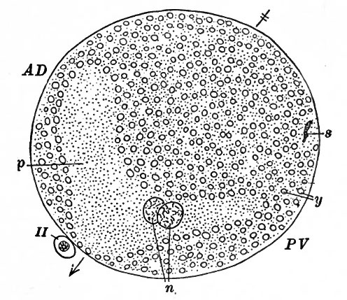
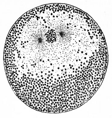
The ovum of Amphioxus has certain peculiarities which are important in their effect upon cleavage. While it contains only a small quantity of yolk, being regarded as a meiolecithal ovum, this material is situated slightly off center and the nucleus lies outside of the yolk (Fig. 18). This condition really effects a polarity of the cell. The first polar body is given off from the yolk-free portion of the egg. This marks the animal pole and also the side which will be the anterior part of the embryo. The sperm enters the egg at the vegetative pole and seems to stimulate the formation of the second polar body. The sperm nucleus and centrosome then traverse the yolk area to meet the mature egg nucleus which in the meantime has migrated toward, but not quite to, the center of the egg. The division of the sperm centrosome to form a disaster and the arrangement of the chromosomes of the two pronuclei in the equatorial plane comprise the preparatory step for the first cleavage. These phenomena are identical with the prophase of mitosis (Fig. 19).
Fig. 18. Diagram of a median sagittal section through an Amphioxus ovum. Cerfontaine, from Kellicott. The arrow indicates the direction of the polar axis. AD, antero-dorsal region; PV, postero-ventral region; N, male and female pronuclei; p, yolk-free area; S, tail of sperm; y, yolk area; II, second polar body.
The position that the spindle assumes is determined by three factors: the point where the first polar body is extruded, the point where the sperm enters, and the location of the yolk-free area. A plane bisecting this area and passing through the other two points will divide the egg into symmetrical halves. The spindle takes its position at right angles to this plane. The first cleavage therefore will produce two equal and symmetrical daughter cells, or blastomeres, the first cleavage plane coinciding with the plane of symmetry of the ovum. These two blastomeres will become the right and left halves of the embryo, the plane of symmetry of the ovum representing the sagittal plane of the embryo. With the anterior portion already indicated by the point of extrusion of the first polar body, the orientation of the first two blastomeres relative to the future embryo is now complete.
Fig. 19. Prophase of first cleavage figure in the ovum of Amphioxus. The chromosomes of the male and female pronuclei are mingled in the equatorial plane. Sobotta, from Kellicott.
The second cleavage plane falls at a right angle to the first, cutting both the animal and the vegetative pole. The division is slightly unequal, however, the result being two slightly smaller blastomeres and two slightly larger blastomeres (Fig. 20, A ) . These are arranged symmetrically on the two sides of the median plane. The third cleavage plane lies at right angles to the other two, and division of the cells is again slightly unequal (a condition often called subequal), the result being four pairs of cells of four different sizes (Fig. 20, B) . The smallest cells are those derived from the portion of the ovum which contained less yolk, the largest are those derived from the portion which contained more yolk. All the cells have divided completely, a circumstance which gives rise to the term total cleavage; and this condition obtains throughout the later stages. All the cells at a given cleavage thus far have divided at the same time, a fact which is expressed in the term regular cleavage. If cleavage were to continue regularly the result at succeeding divisions would be 16, 32, 64, 128 cells, and so on. Regularity is lost, however, during the fourth cleavage, some of the cells dividing before others, with the result that numbers other than those just given will be found. The smallest cells, with the least amount of yolk are the first to divide and they divide more rapidly than the large cells with a greater yolk content; the inert non-protoplasmic substance retards the progress of division.
Fig. 20. Cleavage in Amphioxus. Cerfontaine, from Kellicott. A, four-cell stage seen from animal pole; B, eight-cell stage seen from animal pole, showing four sizes of blastomeres; C, sixteen-cell stage seen from left side; A, thirty-two-cell stage seen from vegetal pole; E, 32-64 cells seen from antero-dorsal region; F, half of early blastula containing about 128 cells, a, Animal pole; ad, antero-dorsal; I, left; pv, postero-ventral; r, right; v, vegetal pole.
Division succeeds division in the blastomeres, with the irregularity noted in the preceding paragraph. The cleavage planes vary considerably in direction in different individuals. At the i6-cell stage the micromere group assumes a sort of dome form and the macromere group in similar form fits into the hollow of the dome (Fig. 20, C) . The early blastomeres remain well rounded so that even at the four-cell stage there is a small central cavity (Fig. 20, A). As cleavage progresses the cells become more closely arranged and pushed away from the central cavity (Fig. 20, D, E, F). At the i28-cell stage all the cells are arranged in a simple epithelial layer around a relatively large central cavity, the segmentation cavity or Uastoccel. The entire structure is now the bias tula. Other divisions occur until the blastula contains about 256 cells. There is a gradual transition from the micromeres at one pole of the hollow sphere to the macromeres at the opposite pole. It should be recalled here that, on account of the position of the yolk-free portion of the ovum, the micromeres lie where the anterior region of the embryonic body will arise and the macromeres where the posterior region will develop. About four hours elapse between the time the first cleavage occurs and the time the 256-cell blastula is formed.
Gastrulation
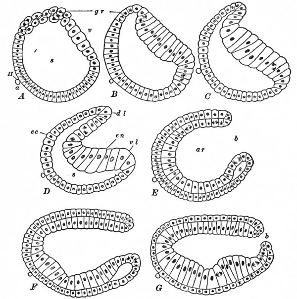
This process comprises the conversion of the single walled blastula into the double walled gastrula. The vegetative pole becomes flattened, the macromeres assuming columnar form. The cells at the dorsal margin of the flattened pole begin to proliferate more rapidly than elsewhere, as shown by the increased number of mitotic figures (Fig. 21, A, B). This area of accelerated division then extends in both directions around the margin of the flat pole, forming the germ ring. Beginning at the dorsal margin the macromeres are folded, or invaginated, into the blastocoel until the blastoccel is obliterated (Fig. 21, C, D, E, F, G). A rough analogy is the pushing in of one side of a hollow rubber ball. The invagination, however, is more rapid along the dorsal margin of the plate of macromeres, and as the infolding progresses there is formed a plate of small cells which arise through the more rapid proliferation in the germ ring (Fig. 21, D, E). On the ventral side the ingrowth is but slight, the whole plate of macromeres behaving as if hinged at this point. By these processes the blastula, with a single layer of cells, has been converted into the gastrula, with a double layer of cells and a new cavity which opens to the exterior.
The outer layer of cells is the ectoderm which is in direct contact with the environment of the developing organism. The inner layer is the entoderm which forms the lining of the new cavity, or archenteron, in the interior of the organism. The entoderm consists of two types of cells, the larger cells with considerable yolk content which lie on the ventral side or in the floor of the archenteron and the smaller cells forming the dorsal lining of the archenteron which were produced by the rapid divisions in the germ ring. This latter group in part really had a brief existence as ectodermal cells and then contributed to entoderm by being inflected round the rim of the opening between the archenteron and the exterior. The inflection of the cells in question, often called involution is therefore one of the factors in gastrulation. The circular opening between the archenteron and the exterior is the blastopore. Its margins are its lips which can be differentiated into dorsal, ventral and lateral lips. At these lips the entoderm and ectoderm are continuous.
Another factor in gastrulation is a process known as epiboly. When invagination is complete, that is, when the macromere pole of the blastula has infolded until the blastoccel is obliterated, the gastrula approximates a hemisphere and the form of the archenteron coincides. Then, along with the rapid cell proliferation in the dorsal part or the germ ring and the formation of the plate of entodermal cells mentioned in the preceding paragraph, the dorsal lip of the blastopore extends backward. The lip protrudes, one might say. The extension gradually affects also the lateral lips and finally to a slight degree the ventral lip. This whole process of growth backward, which is due to the rapid cell division in the germ ring most rapid dorsally, less rapid laterally, least rapid ventrally, effects a posterior elongation of the gastrula and a diminution in the size of the blastopore (Fig. 21, E, F, G). This is the first step in the lengthwise growth of the animal as a whole. The whole process of gastrulation has occupied about three hours.
Fig. 21. Gastrulation in Amphioxus. Cerfontaine, from Kellicott.
- A, blastula with slightly flattened vegetal pole, showing rapid cell division in postero-dorsal region (germ ring); 5, more pronounced flattening of the vegetal pole; C, beginning of invagination in postero-dorsal region; D, further invagination, showing obliteration of the blastocoel and formation of the archenteron as the result of invagination; E, invagination almost complete; F, beginning elongation of gastrula and narrowing of blastopore; G, continued elongation of gastrula and narrowing of blastopore. Observe the mitotic figures in the germ ring in all stages. In D and E the inflection of cells (involution) around the dorsal lip of the blastopore can be appreciated. In F and G the process of epiboly is represented in the backward growth of the lip of the blastopore. a, Animal pole; ar, archenteron; b, blastopore; dl, dorsal lip of blastopore; ec, ectoderm; en, entoderm; gr, germ ring; s, blastocoel; v, vegetal pole; vl, ventral lip of blastopore; Il, second polar body.
The account here given differs in one respect from that of the British investigator, MacBride. It has been stated that inflection, or involution, is one of the factors in gastrulation. MacBride maintains that involution does not occur, but that the rapid cell division occurring in the lips of the blastopore produces both ectoderm and entoderm in equal amounts. Cell proliferation is the only process which adds to the number of entodermal as well as of ectodermal components, and this at the same time produces the backward extension of the lips of the blastopore which is recognized as epiboly. He bases his conclusion on nuclear characters. In the bias tula all the nuclei are vesicular. Soon after gastrulation begins the nuclei of the ectodermal cells become more intensely stainable while those of the entodermal cells retain their vesicular nature, all the invaginated cells possessing the vesicular nuclei. This probably indicates a physiological differentiation. In the germ ring two types of the rapidly dividing cells can be distinguished, one with vesicular nuclei and the other with deeply staining nuclei. The former are added to the entoderm, the latter to the ectoderm. There is therefore a zone of growth in which cells are produced and added directly to the two layers without inflection round the lip of the blastopore.
The gastrula is now somewhat elongated antero-posteriorly, somewhat flattened on the dorsal side and is bilaterally symmetrical, with the archenteron opening to the exterior at the caudal end through the small blastopore (Fig. 21 , G) . Even at this time it is not amiss to note a certain fundamental arrangement of structure and anticipate in a measure its biological significance when carried over into later stages. The ectoderm, the outer layer of the gastrula, is in immediate contact with the environment, which fact implies that response to external stimuli and protection are effected through this layer. In Amphioxus, as well as in certain other lower forms, strong cilia develop on the ectodermal cells by the motion of which the gastrula changes its position. In later stages it will be seen that the nervous sytem, that complex mechanism for transmitting stimuli from one part of the body to another, is developed from ectoderm. The outer layer of the integumentary system with certain of its derivatives, primarily protective in nature, is also a product of ectoderm. The archenteron with its lining of entoderm constitutes the primitive gut, the only opening of which is the blastopore, serving as both mouth and anus. Already the simple alimentary system is confined to the interior of the organism, shut off from the outside except through an opening for the intake of food and output of waste. Among the invertebrates the sponges and corals never develop beyond the two layered, or didermic, gastrula stage such as we here see in Amphioxus. It is worth noting also that in Amphioxus the cells with yolk content are members of the entoderm group; in other words, a temporary food supply, scanty as it is here, is stored in the lining of the gut. From this simple primitive gut the whole alimentary system is elaborated, complex as it may become. The mouth, however, is not a derivative of the blastopore, but develops as a new opening into the cephalic end of the gut cavity. The anal opening too in most vertebrates arises independently.
Fig. 22. From transverse sections through Amphioxus embryos, showing successive stages in formation of mesoderm, neural tube and notochord. Bonnet.
Before considering the formation of the middle germ layer, or mesoderm, it is desirable to observe certain changes affecting the exterior of the gastrula which are correlated with the development of the nervous system, because they occur prior to the appearance of the mesoderm and produce a setting for part of this layer. Along the flattened dorsal surface of the gastrula a piate of ectodermal cells sinks slightly below the general surface level and becomes demarkated from the surrounding ectoderm. The plate extends from almost the cephalic (anterior) extremity of the gastrula to the dorsal lip of the blastopore and even slightly affects the lateral lips. These cells thus circumscribed constitute the neural plate; in this manner the rudiment of the nervous system appears (Fig. 22, a). The ectoderm bordering the margins of the neural plate becomes elevated above the general surface level to form the neural ridges* These also form a rim around the blastopore. The neural plate then sinks farther below the surface level and at the same time the ridges slide across it toward the mid-dorsal line until they meet and fuse with each other. Thus a roof is made over the neural plate, with a small space between the two structures (Fig. 22, b, c). The median fusion begins some distance in front of the blastopore and from there progresses both forward and backward. The closure is not complete in front for some time, and the opening thus left is called the neuropore (Fig. 23). The neural ridges close in over the blastopore as they do over the neural plate, so that the blastopore no longer opens to the exterior but into the space between the neural plate and its ectodermal roof (Fig. 23).
Fig. 23. From vertical section through Amphioxus embryo with 5 primitive segments. Hatschek.
Mesoderm Formation. Closely following the appearance of the neural plate in the elongated gastrula, one may observe the rudiment of the middle germ layer and the first indication of the axial structure, the notocord, that gives the name Chordata to the great division of the animal kingdom which includes not only the true vertebrates but also such forms as Amphioxus, Balanoglossus and the Tunicata. In a transverse section of the gastrula, in the roof of the archenteron the entoderm exhibits a change which produces three distinguishable parts. An axial part, lying beneath the center of the neural plate, is the rudiment of the notocord. Two dorso-lateral parts, bilaterally symmetrical, are the rudiments of the mesoderm (Fig. 22). The notocord rudiment advances to the cephalic extremity of the gastrula, and extends caudally to the blastopore. The mesoderm rudiment reaches from the forward end of the archenteron to the blastoporal region where the two parts diverge in the lateral lips of theaperture\ The portion along the archenteron is the gastral mesoderm, that around me" blastopore the peristomaLj
The neural plate becomes depressed along its center and the edges turned^ upward, forming the neural groove. Depression and elevation continue until the two edges meet dorsally in the median plane. Fusion of the edges begins not far from the anterior end and progresses both forward and backward until the entire structure becomes tubular. Thus the neural tube with its central canal is formed (Fig. 22, d}. At the caudal end the central canal remains in open communication with the archenteron owing to the fact that when the ectoderm grew over the neural plate it also grew over the blastopore. The opening thus left is the neurenteric canal (Fig. 23). So long as the neuropore also persists at the cephalic end of the neural tube there is direct communication between the exterior and the archenteron via the central canal and the neurenteric canal. In Amphioxus the neuropore persists until the mouth is formed.
The depression of the center of the neural pjate produces a depression *also of the notocord rudiment and the mesial edges of the mesoderm bands. One effect of this is an inverted groove, the enteroccel, along each side of the notocord, so that the mesoderm appears to bulge outward (Fig. 22, a, b). The grooves extend almost the entire length of the embryo and speedily grow deeper, the mesoderm intruding between entoderm and ectoderm and becoming clearly differentiated from the notocord and the remainder of the entoderm (Fig. 22, c). Near the cephalic end of the embryo a transverse fold drops from the dorsal part of the mesoderm on each side, which closes the groove and delimits the most anterior portion from that immediately behind it. The portion thus delimited, with its fellow of the opposite side, constitutes the first pair of mesodermal somites. Another portion is delimited in the same manner to form the second pair of somites. Then the third pair is formed; and so on toward the caudal end of the embryo (Fig. 23 and Fig. 24). The development of mesodermal somites therefore takes place from before backward.
Each somite assumes a cuboidal form and is hollow, the cavity being a portion of the original groove-like enteroccel, and the cells surrounding the cavity comprise a simple cuboidal epithelium. For a short time an opening between the enteroccel and gut cavity remains, but later this is closed as the mesoderm becomes entirely cut off from the entoderm and the latter again forms a continuous lining of the gut. These processes too occur from before backward.
The fact that the formation of mesodermal somites progresses from before backward, that is, from the cephalic end of the body toward the caudal end, illustrates a fundamental principle of growth. The distinction between gastral and peristomal mesoderm has already been stated, and since mesoderm development is initiated shortly after the gastrula begins to elongate the true gastral portion is relatively short. Whatever is added to this comes from the region of the blastopore. In the germ ring cell proliferation continues rapidly and from the cells thus produced components of all three germ layers are differentiated. In other words, the elongation of the embryo as a whole, with its three germ layers, is due chiefly to this cell proliferation and differentiation at its caudal end. Not only the mesoderm but also the gut, the neural tube and other structures which will subsequently appear, increase and develop from before backward.
Fig. 24. From horizontal section through Amphioxus embryo with 5 primitive segments; seen from dorsal side. Hatschek. The communication between the cavities of the primitive segments (ccelom) and the archenteron can be seen in the last 4 segments.
The original gastral mesoderm gives rise to perhaps not more than the first two pairs of somites. The succeeding somites arise from mesoderm that originates in the region around the blastopore. By the time about fourteen pairs of somites have developed the mesoderm no longer arises as outgrowths from the entoderm of the gut wall but directly from the proliferating cells in the region around the neurenteric canal. As a matter of fact the formation of somites now does not quite keep pace with the differentiation of the middle layer and just in front of the blastoporal region there is a short band of undivided mesoderm (Fig. 23 and Fig. 24). As this band grows at its caudal end it is gradually being cut up into somites from its anterior end. The somites appear as bilaterally symmetrical structures, but when five or six pairs have arisen the symmetry is disturbed since each somite on the right comes to lie a little behind its fellow on the left thus giving an alternation which is carried on into the adult.
Only the first few somites develop with enteroccelic cavities, the remainder originating as solid structures although the cells are arranged radially around a central point. However, the solid ones subsequently acquire cavities. The enteroccel has been regarded as an indication of a primitive character, since in the higher animals the somites do not contain any cavities derived from the gut cavity but arise as solid structures. On the other hand the solid somites may indicate the primitive condition and the appearance of enteroccelic cavities may be a secondary character in Amphioxus.
The rudiment of the notocord, mentioned previously, which is composed of the entodermal cells immediately ventral to the neural tube and between the two mesodermal outgrowths, extending from the cephalic extremity of the embryo to the blastoporal region, requires brief attention. While the mesodermal rudiments are being cut off from the parent entoderm the notocordal cells become rearranged into a compact rod-like structure lying between the somites of the two sides (Fig. 22, d). As the somites enlarge this rod is constricted from the adjacent entoderm, which then closes across the top of the gut cavity, and occupies its definitive position ventral to the neural tube. Clearly the notocord in Amphioxus originates from entoderm. As the embryo continues to grow in length the notocord too is lengthened by the addition of cells to its caudal end in the region of the neurenteric canal.
Continued development of the mesodermal somites comprises their farther intrusion between ectoderm and entoderm and changes in their component cells. When first formed, the somites are composed of columnar or cuboidal epithelial cells in a single layer surrounding the central cavity if present, or, if the enteroccel is absent, radiating from a common center (Fig. 23 and Fig. 24). The somites are block-like in shape and located lateral to the developing notocord and neural tube. The changes to be described begin in the anterior somites and, in accordance with the principle of growth already mentioned, progress from there backward. The cavity in the somite becomes larger and the surrounding cells become flatter. With the enlargement of the cavity the ventral portion of the somite extends ventrally between ectoderm and entoderm (Fig. 22, d). It seems that the whole structure becomes dilated in the direction of least resistance. The outer portion of the wall is apposed to ectoderm and is called the somatic or parietal mesoderm; the inner layer is in contact with entoderm and is spoken of as splanchnic or visceral mesoderm. The dilated cavity is the codomic space (Fig. 22 and Fig. 25). Continued ventral extension brings the dilating structure around the ventral aspect of the gut until it meets its fellow of the opposite side in the sagittal plane, thus separating ectoderm from entoderm. The sagittal partition between the ccelomic spaces of the two sides then breaks down and each side is in free communication with the other ventral to the gut. The cells of the entire dilated structure have become decidedly flattened except those in contact with the notocord and neural tube which become more elongated columns and comprise the muscle plate or myotome (Fig. 25). The portion of the cavity contiguous to the myotome is now known as the myocoel while the remainder of the coelomic space is the splanchnocoel. Subsequently a partition appearing between the myoccel and splanchnocoel completely separates the two cavities. The myotomes, in the sites of the original somites, retain their segmental character. The partitions between adjacent splanchnoccelic cavities, on the other hand, break down and the common cavity thus produced, which is now known as the coelom, no longer bears the segmental character but is continuous on both sides of and below the gut.
Fig. 25. Diagram to show differentiation of primitive segment into muscle plate (myotome) and cutis plate and relation of myocoel and splanchnocoel. Bonnet, Compare with Fig. 22, d.
The biological significance of ectoderm and entoderm has been briefly noted. Between these two layers the mesoderm appears and presently begins to elaborate and to contribute to their support ; support in the broadest sense of the term. As the organism continues to develop, the middle germ layer becomes a framework within and around which the refinements of the two primary layers are suspended. The whole series of connective tissues is of mesodermal origin, and this applies even to the cartilaginous and bony skeleton. The muscles, all three varieties, whose activities are associated with motion and locomotion are derivatives of the mesoderm. The blood vessels and lymphatics, the tubes through which substances are carried from one part of the body to another, the blood and lymph also which are the vehicles for these substances, all are mesodermal in origin. The organs of excretion too arise from this intermediate layer. The reproductive organs, growth centers of the germ cells., originate here. It is not difficult to see, therefore, that in the higher and more complex animal forms many of the activities of the ectodermal and entodermal derivatives which are correlated with response to external stimuli and with alimentation are made possible by structures elaborated from the mesoderm.
While Amphioxus is not a true vertebrate because it never acquires a vertebral column, yet we may observe in it a relatively simple arrangement of structure which foreshadows the fundamental vertebrate organization. After the development of the mesoderm and ccelom the embryo as a whole obviously comprises a tube within a tube; the gut, extending from mouth to anus, is the inner tube, the body wall is the outer tube, and the two are separated by the ccelom or body cavity. This is a typical vertebrate characteristic. The neural tube or central nervous system, situated in the dorsal body wall, is another feature which links Amphioxus with the vertebrates. The notocord which is regarded as the axial supporting structure in Amphioxus appears also in higher animal forms. In the true vertebrates the notocord is not transformed into the axial skeleton which is the chief longitudinal supporting skeleton, but the axial mechanism is built around the notocord. Another impressive attribute of the vertebrates is the series of mesodermal somites, although it must be remembered that this is not exclusively chordate property, for some of the invertebrates, for instance the worms, possess it. This transverse segmentation, or metamerism, affects not only the mesoderm and certain of its derivatives but involves also structures that arise from ectoderm. In the vertebrates the units of the spinal column, arising from the somites, maintain their integrity throughout the life of the animal. The ribs and intercostal muscles are expressions of metamerism. Many of the blood vessels are arranged segmentally. Even the primitive kidney arises as a segmental organ. Among the ectodermal derivatives, the nervous system reflects the metameric quality in the development of the spinal nerves. Obviously many features of vertebrate organization depend upon the principle of metamerism.
- Next: Frog
Amphioxus (Greek, "both pointed" referring to their body shape), are commonly called lancelets or small, eel-like animals that spend much of their time buried in sand. They are simple members of the cephalochordates.
References for Further Study
CERFONTAINE, P.: Recherches sur le development de 1' Amphioxus. Archives de Biologie, tome 22, 1907.
HATSCHEK, B.: Studien uber die Entwickelung des Amphioxus. Arbeiten aus dem 2007. Institut zu Wien, Bd. 4, 1881.
HERTWIG, R.: Furchungsprozess. In Hertwig's Handbuch der vergleichenden und experimentellen Entwickelungslehre der Wirbeltiere, Bd. I, Teil I, Kap. Ill, 1903. Contains extensive bibliography.
KELLICOTT, W. E.: Chordate Development. Chap. I, 1913.
MACBRIDE, E. W.: Text-book of Embryology. Vol. I, 1914.
MORGAN, T. H. and HAZEN, A. P.: The Gastrulation of Amphioxus. Journal of Morphology, Vol. 16, 1900.
WILLEY, A.: Amphioxus and the Ancestry of the Vertebrates. 1894.
WILSON, E. B.: Amphioxus and the Mosaic Theory of Development. Journal oj Morphology, Vol. 8, 1893.
| Historic Disclaimer - information about historic embryology pages |
|---|
| Pages where the terms "Historic" (textbooks, papers, people, recommendations) appear on this site, and sections within pages where this disclaimer appears, indicate that the content and scientific understanding are specific to the time of publication. This means that while some scientific descriptions are still accurate, the terminology and interpretation of the developmental mechanisms reflect the understanding at the time of original publication and those of the preceding periods, these terms, interpretations and recommendations may not reflect our current scientific understanding. (More? Embryology History | Historic Embryology Papers) |
Text-Book of Embryology: Germ cells | Maturation | Fertilization | Amphioxus | Frog | Chick | Mammalian | External body form | Connective tissues and skeletal | Vascular | Muscular | Alimentary tube and organs | Respiratory | Coelom, Diaphragm and Mesenteries | Urogenital | Integumentary | Nervous System | Special Sense | Foetal Membranes | Teratogenesis | Figures
Glossary Links
- Glossary: A | B | C | D | E | F | G | H | I | J | K | L | M | N | O | P | Q | R | S | T | U | V | W | X | Y | Z | Numbers | Symbols | Term Link
Cite this page: Hill, M.A. (2024, April 25) Embryology Book - Text-Book of Embryology 4. Retrieved from https://embryology.med.unsw.edu.au/embryology/index.php/Book_-_Text-Book_of_Embryology_4
- © Dr Mark Hill 2024, UNSW Embryology ISBN: 978 0 7334 2609 4 - UNSW CRICOS Provider Code No. 00098G


