Book - Text-Book of Embryology (1921) - Figures: Difference between revisions
mNo edit summary |
|||
| (36 intermediate revisions by 3 users not shown) | |||
| Line 1: | Line 1: | ||
{{Template:Bailey 1921}} | {{Template:Bailey 1921}} | ||
[[File:Bailey_and_Miller_1921.jpg|right|250px]] | |||
==The germ cells== | ==The germ cells== | ||
| Line 40: | Line 40: | ||
File:Bailey017.jpg|Fig. 17. Polyspermy in sea-urchin eggs treated with 0.005 per cent, nicotine solution | File:Bailey017.jpg|Fig. 17. Polyspermy in sea-urchin eggs treated with 0.005 per cent, nicotine solution | ||
</gallery> | </gallery> | ||
:'''Links:''' [[Fertilization]] | [[:Category:Fertilization|Category:Fertilization]] | |||
==Early development of amphioxus== | ==Early development of amphioxus== | ||
| Line 61: | Line 64: | ||
<gallery> | <gallery> | ||
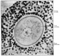
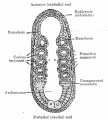
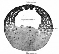
File:Bailey026.jpg | File:Bailey026.jpg|Fig. 26. Section through the fully formed ovarian egg of a frog. | ||
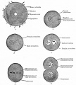
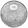
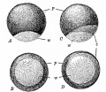
File:Bailey027.jpg | File:Bailey027.jpg|Fig. 27. A frog's egg before and after fertilization, showing the formation of the gray crescent. | ||
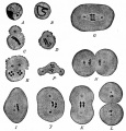
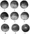
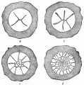
File:Bailey028.jpg | File:Bailey028.jpg|Fig. 28. Cleavage of the frog's egg. | ||
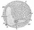
File:Bailey029.jpg | File:Bailey029.jpg|Fig. 29. From a sagittal section through blastula of frog. | ||
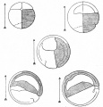
File:Bailey030.jpg | File:Bailey030.jpg|Fig. 30. Diagrams showing the position of the blastopore at successive stages of gastrulation in the frog's egg. | ||
File:Bailey031.jpg | File:Bailey031.jpg|Fig. 31. Median sagittal sections showing successive stages of gastrulation in the frog's egg | ||
File:Bailey032.jpg | File:Bailey032.jpg|Fig. 32. Transverse section of embryo of frog (Rana fusca). | ||
File:Bailey033.jpg | File:Bailey033.jpg|Fig. 33. Transverse section through embryo of frog (Rana fusca). | ||
File:Bailey034.jpg | File:Bailey034.jpg|Fig. 34. Portion of a transverse section still continuous at the lower lateral angles of the larva of a frog (Rana fusca) | ||
File:Bailey035.jpg | File:Bailey035.jpg|Fig. 35. Diagrams of median sagittal sections through an eight-cell stage and four stages during gastrulation of the frog's egg. | ||
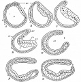
File:Bailey036.jpg | File:Bailey036.jpg|Fig. 36. Postero-lateral views of successive stages following gastrulation in the frog. | ||
File:Bailey037.jpg|Fig. 37. Median sagittal sections of frog larvae. | File:Bailey037.jpg|Fig. 37. Median sagittal sections of frog larvae. | ||
</gallery> | </gallery> | ||
| Line 80: | Line 83: | ||
<gallery> | <gallery> | ||
File:Bailey038.jpg|Fig. 38 | File:Bailey038.jpg|Fig. 38. Cleavage in hen's egg | ||
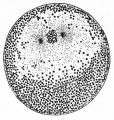
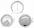
File:Bailey039.jpg|Fig. 39 | File:Bailey039.jpg|Fig. 39. Vertical section germ disk of a fresh-laid hen's egg. | ||
File:Bailey040.jpg|Fig. 40 | File:Bailey040.jpg|Fig. 40. Cross section blastoderm of a pigeon 14.5 hours after fertilization. | ||
File:Bailey041.jpg|Fig. 41 | File:Bailey041.jpg|Fig. 41. Median longitudinal section blastoderm of a pigeon 31 hours after fertilization. | ||
File:Bailey042.jpg|Fig. 42 | File:Bailey042.jpg|Fig. 42. Median longitudinal section blastoderm of a pigeon 36 hours after fertilization. | ||
File:Bailey043.jpg|Fig. 43 | File:Bailey043.jpg|Fig. 43. Surface views of blastoderms of the pigeon. | ||
File:Bailey044.jpg|Fig. 44 | File:Bailey044.jpg|Fig. 44. Surface views of blastoderms of Haliplana primitive streak. | ||
File:Bailey045.jpg|Fig. | File:Bailey045.jpg|Fig. 45. Surface view of embryonic disk of chick. | ||
File:Bailey046.jpg|Fig. 46 | File:Bailey046.jpg|Fig. 46. Surface view of chick blastoderm. | ||
File:Bailey047.jpg|Fig. 47 | File:Bailey047.jpg|Fig. 47. Transverse sections of blastoderm of chick 21 hours. | ||
File:Bailey048.jpg|Fig. 48 | File:Bailey048.jpg|Fig. 48. Transverse section of blastoderm of chick 21 hours. | ||
File:Bailey049.jpg|Fig. | File:Bailey049.jpg|Fig. 49. Median longitudinal section blastoderm of chick after the primitive axis | ||
File:Bailey050.jpg|Fig. 50 | File:Bailey050.jpg|Fig. 50. Transverse section of blastoderm of chick 40 hours. | ||
File:Bailey051.jpg|Fig. 51 | File:Bailey051.jpg|Fig. 51. Dorsal view of chick embryo with ten pairs of mesodermal somites. | ||
File:Bailey052.jpg|Fig. 52 | File:Bailey052.jpg|Fig. 52. Transverse section of chick embryo 2 days incubation. | ||
</gallery> | </gallery> | ||
:'''Links:''' [[Chicken Development]] | |||
==Early mammalian development== | ==Early mammalian development== | ||
| Line 102: | Line 108: | ||
<gallery> | <gallery> | ||
File:Bailey053.jpg|Fig. 53 | File:Bailey053.jpg|Fig. 53. Four stages in the cleavage of the ovum of the white rat. | ||
File:Bailey054.jpg|Fig. 54 | File:Bailey054.jpg|Fig. 54. Four stages in cleavage of the ovum of the mouse. | ||
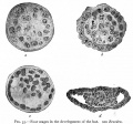
File:Bailey055.jpg|Fig. 55 | File:Bailey055.jpg|Fig. 55. Four stages in the development of the bat. | ||
File:Bailey056.jpg|Fig. 56 | File:Bailey056.jpg|Fig. 56. Sections of blastocysts of the white rat 5 days. | ||
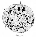
File:Bailey057.jpg|Fig. 57 | File:Bailey057.jpg|Fig. 57. Section of a 16-cell stage of an ovum of the opossum. | ||
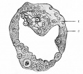
File:Bailey058.jpg|Fig. 58 | File:Bailey058.jpg|Fig. 58. Section of the blastocyst of the lemur Tarsius spectrum. | ||
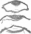
File:Bailey059.jpg|Fig. 59 | File:Bailey059.jpg|Fig. 59. Sections of blastodermic vesicle of bat. | ||
File:Bailey060.jpg|Fig. 60 | File:Bailey060.jpg|Fig. 60. Three stages in the formation of the germ layers in the lemur Tarsius spectrum. | ||
File:Bailey061.jpg|Fig. 61 | File:Bailey061.jpg|Fig. 61 | ||
File:Bailey062.jpg|Fig. 62 | File:Bailey062.jpg|Fig. 62 | ||
| Line 139: | Line 145: | ||
<gallery> | <gallery> | ||
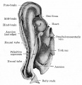
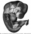
File:Bailey083.jpg|Fig. 83 | File:Bailey083.jpg|Fig. 83. Human embryo with 8 pairs of mesodermal somites | ||
File:Bailey084.jpg|Fig. 84. Human embryo with 14 pairs of mesodermal somites. | |||
File:Bailey084.jpg|Fig. 84 | File:Bailey085.jpg|Fig. 85. Human embryo of 2.6 mm. | ||
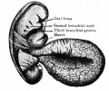
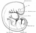
File:Bailey085.jpg|Fig. 85 | File:Bailey086.jpg|Fig. 86. Human embryo of 4 mm. | ||
File:Bailey086.jpg|Fig. 86 | File:Bailey087.jpg|Fig. 87. Human embryo 27 primitive segments. | ||
File:Bailey087.jpg|Fig. 87 | File:Bailey088.jpg|Fig. 88. Human embryo with 28 primitive segments. | ||
File:Bailey088.jpg|Fig. 88 | File:Bailey089.jpg|Fig. 89. Human embryo 11 mm. | ||
File:Bailey089.jpg|Fig. 89 | File:Bailey090.jpg|Fig. 90. Human embryo of 15.5 mm. | ||
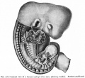
File:Bailey090.jpg|Fig. 90 | File:Bailey091.jpg|Fig. 91. Human embryo of 17.5 mm (47-51 days). | ||
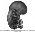
File:Bailey091.jpg|Fig. 91 | File:Bailey092.jpg|Fig. 92. Human embryo of 18.5 mm (52-54 days). | ||
File:Bailey092.jpg|Fig. 92 | File:Bailey093.jpg| Fig. 93. Human embryo of 23 mm (2 months). | ||
File:Bailey093.jpg|Fig. 93 | File:Bailey094.jpg|Fig. 94. Human embryo of 78 mm (3 months). | ||
File:Bailey094.jpg|Fig. 94 | File:Bailey095.jpg|Fig. 95. Human embryo of 4 months. | ||
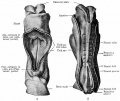
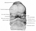
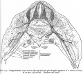
File:Bailey095.jpg|Fig. 95 | File:Bailey096.jpg|Fig. 96. Ventral view of head of 8 mm human embryo. | ||
File:Bailey096.jpg|Fig. 96 | File:Bailey097.jpg|Fig. 97. Ventral view of head of 11.3 mm human embryo. | ||
File:Bailey097.jpg|Fig. 97 | File:Bailey098.jpg|Fig. 98. Ventral view of head of 13.7 mm human embryo. | ||
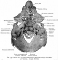
File:Bailey098.jpg|Fig. 98 | File:Bailey099.jpg|Fig. 99. Ventral view of head of human embryo of 8 weeks. | ||
File:Bailey099.jpg|Fig. 99 | |||
</gallery> | </gallery> | ||
| Line 276: | Line 281: | ||
File:Bailey202.jpg|Fig. 202 | File:Bailey202.jpg|Fig. 202 | ||
File:Bailey203.jpg|Fig. 203 | File:Bailey203.jpg|Fig. 203 | ||
File:Bailey204.jpg|Fig. 204 | File:Bailey204.jpg|Fig. 204 Fig. 205 | ||
File:Bailey206.jpg|Fig. 206 | File:Bailey206.jpg|Fig. 206 | ||
File:Bailey207.jpg|Fig. 207 | File:Bailey207.jpg|Fig. 207 | ||
| Line 298: | Line 302: | ||
File:Baileytable02.jpg|Table 2 | File:Baileytable02.jpg|Table 2 | ||
</gallery> | </gallery> | ||
:'''Links:''' [[Cardiovascular System Development]] | |||
==The muscular system== | ==The muscular system== | ||
| Line 316: | Line 323: | ||
<gallery> | <gallery> | ||
File:Bailey228.jpg|Fig. 228 | |||
File:Bailey229.jpg|Fig. 229 | |||
File:Bailey230.jpg|Fig. 230 | |||
File:Bailey231.jpg|Fig. 231 | |||
File:Bailey232.jpg|Fig. 232 | |||
File:Bailey233.jpg|Fig. 233 | |||
File:Bailey234.jpg|Fig. 234 | |||
File:Bailey235.jpg|Fig. 235 | |||
File:Bailey236.jpg|Fig. 236 | |||
File:Bailey237.jpg|Fig. 237 | |||
File:Bailey238.jpg|Fig. 238 | |||
File:Bailey239.jpg|Fig. 239 | |||
File:Bailey240.jpg|Fig. 240 | |||
File:Bailey241.jpg|Fig. 241 | |||
File:Bailey242.jpg|Fig. 242 | |||
File:Bailey243.jpg|Fig. 243 | |||
File:Bailey244.jpg|Fig. 244 | |||
File:Bailey245.jpg|Fig. 245 | |||
File:Bailey246.jpg|Fig. 246 | |||
File:Bailey247.jpg|Fig. 247 | |||
File:Bailey248.jpg|Fig. 248 | |||
File:Bailey249.jpg|Fig. 249 | |||
File:Bailey250.jpg|Fig. 250 | |||
File:Bailey251.jpg|Fig. 251 | |||
File:Bailey252.jpg|Fig. 252 | |||
File:Bailey253.jpg|Fig. 253 | |||
File:Bailey254.jpg|Fig. 254 | |||
File:Bailey255.jpg|Fig. 255 | |||
File:Bailey256.jpg|Fig. 256 | |||
File:Bailey257.jpg|Fig. 257 | |||
File:Bailey258.jpg|Fig. 258 | |||
File:Bailey259.jpg|Fig. 259 | |||
File:Bailey260.jpg|Fig. 260 | |||
File:Bailey261.jpg|Fig. 261 | |||
File:Bailey262.jpg|Fig. 262 | |||
File:Bailey263.jpg|Fig. 263 | |||
File:Bailey264.jpg|Fig. 264 | |||
File:Bailey265.jpg|Fig. 265 | |||
File:Bailey266.jpg|Fig. 266 | |||
File:Bailey267.jpg|Fig. 267 | |||
File:Bailey268.jpg|Fig. 268 | |||
File:Bailey269.jpg|Fig. 269 | |||
File:Bailey270.jpg|Fig. 270 | |||
File:Bailey271.jpg|Fig. 271 | |||
File:Bailey272.jpg|Fig. 272 | |||
File:Bailey273.jpg|Fig. 273 | |||
File:Bailey274.jpg|Fig. 274 | |||
File:Bailey275.jpg|Fig. 275 | |||
File:Bailey276.jpg|Fig. 276 | |||
File:Bailey277.jpg|Fig. 277 | File:Bailey277.jpg|Fig. 277 | ||
File:Bailey278_279.jpg|Fig. 278 279 | File:Bailey278_279.jpg|Fig. 278 279 | ||
| Line 331: | Line 387: | ||
File:Bailey284.jpg|Fig. 284 | File:Bailey284.jpg|Fig. 284 | ||
File:Bailey285.jpg|Fig. 285 | File:Bailey285.jpg|Fig. 285 | ||
File:Bailey286.jpg|Fig. 286 | File:Bailey286.jpg|Fig. 286. Transverse section of a 14 mm. pig embryo, at the level of the upper limb buds, showing especially the two bronchi | ||
File:Bailey287.jpg|Fig. 287 | File:Bailey287.jpg|Fig. 287. Anlage of lungs of a human embryo of 4.3 mm. His. | ||
File:Bailey288.jpg|Fig. 288 | File:Bailey288.jpg|Fig. 288. Anlage of lungs of a human embryo of 8.5 mm. His. | ||
File:Bailey289.jpg|Fig. 289 | File:Bailey289.jpg|Fig. 289. Anlage of lungs of a human embryo of 10.5 mm. His. | ||
File:Bailey290.jpg|Fig. 290 | File:Bailey290.jpg|Fig. 290. Transverse section of a pig embryo of 35 mm, showing the developing lungs (bronchial rami surrounded by mesoderm). | ||
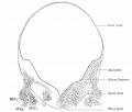
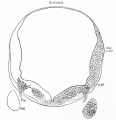
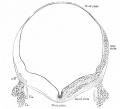
File:Bailey291.jpg|Fig. 291 | File:Bailey291.jpg|Fig. 291. Transverse sections of a rabbit embryo Showing how the omphalomesenteric veins (vom) push outward across the ccelom and fuse with the lateral body wall. | ||
File:Bailey292.jpg|Fig. 292 | File:Bailey292.jpg|Fig. 292 | ||
File:Bailey293.jpg|Fig. 293 | File:Bailey293.jpg|Fig. 293 | ||
File:Bailey294.jpg|Fig. 294 | File:Bailey294.jpg|Fig. 294 | ||
File:Bailey295.jpg|Fig. 295 | File:Bailey295.jpg|Fig. 295 | ||
File: | File:Bailey296_297.jpg|Fig. 296-297 | ||
File:Bailey298.jpg|Fig. 298 | File:Bailey298.jpg|Fig. 298 | ||
</gallery> | </gallery> | ||
| Line 352: | Line 406: | ||
<gallery> | <gallery> | ||
File: | File:Bailey299_300.jpg|Fig. 299-300 | ||
File:Bailey301.jpg|Fig. 301 | File:Bailey301-303.jpg|Fig. 301-303 | ||
File:Bailey304.jpg|Fig. 304 | File:Bailey304.jpg|Fig. 304 | ||
</gallery> | </gallery> | ||
| Line 398: | Line 450: | ||
File:Bailey339.jpg|Fig. 339 | File:Bailey339.jpg|Fig. 339 | ||
File:Bailey340.jpg|Fig. 340 | File:Bailey340.jpg|Fig. 340 | ||
File:Bailey341.jpg|Fig. 341 | |||
File:Bailey342.jpg|Fig. 342 | |||
File:Bailey343-344.jpg|Fig. 343-344 | |||
File:Bailey345-346.jpg|Fig. 345-346 | |||
File:Bailey347-348.jpg|Fig. 347-348 | |||
File:Bailey349.jpg|Fig. 349 | |||
File:Bailey350.jpg|Fig. 350 | |||
File:Bailey351.jpg|Fig. 351 | |||
File:Bailey352.jpg|Fig. 352 | File:Bailey352.jpg|Fig. 352 | ||
</gallery> | </gallery> | ||
| Line 427: | Line 487: | ||
File:Bailey368.jpg|Fig. 368 | File:Bailey368.jpg|Fig. 368 | ||
File:Bailey369.jpg|Fig. 369 | File:Bailey369.jpg|Fig. 369 | ||
File:Bailey370.jpg|Fig. 370 | |||
File:Bailey371.jpg|Fig. 371 | |||
File:Bailey372.jpg|Fig. 372 | |||
File:Bailey373.jpg|Fig. 373 | |||
File:Bailey374.jpg|Fig. 374 | |||
File:Bailey375.jpg|Fig. 375 | |||
File:Bailey376.jpg|Fig. 376 | |||
File:Bailey377.jpg|Fig. 377 | |||
File:Bailey378.jpg|Fig. 378 | |||
File:Bailey379-382.jpg|Fig. 379-382 | |||
File:Bailey383.jpg|Fig. 383 | |||
File:Bailey384.jpg|Fig. 384 | |||
File:Bailey385.jpg|Fig. 385 | |||
File:Bailey386.jpg|Fig. 386 | |||
File:Bailey387.jpg|Fig. 387 | |||
File:Bailey388.jpg|Fig. 388 | |||
File:Bailey389.jpg|Fig. 389 | |||
File:Bailey390.jpg|Fig. 390 | |||
File:Bailey391.jpg|Fig. 391 | |||
File:Bailey392.jpg|Fig. 392 | |||
File:Bailey393.jpg|Fig. 393 | |||
File:Bailey394.jpg|Fig. 394 | |||
File:Bailey395.jpg|Fig. 395 | |||
File:Bailey396.jpg|Fig. 396 | |||
File:Bailey397.jpg|Fig. 397 | |||
File:Bailey398.jpg|Fig. 398 | |||
File:Bailey399.jpg|Fig. 399 | |||
File:Bailey400.jpg|Fig. 400 | |||
File:Bailey401.jpg|Fig. 401 | |||
File:Bailey402.jpg|Fig. 402 | |||
File:Bailey403.jpg|Fig. 403 | |||
File:Bailey404.jpg|Fig. 404 | |||
File:Bailey405.jpg|Fig. 405 | |||
File:Bailey406.jpg|Fig. 406 | |||
File:Bailey407.jpg|Fig. 407 | |||
File:Bailey408.jpg|Fig. 408 | |||
File:Bailey409.jpg|Fig. 409 | |||
File:Bailey410.jpg|Fig. 410 | |||
File:Bailey411.jpg|Fig. 411 | |||
File:Bailey412.jpg|Fig. 412 | |||
File:Bailey413.jpg|Fig. 413 | |||
File:Bailey414.jpg|Fig. 414 | |||
File:Bailey415.jpg|Fig. 415 | |||
File:Bailey416.jpg|Fig. 416 | |||
File:Bailey417.jpg|Fig. 417 | |||
File:Bailey418.jpg|Fig. 418 | |||
File:Bailey419.jpg|Fig. 419 | |||
File:Bailey420.jpg|Fig. 420 | |||
File:Bailey421.jpg|Fig. 421 | |||
File:Bailey422.jpg|Fig. 422 | |||
File:Bailey423.jpg|Fig. 423 | |||
File:Bailey424.jpg|Fig. 424 | |||
File:Bailey425.jpg|Fig. 425 | |||
File:Bailey426.jpg|Fig. 426 | |||
File:Bailey427.jpg|Fig. 427 | |||
File:Bailey428.jpg|Fig. 428 | |||
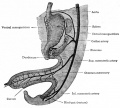
File:Bailey429.jpg|Fig. 429 | |||
File:Bailey430.jpg|Fig. 430 | |||
File:Bailey431.jpg|Fig. 431 | |||
File:Bailey432.jpg|Fig. 432 | |||
File:Bailey433.jpg|Fig. 433 | |||
File:Bailey434.jpg|Fig. 434 | |||
File:Bailey435.jpg|Fig. 435 | |||
File:Bailey436.jpg|Fig. 436 | |||
File:Bailey437.jpg|Fig. 437 | |||
File:Bailey438.jpg|Fig. 438 | |||
File:Bailey439.jpg|Fig. 439 | |||
File:Bailey440.jpg|Fig. 440 | |||
File:Bailey441.jpg|Fig. 441 | |||
File:Bailey442.jpg|Fig. 442 | |||
File:Bailey443.jpg|Fig. 443 | |||
File:Bailey444.jpg|Fig. 444 | |||
File:Bailey445.jpg|Fig. 445 | |||
File:Bailey446.jpg|Fig. 446 | |||
File:Bailey447.jpg|Fig. 447 | |||
File:Bailey448.jpg|Fig. 448 | |||
File:Bailey449.jpg|Fig. 449 | |||
File:Bailey450.jpg|Fig. 450 | |||
File:Bailey451-452.jpg|Fig. 451 452 | |||
File:Bailey453.jpg|Fig. 453 | |||
File:Bailey454.jpg|Fig. 454 | |||
File:Bailey455.jpg|Fig. 455 | |||
</gallery> | </gallery> | ||
| Line 436: | Line 578: | ||
File:Bailey456.jpg|Fig. 456 | File:Bailey456.jpg|Fig. 456 | ||
File:Bailey457.jpg|Fig. 457 | File:Bailey457.jpg|Fig. 457 | ||
File:Bailey458.jpg|Fig. 458 | File:Bailey458-459.jpg|Fig. 458-459 | ||
File:Bailey460.jpg|Fig. 460 | File:Bailey460.jpg|Fig. 460 | ||
File:Bailey461.jpg|Fig. 461 | |||
File:Bailey462.jpg|Fig. 462 | |||
File:Bailey463.jpg|Fig. 463 | |||
File:Bailey464.jpg|Fig. 464 | |||
File:Bailey465.jpg|Fig. 465 | |||
File:Bailey466.jpg|Fig. 466 | |||
File:Bailey467.jpg|Fig. 467 | |||
File:Bailey468.jpg|Fig. 468 | |||
File:Bailey469.jpg|Fig. 469 | |||
File:Bailey470.jpg|Fig. 470 | |||
File:Bailey471.jpg|Fig. 471 | |||
File:Bailey472.jpg|Fig. 472 | |||
File:Bailey473.jpg|Fig. 473 | |||
File:Bailey474.jpg|Fig. 474 | |||
File:Bailey475.jpg|Fig. 475 | |||
File:Bailey476.jpg|Fig. 476 | |||
File:Bailey477.jpg|Fig. 477 | |||
</gallery> | </gallery> | ||
| Line 449: | Line 607: | ||
File:Bailey479.jpg|Fig. 479 | File:Bailey479.jpg|Fig. 479 | ||
File:Bailey480.jpg|Fig. 480 | File:Bailey480.jpg|Fig. 480 | ||
File:Bailey481.jpg|Fig. 481 | |||
File:Bailey482.jpg|Fig. 482 | |||
File:Bailey483.jpg|Fig. 483 | |||
File:Bailey484.jpg|Fig. 484 | |||
File:Bailey485.jpg|Fig. 485 | |||
File:Bailey486.jpg|Fig. 486 | |||
File:Bailey487.jpg|Fig. 487 | |||
File:Bailey488.jpg|Fig. 488 | |||
File:Bailey489.jpg|Fig. 489 | |||
File:Bailey490.jpg|Fig. 490 | |||
File:Bailey491.jpg|Fig. 491 | |||
File:Bailey492.jpg|Fig. 492 | |||
File:Bailey493.jpg|Fig. 493 | |||
File:Bailey494.jpg|Fig. 494 | |||
File:Bailey495.jpg|Fig. 495 | |||
File:Bailey496.jpg|Fig. 496 | |||
File:Bailey497.jpg|Fig. 497 | |||
File:Bailey498.jpg|Fig. 498 | |||
File:Bailey499.jpg|Fig. 499 | |||
File:Bailey500.jpg|Fig. 500 | |||
File:Bailey501.jpg|Fig. 501 | |||
File:Bailey502.jpg|Fig. 502 | |||
File:Bailey503.jpg|Fig. 503 | File:Bailey503.jpg|Fig. 503 | ||
</gallery> | </gallery> | ||
Latest revision as of 10:12, 13 January 2015
| Embryology - 16 Apr 2024 |
|---|
| Google Translate - select your language from the list shown below (this will open a new external page) |
|
العربية | català | 中文 | 中國傳統的 | français | Deutsche | עִברִית | हिंदी | bahasa Indonesia | italiano | 日本語 | 한국어 | မြန်မာ | Pilipino | Polskie | português | ਪੰਜਾਬੀ ਦੇ | Română | русский | Español | Swahili | Svensk | ไทย | Türkçe | اردو | ייִדיש | Tiếng Việt These external translations are automated and may not be accurate. (More? About Translations) |
Bailey FR. and Miller AM. Text-Book of Embryology (1921) New York: William Wood and Co.
- Contents: Germ cells | Maturation | Fertilization | Amphioxus | Frog | Chick | Mammalian | External body form | Connective tissues and skeletal | Vascular | Muscular | Alimentary tube and organs | Respiratory | Coelom, Diaphragm and Mesenteries | Urogenital | Integumentary | Nervous System | Special Sense | Foetal Membranes | Teratogenesis | Figures
| Historic Disclaimer - information about historic embryology pages |
|---|
| Pages where the terms "Historic" (textbooks, papers, people, recommendations) appear on this site, and sections within pages where this disclaimer appears, indicate that the content and scientific understanding are specific to the time of publication. This means that while some scientific descriptions are still accurate, the terminology and interpretation of the developmental mechanisms reflect the understanding at the time of original publication and those of the preceding periods, these terms, interpretations and recommendations may not reflect our current scientific understanding. (More? Embryology History | Historic Embryology Papers) |
The germ cells
Maturation
Fertilization
- Links: Fertilization | Category:Fertilization
Early development of amphioxus
Early development of amphioxus
Early development of the frog
Early development of the chick
Early development of the chick
- Links: Chicken Development
Early mammalian development
Development of the external form of the body
Development of the external form of the body
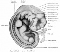
- Bailey092.jpg
Fig. 92. Human embryo of 18.5 mm (52-54 days).
The development of connective tissues and the skeletal system
The development of connective tissues and the skeletal system
- Bailey108.jpg
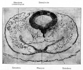
Fig. 108
- Bailey134.jpg
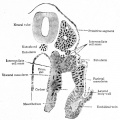
Fig. 134
The development of the vascular system
The development of the vascular system
- Bailey186.jpg
Fig. 186
The muscular system
The development of the muscular system
The alimentary and organs
The development of the alimentary tube and appended organs
- Bailey239.jpg
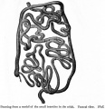
Fig. 239
- Bailey240.jpg
Fig. 240
The respiratory system
The development of the respiratory system
The coelom, pericardium, pleuroperitoneum, diaphragm and mesenteries
The development of the coelom, the pericardium, pleuroperitoneum, diaphragm and mesenteries
The urogenital system
The development of the urogenital system
The integumentary system
The development of the integumentary system
The nervous system
The organs of special sense
Foetal membranes
Teratogenesis
No images in this chapter.
| Historic Disclaimer - information about historic embryology pages |
|---|
| Pages where the terms "Historic" (textbooks, papers, people, recommendations) appear on this site, and sections within pages where this disclaimer appears, indicate that the content and scientific understanding are specific to the time of publication. This means that while some scientific descriptions are still accurate, the terminology and interpretation of the developmental mechanisms reflect the understanding at the time of original publication and those of the preceding periods, these terms, interpretations and recommendations may not reflect our current scientific understanding. (More? Embryology History | Historic Embryology Papers) |
Text-Book of Embryology: Germ cells | Maturation | Fertilization | Amphioxus | Frog | Chick | Mammalian | External body form | Connective tissues and skeletal | Vascular | Muscular | Alimentary tube and organs | Respiratory | Coelom, Diaphragm and Mesenteries | Urogenital | Integumentary | Nervous System | Special Sense | Foetal Membranes | Teratogenesis | Figures
Glossary Links
- Glossary: A | B | C | D | E | F | G | H | I | J | K | L | M | N | O | P | Q | R | S | T | U | V | W | X | Y | Z | Numbers | Symbols | Term Link
Cite this page: Hill, M.A. (2024, April 16) Embryology Book - Text-Book of Embryology (1921) - Figures. Retrieved from https://embryology.med.unsw.edu.au/embryology/index.php/Book_-_Text-Book_of_Embryology_(1921)_-_Figures
- © Dr Mark Hill 2024, UNSW Embryology ISBN: 978 0 7334 2609 4 - UNSW CRICOS Provider Code No. 00098G

