Book - Stoehr's Histology Figures: Difference between revisions
mNo edit summary |
mNo edit summary |
||
| (16 intermediate revisions by the same user not shown) | |||
| Line 1: | Line 1: | ||
{{Lewis1906 header}} | {{Lewis1906 header}} | ||
| Line 8: | Line 7: | ||
<gallery> | <gallery> | ||
File:Lewis1906 fig001.jpg|Fig. 1. Diagram of a Cell. | File:Lewis1906 fig001.jpg|Fig. 1. Diagram of a Cell. | ||
File:Lewis1906 | File:Lewis1906 fig076.jpg|Fig. 76. | ||
File:Lewis1906 fig202.jpg|Fig. 202. | File:Lewis1906 fig202.jpg|Fig. 202. | ||
</gallery> | |||
==Eye== | |||
<gallery> | |||
File:Lewis1906 fig412.jpg|Fig. 412 | |||
File:Lewis1906 fig413.jpg|Fig. 413. | |||
File:Lewis1906 fig414.jpg|Fig. 414. | |||
File:Lewis1906 fig415.jpg|Fig. 415. | |||
File:Lewis1906 fig416.jpg|Fig. 416. | |||
File:Lewis1906 fig425.jpg|Fig. 425. | |||
File:Lewis1906 fig427.jpg|Fig. 427. | |||
File:Lewis1906 fig428.jpg|Fig. 428. | |||
</gallery> | |||
==Ear== | |||
<gallery> | |||
File:Lewis1906 fig429.jpg|Fig. 429. | |||
File:Lewis1906 fig430.jpg|Fig. 430. | |||
File:Lewis1906 fig431.jpg|Fig. 431. | |||
File:Lewis1906 fig432.jpg|Fig. 432. | |||
File:Lewis1906 fig433.jpg|Fig. 433. | |||
File:Lewis1906 fig434.jpg|Fig. 434. | |||
File:Lewis1906 fig435.jpg|Fig. 435. | |||
File:Lewis1906 fig436.jpg|Fig. 436 | |||
File:Lewis1906 fig437.jpg|Fig. 437. | |||
File:Lewis1906 fig438.jpg|Fig. 438. | |||
File:Lewis1906 fig439.jpg|Fig. 439. | |||
</gallery> | |||
==Preparation of Specimens== | |||
<gallery> | |||
File:Lewis1906 fig450.jpg|Fig. 450. | |||
File:Lewis1906 microscope.jpg|microscope | |||
</gallery> | </gallery> | ||
{{Lewis1906 footer}} | {{Lewis1906 footer}} | ||
Latest revision as of 20:44, 9 November 2015
| Embryology - 23 Apr 2024 |
|---|
| Google Translate - select your language from the list shown below (this will open a new external page) |
|
العربية | català | 中文 | 中國傳統的 | français | Deutsche | עִברִית | हिंदी | bahasa Indonesia | italiano | 日本語 | 한국어 | မြန်မာ | Pilipino | Polskie | português | ਪੰਜਾਬੀ ਦੇ | Română | русский | Español | Swahili | Svensk | ไทย | Türkçe | اردو | ייִדיש | Tiếng Việt These external translations are automated and may not be accurate. (More? About Translations) |
Lewis FT. Stoehr's Histology. (1906) P. Blakiston's Son & Co., Philadelphia.
| Stoehr's Histology 1906: 1 Microscopic Anatomy | 1-1 Cytology | 1-2 General Histology | 1-3 Special Histology | 2 Preparation of Specimens | Figures | Histology | Embryology History |
| Historic Disclaimer - information about historic embryology pages |
|---|
 |
- Note - This historic textbook is currently in an early stage of online preparation. (this notice removed when completed)
Template page - content to be added.
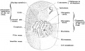
Fig. 1. Diagram of a Cell.
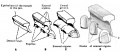
Fig. 76.
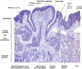
Fig. 202.


Eye
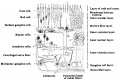
Fig. 412
- Lewis1906 fig413.jpg
Fig. 413.
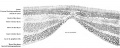
Fig. 414.
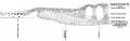
Fig. 415.
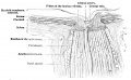
Fig. 416.
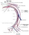
Fig. 425.
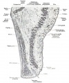
Fig. 427.
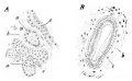
Fig. 428.


Ear
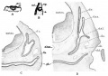
Fig. 429.
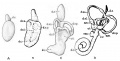
Fig. 430.

Fig. 431.
- Lewis1906 fig432.jpg
Fig. 432.
- Lewis1906 fig433.jpg
Fig. 433.
Fig. 434.
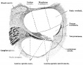
Fig. 435.
Fig. 436
Fig. 437.
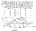
Fig. 438.
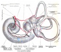
Fig. 439.


Preparation of Specimens
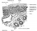
Fig. 450.
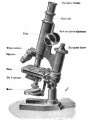
microscope


| Historic Disclaimer - information about historic embryology pages |
|---|
|
| Stoehr's Histology 1906: 1 Microscopic Anatomy | 1-1 Cytology | 1-2 General Histology | 1-3 Special Histology | 2 Preparation of Specimens | Figures | Histology | Embryology History |
Cite this page: Hill, M.A. (2024, April 23) Embryology Book - Stoehr's Histology Figures. Retrieved from https://embryology.med.unsw.edu.au/embryology/index.php/Book_-_Stoehr%27s_Histology_Figures
- © Dr Mark Hill 2024, UNSW Embryology ISBN: 978 0 7334 2609 4 - UNSW CRICOS Provider Code No. 00098G