|
|
| (45 intermediate revisions by the same user not shown) |
| Line 1: |
Line 1: |
| {{Historic Disclaimer}} | | {{Lewis1906 header}} |
|
| |
|
| STOHR'S HISTOLOGY
| | {| class="wikitable mw-collapsible mw-collapsed" |
| | ! Online Editor |
| | |- |
| | | [[File:Mark_Hill.jpg|90px|left]] This historic 1906 textbook translated by [[Embryology History - Frederic Lewis|Frederic T. Lewis]] from the original German of Stöhr, describes histology organised upon an embryological basis. Note that linked terms within the textbook go to the modern pages and the "Online Editor" sections provide additional information.<br> |
| | See also the 1913 second edition: {{Ref-LewisStöhr1913}} |
| | <br><br> |
| | [[Media:1906 A Text-book of Histology - Arranged Upon an Embryological Basis.pdf|PDF version]] | [https://archive.org/details/atextbookhistol00schugoog Internet Archive] |
| | <br> |
| | [[Historic Embryology Textbooks]] |
| | <br> |
| | |} |
| | =Stöhr's Histology= |
| | [[File:Stoehr's Histology 1906 titlepage.jpg|thumb|400px|Stoehr's Histology titlepage]] |
| | Arranged Upon An Embryological Basis |
|
| |
|
| ARRANGED UPON AN EMBRYOLOGICAL BASIS
| | By |
|
| |
|
| | [[Embryology History - Frederic Lewis|Dr. Frederic T. Lewis]] |
|
| |
|
| | Assistant Professor of Embryology at the Harvard Medical School |
|
| |
|
| BY
| | From the Twelfth German Edition |
|
| |
|
| DR. FREDERIC T. LEWIS
| | By |
|
| |
|
| ASSISTANT PROFESSOR OP EMBRYOLOGY AT THE HARVARD MEDICAL SCHOOL
| | Dr. Philipp Stoehr (Stöhr) |
|
| |
|
| | Professor of Anatomy at the University Of Würzburg |
|
| |
|
| | Sixth American Edition |
|
| |
|
| FROM THE TWELFTH GERMAN EDITION
| |
|
| |
|
| |
|
| |
| BY
| |
|
| |
| DR. PHILIPP STOHR
| |
|
| |
| PROPRSSOR OF ANATOMY AT THR UNIVERSITY OF WÜRZBURG
| |
|
| |
|
| |
|
| |
| Sixtb american edition
| |
| With 450 illustrations | | With 450 illustrations |
|
| |
|
| | Philadelphia |
|
| |
|
| | | P. Blakiston's Son & Co. 1012 Walnut Street 1906 |
| PHILADELPHIA
| |
| | |
| P. BLAKISTON'S SON & CO. | |
| | |
| 1012 WALNUT STREET | |
| | |
| 1906 | |
| | |
| Copyright, 1903, by Dr. Alfred Schaper | | Copyright, 1903, by Dr. Alfred Schaper |
|
| |
| Copyright, 1906, by Estatr of Dr. Alfred Schaper | | Copyright, 1906, by Estatr of Dr. Alfred Schaper |
| | Press of WM. Fell Company, 1820-24 Sandom Street, PHILADELPHIA, PA |
| | [[File:Philipp Stöhr.jpg|thumb|alt=Philipp Stöhr|Philipp Stöhr (1849-1911)]] |
| | ==Note== |
| | In the new edition of the American translation of my hand-book a number of additions and changes have been made by the translator with my permission. It is therefore reasonable that I should not take the same responsibility for the translation as for the text of the German original, and I would ask those of my colleagues who wish to question the correctness of my assertions in their papers, to convince themselves, by making comparisons with my last German edition, that the paragraphs in question were written by me. |
|
| |
|
| Press Of WM. Fell Company
| |
| 1820-24 Sandom Street
| |
| PHILADELPHIA, PA
| |
|
| |
|
| | Philipp Stöhr. |
|
| |
|
| ==Note== | | ==Preface== |
| | [[File:Frederick Thomas Lewis.jpg|thumb|alt=Frederick Thomas Lewis|link=Embryology History - Frederic Lewis|Frederick Thomas Lewis (1875-1951)]] |
| | The need of a text-book of histology arranged upon an embryological basis has long been felt. At the Harvard Medical School this need has been urgent. There Professor Schaper, the editor of the five previous American editions of Stohr's Histology planned such a book, and after his return to Germany its preparation was begun. It is greatly to be regretted that at the time of his death the work was only commenced, for there was promise of a notable production. |
|
| |
|
| In the new edition of the American translation of my hand-book a number of additions and changes have been made by the translator with my permission. It is therefore reasonable that I should not take the same responsibility for the translation as for the text of the German original, and I would ask those of my colleagues who wish to question the correctness of my assertions in their papers, to convince themselves, by making comparisons with my last German edition, that the paragraphs in question were written by me.
| |
|
| |
|
| Philipp Stohr.
| | When the writer was informed that Professor Stohr had given generous permission to adapt a new edition of his Histology to American needs it was decided to rearrange the book upon an embryological plan. This has been accomplished with the loss of some characteristic features of the German edition, for which the added material will, it is hoped, make compensation. Thus in order to have space for describing the controlling developmental features of the organs, and for presenting their adult structure somewhat more fully, the directions for preparing sections have been reduced to the minimum. These may be supplemented by directions in the class room; and for the small proportion of students who intend to practice elaborate microscopical methods, a special text-book may be recommended. It is not essential that a physician should be familiar with the details of many staining processes, but the structure of the adult organs and the developmental possibilities of their constituent tissues must be known. |
|
| |
|
|
| |
|
| | The nomenclature adopted is that published by the committee of the German Association of Anatomists m 1895 (-^rcA. /. Anat, u. Phys.; Anat, Abth.; Supplement-Band) J and which is now widely used. It is founded upon the sound principle that the name of a structure should be the simplest possible descriptive Latin term or phrase. Since the Latin names may be translated into the various modem languages the nomenclature is international. Moreover a large number of the names are conmionly used in their Latin forms. Personal names have been discarded (except Wolffian and Milllertan), thus greatly assisting the student. It is obviously easier to learn intestinal glands , duodenal glands, parotid duct, etc., rather than Lieberkiihn's glands, Brunner's glands, Stenson's duct, and the like. It has been estimated that five thousand synonyms have been rejected and are to be removed from the anatomist's vocabulary as soon as possible. In the following pages the more common of the rejected names have been placed in square brackets, [ ]. However difficult it may be for the older anatomists to conform to this nomenclature, it seems clearly a duty to the overworked medical students to adopt it. |
|
| |
|
| ==Preface==
| |
|
| |
|
| | Excellent as the German nomenclature is, as a whole, it is not beyond improvement, and it may be desirable for a conMnittee of the Association of American Anatomists to publish in their English forms a corresponding list of names.<ref>The writer has since been informed that Messrs. Blakiston's Son & Co. have in press such a list prepared by Professor Barker and entitled "Anatomical Terminology" The orderly arrangement of these descriptive names makes the Latin list - and undoubtedly their English version also - an excellent means by which students may review anatomy.</ref> As few changes as possible should be made, but it is certain , for example, that the ventral surface of the body will not be called anterior , or the dorsal surface posterior. In the following pages anterior always means toward the head. Conunon general terms should be made even more specific. For instance, it is questionable whether follicle (Latin, a small leather bag, a husk or shell) should be applied to anything other than closed cysts like the follicles of the ovary and thyreoid gland. Its application by the Germans to the sheath of the hair and by many Americans to solid nodules of lymphoid tissue may lead the student to wonder if ** follicle" is not a colloquial rather than a scientific term. |
|
| |
|
| The need of a text-book of histology arranged upon an embryological basis has long been felt. At the Harvard Medical School this need has been urgent. There Professor Schaper, the editor of the five previous American editions of Stohr^s Histology ^ planned such a book, and after his return to Germany its preparation was begun. It is greatly to be regretted that at the time of his death the work was only commenced, for there was promise of a notable production.
| |
|
| |
|
| When the writer was informed that Professor Stohr had given generous permission to adapt a new edition of his Histology to American needs it was decided to rearrange the book upon an embryological plan. This has been accomplished with the loss of some characteristic features of the German edition, for which the added material will, it is hoped, make compensation. Thus in order to have space for describing the controlling developmental features of the organs, and for presenting their adult structure somewhat more fully, the directions for preparing sections have been reduced to the minimum. These may be supplemented by directions in the class room; and for the small proportion of students who intend to practice elaborate microscopical methods, a special text-book may be recommended. It is not essential that a physician should be familiar with the details of many staining processes, but the structure of the adult organs and the developmental possibilities of their constituent tissues must be known.
| | The attention of all students should be called to the American Journal of Anatomy, the quarterly publication of the Association of American Anatomists, which contains the results of current American anatomical and histological investigations. It probably aflfords the most satisfactory means by which a physician may keep in touch with these sciences. |
|
| |
|
| The nomenclature adopted is that published by the committee of the German Association of Anatomists m 1895 (-^rcA. /. Anat, u. Phys.; Anat, Abth.; Supplement-Band) J and which is now widely used. It is founded upon the sound principle that the name of a structure should be the simplest possible descriptive Latin term or phrase. Since the Latin names may be translated into the various modem languages the nomenclature is international. Moreover a large number of the names are conmionly used in
| |
| their Latin forms. Personal names have been discarded (except Wolffian and Milllertan), thus greatly assisting the student. It is obviously easier to learn intestinal glands , duodenal glands, parotid duct, etc., rather than Lieberkiihn's glands, Brunner's glands, Stenson's duct, and the like. It has been estimated that five thousand synonyms have been rejected and are to be removed from the anatomist's vocabulary as soon as possible. In the following pages the more common of the rejected names have been placed in square brackets, [ ]. However difficult it may be for the older anatomists to conform to this nomenclature, it seems clearly a duty to the overworked medical students to adopt it.
| |
|
| |
|
| Excellent as the German nomenclature is, as a whole, it is not beyond improvement, and it may be desirable for a conMnittee of the Association of American Anatomists to publish in their English forms a corresponding list of names.<ref>The writer has since been informed that Messrs. Blakiston's Son & Co. have in press such a list prepared by Professor Barker and entitled "Anatomical Terminology" The orderly arrangement of these descriptive names makes the Latin list - and undoubtedly their English version also - an excellent means by which students may review anatomy.</ref> As few changes as possible should be made, but it is certain , for example, that the ventral surface of the body will not be called anterior , or the dorsal surface posterior. In the following pages anterior always means toward the head. Conunon general terms should be made even
| | The writer has many acknowledgments to make for help received. Messrs. P. Blakiston's Son & Co., and Mr. William T. Oliver, the artist who has drawn the more elaborate of the new figures, have rendered all the assistance possible. Members of several departments at the Harvard Medical School have given valuable advice, and Dr. G. H. Wright, Assistant in Dental Histology, has arranged a considerable portion of the section on the teeth. It is a privilege to present for the first time in a textbook, the discoveries of Dr. James H. Wright regarding the origin of blood plates. His remarkable conclusion that they are fragments of pseudopodia of the giant cells seems established beyond doubt by an examination of his specimens. |
| more specific. For instance, it is questionable whether follicle (Latin, a small leather bag, a husk or shell) should be applied to anything other than closed cysts like the follicles of the ovary and thyreoid gland. Its appUcation by the Germans to the sheath of the hair and by many Americans to solid nodules of lymphoid tissue may lead the student to wonder if ** follicle" is not a colloquial rather than a scientific term.
| |
|
| |
|
| The attention of all students should be called to the American Journal of Anatomy, the quarterly publication of the Association of American Anatomists, which contains the results of current American anatomical and histological investigations. It probably aflfords the most satisfactory means by which a physician may keep in touch with these sciences.
| |
|
| |
| The writer has many acknowledgments to make for help received. Messrs. P. Blakiston^s Son & Co., and Mr. William T. Oliver, the artist who has drawn the more elaborate of the new figures, have rendered all the assistance possible. Members of several departments at the Harvard Medical School have given valuable advice, and Dr. G. H. Wright, Assistant in Dental Histology, has arranged a considerable portion of the section on the teeth. It is a privilege to present for the first time in a textbook, the discoveries of Dr. James H. Wright regarding the origin of blood plates. His remarkable conclusion that they are fragments of pseudopodia of the giant cells seems established beyond doubt by an examination of his specimens.
| |
|
| |
|
| Finally it is a pleasure to record that after studying histology and embryology under [[Embryology History - Charles Minot|Professor Charles S. Minot]], the writer has for several years enjoyed the closest association with him in his scientific work. The results of such unusual privilege should be found reflected in this edition of Professor Stohr's Histology. | | Finally it is a pleasure to record that after studying histology and embryology under [[Embryology History - Charles Minot|Professor Charles S. Minot]], the writer has for several years enjoyed the closest association with him in his scientific work. The results of such unusual privilege should be found reflected in this edition of Professor Stohr's Histology. |
| Line 86: |
Line 79: |
|
| |
|
| <references/> | | <references/> |
| | | {| class="wikitable mw-collapsible mw-collapsed" |
| | ! Other Texts by Frederic T. Lewis |
| | |- |
| | | |
| | * [[Book - Manual of Human Embryology 17|Manual of Human Embryology (1912) Chapter XVII. The Development of the Digestive Tract and of the Organs of Respiration]] |
| | * Lewis, F. T., 1906. The development of the lymphatic system in rabbits. Amer. Jour. Anat., vol. 5, p. 95. |
| | |} |
|
| |
|
| ==Contents== | | ==Contents== |
| | ===Part I. Microscopic Anatomy=== |
| | [[Book - Stoehr's Histology 1|Part I. Microscopic Anatomy]] |
| | ====I. Cytology==== |
| | [[Book - Stoehr's Histology 1-1|I. Cytology]] |
| | * The Cell, |
| | ** Protoplasm. |
| | ** Nucleus. |
| | ** Centrosome. |
| | ** Cell Wall. |
| | * Form and Size OF Cells |
| | * Vital Phenomena |
| | ** Amoeboid Motion. |
| | * Formation and Reproduction of Cells |
| | ** Mitosis |
| | ** Amitosis |
| | * Cytomorphosis |
|
| |
|
| PART I. MICROSCOPIC ANATOMY.
| | ===II. General Histology=== |
| | | [[Book - Stoehr's Histology 1-2|II. General Histology]] |
| I. CYTOLOGY.
| | * Histogenesis |
| | | ** Segmentation and the Formation of the Germ Layers. |
| The Cell,
| | ** The Fundamental Tissues. |
| | | * Epithelia |
| Protoplasm.
| | ** Origin |
| | | ** Shapes of Epithelial Cells. |
| Nucleus.
| | ** Number of Layers. |
| | | ** Differentiation. |
| Centrosome.
| | ** Processes of Secretion. |
| | | ** The Nature and Classification of Glands |
| Cell Wall.
| | * Mesenchymal Tissues |
| | | ** Reticular Tissue. |
| Form and Size OF Cells, 7
| | ** Mucous Tissue. |
| | | ** Connective Tissue. |
| Vital Phenomena, 7
| | ** Tendon |
| | | ** Cartilage. |
| Amoeboid Motion.
| | ** Bone. |
| | | ** Joints. |
| Formation and Reproduction of Cells, 9
| | ** Teeth (including the Ectodermal Enamel Organs). |
| | | * Muscle Tissue |
| Mitosis.
| | ** Smooth Muscle |
| | | ** Cardiac Muscle |
| Amitosis.
| | ** Striated Muscle |
| | | * Nerve Tissue, |
| Cytomorphosis, 15
| | ** Development of, |
| | | ** The central tract. |
| II. GENERAL HISTOLOGY. | | ** The spinal ganglia. |
| | | ** The ventral roots. |
| Histogenesis | | ** The sympathetic system. |
| | | ** The cerebral nerves. |
| Segmentation and the Formation of the Germ Layers. | | ** Structure of Nerve fibers and nerves |
| | | ** Structure of Sensory endings. |
| The Fundamental Tissues. | | ** Structure of Motor endings. |
| | | ** Structure of Gangiia |
| Epithelia | | ** The spinal cord. |
| | | * Vascular Tissue |
| Origin | | ** Blood Vessels. |
| | | ** Development |
| Shapes of Epithelial Cells. | | ** Capillaries. |
| | | ** Arteries. |
| Number of Layers. | | ** Veins. |
| | | ** The heart. |
| Differentiation. | | ** Lymphatic Vessels. |
| | | ** Red corpuscles. |
| Processes of Secretion. | | ** White corpuscles. |
| | | ** Blood plates. |
| The Nature and Classification of Glands | | ** Plasma. |
| | | * Lymph. |
| Mesenchymal Tissues | | ====III. Special Histology==== |
| | | [[Book - Stoehr's Histology 1-3|III. Special Histology]] |
| Reticular Tissue. | |
| | |
| Mucous Tissue. | |
| | |
| Connective Tissue. | |
| | |
| Tendon | |
| | |
| Cartilage. | |
| | |
| Bone. | |
| | |
| Joints. | |
| | |
| Teeth (including the Ectodermal Enamel Organs). | |
| | |
| Muscle Tissue, | |
| | |
| Smooth Muscle | |
| | |
| Cardiac Muscle | |
| | |
| Striated Muscle | |
| | |
| Nerve Tissue, | |
| | |
| Development of, - | |
| | |
| The central tract. | |
| | |
| The spinal ganglia. | |
| | |
| The ventral roots. | |
| | |
| The sympathetic system. | |
| | |
| The cerebral nerves. | |
| | |
| Structure of - | |
| | |
| Nerve fibers and nerves | |
| | |
| Sensory endings. | |
| | |
| Motor endings. | |
| | |
| Gangiia | |
| | |
| The spinal cord. | |
| | |
| | |
| Vascular Tissue | |
| | |
| Blood Vessels. | |
| | |
| Development. | |
| | |
| Capillaries. | |
| | |
| Arteries. | |
| | |
| Veins. | |
| | |
| The heart. | |
| | |
| Lymphatic Vessels. | |
| | |
| Red corpuscles. | |
| | |
| White corpuscles. | |
| | |
| Blood plates. | |
| | |
| Plasma. | |
| | |
| Lymph. | |
| | |
| | |
| | |
| | |
| | |
| | |
| III. SPECIAL HISTOLOGY. | |
| | |
| | |
| PAOB
| |
| | |
| Blood Foriung and Blood Destroying Organs, 152
| |
| | |
| Bone Marrow.
| |
| | |
| Lymph Nodules and Lymph Glands.
| |
| | |
| Haemolyroph Glands.
| |
| | |
| Spleen.
| |
| | |
| The Entodermal Tract, 165
| |
| | |
| The Mouth and Pharynx, 165
| |
| | |
| Development.
| |
| | |
| Palatine tonsils.
| |
| | |
| Thymus.
| |
| | |
| Thyreoid gland.
| |
|
| |
|
| Parathyreoid glands. | | * Blood Forming and Blood Destroying Organs |
| | * Bone Marrow |
| | * Lymph Nodules and Lymph Glands |
| | * Haemolyroph Glands |
| | * Spleen |
| | * The Entodermal Tract |
| | ** The Mouth and Pharynx |
| | ** Development. |
| | ** Palatine tonsils. |
| | * Thymus. |
| | * Thyreoid gland. |
| | * Parathyreoid glands. |
| | * Glomus caroticum. |
| | * Tongue. |
| | * Oral and pharyngeal cavities. |
| | * Glands of the oral cavity. |
|
| |
|
| Glomus caroticum.
| | * The Digestive Tube |
| | * Development. |
| | * Oesophagus. |
| | * Stomach. |
| | * Small Intestine. |
| | * Large Intestine. |
| | * Rectum and Anus. |
| | * The Liver |
| | * The Pancreas |
| | * The Respiratory Tract |
| | ** Development. |
| | ** Larynx. |
| | ** Trachea, Bronchi. |
| | ** Lungs. |
| | * Urinary Organs |
| | ** Wolffian Body. |
| | ** Pronephros. |
| | ** Kidney. |
| | ** Renal pelvis and ureter. |
| | ** Bladder. |
| | ** Urethra (in the female) |
| | * Male Genital Organs |
| | ** Development. |
| | ** Testis. |
| | ** Epididymis. |
| | ** Ductus deferens. |
| | ** Seminal Vesicles and Ejaculatory Ducts. |
| | ** Appendices and Paradidymis. |
| | ** Prostate. |
| | ** Urethra and Penis. |
| | * Female Genital Organs |
| | ** Development. |
| | ** Ovary. |
| | ** Epoophoron. |
| | ** Uterine Tubes. |
| | ** Uterus. |
| | ** Menstruation. |
| | ** Development of the decidual membranes. |
| | ** Structure of the membranes and placenta. |
| | ** Umbilical Cord. |
| | ** Vagina and External Genital Organs. |
| | * Skin |
| | ** Nails. |
| | ** Hair. |
| | ** Sebaceous glands. |
| | ** Sweat glands. |
| | ** Mammary glands. |
|
| |
|
| Tongue.
| | Suprarenal Glands |
|
| |
|
| Oral and pharyngeal cavities.
| | Brain and Sense Organs |
|
| |
|
| Glands of the oral cavity.
| | Brain |
| The Digestive Tube 193
| |
| | |
| Development.
| |
| | |
| Oesophagus.
| |
| | |
| Stomach.
| |
| | |
| Small Intestine.
| |
| | |
| Large Intestine.
| |
| | |
| Rectum and Anus.
| |
| | |
| The Liver, 218
| |
| | |
| The Pancreas, 230
| |
| | |
| The Respiratory Tract, 234
| |
| | |
| Development.
| |
| | |
| Larynx.
| |
| | |
| Trachea, Bronchi.
| |
| | |
| Lungs.
| |
| | |
| Urinary Organs, 244
| |
| | |
| Wolffian Body.
| |
| | |
| Pronephros.
| |
| | |
| Kidney.
| |
| | |
| Renal pelvis and ureter.
| |
| | |
| Bladder.
| |
| | |
| Urethra (in the female).
| |
| | |
| Male Genital Organs, 263
| |
| | |
| Development.
| |
| | |
| Testis.
| |
| | |
| Epididymis.
| |
| | |
| Ductus deferens.
| |
| | |
| Seminal Vesicles and Ejaculatory
| |
| | |
| Ducts.
| |
| Appendices and Paradidymis.
| |
| Prostate.
| |
| Urethra and Penis.
| |
| | |
| | |
| | |
| FAHE
| |
| | |
| Female Genital Organs, 285
| |
| | |
| Development.
| |
| Ovary.
| |
| Epoophoron.
| |
| Uterine Tubes.
| |
| Uterus.
| |
| | |
| Menstruation.
| |
| | |
| Development of the decidual
| |
| | |
| membranes.
| |
| Structure of the membranes
| |
| | |
| and placenta.
| |
| Umbilical Cord.
| |
| Vagina and External Genital Organs.
| |
| | |
| Skin, 3
| |
| | |
| Nails.
| |
| | |
| Hair.
| |
| | |
| Sebaceous glands.
| |
| | |
| Sweat glands.
| |
| | |
| Mammary glands.
| |
| | |
| Suprarenal Glands, 33 r
| |
| | |
| Brain and Sense Organs, 334 | |
| | |
| Brain, 334
| |
|
| |
|
| Development. | | Development. |
| Line 375: |
Line 244: |
|
| |
|
| Meninges. | | Meninges. |
| Eye, 353
| |
|
| |
| Development.
| |
|
| |
| Retina.
| |
|
| |
| Optic nerve.
| |
|
| |
| Lens.
| |
|
| |
| Vitreous body.
| |
|
| |
| Tunica vasculosa.
| |
|
| |
| Tunica fibrosa.
| |
|
| |
| Vessels, chambers, and nerves.
| |
|
| |
| Eyelids.
| |
|
| |
| Lachrymal glands.
| |
| Ear, 378
| |
|
| |
| Development.
| |
|
| |
| Internal ear, —
| |
|
| |
| Sacculus.
| |
|
| |
| Utriculus.
| |
|
| |
| Semicircular ducts, and
| |
|
| |
| Brain and Sense OTgnns— Continued, 'aqe Brain and Sense Organs — Continued, 'aob
| |
|
| |
| Cochlea. Nose, 395
| |
|
| |
| Middle ear. Respiratory region.
| |
|
| |
| External ear. Olfactory region.
| |
|
| |
|
| |
|
| |
| PART II. THE PREPARATION AND EXAMINATION OF MICROSCOPICAL SPECIMENS.
| |
|
| |
|
| |
|
| |
| Fresh Tissues,
| |
|
| |
|
| |
| Staining and Mounting
| |
|
| |
|
| |
| 408
| |
|
| |
|
| |
| Isolation.
| |
|
| |
|
| |
| General Stains.
| |
|
| |
|
| |
| Sectioning Fresh Material.
| |
|
| |
|
| |
| ' Special Stains.
| |
|
| |
|
| |
|
| |
|
| |
| Fixation.
| |
|
| |
| The Microscope.
| |
|
| |
| Decalcification.
| |
|
| |
| ' Drawings.
| |
|
| |
| Imbedding.
| |
|
| |
| Reconstructions.
| |
|
| |
| ++++++++++++++++++++++++++++++++++++++++++++++
| |
|
| |
| PART I. MICROSCOPIC ANATOMY.
| |
|
| |
| I. CYTOLOGY. ,
| |
|
| |
| ==The Cell==
| |
|
| |
| Since 1839 it has been known that all plants and animals are composed of small structural elements called cells (Latin, cellula; Greeks kOtoc). The lowest forms of animals and of plants are alike in being single cells throughout life. The more complex organisms are groups of cells which have been derived, by process of repeated division, from a single cell, the fertilized ovum. Thus the human body, which begins as one cell, becomes in the adult an aggregation of cells variously modified and adapted to special functions. Since the liver is a mass of essentially similar cells, the problems of its functional activity are the problems of the functions of a single one of its cells. The diseases of the liver are the result of changes occurring in these cells, which must be restored to a normal condition to effect a cure. As this is equally true of other organs, it is evident that cytology, the science of cells, is a basis for both physiology and pathology.
| |
|
| |
| A cell may be defined as a structural element of limited dimensions which under certain conditions can perform the functions of assimilation,
| |
| growth, and reproduction.^ Because of these possibilities a cell may be
| |
| considered an elementary organism. It is described as a mass of protoplasm containing a nucleus. A third element, the centrosome, is found
| |
| in the cells of animals, but not in those of the higher plants. The centrosome becomes prominent when a cell is about to divide. At other times,
| |
| in many kinds of cells, it has been found as a minute granule which may
| |
| be in the center of a very small clear spot in the protoplasm. Ordinarily
| |
| it cannot be seen unless cell division is about to occur. Some authorities
| |
| regard the centrosome as a temporary structure which forms shortly
| |
| before division begins and disappears after it is completed. Others regard it as a permanent and essential part of a cell, which accordingly
| |
| consists of protoplasm, nucleus, and centrosome.
| |
|
| |
| ==Protoplasm==
| |
| Protoplasm is the living substance of which cells are composed.
| |
| More specifically the term is applied to this living substance exclusive of
| |
| the nucleus, or to the corresponding dead material, provided that death
| |
| has not changed its physical properties. It has been proposed to substitute
| |
| the name cytoplasm for protoplasm in the restricted and earlier sense of
| |
| the][term, to call the nuclear substance nucleoplasm [karyoplasm], and
| |
| to consider both cytoplasm and nucleoplasm as varieties of protoplasm.
| |
| Although these names are often employed, the cell substance apart from
| |
| the nucleus is ordinarily called protoplasm.
| |
|
| |
|
| |
|
| |
| Fig. 1. Diagram of a Cell. Microsomes and filar mass only partly sketched.
| |
|
| |
| Protoplasm is a heterogeneous mixture of substances forming a soft, viscid mass of neutral reaction. In distilled water it swells but does not disappear. It consists of water, salts and organic substances, some in solution, and some in a colloidal state. The organic bodies are classed as proteids, glycogen or some allied carbohydrates, and lipoid (fat-like) bodies. Protoplasm may exist in a numberless variety of forms.
| |
|
| |
| On microscopic examination protoplasm is seen to contain small granules, microsomes. In different cells these vary in abundance and in character. They may be absent from the outer layer of protoplasm, the exoplasm, which is firmer and chemically different from the inner endoplasm, and perhaps has a separate function. The microsomes have been considered both as inert bodies and as the essential living basis of protoplasm. The simplest description of protoplasmic structure is that it consists of a fluid ground substance in which microsomes are embedded.
| |
|
| |
| With high magnification it appears that the protoplasm contains a
| |
| network of filaments (called mitome, or the filar mass, from the Greek
| |
| fihoc and Latin filum, both meaning "a thread," — sppngioplasm is
| |
| another s3nionym). This network is embedded in a more or less homogeneous and chemically different ground substance (paramitome, interfilar
| |
| mass, or hypoplasm). Some of the filaments appear as rows of microsomes, but small particles may also be found in the ground substance
| |
| between the filaments. The conception of protoplasm as fibrillar or
| |
| reticular has been considered at variance with the "granular theory,"
| |
| yet undoubtedly both fibrils and granules occur in protoplasm.
| |
|
| |
| According to a third interpretation protoplasm has the structure of
| |
| foam, or of an emulsion, — that is, it consists of minute droplets of one
| |
| substance completely surrounded by walls of a different substance. This
| |
| view, which has much in its favor, is not inconsistent with the presence
| |
| of granules or of fibrils scattered through the mass.
| |
|
| |
| In addition to these general characteristics the protoplasm of particular
| |
| cells may contain other structures of
| |
| various significance. These may be
| |
| grouped as follows:
| |
|
| |
| I. Fibrils. Although an obscure
| |
| fibrillar network may be characteristic
| |
| of all cells, a high development and
| |
| orderly arrangement of fibrils opcurs
| |
| only in certain specialized cells, as for
| |
|
| |
| example, in muscle, nerve, and connective tissue cells. These fibrils are of very different
| |
| sorts and will be described more fully in the
| |
| section on General Histology.
| |
|
| |
| 2. Granules. These are not the microsomes found in all protoplasm, but are larger
| |
| bodies of definite staining reaction, which often
| |
| are important secretory products elaborated by
| |
| the cell. In many gland cells, and in the
| |
| "granular" white blood corpuscles, these
| |
| structures are conspicuous. Other granules
| |
| may be excretory or waste products of the cell,
| |
| and some of these, which, without being
| |
| stained, are deeply colored, are called pigment granules.
| |
|
| |
|
| |
|
| |
|
| |
| Fig. 2. Fibrils in a Nkrve Cell.
| |
|
| |
|
| |
|
| |
| Fic. 3. Clumps of Granules (NissL's Bodies) in a Nerve Cell.
| |
|
| |
|
| |
|
| |
| HISTOLOGY
| |
|
| |
|
| |
| Fig. 4.Vacl-oi.^ in Young
| |
|
| |
| Fat cells.
| |
|
| |
|
| |
|
| |
| Nuelem. Nucleolus,
| |
|
| |
|
| |
|
| |
|
| |
| Cs|i9ttk* Caiialicijli.
| |
|
| |
| %N>KGtUM> IH A KEttVe
| |
|
| |
|
| |
|
| |
| Retkul»r upparatiis,
| |
|
| |
| Fig 6, ReTictijLAM Nktwork iN A NsKVh CmLL, C After
| |
|
| |
|
| |
|
| |
| the cell. It consists of a
| |
| or nuclear sapi in which
| |
|
| |
|
| |
|
| |
| 3* Vacuoles. Well defined, round spaces,
| |
| apparently empty, may occur in the protoplasm
| |
| due to the formation of droplets of fat or of watery
| |
| fluids. They vary greatly in size, and one or several of these vacuoles may be found in a single cell.
| |
|
| |
| 4. Canals, of two sorts* (a) Secreton^ canals,
| |
| which occur in protoplasm of gland cells and
| |
| empty into the gland cavity or lumen; (i) fine
| |
| tubes which communicate with lymphatic spaces
| |
| outside of the cell* They are found in all cells of
| |
| higher physiological importance, but are lacking
| |
| in most of the supporting tissues and in stratified
| |
| epithehum. They presumably share in nourishing tlie cell and have been called the ** trophospongium," This name, implying a network,
| |
| is due to the opinion, not established however,
| |
| that the Uttle canals are occupied by cell processes extending into the protoplasm from adjoining '^capsule cells." Other investigators consider
| |
| that the trophospongium canals are wholly within
| |
| the cell and constitute a form of vacuole.
| |
|
| |
| 5. Closed networks, which do not open at
| |
| the peripher}* of the cell. This ** reticular apparatus'* has been found in nen-e, cartilage and
| |
| many gland cells. Its significance is unknown,
| |
|
| |
| 6. Inclusions. These are foreign bodies
| |
| which have been ingested by the cell and are
| |
| found in the protoplasm. Inert crystalloid substances formed within the cell are also called
| |
| inclusions. The name "paranucleus" has been
| |
| applied to various structures, such as a dead cell ^
| |
| ingested by a living one, a transformation of the
| |
| centrosome, or a mass of secretion. Some of the
| |
| paranuclei are still obscure.
| |
|
| |
| ==Nucleus==
| |
| The nucleus (Latin, nuclms, '*the kernel of a nut"; Greek, ^i^jov, "a nut'*) is a well defined,
| |
|
| |
| refractive body of vesicular form situated within
| |
| membrane enclosing a mass of ground substance,
| |
| there is a fibrillar network associated with some deeply staining bodies. The ground substance and network are closely
| |
| related to the corresponding structures in the protoplasm. In fact, at the
| |
| time of cell division, when the nuclear membrane disappears, the network
| |
| and ground substance of nucleus and protoplasm are respectively continuous with one another. The ground substance and a delicate, fibrillar
| |
| portion of the network do not stain readily; therefore they are called
| |
| achromatic substances. The fine achromatic fibrils of the network are
| |
| further designated as linin fibrils. Since the linin fibrils cannot be isolated
| |
| for chemical analysis, their composition is unknown. There are two
| |
| deeply staining or chromatic substances found in the nucleus. One
| |
| of these, chromatin, is its most essential and characteristic element. Chemically it is a nucleo-proteid, but undoubtedly it exists in several varieties.
| |
| A portion which responds to acid dyes is called oxychromatin, in distinction from the ordinary form which takes the basic stains. Chromatin is
| |
| distributed as irregular granules or coarse strands along the linin fibrils,
| |
| thus tending to form a network (Fig. i, and Fig. i8, p. i6). Often a
| |
| nucleus presents from one to several large clumps of chromatin, known
| |
| as chromatin "knots." These are to be distinguished from the round
| |
| masses of pyrenin, called nucleoliy which are found between the meshes
| |
| of the nuclear network. All nuclei contain chromatin, but many are
| |
| without nucleoli. The latter are present with great regularity in certain
| |
| kinds of cells. Usually only one is found in a nucleus, although several
| |
| may occur (Fig. i8). They difiFer from chromatin chemically, as is
| |
| evident from differences in staining, and also functionally, as is seen during
| |
| cell division. Pyre nin, of which the nucleolus is said to be composed, is,
| |
| however, a cytological rather than a chemical term.
| |
|
| |
| The nuclear membrane is usually described as formed of amphipyrenin, a term of questionable value. The membrane may consist of a distinct chemical substance as the name suggests, or it may be rather a condensation of the nuclear reticulum, in which the linin fibrils terminate. A nuclear membrane may be simulated by a thin superficial layer of chromatin.
| |
|
| |
| Every cell contains a nucleus consisting, as has been shown, of nuclear
| |
| membrane, ground substance, a network of linin fibrils and of chromatin,
| |
| with perhaps a nucleolus. Non-nucleated bodies like the mammalian
| |
| red blood corpuscles, and the dead outer cells of the skin have lost their
| |
| nuclei in the course of development. Occasionally a single cell contains
| |
| two nuclei, as is frequent in the liver, or even several nuclei, as in certain
| |
| bone cells.
| |
|
| |
| ==Centrosome==
| |
|
| |
| The centrosome is a minute body consisting of a homogeneous or sometimes reticular mass, the centroplasm, which contains a much smaller body, the ceniriole. Such centrosomes have been observed in the invertebrate animals. The cells of vertebrates are not regarded as favorable
| |
| for investigations of the finer structure of centrosomes. In them generally
| |
| both centroplasm and centriole appear as a single small granule, the
| |
| centrosome. This granule is usually, but not always, surrounded by a
| |
| zone of protoplasm which is so modified as to form a darker or a lighter
| |
| area, the archoplasm (Fig. i). (The archoplasm of certain spermatic cells
| |
| is called the idiozome,) The centrosome may be near the nucleus or
| |
| distant from it, frequently being found between the nucleus and the free
| |
| surface of the cell. Rarely, as in a few invertebrates and in cancer cells,
| |
| the centrosome has been found within the nucleus. In many gland cells
| |
| it lies where the secretion accumulates, the expulsion of which is accomplished by the contraction of the protoplasmic framework between the
| |
| masses of secretion. In the intestinal epithelial cells which send out
| |
| motile projections of protoplasm (pseudopodia), the centrosome lies just
| |
| beneath the place of origin of these projections. If one considers also the
| |
| relation of the centrosome in the spermatozoa as well as its r61e in cell
| |
| division, it seems almost certain that the centrosome is the active or passive
| |
| center of the motor functions. In connection with cell division, the
| |
| centrosome undergoes a cycle of changes of varying duration. That
| |
| stage which is continued longest is characterized by a doubling of the
| |
| centrosome, following the division of the centriole in two. The double
| |
| body thus formed is the diplosome. In many resting cells, or those not
| |
| actually in the process of division, a diplosome is found, and this is significant as indicating the readiness of the cell for undergoing division without
| |
| delay.
| |
|
| |
| ==Cell Wall==
| |
|
| |
| A cell wall or cell membrane is an independent membranous layer
| |
| covering a cell and being clearly distinct from the underlying protoplasm.
| |
| It is not an essential constituent of a cell. Often it is lacking, and when
| |
| present it is either a modification or a secretion of the peripheral protoplasm.
| |
| If the membrane surrounds the cell on all sides it is called a pellicula;
| |
| if it is on only one side, covering the free surface, it is a cuiicula. (The
| |
| former term is seldom used.) Cells may unite with one another by protoplasmic processes of varying length and width, thus forming cellular
| |
| networks; or they may completely fuse so that their nuclei appear irregularly distributed through a single mass of protoplasm. Such a formation
| |
| is a syncytium [plasmodium]. This name is applied also to such structures
| |
| as the striated muscle fiber, due not to the fusion of cells but to the multipUcation of nuclei in an undivided mass of protoplasm.
| |
|
| |
|
| |
| Although cell membranes are usually lacking, or if present are often inconspicuous in animal cells, they are highly developed in plants. Thus cork
| |
| is a mass of dead cells from which nuclei and protoplasm have disappeared,
| |
| leaving only the cell walls. In describing cork, Robert Hooke introduced the
| |
| name "cell," in 1667. He wrote: "I took a good clear piece of Cork and with a
| |
| Pen-knife sharpened as keen as a razor, I cut a piece of it off and thereby left
| |
| the surface of it exceedingly smooth, then examining it very diligently with a
| |
| microscope, me thought I could perceive it to be a little porous. . . . These pores or cells were not very deep but consisted of a great many little Boxes In this way one of the briefest and most important of scientific terms was introduced.
| |
|
| |
| ==Form and Size of Cells==
| |
|
| |
| Cells are regarded as typically spherical in form. Spherical cells
| |
| are comparatively numerous in the embryo, and in the adult the resting
| |
| white blood corpuscles which float freely in the body fluids assume this
| |
| form. Such cells are circular in cross section. When spherical cells
| |
| are subjected to the pressure of similar neighboring cells they become
| |
| polyhedral and usually appear six-sided in cross section. Such cells, as
| |
| a whole, may be cuboidal, columnar, or flat. Certain cells become fusiform (spindle-shaped) or are further elongated so as to form fibers; others
| |
| send out radiating processes and are called steUate. Thus the form of
| |
| cells is extremely varied. The shape of the nucleus tends to correspond
| |
| with that of its cell. It is usually an elliptical body in elongated cells,
| |
| and spherical in round or cuboidal cells. In stellate cells it is either
| |
| spherical or somewhat elongated. Crescentic nuclei and others more
| |
| deeply and irregularly lobed are found in some of the white blood corpuscles
| |
| and in giant cells.
| |
|
| |
| The size of cells ranges from that of the yolks of birds' eggs —
| |
| which are single cells at least shortly before being laid — down to microscopic structures four thousandths of a millimeter in diameter. The
| |
| thousandth of a millimeter is the unit employed in microscopic measurements. It is called a micron, and its symbol is the Greek letter /jl. The
| |
| small cells referred to are therefore four microns, 4 /i, in diameter. \The
| |
| size of any structure in a section of human tissue may be roughly estimated
| |
| by comparing its dimensions with the diameter of a red blood corpuscle
| |
| found in the same section. These red corpuscles are quite uniformly
| |
| 7.5 /i in diameter.;
| |
|
| |
| ==Vital Phenomena==
| |
|
| |
| The vital properties of cells are more fuUy treated in text-books of physiology. They include the phenomena of irritability, metabolism, contractility, conductivity, and reproduction. Under irritability may be grouped the response of cells to stimuli of various sorts such as heat,
| |
| light, electricity, chemical reagents, the nervous impulse, or mechanical
| |
| interference. Metabolism, in a wide sense, includes the ingestion and assimilation of food, the elaboration and secretion of desirable products,
| |
| together with the elimination of waste products. Contractility may
| |
| be manifest in the locomotion of the entire cell, in the vibratile action of
| |
| slender, hair-hke processes, the cilia, or in contraction of the cell body.
| |
| Conductivity is the power of conveying impulses from one part of the cell
| |
| to another. Reproduction is seen in the process of cell division. Many
| |
| phases of these activities are observed in microscopic sections and as such
| |
| they will be referred to in later chapters. A few which are of general
| |
| occurrence will be described presently.
| |
|
| |
| ==Amoeboid Motion==
| |
|
| |
| The imicellular animal, Amoeba, exhibits a type of motility known
| |
| as amoeboid, which has been observed in many sorts of cells in the vertebrate body. In marked cases, as
| |
| in certain white blood corpuscles
| |
| (the leucocytes), the cell protoplasm
| |
| sends out fine or coarse processes
| |
| which divide or fuse with one an/^^ ^5L ^^ ^fe d i ^^JP other, causing the cell to assume a
| |
|
| |
| s«', 5 6 8 10 Minuses. g^eat Variety of forms. The processes may be retracted, or they may
| |
|
| |
| Fig. 8. Leucocytes of a Frog. X 560. , - - ,
| |
| Changes in form obsen ed duriiiR ten minutes : bCCOme attached SOmCWhcrC and
| |
| o, at the beeinninj; of the observation; J4, a j .1 • j ^ ^i_ n i_ j
| |
|
| |
| half minute later, etc. draw the rcmamdcr of the cell body after them, the result of which is
| |
| locomotion or the so-called wandering of the cell. Such wandering cells
| |
| play an important part in the economy of the animal body. Their processes can flow around granules or cells and thus enclose them in protoplasm. Some of these ingested bodies may be assimilated by the cell as
| |
| a result of complex chemical and osmotic reactions. Cells which feed on
| |
| foreign particles and can alter or digest them are known as phagocytes.
| |
| Amoeboid movements take place very slowly. In preparations from warmblooded animals they may be accelerated by gently heating the object.
| |
|
| |
| Another form of motion, which, however, does not occur in living
| |
| cells, consists in an oscillation of minute granules within the cell. This
| |
| may be due to diffusion currents or to the Brownian phenomena. It
| |
| may often be seen in salivary corpuscles.
| |
|
| |
|
| |
| ==Formation and Reproduction of Cells==
| |
|
| |
| In the past, two sorts of cell formation have been recognized, namely
| |
| the spontaneous generation of cells, and the origin of cells through the
| |
| division of pre-existing cells. According to the theory of spontaneous
| |
| generation it was once thought that animals as highly organized as intestinal worms came into existence from the fermentation of the intestinal
| |
| contents. After this had been disproved it was still thought that cells
| |
| might be formed directly from a suitable fluid, the cytoblastema. Something of the sort may have occurred when life began, and it is the expectation of certain investigators that conditions may yet be produced which
| |
| shall lead to the formation of organic bodies capable of growth and reproduction. At present, however, only one source of cells is recognized, —
| |
| the division of existing cells. "Omnis cellula e cellula." A nucleus
| |
| likewise can arise only by the division of an existing nucleus. There
| |
| is no satisfactory evidence that a nucleus may be formed from nonnudeated protoplasm. In cell division the nucleus divides first and then the
| |
| protoplasm, generally into two nearly equal parts. During the process
| |
| a special grouping and transformation of the nuclear substance occurs
| |
| in accordance with fixed laws. The ordinary mode of cell division is
| |
| called mitosis or indirect division [karyokinesis] and the characteristic
| |
| groups of nuclear material are commonly known as mitotic figures. Mitosis
| |
| is arbitrarily but conveniently divided into three successive phases, the
| |
| prophase, metaphase, and anaphase, in which respectively the nuclear
| |
| material prepares for division, divides, and returns to its usual condition.
| |
| (The final stages of reconstruction are often grouped as a fourth phase, the
| |
| telophase.) In the details of mitosis there are considerable variations,
| |
| not only in different animals but also in different kinds of cells in a single
| |
| species. The account of the process which follows will apply only in a
| |
| general way to a particular case of cell division which the student may
| |
| be examining.
| |
|
| |
| ==Mitosis==
| |
|
| |
| Prophase, The centrosome and nucleus approach one another until
| |
| the centrosome is close to the nuclear membrane where it lies surrounded
| |
| by the clear zone of archoplasm. The archoplasm contributes to the
| |
| formation of radiating fibrils which extend from the centrosome in all
| |
| directions, and are known collectively as the centrosphere [astrosphere].
| |
| The two parts of the centrosome which had formed in the '* resting stage"
| |
| by the division of one, are in the midst of the centrosphere. They move apart, the diplosome thus separating into two centrosomes, and ^ the centrosphere becoming divided into two spheres, each of which contains a centrosome. Fig. 9.
| |
|
| |
| The nucleus meanwhile enlarges and its chromatin stains much more
| |
| deeply. The branching portions of the chromatin network are withdrawn,
| |
| so that instead of a net, the entire chromatic material forms one convoluted
| |
| thread, a monospireme, as this mitotic figure is called. The thread is at
| |
| first more closely coiled than it is later. It divides transversely into a
| |
| definite number of segments, called chromosomes. These bodies may be
| |
| spherical or rod-like, but generally they are U- or V-shaped. The apices
| |
| of all the V's may at first point toward the centrosome with their free ends
| |
| directed away from it as shown in the diagram. Fig. 9. Instead of being
| |
| arranged in the orderly manner of the diagram, however, the chromosomes are so massed that they can scarcely be counted.
| |
|
| |
|
| |
| Fig. 9.Early Prophase: Mono-spireme.
| |
|
| |
| Fig.10. Later Prophase: Mono-spireme.
| |
|
| |
| This is shown in Fig. 15,
| |
| representing mitoses in the salamander. In man they are even harder to
| |
| count and have been estimated both as sixteen and twenty-four. This
| |
| is of importance, since in any one species the number of chromosomes
| |
| is believed to be constant for all the cells except the sexual cells. Certain
| |
| worms in which the chromosomes are only two or four in numter and hence
| |
| can be followed with certainty, have furnished the strongest evidence
| |
| for this. Except in the sexual cells, the number of chromosomes is always
| |
| even. Since it has been found that the same number of chromosomes
| |
| which entered into the formation of the chromatin network of the resting
| |
| nucleus, will emerge from that net preceding mitosis, the suggestion is
| |
| made that the chromosomes retain their individuality in the quiescent
| |
| nucleus. They are regarded as disguised by numerous branches. In
| |
| the prophase of mitosis the chromatin in many cases does not form a continuous thread but passes from the network condition directly into that
| |
| of a group of chromosomes. Such a group is, however, properly called
| |
| a monospireme.
| |
|
| |
| The centrosomes, in moving apart from one another, travel along
| |
| the nuclear membrane to points 90° from their original position. Thus
| |
| if before division the centrosome was on one side of the nucleus, now the
| |
| two centrosomes into which it has divided will be found one at either end
| |
| of the nucleus. Fine fibrils extend between them as they separate, constituting the central syndic. Outside of these, there are other fibrils
| |
| passing from the chromosomes to the centrosomes (Fig. 10). These
| |
| fibrils, which are sometimes derived from those of the centrosphere and
| |
| sometimes from the linin framework of the nucleus, are known as mantle
| |
| fibrils. Toward the end of the prophase the nuclear membrane disappears,
| |
| together with the nucleoli.
| |
|
| |
| Metaphase. The V-shaped chromosomes become arranged about
| |
|
| |
| Polar radiation. Nuclear spindle.
| |
|
| |
|
| |
|
| |
|
| |
|
| |
| Fig. 11. Early Metaphase: Mon-
| |
|
| |
| Fig. 12. Metaphase : Division
| |
|
| |
| ASTER. OF THE ChROMCSO.MES.
| |
|
| |
| the equator of the spindle in such a way that their apices point toward the
| |
| axis of the spindle and their free ends radiate from it in all directions, Fig.
| |
| II. At either apex of the spindle is the centrosome surrounded by the
| |
| centrosphere, the radiating fibrils of which are now called polar radiations.
| |
| K the cell at this stage is viewed from one of its ends or poles, the chromosomes together constitute a single star and this mitotic figure is accordingly
| |
| called the monaster. Fig. 15 shows the monaster both in side and polar
| |
| views.
| |
|
| |
| In the prophase, before the chromosomes have formed, the convoluted
| |
| thread of chromatin is sometimes seen to be split longitudinally into
| |
| halves. During the prophase, therefore, each V-shaped chromosome
| |
| may consist of parallel portions which remain together until the monaster
| |
| is complete. Then, beginning at the apex of the V, the halves of each chromosome axe drawn apart as if by means of the outer spindle, or mantle
| |
| fibrils. In an unusual but important form of mitosis, known as helerotypical mitosis, the partially divided chromosomes remain for some time
| |
| united by their ends, in the form of rings. How such ring-shaped chromosomes may occur is shown in Fig. 12. Ordinarily the V's are completely
| |
| divided, and the separate halves travel, apex forward, toward their respective poles. Two stellate groups are now observed and this stage is
| |
| called the dyaster (Fig. 13). Stretching between these groups are the
| |
| central fibrils of the spindle, not shown in the drawing. A development
| |
| of granules in these fibrils along the equatorial plane may take part in
| |
| forming a new transverse cell wall.
| |
|
| |
| The metaphase is the stage of division of the chromosomes, and by
| |
| some writers it is considered very brief, the monaster being counted the last
| |
| of the prophase, and the dyaster being included in the anaphase.
| |
|
| |
|
| |
|
| |
|
| |
|
| |
| Fig. 13. Latk Metaphase: Fig. 14. Anaphase : Di
| |
| Dyaster. spireme.
| |
|
| |
| Anaphase. The chromosomes of either portion of the dyaster are the
| |
| same in number as those of the nucleus from which they came. Each
| |
| group represents half of the chromatic material. These new chromosomes
| |
| unite with one another, each group forming a spireme. The mitotic
| |
| figure thus produced is the dispireme (Fig. 14). The centrosphere loses
| |
| its radiations, becoming reduced to a zone of archoplasm, and the centrosome often divides to form a diplosome. A nuclear membrane forms,
| |
| beginning at a point opposite the centrosomes. The nucleoli reappear
| |
| as the chromatin thread returns to a network by sending out branches.
| |
| Thus two resting nuclei have formed. Meanwhile the protoplasm along
| |
| the equator constricts, and here, sometimes aided by the granules of the
| |
| central spindle, the new cell wall develops to complete the process of mitosis.
| |
|
| |
| Summary. The stages described have been successively the reticular
| |
| quiescent stage, the monospireme, monaster, dyaster, dispireme, and .the
| |
| return to the reticular condition. These terms refer to the arrangement of the chromatic material. The achromatic structures were successively
| |
| the centrosome surrounded by archoplasm; the diplosome in a centrosphere; two centrosomes connected by a spindle and each surrounded by
| |
| polar radiations; the division of this amphiaster, as it is called, into two centrospheres each with its centrosome; and, finally, the reduction of the centrosphere to archoplasm. Each new cell ordinarily receives half of the
| |
| protoplasm, spindle, centrosome and chromatic material of its parent,
| |
| and becomes a cell of the same sort.
| |
|
| |
| The process of mitosis requires probably about half an hour, but
| |
| the time is variable and it may last several hours. In the blood cells of
| |
| amphibia it is said to take two hours and a half. Mitoses will be found
| |
|
| |
|
| |
|
| |
| Close monospireme
| |
| (viewed from
| |
| the side).
| |
| Polar side.
| |
|
| |
|
| |
|
| |
| Loose monospireme
| |
|
| |
| (viewed from above —
| |
|
| |
| ». g., from the pole).
| |
|
| |
|
| |
|
| |
| Monaster (viewed from the side).
| |
|
| |
|
| |
|
| |
|
| |
|
| |
|
| |
|
| |
| Monaster (viewed
| |
|
| |
|
| |
|
| |
| Dyaster.
| |
|
| |
|
| |
|
| |
| Beginning:, Completed,
| |
|
| |
| from above). ' Division of the protoplasm (Dispiremes).
| |
|
| |
| Fig. 15. Mitotic Figures from the Epithelium of the Oral Cavity of Triton
| |
|
| |
| Alpestris. X 560.
| |
|
| |
| in all well preserved, rapidly developing tissues. They are abundant
| |
| in embryos; and if numerous in tumors they furnish evidence of rapid
| |
| growth and malignancy. After death, if the tissues are not hardened by
| |
| cold or reagents, it is thought that mitoses may go on to completion, as
| |
| they are absent from specimens which are not properly preserved.
| |
|
| |
| Varieties 0} mitosis. In connection with the formation of sexual
| |
| cells (the ova and spermatozoa) there occur two successive mitotic divisions
| |
| of a unique sort. A cell which had itself been formed by ordinary mitosis,
| |
| in preparing for division converts its chromatic material into one half 0}
| |
| the usual number 0} chromosomes. It divides into two cells, each with the
| |
| reduced number, and these divide once more in the same way. Thus four cells, each having one half the usual number of chromosomes, arise
| |
| from the one which first presented this peculiarity. With some modifications but without further division they may become the mature sexual
| |
| cells. The process of their formation is called maturation , and the two
| |
| peculiar and final mitoses through which every mature sex cell has passed
| |
| are called reduction division s. In the process of fert Uizaiion two mature
| |
| sexual cells, •a spermatozoon and ovum respectively, fuse, and the normal
| |
| number of chromosomes is restored. Thus each parent contributes an
| |
| equal number of chromosomes to the fertilized ovum and these have been
| |
| considered bearers of hereditary qualities. The reduction divisions will
| |
| be further considered under Testis and Ovary.
| |
|
| |
| An unusual form of mitosis is that in which the centrosome divides
| |
| into more than two parts and the cell correspondingly divides at once into
| |
| several. These pluri- or multi-polar mitoses are said to occur normally
| |
| in parts of certain higher plants; they have been induced by injecting
| |
|
| |
|
| |
|
| |
|
| |
| Fig. i6.— Mitosks in Human Cancer Cells. (From Wilson, after Galeotti.)
| |
| a. Asymmetrical mitosis with unequal distribution of chromatin; b» tripolar mitosis; c* quadripolar mitosis.
| |
|
| |
| drugs into the skin of salamanders; and are sometimes found in human
| |
| cancer cells and in the rapidly growing connective tissue of scars. They
| |
| may lead to an unequal distribution of the chromatic material in the cells
| |
| which they produce.
| |
|
| |
| For further information regarding mitosis, and for definition of the
| |
| many terms frequently employed but not mentioned in this account, the
| |
| student is referred to Prof. E. B. Wilson's book entitled "The Cell."
| |
|
| |
|
| |
|
| |
| ==Amitosis==
| |
|
| |
| Amitosis or direct cell division takes place without spindle formation
| |
| or the rearrangement of nuclear material. The nucleolus, nucleus, and
| |
| cell body successively divide by fission, or by elongation and constriction,
| |
| into two parts. The r61e of the centrosome has not been determined.
| |
| This form of divison is rare and its significance unknown. The suggestion
| |
| that it is more primitive than mitosis lacks support. Generally it is
| |
| regarded as a sign of cell degeneration, since it occurs in old cells - leucocytes and the superficial cells of the bladder — the cell bodies of which often
| |
| fail to divide following the division of their nuclei. Thus cells with two
| |
| or more nuclei may be produced by amitosis. It occurs in wounded
| |
|
| |
|
| |
|
| |
|
| |
| Beginning^ Completed Beginning Completed
| |
|
| |
| Division of the nucleolus. Division of the nucleus.
| |
|
| |
| Fig. 17.— Amitosis in Epithelial Cells from the Bladder of a Mouse. X560.
| |
|
| |
| tissues where it has been interpreted both as a result of injury and as
| |
| evidence of activity toward repair. In the egg tubes of certain insects
| |
| amitosis is a common and normal process.
| |
|
| |
| CYTOMORPHOSIS.
| |
|
| |
| Cytomorphosis is a comprehensive term for the structural modifications
| |
| which cells or successive generations of cells may undergo from their
| |
| origin to their final destruction. It implies that the life of a cell is limited,
| |
| and that during its life it may change in structure by becoming dijferentiated^
| |
| or adapted to the performance of special functions, and that finally it will
| |
| pass through regressive changes to its death. Successive generations of
| |
| cells may represent stages along a certain line of diflFerentiation. The
| |
| cells resulting from mitotic division begin their specialization where the
| |
| parent cell left off, and the phenomena of regression are then reserved
| |
| for the final generations in the series. Four successive stages of cytomorphosis have been recognized: First, the undifferentiated stage; second,
| |
| that of progressive differentiation; third, the stage of regression; fourth,
| |
| the removal of dead material. These may be considered in turn.
| |
|
| |
| Undifferentiated cells, as can be seen in sections of young embryos,
| |
| are characterized by large nuclei and relatively little protoplasm. They
| |
| have great power for undergoing division. The subsequent increase
| |
| of cytoplasm which makes functional differentiation possible, retards the
| |
| rate of mitosis. In the adult, relatively undifferentiated cells are found
| |
| in many situations, as, for example, in the deep layer of the epidermis.
| |
| These cells are a source of supply to replace the outer cells as they differentiate, die, and are cast ofiF. Since they can produce only epidermal cells,
| |
| they are themselves partly differentiated. The fertilized ovum which can
| |
| produce all kinds of cells must be regarded, in spite of its size and great
| |
| mass of yolk-laden cytoplasm, as the least diflferentiated.
| |
|
| |
| The progressive specialization of cells concerns chiefly their protoplasm, yet in the case of the muscle fibers of the salamander it is accompanied also by marked nuclear changes. Typical muscle nuclei from
| |
| Necturus embryos 7 mm. and 26 mm. long, respectively, are shown in Fig.
| |
| 18. The significance of the differences between them is not known, as
| |
|
| |
| they have been but recently detected. The cytoplasm of muscle
| |
| cells differentiates its contractile
| |
| function beyond all others, and
| |
| becomes filled with contractile
| |
| fibrils. Many kinds of cells are
| |
| specially modified for producing
| |
| secretions which may either be
| |
| discharged, as from gland cells,
| |
| or in a somewhat solid state
| |
| may remain in contact with the
| |
| cell, thus forming certain of the
| |
| intercellular substances. Small
| |
| amounts of structureless intercellular substance, such as is sometimes found between epithelial
| |
| cells, are caUed cement substance^
| |
| even though it may be fluid.
| |
| Between connective tissue cells
| |
| the intercellular substances are
| |
| formed in such quantity that they
| |
| far exceed the bulk of the cells
| |
| which produced them. These
| |
| ground substances may be homogeneous, or permeated with fibrils and
| |
| granules, formed either by the exoplasm or by the transformation of
| |
| the intercellular substance. The remnant of ground substance between
| |
| the fibrils is another so-called cement substance. In cartilage and bone,
| |
| the cells appear scattered through the ground substance which by their
| |
| differentiation they have produced.
| |
|
| |
| Regression or degeneration is the manifestation of approaching death.
| |
| Normally it is not seen in nerve cells and probably not in the voluntary
| |
| muscle cells. Subtle and unrecognized changes may occur in them in old age, but they remain active throughout life; if destroyed, they can
| |
| never be replaced. In many glands, in the blood and in the skin, however,
| |
| the cells are constantly dying and new ones are being dififerentiated.
| |
|
| |
|
| |
| Fig. iS.—Nuclki of Striated Muscle Fibers from
| |
| Young Salamanders (Necturus). (Eycleshymer.)
| |
|
| |
| A, From a 7 mm. embryo ; B, from one of 26 mm.; ch.,
| |
| chromatin knot ; g. s., ground substance ; I, linin
| |
| fibril; n., nucleolus ; n. m., nuclear membrane.
| |
|
| |
|
| |
| In a few organs the cells perish, but no new ones form, so that the organ to
| |
| which they belong atrophies. Thus the mesonephros (Wolffian body)
| |
| largely disappears during fetal life; the thymus becomes vestigial in the
| |
| adult; and the ovary in later years loses its chief function through the
| |
| degeneration of its cells.
| |
|
| |
| The optical effects of regression cannot at present be properly classified. In a characteristic form, known as "cloudy swelling," the cell enlarges, becoming pale and opaque. In another form the cjbII shrinks and
| |
| stains deeply, becoming either irregularly granular or homogeneous and
| |
| hyaline. The nucleus may disappear as if in solution (karyolysis, chromatolysis), or it may fragment and be scattered through the protoplasm (karyorhexis). If the process of degeneration is slow, the cell may divide by
| |
| amitosis. It may be able to receive nutriment which it cannot assimilate,
| |
| and thus its protoplasm may be infiltrated with fat and appear vacuolated.
| |
| It may form abnormal intercellular substances, for example, amyloid, or
| |
| the existing intercellular substances may become changed to mucoid
| |
| masses or have lime salts deposited in them. In short, together with
| |
| optical changes in the cell substance there is often an impairment or
| |
| perversion of function.
| |
|
| |
| The removal of dead cells is accomplished in several ways. Those
| |
| near the external or internal surfaces of the body are usually shed or desquamated, and such cells may be found in the saliva and urine. Those
| |
| which are within the body may be dissolved by chemical action or devoured
| |
| by phagocytes.
| |
|
| |
| Every specimen of human tissue exhibits some phase of cytomorphosis.
| |
| In some sections a series of cells may be observed from those but slightly
| |
| differentiated, to the dead in process of removal. Because of the similarity
| |
| and possible identity of this normal, "physiological" regression, with that
| |
| found in diseased tissues, such specimens should be studied with particular
| |
| care.
| |
|
| |
|
| |
| ++++++++++++++++++++++++++++++++++++++++++++++++++++++++
| |
|
| |
|
| |
| II. GENERAL HISTOLOGY.
| |
|
| |
| HISTOGENESIS.
| |
|
| |
| Segmentation and the Formation of the Germ Layers.
| |
|
| |
| The body is composed of groups of similarly differentiated cells,
| |
| similar therefore in form and function. Such groups are known as tissues.
| |
| 'gjcjlolocry (Greek, ^(nSc, "a te x tile fabric") is the sdence of tissues ,
| |
| and histop^enesis deals with their origin . There are as many tissues in the
| |
| body as there are "sorts of substance"; thus the liver consists essentially
| |
| of hepatic tissue, and the bones of osseous tissue. All of these, however, are
| |
| modifications of a small number of fundamental tissues, the histogenesis
| |
| of which may now be considered.
| |
|
| |
| It has already been noted that a new human individual begins existence
| |
| as gL single cell, the fertilized q , Y pm , formed by the fusion of two mature
| |
| sexual cells, the spermatozoon and ovum respectively. The fertilized
| |
| ovum then divides by mitosis into a pair of cells, Fig. 19, A; these again
| |
| divide making a group of four, Fig. 19, B; by repeated mitosis a mulberrylike mass of cells results, called the morula, Fig. 19, C. Development to
| |
| this point is known as the segmentation of the ovum.
| |
|
| |
| A section through the morula is shown in D. An outer layer of cells
| |
| surrounds the inner cell mass. Soon a cup-shaped cleft, crescentic in
| |
| vertical section, forms between the outer and inner cells as shown in E, and
| |
| this enlarges until the entire structure becomes a single-layered, thin- walled
| |
| vesicle, within and attached to one pole of which is the inner cell mass.
| |
| This mass gradually spreads beneath the outer layer until it forms a
| |
| complete lining for the vesicle, which becomes consequently two layered,
| |
| Fig. 19, G. The inner layer is called entoderm, and the outer layer,
| |
| ectoderm.* The entire embryonic structure at this stage is called a
| |
| blastodermic vesicle.
| |
|
| |
| On the upper surface of the vesicle the future axis of the embryo is
| |
| indicated by a thickened streak called the primitive streak. In front of
| |
| this there is a groove in the ectoderm, also in the axial line of the future
| |
| body. It is named the medullary groove, and just beneath it is a rod
| |
|
| |
|
| |
|
| |
| * The ectoderm is in part derived from the superficial cells of the inner cell mass, and
| |
| in part from the primary outer layer of the vesicle. The former portion is to cover the body
| |
| of ^ the embryo, and the latter [named trophoblast] covers the fetal membranes. These
| |
| membranes are to be described in a later chapter. They are omitted in the diagrams
| |
| of Fig. 19.
| |
|
| |
|
| |
|
| |
| of entodermal cells called the notochord. These may be seen in cross
| |
| section in Fig. 19, G and H. In G, on either side of the medullary groove
| |
|
| |
|
| |
|
| |
|
| |
|
| |
|
| |
|
| |
| Neph.
| |
|
| |
|
| |
|
| |
|
| |
| Fig. 19.— Diagrams showing the Development of thk Germ Layers. (A to F, after van Beneden's
| |
|
| |
| fig^ures of the rabbit.)
| |
|
| |
| A. Two-celled stage ; B, four-celled stage ; and C, morula stage of the segmenting ovum, all being surface
| |
| views. D to I represent sections described in the text. The innfr crll mass and entoderm are heavily
| |
| shaded ; the outer layer and ectoderm are light ; and the mesoderm is represented by dashes. Coe.,
| |
| coelom or body cavity. Int., intestinal cavity. Neph., nephrotome. Seg., mesodermic segment.
| |
|
| |
| and notochord a third layer of cells appears between the ectoderm and
| |
| entoderm, and it gradually encircles the vesicle as did the entoderm. It
| |
| is the mesoderm^ which has an obscure origin near the primitive streak.
| |
|
| |
| As it spreads out around the vesicle it divides into two layers, one of which
| |
| is closely applied to the ectoderm (the somatic layer) and the other to the
| |
| entoderm (the splanchnic layer) . Between them is the body cavity or coelom,
| |
| which in the adult is subdivided into the peritoneal, pleural, and pericardial
| |
| cavities. The ectoderm and the somatic mesoderm constitute the somalopleure, or body wall; the entoderm and splanchnic mesoderm form the
| |
| intestinal wall, or splanchnopleure. The coelom has appeared in Fig. 19,
| |
| H, and in I it has attained a full development. On the ventral side of the
| |
| intestine it crosses the median line. Dorsally the medullary groove,
| |
| which has now become the medullary tube by the fusion of its upper
| |
| margins, separates the coelom into right and left portions. Fig. 19, I,
| |
| may be regarded as showing the fundamental relations to be observed in
| |
| the cross section of an adult, made through the abdominal cavity.
| |
|
| |
| Reviewing the preceding paragraphs it is seen that the fertilized
| |
| ovum through segmentation forms a morula, and later a blastodermic
| |
| vesicle composed of three germ layers, the ectoderm or outer, the mesoderm
| |
| or middle, and the entoderm or inner. For studying the transformation
| |
| of these layers into the organs and tissues of the adult, chick embryos
| |
| are more available than those of mammals. *The structure of a chick
| |
| embryo of about thirty hours' incubation may therefore be briefly reviewed.
| |
| Fig. 20, A, represents a dorsal view of such an embryo, various portions
| |
| of which have been removed, and Fig. 20, B, is a median sagittal section of
| |
| a similar embryo. On the dorsal side the ectoderm forms a continuous
| |
| layer covering the embryo, and it becomes a part of the skin, — the epidermis and its appendages. In the figure (A) it has been cut away except
| |
| a portion folded in under the head and the part surrounding the rhomboida l
| |
| sinus, rh.s . Besides the epidermis the ectoderm forms the medullary
| |
| groove, the edges of which unite to form the tube beginning near the
| |
| head. The union of these edges proceeds in both directions. The
| |
| anterior neuropore is the last portion to close anteriorly (there are two
| |
| small anterior openings in B), and the rhomboidal sinus is the expanded
| |
| open part behind. Later these openings are closed over^d the medullary tube becomes detached from the epidermis. ^Its anterior part
| |
| enlarges to form the brain and the two optic vesicles {op, v.), each of which
| |
| it -ut rj. becomes the retina of an eye . Its posterior part forms the spinal cordj
| |
| The entoderm in dorsal view is the deepest layer, exposed by removing
| |
| the ectoderm and mesoderm. Under the head it forms a broad fingerlike pocket, the pharynx (ph.). Its relations are seen in the median section.
| |
| Later its anterior end fuses with an inpocketing of the adjacent ectoderm
| |
| to form the ^ql ^laie. When this plate becomes thin and ruptures, the
| |
| pharynx opens to the exterior at the mouth. Posteriorly the entoderm envelops theyolk mass which may be regarded as occupying a distended
| |
| intestine. f£he entoderm fo rms the lining of the pharynx and intestine,
| |
| together with their appendages which include the lungs, liver, pancreas,
| |
| and bladderT^These develop later. The intestine acquires its anal
| |
| opening by the rupture of an anal plate, formed, like the oral plate, by the
| |
| meeting of entoderm and ectoderm. The entoderm also gives rise to the
| |
|
| |
|
| |
|
| |
|
| |
|
| |
| Fig. 20. — Diagrams Based upon Reconstructions op a Chick of 30 Hours.
| |
| A, Dorsal view. B. Median sagittal section but with the entire heart, int. n.. Anterior neuropore:
| |
| ao.. aorta; ect., ectoderm; ent., entoderm; Ht., heart; meil.gr., med. tube, medullary s^roove and
| |
| tube ; mes. seg.» mesodermic segment ; nch., notochord ; neph., nephrotume ; op. v., optic vesicle ;
| |
| p. cav., pericardial cavity; ph., pharynx; pr. St., primitive streak; rh. 8.. rhomboidal sinus; som.
| |
| mes., spl. mes., somatic and splanchnic mesoderm ; v. v., vitelline vein.
| |
|
| |
| notochord . a supporting rod of cells extending from the anterior end of
| |
| the^rimitive streak, along the axial line to the head (B, nch,). It is the
| |
| only skeletal element in some animals. In fishes it is retained as a gelatinous
| |
| cord running through the bodies of the vertebrae which have formed about
| |
| it, and expanding in the intervertebral spaces. In man, if it remains at all it Is vestigial in the adult. It sometimes develops abnormally, forming a
| |
| peculiar tumor.
| |
|
| |
| The mesoderm has been described as forming splanchnic and somatic
| |
| layers which imite with one another toward the median line. Where the
| |
| layers come together they are greatly thickened, and the thickened portion,
| |
| by a series of transverse constrictions, becomes cut into block-like masses
| |
| called mesodermic segments (protovertebrae ). They are paired structures
| |
| bordering upon the medullary tube and increasing in number by the formation of new segments, chiefly posteriorly. A portion of them is seen on
| |
| the right of Fig. 20, A; the rest have been removed. There is a longitudinal depression separating the segments from the splanchnic and so
| |
|
| |
|
| |
|
| |
|
| |
|
| |
|
| |
|
| |
|
| |
|
| |
| [Fig. 21.— Transversk Section ok a 2.5 mm. Hu.man Embryo. (After von Lenhossek.)
| |
| (Compare this section with the upper part of the diagram, Fig. 19, I.)
| |
| Ao., aorta ; coe., coelom ; ecL, ectoderm ; ent., entoderm ; Int., intestinal cavity ; med. t., medullary tube ;
| |
| nch., "olochord; neph., nephrotome; seg., »"espdermic segment ; som., somatic mesoderm; spl.,
| |
| splanchnic mesoderm.
| |
|
| |
| matic layers, and the part of mesoderm which crosses the depression is
| |
| called the intermediate cell mass, or nephrolome . The coelom at first
| |
| extends through the nephrotome into the segments, as shown in the cross
| |
| section. Fig. 19, 1. Later the segments and nephrotome become separated
| |
| from the lateral layers and from each other, and lose their cavities. This
| |
| has occurred in the nephrotome of Fig. 21. vjFrom the cells o f the segments
| |
| the volu nt p^r y. s tria t ed muscles are derived, and from the nephrotomes ,
| |
| come me JIii]S^ Q^ ^h!^ ? lPj J^^^ ^^^ urinaryjd u cts an d kidnevsTJ From
| |
|
| |
| all parts of the mesoderm certain cells become detached, and then unite
| |
| with one another by branching protoplasmic processes. Thus they form
| |
| a network, in the. meshes of which is a clear intercellular fluid.
| |
|
| |
|
| |
|
| |
| Fig. 22.— Section from the Head of a Rabbit Embryo of 10^
| |
| Days, 4.4 mm., to Show Mf.senchyma.
| |
|
| |
| EpI. and M. T., Ectodermal epithelium of the epidermis and medullary tube, respectively. N., nucleus; P., protoplasm ; and I. 8.,
| |
| intercellular substance of a mesenchymal cell. Two of these cells show mitotic figures. B. V., Blood vessel, lined by endothelium. One of the blood vessels contains an embryonic red
| |
| blood corpuscle.
| |
|
| |
|
| |
|
| |
| Such tissue is called mesenchyma (Fig. 22). It fills the intervals between the
| |
| layers already described and surrounds the notochord and medullarytube. Mesenchymal cells, however, do not enter the coelom. In the
| |
| chick embryo of Fig. 20, A, the greatest accumulation of mesenchyma
| |
| would be found between the ectoderm covering the head and the medullary
| |
| tube. Both the cells and the intercellular substance of mesenchyma
| |
| undergo transformations; the latter may become a more or less solid
| |
| fpialrix ^^ tius mesenchyma produces cartilage and bone , teadon , fascia ,
| |
| and the loose connective
| |
| tissue throujB[h which the
| |
| vessel«> ^^ nervef^e^^ tend.
| |
| together with smooth
| |
| muscl e fib eig i
| |
|
| |
| IL \}\f} splanrhno pleure , between the mesodermaf and entodermal
| |
|
| |
| I ij^y^ ^s, a network of blood
| |
|
| |
| [ [vessels, lined
| |
|
| |
| in embryonic life (Fig.
| |
| 23). Its first indication
| |
| is the formation of irregular dark patches of cells,
| |
| calle d blood islands , which
| |
| surround the embryo as
| |
| a mottled layer. The
| |
| islands consist of cells
| |
| which form the blood corpuscles, and perhaps also
| |
| the lining of the blood
| |
| vessek which surround
| |
| them. So distinct is this
| |
| vascular layer that it has
| |
| been called th e angioblasly
| |
| and regarded as a sep arate germ layer. Usually it is considered to be defivedjroii^die mesenchyTna r Aiter the angio blast has once been Seveloped n -^
| |
| it sends prolongations into the embryo to form the bloodvessels. The latter • '
| |
| thereafter never arise from mesenchymal spaces, but always as sprouts
| |
| from the pre-existing vessels, growing through mesenchyma like roots
| |
| through the soil. In single sections the lining of the vessels may appear
| |
| inseparable from the cells around them, as in Fig. 22, but by following the vessels from section to section they will be found to be branches.
| |
| The red blood corpuscles of the adul t are thought to be descendants of
| |
|
| |
| ! those which form the blood islands, f^ ey multiply in places to which
| |
| they have been carried by the circulatmg blood, for example in the liver
| |
| in later embryonic life, and in the red bone marrow of the adult.J^ The
| |
| white corpuscle s may be derived from the same parent form asthe red,
| |
| or ttiey may have several origins. The corpuscles pass out between the
| |
| cells of the. vessel walls into the mesenchyma, where they wander about.
| |
| Whether some of them are formed by the transformation of mesenchymal
| |
| cells is still discussed. Their earliest origin like that of the vessel walls
| |
| is obscure.
| |
|
| |
| The vascular system in the chick embryo (Fig. 20) consists of the
| |
| network in the splanchnopleure just over the yolk, from which nutriment
| |
| is received by the blood. This is conveyed by the vitelline veins, one on
| |
|
| |
|
| |
|
| |
|
| |
|
| |
|
| |
|
| |
| Fig. 23. — Wall of the Yolk Sac (Intestine) from a Chick of the Second Day of
| |
|
| |
| Incubation. (Minot.)
| |
|
| |
| Mes., Splanchnic mesoderm ; Ent., entoderm, four distinct cells of which are shown at c; V. V., blood
| |
|
| |
| vessels containing a few young blood cells.
| |
|
| |
| either side, to the heart, a single median vessel under the pharynx made
| |
| by the junction of the veins (Fig. 20, B). The heart divides into two
| |
| aortae which pass around the anterior end of the pharynx to its dorsal side
| |
| and then extend through the body posteriorly, lying und er the segments .
| |
| Their branrh^^ P^*^^ off If^^f^^^^y to the yitellinfi jxet^^oxL U)WcoxQplelin&
| |
| Ihe circujation, All future vessels in the body are branches of this simple
| |
| system.
| |
|
| |
| The Fundamental Tissues.
| |
|
| |
| It has been said that there are two fundamental tissues, epithelium
| |
| ana mesenchyma. Epithelium is a layer of cells covering an external or
| |
| ; I an internal surface of the body, having one side free and the other resting
| |
|
| |
|
| |
| .'I on underlying tissue. TEe epidermis, and the linings of the mtestina l
| |
| [ / Tract, of the blood vessels,, of the peritoneal cavity and of the joint cavities
| |
|
| |
|
| |
| are all examples of epitheiiO{^The epidermis is ectoderm;, the lining of
| |
| the intestine is entoderm; that of the blood vessels, called endothelium, is
| |
| from the suigioblast; the peritoneal epit helium (m esolhelium) is part of
| |
| t he splanchnic and somat ic layers of mesoderm; anTthe' Jomt cavities are
| |
| lined by flattened mesenchymal cells, the cavity being, as it were, a large
| |
| intercellular space. Thus epithelia are derived from all the germ layS!?^
| |
|
| |
| <\ Mesenchyma is a non-epunenaTportion of the mesoderm, which
| |
| has jusPoeen described as consisting of branched cells, the protoplasmic
| |
| processes of which form a continuous network. In its meshes is a clear
| |
| intercellular fluid. Mesenchyma is essentially a tissue of the embryo.
| |
| In the adult it is represented by connec ti ve tissue , bone, and o ther d eriva- f
| |
| tives w|iich preserve certain of t he characteristics of mesenchy maT^
| |
|
| |
| Cjbree other forms of tissue depart so far from the epithelial andji fr
| |
| mesenchymal types that they are naturally placed by themselves. These ,'
| |
| are muscle, nerve, and vascular tissue. MusdeHssm exists in three forms, \
| |
| of which the smooth and cardiac varieties are derived from mesenchyma
| |
|
| |
|
| |
| and the s triated ( voluntary) muscles from the mesodermic segments.^ The • i
| |
| epithelial character of the latter is lost. Nerve tiss ue^is ectodermal, consisting of an epithelial tube which later becomes essentially non-epithelial,
| |
| and of detached masses of cells which send processes to all parts of the .
| |
| body, forming the nerves. These are never epithelial. Vascu lar^ ^^^f^ 1
| |
| includes the blood and the lymph , which are of obscure origin, perhaps
| |
| mesenchymal; also the end othelium which lines the vessels, provided that
| |
| the blood and the endothelium have a common origin. It will be convenient to describe t he en tire blood vessels and lymphatic vessels in connection with their contents^)
| |
|
| |
| In the following pages the several tissues will be considered in the r \
| |
| order above outlined. In connection with them, certain organs may be \ ,
| |
| examined. ^Sn organ is a more or less independent portion of the body, /
| |
| having its own blood, lymphatic and nerve supply, and connective tissue j
| |
| framework, together with its characteristic essential c ells!;} Thus an organ •
| |
| should consist of several tissues. The pancreas or lungs are obviously \
| |
| organs. An individual muscle or a particular bone has a connective .
| |
| tissue framework or covering, blood vessels and ner\'es, besides its essential ,
| |
| substance. Thus it is an organ. Even a blood vessel of ordinary size
| |
| comes within the definition. The organs which are of wide occurrence '
| |
| like the bones, muscles, tendons, nerves and vessels, may be described
| |
| with their essential tissues. The more complex organs are reserved for
| |
| the later section entitled "Special Histology."
| |
|
| |
| Before presenting in summary form the derivatives of the germ layers
| |
| it should be noted that the ectoderm becomes continuous with the entoderm at the mouth, anus, and urogenital opening. The line of separation
| |
| is not that of transition from skin to mucous membrane, but is indicated
| |
| by the transient membranes (the oral and anal plates) found in young
| |
| embryos. Nothing in the adult remains to show where the layers join.
| |
|
| |
|
| |
|
| |
| +++++++++++++++++++++++++++++++++++++++++++++++++++++++++++++++++++++++
| |
|
| |
| ORIGIN OF THE TISSUES FROM THE GERM LAYERS.
| |
| The ectoderm produces:
| |
|
| |
| 1. Epithelium of the following organs: — the skin, including its glands,
| |
| hair and nails; the cornea and the lens; the external and internal ear; the nasal
| |
| and oral cavities, including the salivary glands, the enamel of the teeth and aii;terior lobe of the hypophysis; the anus; the cavernous and membranous parts of
| |
| tEe male ureflira; together with that epithelium of the chorion which is toward
| |
| the uterus and of the amnion which is toward the fetus.
| |
|
| |
| 2. Nerve tissue forming the entire nervous system, central, peripheral and sympathetic.
| |
|
| |
| 3. Muscle tissue, rarely, as of the sweat glands, and perhaps also some muscle fibers of the iris.
| |
|
| |
| The mesoderm produces:
| |
|
| |
| 1. Epithelium of the following structiurs: — the urogenital organs except
| |
| most of the bladder and the urethra; the pericardium, pleurae, and peritonaeum
| |
| and the continuation of this layer over the contiguous surfaces of amnion and
| |
| chorion; the blood and lymphatic vessels; and the joint cavities and bursae .
| |
|
| |
| 2. Muscle tissue, striated (voluntar}'), cardiac, and smooth (involuntary;.
| |
|
| |
| 3. Mesenchyma, an embryonic tissue, which forms in the adult, connective
| |
| and adipose tissue, bone (including the teeth except their enamel), cartilage,
| |
| tendon, and various special cells.
| |
|
| |
| 4. Vascular tissue, the cells of the blood and lymph, consequently the
| |
| essential elements of the lymph glands, red bone marrow and spleen.
| |
|
| |
| The entoderm produces:
| |
|
| |
| 1 . Epithelium of the following organs : — the pharynx, including the auditory
| |
| tube and middle ear, thyreoid and thymus glands; the respiratory tract, including
| |
| larynx, trachea, and lungs; the digestive tract, including the oesophagus, stomach,
| |
| small and large intestine, rectum, liver, pancreas, and the fetal yolk sac; and
| |
| part of the urinary organs, namely most of the bladder, the female urethra, and
| |
| prostatic part of the male urethra.
| |
|
| |
| 2. NoTOCHORDAL TISSUE, which disappears (?) in the adult.
| |
|
| |
| EPITHELIA.
| |
|
| |
|
| |
| Epithelium has already been defined as a layer of cells covering an
| |
| external or an internal surface of the body, having one side therefore free,
| |
| and the other resting on underlying tissue, ^pithelia differ from one
| |
| another in embryoni c orijyin^ in the shape of llie'ir cells, in the number
| |
| of layers ^ o f cellg of which they are composed, and in the differen tiatip fl
| |
| o£ these^jcensr^;,(Ul of these features should be recorded in any complete
| |
| description of an epithelium, and, except the origin, something of each
| |
| is to be observed in a single specimen. These four characteristics may
| |
| be considered in order. ^
| |
|
| |
|
| |
| Origin.
| |
|
| |
| Epithelia arise from all three of the germ layers as described in the '
| |
| section on Histogenesis. The terms which may be applied to adult epithelia
| |
| indicating their origin are ectodermal^ entodermal, mesodermal, mesothelial,
| |
| and mesenchymal, Mesothelium is a term applied sometimes to all mesodermal epithelia except the mesenchymal. There is a tendency, however,
| |
| which seems desirable, to limit its application in the adult to the pericardial ,
| |
| pleural, and peritonaeal epithelia. Endothelium is from the '* angioblast'*
| |
| and lines the heart , the blood vessel s and the lymphatic vessels only .
| |
| The loose but rather common application of this name to mesothelium
| |
| and mesenchymal epithelium is much to be regretted. Mesenchymal [or
| |
| false] epithelia are formed by flattened mesenchymal cells, developing n
| |
|
| |
| relatively late in embryonic Ufe. They line. the bursae, tendon sheath s, /^^ »
| |
| joint cavities, the chambers of the eve , and the scalae tympani and vestibul iV
| |
|
| |
|
| |
|
| |
| of the ear. The table on page 26 indicates to which germ layer the
| |
| epithelia belong.
| |
|
| |
| Shapes of Epithelial Cells.
| |
|
| |
| Epithelial cells may be grouped, according to their shape, in three
| |
| classes, flat, cuboidal, and columnar. These names apply to the appearance
| |
|
| |
|
| |
|
| |
|
| |
| Fic;. 24.— Amnion of Pig. (A Fetal Mkmbrane Covering thk Embryo.)
| |
| S. C. EpI., Simple cuboidal epithelium ; Mes^n., a mesenchymal tissue ; Meso., mesothelium, a simple
| |
|
| |
| flat epithelium.
| |
|
| |
| of the cells when cut in a plane perpendicular to the free surface. On
| |
| surface view all three kinds are usually polygonal and often six sided.
| |
| If the epithelium consists of but a single layer of cells it is called simple.
| |
| Fig. 24 shows along its upper surface a simple cuboidal epithelium, ^ The
| |
| sections of its cells are approximately square. On the lower surface is a
| |
| simple fiat epithelium, which, being an extension of the layer lining the
| |
| coelom, is a mesothelium. A surface view of mesothelial cells on a smaller
| |
| scale is shown in Fig. 25, A. Endothelium, Fig. 25, B, is quite like mesothelium in appearance; its cells, however, are usually more elongated,
| |
| parallel with the course of the vessel which they line. It is a simple epithelium, so flat that the thickest part of its cell is that which accommodates the nucleus. In Fig. 26 there is both a surface view and a section of
| |
| simple columnar epithelium. Often columnar cells are nearly cuboidal
| |
| and are described as low columnar.
| |
| Gradations between all the types
| |
| described are to be expected. (The
| |
| following synonyms are in common
| |
|
| |
|
| |
| Cuticula.
| |
|
| |
| Cross section
| |
| of a
| |
| terminal bar.
| |
|
| |
|
| |
| Fic. 25. Fig. 26.— Simple Columnar Epithklium from
| |
|
| |
| A. Surface view of mcsothelium from the raesen- ^hf Intkstinal Villus of Man.
| |
|
| |
| ter>'; B, surface view of endothelium from an A, Surface view; B, vertical section. The proraiartery. neiit cell outlines in A are due to terminal dars,
| |
|
| |
| shown in cross section at the left of B, and in
| |
| de view at the right.
| |
|
| |
| use: — cylindrical for columnar; pavement for cuboidal or flat; and
| |
| squamous, meaning scale-like, for flat.)
| |
|
| |
| Number of Layers.
| |
| A simple epithelium may be so arranged that it appears to consist of
| |
| several layers (Fig. 27). All of the cells start from the connective tissue
| |
|
| |
|
| |
|
| |
| Fig i;. I)l\gramof PskudosiRATiFiKD Epithelium.
| |
|
| |
|
| |
|
| |
| Fig. 2S.— Stratifikd Epitmf.lhtm from thk
| |
| Human Larynx, v 240.
| |
|
| |
| 1, Columnar cells : 2, polygonal cells: 3, Hal ( squamous) cells.
| |
|
| |
|
| |
|
| |
|
| |
| Fig. 29. 1)i:tachf.d Sqia
| |
| MOLS CliLLS from THE
| |
|
| |
| Mouth.
| |
|
| |
|
| |
|
| |
| below, but may fail to reach the free surface. Their nuclei are at difl^erent
| |
| levels. Such pseudo-straiified epithelium is found in parts of the respiratory and genital tracts. A stratified epithelium is one which actually
| |
| consists of several layers of cells (Fig. 28). In descriptions of stratified
| |
| epithelia the number of layers should be recorded, especially if few. We
| |
| may say that it is 2-layered, 4-6-layered, or many layered, as the case may
| |
| be. The shapes of the cells in the basal, middle, and superficial strata
| |
| should be noted. The cells are formed in the basal layer, and as they are
| |
| pushed outward they become changed in shape and character. The
| |
| superficial cells, for which the entire stratified epithelium is often named,
| |
| may be columnar, cuboidal, or flat. The flat ones are called squamous^
| |
| especially when they have become detached and are found in urine or
| |
| saliva (Fig. 29). (Transitional epithelium is an 'undesirable name for that
| |
| form of stratified epithelium found in the bladder, ureter, and pelvis of
| |
| the kidney. It will be described in connection with those organs.)
| |
|
| |
|
| |
|
| |
| Network of
| |
| terminal
| |
| bars.
| |
|
| |
|
| |
|
| |
| Cuticula. f
| |
|
| |
|
| |
|
| |
| Intercellular
| |
| substance.
| |
|
| |
|
| |
|
| |
|
| |
| Peripheral Differentiation.
| |
|
| |
| Crhe dijfferentiation of epit helial jcellsjs chiefly SJ^PS ^^^^^ lines, —
| |
| first, the transformation of entire cells into comified masses as in the
| |
| outer cells of the skin, in the nails,
| |
| and hair; second, the development
| |
| of various structures around the
| |
| borders of the cells, particularly
| |
| along the free surface; and third,
| |
| the elaboration of secretion within
| |
| the protoplasm. The last two forms
| |
| mU be considered in detail.
| |
|
| |
| "Cell walls in young epithelia
| |
| are generally lacking. In the early
| |
| embryonic skin and in its basal
| |
| layers in the adult, they are often
| |
| absent, so that the cells are in very
| |
| close contact. Later they become
| |
| separated from one another by "cement substance," probably fluid. This is true of mesothelial and endothelial cells also. Since silver nitrate is precipitated by the intercellular
| |
| substance, their cell boundaries become very distinct after treatment with
| |
| this reagent. Lymph corpuscles and leucocytes may pass out from thinwalled blood vessels, between the endothelial cells, into the mesenchymal
| |
| spaces. They may enter the intercellular substance between the columnar
| |
| cells of the intestinal epithelium. Here they are prevented from reaching
| |
| the free surface by terminal bars.
| |
|
| |
| Ik U.V. U
| |
|
| |
|
| |
|
| |
| Fig. 30. — Diagram ok thk Nktwork of
| |
| Terminal Bars.
| |
| The two cells on the left are divided lenj^thwise
| |
| into halves ; the two on the right are drawn as
| |
| complete cylinders or prisms.
| |
|
| |
|
| |
|
| |
|
| |
| The diagram. Fig. 30, illustrates how these bars encircle each cell near its top, binding it to the adjoining cells. The bars are regarded as a form of cement substance. In
| |
| sections of the intestine, Fig. 26, or of the epidid^iS; Fig. 33, b, they
| |
| may be seen with ordinary high power lensesIXPccasionally, as in the
| |
| deeper layers of the skin, the spaces between the cells are crossed by delicate
| |
| prptoplasmic bridges, so that the cells have a spiny appearance (Fig. 31).
| |
| Fine fibrils may pass from cell to cell through these bridges which are
| |
| themselves so slender as scarcely to be defined without oil immersion
| |
| objectives. The spaces are smaller and the .bridges shorter in simple
| |
| than in stratified epithelium. Therefore the spaces have been regarded
| |
| as canals to convey nutriment to the outer cells. Nutriment copaes to 1
| |
| O epithelia through blood vessels in the tissue just beneath them^) CExcept
| |
| ' ^possibly in the bladder and renal pelvis the vessels do not enter an epithelium, nor are lymphatic vessels found within Jt) (Whatever nutriment
| |
| the outer cells receive must come through the cells
| |
| '\ below or through the intercellular spaces/^
| |
|
| |
| .**; " Intercellular spaces have been said to arise
| |
|
| |
| through coalescence of vacuoles in the exoplasm.
| |
| The fact that the spinous cells, with intercellular substance between them, present a form intermediate
| |
| between ordinary epithelium and mesenchyma has
| |
| been emphasized. The basal cells of an epithelium
| |
| sometimes seem to send out processes which connect with the underlying mesenchymal cells. In
| |
| glands especially, a thin, well-defined membrane is often found just under
| |
| the epithelium, and it is called a hasement membrane (membrana^^ro^a) .
| |
| It is usually homogeneous and without nuclei, often being of elastic substance. Some basement membranes are held to be formed by the basal
| |
| processes of epithelial cells, but generally they are considered of mesenchymal derivation.
| |
|
| |
| (^long their free surface, epithelial cells often have a thick wall called a
| |
| cuticular borde r (top plate, or if very thick, a crusta). Under high magnification some cuticular borders appear perpendicularly striated and consist
| |
| of protoplasmic processes or pseudopodia, which may be sent out or
| |
| retracted, thus causing the border to vary in width. This has been
| |
| observed in the human large intestine, and in the efferent ducts of the testis
| |
| of a mouse. Fig. 32, a and b. x Longer processes which are vibratile but not
| |
| retractile are known as cilia . They cover the free surfaces of many
| |
| epithelia either simple or stratified. In the living condition the motion
| |
| of cilia may be observed in certain unicellular animals, along the gills of
| |
| fresh water clams or in pieces of oral epithelium from a frog. The stroke
| |
|
| |
|
| |
|
| |
| Fig. 31.— Intercellular
| |
|
| |
|
| |
| Bripgk?
| |
|
| |
|
| |
| . as seen in
| |
|
| |
|
| |
| Vertical Sections
| |
|
| |
|
| |
| OF THE
| |
|
| |
|
| |
| Gp:rminative
| |
|
| |
|
| |
| Layer
| |
|
| |
|
| |
| OF THE EpI
| |
|
| |
| DERMIS.
| |
|
| |
|
| |
|
| |
|
| |
|
| |
| CILIATED EPITHELIA.
| |
|
| |
|
| |
|
| |
| 31
| |
|
| |
|
| |
|
| |
|
| |
| Fu;. ^2.— Cki.lsofthh Efferent
| |
| Ui'CTS OF THK Testis of a
| |
| MousK. (After Fuchs.)
| |
|
| |
| To show terminal bars; cuticular
| |
| border (in b); diplosoffles; and
| |
| cilia (in c).
| |
|
| |
|
| |
|
| |
| of cilia is eflfective in one direction only, so that mucus or solid particles
| |
| may be swept by their action across the surface of the epithelium, for
| |
| example from the trachea to the mouth. In the lower animals the stroke
| |
| may be reversed under certain conditions. Ordinarily the student can merely detect the presence or absence of cilia in a given specimen.
| |
| Under favorable conditions investigators have
| |
| observed that each cilium is connected with a
| |
| granule or pair of granules, the basal body,
| |
| near the upper surface of the cell, and several
| |
| agree that these arise by division of the centrosome. In Fig. 32, a, the cell contains a
| |
| single diplosome (centrosome) in characteristic
| |
| position; b has four diplosomes; and c is ciliated with basal bodies similar to diplosomes.
| |
| Apparently no ciliated cell has been observed
| |
| in mitosis. Fig. 33, a, is a diagram to show that cilia may extend through
| |
| the top plate into the protoplasm, and obscure modifications of the upper
| |
| part of the protoplasm may sometimes be seen with ordinary magnification. The row of diplosomes may appear to form a single or double
| |
| transverse line.
| |
|
| |
| The cells known as spermatozoa are each provided with a single,
| |
| very long, motile process, such as is called a
| |
| flageUum. It develop s in relat ion to the centrosome, as will be described in connection with
| |
| tlie testis. Some so-called cilia are non-motile
| |
| prolongations of the filar mass of the protoplasm, and seem to be concerned with the discharge of secretion. They have no basal bodies
| |
| and lack the distinctness of true cilia, generally
| |
| appearing in conical clumps like the hairs of
| |
| a wet paint brush. Such cilia are found in
| |
| the epididymis (Fig. 33, b). In certain of the
| |
|
| |
|
| |
|
| |
|
| |
|
| |
|
| |
|
| |
| the epiQig ymis
| |
| kidney cells th
| |
|
| |
| ey cells there are short, thick, non-motile
| |
| processes, described sometimes as rudimentary
| |
| cilia, sometimes as a cuticula, and known as
| |
| the "brush border.'' The cells which line the
| |
| central canal of the nervous system develop
| |
| processes which are not true cilia. Finally, in what is called neuro-epithdium, as in the taste buds, the epithelial cells have one or more slender
| |
| processes apparently designed to receive stimuli, and the function of the
| |
|
| |
|
| |
|
| |
| Fig. 33a, Diag:ram of a ciliated cell (after
| |
| Prenant). showing vibratile
| |
| cilia; b, cells of the human epididymis (after Fuchs), show inj?
| |
| m n-moiile cilia.
| |
|
| |
|
| |
| neuroepithelial cell is to transmit this stimulus to the nerve fibers which
| |
| branch around its lower part.
| |
|
| |
|
| |
| Processes of Secretion.
| |
|
| |
| I^Many cells can elaborate and discharge certain substances which do
| |
| not become parts of the tissue. Such cells are called glan^ells and their
| |
| products are either used jn the body (secretions) or, being of no value, they
| |
| are removed (excreti ons)^ The processes of elaboration and discharge
| |
| of either secretion or excretion may often be recognized by changes in
| |
| the form and contents of the cell, indicating that it is empty or full of
| |
| secretion, as the case may be. A gland cell which is full of elaborated
| |
| secretion is called "active," and one in which the secretion is not apparent,
| |
| though it may be in process of formation, is called "resting." The
| |
| appearances of secretion differ in two types of gland cells, the serous, which
| |
| produce a watery secretion like that of the parotid salivary gland, and the
| |
|
| |
|
| |
|
| |
| Granule.
| |
|
| |
|
| |
|
| |
| Protoplasm.
| |
|
| |
|
| |
|
| |
| Basal filaments.
| |
|
| |
|
| |
|
| |
| Nucleus.
| |
|
| |
|
| |
|
| |
|
| |
| Nucleolus.
| |
|
| |
|
| |
|
| |
| Fig. 34.— Two Skrois Gland-cells from the Submaxillary Gland of a Guinea-pig. x 1260.
| |
|
| |
| In cell B the granules have passed into the unstainable state ; new stainable granules are beginning
| |
|
| |
| to develop in the protoplasm.
| |
|
| |
| I mucousy which form a thick secretion such as occurs in the nose and
| |
|
| |
| j throat.
| |
|
| |
| The nucleus of empty gland cells often has a fine chromatin network
| |
| together with distinct nuclear granules. The granules are lacking when
| |
| the cell is full of secretion and the chromatin takes the form of coarse
| |
| fragments. Doubtless the granules pass from the nucleus into the protoplasm, but whether they become true secretion granules is uncertain,
| |
| since similar phenomena have been observed in nerve cells.
| |
|
| |
| The protoplasm of serous gland cells at the beginning of secretion
| |
| exhibits distinct granules, coarser than microsomes, staining intensely
| |
| with certain dyes (Fig 34, A). The granules enlarge, lose their staining
| |
| capacity, and are transformed into drops of secretion with which the cell
| |
| becomes charged. As a whole, the cell is larger and clearer than before.
| |
|
| |
|
| |
|
| |
| Fig. 35.— Epithelial Cells Secreting Mucus.
| |
| From a section of the mucous membrane of the stomach of man. X 560.
| |
| p. Protoplasm ; 8, secretion ; a. two empty cells ; the cell between
| |
| them shows beginning mucoid metamorphosis; e, the cell on the
| |
| right is discharging its contents ; the granular protoplasm has increased and the nucleus has become round again.
| |
|
| |
|
| |
|
| |
| The fluid secretion and sometimes the granules are discharged from the
| |
| free surface. ' ^
| |
|
| |
| Such cells are
| |
| striking examples of
| |
| the ^g/g^2ig^^ells,by
| |
| which is meant a differentiation of protoplasm along the axis
| |
| of the cell. The basal
| |
| portion receives the
| |
|
| |
| nutriment to be made into secretion. It often exhibits striations, rods, or
| |
| filaments known as ergastoplasm (Fig. 34, A). The distal portion which
| |
|
| |
| contains the elaborated product
| |
|
| |
| '^' ''^ ^^ is obviously of a diflferent nature.
| |
|
| |
| ^^ ^^ Very many kinds of cells give evi
| |
| 4 ^ ,^b dence of polarity. The nuclear
| |
|
| |
| constituents also may be arranged
| |
| in relation to this same axis or
| |
| to another, but nuclear polarity
| |
| which is manifest during mitosis
| |
| may be disguised or lost at other
| |
| times.
| |
|
| |
| In mucous cells as in serous, '^^.^^cM'^^^
| |
| secretion begins with granule ^
| |
| formation. The granules soon
| |
| change into clear masses of mucus, which accumulate toward
| |
| the free surface and are more or
| |
| less sharply separated from the
| |
| unchanged protoplasm beneath.
| |
| The mucus is, however, penetrated by strands of protoplasm
| |
| which contain the centrosome. As
| |
| elaboration of mucus continues
| |
| the nucleus is crowded to the base
| |
| of the cell, and may become round
| |
| or flattened (Fig. 35). Then the
| |
| secretion is gradually discharged,
| |
| apparently with the rupture of the
| |
| top plate. If the cell is not destroyed the nucleus returns to its
| |
|
| |
|
| |
|
| |
| Secretion.
| |
|
| |
|
| |
|
| |
| Protoplasm
| |
| and nucleus.
| |
|
| |
|
| |
|
| |
| <^land lumen.
| |
|
| |
|
| |
|
| |
|
| |
| -,.K
| |
|
| |
|
| |
| Fig. 36.— Intestinal Gland from a Section of
| |
| THE Large Intestine of Man. X 165.
| |
|
| |
| The secretion formed in the goblet-cells is colored
| |
| blue. In zone 1 the globlei-cells show the beginning of secretion ; that expulsion has bejarun is
| |
| evident from the presence of^ drops of secretion in
| |
| the lumen of the gland. 3, Goblet-cells with much
| |
| secretion. 3, Goblet-cells containing less secretion.
| |
| 4, Dying goblet-cells, some of which still contain
| |
| remnants of secretion.
| |
|
| |
|
| |
| central position and the protoplasm refills the cell now greatly reduced in
| |
| size. Most gland cells are not destroyed by the discharge of secretion,
| |
| but may repeat the process several times. In the sebaceous glands,
| |
| however, cells and secretion are cast off together, and many of the mucusproducing goblet cells, such as have just been described, are thought to
| |
| perish after once filling with secretion. In the large intestine, goblet cells
| |
| are formed near the bottom of tubular depressions in a simple columnar
| |
| epithelium. Fig. 36. By the addition of new cells below them, they are
| |
| pushed toward the outlet of the tube where the oldest cells are found.
| |
| Mucus is discharged while its formation continues. For a time the secretion develops faster than it is discharged, so that it accumulates within the
| |
| cell (Fig. 36, 2), but later, as elimination exceeds production, the cell
| |
| becomes emptied and dies (Fig. 36, 4). In stratified epithelium, mucus
| |
| may be formed in the deeper cells, but it cannot be discharged until these
| |
| have reached the surface.
| |
|
| |
| The Description of an Epithelium.
| |
|
| |
| I , In describing an epithelium the student should record its origin if it
| |
|
| |
| is remembered, and should note from observation, first, the number of
| |
|
| |
| layers (whether the epithelium is simple or stratified; in the latter case,
| |
|
| |
| , the number of strata) ; second, the shapes of the cells (columnar, cuboidal,
| |
|
| |
| I ' or flat, and in a stratified epithelium the layers, basal or superficial, in
| |
|
| |
| . which such shapes occur) ; finally, the special structures should be sought,
| |
|
| |
| i J including basement membran es, intercellular bridges, terminal bars,
| |
|
| |
| I striated, brush , or filiated J)order^, and forms of secretioi^ within the
| |
|
| |
| i protoplasm. A detailed description of nucleus""anj protoplasm should
| |
|
| |
| ' be given of such epithelial cells as are of special importance.
| |
|
| |
| THE NATURE AND CLASSIFICATION OF GLANDS.
| |
|
| |
| A preliminary description of glands may be inserted at this point,
| |
| v^. since glands in the strictest sense are groups of such secreting epithelial
| |
| cells as have just been described. ^Two other classes of structures are called
| |
| glands, however. In one of these, cells instead of secretions are formed
| |
| and set free. Cell-producing glands are called specifically cytosjenic
| |
| "TT* SiS^UiS- These include, first, the ovary and testis^ which produce sexual
| |
| cells; and, second, the lymph gla nds, haemdjmph^ glands, sp leen^ and
| |
| jed bone inarrow, all of which produce blood corpuscles. Tissue similar
| |
| to that of the lymph glands when found in a diffuse form is not called glandular, but merely lymphoid tissue. The term gland, as here employed,
| |
| suggests a well-defined, macroscopic mass of cell-producing tissue, epithelial in the sexual glands, and non- epithelial in the lymphoid grpu^
| |
|
| |
|
| |
|
| |
|
| |
| Excretory
| |
| duct.
| |
|
| |
|
| |
| \j (^esid es the cytogenic glands, there are e fUhdioid elands consisting!
| |
| of clumps or cords of cells resembling epithelium, yet having no free surface.
| |
| These masses of cells, which may be detached from an epithelium or
| |
| formed from mesenchyma, are generally penetrated by blood or lymphatic
| |
| vessels into which their secretions are discharged. Secretions eliminated
| |
| in this manner are called internal secr etions ^ The epithelioid glands can
| |
| produce only internal secretions. The suprarenal gland is a large example
| |
| of this cl ass?^
| |
|
| |
| Epithelial ttlands are such as consist of true epithelium, discharging
| |
| their secretions from its free surface. Most glands are of this nature.
| |
| In simplest form they are merely the occasional mucous or other secreting cells found scattered over an epithelium. These are
| |
| sometimes called
| |
| unicellular glands.
| |
| Others are simple
| |
| tubular or saccular
| |
| depressions in the
| |
| epithelium, lined
| |
| with secreting cells
| |
| as shown in Fig. 36.
| |
| Glands of this description, perhaps
| |
| coiled at their lower
| |
| end, or having a few
| |
| branches, or consisting of a cluster
| |
| of saccular secreting spaces, often
| |
| occur in large numbers as parts of some organ. Thus they are found in
| |
| the intestine, the uterus, and the skin, where they are named intestinal
| |
| glands, uterine glands, sebaceous and sweat glands respectively, each
| |
| kind having its special characteristics, ^liey are named as classes and
| |
| not as individuals, and have been grouped as the simUe ({ If^nds, JOn the
| |
| other hand, there are epithelial glands which occur singly or m circumscribed groups, having their own connective tissue capsule, blood, nerve,
| |
| and lymph supply. Such forms are considered as separate^rgans, for
| |
| example, the liver, pancreas, mammary gland, and^rostate, and for this
| |
| group the name compound glg nds has been introduced!)
| |
|
| |
| These glands develop in the embryo generally as a solid downgrowth
| |
|
| |
|
| |
|
| |
|
| |
| End pieces.
| |
|
| |
|
| |
|
| |
| Fig. 37.— Diagram of the Dhvelop.ment of a Compound Gland.
| |
| The arrangement of ducts in is that of the human submaxillary gland.
| |
|
| |
|
| |
|
| |
| of the epithelium. ("This divides by branching, and subdivides as shown
| |
| in the diagram, Fig. 37, A, B, and C. A cavity appears in the cord of cells
| |
| which then become clearly epithelial. Simple glands, as in the intestine,
| |
| may remain in the stage A, and be lined throughout with secreting cells;
| |
| in glands of greater size and complexity only the terminal portions contain
| |
| the essential secreting cells. The trunk and its main branches serve to
| |
| convey the products of the "end pieces" to the surface, and constitute the
| |
| ducts. Stage B is permanent in such simple glands as those of the stomach,
| |
| in which a short duct without branches is formed by the union of a few
| |
| tubular end pieces^ The compound glands generally have branching
| |
| ducts as in C and D. )
| |
|
| |
| The secreting portions of the gland may be tubular, spheroidal,
| |
| or of some intermediate shape. A round termination is called either an
| |
|
| |
| acinus (Latin, a grape) or an alveolus (Latin,
| |
| a small rounded vessel). The intermediate
| |
| forms are called alveolo-lubular [tubuloacinar, etc.]. The cavity of these parts is
| |
| called the lumen of the gland, and is directly
| |
| continuous with the cavity of the ducts.
| |
| The secretion may pass from the cells directly into the gland lumen, or it may enter
| |
| extensions of the lumen found either between
| |
| the cells or actually within their protoplasm.
| |
| These are the intercellular and intracellular
| |
| Fig. 38.-D1AGRAM OF A Simple al- secretorv capUlaries respectively. They may
| |
|
| |
| VEOLAR Gland, Showing Inter- -^ -* ^ ^ •' •'
| |
|
| |
| "e's'' oR^ CANALro'i^VHE^ R^Gir; ^^ branched or anastomosing,— that is, formTHE [7/^^^*^^^^^^* ^'^^•^^^ ""^ ing networks by the union of their branches.
| |
|
| |
| The intracellular capillaries have less distinct
| |
| walls than the others, and are considered transient formations related
| |
| to vacuoles. The diagram, Fig. 38, represents one half of a simple alveolar
| |
| gland with intercellular secretory capillaries on the right, and intracellular
| |
| ones on the left. Both kinds are found in the sweat glands, the liver,
| |
| and the gastric glands. Intercellular capillaries only are found in the
| |
| serous glands of the tongue and in the serous portions of the salivary glands,
| |
| also in the bulbo-urethral, pyloric and lachrymal glands. Secretory capillaries are apparently absent from mucous, duodenal, intestinal, uterine
| |
| and thyreoid glands, and from the kidney and h>^ophysis.
| |
|
| |
| Having reached the gland lumen, the secretion may pass into a narrow
| |
| duct lined with simple cuboidal or flat epithehum, the intercalated duct
| |
| of Fig. 37, D. The transition from this to the larger duct, lined perhaps
| |
| with columnar epithelium, is not as abrupt as in the diagram. In certain glands the cells here show basal striations, due to rows of granules, which
| |
| indicate that this portion of th e du cts produces a secretio n. The terminal
| |
| part of the ducts of a large gland may be formed of stratified epithelium,
| |
| perhaps containing mucous cells. The ducts of the liver produce a
| |
| considerable quantity of mucus, and the bronchi, which from their develop ment and form may be cons) der<^<;^ the f^n^-^c r^f tht^ ^"ng*^, ^^^^ contain
| |
| scattered mucous cells and small secondary mucous glands. Important
| |
| secretions are elaborated by the efferen t and some other ducts of the testis.
| |
| In the kidney there is no terminal se"cretirig "pbrtTon as in most glands.
| |
| The duct-like tubules serve rather to transfer selected materials from the
| |
| blood to the lumen than to form new substances. This is more obviously
| |
| true of the alveoli of the lung which merely transmit oxygen and other
| |
| substances through inert modified cells. Morphologically, that is, in their
| |
| form and development, both the kidneys and the lungs are glands.
| |
|
| |
| All epithelial glands arise as outgrowths from an epithelium, as has
| |
| been described. A few, by the obliteration of their ducts, become separated
| |
| from their place of origin. This occurs in some small glands associated
| |
| with the brain and in the thyreoid gland. The closed end pieces of the
| |
| thyreoid become filled with a secretion that cannot escape. Derived from
| |
| or in addition to this, there is an internal secretion which is taken up by
| |
| the vessels adjacent to the basal surfaces of the cells.
| |
|
| |
| For completely 9l9sed ep itbdirll f^^r?i t such as occur in the thyreoid gland aM M tUfe 6varv. t he term joduTe^ is used (Latin, jolliculus, "a little
| |
| bag"). If such closed spaces are^ainological or degenerative, they are
| |
| called c^sts. Small round solid masses of lymphoid tissue, occurring
| |
| singly or as parts of lymph glands, are called nodules (Latin, nodulus,
| |
| **a little knot"). Very often and improperly lymph nodules are called
| |
| follicles.
| |
|
| |
| ^^In examining sections of glands the student should observe to what
| |
| dass tliey belong, and should record in case of epithelial glands whether
| |
| they are unbranched or branche d, together with the shape of the endj ; ^iere«=; .
| |
| It is often difl&cult to determine this shape without resort to reconstructions
| |
| from a series of sections. The various appearances of the ducts should
| |
| be studied with the idea of picturing the gland as a whole.
| |
|
| |
| As a summary of the preceding paragraphs the following tabular
| |
| classification of glands may be presented:
| |
|
| |
| I. Epithelial glands, with persistent ducts, producing external secretions.
| |
|
| |
| I. Unicellular glands.
| |
| (J^Simple glands.
| |
|
| |
| a. Ectodermal, e. g., sweat and sebaceous glands.
| |
|
| |
| b. Mesodermal, e. g., uterine glands.
| |
|
| |
| c. Entodermal, e.g., gastric and intestinal glands.
| |
| ^3^ Compound glands.
| |
|
| |
| a. Ectodermal, e. g., mammary and lachrymal glands.
| |
|
| |
| b. j^sodermaK e. j^.^ epididymis and kidney .
| |
|
| |
| c. Entodermal, e. g., pancreas and liver.
| |
|
| |
| II. Epithelial glands, with obliterated ducts, producing internal secretion.
| |
|
| |
| a. Ectodermal, pineal body ; both lobes of the hypophysis.
| |
| 6. Entodermal, thyreoid gland.
| |
|
| |
| III. Epithelioid glands, never having duct or lumen, producing internal
| |
| secretions only.
| |
|
| |
| a. Ectodermal (through their relation to the sympathetic
| |
|
| |
| nerves ), glomus caroticum; glomus coccygeum; and medulla of the suprarenal gland.
| |
|
| |
| b. Mesodermal, cort ex of suprarenal gland; interstit jal cells Qf
| |
|
| |
| the testis; corpus JuteunT
| |
|
| |
| c. Entodermal, islands of the pancreas; epithelioid bodies in
| |
|
| |
| relation wit h the thyreoid glahH;" thymus in its earlystageg.
| |
|
| |
| IV. Cytogenic glands, producing cells.
| |
|
| |
| a. Mesodermal, epithelial, — th e ovary and testis^ ^
| |
|
| |
| b. Mesode r mal, mesenc hymal, — the l)miph glands, haemolymph
| |
|
| |
| glands, spleen, red' bone marrow, arid many smaller structures.
| |
|
| |
| THE MESENCHYMAL TISSUES.
| |
|
| |
| In an early stage the embryo is composed of two tissues, epithelium
| |
| and mesench)mia. Mesenchyma has already been defined as a non-epithelial
| |
| portion of the mesoderm composed 0} branching cells. Their protoplasmic
| |
| processes atuistomose, forming a continuous network of protoplasm, — a
| |
| syncytium, in the meshes of which is a homogeneous intercellular substance
| |
| or matrix (Fig. 22, page 23). Those derivatives of mesenchyma which
| |
| diverge greatly from this embryonic type will be reserved for later consideration. Such are the vascular systems, smooth muscle and certain epithelioid
| |
| cells. Reticular tissue^ mucous tissue, connective tissue, tendon, cartilage
| |
| and bone, sometimes grouped as the supporting tissues, may now be considered in turn. They are all mesenchymal tissues which have undergone
| |
| transformations both of their cells and of the intercellular substance.
| |
|
| |
| RETicuLAit Tissue.
| |
|
| |
| Reticular tissue is that form of adult tissue which most closely resembles mesenchvma. It is a network of cells with a fluid intercellular substance.(^The protoplasmic processes, however, have been transformed
| |
| into stiff slender fibrils containing a substance known chemically as
| |
| reiiculin.) Whereas ordinary connective tissue may be made to yield
| |
| geTailn, reticular tissue gives both gelatin and reticulin. Since connec ive
| |
| and reticular tissues occur so closely associated that it would be difficult
| |
| to obtain pure specimens of the latter, the gelatin has been ascribed to
| |
| a mixture with connective tissue elements. On the other hand, it has been
| |
| asserted that, reticulin is merely a variety of gelatin due to the method of
| |
| analysis. (Reticular fibers, by their greater resistance to pancreatic '
| |
| digestion and by dissolving in dilute acid, differ from the elastic elements
| |
| of connective tissue.^ They are said to be more resistant to acids or alkalies \
| |
| than the fibrillar part of connective tissue. Such a distinction is hard to
| |
| establish, especially since some reticular tissues are more resistant than
| |
| others, (^hemically, therefore, the validity of reticulin
| |
| is questionaWeTXjIistologically reticular tissue is quite
| |
| clearly defined by^ the abundance and fluidity of its
| |
| matrix. Small round cells, the l)miphocytes, which
| |
| may be scattered through ordinary connective tissue,
| |
| are always abundant in the meshes of reticular tissue.
| |
| They are so numerous and closely packed that the
| |
| delicate reticular fibers are mostly hidden, and can be
| |
| studied to advantage only after the loose cells have
| |
| been disengaged from their meshes. This may be accomplished by shaking or brushing the sections, or by
| |
| artificially digesting: the specimen (which destroys the fig. 39.-reticular
| |
|
| |
| •^ ^ *^ *^ ^ '^ -.^^ Tissue from the
| |
|
| |
| reticular cells along with the others, but leaves the fibers) / spleen of the pig.
| |
|
| |
| . ... ' — n.. Nucleus; f., fiber, of
| |
|
| |
| or by the method of Prof. Mall, used in obtaining Fig. ^g. retjcuim ; i. §.. inter
| |
| "' o o v^^ cellular Space.
| |
|
| |
| A piece of fresh spleen was distended by injecting
| |
| gelatin into its substance; then frozen and sectioned. The sections were
| |
| put in warm water which dissolved out the gelatin, ca^ying the loose
| |
| cells with it, and leaving areas of clear reticular tissue, ( in^ord inary sections the student will recognize reticular tissue by the cells in its meshes,
| |
| but some of its nuclei and fibers can always be detected upon close examination. It may contain cells other than lymphocytes, for it forms the
| |
| framework not only of lymph glands, but of red bone marrow and the
| |
| spleen. A layer of reticular tissue is found under the epithelium of the
| |
| digestive tract, and it has been reported in many organs. ^^^
| |
|
| |
| Mucous Tissue.
| |
|
| |
| Mucous tissue forms the substance of the umbiUcal cord, where it
| |
| was formerly called Wharton's jelly. There it occurs as a gelatinous tissue of pearly luster, containing neither capillary nor lymphatic vessels,
| |
| nor nerves. In the umbilical cords of young embryos it closely resembles
| |
| mesenchyma. At birth its cells, which retain their protoplasmic connections with one another, appear fusiform (spindle-shaped) or triangular
| |
| rather than stellate. The intercellular substance consists of fibrils in
| |
| irregular bundles, embedded in a matrix containing mucus. It has long
| |
| been debated whether these fibrils originate in the matrix directly, by a
| |
| sort of precipitation or coagulation, or develop in the outer protoplasm
| |
| (exoplasm) from which they later become separated. The tendency is
| |
| toward the latter interpretation. In specimens specially stained, Fig. 40,
| |
| the protoplasm may present a sharp fibril-like border staining differently
| |
| from the intercellular fibrils. Chemical changes in the fibrils may occur
| |
| after they have left the cells. Elastic fibers, to be described under connective tissue, are not found in the
| |
| mucous tissue of the umbilical cord.
| |
|
| |
| The mucins are a group of compound proteid bodies containing a carbohydrate complex in their molecule
| |
| and therefore known as glycoproteids.
| |
| There are many varieties, the mucus
| |
| of gland cells and of the mucous tissue
| |
| just described both containing true
| |
| mucins. Related substances, called
| |
| mucoids, have been obtained from ten
| |
| Fu;. 40— Mucous Tissue from the Human ^^v^ pRrtilflcrp and hone The develoDUmbilical Cord, at Birth. Mallorys con- ^^"> CanUdgC anu UOnc. X UC UCVClup
| |
|
| |
| ment of mucus by connective tissue
| |
| cells does not produce anything corresponding with goblet cells. It is only
| |
| in connection with other sorts of secretion that connective tissue cells are
| |
| said to elaborate granules which are converted into vacuoles.
| |
|
| |
| All embryonic connective tissues are thought to contain mucus, and
| |
| a variety of tumor (myxoma) is of this type. The peculiar connective
| |
| tissue of the cornea, to be described in connection with the eye, contains
| |
| no elastic fibers and isj:ich in mucin; nevertheless its structure is very
| |
| different from that ofi, the substance of the umbilical cord, to which the
| |
| name "mucous tissue'' is particulary applicable.
| |
|
| |
|
| |
|
| |
|
| |
| nective tissue stain,
| |
| d. f., Dense bundle of fibrils ; m., mucus— containing intercellular substance; l.f., loose
| |
| fibrils; c, cell with fibril-like border.
| |
|
| |
|
| |
|
| |
| Connective Tissue.
| |
|
| |
| Connective tissue is that derivative of mesenchyma which consists of
| |
| cells either connected with one another or disconnected, and of intercellular
| |
| spaces largely occupied by fibers of two sorts, white and elastic fibers respectively. In the dense forms of connective tissue the fiber-bundles
| |
| tend to be parallel and are closely packed. In loose or areolar connective
| |
| tissue the fibers run in various directions, and among them are cells which
| |
| have become charged with fat. When these are numerous they constitute
| |
| jot tissue (adipose tissue). Areolar connective tissue ordinarily contains
| |
| fat cells. In every specimen of connective tissue three features should
| |
| be examined: the fibers^ the cells ^ and the remains of the intercellular
| |
| substance.
| |
|
| |
| Fibers. If a small piece of fresh connective tissue, such as envelops
| |
| the muscles of a guinea pig, be pulled apart on a shde and examined in
| |
| water, it will exhibit the structures shown in Fig. 41 . Most of the specimen
| |
| may be obscure, but in such parts as were properly spread out the white
| |
| fibers can be seen as pale, wavy
| |
| bands, without sharp borders.
| |
| They are faintly striated longitudinally, due to the fact that
| |
| they are bundles of minute
| |
| fibrils bound together by a
| |
| small amount of cement substance. The addition of picric
| |
| acid causes them to separate
| |
| into their constituent elements.
| |
| The white fibers divide, as
| |
| shown in the figure, by the
| |
| separation of the fibrils into
| |
| smaller groups; the fibrils
| |
| themselves do not branch. If
| |
| dilute acetic acid is put upon
| |
| the specimen, these fibers swell,
| |
| as shown in Fig. 41, B, often presenting a series of constrictions ascribed
| |
| to the remains of encircling cells, to rings of elastic fiber, or to remnants of
| |
| a sheath which enveloped the bundle. Ultimately the white fibers disappear in acids or in alkalies. Chemically they are said to consist of collagen,
| |
| an albuminoid body which on boiling yields gelatin (glutin, the source of
| |
| glue). The white fibers are supposed to arise in the exoplasm. Those
| |
| seen in mucous tissue were of this variety.
| |
|
| |
| Elastic fibers are probably always present in connective tissue, though
| |
| varying greatly in their abundance. They are said to develop later than
| |
| the white fibers and are absent from corneal tissue, mucous tissue, and
| |
| generally, though not always, from reticular tissue. In Fig. 41 they are
| |
| seen as sharply defined, straight or stiflSy bent, homogeneous structures
| |
|
| |
|
| |
|
| |
|
| |
| Wh.F
| |
|
| |
| —Fresh Connective Tissue from Around the
| |
| Shoulder Muscles of a Guinea Pig.
| |
|
| |
|
| |
|
| |
| A, Before and B, after adding dilute acetic acid. El. F., Elas" ", while fiber ; n., nucleus of connective
| |
|
| |
|
| |
|
| |
| tic fiber; Wh.F.,
| |
| tissue cell.
| |
|
| |
|
| |
|
| |
| which are highly refractive, — that is, they so reflect light as to change from
| |
| bright to very dark objects on varying the focus. They may be extremely
| |
| fine, or quite broad, but the latter are not divisible into smaller elements
| |
|
| |
| or fibrils. Seen in specimens which have
| |
| not been torn apart, the elastic fibers form a
| |
| network, Fig. 42, A, and the smooth manner
| |
| in which they fuse at its angles is characteristic. When the net is broken the fibers
| |
| retract in irregular spirals. The elastic
| |
| fibers are thought to be of exoplasmic origin,
| |
| as is suggested by Fig. 42, B. Elastic substance may appear within the cell as
| |
| ments, or as granules which later fuse,
| |
| some cases the fibers forming the elastic net
| |
| are wider than its apertures, as shown in
| |
| the lower part of Fig. 43, A. Here they
| |
| constitute a perforated elastic plate, called a
| |
| fenestrated membrane, and such occur in
| |
| many blood vessels) B and C of the same figure present elastic elements
| |
| from the ligatnefttum nuchae, a structure containing relatively little white
| |
|
| |
|
| |
|
| |
|
| |
|
| |
| Fig. 42.
| |
| A, Elastic fibers of the subcutaneous
| |
| areolar tissue of a rabbit. (After
| |
| Schafer.) B, Cells in relation with
| |
| elastic fibers, after treatment with
| |
| acetic acid. Subcutaneous tissue of
| |
| a fetal pig. (After Mall.)
| |
|
| |
|
| |
|
| |
|
| |
| Fig. 43.— Elastic Fibers.
| |
| A, Network of thick fibers below, passing into a fenestrated membrane above. (From the endocardium
| |
| of man.) B, Thick elastic fibers, f, from the ligamcntum nuchae of the ox; b, white fibers.
| |
| C, Cross section of the ligamentum nuchae, lettered as in B.
| |
|
| |
|
| |
|
| |
| fiber, and hence used for the chemical analysis of elastic fiber. The
| |
| stylo-hyoid ligament and the ligamenta flava are also elastic ligaments.
| |
|
| |
| Elastic fibers are not destroyed by dilute acids (Fig. 41, B) or alkalies.
| |
| They consist of elastin, an albuminoid body which does not yield gelatin on boiling. Because of the difference in chemical composition, elastic
| |
| fibers may be stained with dyes which fail to color white fibers: thus
| |
| resorcin-fuchsin stains them dark purple, but scarcely affects the white
| |
| fibers; on the other hand, Mallory's connective tissue stain makes the
| |
| white fibers deep blue, the elastic elements remaining colorless or pale
| |
| pink. These special stains are of the greatest importance in stud)ang
| |
| connective tissue. In ordinary specimens white fibers appear blended
| |
| in masses and the small elastic fibers are invisible. There may be other
| |
| sorts of fibers than the white and elastic, such as the fibroglia of Prof.
| |
| Mallory, but these are still very little understood.
| |
|
| |
| Cells. Usually the cells of connective tissue are conspicuous only
| |
| through their flattened nuclei, which are broadly elliptical on surface
| |
| view, and rod shaped when seen on edge. The protoplasm forms a wide,
| |
| thin layer, and since it is closely applied to the fiber bundles which it may
| |
| encircle, and ordinarily stains like them, very
| |
| often it can scarcely be distinguished. As a
| |
| whole, the cells are irregularly polygonal, flat- \ /
| |
|
| |
| tened, and bent to conform with the fibers. In ^ '
| |
| some lamellar tissues these flat cells are in con- -. -JP tact with one another along their edges, thus | /^•:
| |
| simulating an epithelium. In loose connective ^^|| I -^ %/:^
| |
| tissue they may be widely separated. They ^^pr • '^ •
| |
| possess processes which may or may not unite
| |
| with those from other cells, and in their proto- '^"neSus fat^cSlls. hlman
| |
|
| |
| -.,-.- . - ,, , Fhtus ok Five Months.
| |
|
| |
| plasmic bodies there are often a few small fat n., Nucleus; t. v.. fat vacuole; p. r..
| |
|
| |
| J -t . protoplasmic rim.
| |
|
| |
| Fat cells, as may be seen in the subcutaneous tissue of a five months'
| |
| fetus (Fig. 44) arise from mesench}'Tnal cells by the development of
| |
| vacuoles of fat within their protoplasm. The vacuoles enlarge and coalesce,
| |
| so that the nucleus is crowded to one side, lying in a rim of unaltered
| |
| protoplasm. Gradually the protoplasmic processes disappear. The resulting form of cell has often been compared with a "signet ring," referring
| |
| to its appearance when seen in section. The vacuole of fat further enlarges
| |
| so that the nucleus is flattened and the protoplasmic layer becomes very
| |
| thin. In fresh cells it cannot be seen. The entire structure appears as a
| |
| large refractive drop of oil, Fig. 41, spheroidal if occurring singly, or
| |
| polyhedral if compressed by adjoining cells. Small fat drops may be
| |
| scattered through the specimen due to rupture of the cells. In order to
| |
| study fat in sections it is necessary to employ special reagents. The tissue
| |
| may be preserved either in osmic acid which blackens the fat, or in a
| |
| formalin solution and afterwards stained with Sudan III or Sharlach R,
| |
|
| |
| which color the fat droplets red and demonstrate them even when minute.
| |
| In ordinary sections all the fat has been dissolved by treatment with alcohol,
| |
| leaving the protoplasmic rims enclosing empty spaces. The spaces,
| |
| however, correspond in size and shape with the droplets of fat which have
| |
|
| |
|
| |
|
| |
| Surface view of fat cells, in the nuclei of which fat droplets are visible.
| |
|
| |
|
| |
|
| |
| Connective tissue Blood vessel containcells. ine corpuscles.
| |
|
| |
|
| |
|
| |
|
| |
| Fat cell and its nucleus in side view. Blood capillary. Connective tissue.
| |
|
| |
| Fig. 45.— Fat Tissue from the Hu.man Scalp.
| |
|
| |
| been removed. Provided that the cells have not collapsed, they appear
| |
| as large, round or polygonal structures (Fig. 45). Some are seen in
| |
| surface view, as if looked do\\Ti upon, and may show a broadly elliptical
| |
| nucleus containing perhaps one or two small vacuoles. Most of the cells
| |
| in thin sections are cut across. The protoplasmic rim, reduced to a line,
| |
|
| |
| may be seen to widen and enclose the nucleus,
| |
| but often no nucleus is found. This is because
| |
| the fat cells are so large that they may be cut
| |
| into several slices, only one of which carries
| |
| with it the nucleus. Filling the spaces between
| |
| the cells there is more or less connective tissue
| |
| containing blood vessels. The student should
| |
| distinguish the nuclei within the fat cells from
| |
| such connective tissue nuclei as may be closely
| |
| adjacent to them. In some sections, radiating
| |
| slender crystals, often ill defined, will be seen
| |
| within the fat vacuole. These are fat crystals
| |
| [margarin crystals] which formed as the fat
| |
| cooled and solidified; in the living body fat is fluid.
| |
|
| |
| All fat cells do not contain a single large vacuole. As described by
| |
| Dr. H. A. Christian there occur both at birth and in the adult such fat
| |
|
| |
|
| |
|
| |
|
| |
| Fir.. 46.— Fat Gems from Near
| |
|
| |
| THE KiD.NEV OF A NeW-BORN
| |
|
| |
| Child.
| |
|
| |
|
| |
| cells as are drawn in Fig. 46.(^Their protoplasm contains a number of
| |
| large vacuoles and the nucleus is sometimes central. Such cells may be
| |
| found in subcutaneous tissue, but are more often seen in the omentum /
| |
| or around the kidneys. In extreme emaciation the fat cells become
| |
| flattened and several small vacuoles replace the one large one. These
| |
| cells are said to produce a mucoid substance appearing both between and
| |
| in the cellsT^
| |
|
| |
| Fat cells develop in the fetus in lobular groups around small blood vessels.
| |
| They are always foimd under the skin, behind the eye and in other definite places,
| |
| so that they have been regarded as secretory organs. Like gland cells they take
| |
| material from the vessels near by, either fat which is stored with but little change,
| |
| or sugar and probably albuminoid bodies which are transformed into fat by the
| |
| activities of the cell. The process has been said to begin in or near the nucleus
| |
| with the formation of granules, which disappear a3 the vacuoles develop aroimd
| |
| them. The small vacuoles in the nucleus have been
| |
| described as containing an alkaline fluid which is
| |
| not fat, and which is discharged into the protoplasm. They are also described as fat droplets and
| |
| are observed in cells full of fat rather than in those
| |
| beginning its formation. Like an internal secretion, fat is taken from the cells into the vessels,
| |
| though probably not in the form in which it is
| |
| stored. It should be remembered, however, that
| |
| most cells take material from the blood and transform it into new substances. They also very generally may effect the body by the products of their
| |
| activity. Unless the term "gland cell" is to be so
| |
| extended as to lose its significance, lobules of fat
| |
| should not be considered glands. fig. 47 -fat Cells from the
| |
|
| |
| *=* Axilla of an Extremely
| |
|
| |
| Emaciated Individual.
| |
|
| |
| Besides the mesenchymal cells which early x 240.
| |
|
| |
| '^ . , . - 11 1 11 i? k, Nucleus ; f, fat droplets ; c, cap
| |
| become differentiated mtO fat cells, the cells of illaryblood vessels; b.connec
| |
|
| |
| live tissue.
| |
|
| |
| adult connective tissue, of cartilage, and the
| |
|
| |
| epithelium of the liver all form fat vacuoles which may or may not coalesce.
| |
| Pathologically fat appears in many kinds of cells, sometimes representing
| |
| an accumulation of nutrient material which the cells are unable to assimilate, sometimes resulting from the breaking down of the normal
| |
| combined fats into vacuoles of free fat. (^t is customary to speak of such
| |
| cells as ''fatty liver cells,'' '^cartilage cells containmg fat," etc., and to restrict
| |
| the term **fat cell" to those of mesenchymal origin distended with one or a
| |
| few large vacuoles. »
| |
|
| |
| Pigment cells are cells of mesenchymal type the protoplasm of which
| |
| contains colored granules. The granules, which are generally unaffected
| |
| by stains, appear brown or black in sections, and are composed of melanin
| |
| in some of its various forms. The changes of color in the chameleon
| |
| are largely due to the contraction or extension of the processes of such pigment cells. In man this type of cell is of limited occurrence, being
| |
| found chiefly around the eye (Fig. 48, A). The same sort of pigment
| |
| may be found in epitheUal cells. Thus it appears in the epithelium of
| |
| that part of the conjunctiva which covers the bulb of the eye in the guinea
| |
| pig (Fig. 48, B), and as has recently been noted, it occurs there in all human
| |
| races but the European. The pigment of the skin in the negro races and
| |
| of the nipple in others is of this sort. It has been discussed whether such
| |
| pigment arose in epithelial cells or was transferred to them from underlying
| |
| connective tissue cells, or actually remained in such underlying cells
| |
| (Fig. 48, C). The retina affords positive evidence that pigment may
| |
| develop in epithelial cells, and it has even been said that some of these
| |
| become detached and send out branches. The term "pigment cell'* as
| |
| ordinarily used refers to a branched cell of mesenchymal origin. Others
| |
| are said to "contain pigment granules," or to be "pigmented epithelial cells.'*
| |
| Finally, it should be added that the melanin series of pigments is one of
| |
| three which give color to the body. The others are the fat pigments, or
| |
| lipochromesy and the blood pigments, or haemoglobin derivatives. Cells
| |
| containing these other pigments are seldom called pigment cells.
| |
|
| |
|
| |
|
| |
|
| |
|
| |
|
| |
|
| |
|
| |
| ;-S^.
| |
|
| |
|
| |
|
| |
| -CL
| |
|
| |
|
| |
|
| |
| Fig. 48.
| |
| A, Two pipnent cells from the deep, peripheral part of the cornea of the rabbit. B, Pigmented epithelium
| |
| from the conjunctiva of the guinea pig. The pigment is chiefly in the basal layer. C, Pigment cells
| |
| sending processes between the epithelial cells of the skin of an embryo lizard, Lacerta. (After
| |
| Prenant.)
| |
|
| |
|
| |
|
| |
| Besides the pigment ceUs, fat cells, and fiber-producing cells {fibroblasts) several other forms occur in the meshes of connective tissue. These
| |
| are free from one another and are merely lodged in the connective tissue
| |
| meshes. Some of these cells emigrate from the blood vessels in adult life.
| |
| Others may be descendants of cells which emigrated from the vessels in
| |
| the young embryo, or else they may have arisen directly from mesenchyma
| |
| in the neighborhood of the vessels. A more definite statement concerning
| |
| them is not justified. The free cells in connective tissue have been recently
| |
| classed as lymphocytes, plasma cells, ^'resting wandering cells,'* mast cells,
| |
| and eosinophUes. All of these types' except the resting wandering cells
| |
| are well known and generally recognized.
| |
|
| |
|
| |
| Lymphocytes (Fig. 49, 1) are a form of blood corpuscle consisting
| |
| of a round nucleus containing block-like masses of chromatin, and of
| |
| a narrow rim of protoplasm. Plasma cells (Fig. 49, p) are derived from
| |
| lymphocytes by an increase in their protoplasm which stains deeply with
| |
| most stains, but especially with basic dyes such as methylene blue. It
| |
| is a dense protoplasm which contains no distinct coarse granules. A clear
| |
| area around a diplosome or a group of centrosome granules may be found
| |
| in favorable specimeils. The resting wandering cells (Fig, 49, r. w.) are
| |
| said also to be derived from lymphocytes. They reseipble connective
| |
| tissue cells (fibroblasts) but do not produce fibers. Their nuclei are smaller,
| |
| darker, and more irregular. Their protoplasm, which extends in irregular
| |
| processes, contains scattered coarse granules staining deeply with basic
| |
| stains. These cells have been called clasmahcytes. In amphibia there
| |
| are connective tissue cells with slender
| |
| processes full of granules. These are
| |
| described as producing detached fragments, and so were named clasmatocytes. In mammals the fragmentation
| |
| has not been observed and the **clasmatocytes" are so different from those
| |
| of amphibia that the term is scarcely
| |
| applicable. The resting wandering
| |
| cells or clasmatocytes have been considered varieties of mast cells. The
| |
| mast cells (Fig. 49, m) are characterized
| |
| by coarse protoplasmic granules staining intensely with basic stains. These
| |
| granules are soluble in water and are
| |
| poorly preserved in ordinary sections
| |
| Eosinophiles (Fig. 49, e) also have coarse granules, but they do not stain
| |
| with basic dyes; they have great affinity for acid stains, particularly eosine.
| |
| Their nuclei are round or indented.
| |
|
| |
| The free cells of connective tissue occur especially along the courses
| |
| of small blood vessels. They will be better understood by the student
| |
| after examining blood, for they are closely related to the white corpuscles
| |
| to be described later. All forms of blood corpuscles are to be found at
| |
| times in the meshes of connective tissue.
| |
|
| |
| The intercellular spaces of connective tissue are of special importance.
| |
| Between the fibril bundles, the cells and the elastic network, there remain
| |
| spaces filled with fluid. They are extensive in reticular, mucous, and
| |
| loose connective tissue, but are reduced to slender channels in the dense
| |
|
| |
|
| |
|
| |
|
| |
| Fig. 49.— The Cells of Loose Connectivb
| |
| Tissue, the Lowest Row from a Rabbit,
| |
| THE Rest from a Guinea Pig. (After
| |
| Maxhnow.)
| |
|
| |
| e., Eosinophile; f., fibroblast; I., lymphocyte;
| |
| m., mast cell; p., plasma cell ; r.w., resting
| |
| wandering cell.
| |
|
| |
| The nuclei are usually round.
| |
|
| |
|
| |
| forms. Fluids circulate in them, conveying nutriment from the vessels
| |
| to epithelial and other cells and conducting waste products back to the
| |
| vessels. White blood corpuscles pass out between the endothelial cells
| |
| of the vessels to enter these spaces in which they may travel about or
| |
| multiply. Some corpuscles may originate in them, formed from adjacent
| |
| connective tissue cells. The intercellular or tissue spaces (lymph spaces)
| |
| diflFer from small vessels, either blood or lymphatic, in having no endothelial
| |
| walls; and the tissue fluid which they contain ordinarily differs from either
| |
| the blood plasma or the lymph. It undoubtedly resembles lymph with
| |
| which it has been considered identical.
| |
|
| |
| Summary of connective tissue. Connective tissue consists of intercellular spaces and fluid, white fibers, elastic fibers, and cells. It surrounds the various
| |
| organs, and through
| |
| it pass the nerves,
| |
| blood and lymphatic vessels. Its spaces
| |
| are intermediate
| |
| paths between the
| |
| vessels and the cells
| |
| of the organs. Its
| |
| elastic fibers which
| |
| though varying in
| |
| size are not divisible
| |
| into smaller elements, form slender
| |
| networks or coarse
| |
| fenestrated membranes, and are of
| |
| exoplasmic origin.
| |
| Its white fibers are bundles of fibrils cemented together, and either densely
| |
| packed or loose and areolar. Its cells are those which produce the fibers,
| |
| together with fat and pigment cells, and various forms lodged in the
| |
| intercellular spaces. These include lymphocytes, plasma cells, resting
| |
| wandering cells, mast cells, and eosinophils.
| |
|
| |
| Tendon.
| |
|
| |
| Tendons consist essentially of very dense connective tissue with
| |
| parallel fibers. The dense tissue as seen in cross section, Fig. 50, is covered
| |
| by a sheath of ordinarj' connective tissue, prolongations of which extend
| |
| into the substance of the tendon. There .they unite to form a network
| |
|
| |
|
| |
|
| |
|
| |
| Septum. Blood vessel.
| |
|
| |
|
| |
|
| |
| Tendon bundle.
| |
|
| |
|
| |
|
| |
| Fibrous sheath.
| |
|
| |
|
| |
|
| |
| Fig. 50.— From a Cross Section of a Tendon from an Adult Man.
| |
| X40.
| |
|
| |
|
| |
|
| |
| of partitions or septa. This ordinary connective tissue contains nerves
| |
| which supply the tendon, to be further described on page 103; also blood
| |
| vessels in relatively small number, and lymphatic vessels which are
| |
| confined to the outer sheath. The septa surround bundles or fasciculi
| |
| of tendon fibers, called "secondary tendon bundles" in distinction from the smaller "primary
| |
| bundles" of which they are composed. The latter
| |
| are groups of fibers more or less definitely surrounded by wing-like processes of the tendon cells,
| |
| which appear as dots in Fig. 50, but are clearly
| |
| shown in Fig. 51. The tendon cells are characterized by their compressed branches which extend
| |
| between and around the fiber bundles, anastomosing with similar branches of neighboring cells.
| |
| The fibers are white, consisting of collagen (the
| |
| gelatin-producing substance) and of tendo-mucoid
| |
| which may be found in the cementing matrix.
| |
| Elastic elements are said to occur in small quantity especially near the cells and their processes.
| |
| Intercellular spaces are very small and are not
| |
| shown in the figure. In longitudinal sections. Fig.
| |
| 52, the parallel arrangement of the fibers is apparent, and the nuclei are in rows. The protoplasm is often indistinguishable, but in special preparations from delicate tendons it appears as a
| |
| thin folded layer with plate-like projections, Fig. 53.
| |
|
| |
|
| |
|
| |
|
| |
| Fig. 51.— FgoM the CalcaNEAN Tendon [Tbndo
| |
| AcHiLi.is] OK A Rabbit.
| |
| ( After Prenant.)
| |
|
| |
| p. b.. Primary bundle; sh.,
| |
| sheath of the bundle; p.,
| |
| process from a tendon cell,
| |
| t.C, extending into a primary bundle. The entire
| |
| figure is a portion of a secondary bundle.
| |
|
| |
|
| |
|
| |
| Fig. 5x Longitudinal Section of a CalcaNBAN Tendon of Man.
| |
|
| |
|
| |
|
| |
|
| |
| Fig. 53.— Tendon Cells from the Tail of
| |
| A Rat. Stained with Methylene Blue,
| |
| Intra ViTAM. (Huber.)
| |
|
| |
|
| |
|
| |
| The fibrous sheath, vagina fibrosa, which surrounds the tendon, may
| |
| contain a cavity filled with fluid. Such a tendon sheath is called a mucous
| |
| sheath, vagina mucosa. The cavity arises as a cleft in the embryonic
| |
| connective tissue and its walls are formed of mesenchymal epithelium.
| |
| The cells have become flattened and the fibers felted together to bound the space. It contains a fluid like that of the joint cavities, being chiefly
| |
| water and a mucoid substance (not a true mucin) which renders it viscid,
| |
| together with proteid and salts. The function of the mucous sheath is
| |
| to facilitate the movements of the tendon. By its formation the tendon
| |
| is freed from the local connection with surrounding tissue, and the sheath
| |
| generally occurs where such connection would especially interfere with
| |
| motion. The mucous bursae are similar structures in relation with muscles
| |
| or bones. The joint cavities, to be described later, belong in the same class,
| |
| having a similar origin and function.
| |
|
| |
| Aponeuroses and fasciae are connective tissue formations, resembling
| |
| tendon in possessing a more or less regular arrangement of cells and
| |
| fibers. Elastic elements may be abundant.
| |
|
| |
|
| |
|
| |
| Mes
| |
|
| |
|
| |
|
| |
| Pre. Cart,
| |
|
| |
|
| |
|
| |
| Cart
| |
|
| |
|
| |
|
| |
|
| |
| Cartilage.
| |
|
| |
| Cartilage is a derivative of mesenchyma which may develop as sho^Ti
| |
| in Fig. 54, A. The mesenchymal cells multiply and become crowded
| |
|
| |
| together so .that the intercellular spaces are obliterated. Thus precartilage is
| |
| formed, consisting of large
| |
| 1 closely adjacent cells sepa
| |
| *f'^*"-A ^ ^ ^J^^^^m^^'/'^r^L^^^r ^^^^^ fro° another by walls staining red with eosin. Precartilage becomes
| |
| cartilage by the thickening
| |
| of these exoplasmic walls
| |
| which become changed
| |
| chemically so that they stain
| |
| blue with haematoxylin.
| |
| The endoplasm may shrink
| |
| from them so that the cell is seen to occupy a little cavity in the
| |
| exoplatmic matrix. The cavity is a lacuna and if the matrix around it
| |
| appears to form a special wall for the lacuna, the wall is called a capsule.
| |
| The cell is the center of matrix formation, producmg it in concentric
| |
| layers; and the capsule, being that portion of the matrix nearest the cell,
| |
| is the part most recently formed. The cells consist of a spongy protoplasm
| |
| due to vacuoles of fat, and to spaces from which glycogen has been removed.
| |
| Within a lacuna the cells may divide by mitosis so that there may be four
| |
| or eight in one capsule. Ordinarily they move apart, by resorbing the
| |
| adjacent matrix CStohr) or by forming new ground substance which forces
| |
| them apart (Mall). New exoplasmic walls develop between them, pro
| |
|
| |
|
| |
| FiG. 54.— Diagrams of the Development of Cartilage
| |
|
| |
| FROM MF.SENCHYMA.
| |
|
| |
| A, Based upon Studnicka's studies of fish; B, upon Mall's
| |
| study of mammals. Mes., Mesenchyma ~
| |
| cartilage; Cart., cartilage.
| |
|
| |
|
| |
|
| |
| Pre. Cirt., pre
| |
|
| |
| ducing characteristic groups and rows of cells such as are shown in the
| |
| diagram. It has been reasserted that some of the cells undergo a mucoid
| |
| degeneration and become lost in the matrix. Around the entire cartilage
| |
| of the adult there is a connective tissue envelope, the perichondrium^
| |
| containing undifferentiated cells which by growth and division become
| |
| cartilage cells. They are added to its surface. The young generations of
| |
| cartilage cells are therefore at the periphery, and the old are in the center
| |
| of the cartilage. Between them an interesting series of cytomorphic
| |
| changes may be seen. The perichondrium contains vessels and nerves.
| |
| Blood vessels may extend into the cartilage of young embryos, and into
| |
| cartilages which are being replaced by bone, but ordinarily cartilage is
| |
| non-vascular, receiving its nutriment by diffusion through the matrix.
| |
| In surgical operations the preservation of the perichondrium may be of
| |
| importance, since it can produce new cartilage.
| |
|
| |
| Fig. 54, B, presents Prof. Mall's idea of the formation of precartilage
| |
| in mammals, differing from that just described which followed Dr. Studnicka's work on fishes. In B, by the development of fibrils which are
| |
| exoplasmic structures staining with eosin, the nuclei, and endoplasm
| |
| become "extruded from the syncytium" and lie in the intercellular spaces.
| |
| The exoplasm becomes transformed into the matrix of the cartilage.
| |
| The crowded condition of the nuclei in precartilage makes it difficult of
| |
| interpretation.
| |
|
| |
| Glycogeny which occurs in cartilage cells, is a carbohydrate resembling
| |
| starch and known as * 'animal starch." It is soluble in water, and soon
| |
| after death is converted into glucose. For these reasons it disappears
| |
| from ordinary sections. Fresh tissues preserved in strong alcohol, and
| |
| stained with tincture of iodine, exhibit glycogen as brownish red masses,
| |
| tending to be round, but often not sharply outlined. Glycogen is abundant
| |
| in embryos in the epithelium of the skin, in liver cells and striated muscles
| |
| and in cartilage cells. It is found in similar situations in the adult, especially in well-nourished individuals, but is apparently not as abundant
| |
| relatively as in the embryo. It occurs also in other cells. Its production,
| |
| like that of fat, may be considered a nutritive rather than a glandular
| |
| phenomenon.
| |
|
| |
| The matrix of cartilage chemically is a mixture of collagen, chondromucoid, chondroitin sulphuric acid (in combination), and albuminoid substances (albumoid). [The old term "chondrin" really means little else
| |
| than the matrix of cartilage.] The collagen may occur in white fibers
| |
| which abound in the matrix of that form of cartilage called fibro-carUlage,
| |
| Elastic fibers predominate in the matrix of elastic cartilage. If, however,
| |
| on ordinary microscopic examination the matrix appears homogeneous, it denotes a hyaline cartilage. Hyaline, elastic, and fibro-cartilages
| |
| require special examination.
| |
|
| |
| Hyaline cartilage macroscoplcally is a pale bluish or pearly translucent substance, firm and elastic. It forms some of the cartilages of the
| |
| larynx, and those of the trachea and bronchi, the nose, ribs and generally
| |
|
| |
|
| |
|
| |
|
| |
|
| |
| Fig. 55. — The Three Types of Cartilage: A, Hyaline; B, Elastic; C, Fibrous. (Radasch.)
| |
| t, b, Outer and inner layers of perichondrium ; c, vounR cartilage cells ; d, older cartilage cells ; e, f, capsule; g, lacuna.
| |
|
| |
| the covering of the joint surfaces, together with the cartilaginous skeleton
| |
| of the embyro. Its matrix, though apparently homogeneous. Fig. 55, A,
| |
| is actually fibrillar, as shown by its behavior under polarized light, and by
| |
| its separation into fibers after artificial digestion. Whether its lacunae
| |
| are connected with each other by small canals as in bone and in the cartilage of some invertebrates, is very doubtful. Such canals as have been
| |
| observed are ascribed to shrinkage caused by reagents. Sometimes, as
| |
| in portions of the laryngeal and costal cartilages, the matrix may develop
| |
| coarse fibers, neither white nor elastic, which have a luster like asbestos.
| |
| In old age, and even by the twentieth year in the case of some laryngeal
| |
| cartilages, lime salts may be deposited in the matrix, first as granules
| |
| but later combining to form shells enclosing the cartilage cells. Calcified
| |
| cartilage, together with calcified tendon and other structures, should not,
| |
| however, be regarded as bone.
| |
|
| |
| Elastic cartilage is a pale yellowish structure containing in its matrix granules, fibers, or networks of elastic material, Fig. 55, B, and Fig. 56.
| |
| Specific elastic tissue stains are as applicable to cartilage as to connective
| |
| tissue, and should be used in all cases of doubt as to the nature of the fibers.
| |
| The elastic elements are found near the cells, but agreement has not been
| |
| reached as to whether they arise in the matrix or in the exoplasm. Elastic
| |
|
| |
|
| |
|
| |
|
| |
| Fig. 56.— Elastic Cartilage, x 240.
| |
| I, Portion of a section of the vocal process of an ar>taenoid cartilage of a woman thirty years old ; the
| |
| elastic substance is in the form of granules. 2 and 3, Portions of sections of the epif^lbttis of a woman
| |
| sixty vears old ; a fine network of elastic fibers in 2, a denser network in 3. z, Cartilage-cell, nucleus
| |
| invisible; k, capsule (?).
| |
|
| |
| cartilage occurs in the external ear and the auditor}' (Eustachian) tube; also
| |
| in the epiglottis, the cuneiform and comiculate cartilages, and the vocal
| |
| process of thearytaenoid cartilages, the last group being parts of the larjoix.
| |
| Fibrocartilage, Fig. 55, C, appears as a cartilaginous modification
| |
| of dense connective tissue. A chondro-mucoid matrix forms among the fibers, and the cells which occur singly or
| |
| in small groups at considerable intervals,
| |
| are surrounded by capsules. Fibrocartilage is found in the intervertebral ligaments, Fig. 57, in the symphysis pubis,
| |
| around the mandibular and sternoclavicular joints, at the head of the ulna, in the
| |
| ligamentum teres of the hip-joint and in
| |
| other places associated wath joints. Vesicidar supporting tissue is the name given
| |
| to a tissue found in lower animals, resembling precartilage, and consisting of vesicular cells with firm resistant walls. Such
| |
| cells may occur singly. They have been
| |
| described in various tendons, and m the sesamoid bone in the tendon of
| |
|
| |
| the human peroneus longus.
| |
|
| |
| Bone.
| |
|
| |
| Bone develops relatively late in embryonic life, after the muscles,
| |
| nerves, vessels, and many of the organs have been formed. At this time
| |
|
| |
|
| |
|
| |
| Fig. 57. — From a Horizontal Skction
| |
| OF the Intervertebral Disk ok Man,
| |
|
| |
| g, Fibrillar connective tissue; z. cartilagecell (nucleus invisible) ; K, capsule
| |
| surrounded by calcareous granules.
| |
| X -240.
| |
|
| |
|
| |
| the skeleton consists of hyaline cartilages which correspond with the bones
| |
| of the adult. Around the cartilages, or in some places quite apart from
| |
| them, the bone is formed in the following manner:
| |
|
| |
|
| |
|
| |
| Calcifying connective
| |
| tissue Dundles.
| |
|
| |
|
| |
|
| |
| Bone cells.
| |
|
| |
|
| |
|
| |
|
| |
| Fic. 58.— From a Section of the Mandible of a Human Fetus Four Months Old. x ^^o.
| |
|
| |
|
| |
|
| |
| ( ^"
| |
|
| |
|
| |
|
| |
| Bone matrix.
| |
|
| |
|
| |
|
| |
| Osteoblasts.
| |
|
| |
|
| |
|
| |
| Bone cell.
| |
|
| |
|
| |
|
| |
| Osteoblast
| |
|
| |
| becomine a
| |
|
| |
| bone cell.
| |
|
| |
|
| |
| In the embryonic connective tissue certain homogeneous strands
| |
| becoHTe apparent, st^ining-de^ly with eosin . Fig. 58. These represent
| |
| the matrix or ground substanc e o f bone) and are considered either transformations of the exoplasm of the neighboring cells, or as secretions of those
| |
|
| |
| cells, or as modifications
| |
| of connective tissue fibrils.
| |
| They blend with the connective tissue as shown in
| |
| the lower part of the figure.
| |
| (As these strands become
| |
| oistinct, they are seen to be
| |
| covered with peculiar cells of
| |
| mesenchymal origin which
| |
| tend to form a distinct
| |
| layer. Since they produce
| |
| bone they are called osieo'
| |
| blasts.^ { Blast is a design ation for^a formative cell, and
| |
| is used in many combinations with a prefix denoting
| |
| the structure which it produces.)^ (Osteoblasts are
| |
| Thpy prfi rp]k wifh rounded nucki and ,^]ii^flat to columnar, often being
| |
|
| |
|
| |
| Fig. 59.— Part ok a Cross Section of thk Shaft ok thk
| |
|
| |
| Hi MERUS OF A HU.MAN E-MURYO FoUR MONTHS OlD.
| |
|
| |
| X560.
| |
|
| |
|
| |
|
| |
| shown in Figs. 58 and 59.
| |
|
| |
| dant protoplasm, varying in shape from
| |
|
| |
|
| |
| triangular and resting against the strand of bone either by their base or
| |
| apex. They form bone only along that surface which is applied to the
| |
| matrix. As the strand of bone grows broader through their activity, it
| |
|
| |
| encloses here and thprp^jv;tenbl?-<;f whjrh hpr/^mPQ fhprpKy ^ fi^f^^ f^l[^
| |
|
| |
| (Fig. 59). Apparently bone cells do not divide, and if they produce matrix,
| |
| thus becoming more widely s^arated from each other, it is only to a slight
| |
| extent and in young bones l ^ney are therefore quite i nactlYg- Each bone
| |
| cell occupies a space in the liyalihe matrix, called as in cartilage, a lacuna,
| |
|
| |
|
| |
|
| |
| Osteoblasts. .
| |
|
| |
| Haversian canals in
| |
| the process of formation.
| |
|
| |
|
| |
|
| |
| Blood vessels.
| |
|
| |
|
| |
|
| |
| Perichondral bone.
| |
|
| |
|
| |
|
| |
| Finished Haversian
| |
| canal.
| |
|
| |
|
| |
|
| |
| Empty lacunae.
| |
|
| |
|
| |
|
| |
| Osteoclast.
| |
|
| |
|
| |
|
| |
|
| |
|
| |
|
| |
| Endochondral
| |
| line.
| |
|
| |
|
| |
|
| |
| border
| |
|
| |
|
| |
| Endochondral bone.
| |
|
| |
|
| |
|
| |
| Fig. 60. Portion of a Cross Section of a Tubular Bone of a Newborn Kitten.
| |
|
| |
| but unlike the lacunae of cartilag e those in bone are connected by numerous
| |
| delicate canals, the canalictUi. In ordinary specimens the canaliculi are
| |
| visible only as they enter the lacunae, which are thus made to appear stellatjT )
| |
| The matrix around the lacunae resists such acids as destroy the ordinary
| |
| matrix and thus may be isolated in the form of ** bone corpuscles.'' The
| |
| "corpuscles'* correspond with the capsules of cartilage. The bone cells
| |
| nearly fill the lacunae and send out slender processes into the canaliculi.
| |
| These may anastomose with the processes of neighboring cells, as can
| |
| be seen in the embryo, but it is considered doubtful if this condition is retained in the adult. The processes, moreover, are so fine as to be
| |
| invisible ordinarily, and formeriy their existence was denied.
| |
|
| |
| The strands of bone containing bone cells, and beset with osteoblasts,
| |
| increase in size and unite so as to enclose areas of embryonic connective
| |
| tissue containing blood vessels, as shown in the upper part of Fig. 60,
| |
| and in the diagram, Fig. 61. The connective tissue surrounding the
| |
| entire network of bone becomes differentiated into a distinct layer, the
| |
| ^Qtjjjljg^ This includes an outer stratum of ordinary connective tissue
| |
| (not drawn in the figures), a middle layer of dense fibrous tissue, and an
| |
| inner cellular layer including the osteoblasts in contact with the outer
| |
| surface of the bone. Fig. 61 shows the way in which a portion of this
| |
| inner stratum may be enclosed in the bone matrix. It is about to occur
| |
| . around the blood vessel,
| |
|
| |
| B. v., and has taken place
| |
| in the space H. C^ Within
| |
| such an enclosure the osteoblasts continue to form
| |
| bone in concentric layers or
| |
| lamellaey thus gradually reducing the central space
| |
| until it contains only a few
| |
| cells and the blood vessels
| |
| as in H. C^. Such spaces
| |
| occur abundantly in adult
| |
| bone, and are called Havef sian canals (in recognition
| |
| of the anatomist Havers).
| |
| They are always surrounded
| |
| by concentric lamellae^ or
| |
| layers of bone, of which the innermost is the youngest. Between these
| |
| Haversian systems there are irregular lamellae, called interstitial lamellacy
| |
| and sometimes a blood vessel runs through them, not surrounded by concentric layers. It is said to occupy a Volkmann^s canal. Transitions from
| |
| a Volkmann's to an Haversian canal are gradual, and are made not by a
| |
| change in the canal but by a rearrangement of the surrounding lamellae.
| |
| Coarse fibers may extend from the periosteum into the mterstitial lamellae,
| |
| known as Sharpey^s fibers. They consist of more or less calcified bundles
| |
| of connective tissue fibers, including both white and elastic elements, though
| |
| chiefly the former. If abundant, the periosteum is most closely adherent
| |
| to the bone. They are absent from the Haversian systems. Besides the
| |
| interstitial and concentric lamellae, another set is deposited under the periosteum, parallel with the surface of the bone, the periosteal lamellae [outer
| |
| circumferential or outer ground lamellae]. If the bone is hollow, having a
| |
| marrow cavity, similar lamellae may be deposited over the inner surface of
| |
| the shaft by a formative layer called the endosteum. These lamellae are endosteal lamellae [inner ground or circumferential lamellae, marrow lamellae]
| |
| and they line the marrow cavity. The four sets of lamellae are shown in
| |
| Fig. 62.
| |
|
| |
|
| |
|
| |
| FiG. 61.— Diagram of the Dkvklopment of Bone.
| |
| (In part, after Duval.)
| |
| !., Fibrous layer of periosteum ; o., osteogenic laverof periosteum ; OS., osteoblast ; b. C, bone cell ; B. Vl,, blood vessel ; H. C'm beginning Haversian canal ; H. C*., complete
| |
| Haversian canal; i. I., interstitial lamellae; c. I., concentric lamellae ; Sh., Sharpey's fibers.
| |
|
| |
|
| |
|
| |
| Lamellar bone is compact, differing notably from the spongy network
| |
| of trabeculae seen in the embryo. Compact bone is found in the outer parts
| |
| of the long and flat bones and as a thin outer layer in short bones. Spongy
| |
| bone is found in the interior of long bones, and of flat bones (where it is
| |
| called diploe), and it constitutes the greater part of short bones and epiphyses. It is due in part to the persistence of the embryonic trabeculae,
| |
| and in part to the reduction
| |
| of compact bone to slender
| |
| spicules through processes
| |
| of absorption. Scarcely has
| |
| bone formed before portions
| |
| of it begin to be resorbed.
| |
| The osteoblasts disappear
| |
| locally and in place of them
| |
| there are large irregular
| |
| masses of protoplasm containing several separate nu
| |
| clei. The idea that these
| |
| structures arise by the fusion
| |
| of several osteoblasts is not
| |
| accepted; the nuclei are thought to arise by repeated division within a
| |
| mass of protoplasm which enlarges but does not divide. The form of
| |
| giant cell resulting is called an osteoclas t, from its supposed function of
| |
| destroying bone. The osteoclasts. Fig. 60, are often seen in hollows
| |
| which they are thought to have excavated in the ground substance, and
| |
| which are called Howship^s lacunae. Ctbere seems to be no satisfactory\
| |
| evidence that the osteoclasts are the cause rather than a product of those '
| |
| conditions which lead to the dissolution of bqnp The process of resorption is of the greatest importance, smce it prevents bones from becoming
| |
| solid and heavy. While new bone is forming on the periosteal surface,
| |
| old bone is being dissolved, both around the marrow cavity and in the
| |
| deeper Haversian canals. This process produces most of the spongy bone
| |
| of the adult.
| |
|
| |
|
| |
| Endosteal
| |
| lamellae.
| |
|
| |
|
| |
|
| |
| Marrow.
| |
|
| |
|
| |
|
| |
| Fig. 62.— From a Cross Section of a Mhtacarpal of
| |
|
| |
| Man. X 50
| |
| Resorption line at A.
| |
|
| |
|
| |
| Reviewing the preceding paragraphs, it may be said that bone appears
| |
| first as strands of ground substance produced by osteoblasts derived from mesenchyma. The osteoblasts may be enclosed by the matrix which they
| |
| form, thus becommg bone cells. The trabeculae of bone produced in
| |
| this manner unite in a network, described as spongy bone. By the deposition of new layers or lamellae of bone, which conform with the surfaces
| |
| on which they are laid down, the spongy bone becomes compact. By
| |
| resorption of the inner part, the marrow cavity forms and parts of the
| |
| compact bone become spongy. It remains to consider the substances
| |
| , and appearances of adult bonej and to describe the manner in which the
| |
| cartilages are replaced by bone, j
| |
|
| |
| The matrix of bone is at first uncalcified and soft, apparently homogeneous, but actually con*fg'^^-j:v^\-n'gj sisting of cemented fibrils.
| |
|
| |
| It consists chiefly of collagen — ^the gelatin-producing substance, and of
| |
| a mucoid called osseomucoid. Through it there
| |
| may be distributed fine
| |
| elastic fibers (said to be
| |
| lacking in the bones of
| |
| the vertex of the skull)
| |
| besides the coarser connective tissue bundles of
| |
| Sharpey. Soon after this
| |
| organic matrix is established, calcification begins
| |
| by the deposition of lime
| |
| salts either in or between
| |
| the fibrils. Over 80 %
| |
| of the inorganic matter
| |
| is calcium phosphate,
| |
| Ca3(POj2, the remainder including chlorides, carbonates, fluorides and
| |
| sulphates of calcium, sodium, potassium, and magnesium. The properties of bone depend largely upon the intimate blending of the organic
| |
| and inorganic constituents, possibly in chemical combination. The two
| |
| parts may be separated, however. Acids remove the salts leaving the
| |
| organic portion as a flexible counterpart of the entire bone. Heat or
| |
| maceration may be employed to destroy the organic part. Microscopic
| |
| preparations are made in either way, but usually from decalcified bones.
| |
| All of the drawings thus far referred to were of such specimens.
| |
|
| |
| The cross section of a decalcified long bone of an adult. Fig. 62,
| |
|
| |
|
| |
| Fig. 63. From a Longitudinal Section of a Human
| |
|
| |
| Metacarpal. X 30.
| |
|
| |
| Fat drops arc seen in the Haversian canals. At x Haversian canals
| |
|
| |
| open on the outer, and at xx on the inner surface of the bone.
| |
|
| |
|
| |
|
| |
| shows the perios te um on its outer surface. In favorable specimens it is
| |
| seen to include an outer vascular, rather loose connective tissue layer,
| |
| and an iimer dense fibro-elastic layer, in which elastic elements predominate. Into this layer the tendons are inserted, which means that they
| |
| blend with it and may contribute to the ffbers'penetrating the bone, ^he
| |
| mhermost cellular layer of the periosteum has become reduced to occasional osteoblasts. These may multiply after an injury; in young
| |
| individuals, if the periosteum is slit and the shaft of bone shelled out, they
| |
| may produce a new bone. The cross section further shows the contents
| |
| of the Haversian canals, which include one or two blood vessels, and a
| |
| few connective tissue or fat cells.
| |
| Nerve fibers which are found in
| |
| the periosteum, where they sometimes terminate in lamellar corpuscles (page 107), have been described as extending into the
| |
| Haversian canals. They are not
| |
| easily detected there. Lamellae
| |
| may be observed as indistinct
| |
| layers. They are said to be due
| |
| to the diflferences in direction of
| |
| the fibrils which they contain,
| |
| as shown under polarized light.
| |
| They may also represent differences in texture, from variations
| |
| in the food supply at the time of
| |
| their formation. The lacunae
| |
| may appear either in or between
| |
| the lameUae. They are nearly
| |
| filled by the bone cells, which,
| |
|
| |
|
| |
|
| |
|
| |
| Fig. 64.— Cross Section of Compact Bone, from
| |
| THE Shaft of the Humerus, showing Three
| |
| Haversian Systems and Part of a Fourth.
| |
| {Sharpe'y,hom Bailey's " Text-book of Histology.")
| |
|
| |
| however, are seldom well preserved. The cells are generally flattened, parallel with the lamellae, and
| |
| are provided with processes extending into the canaliculi. They do not
| |
| fill them and it is supposed that tissue fluids may circulate through the
| |
| lacunae and canaliculi. Wandering blood cells are too large to enter them.
| |
| The Ijrmphatic vessels are limited to the superficial layer of the periosteum.
| |
| The blood supply of bone is abundant. One or more nutrient arteries
| |
| enter a bone through its periosteum and break into branches which run
| |
| in the Haversian canals, thus extending through to the marrow cavity
| |
| in which they ramify freely. The blood vessels and Haversian systems
| |
| are parallel with the long axis of the bone, so that they are cut across in cross sections. In longitudinal sections they appear as in Fig. 63. Veins
| |
| pass back from the marrow, through the Haversian canals, emerging
| |
| through the periosteum. It will be noticed that in longitudinal sections
| |
| the lamellar systems are scarcely distinguishable. On the marrow side, the
| |
| endosteum forms a thin fibrous layer containing occasional osteoblasts and
| |
| osteoclasts. The marrow will be described with the blood-forming organs.
| |
|
| |
|
| |
|
| |
| Hyaline .
| |
| cartilage N
| |
|
| |
|
| |
|
| |
|
| |
| Osleoj^enic
| |
| tissue.
| |
|
| |
|
| |
|
| |
| Center of cal--^ , /^
| |
|
| |
| cification.
| |
|
| |
|
| |
|
| |
| Primary
| |
|
| |
| marrow
| |
|
| |
| space.
| |
|
| |
|
| |
|
| |
| Perichondral y
| |
| bone.
| |
|
| |
|
| |
|
| |
|
| |
| ^
| |
|
| |
|
| |
|
| |
| Fig. 65.— From a Dorso-palmar Longitudinal Skction of a Phalanx of the Little
| |
| Finger of a Hl.man Fetis Six Months Old. X 60.
| |
|
| |
|
| |
|
| |
| Preparations from washed and dried bones show only the calcareous
| |
| framework. Sections made by sawing show macroscopically an arrangement of the spongy bone in arches and trusses to resist compression.
| |
| Microscopic sections are made by grinding thin sawed slices until they
| |
| become translucent, and mounting them so that the lacunae and canaliculi
| |
| remain full of air. Since the air is refractive it appears black. Thus the
| |
| canaliculi are clearly demonstrated, as in Fig. 64. They extend from one lacuna to another, connecting the different Haversian systems, and opening
| |
| into the Haversian canals.
| |
|
| |
|
| |
|
| |
| 4^^'
| |
|
| |
| /'M
| |
|
| |
|
| |
| Enlarged
| |
|
| |
| cartilage
| |
|
| |
| cells.
| |
|
| |
|
| |
|
| |
| Calcified
| |
| matrix of
| |
| cartilage.
| |
|
| |
|
| |
|
| |
| ==The Relation of Bone to Cartilage==
| |
|
| |
| Some bones develop quite independently of cartilage. These include,
| |
| besides the teeth, the so-called membrane bones [intramembranous, connective tissue or secondary bones]. In the midst of the embryonic connective
| |
| tissue, spicules of bone
| |
| are formed in the manner already described,
| |
| and they unite to form
| |
| a bone. The membrane
| |
| bones are the bones of
| |
| the face, and the flat
| |
| bones of the skull; — the
| |
| interparietal or upper
| |
| part of the occipital, the
| |
| squamous and tympanic
| |
| parts of the temporal,
| |
| the medial pterygoid
| |
| plate of the sphenoid,
| |
| the parietal, frontal,
| |
| nasal, lachrymal, zygomatic (malar), and palate bones, together with
| |
| the vomer, maxilla, and
| |
| almost the entire mandible. The remaining
| |
| bones, being preformed
| |
| in cartilage, are grouped
| |
| as cartilage bones [primary bones]. They develop like membrane
| |
| bones except that the matrix is in part deposited in contact with cartilage
| |
| in the following manner.
| |
|
| |
| (^ig. 65 shows a longitudinal section of a developing phalanx. On
| |
| either side of the shaft a strip of bone is seen, formed from undifferentiated
| |
| cells of mesenchymal origin, situated in the perichondrium. It is called
| |
| perichondral or periosteal bone, and arises like membrane bone. As a
| |
| whole, it forms a band encircling the shaft of cartilage. Within it, the
| |
| cartilage cells have enlarged, and divided so that several cells may be in one lacuna. The lacunae also have increased in size. The matrix in
| |
| this region stains a deeper blue with haematoxylin than elsewhere, due
| |
| to the deposition of lime salts within it. On the left a cavity is seen ex
| |
|
| |
|
| |
|
| |
|
| |
|
| |
| Blood vessel
| |
| containing
| |
| corpuscles.
| |
|
| |
|
| |
|
| |
|
| |
| Endochondral bone.
| |
|
| |
|
| |
| *â–
| |
|
| |
|
| |
| Perichondral bone.
| |
|
| |
|
| |
|
| |
|
| |
| Periosteum.
| |
|
| |
|
| |
|
| |
| Fig. 66.— Fro.m a D(
| |
|
| |
|
| |
|
| |
|
| |
| Perichondral bone.
| |
|
| |
|
| |
|
| |
| dorso-pal.mar longitudinal section of a
| |
| Middle-finger Phalanx of a Human Fetus Four Months
| |
| Old. X 60.
| |
|
| |
|
| |
|
| |
| Hsaline carii^ I -l:i* Kclls in
| |
| groups).
| |
|
| |
|
| |
|
| |
| Hyilinc cariilflge (cells
| |
| â– .Milarged).
| |
|
| |
|
| |
|
| |
|
| |
|
| |
|
| |
| •\ Calcified
| |
| \ matrix
| |
| •• / of h>-a* line car
| |
| tilage.
| |
|
| |
|
| |
|
| |
| Ft:rK4«ieiiiii
| |
|
| |
|
| |
|
| |
| Fii>|... iKthdral bone
| |
|
| |
|
| |
|
| |
| Fig. 67— From a Longitluinai. SKriioN of thr Phai anx of the First Fingrr of a Human
| |
| Fkiis of Folk Months. » 220.
| |
|
| |
| cavated by the perichondral tissue. Several such buds of tissue will form,
| |
| invading the cartilage from all sides, and uniting in the center of its shaft.
| |
| The calcified matrix of cartilage dissolves before their advance, setting free the cartilage cells as the lacunae are broken down. This has occurred
| |
| in Fig. 66. The tissue which enters the cartilage is a vascular, embryonic
| |
| connective tissue, containing osteoblasts, and forming the prima ry marrowT)
| |
| Meanwhile the cartilage has continued to grow, especially in length, and (
| |
| the cells in the calcified region have divided so as to form rows. The
| |
| tr ansverse walls of the Jacun ae are dissolved,Jeaving_deep blue spicules
| |
| of calcified matrix extending from the ends of the cartilage toward _it§ '
| |
| c enter . "Osteoblasts arrange themselves on these spicules and form bone,
| |
| the matrix of which stains red with eosin. It was formerly thought that
| |
|
| |
|
| |
|
| |
| Periosteum.
| |
|
| |
|
| |
|
| |
| Haversian
| |
| depressions.
| |
|
| |
|
| |
|
| |
| Endochondral Perichondral
| |
| bone. bone.
| |
|
| |
|
| |
|
| |
| Haversian canal.
| |
|
| |
|
| |
|
| |
| Calcified matrix
| |
| between endochondral and
| |
| perichondral
| |
| Done.
| |
|
| |
|
| |
|
| |
| Blood vessel.
| |
|
| |
|
| |
|
| |
|
| |
| Marrow. -^^ i.'f , • ^^''-IT %i ^^ ' * * '^^ \ 1
| |
|
| |
|
| |
|
| |
|
| |
|
| |
|
| |
| Fig. 68.— From a Cross Sfxtion of the Shaft of the Hlmbrls. from a Four Months"
| |
|
| |
| HiMAN Fetts. X So.
| |
|
| |
| the cartilage cells set free by the absorption of the walls of the lacunae
| |
| became osteoblasts, but now they are considered as dying cells without
| |
| further function. The osteoblasts belong with the invading cells. As
| |
| seen in Fig. 67, both the perichondral bone on tfiie surface of the cartilage
| |
| and the endochondral bone forming within it, develop like membrane bone.
| |
| As the bone grows, the older parts which have formed around the calcified
| |
| cartilage are resorbed, and in the shafts of adult bones probably
| |
| no trace of the cartilage remains. In the ear bones, however, calcified cartilage majT be found throughout life. Fig. 68 shows a part of the
| |
| humerus of a fetus in which the calcified cartilage remains, forming in one
| |
| place a boundary between endochondral and perichondral bone. The
| |
| vascular tissue within the shaft becomes marrow, — a reticular tissue
| |
| associated with fat cells, and having developing blood corpuscles in its
| |
| meshes, to be described later.
| |
|
| |
| • ^^ brief review it may be said that cartilage bones are formed by the
| |
| deposition of perichondral bone on the outside of a hyaline cartilage,
| |
| and of endochondral bone upon the lining of excavations within the cartilage. The cartilage is not transformed into bone, although the matrix
| |
| in part becomes calcified and encased in bone. In the long bones this
| |
| process of ossification produces a shaft of bone tipped with a mass of
| |
|
| |
| cartilage at either end, Fig. 69, A, B, C.
| |
| The shaft is the diaphysis; the cartilage
| |
| ends are epiphyses. At various times after
| |
| birth, or in the tibia shortly before birth,
| |
| osteogenic tissue invades the epiphysis and
| |
| gradually replaces its cartilage by bone.
| |
| A layer of epiphyseal cartilage between
| |
| the epiphysis and diaphysis, and a layer
| |
| of articular cartilage covering the joint
| |
| surface persist longest. Until adult life
| |
| the epiphyseal cartilage grows, chiefly toward the diaphysis, and the addition as
| |
| ^ , „ ^ fast as it forms is replaced by bone. Thus
| |
|
| |
| Fig. 69.— Plan of Ossification in a r j
| |
|
| |
| Long Bone, Based upon the Tibia. ^he epiphyseal Cartilage is an essential
| |
|
| |
| Cartilage is drawn in black, and bone is ,.- iit
| |
| stippled. Art., Articular cartilage; ep., prOVlSlOn for the IcngthwiSe CTOWth of
| |
| epiphysis ; diaph., diaphysis. ^ o o
| |
|
| |
| bones. The epiphyseal cartilages become entirely calcified at different ages in the various bones, generally
| |
| from 18 to 22 years, at which time the epiphysis is said to unite with the
| |
| diaphysis. After that the articular cartilages are all that remain of the
| |
| original cartilaginous structure which preceded the corresponding bone.
| |
|
| |
| ==The Joints==
| |
|
| |
| Bones may be joined in two ways, either by a synarthrosis which
| |
| allows little or no motion between them, or by a diarthrosis which permits
| |
| them to move freely upon one another.
| |
|
| |
| In a synarthrosis the mesenchymal tissue between the adjacent bones
| |
| may become a dense connective tissue, either like a fibrous tendon or an
| |
| elastic ligament, thus forming a syndesmosis; or it may become cartilage,
| |
| usually of the fibrous type, making a synchondrosis. The sutures are forms
| |
|
| |
|
| |
|
| |
|
| |
| JOINTS.
| |
|
| |
|
| |
|
| |
| 65
| |
|
| |
|
| |
|
| |
| of syndesmosis in which the serrate borders of bones are connected by
| |
|
| |
| short fibrous ligaments. The intervertebral ligaments are synchondroses,
| |
|
| |
| each consisting of a fibrocartilage which has at its center a soft mucoid
| |
|
| |
| substance containing large groups of cartilage ^ -^
| |
|
| |
| cells. This nucleus pulposus is usually inter- / - ^
| |
|
| |
| preted as the remains of the notochord, but
| |
|
| |
| some consider that the notochord is entirely
| |
|
| |
| absorbed, iliaking the nucleus pulposus an
| |
|
| |
| independent formation. The term ligament, it
| |
|
| |
| will be noted, is applied to bands of various
| |
|
| |
| sorts, fibrous, elastic, or cartilaginous.
| |
|
| |
| In a diarthrosis the mesenchymal tissue
| |
| between the bones remains comparatively loose
| |
| in texture and a cleft forms in it, containing
| |
| tissue fluid. This is the joint cavity, Fig. 70.
| |
| It is bounded by mesenchymal cells which
| |
| spread out and form an epithelium, shown in
| |
| Fig. 71. The epithelium may fuse with the articular cartilage so][that the
| |
| latter, uncovered by perichondrium, forms a part of the wall of the joint
| |
|
| |
|
| |
|
| |
|
| |
|
| |
|
| |
|
| |
| i
| |
|
| |
|
| |
|
| |
|
| |
| ^
| |
|
| |
|
| |
| 5
| |
|
| |
| s.a.- — """^
| |
|
| |
|
| |
|
| |
|
| |
| •
| |
|
| |
|
| |
|
| |
|
| |
|
| |
|
| |
| V
| |
|
| |
|
| |
| J"
| |
|
| |
|
| |
| i
| |
|
| |
|
| |
| A .,
| |
|
| |
|
| |
|
| |
|
| |
| .li
| |
|
| |
|
| |
| Fig. 70.— Phai.angeal
| |
| A Four Months'
| |
|
| |
|
| |
| Joint prom
| |
| Fetus.
| |
|
| |
|
| |
| Car., Carlilajce; j. c.» joint cavity;
| |
| 8. f., stratum (ibrosutn ; s. 8.,
| |
| stratum synoviale.
| |
|
| |
|
| |
|
| |
| JC^J^^:-:M 'U. ^ • o *^ <l
| |
|
| |
|
| |
|
| |
|
| |
| Fig. 71,— An Enlarged Drawing of thk Left Part of the Joint shown in Fig. 70.
| |
| b.*.. Blood vessel ; car., cartilage ; j. c, joint cavity ; mes. epi., mesenchymal epithelium.
| |
|
| |
| cavity. Articular cartilages are usually hyaline layers from 0.2 mm. to
| |
| 5 mm. thick, becoming thin at the periphery. The cells near the joint
| |
| are flattened parallel with the free surface, and some of the deeper of these
| |
|
| |
|
| |
|
| |
| 66
| |
|
| |
|
| |
|
| |
| HISTOLOGY.
| |
|
| |
|
| |
|
| |
| arc said to have lobcd nuclei. The flat cells are succeeded by groups
| |
| of rounded ones which are described as having protoplasmic processes.
| |
| In the deepest layers the cells tend to be in rows perpendicular to the joint
| |
| surface and the matrix is calcified. In Fig. 72 a line is seen separating
| |
| the calcified from the uncalcified part.
| |
|
| |
|
| |
|
| |
| Hyaline
| |
| cartilage.
| |
|
| |
|
| |
|
| |
|
| |
|
| |
| Marrow
| |
|
| |
| (fat cells).
| |
| Blood vessel.
| |
|
| |
| Fig. 72. — Vertical Section throigh the Head of a
| |
| Metacarpal of an Adult Man. X 50.
| |
|
| |
|
| |
|
| |
| i
| |
|
| |
|
| |
|
| |
| Fig. 73.— Synovial Vii.i.i with
| |
| Blood Vessels from a Human Knee Joint, y 50.
| |
|
| |
| The epithelium has fallen from the
| |
| apex of the left villus, exposing
| |
| the connective tissue.
| |
|
| |
|
| |
|
| |
| The joint capsule consists of an outer layer of dense connective tissue,
| |
| the stratum fibrosum; and an inner loose layer of which the mesenchymal
| |
| epithelium is a part, the stratum synoviale (Fig. 70). The fibrous layer
| |
| is specially thickened in various places to form the ligaments of the joint.
| |
| It may cover the end of the bone, coming* between it and the joint cavity;
| |
| thus the distal articular surface of the radius is covered with dense fibrous
| |
| tissue. In other joints, as in the shoulder and hip, such tissue forms a rim,
| |
| deepening the socket of the joint. These rims are called labra glenoidalia.
| |
| The synovial layer consists of loose tissue, generally with abundant elastic
| |
| elements, and in places containing fat cells. It has nerves which may
| |
| terminate in lamellar corpuscles, numerous blood vessels, and lymphatic
| |
| vessels which extend close to the epithelium. The epithelium is a smooth,
| |
| flrlossy layer of connective tissue with parallel fibers and small round or
| |
|
| |
|
| |
|
| |
| TEETH.
| |
|
| |
|
| |
|
| |
| 67
| |
|
| |
|
| |
|
| |
| Stellate cells containing large nuclei. They may be spread in a single thin
| |
| layer, or heaped together, making an epithelium of three or four layers.
| |
| The synovial membrane may be thrown into coarse folds {plicae) or into
| |
| slender projections often microscopic {villi). The synovial villi. Fig. 73,
| |
| are variously shaped but are usually finger-like; they ordinarily contain
| |
| blood vessels and impart a reddish velvety appearance to the membrane.
| |
| The large folds of embryonic tissue projecting into the joint, but always
| |
| covered with the mesenchymal epithelium, may become dense fibrous
| |
| articular discs such as are interposed
| |
| in the sternoclavicular and mandibular joints, or they may form the
| |
| fibrous cartilage-like menisci of the
| |
| knee joint. Nerves and blood vessels are absent from the discs, menisci, and labra^lenoidalia.
| |
|
| |
| Synovia [synovial fluid] is 94 %
| |
| water, the remainder being salts,
| |
| proteids, and mucoid substances,
| |
| together with fat drops and fragments of cells shed from the membrane.
| |
|
| |
|
| |
|
| |
| Enamel
| |
|
| |
|
| |
|
| |
| Dentine.
| |
|
| |
|
| |
|
| |
| Root.
| |
|
| |
|
| |
|
| |
| Cement.
| |
|
| |
|
| |
|
| |
| Teeth.
| |
|
| |
| A tooth consists of three parts,
| |
| crown, necky and* root or roots. The
| |
| crown is that portion which projects
| |
| above the gums; the root is the part
| |
| inserted into the alveolus or socket
| |
| in the bone of the jaw; and the neck,
| |
| which is covered by the gums, is the
| |
| connecting portion between the root
| |
| and crown. A tooth contains a dental
| |
| cavity filled with pulp. The cavity
| |
| is prolonged through the canal of the
| |
| root to the apex of the root where
| |
| it opens to the exterior of the tooth at the foramen apicis dentis. The
| |
| foramen is shown, but is not labelled, in Fig. 74. The solid portion of
| |
| the tooth consists of three calcified substances, the dentine or ivory {substantia eburnea)f the enamel {substantia adamantina), and the cement
| |
| {substantia ossea). Of these the dentine is the most abundant. It forms
| |
| a broad layer around the dental cavity and root canal, and is interrupted
| |
|
| |
|
| |
|
| |
|
| |
| Crown.
| |
|
| |
|
| |
|
| |
| — Neck.
| |
|
| |
|
| |
|
| |
| VJti/
| |
|
| |
|
| |
|
| |
| Fig. 74.
| |
|
| |
|
| |
| -longiti'dinal ground section of a
| |
| Human Incisor Tooth, x 4.
| |
|
| |
|
| |
|
| |
| 68
| |
|
| |
|
| |
|
| |
| HISTOLOGY.
| |
|
| |
|
| |
|
| |
| only at the foramen. Nowhere does the dentine reach the outer surface
| |
| of the tooth. In the root it is covered by the cement layer which increases
| |
| in thickness from the neck toward the apex; and in the crown it is enclosed
| |
| by the broad layer of enamel. The enamel, however, becomes thin toward
| |
| the neck, where it meets and is sometimes overlapped by the cement. The
| |
| pulp, dentine, and cement are of mesenchymal origin, the dentine and cement
| |
| being varieties of bone. The enamel is an ectodermal formation, but so
| |
| intimately associated with the others that it may be described with them.
| |
|
| |
| In the human fetus of about two months the ectoderm covering the
| |
| jaws is continuous with the entoderm lining the mouth and throat, as
| |
| shown in Fig. 75, and there is nothing to indicate where they join. Toward the front of the mouth, in either jaw, the epitheUum sends a plate-like
| |
| prolongation into the underlying mesenchyma. This is called the dental
| |
|
| |
| ridge. There is a continuous ridge parallel
| |
| with the circumference of each jaw, and that
| |
| it is entirely ectodermal is known from the
| |
| study of earlier stages when the oral plate is
| |
| still present. In the diagram, Fig. 76, at A, a
| |
| part of the ridge in the lower jaw and of the
| |
| oral epithelium from which it grows, is represented as free from the surrounding mesenchyma. The labial side of the ridge is toward
| |
| the left and the lingual side toward the right.
| |
| The ridge later produces a series of inverted
| |
| cup-shaped enlargements along its labial surface and these become the enamel organs.
| |
| There is an enamel organ for each of the ten
| |
| deciduous or temporary teeth in either jaw.
| |
| Within the inverted cups the mesenchyma becomes very dense, producing
| |
| in each a dental papilla from which the pulp and dentine are derived. The
| |
| enamel organ produces the enamel, and perhaps controls the shape of the
| |
| tooth. The cement is derived from the surrounding mesenchyma.
| |
|
| |
| Three stages in the formation of enamel organs and papillae are
| |
| shown in Fig. 76. The dental groove in C is a transient depression which
| |
| is relatively unimportant. In D the enamel organs are connected with
| |
| the dental ridges by slender necks of epitheHal tissue which subsequently
| |
| become severed. At about eleven weeks all the papillae and enamel
| |
| organs of the deciduous teeth have formed. The permanent teeth develop
| |
| from similar organs and papillae which arise later; the first molars are
| |
| indicated at five months, and in embryos of six months (30-40 cms.)
| |
| all of the permanent front teeth may be found. Their enamel organs
| |
|
| |
|
| |
|
| |
|
| |
| Hit;. 75.
| |
| Part of a sagittal section of a human
| |
| embryo, to show the position of
| |
| the dental ridges, D. R.; M.,
| |
| mouth; Md., mandible; My.,
| |
| maxilla; N.. median nasal septum ; P., palate.
| |
|
| |
|
| |
|
| |
| TEETH.
| |
|
| |
|
| |
|
| |
| 69
| |
|
| |
|
| |
|
| |
| appear on the labial side of the deep portion of the dental ridge, as shown
| |
| in Fig. 77, but they are on the inner side of the deciduous teeth. The
| |
| portion of the dental ridge which is not included in the enamel organs
| |
|
| |
|
| |
|
| |
| Epithelium of the margin
| |
| ol the jaw.
| |
|
| |
|
| |
|
| |
| Enamel
| |
| organs.
| |
|
| |
|
| |
|
| |
|
| |
| Dental noge
| |
|
| |
|
| |
|
| |
| Enamel organs.
| |
| C
| |
|
| |
|
| |
|
| |
| Necks of enamel organs.
| |
|
| |
|
| |
|
| |
| Fig. 76.— Diagrams showing fhr Early Development of Thrkk Treth, One of
| |
|
| |
| which is shown in vertical section.
| |
|
| |
| k. Free border of the dental ridge.
| |
|
| |
| sends irregular projections into the mesenchyma and becomes perforated
| |
| and detached from the oral epithelium. Its remnants found in the gums
| |
| at birth have been mistaken for glands. A
| |
| portion of the ridge extends beyond the necks
| |
| of the enamel organs for the permanent teeth,
| |
| and this has been said to indicate the possibility
| |
| of a third set of teeth, — a possibility never realized in mammals. The second and third
| |
| molars are formed from a dorsal or backward
| |
| extension of the dental ridge free from the oral
| |
| epithelium. The second molars appear in a
| |
| child of six months, and the third or late molars
| |
| (wisdom teeth) at five years. The latter are not
| |
| at the extremity of the dental ridge but are on
| |
| the labial side of it, so that there is a theoretical possibility of fourth molars.
| |
|
| |
|
| |
|
| |
|
| |
| Fig. 77.— Teeth from a Himan
| |
| Feti'r of 30 CMS. (Modified,
| |
| from Rose.)
| |
|
| |
| E. and E. 0., Enamel organs of a
| |
| deciduous and of a permanent
| |
| tooth respectively; O.R., dental
| |
| ridge; 0. E., oral epithelium;
| |
| P., papilla.
| |
|
| |
|
| |
|
| |
| ENAMEL ORGANS AND ENAMEL.
| |
|
| |
| The enamel organ is at first a mass of undifferentiated epithelial cells,
| |
| but soon it becomes divisible into three parts as shown in Fig. 78. The
| |
| inner enamel cells are applied to the dense mesenchymal papilla; the
| |
| outer enamel cells, continuous at the rim of the cup with the inner cells,
| |
| are toward the loose mesenchyma; and the enamel pulp fills the space
| |
| between the outer and inner layers. The outer enamel cells form a single
| |
| layer of cuboidal cells, with which some flattened cells of the enamel pulp
| |
| are in close contact. In later stages the layer appears as a feltwork of
| |
|
| |
|
| |
|
| |
| 70 HISTOLOGY.
| |
|
| |
| flattened elements. It is in close relation with the surrounding vascular
| |
| mesenchyma, but no blood vessels penetrate it. The enamel pulp is at
| |
| first a compact mass of ectodermal cells, but by peripheral vacuolization
| |
| or by the enlargement of intercellular spaces it forms a network considerably resembling mucous connective tissue (Fig. 79). Its slender
| |
| fibers have been considered as elongated intercellular bridges. The inner
| |
| enamel cells form a single layer of cylindrical cells separated from the
| |
| enamel pulp by a cuticular plate, yet connecting with the pulp cells by
| |
| bridges. Beginning at the summit of the cfown the inner enamel cells
| |
|
| |
|
| |
|
| |
| Thickened
| |
|
| |
| oral
| |
| epithelium.
| |
|
| |
|
| |
|
| |
| .....•*'*-*"
| |
|
| |
|
| |
|
| |
|
| |
|
| |
|
| |
| -^^
| |
|
| |
|
| |
|
| |
| Outer enamel cells.
| |
|
| |
|
| |
|
| |
| Enamel pulp.
| |
|
| |
|
| |
|
| |
| Inner enamel cells. —
| |
|
| |
|
| |
|
| |
|
| |
| V^
| |
|
| |
|
| |
|
| |
|
| |
| Free edge of
| |
| the dental ridge.
| |
|
| |
| Papilla.
| |
|
| |
|
| |
|
| |
| Fig. 78.— From a Cross Skction of thk Upper Jaw of a Human E.mbryo
| |
| Five Mo.nths Old. x 4^.
| |
|
| |
|
| |
|
| |
| produce cuticular basal plates which become long and slender, and later,
| |
| calcified. They extend from the enamel cells toward the dental papilla.
| |
| These are the enamel prisms^ and the cells which produce them are called
| |
| adamantoblasis [ameloblasts]. The formation of enamel prisms spreads
| |
| from the summit over the sides of the crown and neck, but although the
| |
| root is enveloped in the enamel organ, no prisms are formed there. The
| |
| inner enamel cells of the root flatten and by disappearance of the enamel
| |
|
| |
|
| |
|
| |
| TEETH.
| |
|
| |
|
| |
|
| |
| 71
| |
|
| |
|
| |
|
| |
| pulp they come in contact with the outer cells. The two layers form the
| |
| epithelial sheath of the root (Fig. 86).
| |
|
| |
| The adamantoblasts are columnar cells with elongated nuclei toward
| |
| their outer ends. (Since the enamel organ is an inpocketing of ectodermal
| |
| epithelium, it is clear that the basal surfaces of the enamel cells are toward
| |
| the mesenchyma, and the outer surfaces toward the enamel pulp.)
| |
| Diplosomes have been found near the nuclei. There are terminal bars
| |
|
| |
| Cuticular border. Enamel prisms. Cement substance.
| |
|
| |
| . ^ \ Calcified, ..uncalcified dentine.
| |
|
| |
|
| |
|
| |
| /
| |
|
| |
|
| |
|
| |
| #l^i
| |
|
| |
|
| |
|
| |
| 1
| |
|
| |
|
| |
|
| |
|
| |
| •Enamel pulp.
| |
|
| |
|
| |
|
| |
| CJuter enamel cells.
| |
|
| |
|
| |
|
| |
| I Odontoblasts.
| |
|
| |
| Inner enamel cells
| |
| (adamantoblasts ) .
| |
|
| |
|
| |
|
| |
| \
| |
| Pulp.
| |
|
| |
|
| |
|
| |
| Rectangle enclosing the portion
| |
| of the tooth shown highly magnified in the adjoining part of the
| |
| figure.
| |
|
| |
|
| |
|
| |
| Fig. 79.— Portion of a Longitcdinal Section of an Incisor Tooth
| |
|
| |
| OF A Newborn Kitte.n. y 300.
| |
|
| |
| In this section the young enamel prisms have been pulled out of their spaces in the cement substance.
| |
|
| |
| (The cement of the enamel must not be confused with the cement which covers the root.)
| |
|
| |
|
| |
|
| |
| and a cuticular border at the basal surface, toward which the protoplasm
| |
| contains granules which blacken with osmic acid. Between the cells
| |
| there is a cement substance. The long columns (prisms) which grow out
| |
| from the basal surface of the cells are likewise surrounded by cement
| |
| substance. The columns at first are not calcified [and are often called
| |
| Tomes* processes]; they have a honey-comb structure and tend to split
| |
| into longitudinal fibers. They may connect with one another by winglike expansions. Later both the prisms and the cement substance become
| |
| calcified, the former increasing in diameter at the expense of the cement.
| |
| Eventually little (2-5 %) or no organic matter remains in the enamel.
| |
|
| |
| The prisms extend across the enamel from its inner to its outer surface.
| |
|
| |
|
| |
|
| |
| 72
| |
|
| |
|
| |
|
| |
| HISTOLOGY.
| |
|
| |
|
| |
|
| |
| As they increase in length the enamel layer broadens. Their course does
| |
| not remain straight. A vertical median section of the enamel shows in its
| |
| middle part (Fig. 8i, c) alternating layers of prisms in cross and longitudinal section. At the borders of these layers the prisms are in transition
| |
| from one layer to the other. At either end the prisms are said to be perpendicular to the enamel surfaces, but in the midst of their course they
| |
| bend laterally in opposite directions. Thus they reflect hght in such a way
| |
| as to form alternating hght and dark bands (Schreger's lines) which cross
| |
| the enamel, and are related to the layers of prisms as shown on the right
| |
| of Fig. 8i, c. The lines are seen in reflected light. Contour lines (lines
| |
| of Retzius) cross the prisms obliquely. They are due to pauses in the
| |
| enamel formation, and in poorly developed teeth especially they are planes
| |
| along which the enamel may most readily be fractured. Since they often
| |
|
| |
|
| |
|
| |
|
| |
|
| |
|
| |
|
| |
|
| |
|
| |
|
| |
|
| |
| Enamel prisms,
| |
|
| |
| isolated.
| |
|
| |
| Fig. 8o.— Fkom a Child
| |
|
| |
| AT Birth.
| |
|
| |
|
| |
|
| |
| Fig. 8i. — a, Cross section of enamel prisms (after Sliihr); b,
| |
| cross sections of enamel prisms (after Smreker): c. Middle
| |
| part of the enamel from aj^round lonjcitudinal section of a
| |
| canine tooth (after Kolliker). On the right, seen in reflected light, it shows the light and dark lines of Schreger.
| |
|
| |
|
| |
|
| |
| appear brown in sections they have been ascribed to pigment, but it is said
| |
| that they are air spaces in the cement. They tend to be parallel with the
| |
| outer surface of the enamel, on which, however, they terminate between
| |
| the little encircling ridges which may be seen with a hand lens. A few
| |
| contour lines but no ridges are shown in Fig. 74.
| |
|
| |
| In cross section enamel prisms are shown in Fig. 81. They are from
| |
| 3 to 6 /^ in diameter, sometimes five or six sided, but often are concave
| |
| on one surface and convex on the other, being grooved by the pressure of
| |
| adjoining prisms. They are said to increase in diameter from the inner
| |
| toward the outer enamel surface. Nodular enlargements have been
| |
| described, and transverse bands appear in isolated prisms treated with
| |
| dilute acid.
| |
|
| |
| After birth the tooth pushes out through the tissue of the jaw in
| |
| which it is embedded, so that its crown becomes exposed. In this process
| |
|
| |
|
| |
|
| |
| TEETH.
| |
|
| |
|
| |
|
| |
| 73
| |
|
| |
|
| |
|
| |
| of eruption the outer enamel cells and the enamel pulp are broken through
| |
| and disappear. That portion of the inner cells which is applied to the
| |
| enamel prisms remains as an uncalcified but very resistant layer about i n
| |
| thick, the cuticula dentis [Nasmyth's membrane]. It may be detached by
| |
| acids which dissolve the enamel but have little efiFect upon the cuticula.
| |
| The latter, however, yields readily to mechanical erosion, and is. soon
| |
| worn away. The enamel is the hardest portion of the tooth, surpassing
| |
| the dentine which is harder than bone.
| |
|
| |
|
| |
|
| |
| DENTAL PAPILLA AND DENTINE.
| |
|
| |
|
| |
|
| |
| The dental papilla has already been described as a dense mass of
| |
| mesenchyma enclosed and probably moulded by the enamel organ. Its
| |
| cells branch and anastomose, producing fibrils. The cells next to the inner
| |
|
| |
|
| |
|
| |
|
| |
|
| |
|
| |
|
| |
| mTviff?
| |
|
| |
|
| |
|
| |
| 111 9:?^l.* m&
| |
|
| |
| Wmm
| |
|
| |
|
| |
|
| |
| Fig. 82.— Thk Development of Dentine
| |
|
| |
| IN Pig Embryos. (After v, Korff.)
| |
| d.. Calcified dentine; e. C. inner enamel cells ;
| |
|
| |
| !., fibrous jrround substance of dentine;
| |
|
| |
| oil., odontoblasts; p., mesenchymal pulp
| |
|
| |
| cells.
| |
|
| |
|
| |
|
| |
|
| |
| ^/
| |
|
| |
|
| |
|
| |
| Fig. 83.
| |
|
| |
| Six odontoblasts with dental fibers, f. p., pulp processes. From the pulp at birth. X 240.
| |
|
| |
|
| |
|
| |
| enamel layer become elongated as shown in Fig. 82, A, and soon constitute
| |
| a simpte epitheUoid layer as in B. Between them there are groups of fibrils
| |
| which spread beneath the enamel layer. Calcareous granules are deposited between the fibrils and produce the matrix of the dentine. The
| |
| elongated cells which are comparable with osteoblasts are called odontoblasts. Unlike the former they never become buried in the matrix, but
| |
| remain on its inner surface. Long processes extend from the odontoblasts radially through the dentine as seen in the isolated cells in Fig.
| |
| 83. These processes are lodged in the dental canaliculi and are called
| |
| dental fibers [Tomes' fibers]. As in bone the canaUculi have an incompletely calcified lining which resists acids. [The canaliculi of the teeth
| |
| have therefore been described as bounded by Neumann's membrane.]
| |
| They follow a wavy or spiral course from the outer to the inner surface of
| |
| the dentine, often being S-shaped as seen in median longitudinal sections.
| |
|
| |
|
| |
|
| |
| 74
| |
|
| |
|
| |
|
| |
| HISTOLOGY.
| |
|
| |
|
| |
|
| |
| Their diameter increases toward the inner surface where it is from 2 to 4 /^.
| |
| They branch freely, as shown in Figs. 84 and 85, and terminate blindly
| |
| or by connecting with neighboring canaliculi. Sometimes they are prolonged into the enamel for a short distance; they may end abruptly as if
| |
| the terminal part had been destroyed or, in the permanent teeth, the
| |
| enamel may form knobs invading the dentine. Ordinarily the contact
| |
| between enamel and dentine is smooth.
| |
|
| |
| The calcification of dentine begins shortly before the formation of
| |
| enamel and spreads from the crown over the neck and root (Fig. 86).
| |
| The calcified portion increases in thickness, and contour lines, indicative
| |
| of stratification, are sometimes seen. Near the enamel there are large
| |
| irregular spaces of uncalcified matrix which occur in the course of the
| |
|
| |
|
| |
|
| |
| Enamel prisms.
| |
|
| |
|
| |
|
| |
|
| |
|
| |
|
| |
|
| |
| Dentine.
| |
|
| |
|
| |
|
| |
| Cement.
| |
|
| |
|
| |
|
| |
| Dentine.
| |
|
| |
|
| |
|
| |
| Enamel.
| |
|
| |
|
| |
|
| |
| Fig. 84.— From a Longitidinal Skction of the Lateral Part of the Crown of a Human Molar
| |
| Tooth, x 240.
| |
|
| |
| 1, Dental canaliculi, some extending into the enamel ; 2,
| |
| dental crlobules projecting toward the interglobular
| |
| spaces, 3.
| |
|
| |
|
| |
|
| |
| Fig. 85.— F"rom a Longitudinal Section of the Root of a Human
| |
| Molar Tooth. X 240.
| |
|
| |
| 1, Dental canaliculi interrupted by a stratum with many small interglobular
| |
| spaces, 2. 3, bone lacunae and canaliculi.
| |
|
| |
|
| |
|
| |
| contour lines if such are apparent. The spaces, which in section suggest
| |
| bone lacunae, are bounded by rounded masses of calcified dentine, and
| |
| are therefore called interglobular spaces (Fig. 84). The reason for their
| |
| persistence is unknown. The interglobular spaces of the root are much
| |
| smaller and more numerous than in the crown. As seen in Fig. 85 they
| |
| occur in a layer of dentine not far from its outer surface, and because
| |
| with low magnification they appear as dots this layer is sometimes named
| |
| the "granular layer." The compact dentine beyond it is closely joined to
| |
| the substantia ossea, their canaliculi having been said to communicate.
| |
| The epithelial sheath which bounded the dentine in early stages becomes
| |
| reduced to fragments, thus allowing the cement and dentine to unite.
| |
|
| |
| The pulp of the adult tooth is a very vascular connective tissue of
| |
| embryonic type. It suggests reticular tissue since its fibrils do not form
| |
|
| |
|
| |
|
| |
| TEETH.
| |
|
| |
|
| |
|
| |
| 75
| |
|
| |
|
| |
|
| |
| coarse bundles, and the cell processes remain evident. Elastic elements
| |
| are absent. The small arteries entering the apical foramen send capillaries
| |
| close to the odontoblasts, but they do not enter the dentine. There are no
| |
| lymphatic vessels in the pulp. The medullated dental branches of the
| |
|
| |
| Dental sac.
| |
|
| |
|
| |
|
| |
| Outer layer. Inner layer.
| |
|
| |
|
| |
|
| |
| Oulcr enamel cells.
| |
|
| |
|
| |
|
| |
| Enamel pulp.
| |
|
| |
|
| |
|
| |
| r enamel cells.
| |
|
| |
|
| |
|
| |
| Enamel.
| |
|
| |
|
| |
|
| |
| Dentil
| |
|
| |
|
| |
|
| |
| Odotitobla^LP.
| |
|
| |
|
| |
|
| |
| Dental papilla (future pulp).
| |
|
| |
|
| |
|
| |
|
| |
| y
| |
|
| |
| ,.''
| |
|
| |
|
| |
| "^^
| |
|
| |
|
| |
| K" 'â– '^^^^^5^'^^?^
| |
|
| |
|
| |
| Blood vesseL "^
| |
|
| |
|
| |
| tI
| |
|
| |
|
| |
| ^^^:^. ^^^
| |
|
| |
|
| |
| Bony tmbecula of ihu luwci
| |
|
| |
|
| |
| jaw.
| |
|
| |
|
| |
|
| |
|
| |
|
| |
| *^r'--.
| |
|
| |
|
| |
|
| |
| Fic. 86.— Longitudinal Section ok a DECiDuors Tooth of a Newborn Dog. X 42.
| |
| The white spaces between the inner enamel cells and the enamel are artificial, and due to shrinkage.
| |
|
| |
| alveolar nerves pass through the foramen, lose their sheaths and form a loose
| |
| plexus beneath the odontoblasts, between which they terminate in free
| |
| endings. Odontoblasts persist throughout the life of a tooth, and in case
| |
| of disease or injury they may deposit dentine as a reparative process.
| |
|
| |
|
| |
|
| |
| 76 HISTOLOGY.
| |
|
| |
| DENTAL SAC, CEMENT AND ALVEOLAR PERIOSTEUM.
| |
|
| |
| The papilla and enamel organ together are surrounded by loose
| |
| mesenchyma extending to the oral epithelium and to the bone trabeculae
| |
| of the developing jaws, as shown in Fig. 87. The portion of mesenchyma
| |
| between the trabeculae and the teeth forms the so-called dental sacs.
| |
| Toward the enamel organ the sac is a vascular and very loose tissue (Fig.
| |
| 86) which may form elevations between projections of the outer enamel
| |
| layer. The peripheral part of the sac is much denser. After birth,
| |
|
| |
|
| |
|
| |
|
| |
| Cross section of the
| |
| orbicularis oris muscle.
| |
|
| |
|
| |
|
| |
| Labial gland.
| |
|
| |
|
| |
|
| |
| ^'^â– ^ mSs:r^~
| |
|
| |
|
| |
|
| |
| Dental ridge.
| |
|
| |
| '\^
| |
|
| |
| '^- > _ Enamel
| |
| â– i organ.
| |
|
| |
| _ Enamel.
| |
|
| |
| '" "— — . Dentine.
| |
|
| |
| Pulp.
| |
|
| |
| t Bone.
| |
|
| |
| F'lG. 87.— Vkrtical Section through the Lip and Jaw of a Human Fktus
| |
| OF Six AND A Half Months. X 9.
| |
|
| |
| but»before the eruption of the teeth, the sac surrounding the root produces
| |
| the cement or substantia ossea. This is a layer of bone, containing typical
| |
| lacunae and canaliculi and penetrated by many uncalcified connective tissue
| |
| fibersi(Sharpey's fibers). These may be so numerous as to suggest the
| |
| columnar appearance of enamel. Their direction is generally radial.
| |
| Lamellae in the cement are parallel with the surface of the root. Haversian
| |
| canals^are absent except in the outer part of the cement of old teeth.
| |
|
| |
| As the footh grows and fills the alveolar socket in the jaw bone, the
| |
| dental sac is reduced to a vascular fibrous layer, continuous with the
| |
| connective tissue of the gums at the neck of the tooth. Elastic fibers are
| |
|
| |
|
| |
|
| |
| MUSCLE.
| |
|
| |
|
| |
|
| |
| 77
| |
|
| |
|
| |
|
| |
| absent. It is a single layer serving as the periosteum of the cement on
| |
| one side and of the alveolus on the other and being intimately joined to both
| |
| bones by Sharpey's fibers. It is named the alveolar periosteum [peridental
| |
| membrane]. Its numerous blood vessels are branches of those which enter
| |
| the apical foramen together with vessels from the gums and perhaps from
| |
| the mandible and maxilla. Its nerve endings are the terminations of
| |
| branches from the dental and alveolar nerves. Lymphoid tissue has been
| |
| found in the gums, but apparently it does not extend into the alveolar periosteum.
| |
|
| |
| MUSCLE TISSUE.
| |
|
| |
| Contractility is a fundamental property of protoplasm. Muscle cells
| |
| are those in which the contractile function has become predominant.
| |
| They are elongated cells containing fibrils parallel with their long axes.
| |
| By the shortening of these fibrillated cells muscular action results. Embryologically muscles arise either from mesenchyma or from epithelium.
| |
| Mesenchyma produces two types of muscle, smooth (non-striated, involuntary) and cardiac (the striated, involuntary muscle of the heart). Mesodermal epithehum produces one type, the striated voluntary skeletal
| |
| muscles, ordinarily called striated. In the invertebrates ectodermal and entodermal epithelia also produce muscle cells. In mammals the
| |
| muscle fibers of the sweat glands are generally
| |
| recognized as ectodermal, and some in the iris
| |
| have been described as such; entodermal muscles have not been observed.
| |
|
| |
| The three principal classes of muscles,
| |
| smooth, cardiac, and striated, may be described
| |
| in turn.
| |
|
| |
|
| |
|
| |
| ^^^i^^^^-^.'OT!^^?^
| |
|
| |
|
| |
|
| |
|
| |
| SJ^,^^'
| |
|
| |
|
| |
|
| |
| Smooth Muscle.
| |
|
| |
|
| |
|
| |
| 0" ::)e»^-::::^
| |
|
| |
|
| |
|
| |
|
| |
|
| |
|
| |
| Lm.
| |
|
| |
|
| |
|
| |
| : '6!0
| |
|
| |
|
| |
|
| |
| Ct
| |
|
| |
|
| |
|
| |
| ^::jb^I
| |
|
| |
|
| |
|
| |
| .^•^
| |
|
| |
|
| |
|
| |
| (^mooth muscle develops around the large
| |
| lymphatic and bloodvessels; around the intestinal canal, including the principal gland ducts
| |
| opening into it and the large respiratory tubes';
| |
| also around the bladder and ureters, the uterus
| |
| and ducts of the genital system; and, finally, in
| |
| connection with the hairs, in the capsule of the spleen, and in other minor
| |
| places. In general terms, it form s the musculature of the viscera. >
| |
|
| |
| The development of smooth muscle may be studied in a cross section
| |
| of an i8nmi. pig embryo (Fig. 88). The stratified entodermal epithelium
| |
|
| |
|
| |
|
| |
| Fig. 88— From a Cross Section
| |
| OF THE Oesophagus of an
| |
| i8 MM. Pig.
| |
|
| |
| epi.. Epithelium; b. m., basement
| |
| membrane; c. t., connective
| |
| tissue; c. m., circular smooth
| |
| muscle cut leng:thwise ; n. C,
| |
| nerve cells; I. m.,lonsritudinal
| |
| smooth muscle cut across.
| |
|
| |
|
| |
|
| |
|
| |
| 78 HISTOLOGY.
| |
|
| |
| which lines the oesophagus, a part of which is shown in the figure, is seen
| |
| to be surrounded by mesenchymal tissue in which the smooth muscle cells
| |
| are being differentiated. There is a layer, c. m., in which the cells have
| |
| become spindle-shaped, and since they are parallel and close together,
| |
| they form a band encircling the oesophagus. Outside of this there is a
| |
| broader layer of elongated cells, 1. m., all running lengthwise of the oesophagus and therefore cut across in this section. This layer of longitudinal
| |
| muscle passes into mesenchymal tissue on the outside. (The figure illustrates that smooth muscle cells are^elongated^ mesenchymal cells; gener
| |
| ally parallel and arranged in layers.
| |
| In the embryonic
| |
| stage they are con
| |
| FiG. 89.— Smooth Musci.b Fibers from the Small Intestinr «o/*fo/1 Kv T\rr\f/\_
| |
|
| |
| OF A Frog. X 240. neciea oy proio
| |
| plasmic processeslN
| |
| Smooth muscle cells in the adult may occur singly or in the form of
| |
| interlacing networks. Generally they are in layers and so closely packed
| |
| that separate cells are hard to follow. Moreover they often extend beyond the planes of the section so that only portions of them are included
| |
| in the specimen examined. If a piece of fresh tissue is treated with a
| |
| 35 % aqueous solution of potassium hydrate or 20 % nitric acid, the cells
| |
| may be shaken apart, and appear as in Fig. 89. They vary in length from
| |
| 0.02 mm. in some blood vessels to 0.5 mm. in the pregnant uterus; in the
| |
| intestine they are said to be about 0.2 mm. Their width ranges around
| |
| 0.005 mm. (5 fJL). They are fusiform or cylindrical,
| |
| rarely being branched as has been recorded for muscle
| |
| cells in the bladder, the ductus deferens, and the aorta
| |
| (Fig. 156, p. 131).
| |
|
| |
| (The nucleus , situated near the center of the cell,
| |
| is cyhndrical^ith its chromatin in a network and in ^^^smoo^h muLcle
| |
| masses lining the nuclear membrane. In favorable prep- ART*'F!ifY ^ofa dog^
| |
| arations it has been observed to contain several nucleoli,
| |
| and a diplosome has been found just outside of its longitudinal border.
| |
| When the muscle cell contracts the nucleus shortens and may be bent
| |
| or spirally twisted, Fig. 90. (Such nuclei have been interpreted as distortions of resting nuclei caused by the contraction of neighboring cells.)
| |
|
| |
| The protoplasm of the smooth muscle cells early produces coarse
| |
| fibrils called border fibrils [myoglia], since they tend to be at the periphery
| |
| of the cell. They are said to extend from cell to cell, which is made possible by the syncytial arrangement of mesenchyma. In one interesting
| |
| but unique instance, the fibrils from the mesentery of a salamander showed
| |
|
| |
|
| |
|
| |
|
| |
| SMOOTH MUSCLE.
| |
|
| |
|
| |
|
| |
| 79
| |
|
| |
|
| |
|
| |
| 4^' ;-r .W^-'i
| |
|
| |
|
| |
|
| |
| -^^^^^â– :.ri-'
| |
|
| |
|
| |
|
| |
|
| |
| End of a muscle fiber. Nerve cell.
| |
|
| |
| Fig. 91,— Apparent Intercellular Bridges of Smooth Muscle Fibers.
| |
| A, Transverse section of the intestine of a rabbit. B, Longitudinal section of the intestine
| |
| of a guinea pig. X 420.
| |
|
| |
| alternating light and dark bands, very distinct in photographs. The
| |
| fibrik of cardiac and striated muscles are always banded in this way.
| |
| Some investigators consider that the border
| |
| fibrils are the contractile elements. Others hold
| |
| that by their elasticity they cause the muscle
| |
| cells to elongate after contraction, thus being an
| |
| obstacle to contraction. The elongation of the
| |
| relaxed muscles, either in the blood vessels or in
| |
| the intestinal wall, may be accomplished by the
| |
| pressure of the contents of these organs, or by the
| |
| elastic connective tissue which is outside of the
| |
| muscle cells. In the endoplasm of smooth muscle
| |
| cells, and thus surrounded by the border fibrils,
| |
| minute inner fibrils have been described and said
| |
| to be contractile. Among them is the unaltered
| |
| protoplasm. Where the fibrils diverge to pass
| |
| around the nucleus, that is, at the ends of the
| |
| nucleus, the granular protoplasm is most readily
| |
| distinguishable. In the intestine it has been
| |
| observed to contain pigment. Surrounding the
| |
| smooth muscle cells there is probably a delicate
| |
| cell membrane, but the nature of the structures
| |
| observed is still under discussion. The cell membrane of a muscle cell is called a sarcolemma; its
| |
| protoplasm is named sarco plasm; and the entire
| |
| cell is called a muscle fiber. Fibril is applied to the
| |
| filaments within the fibers.
| |
|
| |
| (Smooth muscle cells are bound together so
| |
| that they may act in unison. They may be joined end to end by the border
| |
| fibrils. ) Protoplasmic bridges have been described between them (Fig. 91).
| |
|
| |
|
| |
|
| |
|
| |
| Fig. q2.— FiBRors TissrK in
| |
| Rkiationwiih S.mooth
| |
| Muscle Fibeks. from the
| |
| Bladder of a Pike. (After
| |
| Prenanl.)
| |
|
| |
| C, Connective tissue network ;
| |
| n., p., f., nucleus, eraiiular
| |
| protoplasm, and fibrillar protoplasm uf a muscle cell.
| |
|
| |
|
| |
|
| |
| 8o HISTOLOGY.
| |
|
| |
| They are certainly closely invested by connective tissue membranes or networks (Fig. 92), consisting of white and elastic elements and extending from
| |
| cell to cell. These may be formed from the protoplasmic processes of the
| |
| mesenchymal muscle cells, or from distinct interspersed connective tissue
| |
| cells. Tissue spaces exist in this network between the muscle fibers.
| |
| The loose muscular coat of the blood vessels in the umbilical cord is a
| |
| particularly favorable place for the study of fibrous tissue in relation to
| |
| smooth muscle.
| |
|
| |
| (^ ordinary sections the student should recognize smooth muscle
| |
| by the parallel arrangement of its cells, with which the nuclei correspond,
| |
| and by the protoplasmic appearance of muscle substance as compared
| |
| with fibrous connective tissue?) In doubtful cases Mallory's connective
| |
| tissue stain may be used, making the muscle substance red and the white
| |
| fiber blue. In cross section smooth muscle appears as in Fig. 93. Since
| |
| the cells taper the sections near their ends are smaller than the others.
| |
| Only those cut near their centers show nuclei. Between groups of muscle
| |
| V , . cells there are generally bands of
| |
|
| |
| . . IhY^xyv^^^^TiT^Tri connective tissue containing lym
| |
| Conneclive tissue ^JlU.^djrCjW °
| |
|
| |
| septum. ^'ciC^ phatic and blood vessels, and nerves
| |
|
| |
| (ji|A?^^n^ which terminate in contact with the
| |
|
| |
| Smooth muscle fibers) ^2ij!ia>t?VG>t^^ ^ells in a manner to be considered
| |
|
| |
| and nuclei in cross V ~\JIII^p^ \^i^^'-^ i >-*^> i m •
| |
|
| |
| section. j _i4C3Hb>»^^^ later. (In describmg smooth muscle
| |
|
| |
| Fig. 93.-SECT.0N OF TH« CIRCULAR MUSCLE the student should always record
| |
|
| |
| Coat of the Hu.man Intestine, x 560. whether it is ckcukr, longitudinal,
| |
|
| |
| or oblique in relation to the organ of which it forms a parO This relation
| |
| is independent of the plane in which the organ has been sectioned, and in
| |
| many small sections it cannot be determined from observation. He should
| |
| add the way in which the fibers are cut, whether lengthwise or across, and
| |
| this depends entirely on the way in which the sections happened to be made.
| |
| It can always be observed in the specimen. Thus in Fig. 88 the student
| |
| should observe an inner layer of muscle fibers cut lengthwise and an outer
| |
| layer cut across. If he knows that the inner layer of intestinal muscles
| |
| is generally circular, and the outer layer is longitudinal, he infers that
| |
| Fig. 88 is from a cross section of the oesophagus. If the oesophagus had
| |
| been split, the inner circular fibers would have been cut across and the
| |
| outer ones cut lengthwise. Being told that Fig. 93 represents the circular
| |
| layer of muscle, he can state whether it is from a transverse or a longitudinal
| |
| section of the intestine.
| |
|
| |
| Cardiac Muscle.
| |
|
| |
| Cardiac muscle begins as a mesenchyma with very broad protoplasmic
| |
| connections between its cells. This syn cytial c ondition is retained in the
| |
|
| |
|
| |
|
| |
|
| |
|
| |
|
| |
| *• / #Kl"*4« ^
| |
|
| |
|
| |
|
| |
|
| |
|
| |
|
| |
| CARDIAC MUSCLE.
| |
|
| |
|
| |
|
| |
| 8l
| |
|
| |
|
| |
|
| |
| adult, cardiac muscle being a network of broad protoplasmic bands, in
| |
|
| |
| and near the centers of which nuclei are situated at irregular intervals
| |
|
| |
| liberal union. (Fig- 94) • The intcrc^Uular spaces
| |
|
| |
| are reduced to clefts occupied by a
| |
|
| |
| small am ^^jJlt Pf rny^r^t^n\\xrt^ ^^'^^^r
| |
|
| |
| which is either a part of the original
| |
|
| |
|
| |
|
| |
|
| |
| Nurle»i«i of Nucleus of Intercalated
| |
| a muscle a connective disc,
| |
|
| |
| fiber. tissue cell.
| |
|
| |
| Fig. 04— From a LoNr.iTrniNAL Skction of
| |
| \ Papillary Mrsci.K of thk Human
| |
| Heart. X ^f-o
| |
|
| |
|
| |
|
| |
|
| |
| Fig. 95.— Part of the Miscular Syncytium
| |
| FROM THK Heart of a Duck Embryo of
| |
| 3 Days. {M. Hfidrnhatn, from McMurrich's
| |
| " Embryolojfy. ")
| |
|
| |
|
| |
|
| |
| i
| |
|
| |
|
| |
|
| |
| mesenchjTTia or a later ingrowth accompanying the blood vessels.^
| |
|
| |
| ^ht protoplasm of cardiac muscle contains longitudinal fibrils/) Early
| |
| in development they are few in number and sityated
| |
| near the periphery of the bands of protoplasm. <^hey %-^^f
| |
|
| |
| extend for considerable distances through the syncytium
| |
| regardless of cell areas (Fig. 95)?) Their ^rigin is a
| |
| subject for speculation. It has been suggested (i) that
| |
| they are bundles of ultra-microscopic molecular fibrils;
| |
| (2) that they develop by the coalescence of granules
| |
| in the hyaloplasm between the reticular network of
| |
| protoplasm; and (3) that they are parts of this network,
| |
| supposed to be retractile, which is irregularly arranged
| |
| in ordinary cells but which in muscle cells has acquired
| |
| rectilinear meshes. At first homogeneous, they soon
| |
| become marked by alternating light and dark bands.
| |
| They increase in number by longitudinal splitting.
| |
|
| |
|
| |
|
| |
|
| |
|
| |
|
| |
| I
| |
|
| |
|
| |
|
| |
| FIG.06.— MUSCI.K FiRKR
| |
|
| |
| oi-- A Frog. X 240.
| |
| f., F'ibrillae ; k., nucleus.
| |
|
| |
|
| |
|
| |
| The protoplasm
| |
|
| |
|
| |
|
| |
| 82
| |
|
| |
|
| |
|
| |
| HISTOLOGY.
| |
|
| |
|
| |
|
| |
| becomes nearly full of these fibrils, so arranged that their light and dark
| |
| bands appear to form continuous stripes across the muscle fiber (Fig. 94).
| |
| ^hat the transverse striations are optical effects is shown by the readiness
| |
| with which they may be broken up by the separation of the longitudinal
| |
| fibrils (Fig. 96)!) The dark bands stain more deeply than the light ones,
| |
| / which perhaps is not due to chemical differences but is because they are
| |
| ' denser, containing less water. In polarized light the dark bands are
| |
| ["doubly refractive" or anisotropic and the hght ones are "singly refractive" or isotropic.
| |
|
| |
| The finer structure of the fibrils such as occur both in cardiac and in
| |
| the skeletal muscles, is shown in the diagram. Fig. 97. The light band
| |
| is bisected by a slender dark one said to be continuous from one side of
| |
| the fiber to the other, thus connecting the fibrils with one another. Since
| |
| such a transverse membrane is not present from the first it has been
| |
| suggested that it forms by lateral outgrowths of the fibrils. It is named
| |
| the ground membrane of^Krause, and is always designated by the letter
| |
| Z. The light band is /. The large dark band seen with ordinary lenses
| |
|
| |
|
| |
|
| |
|
| |
| Fic. 97.— Diagram of Muscle Striations.
| |
| The fibrils consist of alternating dark bands, q, and light bands, j. i is traversed by the ground raeoibrane z, and q by the median membrane m. In the right of the three muscle segments shown
| |
| in the figure the bands, n, have been drawn. (The portion of j between n and z is designated a.)
| |
|
| |
| is called Q. It grows lighter toward its middle part where it is sometimes
| |
| crossed by the median membrane of Heidenhain, M. This is thought to
| |
| be similar to the ground membrane Z, but more delicate. The light
| |
| portion of Q through which it passes is designated H. In some highly
| |
| developed muscles of insects a dark band N is found in J. It is of uncertain nature. The fiber as a whole is divided by the ground membranes
| |
| which cross it, into a series of similar compartments called muscle segments
| |
| ( sarcomeres) . Additional sarcomeres may be formed at the ends of
| |
| muscle fibers; it has not been found that the median membrane can become
| |
| a ground membrane, thus producing two segments from one.
| |
|
| |
| (The contraction of muscles corresponds in its rate with the complexity
| |
| of the striae) Thus smooth muscles which are non-striated contract
| |
| slowly. TEe more rapidly acting muscles of some invertebrates have
| |
| banded fibrils but lack the orderly arrangement which produces transverse
| |
| striations. The highest development of striated structure is perhaps in
| |
| the wing muscles of insects which contract with great rapidity, ^s the
| |
|
| |
|
| |
|
| |
| CARDIAC MUSCLE.
| |
|
| |
|
| |
|
| |
| 83
| |
|
| |
|
| |
|
| |
| muscle cell contracts it broadens, and shortens, even to one tenth of its
| |
| length when at rest (Prenant). The ground membranes approach one
| |
| another (Fig. qSJT) It has been said that by a transfer of light substance
| |
| to the dark the staining reactions are reversed, but this has been denied.'
| |
| The retreat of the protoplasm into capillary spaces between the dark
| |
| fibrils has been described. The process is known to be most complex,
| |
| involving physical (electrical) and chemical changes which are but imperfectly expressed in the histological pictures. With prolonged activity
| |
| g the muscle nuclei are said to shrink
| |
|
| |
| w| and to stain less deeply.
| |
|
| |
| â– p In ordinary specimens of cardiac
| |
|
| |
| ^^ c muscles the student will observe only
| |
|
| |
| the alternating light and dark bands,
| |
| with possibly the ground membrane
| |
| Z. On changing the focus the dark
| |
| bands may appear light and vice
| |
| versa, but in the proper focus for adjacent nuclei and connective tissue,
| |
|
| |
|
| |
|
| |
| A
| |
|
| |
| S
| |
| §
| |
|
| |
|
| |
|
| |
|
| |
| Fig. 98.— Fibrils from the Wing Miscles
| |
| OF A Wasp. (Schafer.)
| |
|
| |
| A, Contracted ; B, stretched ; C. uncontracted.
| |
| The dark bands are bisected by the li^ht
| |
| stripes { H ) , but they do not show the median .
| |
| membranes (M).
| |
|
| |
|
| |
|
| |
| Fig. 99.— INTERCALATKD DiSC (d) FRO.M HUMAN
| |
|
| |
| Cardiac Muscle, Stained with Thiazin
| |
| Red and Toluidin Blue. The Ground
| |
| Membranes are Lettered z. (Hcidcnhain.)
| |
|
| |
|
| |
|
| |
| the bands appear as has been described. At irregular intervals, in cardiac
| |
| muscle only, transverse lines of another sort may be found, called inlercalated discs and formerly known as cement lines.- /('ft I » rut r^"^"Intercaiated discs are seen in Fig. 94, and as pictured by Prof. Heidenhain, in Fig. 99. He describes them as deeply staining plates ahnost
| |
| invariably not as wide as a muscle segment. The segment in the human
| |
| heart is 2 /i, whereas the intercalated discs vary from i to 1.7 /^. A disc
| |
| may extend straight across a fiber, or it may be interrupted so as to form
| |
| a succession of steps, usually from two to four. The discs are always
| |
|
| |
|
| |
|
| |
| 84
| |
|
| |
|
| |
|
| |
| HISTOLOGY.
| |
|
| |
|
| |
|
| |
| <
| |
|
| |
|
| |
|
| |
| X Capillaries.
| |
|
| |
|
| |
|
| |
| connected with ground membranes. It may be said that here and there
| |
| within the cardiac muscle two successive ground membranes are closer
| |
| together than usual and the fibrils in crossing such an interval become
| |
| expanded and more stainable, thus making an intercalated disc. The
| |
| discs have been variously interpreted, for example, as locally contracted
| |
| segments; as lines where the fibrils are inserted and upon which they may
| |
| pull in contracting; or as places where the fibrils may grow to form ngw
| |
| segments, being comparable with the unhanded embryonic fibrils, ^^e
| |
| older idea that they are cell boundaries, either cement lines, or protoplasmic
| |
|
| |
| bridges, is supported by
| |
| ~ \ ^-ff'^^*^^^ ^^^ tendency of heart
| |
|
| |
| musj je to rupture alonp
| |
| their course^ They mark
| |
| off irregular spaces, however, some containing
| |
| more than one nucleus,
| |
| and others non- nucleated. Intercalated discs
| |
| should be distinguished
| |
| from the cut edges of
| |
| fiber, made where a
| |
| branch of the syncytium
| |
| extending toward the observer, passed out of the
| |
| plane of section.
| |
|
| |
| ^h e nuclei of cardiac ^
| |
| musc!e"are round or oval
| |
| and are found near the
| |
| central axes of the fibers^
| |
| ^s the fibrils spread out
| |
| to pass around them,
| |
| often a considerable quantity of granular p rotoplasm may be seen, containing fat droplets and pigment granule s which increase with ag^ A
| |
| delicate membrane (sarcolemma) has been described as surrounding the
| |
| cardiac fibers, and in it the ground and median membranes are said to
| |
| terminate. Some of the clefts in cardiac muscle are protoplasmic (sarcoplasmic) intervals between bundles of fibrils. Others, bounded by the
| |
| sarcolemma, are spaces which contain capillary vessels closely applied to
| |
| the muscle. Probably always a little connective tissue intervenes between
| |
| the vessel and sarcolemma. The connective tissue, which is more abundant
| |
| toward the surfaces of the heart, contains tissue spaces and the nerves
| |
|
| |
|
| |
|
| |
|
| |
| 1 1\^.. ^ - •
| |
|
| |
|
| |
| >
| |
|
| |
|
| |
| ^ â– â– .' w '
| |
|
| |
|
| |
|
| |
|
| |
| /m- V
| |
|
| |
| "1
| |
|
| |
|
| |
| Elulo- Elaslic Nucleus Cross Nucleus
| |
| helium. fibers. of a sections of a
| |
| connective of muscle
| |
| tis^iue muscle fiber,
| |
| cell. fibers.
| |
|
| |
|
| |
| Nuclei ot connec
| |
| live tissue cells
| |
|
| |
|
| |
|
| |
| Fig, loo. — From a Cross Section of a Papillary Miscle
| |
| OF THE Human Heart. X 3^«.
| |
|
| |
|
| |
|
| |
| y:
| |
|
| |
|
| |
|
| |
| STRIATED MUSCLE. 85
| |
|
| |
| which terminate in contact with the cardiac muscle fibers. Lymphatic
| |
| vesseb are found in the larger layers and bands of connective tissue, but
| |
| they end before penetrating between the separate fibers.
| |
|
| |
| Although the cardiac muscle fibers form a network, they are in layers, ^
| |
| each having one general direction. . Since the predominant direction varies
| |
| in different parts of a single section it is possible to find places where |
| |
| the fibers are mostly cut lengthwise as in Fig. 94, and others where they I
| |
| are cut across (Fig. 100). Here transverse bands and intercalated discs \
| |
| cannot be seen. The nuclei surrounded by some protoplasm are near(
| |
| the centers of the fibers. The fibrils cut across appear as dots which shift j
| |
| about but do not disappear on focusing, since even in thin sections they :
| |
| are not granules but short perpendicular rods. They are arranged in
| |
| radiating lines, or in clumps known as muscle columns, ^los e to the inner
| |
| lining of the heart the muscle fibers may be ipiperfectlv developed, (^p ntaining only a peripheral rinp of fibr ils^ These fibers (of Purkinje ] ) f'^^
| |
| are abundant in the sheep but are infrequent in ma n.3 ' \.J^
| |
|
| |
|
| |
|
| |
| SrCHYMAL MUSCLES.
| |
|
| |
|
| |
|
| |
| Smooth muscles are slender mesenchymal cells containing contractile . J
| |
|
| |
| fibrils which are not banded. The cells, surrounded by a fibro-elastic 1 (29 f^
| |
| network, are gen erally closely associated in layers. If the border fibrils
| |
| actually pass from cell to cell, as has been said, then smooth muscle, like
| |
| other muscle, is s)ntic)rtial in nature.
| |
|
| |
| Cardiac muscle is a syncytium of mesenchymal origin, consisting of
| |
| broad approximately parallel branches. It contains banded contractile f
| |
| fibrils not limited by cell areas. It is distinguished from smooth muscle
| |
| by its cross striations and by the width of its fibers; and from striated j
| |
| (voluntary) muscle by its mesenchymal origin, the branching of i t s syncy tjum, the central position of its n uclei, and the possession of intercalate
| |
| discs. ~ "
| |
|
| |
| Striated Muscle.
| |
|
| |
| Striated muscle, as the term is ordinarily used, does not include the
| |
| striated cardiac muscle, but only the striated muscle which develops from
| |
| Qie epithelium of the mesodermic segments [protovertebrae]. The segments
| |
| form a series of paired masses of cells found on either side of the medullary
| |
| tube. They have been briefly described on page 22. At first they are
| |
| epithelial structures bounding a part of the coelom or body cavity. Later
| |
| they lose their connection with the coelom (Fig. 21) and become rounded
| |
| masses of cells, each mass enclosing a cavity. From the median side
| |
| of the segment, near its ventral border, a stream of mesenchymal cells is
| |
|
| |
|
| |
|
| |
| 86
| |
|
| |
|
| |
|
| |
| HISTOLOGY.
| |
|
| |
|
| |
|
| |
| ii*f>
| |
|
| |
|
| |
|
| |
|
| |
|
| |
|
| |
|
| |
|
| |
|
| |
|
| |
|
| |
|
| |
|
| |
|
| |
| given oflF, which surrounds the notochord and produces the vertebral
| |
| cartilages and intervertebral discs. It also extends around the medullary
| |
| tube. This stream of cells is called the sclerotome. The rest of the segment becomes flattened and plate like, by the approximation of its lateral
| |
| and medial walls. Thus the central -cavity is obliterated. Fig. loi, i,
| |
| shows a cross section of such a segment. Its medial layer is called the
| |
| muscle plate or myotome. Here the cells multiply rapidly by mitosis and
| |
| become elongated lengthwise of the embryo. They are called myoblasts
| |
| and become the striated muscle cells. The lateral layer of the segment,
| |
| named the cutis plate or dermatome, was supposed to form only mesen
| |
| chyma which became the deeper part of the
| |
| skin. It also forms striated muscles, however,
| |
| and in the pig it is said to be concerned only
| |
| with muscle formation. The elongated cells
| |
| of the myotome become separated from one
| |
| another by mesenchyma, containing blood vessels. Thus the myotome is subdivided into
| |
| layers and groups of cells which shift about
| |
| in various directions to become the skeletal
| |
| muscles of the adult. The mesenchyma around
| |
| them forms fascia and tendon, and connects
| |
| with the periosteum which is often derived from
| |
| the sclerotome. In the adult some of the myotomes remain quite clearly defined; thus the
| |
| muscles of each intercostal space are derived
| |
| from a single mesodermic segment, the ribs
| |
| having developed between them. In the abdominal muscles several segments have fused.
| |
| The muscles of the limbs are supposed to
| |
| arise from myoblasts which have migrated into
| |
| them from the myotomes of the adjacent body wall. Apparently they
| |
| come directly from mesenchyma. (^11 the striated skeletal muscles, however, are believed to come directly or'indirectly from the epithelium of the
| |
| mesodermic segments^
| |
|
| |
| In cross section the myoblasts are of rounded outline (Fig. 102),
| |
| bounded by a delicate cell membrane or sarcolemma. This membrane
| |
| is in close relation with processes from the adjacent mesenchymal cells
| |
| and it has been said that the well defined sarcolemma of the adult is
| |
| essentially a product of such cells. The myoblasts consist of granular
| |
| protoplasm (sarcoplasm) with coarse fibrils near the periphery and nuclei
| |
| in the central part. In a given cross section the nuclei of many of the
| |
|
| |
|
| |
|
| |
|
| |
|
| |
|
| |
| Fic. lor. — Three Mksodbrmic
| |
| Segments from Amphibian
| |
| (SiRKDON) Embryos, of Successively Older Stai;ks.
| |
| (Diaj{:ranis after Mauier.)
| |
|
| |
| m., Muscle plate; c, cutis plate;
| |
| the former is resolved into muscle fibers, m. f., the latter in
| |
| part into muscle fibers and in
| |
| part into mesenchyma, met.
| |
|
| |
|
| |
|
| |
| STRIATED MUSCLE.
| |
|
| |
|
| |
|
| |
| 87
| |
|
| |
|
| |
|
| |
| myoblasts will not be included. In becoming muscle fibers the myoblasts
| |
| increase to a diameter of from 10 to 100 //. (The fibrils multiply by longitudinal splitting so as to form groups of fibrils, or muscle columns, which
| |
| in cross section are called Cohnhei m^ are^ Fig. 103 shows four adult
| |
| muscle fibers cut across, in all of. which Cohnheim*s areas are distinct.
| |
| Often such areas are not distinguishable, however, and when present they
| |
| may appear as though due to shrinkage. Between the areas is the sarcoplasm which may show '^interstitial granules" of fat or lecithin. (The ^/
| |
| nuclei of striated muscle fibers, not seen in the figure, are usually flattened I
| |
| and dose to the sarcolemmaT^ The fibers just described belong to the
| |
| pale or white type. In the dark or red form the protoplasm is more
| |
| abundant and granular, the diameter is less, the fibrils fewer, and the
| |
| nuclei may be central or imbedded among the fibrils. Clearly this type
| |
|
| |
|
| |
|
| |
|
| |
| Bundles
| |
|
| |
| of fibrils
| |
|
| |
| (Cohnheim's
| |
|
| |
| areas).
| |
|
| |
|
| |
|
| |
|
| |
| Fig. 102.— Cross Section op Myoblasts
| |
|
| |
| AND MESBNCHYMAL CelLS FROM AN
| |
|
| |
| iS MM. Pig.
| |
| flits.. Mesenchymal cdl ; f.. fibril ; n., nucleus ; t., sarcolcmma, of a myoblast.
| |
|
| |
|
| |
|
| |
| Connective tissue.
| |
|
| |
| Fig. 103.— Cross Skction of Four Muscle Fibers
| |
| OF the Human Vocal Muscle, x 590.
| |
|
| |
|
| |
|
| |
| is intermediate between the myoblast and the pale form. The dark fibers
| |
| contract more slowly than the light ones, but are less easily fatigued.
| |
| They are found in the ocular muscles and in those of mastication and of
| |
| respiration. In some single muscles both types with intermediate forms
| |
| may be observed. Ordinarily striated muscle is of the pale type.
| |
|
| |
| The mesenchyma surrounding the myoblasts becomes connective
| |
| tissue. It envelops each fiber as shown in Fig. 103, and in progressively
| |
| wider bands it surrounds small bundles of fibers, large groups of these
| |
| bundles, and the entire muscle as shown in Fig. 104. The connective
| |
| tissue layer which covers the whole muscle is the external perimysium;
| |
| its prolongations into the muscle form the internal perimysium. It
| |
| contains fine longitudinal elastic elements and sometimes fat, chiefly in
| |
| the outer layer. Elastic substance is particularly abundant in the dia
| |
|
| |
|
| |
| 88
| |
|
| |
|
| |
|
| |
| HISTOLOGY.
| |
|
| |
|
| |
|
| |
| phragm. Lymphatic and blood vessels and nerves extend through the
| |
| perimysium. The lymphatic vessels end before reaching its smaller
| |
|
| |
|
| |
|
| |
|
| |
| Muscle spindle.
| |
|
| |
|
| |
|
| |
| Cross section
| |
| of nerve.
| |
|
| |
|
| |
|
| |
| Connective liisuc.
| |
|
| |
|
| |
|
| |
| Muscle fiber. Connective tissue.
| |
|
| |
|
| |
|
| |
|
| |
|
| |
|
| |
| 'â– ' r^
| |
|
| |
|
| |
|
| |
| Fig. 104.— From a Cross Section of thb Omohyoid Muscle of Man. X 60.
| |
|
| |
| subdivisions. Capillary blood vessels are found between the individual
| |
| fibers, with which they tend to be parallel. The nerves, chiefly motor,
| |
| terminate on the fibers. Sensory nerves are associated with the muscle
| |
|
| |
| spindles (Figs. 104 and 105)
| |
| which in cross section are
| |
| small groups of slender
| |
| fibers, containing many nuclei. (For further description see page 103.)
| |
|
| |
| Since adult striated
| |
| muscle fibers attain a length
| |
| of from 50 to 120 millimeters, complete longitudinal
| |
| sections of them are seldom
| |
| seen, (^single fiber contains
| |
| very many nuclei (scores or perhaps hundreds), gener ally flattened oval
| |
| structures just inside the sarcolemma. "" Sometimes the nuclear membrane
| |
| is indented by the adjacent fibrils.' ""The sarcolemma is most clearly seen
| |
|
| |
|
| |
|
| |
|
| |
| I
| |
|
| |
|
| |
|
| |
| Cross section
| |
| of nerve.
| |
|
| |
|
| |
|
| |
| Fig. 105.
| |
|
| |
|
| |
| Muscle fil»crs Nucleus Nucleus of Ihe
| |
| of the of the sarcolemma.
| |
|
| |
| spindle. perimysium.
| |
| Thf. Mf-scLK Sj'indi.f. shown in Fig. 104. X 240.
| |
|
| |
|
| |
|
| |
| STRIATED MUSCLE.
| |
|
| |
|
| |
|
| |
| 89
| |
|
| |
|
| |
|
| |
|
| |
| Fig. 106.— SiRiATKo MiscLK
| |
| Fiber of Frog, Tkaskd
| |
| Apart in Water, Being
| |
| Torn at x, and showing
| |
|
| |
| THE SaRCOLEMMA AT t
| |
| AND t^
| |
|
| |
|
| |
|
| |
| in fresh fibers within which the fibrils have been ruptured and have dra^\Ti
| |
| away from the membrane (Fig. 106). It resists acetic acid and has been
| |
| considered elastic. These fibers arise from myoblasts which at first have single nuclei within
| |
| their central portions. As the cells elongate their
| |
| nuclei divide rapidly, at first by mitosis and later,
| |
| it is said, by amitosis. It is generally denied that
| |
| the adult fibers are due to a fusion of myoblasts.
| |
| The first fibrils are homogeneous structures at the
| |
| periphery of the cells. It has been observed that
| |
| the activity of certain muscles in living embryos
| |
| begins at the time that their fibrils appear. As
| |
| the fibrils multiply and fill the cell the nuclei
| |
| migrate toward the sarcolemma. The striations
| |
| which have been described under cardiac muscle,
| |
| are most perfectly developed in the voluntary muscles. All that can ordinarily be seen of them,
| |
| however, is shown in Fig. 107, namely, the alternating dark and light bands, the latter bisected
| |
| by the ground membrane. Sometimes, though
| |
| rarely, as a result of treatment with alcohol the
| |
| muscle fiber breaks into transverse discs, called
| |
| sarcous dements^ each having the thickness of a
| |
| muscle segment. These elements are single layers
| |
| of cuboidal blocks, one for every longitudinal fibril,
| |
| and these blocks may separate from one another.
| |
| Neither the elements nor their small pieces are
| |
| now considered significant.
| |
|
| |
| The extremities of the muscle fibers are
| |
| rounded or conical, the end toward the tendon
| |
| being more obtuse than the other. Near the tendon
| |
| the fiber contains many nuclei both peripheral
| |
| and deeply placed. They divide by amitosis and
| |
| provide for lengthwise growth of the fiber. Connection with the tendon is established by the perimysium which is continuous with the tissue of the
| |
| tendon. The sarcolemma ends with the muscle
| |
| substance. Such striated muscle fibers as are inserted in the skin or mucous membranes may be
| |
| pointed or branched (Fig. 108). Their perimysium
| |
| is prolonged in the form of elastic fibers which
| |
| blend with the surrounding connective tissue.
| |
|
| |
|
| |
|
| |
|
| |
| Fig. 107.— Part of a LongiTUDiAAL View of a Human Striated Muscle
| |
| Fiber.
| |
|
| |
| a., Anisolropic ; I.,
| |
|
| |
|
| |
|
| |
| band; k., nucleus;
| |
| ground membrane
| |
|
| |
| X 5^
| |
|
| |
|
| |
| isotropic
| |
|
| |
|
| |
|
| |
| q..
| |
|
| |
| (Z).
| |
|
| |
|
| |
|
| |
|
| |
| Fk;. loS. — Branched Striated Mrsri.K Fiber from
| |
| THE Tom.uk of a Frog.
| |
|
| |
|
| |
|
| |
| go HISTOLOGY.
| |
|
| |
| The diameter of muscle fibers is greater in large animals than in
| |
| small ones; it is increased by functional activity; and varies with the
| |
| general nutrition so that the caliber may become perhaps trebled. It is
| |
| doubtful, however, if any new striated muscle fibers develop in the adult.
| |
| Some have said that they are constantly being worn out and that new ones
| |
| form to take their places, developing from latent myoblasts. It seems
| |
| to be generally considered that the formation of new fibers ceases in the
| |
| embryo; muscle destroyed by injury is not restored in the higher animals.
| |
| The origin of muscle fibers by division of those already formed, rather
| |
| than by the development from myoblasts, is also generally denied.
| |
|
| |
| Striated muscle occurs not only in the mus cles of the limbs and body
| |
| -w^m but^lso in the ocujar and ear muscles, the diaphragm, the to ngue,
| |
| pharynx, larynx and upper half of the oesophagus, and in parts of the
| |
| rectum and genital organs.
| |
|
| |
| NERVE TISSUE.
| |
|
| |
| Irri tability and con d uctivity have already been mentioned as fundamental properties of protoplasm. Response to particular irritants becomes the chief function of certain cells. Thus some cells in the eye are
| |
| diflFerentiated to react to light; some in the ear respond to jound; the
| |
| taste cells of^the ton gue and olfactory cells in the nose are affected by
| |
| solutions]_tactile cells are Influenced by pressure, an3 muscle cells contract
| |
| at the stimulus of the nervous impulse. The effects of irritation may
| |
| be conveyed from one part of the cell 'to another through its power of
| |
| conduction. Thus when a muscle fiber is stimulated at one point, a wave
| |
| of contraction may be transmitted along its whole extent; or when an
| |
| olfactory cell is stimulated, the effects may be conveyed through a long
| |
| fiber- like basal prolongation toward the brain. For the purpose of
| |
| connecting these particularly irritable cells there exists a specially modified
| |
| median longitudinal tract of ectoderm, the nervous system. Some of its
| |
| cells send out slender prolongations, know as nerve fibers^ to meet the taste
| |
| cells, the auditory cells, the processes of the nasal cells, the cells of the
| |
| muscle spindles or the epithelial cells of the skin, and to branch in contact
| |
| with them. The effects of stimulating the various irritable cells enumerated, are conducted along these nerve fibers back to the central nervous
| |
| tract. Such fibers as convey peripheral stimuli to the central system are
| |
| called afjereni or sensory fibers; they are the outgrowths of sensory cells.
| |
| Another set of nerve fibers grows out from the central tract and branches
| |
| in contact with muscle cells, smooth or striated. Since they transmit
| |
| stimuli which cause the muscles to contract ihcy eLvecaMtd motor fibers, and
| |
|
| |
|
| |
|
| |
| NERVE TISSUE. 9I
| |
|
| |
| the cells of which they are a part are the motor cells. The effererU fibers,
| |
| or those which bear impulses from the central tract to the periphery, include the motor fibers, and also some which pass to the epithelium of glands
| |
| to control their activity. Besides the afferent sensory and the efferent
| |
|
| |
| motor fibers there is a third set of cnmmjssurnl c.pRr anH fiwr^^ c;^rvin£ to |.
| |
| connect the other tw o. Senspry and mnfn^ c^^ may rnnnprf withniit
| |
|
| |
| the in tervention of commissural cells, thus j)roviding a path for the simplest ♦ ,
| |
| form of uncon scious^reflex jictifln^ but often one .or more commissural . ,
| |
| cells are interposed and the brain consists essentially of these cells. As ;
| |
| the nervous impulse is transferred from cell to cell, being further removed
| |
| from the primary stimulus, it is suggested that it becomes "more subjective^and personal.''
| |
|
| |
| ( The nervous system, then, is a median longitudinal tract of ectodermal ,
| |
| cells, divisible into afferent (sensory), efferent (motor), and commissural
| |
| cells. The sensory and motor cells send out processes or fibers, which
| |
| in bundles called nerves extend through the mesenchymal tissue to all
| |
| parts of the body. The central tract is called the central nervous system
| |
| and consists of the brain and spinal cord. The nerves constitute the
| |
| peripheral nervous system. Associated with the nerves there are clumps
| |
| of nucleated bodies of nerve cells, known as ganglia. The affer ent aad
| |
| efferent fibers to the viscera and blood vessels, together with numerous
| |
| gan^fi[li a,"constitute tiie"sympathetic nervous system. The nervous system, j
| |
| therefore, Is composed of central, peripheral, and sympathetic portions^)!
| |
|
| |
| Development of Nerve Tissue.
| |
|
| |
| The Central Tract. The ectoderm in an early stage forms a flat
| |
| layer covermg the eml5ryo (Fig. 109 A). Along the axial line and extending on either side of it, the ectoderm thickens to form the medullary plate.
| |
| The plate becomes depressed so as to make a longitudinal groove, the
| |
| medullary groove [or neural groove] (Fig. 109 B). The dorsal edges of
| |
| the groove come together and fuse, transforming it into the medullary [or
| |
| neural] tube (Fig. 109 C). Thus the tube becomes separated from the
| |
| general layer of ectoderm which is to form the epidermis. This medullary
| |
| tube is the central nervous system. In its anterior part the cavity is trans- 1
| |
| formed into a series of connected dilated spaces or ventricles, and its '
| |
| walls become very thick, thus forming the brain. The posterior part ,
| |
| makes the spinal cord; its walls are less extensively but more uniformly
| |
| thickened than those of the brain, and its cavity remains small, becoming
| |
| the central c anal. This canal is continuous with the ventricles of the
| |
| brain and a line of division between the spinal cord and brain must be
| |
|
| |
|
| |
|
| |
| 92
| |
|
| |
|
| |
|
| |
| HISTOLOGY.
| |
|
| |
|
| |
|
| |
| arbitrarily drawn. The relations of the medullary tube to other structures
| |
| in the embryo have been shown in Figs. 19-21, p. 19-22.
| |
|
| |
|
| |
|
| |
| ob
| |
|
| |
|
| |
|
| |
| dr
| |
|
| |
|
| |
|
| |
| \ r
| |
|
| |
|
| |
|
| |
|
| |
| ^ ^''
| |
|
| |
|
| |
|
| |
|
| |
|
| |
|
| |
|
| |
|
| |
|
| |
| T-ccll.
| |
|
| |
|
| |
|
| |
| Fig. 109. — The Development of the Nervous System as seen in Cross Sections of Rabbit
| |
| Embryos: A, 7^4 Days; B, 8^ Days; C, 9 Days; D, ioJ4 Days; E, 14 Days.
| |
| C. C, Central cavity ; d. r., dorsal root ; d. ra., dorsal ramus ; ep.. ependymal layer * g. c, j^nglion cells ;
| |
| g. I., gray layer ; m. g., medullary groove ; m. t., medullary tube ; 0. b., oval bundle ; t. g., sympathetic
| |
| ganglion ; $p. g., spinal ganglion ; t. ra., sympathetic ramus ; v. r., ventral root ; v. ra., ventral ramus ;
| |
| w. I., white layer.
| |
|
| |
| The Spinal Ganglia. At about the time when the medullary tube
| |
| separates from the epidermal ectoderm, some cells which are detached
| |
| from its median dorsal portion pass down on either side of the tube, as
| |
| shown in Fig. 109 C and D. Through mitotic division these cells accumulate in paired masses corresponding in number with the segments
| |
| of the body. They are the spinal ganglia, A typical cell of a spinal
| |
|
| |
| ganglion is at first round, but later becomes
| |
| bipolar by sending out two processes, one
| |
| toward the periphery and the other toward
| |
| the medullary tube. These processes grow
| |
| out from opposite sides of the cell (Fig. 1 10).
| |
| With further growth the nucleated cell body
| |
| passes to one side of its prolongations, with
| |
| which it remains connected by a slender stalk.
| |
| These T-shaped cells are characteristic of
| |
| the spinal ganglia. The fibers which grow toward the medullary tube
| |
| enter its outer part and fork, sending one branch toward the brain and the
| |
| other down the cord. There are many of these parallel fibers extending
| |
| toward the brain so that they form distinct bundles, one on either side of
| |
| the cord, known as oval bundles (Fig. 109, E). Since they receiy^acccs
| |
|
| |
|
| |
|
| |
| Fir. no.— Spinal GAsrji.ioN Ckli.s.
| |
| THK Ripoi AR Forms irom a 6
| |
| Day Chick Embryo.
| |
|
| |
|
| |
|
| |
| NERVE TISSUE.
| |
|
| |
|
| |
|
| |
| 93
| |
|
| |
|
| |
|
| |
| sions of fibers from every spinal ganglion, they enlarge as they approach
| |
| the brain. The fibers of the oval bundle branch freely at their termination
| |
| and also give off collateral branches along their course, which enter the
| |
| deep substance of the cord. The perighejaLfib^FS from the spinal ganglia
| |
| elongate through the mesenchyma, and terminate in branches applied to
| |
| cells in. the. skin or muscle spindles, in ways to be described presently.
| |
| The fibers of the spinal ganglia are essentially afferent or sensory, and they
| |
| proceed from sensory cells.
| |
|
| |
| The Ventral Roots. The efferent, motor fibers arise chiefly from
| |
| cells, the bodies of which remain within the central nervous system. Each
| |
| of these cells sends out one long process called a neuraxon (axone). The
| |
| neuraxons of the motor cells leave the spinal cord, near its ventral surface,
| |
| in bundles which are
| |
|
| |
| segmentally arranged — ^"^
| |
|
| |
| so that they correspond with the spinal
| |
| ganglia. A bimdle of
| |
| motor fibers joins a
| |
| bundle of peripheral
| |
| fibers from a spinal
| |
| ganglion to form a
| |
| sp inal nerve . Every
| |
| spmal nerve consequently has a dorsal
| |
| (sensory) root, and a
| |
| ventral (motor} .root.
| |
| The fibers from the
| |
| two roots travel in the
| |
| same connective tissue sheath, but otherwise they remain entirely distinct.
| |
| The motor fibers terminate in contact with muscle cells. / Soon after a
| |
| spinal ner vp \fi formpH hy ^hfiJiim-Hnn qfits roots^^jt divides, into a dorsal
| |
| ramus and ^ niefitrcU ram us (Tig. 109, E). These_ r^mi,M!^_miXQ(i .Mn:^
| |
| (containing both sensory and motor fibers) an3^upply the stin and muscles
| |
| of thejjack and of the lateral body wall respectively^)
| |
|
| |
| (Within the cord the motor cells send out a large number of comparatively short branching processes called dendrites. By means of the
| |
| dendrites the motor cell is put in communication with the collateral fibers
| |
| of the sensory cells, and with fibers of commissural cells coming either
| |
| from other parts of the cord or from the brain. This arrangement is
| |
| shown in the diagram Fig. iii. A painful stimulus transmitted along the
| |
| sensory fiber, 6, passes through the spinal ganglion into the cord. Through
| |
|
| |
|
| |
|
| |
|
| |
| Diaf^ram of the spinal cord, showing: a motor fiber, a: a sensory fiber,
| |
| b and c; and a commissural fiber, d,frum the brain ; coll., collateral
| |
| fiber; sp.g., spinal ganglion.
| |
|
| |
|
| |
|
| |
| A'Y
| |
|
| |
|
| |
|
| |
| 94 HISTOLOGY
| |
|
| |
| collateral branches it may be transmitted to the motor fiber, a, causing a
| |
| muscle to contract involuntarily. This is the re f,ex path. Or the stimulus
| |
| from b may be conveyed to the brain along the fiber c, and be transferred
| |
| to commissural cells of which cf is a fiber extending down the cord. This
| |
| also may stimulate the motor cell a, causing the muscle to contract voluntari^.^
| |
|
| |
| The terms dendr^ ^^mH fj^^nraynn are of wide application. A nerve
| |
| cell generally has a single process which differs from the others in being
| |
| . clear, non-granular, and sh ar plv defined , often becoming very slender
| |
| ' soon after leaving the cell body. It may have collateral branches, usually
| |
| â– given off at right angles, but except at its termination its branches are
| |
| relatively few. It conducts impulses awa y fmm thp. rpU hQfiy. This process
| |
| is the neuraxon. f xhe_d[£iidntes ^ which develop later , appear as granular,
| |
| protoplasmic proces ses?^ They fork and branch freely, giving the cell a
| |
| great extent of exposed surface. They may serve in obtaining nutriment,
| |
| as well as in providing many opportunities for contact with the processes
| |
| of other nerve cells. Dendrites conduct impu lses toward the cell body.
| |
| .In the sensory cells of the dorsal ganglion the single peripheral fiber is a
| |
| dendrite of unusual form, and the fiber entering the cord is the neuraxonr>
| |
| ^HE Sympathetic S ystem ^ develops chiefly from the visceral o r
| |
| s)rmpathetic branches of the spinal nerves. A spinal nerve typically
| |
| has one such branch, extending ventrally and medially toward the^jprta,
| |
| and ending in a clump of nerve cells (Fig. 109 E). QThese cells, which
| |
| constitute a sympathetic ganglion , are considered to have migrated along
| |
| the nerve bundles from the spmal ganglion, or possibly from the spinal
| |
| corJT^ They multiply by mitosis. The successive ganglia become connected by longitudinal nerve fibers so that they form two sympathetic
| |
| trunks (or cords), one on either side of the vertebral column. The ganglia
| |
| of the sympathetic trunk are cervical, thoracic, lumbar and sacral. There
| |
| are only three cervical ganglia , probably because some in this region have
| |
| fused. <In the adult the sympathetic ganglia are each usually connected
| |
| with the spinal nerves by two bundles of fibers, the white an d gr ay rami
| |
| respectively. The smaller gray ramus is said to convey ^ers from the
| |
| ganglion to the spinal nerve. These rami may be subdivisions of the original visceral branch?)
| |
|
| |
| Besides smaller branches from the three cervical ganglia to neighboring vessels and organs, each of these ganglia sends out a large cardiac
| |
| nerve, the branches of which unite to form the cardiac plexus- From
| |
| this plexus and the associated cardiac ganglion the fibers continue to the
| |
| heart muscle which they innervate. In the lower thoracic region the
| |
| gangha of the sympathetic trunk send out nerve bundles which unite to
| |
|
| |
|
| |
|
| |
| SYMPATHETIC NERVES.
| |
|
| |
|
| |
|
| |
| 95
| |
|
| |
|
| |
|
| |
| 2 ' form the s planchnic ne rves. These pass along the sides of the aorta, in
| |
| front of which they form a large plexus, the coelidc [or soXdJcl^lescus, associated with which is the coeliac [or sem ilunar] ganglion (Fig. 112).
| |
| (^^gjgjjjg is a net of nerves which allows a transfer of Sbers from one
| |
| bundle to another; the individual nerve fibers probably do not anastomose^
| |
| QLn the sympathetic plexuses there are usually nerve cells, called gangUon
| |
| cells, often found at the angles of the network. In contact with them
| |
| the nerve fibers may terminate. When these cells are very abundant
| |
| the plexus becomes a ganglion!) From the coeliac ganglion, fibers pass
| |
| into the intestine and form a ganglionated plexus between the muscle
| |
| layers, called the myenteric plexus. Branches from it innervate the
| |
| muscles and pass on to make another plexus under the intestinal epithelium,
| |
| the submucous plexus. Finally they come very
| |
| close to the epithelium itself.
| |
|
| |
| All of the nerve cells of the sympathetic
| |
| system are believed to be ectodermal, and descendants of those which migrated from the
| |
| spinal ganglia or central nervous system. All
| |
| the sympathetic nerve fibers are processes of
| |
| such cells, and they are found forming plexuses
| |
| around the blood vessels and organs, including
| |
| those of the intestinal tract, the bladder,. kidney, suprarenal gland and spleen. (^Two features of the sympathetic system seem fundamental; their fibers supply the viscera^ and they
| |
| are so connected with peripheral ganglion cellsthat they act more or kss independently of the
| |
| central nervous syste^a,^
| |
|
| |
| The Cerebral Nerves. The nerves connected with the brain are not a series of similar structures like the spinal
| |
| nerves. Four of them possess only ventral motor roots. Four others have
| |
| dorsal sensory roots provided with ganglia, and IcUeral m^tor roots. Lateral
| |
| roots emerge just ventral to, or beneath the dorsal roots. Their fibers are
| |
| the neuraxons of cells, the bodies of which remain within the central nervous system. Lateral root fibers occur as far down the cord as the sixth
| |
| cervical ganglion. Instead of entering the corresponding cervical nerves,
| |
| however, these fibers unite to form a bundle which passes along just outside
| |
| of the spinal cord, through the foramen magnum into the skull where it
| |
| becomes the accessory portion of the vagus nerve. Below the sixth cervical
| |
| ganglion the lateral root elements have not been demonstrated. (It has
| |
| been suggested that they pass out in the dorsal roots, and that they form
| |
| parts of the ventral roots.)
| |
|
| |
|
| |
|
| |
| .-M
| |
|
| |
|
| |
|
| |
|
| |
| Fig. iia.
| |
| Diai^ram of the sympathetic system
| |
| in its relation to the intestine.
| |
| int.; A., aorta; tp. g.. spinal
| |
| ganglion ; w. r., white ramus ;
| |
| g. t., ganglion of the sympathetic trunk; tpl., splanchnic
| |
| nerve; coe.g., coeliac ganglion ;
| |
| my. pi., myenteric plexus; sbm.
| |
| pi., submucous plexus.
| |
|
| |
|
| |
|
| |
| 96
| |
|
| |
|
| |
|
| |
| HISTOLOGY.
| |
|
| |
|
| |
|
| |
| In the diagram Fig. 113, based upon the nerves in a 1 2 mm. pig embryo,
| |
| the roots, ganglia, and fundamental branches of the cerebral nerves are
| |
| indicated. The ventral roots have been shaded by lines. The hypoglossal,
| |
| abducens, trochlear and oculomotor nerves are ventral roots only, the first
| |
| going to muscles of the tongue and throat, the other three supplying
| |
| muscles of the eye. The trochlear nerve is unique in having its neuraxons
| |
| pass to the upper side of the brain and cross to the opposite side before
| |
| emerging. Four cerebral nerves are mixed, consisting of dorsal and lateral
| |
| roots. Beginning posteriorly these are the vagus (its motor part being
| |
|
| |
|
| |
|
| |
|
| |
| Fig. 113.-THH Ckrkbral Nkrves of a 12 mm. Pir.. Xamkd in thh Ordfr of thfir Occurrkncf
| |
|
| |
| Bkginning Antkriorly, with their Ganglia and Chief Branches
| |
| ^'^««S [T ,^«^^'«P^*1^- %"f (fib^'-s in the stalk of the eye. the lens of which is marked L). Ocuhmotor { Oc.) rrot/jrar (tr.) rr/j(r^w/«<z/.-serni lunar KanRlion (f.-l.); ophthalmic (oDh) maxit[.) and mandibular md. branches. AAifurrnx (Ah ^ %/^^^%. ' " * .V"._ 1^?"_:,V_?^_\'.
| |
|
| |
|
| |
|
| |
| and Iar>n|feal branches, rec. heinjr the recurrent nerve; the main stem proceeds to the stomach us
| |
| accessory portion has an external ramus (ex.). Hypoglossal (Hy.). Frorien\ rudrmenS?^- hvii^
| |
| glossal ganRl.on (F.) sometimes sends fibers to the hypoglossal nei^'e. cJ.TS/c.?, epical nervS;
| |
|
| |
| caUed the accessory nerve), the glossopharyngeal, the inlermedius (its motor
| |
| part and its largest branch forming the facial nerve), and the trigeminus.
| |
| In the diagram the lateral roots are in solid black and the dorsal roots are
| |
| not shaded. The accessory nerve is seen passing up the spinal cord to
| |
| join the vagus. A part of its fibers turn aside in the external ramus, ex,
| |
| to supply the trapezius and stemo-cleido-mastoid muscles; others remain
| |
| with the vagus to supply pharyngeal muscles, and to pass down the body
| |
| to the stomach. The vagus and thegIossophar>Tigeus each have two ganglia,
| |
|
| |
|
| |
|
| |
| CEREBRAL NERVES. 97
| |
|
| |
| one above the other. The lower ganglia occur near the epidermis of the
| |
| embryo in positions said to correspond with the epibranchial sense organs
| |
| of fishes. These organs do not develop in man, but the ganglia are
| |
| permanent -structures. Closely united with the geniculate ganglion of
| |
| the intermedius is the gangUon of the acoustic nerve. The latter is a
| |
| purely sensory nerve to the ear. By some comparative anatomists it is
| |
| considered a part of the intermedius. In the trigeminus it is to be noted
| |
| that the lateral root joins the mandibular division only. The peculiar
| |
| optic and olfactory nerves will be considered with the sense organs.
| |
|
| |
| The sympathetic system in the head supplies the smooth muscles of
| |
| the blood vessels and iris, together with parts of the pharyngeal mucous
| |
| membranes and the salivary glands; it sends fibers into the periosteum.
| |
| The plexuses around the large blood vessels are continuous with the
| |
| sympathetic plexuses of the neck. Although the cerebral nerves do not
| |
| have any regularly arranged sympathetic or visceral rami, all of them,
| |
| except the olfactory, optic, and acoustic, are said to communicate with
| |
| the sympathetic system. In the head there are four sympathetic ganglia,
| |
| the ciliary, sphenopalaiiney otic and submaxillary , all of which are connected
| |
| with the trigeminal nerve. They develop later than the semilunar ganglion
| |
| from which their cells may migrate. The sphenopalatine, otic, and submaxillary ganglia are also connected with the intermedius and may receive
| |
| cells from the geniculate ganglion. The otic further receives the continuation of the tympanic branch of the glossopharyngeus.
| |
|
| |
| Structure of Nerve Tissue.
| |
|
| |
| In the following sections the HVture of nerve fibers and of nerves
| |
| will be considered fiirst; then the sensory and the motor endings; next
| |
| the ganglia, spinal and sympathetic; and finally the spinal cord as illustrating the tissue of the central nervous system.
| |
|
| |
| Nerve Fibers. The peripheral processes of nerve cells generally
| |
| appear as slender homogeneous strands varying in diameter. The smallest
| |
| are found in connection with the sympathetic system and near the terminations of the spinal nerves; the largest fibers are the portions near the cord
| |
| of those which have the longest course. There is no characteristic diflFerence in diameter between sensory and motor fibers.
| |
|
| |
| With special methods it has been clearly shown that the nerve fiber
| |
| consists of longitudinal ^ils imbedded in a protoplasmic neurMplasnu
| |
| The fibrils begin in the cell body. At the origin of the neuraxon they may~
| |
| appear as if gathered into one coarse stiff fibril which distally is resolved
| |
| into a bundle. The fibrils are supposed to divide but presumably they
| |
| do not form networks. When the fiber branches the fibrils separate into
| |
| 7
| |
|
| |
|
| |
|
| |
| I'ltO . "y".-s.-t'.
| |
|
| |
|
| |
|
| |
| o,/VV
| |
|
| |
|
| |
|
| |
| 98
| |
|
| |
|
| |
|
| |
| HISTOLOGY.
| |
|
| |
|
| |
|
| |
| >
| |
|
| |
|
| |
|
| |
| corresponding groups. They are considered to be the essential conducting
| |
|
| |
| element of nerves, but it is known that conduction occurs in protoplasm
| |
|
| |
| in which fibrils cannot be demonstrated.
| |
|
| |
| CAs the fibers in the embryo grow out from the central nervous system
| |
|
| |
| they form bundles, in and around which there are numerous nuclei.
| |
|
| |
| Opinions differ as to whether these nuclei belong with the mesenchymal
| |
|
| |
| cells through the meshes of
| |
| which the nerve is growing,
| |
| or with ectodermal cells carried along from the spinal
| |
| ganglia or cord. In either
| |
|
| |
|
| |
|
| |
| Fig. 114.— Non-medullatko Nkrve Fibhrs.
| |
| /( (After Schiifcr.)
| |
|
| |
|
| |
|
| |
| X 400.
| |
|
| |
|
| |
|
| |
| W:-;-m?;m^^
| |
|
| |
|
| |
|
| |
| A
| |
|
| |
|
| |
|
| |
| W?^'-'
| |
|
| |
|
| |
|
| |
|
| |
|
| |
|
| |
| U> ' ". I . case they are called shealh cells, and are "so closely applied to the fibers
| |
| that it becomes a matter of judgment to decide whether the fibrils are surrounded by or imbedded in the sheath celjs." Therefore some writers
| |
| have thought that the nerve fiber was not the outgrowth of a single cell,
| |
| but was produced by the end to end anastomosis of many sheath cells,
| |
| each of which formed that portion of the nerve fiber which it enclosed.
| |
| Since the fiber may be a meter long
| |
| and perhaps ten thousand times the
| |
| diameter of the cell body from which
| |
| it comes, such an assumption seems
| |
| plausible; neyertheleis it is not sustained by recent embryological investigpiiori^
| |
|
| |
| The cells applied to the nerve
| |
| fiber may unite and thus surround ii
| |
| with a delicate homogeneous sheath
| |
| called the neurolemma [sheath of
| |
| Schwann]. Some fibers in the adult,
| |
| especially in the sympathetic system,
| |
| possess only a sheath of this sort,
| |
| and they are called non- medul laled
| |
| ^kers (Fig. 114). Other fibers in the
| |
| sympathetic system and near the
| |
| nerve terminations may be surrounded only by ordinary connective tissue; these axe ncm-medullatedJijstL
| |
| without a neurolemma [ naked axis cylinders]. (Non-medullated fibers of
| |
| the sympathetic system are often called Remakes fibers.) The fibers of the
| |
| spinal nerves are generally characterized by a deposit of myelin, found
| |
| between them and the neurolemma. The fibers with a myelin sheath are
| |
| called meduUated, and the fibers themselves within the myelin sheath,
| |
| whether they are dendrites or neuraxons, are called axis cylinders.
| |
|
| |
|
| |
|
| |
|
| |
| Fig. X15.— Mkdullated Nkrve Fibers.
| |
| A-D, Longitudinal sections ; E-l, cross .sections. (A.
| |
| B, after (;edoelsl; C, E, F, after Hardesty; D
| |
| and I, osmic acid prepRrations. after Pre'nant
| |
| and Scymonowice ; G, alcoholic presen-ation,
| |
| after Koelliker; H, picric acid preservation,
| |
| after Schafer.) t. C, Axis cylinder; In., incisure ; my., myelin ; nu., nucleus of the neurolemma.
| |
|
| |
|
| |
|
| |
| NERVE FIBERS.
| |
|
| |
|
| |
|
| |
| 99
| |
|
| |
|
| |
|
| |
| my—
| |
|
| |
|
| |
|
| |
| fPr-_,
| |
|
| |
|
| |
|
| |
| Myelin is a mixture of complex fats and lipoid substances, some of
| |
| which are combined with sugar. Like fat it is dissolved by ether and
| |
| blackens with osmic acid. It exists as an emulsion, and appears very
| |
| white macroscopically. Between the myelin globu les there is a net woxk
| |
| of neurokeratin, a substance unstained Jby Xismic Acid and not dissolved
| |
| by ether. FigN^s, A and B, show the neurokeratin network after treatment with ether, sqrrounding the axis cyhnder^^.c. The meshes vary
| |
| greatly in diameter, Dteqoming coarse with the rapid, post mortem coalescence of myelin droplets.^^ig. 115, C, shows a heavier framework which
| |
| toward the right of the figure tends to form conical layers, the axis cylinder
| |
| penetrating their apices; in E a cross^^ection of C is drawn showing a myelin
| |
| vacuole, my, encircling the fiber. In specimens stained with osmic acid
| |
| (D), the myelin is very dark and the framework hght. The latter is
| |
| prominent only in obhque lines called incisures [or Lantermann's segments]. The
| |
| lines seen on the opposite sides of the
| |
| fiber are interpreted as optical sections of
| |
| a cone of neurokeratin. A cross section
| |
| of D through an incisure would appear
| |
| as in I. Successive incisures may point
| |
| in opposite directions. X^^Y do ^o^ sill
| |
| represent perfect cones, buf ^in that form
| |
| they are characteristic post m6|-tem figures. Fig. 115, F, G, and H, show other
| |
| cross sections of medullated fibers in which
| |
| the neurokeratin is arranged radially or
| |
| in concentric layers.
| |
|
| |
| v^At regular intervals the myeUn sheath is more or less interrupted by
| |
| nodes [of Ranvier]. , The intervals vary from 80 /i to a milUmeter, being
| |
| shorter in growing fibers and in the distal portions of adult fibers. The
| |
| branching of medullated fibers occurs at these nodes. Fig. 116, A, an
| |
| osmic acid preparation, illustrates one interpretation of the myelin and
| |
| nodes, according to which the sheath cells are thought to be wrapped
| |
| around the axis cyUnders, and to contain within them the myeUn which
| |
| develops like fat in the mesenchymal cells. The nodes (A, no) are at
| |
| the junction of two sheath cells; and there the outer cell membrane or
| |
| neurolemma is continuous with the axolemma or inner cell membrane,
| |
| the latter being in contact with the axis cylinder. It accords with this
| |
| view that the neurolemma usually has but a single nucleus, found midway
| |
| between two nodes. Surrounded by very little protoplasm it occupies
| |
| a depression in the outer surface of the myelin.
| |
|
| |
|
| |
|
| |
|
| |
| no' D
| |
|
| |
| Fig. 116.— Nodes.
| |
| A, Diagrram of the intracellular explanation
| |
| of myelin; B, the cross obtained with
| |
| silver nitrate : C, the biconical enlargement (after Gedoclst); D, intercellular
| |
| myelin (after Hardesty); a. c, axis cylhicler ; ax., axolemma : my., myelin ; ne.,
| |
| neurolemma ; no., node.
| |
|
| |
|
| |
|
| |
| J-:
| |
|
| |
|
| |
|
| |
| -y
| |
|
| |
|
| |
|
| |
| lOO
| |
|
| |
|
| |
|
| |
| HISTOLOGY.
| |
|
| |
|
| |
|
| |
| ( Wh en nerve fibers are treated with a solution of silver nitrate a precipitate occurs at the nodes and spreads along the axis cylinder forming
| |
| a cross (Fig. ii6, B). This has been interpreted as indicating a penetrable
| |
| intercellular substance at the nodes through which nutriment has access
| |
| to the fiber^ Silver nitrate sometimes causes a transverse banding of the
| |
| axis cylmder, which is considered artificial and without significance.
| |
| In crossing the node the fibrils may spread apart forming a "biconical
| |
| enlargement." As shown in C, the fibrils in the midst of the enlargement
| |
| have been said to be thickened. The same figure suggests that the neurolemma is not continuous with an axolemma but passes the node without
| |
| interruption. Q^his is clearly shown in D, where the myelin layer also,
| |
| though constricted, is unbroken. The myelin has therefore been reg ard^
| |
| ^l_^IL^I2EiMflli[i-M ^^ thp^a^ ^Y^inder. The inter-fibrillar subst^ce
| |
|
| |
|
| |
|
| |
| Fat cells.
| |
|
| |
|
| |
|
| |
| Artery.
| |
|
| |
|
| |
|
| |
| Bundles of nerve fibers. ^
| |
|
| |
|
| |
|
| |
|
| |
| Epineuriutn.
| |
|
| |
|
| |
|
| |
| rr- Perineurium.
| |
|
| |
|
| |
|
| |
| "^ Endoneurium.
| |
|
| |
|
| |
|
| |
| Fig. 117.— Mbdillated Nerve. Partook a Cross Sectio.n ok the Human Median Nbrve. x 20.
| |
|
| |
| ^ the n erve fib er has been said to present ma ny c har acterist ics of mye lin.
| |
| The clo se relation of myelin to the_ cylinder is shown in " peripheritl jdcgeneration.''_ When a nerve is severed, that portion of the axis cyUnder
| |
| which is cut off from the cell body from which it grew, degenerates by
| |
| fragmentation. The myelin at the same time breaks up into drops of
| |
| a different chemical nature which later disappear. The sheath cells
| |
| multiply. Recently it has been stated that the myeUn should be considered
| |
| ail intercellular substance* (Tuc to a' transformation of tissue fluid by the
| |
| joint activity of the axis cylinder and sheath cells. It first appears in the
| |
| embryo as vesicles attached to the nerve fiber. These unite to form a
| |
| nodular or beaded layer which later becomes smooth. The axolemma
| |
| is considered a condensation of the myelin framework such as occurs also
| |
| just beneath_ the neurolemma. The myelin itself is said to be derived
| |
| from the blood, j
| |
|
| |
|
| |
|
| |
| NERVES.
| |
|
| |
|
| |
|
| |
| lOI
| |
|
| |
|
| |
|
| |
| Nerves are bundles of nerve fibers enveloped in connective tissue
| |
| sheaths. According to the nature of their constitutents they are classed
| |
| as meduUaled and non-medullated, a distinction which the student should
| |
| remember to record.
| |
|
| |
| The spinal and cerebral nerves consist mostly of medullated fibers
| |
| of varying diameter (2-20 /i), scattered among which are a few that are
| |
| non-medullated. Medullated nerves are white in reflected light. They
| |
| are surrounded by loose connective tissue [the epineurium] which contains
| |
| lymphatic and blood vessels, and small nerves, and has many elastic
| |
| fibers. It extends around the entire nerve and between the several well
| |
| defined bundles of which a large nerve consists (Fig. 117). Each of these
| |
| bundles is covered by a dense lamellar layer of flattened connective tissue,
| |
|
| |
|
| |
|
| |
| Blood vessel
| |
| containiiif^ â– ''^
| |
| coipuscles.
| |
|
| |
|
| |
|
| |
|
| |
| Axis cylinder
| |
|
| |
|
| |
|
| |
| Endoneuriutn.
| |
|
| |
|
| |
|
| |
| I'l'bcr sheath.
| |
|
| |
|
| |
|
| |
| Fic. 118.— -Medullated Nkrve. Part ok a Cross Section of the Himan
| |
| Median Nerve. X 220.
| |
|
| |
| called the perineurium (Fig. ii8). The cells in the perineural layers are
| |
| in contact with one another along their borders so that on surface view
| |
| they resemble a mesothelium. The perineurium sends septa into the
| |
| nerve bundle and becomes continuous with the connective tissue which,
| |
| outside of the neurolemma, surrounds each individual nerve fiber [Henle's
| |
| sheath]. The inner extensions of the perineurium may be called internal
| |
| perineurium (or endoneurium). The perineurium contains capillarie*s,
| |
| generally parallel with the ner\'e fibers, and tissue spaces, but no lymphatic
| |
| vessels. The outer sheaths of the nerves arc continuous with the dura
| |
| mater of the cord and br^in.
| |
|
| |
| The large sympathetic nerves vary in color. The splanchnic nerves
| |
| contain many medullated fibers and are whiter than the nerves of the
| |
| plexuses. Medullated fibers in the latter are few and very slender. Xon medullated nervous ti ssue is gra^. A part of the medullated fibers of the
| |
| sympathetic nerves come directly from the spinal nerves, and a part arc
| |
| medullated processes of the sympathetic ganglion cells. Small non
| |
|
| |
|
| |
| I02
| |
|
| |
|
| |
|
| |
| HISTOLOGY.
| |
|
| |
|
| |
|
| |
|
| |
| Fig. 119.— NoN-MKUi'LLATRD Nervks,
| |
| FROM A Cat's Intkstink.
| |
|
| |
| A, From the submucous and B, from
| |
| the myenteric plexus, c. t.. Connective tissue; n.. sympathetic,
| |
| non-medullated nerve fibers; n. C,
| |
| nerve cell ; s. m., smooth muscle.
| |
|
| |
|
| |
|
| |
| medullated nerves are shown in Fig. 119; A represents a nerve which
| |
| is easily recognized by the two large nerve cells which it contains; B
| |
| is a bundle of fine fibers containing a few nuclei, probably of connective
| |
| tissue.
| |
|
| |
| /"TVip rprnprn^|inn nf <tTnfl11 nprvp<; inj)rdinary sections niay be f acilitated,
| |
| by re membering that they are fibrous bundles extending through connective tissue and found in the same situations as the vesselsT The latter are tubes
| |
| hned with endothelium. Sometimes they are
| |
| filled with corpuscles (Fig. 118) but the corpuscles never appear fibrous and usually
| |
| stain unhke anything else in the specimen.
| |
| Ner\'^es differ in texture from the white fiber
| |
| of connective tissue, which forms a diffuse
| |
| network or layer instead of occasional distinct circumscribed bundles!)
| |
|
| |
| Sensory Endings. The way has already been described, in which ectodermal
| |
| cells become detached from the medullary
| |
| tube to form spinal and cerebral ganglia, afterwards becoming bipolar and
| |
| then T-shaped, sending a long dendrite through the nerve bundle to the
| |
| periphery. Soon after it leaves the cell body, this process becomes surrounded by the neurolemma and myelin sheath. Its branches are very few
| |
| until it nears its distal end when it forks repeatedly at the nodes. Finally
| |
| it loses its sheaths and is resolved into many small fibers which terminate
| |
| in contact with epitheUal, connective tissue or muscle cells. These terminal
| |
| branches of the dorsal root fibers are the sensory nerve
| |
| endings. Apart from those of the special sense organs,
| |
| to be described with the eye, ear, etc., they are as follows.
| |
|
| |
| Free nerve endings. Sensory nerves to the epithelia, such as the epidermis, or that which forms part
| |
| of the mucous membrane of the mouth, or the corneal
| |
| epithehum, lose their myelin sheaths and divide repeatedly in the connective tissue just beneath. The unsheathed slender fibers thus formed pass between the
| |
| epitheUal cells where they ramify further, and terminate with pointed or
| |
| club-shaped ends (Fig. 120). Such free endings are too dehcate to be seen
| |
| in ordinary preparations. Sometimes the terminal fibers in the lower
| |
| layers of the epidermis expand into crescentic structures called tactile
| |
| menisci (Fig. 121). An epidermal cell, the base of which rests upon a
| |
|
| |
|
| |
|
| |
|
| |
| Fig. 120.— FrkkNervk
| |
| Ending, in Epi
| |
| THELIl'M. GOLGI
| |
|
| |
| Preparation.
| |
|
| |
| (After Retzius.)
| |
|
| |
|
| |
|
| |
| SENSORY ENDINGS. IO3
| |
|
| |
| meniscus, may thereby.be modified so that it is larger and clearer, having
| |
| a more vesicular nucleus, than those around it. Cells thus differentiated
| |
| are called tactile cells.
| |
|
| |
| (The sensory nerves to muscles similarly may end freely, or may be
| |
| in special relation with modified muscle fibers^ In the former case (Fig.
| |
| i3i> sensory fibers) the nerves become non-medullated and their fibers y
| |
| arborize extensively, terminating in long slender filamenjts._between the
| |
| muscle cells. . The specially modified muscle fibers in contact with which
| |
| sensory nerves end, constitute the muscle spindle s (Fig. 105, p. 88). ^
| |
| These are bundles of from 3 to 20 muscle fibers, i to 4 mm. long, varying
| |
| in width from 80 to 200//. They are surrounded by a thick connective
| |
| tissue sheath or capsule, continuous with the perimysium and described
| |
| as divided into an inner and an outer portion by a considerable tissue
| |
| space filled with fluid. (Jhe muscle fibers of the spindle are distinctly
| |
| striated toward their tapering and very slender ends/^ In their middle
| |
|
| |
|
| |
|
| |
| Tactile cell
| |
|
| |
|
| |
|
| |
|
| |
| Epidermis
| |
|
| |
|
| |
|
| |
| Corium.
| |
|
| |
|
| |
|
| |
| Fig. 121. — From a Vertical Section of thk Skin of the Great Toe of a Man Twenty-five
| |
|
| |
| Years Old. X 240.
| |
|
| |
| The outlines of the cells and the nuclei of the epidermis can only be indistinctly seen, x, Tactile cells
| |
|
| |
| in the corium, resting upon the ramifications of a delicate nerve fiber.
| |
|
| |
| pK)rtions the striations are obscure; there the sarcoplasm is abundant and
| |
| the muscle nuclei are numerous. Three or four nerves terminate in each
| |
| muscle spindle. Their connective tissue sheaths blend with the perimysial
| |
| capsule, and they branch and lose their myelin as they pass through this
| |
| capsule to the muscle cells. v^They may encircle the muscle fibers of the
| |
| spindle, forming spirals or rings (as in the upper part of Fig. 122) or they
| |
| may form a panicle of branches with enlarged club-shaped endSP Muscle
| |
| spindles are not found in the muscles of the eye, pharynx, larynx, and
| |
| oesophagus, the muscles of expression, the diaphragm and the ischio- and
| |
| bulbo-cavemosus muscles. They are especially numerous in the muscles
| |
| of the hand and foot. (jThe nerves of the spindles are stimulated by pres- '
| |
| sure caused by the contraction of adjoining muscle fibersX
| |
|
| |
| In tendon s there are said to be free nerve endings, but the sensory
| |
| fibers which terminate in tendon spindles are better known. These
| |
| are small portions of the tendon, from i to 3 mm. long, 170 to 250 /^ wide.
| |
|
| |
|
| |
|
| |
| I04
| |
|
| |
|
| |
|
| |
| HISTOLOGY.
| |
|
| |
|
| |
|
| |
| containing many nuclei and staining more deeply than the surrounding
| |
| tendon. They are enclosed in sheaths of ordinary connective tissue.
| |
|
| |
|
| |
|
| |
| Medullated Muscle
| |
| nerves. fibers.
| |
|
| |
|
| |
|
| |
| , : * I T cnn i im I rnni i fi ca i joti. 1 « iid on bu i id ] t
| |
|
| |
|
| |
|
| |
|
| |
| Medullated nerve filter.
| |
|
| |
|
| |
|
| |
|
| |
|
| |
|
| |
| Muscle fil)ers.
| |
|
| |
|
| |
|
| |
|
| |
|
| |
|
| |
| '.JVl^'^^.^^i^;^?^!^; ;^
| |
|
| |
|
| |
|
| |
| Fig. 123.— Tendon Spindle of an Adult Cat. > Sd.
| |
|
| |
|
| |
|
| |
| <^-.
| |
|
| |
|
| |
|
| |
| \
| |
|
| |
|
| |
|
| |
|
| |
|
| |
| ^^___ Medullaled
| |
|
| |
| ^Pmi^^^^^^^ . nerve fiber.
| |
|
| |
| ■—^ ■^~ Axis cvlinder.
| |
|
| |
|
| |
|
| |
| Fig. 124.— Thk Lkit Portion of Fig. 123. X 345
| |
|
| |
|
| |
|
| |
| The few nerve fibers which terminate in a tendon spindle lose their sheaths and branch freely, ending in clubshaped enlargements (Figs. 123 and 124). They are
| |
| found in all tendons and serve to transmit the sensation
| |
| of tension, being active in connection with coordinated
| |
| movements.
| |
|
| |
| In connective tissue, sensory nerves may either end
| |
| free or surrounded with a connective tissue capsule. In
| |
| the subcutaneous tissue near the coils of the sweat
| |
| glands, and in the corium of the fingers and toes, there
| |
| are terminal cylinders [of Ruflmi] which resemble tendon spindles in the
| |
|
| |
|
| |
|
| |
| Fig. 122. — M i' sc l k
| |
|
| |
| SPINDLK Oh AN
| |
|
| |
| Ani'LT Cat. •' 135.
| |
|
| |
|
| |
|
| |
| ENCAPSULATED SENSORY ENDINGS.
| |
|
| |
|
| |
|
| |
| 105
| |
|
| |
|
| |
|
| |
| way that the nerves ramify (Fig. 125). These cylinders lack the distinct
| |
|
| |
| capsules which characterize the nerve corpuscles.
| |
|
| |
| Termi nal corpuscles are nerve endings consisting of a coarse nerve
| |
|
| |
| fiber or knot of small branches surrounded by a semifluid intercellular
| |
|
| |
| substance (which is granular in preserved tissue) and enclosed in a connective tissue capsule. The terminal
| |
| ramifications of the nerve show irregular
| |
| swellings or varicosities^ such as are found
| |
| along terminal nerve fibers generally and
| |
| which are not considered artificial. Some
| |
| authorities describe the interlacing terminal branches as ending blindly, but others
| |
| believe that they unite so as to make a
| |
| network. Often more than one fiber enters a corpuscle and it has been suggested
| |
| that they include afferent and efferent
| |
| fibers. Generally the connective tissue
| |
| sheaths of the entering fibers blend with
| |
| the capsule of the corpuscle, and the myelin sheaths are lost just inside the capsule.
| |
| .Terminal corpuscles have been grouped
| |
| as iactUcy genital , bulbo us^ articular^ {cylindrical), and lamellar.
| |
|
| |
|
| |
|
| |
|
| |
|
| |
|
| |
| Fio. 125. — Tkrminai. Cylinder.
| |
| (After Ruflliiii, from Ferguson's
| |
| llislolog:>',
| |
|
| |
| gH, Medullary sheath ; II, terminal
| |
| ramifications of the axis cylinder ;
| |
| L. connective tissue.
| |
|
| |
|
| |
|
| |
| Fig. 126.--TACTI1.F. CoRPi'scLE from a Pkri-knoicular
| |
| Section ok the Great Toe of a Man Twentyfive Years Old. y 560.
| |
|
| |
| n, Medullated nerve fibers; e, varicosities; h, connective
| |
| tissue sheath. The nuclei are invisible.
| |
|
| |
|
| |
|
| |
| Tactile corpuscles [of Meissner] are elliptical structures, 40-100 /i
| |
| long and 30-60 n broad (Fig. 126). They are characterized by transverse
| |
| markings due to the corresponding elongation of their capsule cells and
| |
| nuclei. From one to five medullated fibers enter the lower end of a tactile
| |
| corpuscle, losing their sheaths on entering. Some fibers may pass straight
| |
|
| |
|
| |
|
| |
| io6
| |
|
| |
|
| |
|
| |
| HISTOLOGY.
| |
|
| |
|
| |
|
| |
| through the axis of the corpuscle, the^others making spiral turns about
| |
| them before breaking up into numerous varicose branches. Tactile
| |
| corpuscles are found in certain of the connective tissue elevations (papillae)
| |
| just beneath the epidermis, being 'especially numerous in the soles and
| |
|
| |
| palms (23 in i sq. mm.) and at the
| |
| finger tips; also "in the nipple, border of the eyelids, lips, glans penis
| |
| and clitoris.''
| |
|
| |
| Genital corpuscles are large,
| |
| round or oval bodies 60-400 // long
| |
|
| |
|
| |
|
| |
|
| |
|
| |
| Fir.. 127.— Genital Corpuscle from thk
| |
| Glans Pknis of Man. Mkihylhne
| |
| Blue Stain. (After Dogiel, from Bohm
| |
| and von Davidoff.)
| |
|
| |
|
| |
|
| |
| Fig. 128.— Bulbous Corpuscle from the Conjunctiva OF Man. Mf-THVi.kne Blue
| |
| Stain. (After Dogiel, from Bc>hm and von
| |
| Davidoff.)
| |
|
| |
|
| |
|
| |
|
| |
| (Fig. 127) which may receive as many as ten nerve fibers. These ramify
| |
| and send branches to neighboring corpuscles and also to the epidermis.
| |
| The genital corpuscles are deeply placed beneath the epithelium of the
| |
| glans penis and clitoris and the adjoining structures.
| |
|
| |
| Bulbous corpuscles [of Krause] are smaller than the genital corpuscles
| |
| and are most numerous (1-4 in a sq. mm.) in the superficial connective
| |
|
| |
| tissue of the glans penis and clitoris.
| |
| Similar structures, either round or
| |
| oval, are found in the conjunctiva
| |
| and "edge of the cornea, in the lips
| |
| and lining of the oral cavity, and
| |
| probably in other parts of the corium." They vary in length from 20
| |
| to 100 /i; they have thiimer capsules
| |
| and receive fewer nerves than the
| |
| genital corpuscles which they resemble (Fig. 128). The articular corpuscles, found near the joints, belong in the same category.
| |
|
| |
| Cylindrical corpuscles [cylindrical end bulbs of Krause] contain a
| |
| single axial nerve fiber with few or no branches, terminating in a knob- like
| |
| or rounded extremity (Fig. 129). The fiber is surrounded by a semifluid
| |
|
| |
|
| |
|
| |
| I ^r_?L_.z:: ;
| |
|
| |
| Fir.. 129.— Cylindrical Corpuscles, from In
| |
| TKK.Mt SCULAR SkPTUM OF CaT. METHYLENE
| |
|
| |
| Blue Stain. (Huber.)
| |
|
| |
|
| |
|
| |
| MOTOR ENDINGS.
| |
|
| |
|
| |
|
| |
| 107
| |
|
| |
|
| |
|
| |
| Axis cylinder.
| |
|
| |
|
| |
|
| |
| Inner bulb.
| |
|
| |
|
| |
|
| |
| substance, sometimes described as an inner bulb, and this is enclosed in
| |
| a few concentric layers of cells which are continuous with the sheath of
| |
| the nerve. Cylindrical corpuscles are found in the mucous membrane
| |
| of the mouth and in the connective tissue of muscles and tendons.
| |
|
| |
| Lamellar corpuscles [Pacinian corpuscles] are macroscopic eUiptical
| |
| structures, 2-4.5 ^nni. long and 1-2 mm. wide (Fig. 130). They may
| |
| have as many as fifty concentric layers of flattened capsule cells between
| |
| which there are spaces containing fluid. A single large nerve fiber enters
| |
| one end of the corpuscle and loses its myelin as it traverses the lamellae.
| |
| It extends through the semifluid inner bulb without obvious branches,
| |
| sometimes being flattened and band-like; it may fork at its further end or
| |
| form a coil of branches. Special methods have shown that the axial
| |
| fiber may possess many short lateral
| |
| branches ending in knobs, and that
| |
| one or more delicate fibers may enter
| |
| (or leave) the corpuscles in addition to
| |
| the large one just described; they form
| |
| a net surrounding the axial fiber. A
| |
| small artery may pass into the corpuscle beside the nerve and supply the
| |
| lamellae with capillaries. Lamellar
| |
| corpuscles are abundant in the subcutaneous tissue of the hand and foot and
| |
| occur in other parts of the skin, in the
| |
| nipple, and in the territory of the pudendal nerve; they are found near the
| |
| joints (particularly on the flexor side)
| |
| and in the periosteum and perimysium,
| |
| the connective tissue around large blood
| |
| vessels and nerves, and in the tendon sheaths ; also in the serous membranes, particularly in the mesenteries. As they are usually cut obliquely
| |
| or transversely the student should expect to find the lamellae completely
| |
| encircling the inner bulb.
| |
|
| |
| Motor Endings . The motor nerve endings are the terminations of \
| |
| efferent nerves in contact with smooth, cardiac or striated muscle fibers.
| |
| The nerves to the s mooth muscles are a part of the sympathetic system.
| |
| They ar e non-meduIlatecT fibers which branch repeatedly^ forining plexuses.
| |
| From th e plexuses very slender varicose fibers proceed to the muscle
| |
| cells, in contact with the surface of which they end in one or two terminal
| |
| or lateral nodular t hicken i ng s. Probably each muscle cell receives a nerve
| |
| termination. Except that the nerve endings in heart" muscle are a little
| |
|
| |
|
| |
|
| |
|
| |
| Fig. 13c.— Small Lamellar Corpusclk from
| |
| THE Mesentery of a Cat. X 50.
| |
|
| |
| The cells lining the capsules can be recognized
| |
| by their shaded nuclei. The myelin of the
| |
| nerve fiber may be traced to the inner bulb.
| |
|
| |
|
| |
|
| |
| io8
| |
|
| |
|
| |
|
| |
| HISTOLOGY.
| |
|
| |
|
| |
|
| |
| larger, often provided with a small cluster of terminal nodules, they are
| |
| like those of smooth muscle. They belong with the sympathetic system.
| |
| .Th^accessory_fibers of the xagua which enter the cardiac plexuses, are not
| |
| known to terminate upon the muscle fibers)
| |
|
| |
|
| |
|
| |
| \'
| |
|
| |
|
| |
|
| |
| J
| |
|
| |
|
| |
|
| |
|
| |
| Medullated ^^
| |
| nerve fibers.
| |
|
| |
|
| |
|
| |
| Nerve fiber /
| |
| bundle.
| |
|
| |
|
| |
|
| |
| â– ^'iJ'lii|'|"|'''| _
| |
|
| |
| Fig. 131.— Motor Nerve Endings of Intkrcostai. Misclk Firkks of a Rabbit. X 150
| |
| S^^gjed musrl^j > are innervated by the neuraxons of the ventral roots,
| |
| which grow out from cell bodies remaining within the central system.
| |
| These neuraxons, as medullated -fibers, extend through the spinal and
| |
| certain cerebral ner\Ts to the muscles. They form plexuses of medullated
| |
| fibers in the perimysiuni^from which branching medullated fibers pass on to
| |
| the muscle (Fig. 131). E ach m uscle fiber receives one of these branches, or
| |
|
| |
| sometimes two placed near together.
| |
| They are usually implanted nearjhe
| |
| middle of the muscle fiber. CThe
| |
|
| |
|
| |
|
| |
|
| |
| Fu;. 132— Motor Pi.atks.
| |
| , Surtruf view, tiom a j^uiiiea pi;;; B, vertical
| |
| section, trt)m a he<lKebo«:. (Atler Hohm and
| |
| von I)a\i<lof!.) g., dranular substance of tbe
| |
| niotf>r plate; m., striate<l muscle; n., iierAe
| |
| fiber ; t. r., terminal ramifications of the neiA e
| |
| fiber.
| |
|
| |
|
| |
|
| |
| connectrve tissue shcatlTof the nerve
| |
| blends with the perimysium; the
| |
| neurolemma is said to be continuous^^itlT the sarcojemma, the nerve
| |
| having become a ttached to jhc embryonic muscle fiber before_the sarcoIcmrnaThad developcdA Under this
| |
| membrane the myelin sheath ends abruptly, and the fiber ramifies in a
| |
| granular mass considered to be modified sarcoplasm. It _may contain
| |
| muscle nuclei. This granular mass with the nerve ending appears as a
| |
| distinct elevated area, estimated to average from 40 to 60 // in diameter,
| |
| and has been named the motor plate. A surface view and a section of a
| |
| motor plate are sho\\Ti in Fig. 132.
| |
|
| |
|
| |
|
| |
| GANGLIA.
| |
|
| |
|
| |
|
| |
| 109
| |
|
| |
|
| |
|
| |
| Ganglia. The ganglia are enlargements, usually macroscopic, occurring in the course of the peripheral or sympathetic nerves. They always
| |
| consist of nerve fibers between which there are rows or rounded groups
| |
| of the bodies of nerve cells. Nerve cell bodies vary in diameter from
| |
| 4 to 150 /i. Thus they include some of the largest cells in the body. Each
| |
| has a single round or oval nucleus which appears vesicular because of its
| |
| small amount of chromatin. It contains usually one large round nucleolus.
| |
| These nuclei are so characteristic that the student should soon learn to
| |
| recognize them. Near the nucleus the centrosome has been detected,
| |
| sometimes represented by a number of granules; but mature nerve cells
| |
| never divide and if destroyed they cannot be replaced. In ordinary
| |
|
| |
|
| |
|
| |
| GanRUon cells
| |
|
| |
|
| |
|
| |
| Ntrve fitwrs.
| |
|
| |
|
| |
|
| |
| rcTiuuurium.
| |
|
| |
|
| |
|
| |
|
| |
| root
| |
|
| |
|
| |
|
| |
| Fig. 133.— Longitudinal Section through a Spinal Ganglion of a Cat. X 18.
| |
|
| |
|
| |
|
| |
| specimens the protoplasm is densely granular. There is no cell membrane.
| |
| Except in the embryo, nerve cells all have one or more processes; and
| |
| according to the number of these, one, two, or several, they are designated
| |
| unipolar, bipolar, and multipolar respectively. The processes cannot be
| |
| traced in ordinary specimens because of their thick entanglement with
| |
| those of other cells. In studying them the special methylene blue and
| |
| silver (Golgi) methods are employed. If pieces of very fresh nerve tissue
| |
| are placed in a dilute solution of methylene blue, after an hour or more
| |
| the processes of certain cells are stained so that they can be followed satisfactorily. By Golgi's silver method a black precipitate occurs in and on
| |
|
| |
|
| |
|
| |
| I lO HISTOLOGY.
| |
|
| |
| individual nerve cells, following their branches to their smallest subdivisions,
| |
| whereas similar adjoining cells are entirely unaffected. This extraordinary
| |
| method is of the greatest value, but it is capricious and the silhouettes
| |
| produced are in part coarse and artificial appearances.
| |
|
| |
| The ganglia are surrounded by connective tissue sheaths, continuous
| |
| with the perineurium, which send prolongations into their interior to
| |
| invest the cell bodies and fibers. They contain an abundance of blood
| |
| vessels so that a cell body may be surrounded with capillaries. The spinal
| |
| and sympathetic gangUa will be described in turn.
| |
|
| |
| Spinal ganglia are found on the dorsal roots of spinal nerves; similar
| |
|
| |
|
| |
|
| |
| Cross section of meduUaled nerve fibers.
| |
|
| |
|
| |
|
| |
|
| |
|
| |
|
| |
| Prolnpljistn \
| |
|
| |
|
| |
|
| |
| ^^>'
| |
|
| |
|
| |
|
| |
|
| |
| \Sk\ view ^ '"^^ - ^^ ' *Ji â–
| |
|
| |
|
| |
|
| |
| LoTi^f^ituflina] view
| |
| of medullated
| |
| nerve fibers. Surface view of
| |
|
| |
| nucleated shealhFiG. 134.— From a Cross Sbction of the Skmilunar Ganglion of Man. X 240,
| |
| The cell processes cannot be seen. At X the protoplasm of the jran>flir>n cell has retracted and simulates
| |
| a process, in the axis of the transversely cut nerve fibers the axis cylinders are seen in section.
| |
|
| |
| Structures occur on the dorsal roots of the cerebral nerves. The general
| |
| relations of the cell bodies and fibers are shown in Fig. 133, a longitudinal
| |
| section through the dorsal and ventral roots. Fig. 134, from the semilunar
| |
| [Gasserian] gangUon of the trigeminus, shows the component structures
| |
| on a large scale. In the upper part of the figure there are characteristic
| |
| nerve cells such as have the T-shaped process, the development of which
| |
| from bipolar cells has already been described. The processes are not
| |
| seen in the section. Each of these cell bodies is surrounded by a nucleated
| |
| capsule said to be continuous with the neurolemma of its fiber. Fig. 135
| |
| shows one of these cell bodies containing canaliculi which have been
| |
| regarded as nutritive passages from the exterior, and as -secretory or
| |
|
| |
|
| |
|
| |
| SPINAL GANGLIA.
| |
|
| |
|
| |
|
| |
| Ill
| |
|
| |
|
| |
|
| |
| Nucleus.
| |
|
| |
|
| |
|
| |
| Nucleolus.
| |
|
| |
|
| |
|
| |
| excretory vacuoles. Fig. 136 is a similar cell containing a reticular network
| |
| within its protoplasm. Nerve fibers branch over the outer surface of
| |
| ganglion cells, forming pericapsular and pericellular nets or baskets,
| |
| and have been said to penetrate the protoplasm. This, however, is denied,
| |
| and such formations as are represented by Fig. 136 are thought not to pass
| |
| outside of the cell. Ganglion cells often contain areas of yellow or brown
| |
| fatty pigment granules which increase with age.
| |
|
| |
| The results of special investigations of the course of the dorsal gangHon
| |
| fibers, made by the methylene blue method, are shown in the diagram,
| |
| F^g- ^37- The large round cells (i) give rise to a single spirally twisted
| |
| process which begins at the apex of a conical elevation on the cell body.
| |
| The spiral fiber has a neurolemma and acquires a myelin sheath. It may
| |
| give off collateral branches (2). At the first or second node, sometimes
| |
| further on, it divides into
| |
| a cdlulipekU or afferent
| |
| branch, which is an axis
| |
| cylinder with a peripheral sensory ending,
| |
| and a cellulifugal or
| |
| efferent branch which
| |
| enters the spinal cord
| |
| (Fig. III). The cellulipetal fiber may have a
| |
| branch in the dorsal
| |
| ramus and another in the
| |
| ventral ramus (2); and
| |
| the cellulifugal fiber may
| |
|
| |
| fork near the cell body (3) or at some distance from it (2). Besides the
| |
| large cells there are similar smaller ones, the fibers from ^ which have little
| |
| or no medullary sheaths (4). It is to be noted that in all these forms the
| |
| cell bodies become virtually appended to single fibers, which in relation to
| |
| the central nervous system are afferent.
| |
|
| |
| A second type of cell, which occurs less frequently, is the round
| |
| unipolar form (6) the process of which divides into many medullated
| |
| branches. After losing their myelin these form pericapsular and pericellular ramifications around the cell bodies of the first type. Each of
| |
| the latter is in relation with branches from several cells of the second type.
| |
| A third form is a multipolar cell with two medullated fibers which are
| |
| thought not to pass beyond the Umits of the ganglion (7).
| |
|
| |
| Fibers from sympathetic cells enter the ganglion from the periphery
| |
| and branch about the blood vessels and cells of the second type. Through
| |
|
| |
|
| |
|
| |
|
| |
|
| |
| sheath. Canallculi.
| |
|
| |
| Fic. 135. — Spinal Ganglion
| |
| Cell of an Adult Cat.
| |
| X 430.
| |
|
| |
|
| |
|
| |
| Reticular apparatus.
| |
|
| |
| Fig. 136. — Spinal Ganglion
| |
| Cell of a Newborn KitTBN.1 (Copied after Golgi.)
| |
|
| |
|
| |
|
| |
| 112
| |
|
| |
|
| |
|
| |
| HISTOLOGY.
| |
|
| |
|
| |
|
| |
| the cells of the second type a few sympathetic fibers are put in communication with a large number of T-cells. Apparently in mammals there are
| |
| no fibers which traverse the spinal ganglion without entering into relation
| |
| with its cell bodies. The observation that there are types of spinal ganglion
| |
|
| |
|
| |
|
| |
| Dorsal root
| |
|
| |
|
| |
|
| |
| Spinal ganglion.
| |
|
| |
|
| |
|
| |
| Motor cell of
| |
| anterior horn (in
| |
| the spinal cord).
| |
|
| |
|
| |
|
| |
|
| |
| Blood vessel.
| |
|
| |
|
| |
|
| |
| Ventral root.
| |
|
| |
|
| |
|
| |
| Dorsal ramus.
| |
|
| |
|
| |
|
| |
| Ventral ramus.
| |
|
| |
| Visceral ramus.
| |
| Cell of a sympathetic ganglion.
| |
|
| |
|
| |
|
| |
| Fig. 137.— Diagram ok vhk Nkrvois Elements of a Spinal Gan(;lio.n, Based upon
| |
| Methylene Blue Preparations.
| |
| The sensory fibers are represented by continuous lines, the sympathetic fibers bv dotted lines, the motor
| |
| fibers by linear series of dashes. The medullary sheaths of the motor fibers of the ventral root have
| |
| not been drawn.
| |
|
| |
| cells with processes confined within the ganglion, and that some of the cells
| |
| have non-medullated fibers, accords with the fact ascertained by counting,
| |
| that the ganglion may contain about six times as many cells as there
| |
| are meduUated fibers in the dorsal root.
| |
|
| |
| Sympathetic ganglia consist of smaller cell bodies, often pigmented,
| |
|
| |
|
| |
|
| |
| SYMPATHETIC GANGLIA.
| |
|
| |
|
| |
|
| |
| 113
| |
|
| |
|
| |
|
| |
| and sometimes having two nuclei, and of fibers some of which merely
| |
| traverse the ganglia. The cells are enveloped in nucleated sheaths. They
| |
| include three types of multipolar cells shown in Fig. 138. Most of the
| |
| cells are of rounded oval form, often flattened, having stellate spiny
| |
| dendrites and a non-meduUated neuraxon with very slender collateral
| |
|
| |
|
| |
|
| |
| Motor, Sensory spinal
| |
| V , ^ nerve fiber.
| |
|
| |
|
| |
|
| |
| Pericellular
| |
|
| |
|
| |
|
| |
| View in section
| |
|
| |
| of pericapsular
| |
|
| |
| plexus.
| |
|
| |
| Surface view
| |
|
| |
| of pericapsular
| |
|
| |
| plexus.
| |
|
| |
|
| |
|
| |
|
| |
| Smooth
| |
| muscle fibers.
| |
|
| |
|
| |
|
| |
| Lamellar corpuscle.
| |
|
| |
|
| |
|
| |
| Fig. 13S.— Diagram of the Elements of Two Sympathetic Ganglia, Based upon
| |
| Mkthvle.ne Bli'E Preparations.
| |
|
| |
|
| |
|
| |
| branches (i). These are motor cells, and their neuraxons terminate in
| |
| contact with smooth muscle cells. The second type (2), possibly sensory,
| |
| includes rounded polygonal cells with slender dendrites which extend in
| |
| the sympathetic nerves even to the neighboring gangUa. Their neuraxons
| |
| may acquire myelin sheaths at some distance from the cell body or may
| |
| remain non-meduUated. They pass to other ganglia but their termination
| |
| 8
| |
|
| |
|
| |
|
| |
| 1 14 HISTOLOGY.
| |
|
| |
| is unknown. Cells of the third typQ (3) are few in the large ganglia and
| |
| are not found in small ones. They have long dendrites which form a * * general
| |
| peripheral plexus " but do not extend beyond the limits of the ganglion.
| |
| Their neuraxons enter the sympathetic nerves as non-medu Hated fibers,
| |
| the destination of which is unknown. Sympathetic gangUa contain also
| |
| stellate connective tissue cells, and chromaffine cells to be considered
| |
| presently. The ganglia may be traversed by sensory medullated fibers
| |
| to lamellar corpuscles, and by medullated motor fibers which lose their
| |
| myelin sheaths and have non-meduUated collateral branches. The motor
| |
| fibers and their collaterals terminate in rather coarse pericellular ramifications about the sympathetic cells of the motor type. There are other
| |
| nerve fibers, non-medullated and varicose, which form pericapsular plexuses,
| |
| and these are considered to be branches of sympathetic cells.
| |
|
| |
| Paraganglia are masses or cords of cells which originate in the embryonic sympathetic gangUa, and are characterized by being colored
| |
| yellowish brown by preserving fluids containing chromic acid or chromium
| |
| salts. The cells are therefore called chromaffine (meaning that they have
| |
| an affinity for chromium, and not, hke ^chromatic material,' for coloring
| |
| matters generally). The paragangUa are either closely or shghtly connected with the sympathetic nerves. In the latter case they are applied
| |
| to large vessels, and in the fetus, between the branches of the spermatic
| |
| vessels, to the paroophoron and paradidymis. The glomus caroticum at
| |
| the bifurcation of the carotid artery, and the glomus coccygeum associated
| |
| with the median sacral arter>% are knots of vessels both of which contain
| |
| clumps of chromaffine cells. The organs discovered by Zuckerkandl at
| |
| the origin of the inferior mesenteric arterj' may be classed with them.
| |
| Single chromaffine cells, or small groups of them, occur diffusely in the
| |
| sympathetic ganglia and nerves. The entire medulla of the suprarenal
| |
| gland in the higher vertebrates is composed of them. Since the extract
| |
| of such cells, on intravenous injection, causes a marked increase in the
| |
| blood pressure, the chromaffine cells are considered to secrete into the
| |
| blood a specific substance which maintains the normal tonus of the vessel
| |
| walls.
| |
|
| |
| Spinal Cord (Medulla spinalis), Dei^elopment. The early development of the medullary tube has been shown in Fig. 109, p. 92. The
| |
| tube at first consists of separate cells but these soon unite to form a syncytium. Those nuclei of the syncytium which border upon the central
| |
| canal divide repeatedly by mitosis and many of them are forced outward
| |
| radially. The protoplasm of the syncytium increases more rapidly than
| |
| the nuclei, and forms a non-nucleated network at the peripher>' of the
| |
| tube; this is the white layer [sometimes called mantle layer]. The fibers
| |
|
| |
|
| |
|
| |
| SPINAL CORD.
| |
|
| |
|
| |
|
| |
| 115
| |
|
| |
|
| |
|
| |
| from the spinal ganglia enter its dorsal portion and grow up and down the
| |
| medullary tube through its meshes, thus forming the oval bundles. Meanwhile the nucleated layer becomes divisible into two portions, a thick
| |
| ependymal layer composed of undiflferentiated cells around the central
| |
| canal; and a gray layer [mantle layer] composed of cells which have
| |
| moved outward and become partly differentiated. The gray layer is at
| |
| first triangular, being thick ventrally and narrow dorsally. It consists of
| |
| two sorts of cells, the neuroglia cells (glia cells), which are the cells of the
| |
| protoplasmic syncytium; and the nerve cells (in their young stage, called
| |
| neuroblasts), which are imbedded in the neuroglia network and send out
| |
| processes to ramify among its meshes. The neuraxons of the motor
| |
| cells grow out from neuroblasts in the ventrolateral part of the gray layer;
| |
| after crossing the white layer, they
| |
| pass out of the medullary tube as
| |
| fibers of the ventral roots. This
| |
| stage of development is shown in
| |
| Fig. 109, E. Blood vessels are seen
| |
| growing into the tube under the
| |
| dorsal roots and near the ventromedian line. They carry some
| |
| connective tissue cells with them,
| |
| to mingle with the neuroblasts and
| |
| neuroglia, both of which are ectodermal.
| |
|
| |
| Fig. 139 represents a later stage
| |
| in which the form of the adult cord
| |
| is clearly suggested. The walls of
| |
| the dorsal portion of the central
| |
| canal have fused and disappeared
| |
| so that the canal is reduced in size.
| |
|
| |
| It is surrounded by an ependymal layer which is becoming thinner, since its
| |
| cells are being added to the gray layer faster than they are replaced by
| |
| mitosis of the inner cells. The gray layer in the preceding stage showed
| |
| two ventral protuberances, one on each side. These extend the length
| |
| of the cord and are known as the ventral columns [horns]. In the present
| |
| stage in addition to these, there are two dorsal columns [horns] which have
| |
| been formed by the dorsal proliferation of the ependymal layer. As a
| |
| whole the gray is shaped like an H. That portion which extends from
| |
| side to side beneath the central canal is the ventral gray commissure. The
| |
| white layer has become wider. Its neuroglia network has a predominant
| |
| radial arrangement. Nuclei are found in its strands of neuroglia which
| |
|
| |
|
| |
|
| |
|
| |
| Fig. 139.— Spinal Cord of a Rabbit Embryo
| |
| OH 20 Days.
| |
| C. C, Central canal ; d. C* dorsal column ; d. m. 8.,
| |
| dorsal median sulcus; d. r., dorsal root; ep.,
| |
| ependymal layer; v. c, ventral column ; v. g. c.
| |
| ventral j^ray commissure; v. m. !.. ventral
| |
| median fissure ; v. r., ventral root ; v. w. C, ventral white commissure; w. I., white layer (lateral funiculus).
| |
|
| |
|
| |
|
| |
| ii6
| |
|
| |
|
| |
|
| |
| HISTOLOGY.
| |
|
| |
|
| |
|
| |
| have become fibrous, but it lodges no nerve cell bodies. It is permeated
| |
| with the processes of nerve cells, the bodies of which remain within the gray
| |
| layer, or the spinal ganglia. On the outer surface of the cord there are
| |
| longitudinal grooves which form the boundaries of certain subdivisions
| |
| of the white layer. These grooves are the dorso-median sulcus; the
| |
| dorso-laieral sulcus, along which the dorsal roots enter the cord; the ventrolateral sulcus, along which the ventral roots leave the cord; and the ventro
| |
|
| |
|
| |
| Dorsal Median ) Portion of
| |
|
| |
| median Dorsal I » dorsal
| |
|
| |
| Entrance rone, septum, funiculus. | Lateral) root. Dorsal root.
| |
|
| |
|
| |
|
| |
|
| |
| Veutf'i-nK'rJiH
| |
| Groups of nurse ctlls
| |
|
| |
| Ventral root.
| |
|
| |
|
| |
|
| |
| V'eutral funiculus.
| |
| Fig. 140.— Cross Section of the Lumbar Enlargement of the Human Spinal Cord. X S.
| |
|
| |
|
| |
|
| |
| White Ventral
| |
|
| |
| commissure, median
| |
|
| |
| fissure.
| |
|
| |
|
| |
|
| |
| median fissure, which unhke the others becomes a very deep narrow
| |
| depression. Between these four grooves the white substance on either
| |
| side of the cord forms the dorsal, the lateral, and the ventral funiculi.
| |
| Each dorsal funiculus receives the entering fibers from the dorsal roots
| |
| on one side of the cord; it represents the oval bundle which has enlarged
| |
| and been folded in toward the median dorsal Une. Later a dorsal median
| |
| septum becomes more evident separating the two dorsal funiculi. Ven
| |
|
| |
|
| |
| SPINAL CORD.
| |
|
| |
|
| |
|
| |
| 117
| |
|
| |
|
| |
|
| |
| trally there is a narrow layer of white substance extending from one side
| |
| of the cord to the other; this is the ventral white commissure.
| |
|
| |
| In the adult cord (Fig. 140) the central canal is usually reduced to
| |
| a cavity 0.5 to i.o mm. broad; sometimes it is obliterated. The canal is
| |
| surrounded by the ependyma which appears as a single layer of neuroglia
| |
| cells. Around the ependyma is the central gray substance, containing
| |
| special neuroglia cells to be described later. In addition to the ventral
| |
| gray commissure of the younger stage, there is now a dorsal commissure,
| |
| by which the vertical portions of the gray H are united dorsal to the central
| |
|
| |
|
| |
|
| |
|
| |
| Fig. 141.— Neuroglia Cri.i.s and Fibers from the Spinal Cord of an Elephant.
| |
|
| |
| { //a f desfy, —Iroin Ferguson's Hislology.)
| |
|
| |
| The letters indicate the neuroglia cells. I., a leucocyte. Benda's stain. X 940.
| |
|
| |
|
| |
|
| |
| canal. Besides the dorsal and ventral columns, a lateral column may now
| |
| be recognized as a bulging of the ventral column on a line with the central
| |
| canal. Lateral colunms are most evident in the upper thoracic part of the
| |
| cord. On the lateral side of the dorsal column there is a network of
| |
| strands of gray substance called the reticular formation (formatio reticularis). Near the dorsal commissure in the dorsal column there is an
| |
| JD^ortant g roup o f nerve cell bodies named the dorsal nucleus [column of
| |
| Clark]. (* Nucleus* is a term applied to many such groups of cell bodies
| |
| m thehjaixL). The dorsal nucleus extends through the _thQracic cprcL
| |
| and is well defined in the anterior lumbar portion; it is not wholly absent
| |
|
| |
|
| |
|
| |
| c/
| |
|
| |
|
| |
| ii8
| |
|
| |
|
| |
|
| |
| HISTOLOGY.
| |
|
| |
|
| |
|
| |
| Hldod vtiJscl.
| |
|
| |
|
| |
|
| |
| from other parts of the cord. Toward the tip of the dorsal column there
| |
| is a macroscopic, apparently gelatinous mass called the gelatinous substance
| |
| (substantia gelatinosa); and dorsal to this there occur successively the
| |
| spongy zone, and the terminal zone (zona spongiosa and zona terminalis).
| |
| The latter consists chiefly of nerve fibers running lengthwise of the cord.
| |
| The dorsal median septum, generally described as formed of compressed
| |
| strands of neuroglia, is well marked; it resembles the ventral median
| |
| fissure since the walls of the latter have been brought so close together.
| |
|
| |
| Structure of the cord. From the preceding account of the development and topography of the cord, it is evident that there are three layers
| |
| tobeexaminedj the white layer, the gray layer, and the ependyma; these
| |
|
| |
| may be considered in
| |
| turn.
| |
|
| |
| The white substance [matter] consists of a sync>1:ial
| |
| framework of neuroglia through which
| |
| pass blood vessels?
| |
| and nerve fibers
| |
| mostly medullated.
| |
| The myelin sheaths
| |
| of the latter produce
| |
| the very white macroscopic appearance of
| |
| this layer when freshly
| |
| cut. The nature of
| |
| the neuroglia syncytium is seen in the longitudinal section. Fig. 141. StifiF fibrils
| |
| have developed in its exoplasm, and they are continuous from one
| |
| cell territory to another. As the nerve fibers which occupy the neuroglia
| |
| meshes increase in number, and" m" "size by becoming mediillatQ^i, the
| |
| neuroglia nuclei surrounded by protoplasm are compressed into stellate
| |
| forms (Fig. 144, A). In^the GolgTpreparations they appear as in Fig. 1 42,
| |
| and are described as long rayed, and short rayed or mossy cells. These
| |
| forms represent clumps of neuroglia fibers, sometimes clogged with
| |
| precipitate, in the center of which there may or may not be a nucleus.
| |
| Fig. 143 shows the appearance of the neuroglia net in ordinary sections.
| |
| Over the outer surface of the cord it makes a dense feltwork, generally
| |
| free from nerv-es. It has been called the external limiting membrane.'
| |
| Outside of it is a very vascular connective tissue layer, the pia mater. The
| |
|
| |
|
| |
|
| |
|
| |
| Short rayed cells.
| |
|
| |
|
| |
|
| |
| Long rayed cells.
| |
|
| |
|
| |
|
| |
| Fig. 142.— Neuroglia Cells from the Brain ok an Adult Man.
| |
| GoLci Method. X 280.
| |
|
| |
|
| |
|
| |
| WHITE AND GRAY SUBSTANCE.
| |
|
| |
|
| |
|
| |
| 119
| |
|
| |
|
| |
|
| |
| White
| |
| substance.
| |
|
| |
|
| |
|
| |
| External limiting
| |
| membrane.
| |
|
| |
|
| |
|
| |
| \'!:^r.:.-K''^^'
| |
|
| |
|
| |
|
| |
| ^^^. ..
| |
|
| |
|
| |
|
| |
|
| |
|
| |
|
| |
| Cross sections of
| |
|
| |
| medii Hated
| |
|
| |
| nerve fibers
| |
|
| |
| consisting of—
| |
|
| |
|
| |
|
| |
| Axis cylinder
| |
|
| |
| and
| |
| ^"^ Medullary sheath.
| |
|
| |
| I
| |
|
| |
| ■—, Neuroglia cells.
| |
|
| |
|
| |
|
| |
| --(/-— ^.^ Connective tissue.
| |
|
| |
|
| |
|
| |
| Blood vessels.
| |
|
| |
|
| |
|
| |
| figure shows a prolongation of the pia mater, containing blood vessels,
| |
| into the white substance. It has not been established beyond doubt
| |
| that such ingrowths of connective
| |
| tissue may not take part in forming supporting tissue around the
| |
| nerves.
| |
|
| |
| The nerv^e fibers of the white
| |
| substance vary in diameter, the
| |
| coarsest being found in the ventral and the lateral parts of the
| |
| dorsal funiculi ; the finest are in
| |
| the median parts of the dorsal
| |
| and lateral funiculi. Elsewhere
| |
| coarse and fine ones are intermingled. Their general direction is parallel with the long axis
| |
| of the cord. Like other nen-e
| |
| fibers they consist of neuroplasm
| |
| and fibrillae. Most of them are
| |
| meduUated and in cross section
| |
| the myelin often forms concentric rings. Although a few
| |
|
| |
| obser\'ers have described nodes it is generally considered that there are no
| |
| nodes in the central nervous system." During the development of the
| |
| ' myelin, fibers have been found en
| |
| circled by sheath cells, Fig. 144, B.
| |
| In longitudinal view, these sheath
| |
| cells are seen in depressions of the
| |
| myelin, where they greatly resemble the neurolemma cells of peripheral nerves. With the increase
| |
| of myelin they become very slender
| |
| and can seldom be detected in the
| |
| adult. It is ordinarily stated that
| |
| the medullated fibers of the central
| |
| ner\^ous system are without a neurolemma.
| |
|
| |
| The gray substance [msLtter] is
| |
|
| |
| composed of a neuroglia framework
| |
|
| |
| containing capillary blood vessels and some larger ones, together with the
| |
|
| |
| cell bodies and non-medullated processes of many nerve cells. The
| |
|
| |
|
| |
|
| |
| Fig. 143. — From a Cross Section of the Human
| |
| Spinal Cord in the Rkgion of the Lateral
| |
| Funiculus, x iSo.
| |
|
| |
|
| |
|
| |
|
| |
| Fig. 144.
| |
| A, Neuroglia cells and nerve fibers from a cross
| |
| st^nion of the spinal cord of an elephant. B,
| |
| Neuroglia cells, nerve fibers and sheath cells,
| |
| from the spinal cord of a pig, 2 weeks after
| |
| birth. C, Isolale<l fiber from the cord of 21 cm.
| |
| i>ig enjbryo, stained with osmic acid. (After
| |
| Hardesty.) t. C, Axis cylinder; my., myelin ;
| |
| n.. neuroglia nuclei; n. !., neuroglia fibrils;
| |
| S. c.« sheath cell.
| |
|
| |
|
| |
|
| |
| I20 HISTOLOGY.
| |
|
| |
| processes run in every direction. It differs from the white substance,
| |
| therefore, in the absence of myelin, the presence of nerve cell bodies and
| |
| the confused courses of the nerve fibers.
| |
|
| |
| The cell bodies belong with three types of cells. The largest are the
| |
| tnotor cells, 67 to 135 /^ in diameter, which form a group in the ventral
| |
| column. (In thecervical and lumbar enlargements of the cor d (F ig. 140)
| |
| the group is divided into dorso-laterai and ventro-mcdial portions.) Cell
| |
| bodies like those of the motor cells are represented in Figs. 145 and 146.
| |
| The former shows the fibrillar structure of their protoplasm, and the latter
| |
| the groups of granules, chro matic bo dies (Nis srs_ bodies) which may
| |
| occur between the fibrils. These are rounded or angular masses which
| |
| are not limited to motor cells. They become reduced or disappear with
| |
|
| |
| Nissl's bodies.
| |
|
| |
|
| |
|
| |
|
| |
|
| |
| Fig. 145.— Nerve Cell of the Spinal Cord Fig. 146.— Nerve Cell 6h the Spinal Cord
| |
|
| |
| OF A Child, x 430. of a Dog. X 600.
| |
|
| |
| fatigue, in old age, and in certain diseases and poisonings. It is supposg d
| |
| t hat they are n utritive rather than nervous elenients. After preservatipn
| |
| in a lcohgl they may.be stained with jnetly^lene blue. In the motor cells
| |
| the Jatty pigment may be abundant, but often in ordinary specimens
| |
| these special features are invisible and the protoplasm seems densely
| |
| granular. The processes of the motor cells are dendrites, which may
| |
| extend into the ventral and lateral funiculi, and even into the dorsal
| |
| funiculi, and neuraxons which leave the cord in the ventral roots and
| |
| proceed to the striated muscles. The neuraxon begins as a slender nonmeduUated fiber at the tip of a clear * implantation cone' and acquires
| |
| its myelin sheath as it crosses the white layer. Ordinarily it has no
| |
| collaterals; when present they are very small.
| |
|
| |
| Cell bodies of the second tj'pe are more numerous and smaller than
| |
| the motor cells. They occur singly and in groups throughout the gray
| |
| substance. Their dendrites are long but with comparatively few branches.
| |
| Their neuraxons give ofT many collaterals in the gray substance and enter
| |
|
| |
|
| |
|
| |
| FIBER TRACTS OF THE CORD.
| |
|
| |
|
| |
|
| |
| 121
| |
|
| |
|
| |
|
| |
| the lateral and ventral funiculi, rarely the dorsal. Sometimes they cross to
| |
| the opposite side of the cord through the gray commissure before entering
| |
| the white substance (Fig. 147). In the white they fork, sending processes
| |
| up and down the cord. These give oflF collaterals which re-enter the cord
| |
| and branch about the motor cells, the main fiber terminating like its collaterals. These cells put the different levels of the cord in communication.
| |
| The neuraxons from the dorsal nucleus (Fig. 147) differ from these in that
| |
|
| |
|
| |
|
| |
| <iin.s.
| |
|
| |
|
| |
|
| |
|
| |
| v.Tn.f.
| |
|
| |
|
| |
|
| |
| Fig. 147.— Diagram of the Spinal Cord.
| |
| The principal fiber bundles are outlined on the left : the predominant courses of the nerves within them
| |
|
| |
| are indicated on the right. ^
| |
|
| |
| Dorsal funiculus :
| |
|
| |
| f. g.y lascicul us gracilis [column of Goll] .
| |
|
| |
| f. c.wL _ " ciiTieaTtrrtc'dTnimT of Burdach].
| |
|
| |
| lateral funiculus :
| |
|
| |
| f.C. I., fasciculus cerebrospinal is lateralis [crossed pyramidal tract].
| |
|
| |
| f. c, '* cerebellospinalis.
| |
|
| |
| f. V. g., •* ventrolateralis superficialis [Gowers' tract].
| |
|
| |
| f. I. p., '* lateralis proprius [ground bundle] .
| |
| Ventral funiculus :
| |
|
| |
| I. ¥. p., fasciculus ventralis proprius.
| |
|
| |
| f. c. v., " cerebrospmalis ventralis [direct pyramidal tract].
| |
|
| |
| Columns,— d. c, dorsal ; I. c, lateral ; v. C, ventral.
| |
|
| |
| d. n., dorsal nucleus.
| |
| Sulci,— d. m. 8., dorsomedian ; d. I. 8., dorsolateral ; v. I. 8., ventrolateral ; v. m f., ventromedian fissure.
| |
|
| |
|
| |
|
| |
| their neuraxons go to the cerebellum in a bundle called the fasciculus
| |
| cerebellospinalis. The spindle shaped cells of the zona spongiosa are also
| |
| of the second type.
| |
|
| |
| The third type is characterized by having all of its processes, the
| |
| dendrites and neuraxon, remain within the gray substance. The neuraxons
| |
| are muc h branched, and may cross to the opposite side of the cord.
| |
|
| |
| Qrhere are therefore three t)^es of nerve cells in the gray substance,
| |
| namely, (i) the cells with processes which enter the peripheral nerves;
| |
|
| |
|
| |
|
| |
| 122 HISTOLOGY.
| |
|
| |
| (2) cells with processes limited to the central nervous system and extending
| |
| through its white substance from one part to another; and (3) cells with
| |
| processes limited to the gray substance.
| |
|
| |
| The fibers of the central nervous system are the processes of these
| |
| three t}pes of cells together with those which enter from the peripheral
| |
| ganglia. These fibers are arranged in bundles or fasciculi as they traverse
| |
| the white substance. The boundaries of the bundles are not indicated in
| |
| ordinary sections and are never sharply outUned. They have been determined in various ways, such as cutting certain parts of the cord and
| |
| observing in sections the path of the fibers which degenerate and lose
| |
| their myelin in consequence. These results are confirmed by the examination of embryos in which certain fiber tracts develop their myelin
| |
| sheaths earlier than others. It has been found that each dorsal funiculus
| |
| includes two large fasciculi, th e cuneale and ^rgct/g ^ respectively. The
| |
| cuneate fasciculus which is the more lateral, receives the fibers of the
| |
| dorsal root. In it they divide into ascending and descending fibers and give
| |
| off the reflex collaterals to the motor cells as shown in the diagram
| |
| (Fig. 147). The ascending fibers in their course up the cord to the brain
| |
| approach the median septum thus entering the gracile fasciculus. The
| |
| manner in which they communicate with the cells of cerebral hemispheres will be considered with the brain.
| |
|
| |
| The lateral funiculus of the cord consists of four fasciculi, (i) The
| |
| cerebellospinal fasciculus consists largely of fibers from the dorsal nucleus
| |
| ascending to the cerebellum. (2) The superficial ventro-lateral fasciculus
| |
| also contains fibers ascending to the cerebellum. Descending fibers from
| |
| the cerebellum, together with large numbers of those connecting the
| |
| different levels of the cord with one another, are found in the lateral fasciculus (3). (4) The lateral cerebrospinal fasciculus is the descending tract
| |
| from the cerebral hemispheres to the motor cells, being the path of voluntary motor action. These tracts cross in the brain so that the right tract
| |
| of the cord is connected with the left hemisphere and vice versa.
| |
|
| |
| The ventral funiculus includes two fasciculi. The ventral fasciculus
| |
| consists chiefly of fibers connecting the lateral halves of the cord and its
| |
| different levels with one another. The small ventral cerebrospinal fasciculus contains descending fibers from the hemispheres, most of which cross
| |
| through the white commissure to connect with motor cells on the opposite
| |
| side of the cord. Some, like the fiber shown in the figure, may have crossed
| |
| at a higher level in the cord. Such fibers as cross in the cord are believed
| |
| not to cross in the brain so that all the motor cells are thus in communication
| |
| with the opposite hemispheres of the brain.
| |
|
| |
| The ependyma is that part of the neuroglia which lines the central
| |
|
| |
|
| |
|
| |
| EPENDYMA.
| |
|
| |
|
| |
|
| |
| 123
| |
|
| |
|
| |
|
| |
| canal. It appears like a simple cylindrical epithelium but the cell-like
| |
| bodies are the ends of strands which may extend clear across the spinal
| |
| cord to the external limiting membrane. A nucleus is generally found in
| |
| the strand near the central canal; there may be others further away.
| |
| Although in the embryo strands from the central canal to the periphery
| |
| are easily traced, in the adult these are largely broken up, giving rise to
| |
| cells with chief processes either to the periphery or to the central canal;
| |
| if the radial strand is lost on both sides, stellate neurolgia cells result.
| |
| These are shown in Fig 148. (The figure also shows the neuroglia cells
| |
| with concentric fibers characteristic of the central gray substance, and a
| |
|
| |
| From the substantia gelatinosa of a newborn rat.
| |
|
| |
| Neuroglia cell.
| |
|
| |
|
| |
|
| |
| Central canal.
| |
|
| |
|
| |
|
| |
| Ependymal cells.
| |
|
| |
|
| |
|
| |
|
| |
| Neuroglia cell of
| |
| the white substance,
| |
| from a cat 6 weeks
| |
| old.
| |
|
| |
|
| |
|
| |
| Chief
| |
| process.
| |
|
| |
|
| |
|
| |
| CoiKeniric neuroglia cdliromacat
| |
|
| |
|
| |
|
| |
| c neuroKjia CUi
| |
| six w'eetcs old.
| |
|
| |
| Fig. 14S.--NKLROGLIA Cklls from thk Spinal Cord. X 280.
| |
|
| |
|
| |
|
| |
| Neuroglia cell of the gray substance of the base
| |
| of the posterior column of a human embryo.
| |
|
| |
|
| |
|
| |
| neuroglia strand with very numerous delicate processes from the substantia
| |
| gelatinosa. These processes are said to be transformed into a granular
| |
| substance. The gelatinous substance contains a few very small ner\^e
| |
| cells, a network of fine nerve fibers and occasional stellate neuroglia cells.)
| |
| The ependjTnal ^cells' at birth and for some time afterward possess cilia
| |
| projecting into the central canal. In the adult they have disappeared.
| |
| It is questionable whether or not they arc motile. Single bodies but not
| |
| diplosomes have been found at their bases. They have been considered
| |
| to be more hke the cilia of the epididymis than like those of the trachea.
| |
| The neurone theory. Years ago it was thought that the central
| |
| nervous system was a continuous network of fibers, prolongations of which
| |
|
| |
|
| |
|
| |
| 124 HISTOLOGY.
| |
|
| |
| formed the peripheral nerves. The dorsal root fibers joined it on entering
| |
| the cord and the motor fibers arose from it; between the two was a diffuse
| |
| net. In opposition to this conception, the neurone theory set forth that
| |
| the nervous system is composed of distinct cells, the neuronesy which are
| |
| related to one another * b^ contact and not by continuity.' Some even
| |
| supposed that the nerve fibers were retractile and by breaking their contact
| |
| produced unconsciousness. In recent years when the syncytial nature
| |
| of many tissues has been shown and fibrils have been found passing from
| |
| cell to cell in smooth muscle (?), neurogha, and some epitheha, it has been
| |
| reasserted that there is fibrillar continuity between nerve cells. The idea
| |
| that the nervous system is an intercellular network with formative or
| |
| nutritive cells appended to it, perhaps comparable with the elastic network
| |
| in connective tissue, is now rejected. Peripheral fibers are not found to
| |
| develop by the anastomosis of chains of cells. It is probable but not certain
| |
| that the connection between nerve cells is merely by the contact of pericellular nets and of spiral terminal fibers wound about the cell bodies.
| |
|
| |
|
| |
|
| |
| VASCULAR TISSUE.
| |
|
| |
| The vascular tissues include the blood vessels and the lymphatic
| |
| vessels, together with the blood and the lymph.
| |
|
| |
| Blood Vessels.
| |
|
| |
| Development. In an early stage the blood vessels of the embryo
| |
| form a network in the splanchnopleure. In mammals, as in the chick
| |
|
| |
| (Fig. 20, p. 21), the portion of the
| |
| net nearest the median Une forms, on
| |
| either side of the body, a longitudinal vessel, the dorsal aorta. The
| |
| part of the net folded under the
| |
| pharynx constitutes successively the
| |
| vitelline veins, the heart, and the ventral aortae continuous in front of the
| |
| pharynx with the dorsal aortae. The
| |
| heart first appears as two dilated
| |
| p'G- ^49. vessels, one on either side, which are
| |
|
| |
| Blood vessels from a ral)bit embryo of 13 days,
| |
|
| |
| developing as endothelial sprouts (en) from parts of the general uetwork. They
| |
|
| |
| pre-existing vessels (b.V.); b.C, blood corpus- 10 ^
| |
|
| |
| cie within a vessel. are brought together in the median
| |
|
| |
| line under the pharynx and fuse.
| |
| At first the heart pulsates irregularly, but with the estabUshment of the
| |
| circulation, its beats become rhythmical. The blood flows from the net
| |
|
| |
|
| |
|
| |
|
| |
| SINUSOIDS.
| |
|
| |
|
| |
|
| |
| 12;
| |
|
| |
|
| |
|
| |
| through the veins to the heart, and thence through the arteries back to
| |
| the net. (AH of the future vessels of the body are believed to be offshoots
| |
| from the endothelial tubes just described.j They grow out, as shown in
| |
| Fig. 149, through the mesenchyma with which they are inseparably
| |
| connected. The sprouts are at first solid but soon become hollow except
| |
| at the growing tips. They may encounter similar offshoots from the !
| |
| same vessel or from other vessels and fuse with them. Through the I
| |
| anastomosis of such sprouts, networks of vessels of small caliber are
| |
| produced which have been divided into two types, the sinusoid and capillary
| |
| types. "" "
| |
|
| |
| Sinusoids are formed as branches or subdivisions of a single vessel.
| |
| A vein passing near a developing epithelial organ may send out branches
| |
| over its surface, and if the organ itself is a ramifying structure its subdi\isions may be nearly enveloped by these venous branches. The liver
| |
|
| |
|
| |
|
| |
| VCl
| |
|
| |
|
| |
|
| |
| inf y 4v
| |
|
| |
|
| |
|
| |
|
| |
| Fig. 150.— Diagram Showing on the Lkft the Liver and its Sinusoids; on the Right the
| |
|
| |
| Pancreas and its Capillaries.
| |
|
| |
| The connective tissue is represented by dots. Ar., Artery ; Int., intestine ; Y., veins ;
| |
|
| |
| Y. C. I., vena cava inferior; Y. P., portal vein.
| |
|
| |
|
| |
|
| |
| is related in this way to the vitelline veins (in which the umbilical veins
| |
| later come to empty). In the left portion of the diagram. Fig. 150, the
| |
| liver is shown in heavy black as a branching outgrowth of the intestine.
| |
| The portal vein (V. P.), which is a persistent part of the vitelline veins,
| |
| forms a net of small branches, the endothelium of which is quite closely
| |
| applied to the hepatic tissue. A thin but important layer of connective
| |
| tissue intervenes, which could not be shown in the figure without great
| |
| exaggeration. The subdivisions of the portal (vitelline) vein are the
| |
| sinusoids and they come together to join the inferior vena cava, this part
| |
| of which is also persistent vitelline vein. A relatively small hepatic artery
| |
| later supplies the connective tissue around the ducts of the liver, but the
| |
| essential vascular system of the liver'is a single large vein which has been
| |
| resolved into a net of sinusoids. (In the human adult, this is perhaps the
| |
| only instance of sinusoidal circulation. In the embryo the mesonephros
| |
| (a renal organ of large size) is supplied by sinusoids derived from the
| |
|
| |
|
| |
|
| |
| 126 HISTOLOGY.
| |
|
| |
| posterior cardinal veins; the musculature of the heart grows into the
| |
| cavity of the ventricle in plates and columns covered with endothelium
| |
| (Fig. i6o), thus producing a net of vascular spaces or sinusoids. Although
| |
| the sinusoidal circulation persists in these organs in lower vertebrates,
| |
| such as the frog, it is not retained in man. The sinusoids of the heart are
| |
| reduced to shallow spaces between the columns of muscle seen on its inner
| |
| surface, and those of the mesonephros disappear with the transformation
| |
| of that organ into the epididymis and epoophoron in the male and female
| |
| respectively. Thick walled subdivisions which may occur in the course
| |
| of a vessel' are not sinusoids. (The latter have essentially the structure of
| |
| broad capillaries, from which they differ in that they arise from a single
| |
|
| |
| vesseL ^They are therefore wholly venous or wholly arterialTN
| |
|
| |
| ^ (papillary circulation arises by the union of vascular outgrowths
| |
| from two vessels, the blood in which flows in more or less opposite directions,
| |
| in other words, from an artery and a veln7> The vessels to the lungs are at
| |
| first a slender bhnd branch from a part of the aorta, and another blind
| |
| outgrowth from the left atrium [auricle] of the heart. These extend
| |
|
| |
| .' through a column of mesenchyma to the epithelial ramifications of the
| |
| lung, over which they branch and become united. The blood flows to
| |
| the lung through the pulmonary arterj^ passes into capillaries and returns
| |
| to the heart through a vein. A similar circulation is shown in the diagram.
| |
| Fig. 150. It is essentially an arttrio- venous circulation. From their
| |
|
| |
| _ jnode of development, capillaries have more connective tissue around them
| |
| than the sinusoids.
| |
|
| |
| (^^/^erw/^^is a round encapsulated knot of small subdivisions of
| |
| an artery which reunite before leaving the capsule, and soon after form
| |
| capillarie^ Glomeruli occur in the kidney and mesonephros. They
| |
| are probably to be regarded as encapsulated capillaries rather than as
| |
| sinusoids.
| |
|
| |
| All the blood vessels of the young embryo, including the aorta and the
| |
| heart, are merely endothehal tubes. Capillaries and certain sinusoids
| |
| retain this structure in the adult, but the larger vessels have thick walls
| |
| formed by transformation of the surrounding mesenchyma. The wall of the
| |
| larger vessels consists of three coats or layers; the tunica intima, which is the
| |
| endothelium with a thin layer of elastic connective tissue; the tunica media j
| |
| which is chiefly smooth muscle with elastic substance intermingled; and
| |
| the tunica externa [adventitial which is a dense layer of elastic connective
| |
| tissue sometimes containing muscle.' In the heart the intima is called
| |
| endocardium; the media, myocardium; and the externa, which there is
| |
| covered with the pericardial mesothelium, is the epicardium. Capillaries,
| |
| arteries, veins, and the heart will be described in order.
| |
|
| |
|
| |
|
| |
| CAPILLARIES.
| |
|
| |
|
| |
|
| |
| I2y
| |
|
| |
|
| |
|
| |
|
| |
| Fig. 151.— Capillary from the Tail
| |
| OF A Tadpole. Silvf.r Nitrate
| |
| Preparation. (After Koelliker.)
| |
|
| |
|
| |
|
| |
| Capillaries are endothelial tubes of varying diameter, the smallest
| |
| being so narrow that the blood corpuscles are distorted in passing through
| |
| them in single file. Their walls are composed of elongated, very flat cells
| |
| with irregularly wavy margins as shown in Fig. 151, from a silver nitrate
| |
| preparation. Between the cells the corpuscles, both red and white, may
| |
| make their way out of the vessel. There
| |
| are no preformed openings for this purpose,
| |
| and the endothelial cells come together after
| |
| the corpuscles have passed out. Two cells
| |
| form the circumference of small capillaries,
| |
| 4-5 to 7 ti in diameter, and three or four
| |
| cells bound the larger ones of 8 to 13 /i.
| |
| Nerves end in contact with them and it is
| |
| possible for the endotheUal cells to contract.
| |
| The bulging of their nuclei into the lumen
| |
|
| |
| of the vessel, often seen in specimens of capillaries and of larger vessels,
| |
| is probably an artificial appearance. The lining in life is thought to be
| |
| smooth. Certain endothelial cells are said to be phagocytic, devouring
| |
| objects which float in the blood, and some endothelial cells have been
| |
| described as becoming detached and entering into the circulation^^mall
| |
| capillaries divide without decrease in caliber, and by anastomosis with
| |
|
| |
| neighboring capillaries
| |
| they form networks differing widely in the size of
| |
| the meshes. The closest
| |
| meshes occur in the secretory organs and in the
| |
| lungs and mucous membranes; the widest are
| |
| in muscles, the serous
| |
| membranes and the sense
| |
| organs. The close networks consist of capillaries of large caliber ; and
| |
| those with wide meshes
| |
| are formed of more slender vessels. Thus the blood supply of glandular organs is particularly
| |
| abundant. The sinusoids of the Uver are close meshed and large. )
| |
|
| |
| Arteries, in approaching their terminal branches, become small
| |
| {arterioles) and as * precapillary vessels' pass without line of demarcation
| |
| into capillaries. The smallest arteries are endothelial tubes encircled
| |
|
| |
|
| |
|
| |
|
| |
|
| |
|
| |
| iM.-^-^ J
| |
|
| |
|
| |
|
| |
|
| |
|
| |
|
| |
|
| |
|
| |
| Fig. 152.— S.MALL Arteries of Man.
| |
| Nuclei of endothelial cells ; m, nuclei of circular muscle fibers,
| |
| at m' seen in optical cross section ; a, nuclei of connective
| |
| tissue. In A, since the endothelium is out of focus, its nuclei
| |
| are not seen. X 240.
| |
|
| |
|
| |
|
| |
| 128 HISTOLOGY.
| |
|
| |
| by occasional smooth muscle fibers. In Fig. 152, C, the oval nuclei of
| |
| the endothelium are seen to be elongated parallel with the course of the
| |
| vessel. As is usually the case, the walls of the endothelial cells are not
| |
| visible. The rod shaped nuclei of the muscle fibers are at right angles
| |
| with the axis of the vessel. In the somewhat larger artery, B, the muscle
| |
| fibers form a single but continuous layer, the media, outside of which the
| |
| connective tissue is compressed to make the externa. Its meshes tend
| |
| to be parallel with the vessel. The walls of such an artery are so thick
| |
| that it is possible to focus on the layers separately; thus in A, the endothehum which with a dehcate elastic membrane beneath it constitutes the
| |
| intima, is not seen, being out of focus. The nuclei of the media and externa are evident.
| |
|
| |
| The structure of the larger arteries is illustrated by the cross section,
| |
| Fig- 154- The intima consists of endothelium resting on a layer of connective tissue containing flattened cells and a network of fine elastic fibers.
| |
|
| |
| Endothelial cell. Indentations made by smooth muscle fibers.
| |
|
| |
|
| |
|
| |
|
| |
| Fig. 153.— Endotheliim of thh Mesenteric Artkry ok a Rabbit. Surface View. X 260.
| |
|
| |
| The meshes of the fibrous and elastic tissue are elongated lengthwise
| |
| of the vessel and on surface view they present a longitudinally striped
| |
| appearance. Toward the media, the intima contains a conspicuous inner
| |
| elastic membrane which is fenestrated and usually thrown into longitudinal
| |
| folds. (Fenestrated membranes have been described on page 42.) In
| |
| the smaller arteries (those under 2.8 nun. in diameter) the endothelium
| |
| rests directly upon the inner elastic membrane; and in such large ones
| |
| as the external iliacs, the principal branches of the abdominal aorta, and
| |
| the uterine arteries in young persons, the subendotheUal connective
| |
| tissue is said to be lacking. The inner elastic layer is very thick in the
| |
| larger arteries of the brain, and may be double.
| |
|
| |
| In the media the number of layers of circular smooth muscle fibers
| |
| increases from the precapillary vessels which have but one, to large arteries
| |
| Uke the brachial which have many. ( Sometimes the media near the intima
| |
| contains a few longitudinal fibers7\ these have been reported in the sub
| |
|
| |
|
| |
| CAPILLARIES.
| |
|
| |
|
| |
|
| |
| 129
| |
|
| |
|
| |
|
| |
| davian, splenic, renal, and dorsalis penis arteries, and in the umbilical
| |
| arteries they form a considerable inner layer. They are said to occur
| |
| especially near the places of branching, c Between the circular muscles
| |
| there is a varymg amount of connective tissue with wide meshed nets of
| |
| elastic fibers, (^he proportion between the muscle and elastic substance
| |
| varies^grgg^yjT^n the aorta and pulmonary arteries the elastic tissue
| |
| far surpasses the muscular, and it predominates also in the carotid, axillary
| |
| and common iliac arteries. Muscular tissue is ascendant in the distal
| |
| arteries. The former group of vessels contains the conducting arteries,
| |
|
| |
|
| |
|
| |
| Endothelium.
| |
| Internal
| |
| elastic
| |
| membcane.
| |
|
| |
|
| |
|
| |
| tntima.
| |
|
| |
|
| |
|
| |
| Media.
| |
|
| |
|
| |
|
| |
|
| |
| Fir., 154.— Portion of a Cross Skc 1 ion ok thk Brachial Artkry of Man. 'X 100.
| |
|
| |
| which always remain freely open; the latter are distributing arteries
| |
| which by changing their caliber control the blood supply in their areas
| |
| of distribution.^) After death these vessels contract, the muscle nuclei
| |
| becoming spirally twisted, and the intima thrown into longitudinal folds.
| |
| The blood is forced on into the capillaries and veins. Then as the rigidity
| |
| of the muscles passes off, the elastic tissue distends the vessel which remains
| |
| comparatively empty of blood; for this reason the ancients supposed that
| |
| arteries contained air. The umbilical arteries are exceptionally deficient
| |
| in elastic tissue and remain contracted, which aids in preventing haemorrhage when the umbilical cord is ruptured at birth.
| |
| 9
| |
|
| |
|
| |
|
| |
| I30
| |
|
| |
|
| |
|
| |
| HISTOLOGY.
| |
|
| |
|
| |
|
| |
| Connective
| |
| tissue.
| |
|
| |
|
| |
|
| |
| Smooth
| |
| muscle fibers.
| |
|
| |
|
| |
|
| |
| Elastic fibers.
| |
|
| |
|
| |
|
| |
| n
| |
|
| |
| a
| |
|
| |
|
| |
|
| |
| .^^ - C ^
| |
|
| |
|
| |
|
| |
| The externa consists of connective tissue, which is denser and contains
| |
| more elastic fibers in its inner portion. A prominent layer of elastic tissue
| |
| near the media is called the outer elastic membrane, and is especially well
| |
| developed in the carotid, brachial, femoral, coeliac, and mesenteric arteries.
| |
| It is absent from the basilar artery and most of those within the skull.
| |
| Sometimes the externa contains scattered bundles of longitudinal muscle.
| |
| In the larger vessels it contains small nutrient blood vessels, the vasa
| |
|
| |
| vasorum. These may
| |
| Endoiheiium. ^-^^^_ ^-, ^,.^^^^ _^^--^- | penetrate the outer
| |
|
| |
| part of the media.
| |
| Lymphatic vessels
| |
| often accompany the
| |
| blood vessels and have
| |
| branches in the externa. Their deeper
| |
| penetration is doubtful, although they
| |
| have been reported in
| |
| the intima of certain
| |
| large vessels. Sensory nerves may terminate in the externa
| |
| with free endings or
| |
| in lamellar corpuscles, the latter being
| |
| numerous in the abdominal aorta; free
| |
| sensory endings are
| |
| also found in the intima. (Thevaso-motor
| |
| nerves are non-medullated sympathetic
| |
| fibers which form plexuses in the media and terminate in contact with the muscle fibers.
| |
| These plexuses are said not to contain ganglion cellsT)
| |
|
| |
| The largest arteries, the pulmonary and the^rta (Fig. 155), have
| |
| a broad intima which increases in thickness 'wnth age. It consists of an
| |
| endothelium of cells less elongated than those of smaller arteries, resting
| |
| on fibrillar connective tissue with flattened round or stellate cells. Its
| |
| elastic fibers are broader toward the media, but there is no distinct inner
| |
| elastic membrane. The media consists of very many concentric elastic
| |
|
| |
|
| |
|
| |
| -' - -^^^-r%l
| |
|
| |
|
| |
|
| |
| Elastic fibers.
| |
|
| |
|
| |
|
| |
| Connective
| |
| tissue.
| |
|
| |
|
| |
|
| |
| Fic;. 155. — From a Cross Section of the Thoracic Aorta
| |
| OF Man. X 100.
| |
|
| |
|
| |
|
| |
| VEINS.
| |
|
| |
|
| |
|
| |
| 131
| |
|
| |
|
| |
|
| |
|
| |
| lamina connected with one another across the muscle layers which lie
| |
| between them, by elastic bands, ^he muscle fibers of the inner portion
| |
| have been described as short, broadand flattened elements joined to one
| |
| another so that they resemble cardiac muscle (Fig. 156). The outer muscle
| |
| is of a more ordinary form. The elastic elements greatly pre^dominate and on section the
| |
| fresh aorta appears yellow, not reddish like
| |
| smaller vessels?) The externa contains no
| |
| outer elastic*Tnembrane. It is relatively and
| |
| absolutely thinner than the externa in some
| |
| medium sized arteries.
| |
|
| |
| Veins. The veins have thinner walls,
| |
| containing less muscle and less elastic tissue
| |
| than the corresponding arteries. Since the
| |
|
| |
| artery to any structure and the returning vein often are side by side, it is frequently possible to make such comparisons in a given specimen. Because of
| |
| thinner walls the veins often collapse, or at least are not as circular as
| |
| the arteries; they may be distended with blood, and frequently have a
| |
| larger lumen than the contracted artery, ^n many large veins the media
| |
|
| |
|
| |
|
| |
| Fig. 156. — Branch HD Smooth Muscle Cki.ls from thk Thoracic
| |
| Aorta of a Child at Birth (a)
| |
| AND AT Four Months (b). (After
| |
| Koeliiker.)
| |
|
| |
|
| |
|
| |
| â– {
| |
|
| |
|
| |
|
| |
| 2\
| |
|
| |
|
| |
|
| |
| Endothelium.
| |
|
| |
| l^'/L Inner elastic
| |
| membrane.
| |
|
| |
|
| |
|
| |
|
| |
| Nuclei of
| |
| smooth
| |
| muscle fibers.
| |
|
| |
|
| |
|
| |
| Connective
| |
| tissue.
| |
|
| |
|
| |
|
| |
| Fig. 157.— Part of a Cross Section of a Vein from a Human Li.mb. x 230.
| |
|
| |
| The elastic elements are drawn very black. 1. Intinia ; 2, media ; 3, externa. (The middle
| |
|
| |
| of the 3 objects labelled nuclei of smooth muscle is apparently an elastic fiber.)
| |
|
| |
| is very thin or even absent, and the externa, containing large bundles of
| |
| longitudinal muscle fibers, becomes the principal muscular coaf/)
| |
|
| |
| Venules and precapillary veins are wider than the corresponding
| |
| arteries. Their endothelial cells are less elongated; the muscle fibers do
| |
|
| |
|
| |
|
| |
| 132 HISTOLOGY.
| |
|
| |
| not form so compact a layer and their nuclei are oval rather than rod
| |
| shaped. For some distance from the capillaries muscle fibers are absent
| |
| although encircling bundles of connective tissue may be present.
| |
|
| |
| In the larger veins (Fig. 157) the intima consists of an endothelium of
| |
| polygonal cells resting on connective tissue and bounded by the inner
| |
| elastic membrane. The latter is structureless in small veins but is represented by elastic nets in the larger ones. In the intima of various veins
| |
| occasional oblique or longitudinal muscle fibers have been found. (These
| |
| occur in the iliac, femoral, saphenous and intestinal veins, the intramuscular part of the uterine veins, and especially in the dorsal vein of the penis
| |
| near the suspensory ligament.)
| |
|
| |
| I CThe media is best developed in the veins of the lower extremity
| |
| (especially in the popliteal), less developed in those of the upper extremity,
| |
| and still less in the larger veins of the abdominal cavity.) It consists of
| |
| • circular muscle fibers, elastic networks, and fibrous connective tissue, the
| |
| last being more abundant than in the arteries. In many veins the media
| |
|
| |
| is represented only by connective tisiiitima. n^srr^^^^wge sue, as in the superior vena cava and
| |
| Media. I j^^==;-:— ^ - j^^ principal tributaries; the veins of
| |
|
| |
|
| |
|
| |
|
| |
| Externa with loiiKitu-l )!^T^8^^^ '> the retina and of the bones; and
| |
|
| |
| diiial smooth muscle ( yS '^F^^^I^WaXwrtfi/" i ^ « j j
| |
|
| |
| fibers cut across. | ^i' !2^^-^ those of the pia and dura mater.
| |
|
| |
| Thin walled veins of large diameter
| |
|
| |
| Fig. 158.— Part of Cross Skction of thk . , . i i * n i
| |
|
| |
| Human Renal Vein. X 50. m the dura and elsewhere are called
| |
|
| |
| sinuses(jThe externa of veins is their most highly developed layer^ It consists of crossed bundles of connective tissue, elastic fibers, and longitudinal
| |
| smooth muscle which, as in the trunk of the portal vein and in the renal
| |
| vein (Fig. 158), form an ahnost complete muscle layer. The blood and
| |
| nerve supply of veins is similar to that of arteries. The vasa vasorum are
| |
| said to be more numerous in veins, into which they empty.
| |
|
| |
| The valves of veins are paired folds of the intima, each shaped like
| |
| half of a cup attached to the wall of the vein so that its convex surface
| |
| is toward the lumen. In longitudinal section thej^ appear like the valves
| |
| of the lymphatic vessel shown in Fig. 164. (The valves are generally
| |
| found distal to the point where a branch empties into the vein, and they
| |
| prevent its blood from flowing away from the heart .J The valves do not occur in small veins. They are most numerous in "the veins of the extremities,
| |
| but appear also in the intercostal, azygos and spermatic veins. Elsewhere
| |
| they are absent. The endothelial cells on the surface of the valve toward
| |
| the lumen of the vein are elongated parallel with the current, but on the
| |
| side toward the wall of the vein they are transversely placed. Under the
| |
|
| |
|
| |
|
| |
| HEART.
| |
|
| |
|
| |
|
| |
| ^33
| |
|
| |
|
| |
|
| |
| former there is a thick elastic network; the transverse cells rest on a
| |
| delicate fibered connective tissue.
| |
|
| |
| The Heart. Development. The heart has already been described
| |
| as a median longitudinal vessel beneath the pharynx, formed posteriorly
| |
| by the union of the vitelline veins and terminating anteriorly in the ,two
| |
| ventral aortae. Such a heart from a rabbit embryo is shown in Fig. 159,
| |
| A. It soon becomes bent like a U, the venous opening being carried
| |
| forward dorsal to the aortic part as shown in B and C. , The ventral or
| |
| aortic limb of the U at the same time is carried to the right of the median
| |
| plane (C). The dorsal limb is divided into two parts by an encircUng
| |
| constriction, the coronary sulcus (s, c). Its thick walled portion ventral
| |
|
| |
|
| |
|
| |
|
| |
| Fig. 159.— Embryonic Hkarts.
| |
| A and B, From rabbits 9 days after coitus ; C. from a human embrso of 3 (?) weeks ; D and E, from a 12
| |
| mm. pig (D sectioned on the left of the median septum, and ton the right of it) ; F, from a 13.6 mm.
| |
| human embryo, sectioned like E. The hearts are all in corresponding positions with the left side
| |
| toward the observer, the anterior end toward the top of the page, the dorsal side to the right, ao.,
| |
| Aorta; c. 8., coronary sinus; f. 0., foramen ovale; I. f.. interventricular foramen; I. a., left atrium;
| |
| p. a., pulmonary artery; p. ¥., pulmonary vein ; r. a., right atrium ; 8. c, coronary sulcus; v., ventricle;
| |
| ». b., Dicuspid valve ; V. t., tricuspid valve ; v. v., vitelline vein ; v. v. 8., valves of'the venous sinus.
| |
|
| |
| to the sulcus is to form the ventricles of the heart; the thin walled dorsal
| |
| portion becomes the cUria [auricles]. In the human embryo of three weeks
| |
| (C) the atria are represented by a single cavity subdivided into right and
| |
| left parts only by an external depression in the median plane. The right
| |
| portion receives all the veins which enter the heart (the vitelline veins and
| |
| their tributaries) and is much larger than the left portion. The cavities of
| |
| the atria not only connect with each other but they have a common outlet
| |
| into the undivided ventricle. From the ventricle the blood flows out of
| |
| the heart through the aortic limb. In a complex manner, described in
| |
| text-books of embryology, a median septum develops, dividing the heart
| |
| into right and left halves.
| |
|
| |
|
| |
|
| |
| 134 HISTOLOGY.
| |
|
| |
| In the heart of a 12 mm. pig embryo the septum has formed (Fig.
| |
| 159, D) *and has been exposed by cutting away most of the left atrium
| |
| and left ventricle. The septum between the atria becomes perforated
| |
| as it develops, so that in embryonic life the atria always communicate.
| |
| The perforation in the septum is the foramen oval e. (The figure shows the
| |
| blind sprout of endothelium (p.v,) growing from the left atrium to form
| |
| the pulmonary veins.) Between the left atrium and ventricle the median
| |
| septum forms a flap-like fold; this and a similar fold from the outer waU
| |
| of the heart constitute the bicuspid valve [mitral]. The median septum
| |
| between the ventricles is never complete. It leaves an intervetUrictUa r
| |
| foramen through which blood passes to the root of the aorta, which is
| |
| shown in E, a section of the same heart made on the right of the median
| |
| septum. The pulmonary artery and the part of the aorta near the heart,
| |
| develop first as a single vessel; they, become separated from one another
| |
| by the formation of a partition across its lumen. As long as the dividing
| |
| wall is incomplete, the blood from either ventricle may pass out through
| |
| either artery as shown in E. In the more advanced human embryo, F,
| |
| the partition between the aorta and pulmonary arteries has extended
| |
| so that it joins the interventricular septum, and causes the interventricular
| |
| foramen to open into the root of the aorta only (s).
| |
|
| |
| The figures E and F further show that the veins which empty into
| |
| the right auricle unite to form the venous sinus just before terminating.
| |
| The outlet is guarded by a valve with right and left flaps. The left is
| |
| said to assist in the closure of the foramen ovale, which occurs at birth,
| |
| and leads to the formation of the fossa ovalis of the adult. The right flap
| |
| of the venous sinus forms the valve of the vena cava [Eustachian valve]
| |
| and the valve of the coronary sinus [Thebesian valve]. The coronary
| |
| sinus ^ Fig. 159, F, c.s, is the persistent terminal portion of a vein which
| |
| conveyed the blood from the left side of the embryo to the right atrium. .
| |
| Most of its branches are lost by anastomosis with other vessels so that in
| |
| the human adult its territory is h'mited to the heart itself. It is found in
| |
| the coronary sulcus. Between the right auricle and ventricle is the tricuspid valve, similar to the bicuspid in its development. These valves
| |
| are seen in section in Fig. 160.
| |
|
| |
| (Embryologically the heart is composed of three layers, the endothelium,
| |
| mesenchyma, and mesothelium^ The endotheUum is continuous with that
| |
| which lines the blood vessels. The mesenchyma which surrounds it,
| |
| becomes in part differentiated into connective tissue which with the
| |
| endothelium makes the endocardium. In part it forms cardiac muscle, the
| |
| myocardium, together with the tendinous rings {annuli fibrosi) between the
| |
| atria and ventricles. As fibrous connective tissue it extends into the valves,
| |
|
| |
|
| |
|
| |
| HEART.
| |
|
| |
|
| |
|
| |
| 135
| |
|
| |
|
| |
|
| |
| and in looser form it unites with the mesothelium to make the epicardium.
| |
| The epicardium or visceral pericardium is continuous with the' parietal
| |
| pericardium in such a way that the two layers form a closed sac which
| |
| envelops all of the heart except its base, where the large vessels enter and
| |
| leave it. The pericardial cavity within this sac was originally continuous
| |
| with the peritonaeal cavity, and in .the adult the walls of these subdivisions
| |
| of the coelom have essentially the same structure. It contains the serous
| |
| pericardial fluid.
| |
|
| |
| Adult structure of the heart.
| |
| The endocardium is a connective
| |
| tissue layer covered with an endothelium composed of irregularly polygonal cells. It contains
| |
| some smooth muscle fibers, and
| |
| elastic networks which, in the
| |
| atria especially, form fenestrated
| |
| membranes. In the deeper part
| |
| of the endocardium, partially
| |
| developed cardiac muscle fibers
| |
| occur in some manmials, but
| |
| rarely in the human adult. Such
| |
| muscle fibers, characterized by
| |
| containing only a peripheral ring
| |
| of banded fibrils, are called
| |
| 'Purkinje's fibers'. They may
| |
| be transformed into typical cardiac muscler The^Yalyes . of *.
| |
| the heart are essentially folds
| |
| of endocardium containing
| |
| dense fibro-elastic tissue continuous with the annuli fibrosi.
| |
|
| |
| In the atrioventricular valves there are smooth muscle fibers, most abundant
| |
| near the attached borders; and some blood vessels. The semilunar valves
| |
| of the pulmonary artery and aorta consist of connective tissue which is
| |
| denser and more elastic on the side toward the^ ventricles, and particularly
| |
| at the periphery and nodules of the valves. The nodules are thickenings
| |
| in the center of the circumference of each segment of the valve, which
| |
| perfect their approximation when closed.^ The endocardium contains
| |
| free sensory nerve endings, associated with modified connective tissue cells,
| |
| and undoubtedly motor nerves to its few muscle fibers. Lymphatic
| |
| vessels have been described in it, together with the terminal capillaries
| |
|
| |
|
| |
|
| |
|
| |
| Fig. 160.— Section of the Heart shown in Fig. 72,
| |
|
| |
| F.
| |
|
| |
| ca., Capillaries ; en., endothelium ; I. a., left atrium ; I. v.,
| |
| left ventricle ; mes., mesothelium (of the epicardium,
| |
| or visceral pericardium); p c, pericardial cavity; p.p.,
| |
| parietal pericardium ; r. a., right atrium ; r. v., right
| |
| ventricle ; si., sinusoids ; v.b., bicuspid valve ; v.t.,
| |
| tricuspid valve ; v. v. s., valves of the venous sinus.
| |
|
| |
|
| |
|
| |
| 136 HISTOLOGY.
| |
|
| |
| of the epicardial blood vessek. The capi llaries of the Jieart are derived
| |
| from venous outgrowths of the coronary sinus ^hich^unite in the epicar*
| |
| dium with arterial outgrowths from the root of the aorta. The branches
| |
| of these vessels invade the myocardium where they form abundant capillary networks and finally reach the endocardium. Some of them, especially
| |
| in the right atrium, empty into the cavities of the heart as small veins,
| |
| the venae minimae [of Thebesius]. Since imder certain conditions the
| |
| blood may flow from the heart cavity to the myocardium through these
| |
| vessels, they are of considerable importance. Their embryological history
| |
| is unknown, so that nothing can be said concerning their possible relation
| |
| to the sinusoids.
| |
|
| |
| The myocardium consists of cardiac muscle, the structure of which
| |
| has been described on pages 81-85, together with intervening connective
| |
| tissue, poor in elastic elements but containing many capillaries, motor
| |
| nprve fibers, and tissue spaces. Some lymphatic vessels pass through it.
| |
| The musculature of the atria is not completely separated from that of the
| |
| ventricles; there is an uninterrupted portion in the median septum. An
| |
| outer oblique layer of muscle covers both atria extending from one to the
| |
| other. Each has a separate inner layer of longitudinal bundles, which,
| |
| as foimd in the prominent ridges seen in the interior of the right atrium,
| |
| are called pectinate muscles. There are similar but less prominent structures in parts of the left atrium. Besides these two layers, more or less
| |
| definite, there are irregularly placed cardiac muscle fibers, and some which
| |
| extend over the terminal parts of the large veins. The annuli fibrosi serve
| |
| for the attachment of the ventricular muscles. The right annulus is larger
| |
| than the left. Similar bands of fibrous tissue surround the openings of the
| |
| arteries. The complex muscle layers of the ventricle may be separated
| |
| by maceration into bands which arise in the annuli, wind spirally around
| |
| the heart, and terminate in the opposite ventricle. The deeper layers
| |
| pass through the septum and are arranged in 8 or S shaped figures. Muscular elevations projecting into the ventricles are called t rabeculae c ameae
| |
| if columnar, or ^pillary muscles j if conical. The latter may be connected
| |
| with the margins of the cuspid valves by fibrous prolongations, mostly
| |
| non-muscular, named the chordae tendineae. These structures represent
| |
| the trabecular framework of the embryonic heart.
| |
|
| |
| The epicardium consists of the single layered, very flat mesothelium
| |
| and the underlying layer of connective tissue, which contains groups of
| |
| fat cells. Its elastic fibers are continuous with those in the externa of
| |
| the large veins, but they cannot be traced beyond the roots of the aorta
| |
| and pulmonary artery. The epicardium contains lympathic vessels, the
| |
| main branches of the coronary blood vessels, and important nerves.
| |
|
| |
|
| |
|
| |
| LYMPHATIC VESSELS. 137
| |
|
| |
| (^he nerves to the heart are the cardiac nerves from the cervical
| |
| sympathetic ganglia, and certain branches of the vagus. Together these
| |
| form the cardiac plexus with the associated cardiac ganglion [of Wrisberg]
| |
| at the base of the heart?) Their fibers extend in plexuses containing
| |
| groups of cell bodies, over the dorsal walls of the atria, along the coronary
| |
| sulcus, and over the ventricles where, however, cell bodies are less numerous.
| |
| They lie in the epicardium but extend into the myocardium and appear
| |
| as bundles of non-medullated fibers. A few medullated fibers, supposed
| |
| to belong chiefly with sensory nerves, are found with them. Free sensory
| |
| endings, comparable with those in tendon, are numerous in the epicardium
| |
| and occur in the connective tissue of the other layers. They include
| |
| vagus fibers, which also terminate in baskets around the cell bodies
| |
| in the plexuses, but none are believed to pass directly to motor endings.
| |
| The motor terminations belong with gangUon cells in or near the heart.
| |
| Fibers from the cervical sympathetic ganglia may end in pericellular
| |
| baskets like the vagus fibers, or may pass directly to the muscles. Their
| |
| exact termination is not known.
| |
|
| |
| Lymphatic Vessels.
| |
|
| |
| The lymphatic vessels are widely distributed through the body and
| |
| physiologically they are perhaps quite as important as the blood vessels.
| |
| They are however far less conspicuous. For this reason they are often
| |
| neglected by the student, who with some study should be able to find
| |
| them in a large proportion of the specimens examined. In a rabbit embryo
| |
| of 14 days and i8 hours. Fig. i6i, the lymphatic system consists of several
| |
| spaces in close relation with the veins, lined with endothelium like that
| |
| of the blood vessels. The largest sac half encircles the internal jugular
| |
| vein and sends a considerable branch into the deep connective tissue of
| |
| the neck. Another large lymph space is near the renal veins; smaller
| |
| ones are with the mesenteric vessels, the azygos, and the external mammaiy
| |
| veins, ^ n exam inatipn of younger embryos indicates that these Ivmphaticll (^j
| |
| vessels are detached branches of the aclj'acent vein^ (They are closed"
| |
| endothelial tiTBes wliich send out ramifying branches mto the subcutaneous
| |
| and other connective tissue, where they anastomose with one another or
| |
| end blindlyT^They do not anastomose with the blood vessels, which they
| |
| resemble, except for thinner walls and larger lumen. All of the lymphatic
| |
| stmctures in the rabbit of 14 days become connected with each other and
| |
| with similar new lymphatic vessels so as to form a system which empties
| |
| into the veins at two points, namely, into the subclavian veins near the
| |
| internal jugulars, on either side of the body. These openings have been
| |
| described as persistent original connections of the lymphatic vessels with
| |
|
| |
|
| |
|
| |
| 138
| |
|
| |
|
| |
|
| |
| HISTOLOGY.
| |
|
| |
|
| |
|
| |
| the veinsj but they cannot be delected in the rabbit figured; they may be
| |
| formed later when the lymphatic system is essentially complete* In the
| |
| adult the lymphatic vessels from the legs follow the femoral, hypogastric
| |
|
| |
| and common iliac vessels
| |
| to the aorta, in front of
| |
| which they form a network. Here they are
| |
| joined by lymphatics from
| |
| the viscera, notably from
| |
| the intestines. The latter
| |
| vessels were called lacteals from their milky
| |
| appearance when filled
| |
| wnth fat obtained from
| |
| the alimentary canaL
| |
| The net is continued into
| |
| the thorax as one large
| |
| vessel, the tkoracic ducK
| |
| wjhich may or may not be
| |
| enlarged at its origin,
| |
| forming the cist em a chyli. "
| |
| The thoracic duct
| |
| receives intercostal
| |
| jJ ^^^-^ -Q^^y^^^ S I branches; in places it
| |
|
| |
| jC^r^^s I j^^y 1^^ irregularly re*
| |
|
| |
| solved into several small
| |
| vessels which reunite.
| |
| Near its termination in
| |
| the left subclavian vein
| |
| the thoracic duct receives
| |
| subclavian and jugular
| |
| trunks from the left arm
| |
| and left side of the head
| |
| On the
| |
| right side there is a right
| |
|
| |
|
| |
|
| |
| I
| |
|
| |
|
| |
|
| |
| Fig, i^Ik^Lvmfhatic Vksski s anp VtttNs tN a KAfi^it op
| |
|
| |
| The lymphatlei are hrflvily »hadirii^i being aloni^ the Irft vag'us
| |
| tuerveatid f sloDK the aorta. The siiVjcJavian vtin is futnicd hs
| |
| the udIqn of the primitivcr ulnar, Pr, Ui., anil exicnijil mammary
| |
| veins, Ex.M. The mhcr veins .ir« l InJ* Eiiid Ek. J., intttnal teSOeCtivelVH
| |
| and pxtcf tia1)ujrutairf< , Ci., cciihaliL? ; Ai., as^ygos ; V.* vitelline; '"^ ^
| |
|
| |
| 6«fgaUrtc: S. H^, »up45Tiur mesenicfic; R. i.^ renat »na£ionii>si&
| |
| rrtial vein); V. C- 1^ iiikxior vena cava; F*., femoral
| |
|
| |
|
| |
|
| |
| . .... , -- V. l;j I
| |
|
| |
| A«. T., anterior tibial ; Pr* FK, pt\
| |
| cannccl with the femora! vein
| |
|
| |
|
| |
|
| |
| iiiit.iv« fibuJar ; €. bp, brancb tci
| |
|
| |
|
| |
|
| |
| lympkaik duct formed
| |
| by the union of vessels
| |
| from the right arm, the right side of the head and heart, and the
| |
| right Iimg. Sometimes the thoracic duct bifurcates in the thorax sending
| |
| a branch to the right lymphatic duct, or its main stem may be on the right
| |
| side. Instead of a single opening of each dtict into the vein, there may be
| |
|
| |
|
| |
|
| |
|
| |
| LYMPHATIC VESSELS.
| |
|
| |
|
| |
|
| |
| 139
| |
|
| |
|
| |
|
| |
|
| |
| Fig. 162.
| |
|
| |
| Connective tissue from the submucous layer of
| |
|
| |
| the small intestine of a cat, showing one
| |
|
| |
| blood vessel, b. v.; three lymphatic vessels,
| |
|
| |
| I. v.; and numerous intercellular spaces, I. s.
| |
|
| |
|
| |
|
| |
| two or more, ^rom its development the lymphatic system is a part of
| |
| , ! the venous system, consisting of endotheUal tubes ramifying in connective
| |
| ; ^ tissue, anastomosing with each other
| |
|
| |
| I or ending blindly. Its striking characteristic is that it is wholly afferent; it \f <
| |
| «l is like a venous system which has no <^<^^--w
| |
| 1' corresponding arteries. The fluid
| |
| ! within it is derived from the intercelluJ lar tissue fluids?)
| |
|
| |
| The smaller lymphatic vessels
| |
| may be studied advantageously in sections of the small intestines from animals in which, intestinal digestion
| |
| is in progress. The lymphatics are then dilated. JTJaey appear as spaces
| |
| in the connective tissue (Fig. 162) which are sharply defined, thus contrasting with the intercellular
| |
|
| |
|
| |
|
| |
| spaces. Their distinct lining
| |
| is due to endotheUum, the
| |
| nuclei of which are often
| |
| seen. Theyjiaye the structure of capillaries but are pf
| |
| larger siz e; blood vessels o f
| |
| £rriilax.caillb^r have, thicker
| |
| walls. The lymphatic vessels often appear empty or
| |
| contain a granular coagulum, whereas red blood corpuscles are to be
| |
| expected in the blood vessels. A structure containing many red corpuscles may be safely regarded as a blood vessel, but obviously an empty
| |
| vesseHsxby no means a lymphatic. Occasional red corpuscW" find their way into
| |
| lymphatic vessels. In silver
| |
| nitrate preparations (Fig.
| |
| 163) the lymphatic endothelium is seen to be similar to that of the blood
| |
| vessels. Valves are numerous even in small lymphatic
| |
| vessels. They are folds of
| |
| endothelium such as would
| |
| result if the distal part of
| |
| proximal part. The vessels are often distended on the proximal side of
| |
|
| |
|
| |
|
| |
|
| |
| Fig. 163.— Silver Nitrate Preparation of a Lymphatic
| |
| Vessel from a Rabbit's Mesentery, Showing the
| |
| Boundaries of the Endothelial Cells, and a
| |
| Bulging Just Beyond a Valvk.
| |
|
| |
|
| |
|
| |
|
| |
| %'*i,iYv^
| |
|
| |
|
| |
| Fig. 164.
| |
| Lymphatic vessel from a section of a human bronchus, showing a valve, v.; distal to the branch , br. Bundles of smooth
| |
| muscle fibers are seen at m. f.
| |
|
| |
|
| |
|
| |
| the vessel were pushed forward into the
| |
|
| |
|
| |
|
| |
| I40 HISTOLOGY.
| |
|
| |
| the valve, as may be shown in injected specimens especially. One of these
| |
| swellings is shown in Fig. 163. The valves of a larger lymphatic vessel
| |
| appear in Fig. 164.
| |
|
| |
| In lymphatic vessels having a diameter of 0.2-0.8 mm. or more,
| |
| three layers may be distinguished very similar to those of thin walled veins.
| |
| The intima consists of endothelium and connective tissue containing delicate elastic nets with longitudinal meshes. The media has circular smooth
| |
| muscle and but little elastic tissue. The externa has bundles of longitudinal muscle fibers, and similarly arranged connective tissue. The nerve
| |
| supply is like that of the blood vessels.
| |
|
| |
| Although the present tendency, baied upon the similar results of several
| |
|
| |
| investigations, is to make a sharp distinction between tissue spaces and lymphatic
| |
|
| |
| vessels, it should be noted that these have long been regarded as inseparable.
| |
|
| |
| Some authorities still consider that the lymphatic vessels open freely at their
| |
|
| |
| J distal ends and blend with connective tissue. Lymphatic vessels have also been
| |
|
| |
| I described as opening into the peritoneal cavity and other parts of the coelom
| |
| through definite mouths or stomala. The stomata are thought to be artificial.
| |
| ^The endothelium remains entirely separate from mesothelium so far as is knowiO
| |
|
| |
| Blood.
| |
|
| |
| Blood consists of rounded cells entirely separate from one another
| |
| floating in an intercellular fluid, the plasma. The plasma also contains
| |
| fragments of cells called blood plates or platelets, together with smaller
| |
| granular bodies. The blood cells or corpuscles are of two sorts, (i) red
| |
| corpuscles (erythrocytes) which become charged with the chemical compound, haemoglobin^ and which lose their nuclei as they become mature;
| |
| and (2) white corpuscles (leucocytes) which are of several kinds, all of them
| |
| retaining their nuclei and containing no haemoglobin. The redness of
| |
| blood is not due to the plasma, but is an optical effect produced by superposed layers of the haemoglobin-filled red corpuscles. Thin films of
| |
| blood, like the individual red corpuscles as seen fresh under the microscope, are yellowish green. Blood has a characteristic odor which has
| |
| been ascribed to volatile fatty acids; it has an oily feeling associated with
| |
| its viscosity, an alkaline reaction and a specific gravity said to average in
| |
| the adult from 1.050 to 1.060.
| |
|
| |
| Red corpuscles. The first cells in the blood are apparently all of one
| |
| sort, derived from the blood islands. They are large, round cells with
| |
| a delicate membrane and a pale granular protoplasmic reticulum; their
| |
| relatively large nuclei contain a chromatin network with several coarse
| |
| chromatin masses. Haemoglobin develops in their protoplasm giving it
| |
|
| |
|
| |
|
| |
| RED CORPUSCLES. 141
| |
|
| |
| a refractive homogeneous appearance. Stained with orange G or eosin
| |
| it is clear and brightly colored, generally quite unlike any other portion
| |
| of the specimen. Often the haemoglobin has been more or less dissolved
| |
| from the corpuscles which then appear granular or reticular. Meanwhile the nucleus becomes smaUer and so dense as to appear a structureless
| |
| mass, stained nearly black with haemotoxylin. This transformation of the
| |
| cells is shown in Fig. 165. Cells which are destined to produce red corpuscles are called eryihroblasts, especially in the stages with reticular
| |
| nuclei. The later stages when the cells are smaller and have dense nuclei
| |
| are called normoblasts. The nuclei of normoblasts have been seen to be
| |
| extruded as in Fig. 165. Before they disappear they may become mulberry, dumb-bell, or trefoil shaped, (as in the group in the lower left hand
| |
| comer of Fig. 174, p. 152) or they may fragment into several dark masses.
| |
| These are said to be extruded so that they lie free, outside of the cell, where
| |
| they are devoured by phagocytes. On the other hand it is believed by
| |
| some that extrusion never occurs as a
| |
| normal process, but that the nuclei are
| |
| dissolved within the cell. The question
| |
| has long been discussed and is not settled. The loss of the nuclei begins in
| |
| human embryos of the second month;
| |
| at the third month nucleated corpuscles
| |
| are still more numerous than the non- fig. 165.— thk development op rkd
| |
|
| |
| , ^ CORPt-SCLES.
| |
|
| |
| nucleated. At birth and afterwards it «, successive stages in the development of
| |
|
| |
| - _ , - , ervthroblasts, from a cat embrvo ; b, the
| |
|
| |
| 15 unusual to find nucleated red corpus- extrusion of the nucleus in cat embryos.
| |
|
| |
| cles in the circulating blood.
| |
|
| |
| The erythroblasts at first divide by mitosis in the blood vessels everywhere. Later they gather about the sinusoids of the liver. Apparently
| |
| they are not only within the blood vessels but also outside of them, in the
| |
| reticular tissue between the endothelium and the hepatic cells. Red blood
| |
| corpuscles both nucleated and non-nucleated are flexible bodies incapable
| |
| of amoeboid movement; accordingly they pass out between endothelial
| |
| cells less readily than the leucocytes. The emigration of red corpuscles
| |
| is called diapedesis, (^ fetal life erythroblasts multiply not only in the
| |
| liver but also in the spleen. Except in a fewmammals the spleen does not
| |
| normally retain this function in the adult. XThe red bone marrow becomes
| |
| the essential permanent location for the production of red corpuscles,
| |
| and throughout life it contains the multiplying erythroblasts. In certain
| |
| important diseases normoblasts leave the marrow and occur in the circulating blood, sometimes together with large forms having reticular nuclei,
| |
| and called megaloblasts. The megaloblasts have been regarded as younger
| |
| erythrocytes than the normoblasts.
| |
|
| |
|
| |
|
| |
|
| |
| 142
| |
|
| |
|
| |
|
| |
| HISTOLOGY.
| |
|
| |
|
| |
|
| |
| {%ith the loss of the nuclei the red corpuscles become smaller and^cup
| |
| sh aped; they are convex on one side and concave on the other. ; (*BeQ
| |
| shaped,' implying a flaring rim, is a less descriptive term; 'sauceF shaped,'
| |
| signifying that they are often shallow cups, has lately been employed.)
| |
| (The protoplasmic reticulum has disappeared and the mature corpuscle
| |
| nas been said to be a drop of dissolved haemoglobin enclosed in a membrafie'.^ With special methods a granular network has been demonstrated
| |
|
| |
| in some apparently homogeneous corpuscles. Others in the same preparation
| |
| may contain no reticulum. The network
| |
| has been interpreted as the remains of the
| |
| original protoplasmic net, and also as an
| |
| artificial decomposition of haemoglobin.
| |
| It occurs especially in the newly formed
| |
| corpuscles (seen in cases of anaemia).
| |
| Instead of a net there may be rings or
| |
| round bodies the nature of which is not
| |
| clear. The_ existence of a m embrane
| |
| around the corpuscles is still debated. It does not stain distinctly^
| |
| and seems to blend with its contents. Sometimes it is described as an
| |
| exoplasmic, fatty layer. f¥he osmotic changes in the corpuscles show
| |
| that they are surrounded ^y structures which are not composed of haemoglobin, and which act as membranes..
| |
|
| |
| Cup shaped corpuscles may be observed circulating in the omentum
| |
| of a guinea pig. The etherized animal should be placed beside the stage
| |
|
| |
|
| |
|
| |
|
| |
| Fig. i66,— Red Corpusclks. Sketched
| |
| while circl'i.ating in thk vessels
| |
| OF THE Omentum of a Guinea Pig.
| |
|
| |
|
| |
|
| |
|
| |
|
| |
| D?n'\)o^
| |
|
| |
|
| |
|
| |
| F'lG. 167.— Rko Corpuscles in Various Conditions.
| |
|
| |
| of the microscope and the omentum spread over the condenser. A cover
| |
| glass is put directly upon it, and the corpuscles are examined with an oil
| |
| immersion lens. Some of them drawn freehand while they were under
| |
| observation are shown in Fig. 166. If a drop of blood from the finger is
| |
| spread upon a slide in a thin layer and examined ai once some cup shaped
| |
| forms are seen. â– They soon flatten into biconcave discs, appearing as in
| |
| Fig. 167, A. Their thin centers appear light in ordinary focus, but become
| |
|
| |
|
| |
|
| |
| RED CORPUSCLES. 1 43
| |
|
| |
| dark if the objective is raised (Fig. 167, C). The biconcave shape is apparent when a corpuscle is seen on edge (Fig. 167, B). This form of the red
| |
| corpuscles is still ordinarily described as normal, since it is observed in
| |
| freshly drawn blood. The making of the thin layer has, however, subjected the blood to very unnatural conditions. Very quickly the corpuscles
| |
| arrange themselves in rows, or rouleaux (Fig. 173), such as are not found
| |
| within the blood vessels. In TnosF" of the sections which the student
| |
| examines, in preparing which various preserving fluids have been used,
| |
| cup shaped corpuscles will be seen like those in Fig. 167, D. Often they
| |
| will show irregular contractions and distortions (E). If the corpuscles
| |
| are placed in a dilute fluid, their haemoglobin is dissolved out and water
| |
| enters them. They become mere flattened membranes or shadows (Fig.
| |
| 167, F). Such barely visible structures are sometimes found in urine. In
| |
| dense solutions, or in ordinary fresh preparations as they begin to dry,
| |
| water leaves the corpuscle, which shrinks, producing nodular, refractive
| |
| masses of haemoglobin called crenated corpuscles (Fig. 167, G). A 0.6 %
| |
|
| |
|
| |
|
| |
|
| |
| 1, Haemin crystals and 3, haemaloidin crystals from human blood ; 2, crystals of common salt (X 560) ;
| |
| 4, haemoglobin crystals from a dog ( X 100).
| |
|
| |
| aqueous solution of common salt is said to cause the least distortion
| |
| from swelling or shrinkage. In life, corpuscles presumably change their
| |
| shape with variations in the plasma and in the nature of the haemoglobin.
| |
| A small number of spherical corpuscles is said to occur normally. When
| |
| a drop of blood is heated to excess the corpuscles form small globules
| |
| united by stalks or entirely separate. This indicates a viscid membrane,
| |
| but does not prove the entire absence of membrane as has been asserted.^'
| |
| In strong picric acid the corpuscles burst, discharging their contents
| |
| through a rent in a capsule which may be largely due to the reagent.
| |
|
| |
| Haemoglobin is an exceedingly complex chemical substance which
| |
| combines readily with oxygen to form oxyhaemoglobin. To the latter the
| |
| bright color of arterial blood is due. Venous blood becomes similarly red
| |
| on exposure to air. Through the oxyhaemoglobin, oxygen is transferred
| |
| from the lungs to the tissues. (JIaemoglobin may be dissolved from the
| |
| corpuscles by mixing blood with ether, and upon evaporation it crystallizes
| |
| in rhombic shapes which vary with diflferent anim^^ Those from the
| |
| dog are shown in Fig. i68, 4; in man they are also chiefly prismatic. Haemo
| |
|
| |
|
| |
| 144
| |
|
| |
|
| |
|
| |
| HISTOLOGY.
| |
|
| |
|
| |
|
| |
| globin is readily decomposed into a variety of substances, some of which
| |
| retain the iron which is a part of the haemoglobin molecule; others lose it.
| |
| Haematoidin, considered identical with a pigment (bilirubin) of the bile,
| |
| is an iron-free substance occurring either as yellow or brown granules,
| |
| or as rhombic crystals. The crystals (Fig. i68, 2) may be found in old
| |
| blood extravasations within the body, as in the corpus luteum of the ovary.
| |
| Haemosiderin, which contains iron, appears as yellowish or brown granules
| |
| sometimes extremely fine, either within or between cells. The iron may
| |
| be recognized b](^ the ferro-cyanide test which makes these minute granules
| |
| bright blue, (if dry blood from a stain is placed on a slide with a crystal
| |
| of common sa3t-the size of a pin-head, and both are dissolved in a large
| |
| drop of glacial acetic acid which is then heated to the boiling point, a combination of a haemoglobin product with hydrochloric acid is formed,
| |
|
| |
| called haemin. It crystallizes in
| |
| rhombic plates or prisms of mahogany brown color (Fig. 168, i).
| |
| Such crystals would show that a
| |
| suspected stain was a blood stain,
| |
| but they aflFord no indication of
| |
| the species of animal from which
| |
| it was derived^)
| |
|
| |
| The dimensions of red corpuscles are quite constant. Those
| |
| in human blood average 7.5 ,« in
| |
| diameter and 'ordinarily vary
| |
| from 7.2 to 7.8 /i. They sometimes surpass these limits. In
| |
| biconcave form they are about 1.6 fi thick. The cups average 7 /^ in
| |
| diameter and are 4 /^ in depth. Spherical corpuscles are said to be 5 /i
| |
| in diameter, ^^he blood of mammals other than man also contains cups
| |
| which become discs. The latter are oval in the camel group but round in
| |
| all others. , Their average diameters are less than in man (7.3 // in the dog,
| |
| 7.48 // in the guinea pig), but the species of animal cannot satisfactorily be
| |
| determined from the diameter of the corpuscles. It should be noted that
| |
| the blood of amphibians, reptiks and birds, in the adult contains only
| |
| nucleated red corpuscles which are oval discs more or less biaonvex.
| |
| They are very large in amphibia (Fig. 169).
| |
|
| |
| (The number of red corpuscles in a cubic millimeter of human blood
| |
| averages five million .for men, and four million five hundred thousand for
| |
| women.^ By diluting a small measured quantity of blood and spreading
| |
| it over a specially ruled slide, the corpuscles may be counted, and the num
| |
|
| |
|
| |
|
| |
| Leucocyte
| |
| in motion ; at rest,
| |
|
| |
|
| |
|
| |
| Side view of
| |
| red corpuscles.
| |
|
| |
|
| |
|
| |
| Fig. 160— Blood Corpi sci.ks from a Frog.
| |
|
| |
| 4, 6, and 6, Surface views of red corpuscles, 6 after
| |
|
| |
| treatment with water. X 600.
| |
|
| |
|
| |
|
| |
| RED CORPUSCLES. 1 45
| |
|
| |
| ber per cubic millimeter calculated. A diminished number is of clinical
| |
| importance.
| |
|
| |
| The duration of the life of mature red corpuscles is unknown, but is
| |
| supposed to be brief. They may be devoured intact by phagocytes, but
| |
| generally they first break into numerous small granules. These may be
| |
| ingested by certain leucocytes, or by the peculiar endothelial cells of the
| |
| liver. Their products are thought to be eliminateH in part as bile pigment, (^he destruction of red corpuscles occurs especially in the spleen
| |
| and haemolymph glands; to a less extent in the lymph'glands and red bone
| |
| marrowT) Pigmented cells in some of these structures derive their pigment from destroyed corpuscles. Sometimes a * stippling' or granule
| |
| formation occurs within the corpuscle, which has been ascribed to degeneration of the haemoglobin. The dissolution of red corpuscles is known
| |
| as haemolysis and follows the injection of certain poisonous substances
| |
| into the blood. It occurs in various diseases. The study of the eflFects
| |
| of mixing the blood of one species of animal with that of another has provided a very perfect means of distinguishing the species from which a
| |
| 'blood stain of unknown origin may have been derived. Such studies are
| |
| not histological, however.
| |
|
| |
| The account of the mammalian red corpuscles may be summarized
| |
| as follows. Erythroblasts with large reticular nuclei, cell membranes7
| |
| and a protoplasmic net, are the first blood cells in the embryo. They
| |
| multiply by mitosis in the circulating blood, and most of them by acquiring
| |
| smaU dense nuclei become normoblasts. Haemoglobin^ has meanwhile
| |
| developed in their protoplasm which loses its reticulum. The membrane is no longer well defined. The nucleus after more or less fragmen- "
| |
| tation becomes either absorbed or extruded from the cell, which thereupon
| |
| is cup shaped. The cups are flexible and very susceptible to osmotic
| |
| changes, swelling or shrinking with alterations in the density of the suf- ;
| |
| rounding plasma. They are destroyed by dissolution or fragmentatioi^, '
| |
| and are often devoured by phagocytic cells. From them pigments with o^ I
| |
| without iron are developed. The red corpuscles in the adult are formed j
| |
| chiefly in the red bone marrow, and are destroyed especially in the spleei !
| |
| and haemolymph glands; some of their products are eliminated in the bile^ I
| |
|
| |
| White corpuscles. The leucocytes are those blood cells which retain ;
| |
| their nuclei and do not contain haemoglobin. About eight thousand
| |
| occur in a cubic miUimeter of human blood. If their number exceeds
| |
| ten thousand the condition is called leucocytosis and becomes of clinical
| |
| importance. There exists, therefore, normally but one leucocyte for five
| |
| or six hundred red corpuscles. . In the circulating blood the two sorts are
| |
| said not to be evenly mixed; the leucocytes are more numerous in the
| |
|
| |
|
| |
|
| |
| 146 HISTOLOGY,
| |
|
| |
| slower peripheral part of the blood stream, near the endothelium. The
| |
| I leucocytes may be divided into three classes according to nuclear characteristics, namely into lymphocytes, large mononuclear leucocytes, and polymorphonuclear leucocytes^
| |
|
| |
| Lymphocytes areTarge and small. The ordinary small ones are
| |
| about the size of red corpuscles, 4 to 7.5 /^ in diameter. Large ones may
| |
| double this diameter. Their protoplasm forms a narrow rim, sometimes
| |
| almost imperceptible, about the dense round nucleus (Fig. 170, A). The
| |
| chromatin is arranged in a network associated with coarse chromatic
| |
| masses such as cause a characteristic checkered appearance. Some of
| |
| the masses rest against the nuclear membrane. Lymphocytes are capable
| |
| of amoeboid motion but not to the extent of the polymorphonuclear type.
| |
| They form from 22 to 25 % of all leucocytes.
| |
|
| |
| Large mononuclear leucocytes, sometimes 20 /^ in diameter, possess
| |
| round, oval, slightly indented, or crescentic nuclei, which are vesicular'
| |
| and usually eccentric in position. Their chromatin occurs in a few large
| |
|
| |
| granules; as a whole the nu
| |
| #^0^ ?f?y^vt .,ji\- . ^^^^ ^^ ^^^^^ ^^^ P^^ (^%'
| |
|
| |
| P^^W^ ^^^c I'flp^^ii^^ ^7^' ^^' '^^^ protoplasm,
| |
|
| |
| ^^J^# ^ImP^ ^ft^ ^P^ which is abundant, usually
| |
|
| |
| A B ^''"' c lacks coarse granules or other
| |
|
| |
| Fic. 170— Leucocytes as seen in a Section of HicHnrtivP fpatiir*>c ^rkmi>
| |
|
| |
| HvMAN Tissue Preserved with Zenker's Fluid. UlSlincuve leaiures. OOme
| |
| A, Lymphocyte; B, large mononuclear leucocyte; C. three timeS it COntainS a fcW nCUtropolymorphonuclear neutrophiles. uiiica IL i.uuLcUii:> d lew IICULTU
| |
|
| |
| philic granules to be described
| |
| presently. These cells are notabW _ phagocytic. They form only
| |
| from I to 3% of The leucocytes. In certain respects they are intermediate between lymphocytes and polymorphonuclear cells.
| |
|
| |
| Polymorphonuclear leucocytes are cells somewhat larger than red
| |
| corpuscles, being from 7.5 to 10 /i in diameter. They are characterized
| |
| by having nuclei with irregular constrictions leading to an endless variety
| |
| of shapes (Fig. 170, C). The nodular subdi\isions may be connected by
| |
| broad bandsjor by slender filaments. /iFis said that in degenerating cells
| |
| the nucleus is divided into several separate masses. Such unusual forms
| |
| can properly be called ' polynucle ar,' an abbreviated term which is a misnomer as applied to the ordinary cells; 'mononuclear' as designating the
| |
| preceding types is also unfortunate since it implies that others have several
| |
| nud^' The irregular shape of the polymorphous nuclei has been ascribed
| |
| to degeneration, comparable with irregularities in the erythroblast nuclei,
| |
| and also to amoeboid changes associated with those of the cell body.
| |
| It has been asserted that the nuclei become rounded when the cells are at
| |
| rest. The latter explanation appears improbable. In the protoplasm a
| |
|
| |
|
| |
|
| |
| WHITE CORPUSCLES.
| |
|
| |
|
| |
|
| |
| 147
| |
|
| |
|
| |
|
| |
| centrosome, or a group of its minute subdivisions, has been found in the
| |
| concavity of the nucleus. (A^ dehcate cell membrane has been described,
| |
| but membranes are usually considered lacking in all forms of leucocytes,
| |
| <^ fundamental characteristic of polymorphonuclear leucocytes is the devel^
| |
| opment of distinct granules in their protoplasmT) These are more definite
| |
| structures than occur in ordinary protoplasm,^ that lymphocytes together
| |
| with the large mononuclear cells are^considered non-granular. Not only
| |
|
| |
|
| |
|
| |
|
| |
| Fig. 171.— The Blood Corpusclks. (Wright's Stain.) (E. F. Faber, from
| |
|
| |
| Da Costa's Clinical Hematology.)
| |
|
| |
| I, Red corpuscles. II, Lymphocytes and laree mononuclear leucocytes. Ill, Neulrophiles.
| |
|
| |
| IV, fcosinophiles. *Y, Myelocytes (not found in normal blood). Yl, Mast cells.
| |
|
| |
| do the granules differ in size but also in staining reaction as may be seen
| |
| by employing the * blood stains.' A drop of blood is spread thinly on a
| |
| cover glass and dried, afterwards being stained with a mixture of acid
| |
| and basic dyes. The details of nuclear structure are not preserved, but
| |
| the granules are clearly differentiated. With several of the blood stains
| |
| the fine granules stain purple or lilac and the coarse ones are red in some
| |
| cells and blue in others. Only one sort of granule occurs in a single cell.
| |
| Figure 171 shows corpuscles from such a preparation.
| |
|
| |
|
| |
|
| |
| 148 HISTOLOGY.
| |
|
| |
| ^ Cells containing coarse blue granules, which often obscure the nucleus,
| |
| are called m ast cells . (The German word mastj meaning food, was applied
| |
| to them because of supposed nutritive functions.) They form about
| |
| 0-5 % of the leucocytes in the blood. Along the blood vessels, especially in
| |
| the mesentery, mast cells may be found in connective tissue if it is hardened
| |
| in alcohol and stained with a basic stain Uke methylene blue. Zenker's
| |
| fluid, a preservative often used, destroys these granulesT) (The mast celk
| |
| of connective tissue are larger than those in the bloodTand generally have
| |
| rounded nuclei. They have been said to arise independently of the "mast
| |
| leucocytes.")
| |
|
| |
| Polymorphonuclear cells with coarse granules which stain red with
| |
| eosin, an acid stain, are called eosinophiles [oxyphiles, acidophiles]. They
| |
| form from 2 to 4 % of the leucocytes m the blood, a proportion greatly
| |
| increased in certain diseases. Eosinophilic cells occurring in connective
| |
| tissue sometimes have round nuclei. It is questionable whether such
| |
| forms are derived from the eosinophiles which migrate from the vessels.
| |
|
| |
| The third type of granular cell, unlike the other kinds, contains fine
| |
|
| |
| eianules, and these stain purple or hlac by taking both stains to some extent.
| |
|
| |
| (They are called neutro philes and form 70 to 72 % of the leucoc3rtes in the
| |
|
| |
| blood. They are actively amoeboid and are the principal wandering cells
| |
|
| |
| of the body, leaving the blood vessels more readily than other forms^
| |
|
| |
| The relation of the various leucocytes to one another has not yet been
| |
| determined. The first forms which appear in embryonic blood have
| |
| rounded nuclei and are perhaps intermediate between lymphocytes and
| |
| large mononuclear leucocytes. They resemble the young erythroblasts
| |
| from which they may be derived. \ Many authorities consider it probable
| |
| that there is a common origin for all 'the blood cells.^(Like the red corpuscles the leucocytes in the adult are produced in tire meshes of reticular
| |
| tissue outside of the blood vessels; the lymphocytes chiefly in the lymph
| |
| glands, and the granular leucocytes chiefly in the red bone marrow where
| |
| the red corpuscles also develop. " The lymphocytes appear in the circulation before the granular leucocytes. An investigator (Engel) of the
| |
| blood in pig embryos found that well defined leucocytes similar to lymphocytes appeared first in pigs of 8 cms. Another investigator (Sabin) has
| |
| recorded that in the lymph glands of an 8 cm. pig the lymphocytes are first
| |
| recognizable. From these independent studies it seems that lymphoc3rtes
| |
| appear in the lymph glands and in the blood at about the same time.
| |
| "In the guinea pig there seems to be a connection between the time of the
| |
| appearance of the polymorphonuclear leucocytes in the marrow and in
| |
| the blood" (Jolly and Acuna). The granular leucocytes appear in the
| |
| blood and in the marrow at first as cells with round nuclei. Such cells
| |
|
| |
|
| |
|
| |
| WHITE CORPUSCLES. 1 49
| |
|
| |
| in the adult are found normally only in the marrow and are called myd&cytes. They enter the blood when their protoplasm is full of the granules
| |
| which develop gradually, and when their nuclei are polymorphous. Only
| |
| in disease are myelocytes and erythroblasts found in the blood of adults
| |
| but they circulate normally in the blood of young embryos. The important
| |
| question, whether the leucocytes arise directly from the mesenchymal
| |
| tissues of lymph gland dud bone marrow, or from cells which have emigrated into them from the blood vessels, has not been determined. /
| |
|
| |
| The large mononuclear cells with round nuclei are thought by some
| |
| to be cells from which both lymphocytes and granular forms arise. The
| |
| granules may be secretory products. Eosinophilic granules were once
| |
| thought to be transformations of the neutrophilic, occurring in old corpuscles. Lately they have been regarded as the ingested fragments of
| |
| red corpuscles, but the fact that they rarely, if ever, are mixed with
| |
| neutrophilic granules is against this view. The form of granule seems
| |
| to be determined by unknown factors early in the dijQFerentiation of the
| |
| leucocytes, and to be fixed for a given cell after the first granules have
| |
| appeared.
| |
|
| |
| (Tn coimection with the terms applied to leucocytes it should be noted
| |
| that those with basophile granules are not called basophiles as would be
| |
| consistent, but mast cells?) The non-granular lymphocytes and large mononuclear cells are, however, sometimes called basophiles because their
| |
| protoplasm takes a pale basic stain. This is undesirable. Mast cells
| |
| were originally called plasma celis, a term now applied to oval cells derived
| |
| from lymphocytes by an increase in their protoplasm (Fig. 49, p. 47).
| |
| \They have eccentric nuclei, and their non-granular protoplasm stains deeply
| |
| with basic dyes. Plasma cells occur in connective tissue, but probably
| |
| not in the bloo^^they are of pathological importance.
| |
|
| |
| The variefies of leucocytes may be reviewed as follows:
| |
|
| |
| Lymphocytes, 22 to 25 % of the leucocytes, are small (about the szie '
| |
| of a red corpuscle) or large (perhaps twice the diameter of a red corpuscle),
| |
| non-granular, with round checkered nuclei.
| |
|
| |
| Large mononuclear leucocytes, i to 3 %, may be two or three times the
| |
| diameter of red corpuscles. They are non-granular, or with few granules,
| |
| and have pale vesicular nuclei, round or crescentic.
| |
|
| |
| Polymorphonuclear leucocytes, larger than red corpuscles, are granular, with nuclei variously constricted or bent. They include, —
| |
|
| |
| Mast cells, 0.5 %, with very coarse basophilic granules obscuring
| |
|
| |
| the nucleus.
| |
| Eosinophils, 2 to 4 %y with coarse eosinophilic granules.
| |
| Neutrophils, 70 to 72 %, with fine neutrophilic granules.
| |
|
| |
|
| |
|
| |
| ISO
| |
|
| |
|
| |
|
| |
| HISTOLOGY.
| |
|
| |
|
| |
|
| |
| (S))Ti
| |
|
| |
|
| |
|
| |
| C?)
| |
|
| |
|
| |
|
| |
|
| |
| Fig. 172. — Blood
| |
| Plates beside
| |
| A Red Corpuscle.
| |
|
| |
|
| |
|
| |
| Bhodplates or platelets are round or irregular protoplasmic structures,
| |
| 2 to 4 // in diameter. From 245,000 to 778,000 have been estimated to occur
| |
| in a cubic millimeter of human blood. They are readily reduced to granular debris in ordinary sections but when well preserved and stained with
| |
| Wright's blood stain it appears that they have dark granular centers and
| |
| clear peripheral zones (Fig. 172). They have formerly
| |
| been interpreted as small nucleated cells, and as fragments
| |
| of leucocytes. \ Dr. J. H. Wright has recently shown that
| |
| they are fragments of elongated pseudopodia of the giant
| |
| cells in the bone marrow. ) Their peripheral zone is ectoplasm and their inner granular part is endoplasm. Consequently they are non-nucleated. The giant cells are not
| |
| always producing blood plates. Only certain of them
| |
| show the pseudopodia, which have been observed extending
| |
| into the blood vessels. In the blood the plates exist for some time, as they
| |
| are found in clots several days old. The function of the plates is unknown.
| |
| In drawn blood they rapidly adhere to one another forming masses, but not
| |
| rouleaux. Sometimes they present irregular projections and so have been
| |
| described as amoeboid.
| |
| In the clotting of blood
| |
| the plasma separates
| |
| into a solid part, the
| |
| fibrin and a thin fluid,
| |
| the serum. The blood
| |
| clot or thrombus consists of fibrin with the
| |
| entangled corpuscles, a
| |
| mass which contracts
| |
| after it forms, squeezing
| |
| out the serum. The
| |
| fibrin is deposited (precipitated?) in slender
| |
| threads which radiate
| |
| from the blood plates
| |
| and form nets shown in
| |
| Fig. 173. Therefore
| |
| the plates have been
| |
| considered active agents
| |
|
| |
| in the clotting of blood and. have been called thrombocytes. In the blood
| |
| of amphibia, spindle shaped nucleated cells smaller than their red corpuscles possess adhesive properties and are also named thrombocytes. Since
| |
|
| |
|
| |
|
| |
|
| |
| Fig. 173. — Red Corpuscles forming Rouleaux. Fibrin in
| |
| Filaments Radiating from Blood Plates. (From Da
| |
| Costa's Clinical Hematology.)
| |
|
| |
|
| |
|
| |
| PLASMA. 151
| |
|
| |
| the plates have been shown to be fragments of giant ceUs they can scarcely
| |
| be homologous with the amphibian thrombocytes.
| |
|
| |
| Plasma is the fluid intercellular substance of the blood. It contains
| |
| various granules some of which are small fat drops received from the
| |
| thoracic duct. Others occurring in variable quantity are refractive particles, not fatty, either round or elongated; they are known as haematokonia (or haemokonia). In ordinary sections the plasma appears as a
| |
| granular coagulum.
| |
|
| |
| Lymph.
| |
|
| |
| The contents of the lymphatic vessels is called Ijmaph. It is a fluid
| |
| which may contain the various cellular elements of blood in small numbers.
| |
| Red corpuscles and pol)miorphonuclear leucocytes are occasional. L)maphocytes are the most abundant cellsj^and some of them have considerable
| |
| protoplasm and are ghagocytic. (^The lymph fluid is not identical with
| |
| plasma or with tissue fluids, yet all three are similar. 1 Nutrient material
| |
| from the plasma traverses the tissue fluids to the epithehal cells, certain
| |
| products of which pass back into the tissue fluids. They may be taken
| |
| up by the blood or by the Ijrmph, first passing through the endothelial cells
| |
| of the vessels. From the intestine much of the absorbed fat is transferred
| |
| across the tissue spaces to the lymphatic vessels in which it forms a milky
| |
| emulsion knoWn as chyle. £rhe small Ijmaphatic vessels containing it have
| |
| been known as lacteals.) ' This example shows that lymph may exist in
| |
| mor^ than one form. In the subclavian veins it mingles with the blood
| |
| plasma. » "
| |
|
| |
| In ordinary sections l)miph appears as a fine coagulum, containing a
| |
| few lymphocytes, and occasionally other corpuscles.
| |
|
| |
|
| |
|
| |
| III. SPECIAL HISTOLOGY.
| |
|
| |
|
| |
|
| |
| A
| |
|
| |
|
| |
|
| |
|
| |
|
| |
|
| |
| r^
| |
|
| |
|
| |
|
| |
|
| |
|
| |
|
| |
| i:^
| |
|
| |
|
| |
|
| |
| BLOOD FORMING AND BLOOD DESTROYING ORGANS.
| |
|
| |
| Bone Marrow.
| |
|
| |
| ^Bone marrow is the soft tissue found within the central cavities of
| |
| bones. Its source in the embryo is the vascular mesenchyma invading a
| |
| cartilage which is being replaced by bone. Early^in its development it
| |
| contains osteoblasts and osteoclasts^^d these cells may be^found in adult
| |
| marrow where it Is ftrTOHtact with the bone. The greater part of the
| |
| mesenchyma becomes reticular tissue with fat cells intermingled. The
| |
| meshes of the reticular tissue are occupied by an extraordiMry variety of
| |
| cells, most of which are called tnyelocytes (m arrow cells). jj [n ordinary
| |
|
| |
| sections the marrow appears as a compact tissue of small cells riddled with
| |
| large round holes. Under high magnification the holes are seen to be fat cells
| |
| the nuclei of which are here and there
| |
| included in the section (Fig. 174). The
| |
| reticular .framework of the marrow consists of flattened cells generally seen cut
| |
| across ; their nuclei then appear slender and
| |
| elongated. The abundant mesh work of
| |
| fibrils associated with these cells is not apparent in ordinary sections.^n the meshes
| |
| are found giant cells; premyelocytes; myelocytes which are neutrophilic^ basophilic
| |
| or eosinophilic; erythrocytes; lymphocytes;
| |
| and mature corpuscles both red and whft^^
| |
| The giant cells of the marrow have a single polymorphous nucleus.
| |
| They have been named therefore * megakaryocytes,' in distinction from
| |
| the multinucleate osteoclasts or * polykarjocytes.' The nucleus is so
| |
| large that it may be cut into several slices, and by combining these it has
| |
| been found that the entire nucleus is a hollow sphere with perforated walls.
| |
| The nuclei, however, are very irregular and some may be of other forms.
| |
| With Wright's stain the protoplasm clearly shows an outer hyaline exoplasm and an inner granular endoplasm. It has been said that the latter
| |
|
| |
| 152
| |
|
| |
|
| |
|
| |
|
| |
|
| |
|
| |
|
| |
| ;/-^— my
| |
|
| |
|
| |
|
| |
| (^
| |
|
| |
|
| |
|
| |
| Fig. 174.— Human Bone Marrow.
| |
| Eosinophilir myelocyte ; e-b., erylhroblasl ; e-C, cr>throc>lc ; f. c, pari of the
| |
| protoplasmic rim of a fat cell ; g. c.,
| |
| giant cell ; mv., ricutrophylic myelocyte;
| |
| n-b., normoblast ; pm., premyelocyte ; r.,
| |
| reticulai tissue cell.
| |
|
| |
|
| |
|
| |
| BONE MARROW. 1 53
| |
|
| |
| is divisible into two concentric zones, which differ from the protoplasm
| |
| within the nuclear sphere. In ordinary preparations these details are
| |
| not evident. A large number of centrosome granules (over one hundred)
| |
| has been found, and pluripolar mitoses have been observed. A phagocytic function has been ascribed to these giant cells, but it has also been
| |
| denied. Their orig;in is unknown, but is said to be from the leucocyte
| |
| series of cells. CCfe^ important function^f producing blood plates has but
| |
| recently been estabUshed (see page 150). J
| |
|
| |
| Premyelojjjg^are cells with large round vesicular nuclei containing
| |
| one or two coarse chromatin masses, and surrounded hv |^p-<iir pi-ft^r^pljiywj
| |
| free from specific granule s. It is possible that these cells are parents of
| |
| myelocytes.
| |
|
| |
| Myelocytes are cells larger than polymorphonuclear leucocytes, having roun^orcrescentic nuclei and protoplasm containing a var)dng quantity of specific granules, either neutrophilic, basophilic, or eosinophilic.
| |
| The young cells have round nuclei and few granules. The oldest become
| |
| the granular leucocytes ready to enter the blood vessels. Several generations derived by mitosis inter\'ene between the young myelocytes and the
| |
| mature leucocytes. Most of the myelocytes are finely granular and neutrophilic. Some are coarsely granular and eosinophiUc; others contain
| |
| the basophilic rhast cell granules, but these are not well preserved in ordinary specimens. In certain diseases myelocytes enter the circulating
| |
| blood, and they appear in smears as shown in Fig. 171, p. 147.
| |
|
| |
| ^are generally found in clusters, some being young with
| |
| vesicular nuclei, others being normoblasts with dense irregular nuclei such
| |
| as have already been described. Rarely a nucleus may be found which
| |
| apparently is partly extruded. Cup shaped corpuscles are seen in the
| |
| tissue meshes.
| |
| L- , ^ 17np^^7Yt^T iF^ not a conspicuous element of the marrow, yet they
| |
| are present and sometimes in disease become abundant.
| |
| ^ The relations of the blood vessels to the reticular tissue are o f ^eat
| |
|
| |
|
| |
|
| |
| interest . It has been thought that the endothelium blends with the retic
| |
| ulum so that no sharp .distinction can be made between the two. It seems
| |
| more probable that the endothelium is merely more permeable thjm usuaL
| |
| ^ a frtftr fifparntinn rf its.cclk; The same problem is presented by th6J
| |
| blood vessels and reticular tissue of the l}Tn£h^landsand spleen^ I
| |
|
| |
| (T ?he f unctio ns of t he marrow are the production and dissolution of the /
| |
| bone, me storing of fat, the formation of granular leucocytes (neutrophiles,
| |
| eosinophiles, and mast cells), of red corpuscles, and to a less extent ofi
| |
| lymphocytes; to these some would add the destruction of red corpuscles a$
| |
| indicated by ingested fragments and intercellular granules. ^> '
| |
|
| |
|
| |
|
| |
| -t.,...
| |
|
| |
|
| |
|
| |
| 154 HISTOLOGY.
| |
|
| |
| (^Such marrow as has just been described is called red marrow. It
| |
| ocaJrsin the bones of embryos and persists in the flat bones of the adult, —
| |
| those of the skull, the bodies of the vertebrae, the ribs and sternum,
| |
| the epiphyses, and the heads of the humerus and femur. The shafts of the
| |
| long bones contain yellow marrow which resembles ordinary fat tissue.}
| |
| B^tjyeen the fat cells an occasional plasma cell or myelocyte may ocair.
| |
| /Yellow marrow is formed from red by the 4evelopment of true fat cells
| |
| aHd not by fatty degeneration of myelocytes. ; In disease it may resume
| |
| its blood forming function and become red. In starvation it becomes
| |
| mucoid like other fat tissue.
| |
|
| |
|
| |
|
| |
| Lymph nodules and Lymph glands.
| |
| QLymph glands, haemolymph glands, and the spleen have a similar
| |
| origin m the embryo. Xhex.are at first small dense areas of mesenchyma
| |
| developing near blood and lyn^)hatic vessels. The blood vessels extend
| |
| into these areas producing a notch on one side of the mass, known as the
| |
| hilus. Here in the adult the arteries enter and veins leave. After the
| |
| invasion of the blood vessels the dense tissue is tranformed into reticular
| |
| tissue containing lymphocytes. The lymphocytes occur especially in that
| |
| part which surrounds the arteries. The veins tend to be at the periphery
| |
| of the compact lymphoid tissue surrounding the arteries and to be associated with a portion of the reticulum which is comparatively free from
| |
| lymphocytes. Lymphatic vessels spread over the surface and into the
| |
| substance of the lymph glands, but^they are absent from haemolymph
| |
| g lands an d from the spleen.
| |
|
| |
| Lymph glands (also called lymph nodes) in early stages of development are shown in Fig^iys, the left half of which represents a younger
| |
| stage than the right. ^ The left portion shows a mass of reticular tissue
| |
| and lymphocytes penetrated by an artery and a vein which join through
| |
| cagUlaries.} It is surrounded by a network" of lympHatuTTessels some of
| |
| which are afferent and others, toward the hilus, are efferent. Such
| |
| structures occurring in the adult are called solitary nodules [follicles]. (They
| |
| are abundant in the walls of the intestine and respiratory tubes., > Each
| |
| is an area of l)rmphocyte production characterized by crowded nuclei
| |
| which stain deeply with haematoxylin. Under low magnification the
| |
| nodule appears as a mass of dark granules (Fig. 244, p. 216) in the center
| |
| of which a lighter area is sometimes seen, the germinalive center. Here
| |
| the cells are larger, resembling the large mononuclear leucocytes of the
| |
| blood, and are frequently found in mitosis. They are thought to give
| |
| rise to lymphocytes. The reticular tissue, which is concealed by the cells
| |
|
| |
|
| |
|
| |
| LYMPH GLANDS.
| |
|
| |
|
| |
|
| |
| 155
| |
|
| |
|
| |
|
| |
| in its meshes, forms a coarser net in the geraiinative centers than in the
| |
| peripheral part of the nodule. Blood vessels within the nodule are inconspicuous and the surroundmg lymphatic vessels are sometimes absent.
| |
|
| |
|
| |
|
| |
| Afferent lymphatic vessels.
| |
|
| |
|
| |
|
| |
| connect I
| |
| tissue.
| |
|
| |
|
| |
|
| |
| L)-tnphoid tissue.
| |
|
| |
|
| |
|
| |
| I Blood vessels. ^ \
| |
|
| |
| Lymphatic vessel. Lymphatic vessel.
| |
|
| |
| Fig. 175.
| |
|
| |
| Afferent lymphatic vessels.
| |
|
| |
|
| |
|
| |
| Peripheral sinus.
| |
|
| |
|
| |
|
| |
| Lymph sinus.
| |
|
| |
|
| |
|
| |
| Capsule.
| |
|
| |
|
| |
|
| |
| Peripheral
| |
| lymph sinus.
| |
|
| |
| Capsule.
| |
|
| |
|
| |
|
| |
|
| |
| Lymph sinus.
| |
|
| |
|
| |
|
| |
|
| |
| Medullary cord.
| |
|
| |
|
| |
|
| |
| Trabecula.
| |
|
| |
|
| |
|
| |
| Efferent lymphatic vessels.
| |
|
| |
| Fig. 176.
| |
| Diagrams Representing Four Stages in the Development of Lymph Glands.
| |
|
| |
| ^Certain of the solitary nodules are merely transient local accumulations
| |
| of lymphocytes which are diffusely distributed in the layer of reticular tissue
| |
| found beneath the intestinal epithelium.
| |
|
| |
|
| |
|
| |
| 150 HISTOLOGY. I
| |
|
| |
| In^the small intestine and in the vermiform process, lymphatic nodules
| |
| occur side by side, so as to form macroscQpic areas visible on the inner
| |
| surface of the intestine. They are broadly elliptical, and usually from
| |
| I to 5 cms. long though occasionally much longer. From two to forty or
| |
| more nodules may enter into the formation of one of these a^greg^e nodules [Peyer^ s patc hes] and they may remain distinct though adjacent, as
| |
| IiiFig7"24i, p. 213, or they may be confluent. In the latter case they may
| |
| be recognized by their germinative centers. Their structure is that of
| |
| the sohtary nodules.
| |
|
| |
| Thg^ l ymph glands are round or bean shaped structures, varying in
| |
| f length from a few milUmeters to a few centimeters, ^^hey occur along the
| |
| • courses of the l)miphatic vesseis,^as is shown in textbooks of anatomy.
| |
| In producing a lymph gland, as" seen on the right of Fig. 175, a connective tissue capsule forms around the lymphoid tissue, into which it later
| |
| sends trabeculae and plate-like prolongations. These may unite with
| |
| similar trabeculae from the region of the hilus, as on the right of Fig. 176,
| |
| thus making colunms of connective tissue extending from one side of the
| |
| gland to the other. (Such a complete trabecular system is found only in
| |
| the larger lymph glands. Y The capsule consists of connective tissue with
| |
| elastic elements whicff increase with age. It contains also scattered
| |
| smooth muscle fibers; the trabeculae are of similar structured
| |
|
| |
| (Beneath the capsule and surrounding the trabeculae, there^s areticular
| |
| meshwork comparatively free from lymphocytes. This is called the lymph
| |
| sinusal It is in free conmiunication with the afferent and efferent lymphatic vessels, and is also continuous with the reticulum of the dense
| |
| lymphoid tissue. -Its embryological relation to the lymphatic vessels has
| |
| not been satisfactorify determined. Some consider that it is a network of
| |
| ^ndothehal tubes closely investing slender strands of reticular tissue; others
| |
| believe that the endotheUal tubes are penetrated by the reticular tissue;
| |
| / and still others that the endothelium blends inseparably with the reticulum,
| |
| j into which the lymphatic vessels therefore open freely. <^It seems justifiable
| |
| to maintain that endotheliuni and reticular tissueare distinct, though in
| |
| close relatTon^ (All of the functions and appearances of the sinus can be
| |
| explained if the endothelial lymphatic vessels are regarded as freely permeable in the gland, by separation of their cells from one another.' ) Fig.
| |
| 178 shows the trabeculae highly magnified; between them and tKe Sense
| |
| lymphdd tissue are the lymph sinuses.
| |
|
| |
| ( Several organs can be divided into an outer and an inner portion,
| |
| called cortex (meaning bark) and medulla (pith) respectively. The lymph
| |
| gland is one of these.^' Its cortical part, shown in Figs. 176 and 179, consists of large lymphoid masses resembhng nodules and containing germi
| |
|
| |
|
| |
| LYMPH GLANDS.
| |
|
| |
|
| |
|
| |
| 157
| |
|
| |
|
| |
|
| |
| native centers. These are sometimes called secondary nodules. The
| |
| medullary portion includes cord-like prolongations of the nodules, called
| |
| medullary cords. The secondary nodules often are incompletely separated
| |
| from one another and the cords join to form a network. Both the nodules
| |
| and the cords are enveloped by the lymph sinuses, and the trabeculae if
| |
| present are in the midst of the sinuses (Figs. 177 and 178). The nodules
| |
| and cords are both composed of lymphocytes in a close-meshed reticular
| |
| tissue^
| |
|
| |
| ^The blood vessels, of the lymph gland in part enter from various
| |
| points in the capsule and r un in the trabeculae^ but the chief vessels enter
| |
| at the hilus. jThe artery divides into several branches which remain in
| |
|
| |
|
| |
|
| |
| -'s^!P*% . -•
| |
|
| |
|
| |
|
| |
|
| |
|
| |
|
| |
|
| |
| Fig. 177. Fi<;. 17S.
| |
|
| |
| From Vhrtical Skctions throigh the Mkollla of a Lymph Gland of an Ox.
| |
| Fig. 177. X 50, shows the medullary cords and trabeculae cut letiKthwise in its upper part, and cut across
| |
| in its lower part. Both the cords and the trabeculae form continuous networks. Fig. 178, x 240.
| |
| shows the fine reticular tissue of the lymph sinus, containing a few leucocytes.
| |
|
| |
| the trabeculae for only a sTiort distance, and then cross the l)miph sinuses
| |
| to the medullary cords.. ' They extend through the axes of the cords into
| |
| the nodules, giving oflF small branches which form capillary networks and
| |
| unite in veins found at the periphery of the nodules and cords. The veins
| |
| soon cross thj^sinuses and enter the trabeculae in which they travel toward
| |
| the hihis) VA_central artery, surrounded by lymphoid tissue together with
| |
| peripheral veins, is found not only in lymph glands but also in the spleen.
| |
| The l)miphatic vessels penetrate the capsule at several points and become
| |
| involved in the lymph sinuses. Through these, partly in endothelial tubes,
| |
| and partly in tissue spaces, the lymph flows toward the hilus which it
| |
|
| |
|
| |
|
| |
| 158
| |
|
| |
|
| |
|
| |
| HISTOLOGY.
| |
|
| |
|
| |
|
| |
| leaves in the eflferent vessels, fewer in number than the afferent. Lymphocytes are added to the lymph as it passes across the gland.
| |
|
| |
| Nerves to the lymph glands are not abundant. They consist of medullated and non-meduUated fibers which form plexuses about the blood
| |
| vessels, and supply the muscle cells in the capsule and trabeculae. They
| |
| have not been found in the nodules and cords.
| |
|
| |
|
| |
|
| |
| Capsule.
| |
|
| |
|
| |
|
| |
|
| |
| Medullary
| |
| substance.
| |
|
| |
|
| |
|
| |
| Fal.
| |
|
| |
|
| |
|
| |
| Fig. 179.— LoNGiTiDi.NAL Shction uf a HiMA.N Cervical Lymph Gland, X 12.
| |
|
| |
| Tlie functi on of t he lymph glands is not only to produce lymphocytes
| |
| which enter the lymphatic vessels, but also to ** filter the lymph." If cerI! tain poisonous substances, inert particles, or bacteria are brought to the
| |
| i gland in the lymph, they may be removed by phagocytic endotheUal or
| |
| ! reticular cells. The gland at the same time may become enlarged by con' egstion, and by multiplication of its cells.
| |
|
| |
|
| |
|
| |
| haemolymph glands. 1 59
| |
|
| |
| Haemolymfh glands.
| |
|
| |
| Haemolymph glands resemble lymph glands in form and also in size,
| |
| ranging from that of a "pinhead to an almond." They occur especially in the retro-peritoneal tissue near the origin of the superior mesenteric
| |
| and renal arteries, but also in the thorax and neck. ^They are darker than
| |
| lymph glands, and on section yield blood in place of lymph. No lymphatic
| |
| vessels are associated with typical haemolymph glands, and instead of a
| |
| lymph sinus they possess a similar structure filled with blood, the blood
| |
| sinus^ The lymphoid tissue with its blood supply, together with the capsule and trabeculae, are like the corresponding structures in lymph glands.
| |
| The capillary blood vessels, however, are readily permeable so that their
| |
| contents, both plasma and corpuscles, escape into the blood sinus. The
| |
| haemol)mph gland is therefore a blood filter. Many blood corpuscles
| |
| fragment and are removed from the circulation by phagocytic cells which
| |
| in consequence become pigmented. Eosinophilic cells which have been
| |
| found in haemolymph glands have been explained as due to the ingestion
| |
| of haemoglobin products. Haemol3m[iph glands have as a second function
| |
| that of producing lymphocytes which may enter the blood vessels.
| |
|
| |
| After accidents accompanied by extravasations of blood, the lymph
| |
| sinuses of lymph glands may be filled with red corpuscles conveyed to them
| |
| by afferent lymphatics. Such glands should not be confounded with
| |
| haemolymph glands which have no l)rmphatic vessels. It has been said,
| |
| however, that intermediate forms between the two sorts of glands occur,
| |
| meaning that some normal lymphatic glands contain blood in their sinuses
| |
| derived from their own blood vessels. C The embryology of haemolymph
| |
| glands is unknown but it is not supposed that they are lymph glands which
| |
| in the course of development have lost their lymphatic vessels. They are
| |
| regarded rather as structures which are distinct from the outset, and which
| |
| are closely related to the spleen. )
| |
|
| |
| Spleen.
| |
| The spleen, being five or six inches long and four inches wide, is much
| |
| the largest organ of the l)inph gland series. It is the first of them to develop, appearing in rabbits of 14 days (10 mm.) as a condensation of the
| |
| mesench)rma in the dorsal mesentery of the stomach. At this stage the
| |
| only lymphatic vessels in the embryo are those near the jugular vein.
| |
| Lymph glands are not indicated until six days l^ter. The blood vessels
| |
| enter the spleen at its hilus and branch freely. \ It is unknown whether or
| |
| not the artery ever connects with the vein. • Surrounding the arterial
| |
| branches there is a zone of lymphoid tissue which is so highly developed
| |
|
| |
|
| |
|
| |
| i6o
| |
|
| |
|
| |
|
| |
| HISTOLOGY.
| |
|
| |
|
| |
|
| |
| in reptilian spleens that they resemble closely mammalian haemolymph
| |
| glands. In the guinea pig the lymphoid sheath of the arteries is continuous,
| |
| though narrow; in man it is so interrupted as to form a succession of spindleshaped or spherical masses called scenic nodules [Malpighian corpuscles],
| |
| f r^ ipn arterial branch passes through each nodule. (Thus, as compared with
| |
| ^-^ (IheJiaemolymph gland, the spleen is deficient in lymphoid tissue (Fig. i^?^
| |
| s jThe bulk of the spleen is composed of splen ic pulp , which corresponds
| |
| J [with the blood sinus of the haemolymph glands?]^ Its framework of reticular tissue is continuous with that of the nodules, and it contains blood corpuscles of all sorts, special phagocytic cells known as splenic cells, and the
| |
| terminal branches of both arteries and veins. There are no lymphatic
| |
| vessels within the spleen. The capsule and trabecular framework are
| |
|
| |
|
| |
|
| |
| ,^
| |
|
| |
|
| |
|
| |
| highly developed as in the largest lymph glands. The following features
| |
|
| |
|
| |
|
| |
|
| |
| spin
| |
|
| |
|
| |
|
| |
|
| |
| "-h^:^
| |
|
| |
|
| |
|
| |
| A B
| |
|
| |
| Fig. 180.— Diagram or a Hakmoi.ymph Gland, A, and of a Part ok the Splki:n, B.
| |
| The arteries are shown as slender lines (art.) and the veins as heavy on^ (v.) ; c, capsule ; b. 8., blood
| |
| sinus, corresponding with the splenic pulp, p.; 8. n., secondary nodule; 8p. n., splenic nodule; tr.,
| |
| trabecula.
| |
|
| |
|
| |
|
| |
| of the spleen may be described in turn; the blood vessels, the pulp, the
| |
| nodules, the capsule and trabeculae, and finally the nerves.
| |
|
| |
| As shown in the diagram. Fig. i8i, the splenic artery enters at the
| |
| hilus and, accompanied by veins, its branches are found in the largest trabeculae. When about 0.2 mm. in diameter the arteries leave the trabeculae in which the veins continue further. The arteries, however, are still
| |
| surrounded by a considerable connective tissue layer, the outer portion of
| |
| which becomes reticular and filled with the lymphocytes of the nodules.
| |
| (The nodules occur near where the artery branches; Small arterial twigs
| |
| ramify in the nodules, in the periphery of whiclTthey anastomose before
| |
| passing on to the pulp. When the main stems are about 15 /^ in diameter
| |
| they lose their surrounding lymphoid layer and pass into the pulp where
| |
| they form brush-like groups of branches (penicilli). These branches do
| |
|
| |
|
| |
|
| |
| SPLEEN.
| |
|
| |
|
| |
|
| |
| l6l
| |
|
| |
|
| |
|
| |
| not anastomose. For a short distance before their termination the walk
| |
| of these branches possess ellipsoid thickenings due to a longitudinal ar
| |
|
| |
|
| |
| Termiiial vein.
| |
|
| |
|
| |
|
| |
| Sheathed artery. Pulp arter>'.
| |
|
| |
|
| |
|
| |
| Pulp vein.
| |
|
| |
|
| |
|
| |
| Resinning of a
| |
| trabecular vein.
| |
|
| |
|
| |
|
| |
| Capillaries of
| |
| a nodule.
| |
|
| |
|
| |
|
| |
|
| |
| Trabecula.
| |
|
| |
|
| |
|
| |
| Penicillus.
| |
|
| |
|
| |
|
| |
| \ Spleen
| |
| f lobule.
| |
|
| |
|
| |
|
| |
| Hilus. Reticulum. Splenic nodule.
| |
|
| |
| Capsule.
| |
|
| |
| Fig. i8i.— Diagram of thk Blood Vessels of the Human Spleen.
| |
| At X is shown the direct connection of terminal arteries with terminal veins (the existence of such a
| |
| connection has been questioned). At XX and XXX are the free endings of the terminal veins in the
| |
| pulp and near the nodules respectively,
| |
|
| |
| rangement of closely applied reticular fibers. These * sheathed arteries'
| |
| are 6-8 fi in diameter, and have been supposed to regulate the amount
| |
| of blood which enters the
| |
| distal portion of the artery.
| |
| Some authorities state that
| |
| this distal part connects
| |
| with the terminal veins,
| |
| meeting them at an acute
| |
| angle. According to others
| |
| such connections are infrequent, and still others
| |
| believe that the arteries
| |
| empty only into the reticular tissue. Numerous careful injections have shown
| |
| the readiness with which the arterial blood mingles with the pulp celfe.
| |
|
| |
|
| |
|
| |
|
| |
| Fig
| |
|
| |
|
| |
|
| |
| 182.— Cross Skction (A) and Surfack View ^B) ok
| |
| Terminal Veins from the Human Spleen.
| |
| Rod shaped endothelial cells, with projecting?: nuclei, n; r.,
| |
| encircling reticular tissue; I., leucocytes passing between
| |
| the endothelial cells. (After Weidenreich.)
| |
|
| |
|
| |
|
| |
| l62
| |
|
| |
|
| |
|
| |
| HISTOLOGY.
| |
|
| |
|
| |
|
| |
| The terminal veins begin as dilated structures (sometimes unfortunately called * splenic sinuses,' or * ampullae,' the latter term being applied
| |
| also to the terminal arteries). Their endotheliial cells are so long and slender as to suggest smooth muscle fibers, and like certain other endothelial
| |
| cells they are contractile. Their edges are not closely approximated, so
| |
| that corpuscles may pass between them freely as shown in Fig. 182. Around
| |
| them are encircling reticular tissue fibers, and a continuous basement membrane-like structure has been described stretching across the intervals between the endothelial cells. The existence of such a membrane has recently been denied. A peculiar feature of the endothelial cells is their
| |
|
| |
|
| |
|
| |
| Pulp
| |
|
| |
|
| |
|
| |
| Spindle-shaped
| |
| nodule.
| |
|
| |
|
| |
|
| |
| Sheathed artery.
| |
|
| |
|
| |
|
| |
|
| |
| Capsule.
| |
|
| |
|
| |
|
| |
| -. Trabeculae.
| |
|
| |
|
| |
|
| |
|
| |
|
| |
|
| |
| )iJ
| |
|
| |
|
| |
|
| |
| Central arteries
| |
| . f' â– in splenic nodules.
| |
|
| |
|
| |
|
| |
|
| |
|
| |
|
| |
| Fig. 1S3.— Part of a Section of the Spleen from an Adllt Man. x 15.
| |
|
| |
|
| |
|
| |
| projection into the lumen of the vessel, their nuclei being at the summits
| |
| of these elevations as shown in Fig. 182. Several terminal veins unite to
| |
| form a pulp vein which enters a trabecula in which it passes toward the
| |
| hilus. The trabecular veins join to form the splenic vein.
| |
|
| |
| The splenic pulp consists of a reticular tissue framework such as has
| |
| been described on p. 39. It supports the terminal arteries and the terminal and pulp veins, and in its meshes are the white and red corpuscles
| |
| passing between them.
| |
|
| |
| ' The pulp appears as a diffuse mass of cells infiltrate d with red corpuscles, and since the vessels within it are thin walled and hard to follow,
| |
| likewise containing corpuscles, it is often impossible in ordinary sections to
| |
|
| |
|
| |
|
| |
| SPLEEN.
| |
|
| |
|
| |
|
| |
| 163
| |
|
| |
|
| |
|
| |
| m
| |
|
| |
|
| |
|
| |
| determine which cells are inside and which are outside of the vessels (Fig.
| |
| 183) .^ The nodules are not sharply separated from the pulp, so that lymphocytes are abundant in their civinity. These lymphocytes enter the terminal veins and thus are removed from the spleen. (^Jhe splenic vein the
| |
| proportion of lymphocytes to red corpuscles is said to be seventy times as
| |
| great as in the splenic arteryT) One for every four red corpuscles has been
| |
| reported by two investigators, but later estimates are lower, ( ^t seems eviAm\ that lY^P^ft^Yf? production is an jm portant function of the spleen
| |
| Another is the filtration of the blood pass in£^^j;^j;^ "g[^ ^hp p iilp^
| |
| Baemolymph gland s granular debris is fou nd, and there are j^hagggj^i^ ,
| |
| pi gm ented, and ysinophilic cell s. (The phagocytes are cells with large
| |
| roun^nuclei and considerable protopFa^m^ They vary in size, but the
| |
| small forms are most numerous; these are called splenk cells. Some are
| |
| described as multinucleate. Erythroblasts are not foundTn the normal
| |
| adult human spleen; in certain blood diseases, however, they occur in it
| |
| and are normal in some adult mammals, as in the skunk. They are abundant in the spleens of human embryos . Giant cells are numerous in the
| |
| spleens of young animals but are seldom found in the human adult. They
| |
| are described as megakaryocytes. The formation of granular leucoc)rtes,
| |
| wh ich h as been asserted, presumably does not occur.
| |
|
| |
| {^The splenic nodu les are quite like the secondary nodules of l)maph
| |
| glan3s!) They consist of a reticular tissue framework continuous with that
| |
| of the pulp, but having coarser meshes. Fine elastic fibers are associated
| |
| with it. It contains lymphocytes, and near the central arteries germinative
| |
| centers are sometimes distinct. The nodules have been regarded as varying in shape from time to time, being but transient accumulations of lym-j
| |
| phocyte§.
| |
|
| |
| (CThe capsule of the spleen is divided into two layers. The outer is
| |
| the tumca serosa and the inner, the tunicajUb^ginea, \ The serosa consists
| |
| of the peritoneal mesotheUum which covers the spleen except around its
| |
| hilus, and of the underlying connective tissue. The albuginea is a dense
| |
| layer of connective tissue, containing elastic networks and smooth muscle
| |
| fibers, gimjlar tissue i?; found in the trab eculae . The iliuscle elements
| |
| are less numerous in the human spleen than in those of many animals.
| |
| ^y contraction they force bloodirpm the pulp ^nd cause the circul ation-la
| |
| follow morf^ i'iyto'tq f |i?»nnpJs . ^>^\v i3en t hey are paralyz eajne^i ^
| |
|
| |
| fiHec^mEfnEBmood corpuscles. ^
| |
|
| |
| The nerves ot the ' spleen, from the right vagus and the coeUac sympathetic plexus, are meduUated and non-medullated fibers, chiefly the
| |
| latter. They form plexuses around the blood vessels (Fig. 184) and send
| |
| fibers into the pulp. Besides supplying the muscles of the vessels and
| |
|
| |
|
| |
|
| |
| n>*'
| |
|
| |
|
| |
|
| |
| OtJil
| |
|
| |
|
| |
| f
| |
|
| |
|
| |
|
| |
| 164
| |
|
| |
|
| |
|
| |
| HISTOLOGY.
| |
|
| |
|
| |
|
| |
| trabeculae, some of them are thought to have free sensory endings. Lymphatic vessels are said to occur in the capsule and trabeculae, but not in
| |
| the pulp or nodules of the spleen.
| |
|
| |
| The spleen is a large organ, without obvious subdivisions. On its surface,
| |
| when fresh, there is a mottled eflFect due to areas bounded more or less definitely
| |
| by trabeculae. Such areas, about i mm. in diameter, have been described by
| |
|
| |
|
| |
|
| |
| **•. Surface blackened by
| |
| precipitate of silver.
| |
|
| |
|
| |
|
| |
| ^
| |
|
| |
|
| |
|
| |
| I *'--- Nerves of
| |
|
| |
|
| |
|
| |
|
| |
| the pulp.
| |
|
| |
|
| |
|
| |
| Ner\'e branches
| |
| for the arterial ~
| |
| wall.
| |
|
| |
|
| |
|
| |
| Small nerve
| |
| bundle.
| |
|
| |
| Branches for the /* ^
| |
| arterial wall. ,r^' ^
| |
|
| |
| Fig. ^84.— Golgi Preparation of the Spleen of a Mouse. X 85,
| |
|
| |
| The boundary between the spleen pulp and the lymphoid tissue is indicated by a dotted line.
| |
|
| |
| The nerves are chiefly in the wall of an artery.
| |
|
| |
|
| |
|
| |
| Professor Mall as * lobules' and he states that they "can easily be seen on the
| |
| surface of the organ or in sections." A lobule as he describes it, has a central
| |
| artery, and its base is where the lymphoid sheath of the artery terminates. There
| |
| are veins in the trabeculae, often three, at the periphery. A lobule is composed
| |
| of some ten structural (or histological) units, imperfectly separated from one
| |
| another by branches of the trabeculae. Each unit contains a central terminal
| |
| artery (branches of the lobular artery) and has peripheral veins (branches of
| |
| *i — « about the lobule). Apparently, therefore, the lobules shown in the dia
| |
|
| |
|
| |
| THE ENTODERMAL TRACT. 165
| |
|
| |
| gram, Fig. 181, except along its lower border, represent groups or pairs of Mall's
| |
| lobules. Professor Stohr notes that " a division into lobules in the interior of
| |
| the spleen is impossible.'' The arrangement of lobules at the periphery suggests
| |
| an ill-defined cortex. Lobes have also been described, corresponding with the
| |
| main branches of the splenic artery, but the lobes are not generally recognized.
| |
| The spleen may present inconstant subdivisions, which sometimes produce
| |
| detached portions called accessory spleens.
| |
|
| |
|
| |
|
| |
| THE ENTODERMAL TRACT.
| |
| Development of the mouth and pharynx.
| |
| In a previous section the early development of the pharyngeal pocket
| |
| of entoderm has been described and illustrated (Fig. 20). This 'pharynx'
| |
| of the young embryo is to produce the fore part of the intestinal tract including the pharynx, oesophagus, and stomach of the adult. Its anterior
| |
| extremity encounters the ectoderm at the bottom of a depression. The
| |
| ectoderm and the entoderm there fuse to make the oral plate (Fig. 185),
| |
| which becomes thin, ruptures, and disappears. Just anterior to the plate,
| |
| in the median line, thfc^ctpderm sends, a j^landlike projection toward the brain. It branches
| |
| and becomes detached from the oral ectoderm,
| |
| - lying in the sella turcica of the adult. It is
| |
| knovni as the anterior lobe of the hypophysis, and
| |
| it will be described with the brain, from which
| |
|
| |
| the posterior lobe develops, ihe ectoderm in p^^ ^s^-diagram showing
| |
| front of the oral plate forms also the epithehum ''^c^^^'il'iZVr^o^^,
| |
| oitht lips and of the peripheral part of the .mouth J^^alian E^?iRYo!''" ^ ^'''''
| |
| including the enamel organs, as has already been •• '-^^st" rij*/ iibcs^o?fhl''hv^^^^^^
| |
| described. No line of separation between the fui^/'ih"phbyrl^f o"T.
| |
| ectoderm and entoderm can be found in the adult. dâ„¢nfwh?chproducestheii^
| |
|
| |
| __, - f. . . , , and teeth of the lower and
| |
|
| |
| The. entoderm of the mouth and pharynx the upper jaw respectively.
| |
| constitutes the epithelium lining a broad cavity
| |
|
| |
| flattened dorso-ventrally. It produces a succession of paired lateral outpocketings which meet corresponding ectodermal depressions. Ectoderm
| |
| and entoderm fuse where these meet, making plates similar to the oral
| |
| plate, and in fishes these rupture to produce the branchial clefts (gill
| |
| clefts). Their arrangement in a young dog-fish is shown in Fig. 186.
| |
| The mouth, m, leads into a cavity, the pharynx, which opens freely
| |
| on the outer surface of the fish through five gill clefts, g.c. It also
| |
| opens to the surface through the spiracle, sp, a structure similar to the gill
| |
| clefts but anterior to them and ha\ang a more dorsal aperture. Gill clefts
| |
| and spiracle occur on both sides of the fish. In mammalian embryos
| |
|
| |
|
| |
|
| |
|
| |
| .'e
| |
|
| |
|
| |
|
| |
| — = iz^a
| |
| - "'list.
| |
|
| |
| .re izu*.
| |
|
| |
| ^ : icnns
| |
| - _-f. Its
| |
|
| |
|
| |
|
| |
| J*
| |
|
| |
|
| |
|
| |
|
| |
| GILL CLEFTS. 167
| |
|
| |
| epithelium may become that of the tonsil. The upper portion of the depression made by the second pouch probably becomes the pharyngeal
| |
| recess [fossa of Rosenmiiller]. The third pouch, near where it meets the
| |
| ectoderm, sends a tubular diverticulum (/A) down the neck behind the
| |
| thyreoid gl^d; it continues into the thorax, lying ventral to the arch of the
| |
| aorta (Fig. 189). The diverticulum loses its lumen and becomes detached
| |
| from the pharynx; it forms the thymus. (^Besides this elongated structure,
| |
| the third pouch produces a rounded clumpl)f cells which becomes separated from the upper or anterior end of the thymus.^ This nodulus thymicus has been said to produce the glomus caroiicim; but the latter is now
| |
| generally regarded as a vascular mesenchymal
| |
| structure. The nodulus th)rmicus has also been said
| |
| to form a small body attached to the posterior surface of the thyreoid gland in the adult, and called
| |
| the parathyreoid jland. The origin of the parathyreoid glands, of which there may be four in man, two
| |
| on either side, is still uncertain; and the fate of
| |
| the nodulus thymicus is obscure. The fourth
| |
| pharyngeal pouch (4) soon becomes Y-shaped by
| |
| union with the postbranchial body {p-h.). The latter ^)id^, t"of 29'mm. human
| |
|
| |
| . , , 1 r 1 1 . embryo; p.. parathyre
| |
| 15 an mdependent outgrowth of the pharynx, ans- °^**K^ndiderivedfrom
| |
| ing near the fourth pouch, and considered either a SvTd^roS^ihc^i^h Luch
| |
| rudimentary fifth pouch, or a structure not related '^J^ J- ^j;^ ^tili^r^fd !
| |
| to the pouches. It elongates and fuses with the thy- JJi^crulJ* ' VAftT ver*reoid gland, from the tissue of which it is scarcely ""^
| |
|
| |
| to be distinguished. Embryologists differ as to whether it forms any of
| |
| the adult thyreoid gland. The fourth pouch itself produces a nodule of
| |
| tissue which has been said to form the anterior pair of parathyreoid glands,
| |
| but it^ fate is still uncertain.
| |
|
| |
| f Since the derivatives of the first pouch are to be described with the ear, ( |
| |
| it remains to consider the palatine tonsils, as related with the second pouch; 1
| |
| the thymus, as derived from the third; the thyreoid, from the floor of the j
| |
| mouth and from the postbranchial bodies; and the parathyreoid glands ii
| |
| from the third ( ?) and fourth ( ?) pouchesT)
| |
|
| |
| Palatine tonsils.
| |
| The palatine tonsils are two rounded masses of lymphoid tissue, one
| |
| on either side of the throat, between the arches of the palate. They are
| |
| covered by the mucous membrane or tunica mucosa, which throughout the
| |
| digestive tract consists of several layers. The entodermal epithelium rests
| |
| on a connective or reticular tissue layer, the tunica propria. A structure
| |
|
| |
|
| |
| l68 HISTOLOGY.
| |
|
| |
| less basement membrane beneath the epithelium is called the membrana
| |
| propria. The epithelium, membrana propria, and tunica propria together
| |
| form the mucous membrane. Beneath it, and sometimes not clearly separable from the tunica propria, is the submucous layer, or tela submucosa.
| |
| It is a vascular connective tissue by which the mucous membrane is attached to underlying muscles or bones. All of the layers named are involved in the tonsils which, however, are essentially lymphoid accumulations in the tunica propria.
| |
|
| |
| The epithelium of the palatine tonsils is a stratified epithelium of
| |
| many^layers, with flattened cells on its smooth free surface, and columnar
| |
|
| |
|
| |
|
| |
|
| |
| ifi%^] .'*&â–
| |
|
| |
|
| |
|
| |
|
| |
|
| |
|
| |
|
| |
|
| |
|
| |
| ^^*^l!;»
| |
|
| |
|
| |
|
| |
| -^»?A*
| |
|
| |
|
| |
|
| |
|
| |
|
| |
|
| |
| A
| |
|
| |
| Fig. 190.— Vertical Skction of a Himan Palatine Tonsil.
| |
| a. Stratified epithelium; b, basement membrane: c, tunica propria; d, trabeculae; e, diffuse lymphoid
| |
| tissue ; f, nodules ; h, capsule ; I, raucous glands ; k, striated muscle ; I, blood vessel ; q, pits.
| |
| (From Radasch.)
| |
|
| |
| cells beneath. Its attached surface is invaded by connective tissue elevations or papillae so that it appears wavy in sections (Fig. 190). The
| |
| stratified epithelium lines from ten to twenty almost macroscopic depressions called tonsillar pits or fossulae fcrypts). These are irregularly cylindrical and sometimes branched. Many lymphocytes penetrate between
| |
| the epithelial cells and escape from the free surface into the saliva, to become * salivary corpuscles.' In places the tonsillar epithelium is so full of
| |
| lymphocytes as to appear disintegrated. In the reticular tissue of the
| |
| tunica propria, especially around the pits, there are many lymph nodules,
| |
| some of which are well defined with germinative centers, but many others
| |
| are fused in indefinite masses. The lymphoid tissue forms the bulk of the
| |
| tonsil.
| |
|
| |
|
| |
|
| |
| PALATINE TONSILS. 169
| |
|
| |
| The submucous layer forms a capsule for the organ, into which it
| |
| sends trabecular prolongations. It contains many blood and lymphatic
| |
| vessels, together with the secreting portions of mucous glands, and the
| |
| branches of the glossopharyngeal nerve and of the spheno-palatine ganglion which supply the tonsil. Some of the small glands empty into the
| |
| pits but most of their ducts terminate in the mucous membrane surrounding the tonsil. They resemble other mucous glands of the mouth which
| |
| are to be described presently. Beyond the submucosa is striated muscle, •
| |
| belonging to the arches of the palate and to the superior constrictor of the
| |
| pharynx.
| |
|
| |
| Except that the palatine tonsils lie in depressions which correspond
| |
| in position with the second phaiyngeal pouches, they afford no evidence of
| |
| their branchial relations. Only their epithelium is entodermal. The
| |
| lymphoid tissue is mesenchymal. In these respects the palatine tonsils
| |
| resemble the median lingual tonsil which forms the posterior part of the
| |
| tongiie (see page 184) and the more diffuse median pharyn geal lonsil on the
| |
| dorsal wall of the nasopharynx between the openings of the auditory tubes.
| |
| Irregular enlargements of the latter may obstruct the inner nasal openings,
| |
| producing the ^ adeno ids' of clinicians ( the a djectiye adenoid being synpnymous with lymphoid}^. The pits of the pharyngeal tonsil are smaller than
| |
| those of the palatine.
| |
|
| |
| Thymus.
| |
|
| |
| (The thymus arises from the two tubular prolongations of the third
| |
| pha ryngeal pou^h es^ which meet in the median line as shown in Fig. 189, ^^' V
| |
| and become bound together by their connective tissue coverinp>>C^e
| |
| lumen is lost, and the cells proliferate/^ They form a broad, flat^, bilobed
| |
| mass with a tapering prolongation up eitlier side of the neck.) * ^The bulk
| |
| of the organ isjn the thorax, beneath the upper part of the sternum. '• At
| |
| birth it weighs generally between 5 and 15 grams (about half an ounce),
| |
| an d is relatively a large organ. It iricreases in size and .weight for some , \ /.
| |
| years after birth, probably until puberty, and then slowly atrophias. At
| |
| i,^ years it is said to wei gh from 40-50 grams. It is considered an ac tiye
| |
| Q r^an even to the" fo rtieth year, losing its functions with beginning old age
| |
| (50^ years). Then it becomes fibrous and fatty. The importance of
| |
| the th^us has apparently been underestimated.
| |
|
| |
| (The th}Tnus is subdivided by connective tissue layers \r)Xo lobes from
| |
| 4 to IT mmi in dinmrtrrj and these are similarly subdivided into lobules
| |
| of about one cubic millimeter eacli> On either side all the lobules are
| |
| attached to a cord of medullary substance, 1-3 mm. in diameter, as may
| |
|
| |
|
| |
|
| |
|
| |
|
| |
|
| |
| 170
| |
|
| |
|
| |
|
| |
| HISTOLOGY.
| |
|
| |
|
| |
|
| |
| be seen if the gland is pulled apart. The medjillary uthsfnttre extends
| |
| from the cord into the lobules (Figs. 191 and 192) where it is partially sur
| |
|
| |
|
| |
|
| |
| Thymic
| |
| corpuscles.
| |
|
| |
|
| |
|
| |
| Z/^ Medullary cord.
| |
|
| |
|
| |
|
| |
| Fig. 191.— From a Cross Section of the Thymus of a Child, i Year and 9 Months Old. X 21.
| |
|
| |
|
| |
|
| |
| X
| |
|
| |
|
| |
|
| |
| rounded by a denser cortical substance.
| |
| tact with interlobular connective tissue
| |
|
| |
|
| |
|
| |
| Cortical substance.
| |
|
| |
|
| |
|
| |
|
| |
| Medullary suhsinnce.
| |
| Blood vtasel.
| |
|
| |
|
| |
|
| |
| In places the medulla is in conThe cor t£X- and medulla are not
| |
| sharply separated from
| |
| one another.
| |
|
| |
| " C^he cells of the thymus have been variously
| |
| interpreted^ (^_According
| |
| to a recent investigatio n
| |
| (by Dr. E. T. Bell) the
| |
| thymus is at first a compact mass of entodermal
| |
| cells. By vacuolizaticm
| |
| the cells form a reticulum,
| |
| and certain of them become lymphocytes. The
| |
| lymphocytes pass 'into
| |
| the cortex where they are
| |
| most abundant, and enter
| |
| the vessels. The lymphoid transformation of
| |
| the thymus "is noticeable
| |
| in pigs of 3.5 cms. and is well advanced at 4.5 cms." It has already been
| |
| stated that lymphocytes are first recognizable in the blood and in the
| |
| lymph glands of pigs of 8 cms. ^The possible first appearance of lympho
| |
|
| |
|
| |
|
| |
| Tangential geclioiis of lobules. - .
| |
|
| |
| Fig. 192.— Part of a Si-ction of the Thymls from
| |
| A 5 Months' Hu.man Fetus, x 50.
| |
|
| |
|
| |
|
| |
| THYMUS.
| |
|
| |
|
| |
|
| |
| 171
| |
|
| |
|
| |
|
| |
| cytes in the thymus and their origin from entoderm are of great intere^^
| |
| ;Tfi at the thymus ceils afe Ty^^phQ^ytesT hQweverTTs^ Zl?^^^?^
| |
|
| |
|
| |
|
| |
| Stohr who regards the cortex as composed of round entodermal cells de - /22 2^^ ^ S
| |
| ceptively similar to lym phocytes, and as forming a degeneration zone of
| |
| thymus tissue. \0i true leucocytes in the thymus he says, — " In the placesj < ■//
| |
| where the medulla is directly in contact with the surrounding connec- '
| |
| tive tissue — and such places become constantly larger and more numer-'
| |
|
| |
|
| |
|
| |
| Vein.
| |
|
| |
|
| |
|
| |
| CoiiDevaive tlsme.
| |
|
| |
|
| |
|
| |
|
| |
| Etilcring
| |
| leucocytes.
| |
|
| |
|
| |
|
| |
| Fig. 193.— Part of a Skction of the Thymus of a Child at Birth. X 50.
| |
|
| |
| ous as the organ grows — many leucocytes wander into the medulla; they
| |
| lie in the connective tissue surrounding the medulla but not in that
| |
| around the cortex (Fig. 193^?^ Still another view is that the cortex
| |
| consists of reticular tissue of mesenchymal derivation, containing l)nnphocytes arising like those in lymph glands. The original entodermal
| |
| pouch is thought to become surrounded by dense mesenchyma and to form
| |
| but an insignificant part of the medulla. The nature of the thymus then
| |
| must still be considered obscure.
| |
|
| |
|
| |
|
| |
| 172
| |
|
| |
|
| |
|
| |
| HISTOLOGY.
| |
|
| |
|
| |
|
| |
| 7
| |
|
| |
|
| |
|
| |
| Not only lymphocytes, but other leucocytes, eosinophilic cells, and
| |
| multinuclear giant cells have been found in the medulla. Erythroblasts
| |
| are said to occur in its outer portion and in the cortex. (The thymus therefore is considered a blood forming organ^ (In ordinary sections it resembles
| |
| a lymph gland, from which it may be distinguished by the presence of
| |
| thymic cpfpy^scles [HassalPs corpuscles] in its medulla. • These corpuscles
| |
| jire found exclusively in the medulla of the thymus. They are rounded
| |
| bodies, at first few in number and small (12-20 fi in diameter), but they
| |
| increase rapidly in size (to a diameter of 180 ft) and new ones are constantly forming. At birth they are numerous, varying in size as shown
| |
| in Fig. 193. To produce them , the nucleus and protoplasm of a reticular
| |
| tissue cell (entodermal) are said to enlarge. The nucleus loses its staining
| |
| capacity by changes in its chromatin, and a layer of deeply staining hyal ine
| |
|
| |
|
| |
|
| |
| Degenerated epithelial cells.
| |
| X
| |
|
| |
| Flat epithelial cells.
| |
|
| |
|
| |
|
| |
| Degenerated nucleus
| |
|
| |
|
| |
|
| |
|
| |
| Fig. 194.— Thymic Corpuscles, in Sfxtion, from a Man 23 Years Old. X 360.
| |
|
| |
| s ubstance develops in the protoplasm. This increases until it fills the entire
| |
| cell, often being arranged in concentric layers. The nucleus becomes
| |
| obliterateoTjCN eighbo ring cells are concentrically compressed by the enlargement of this structureT^nSTBy^ hyaline transformation they may become a part of the corpuscle. The larger corpuscles are due to a. fusion
| |
| of smaller ones, or to hyaline changes occurring simultaneously in a group
| |
| of cells. The central portion of a corpuscle may become calcified. Sometimes it is vacuolated, containing fat. The hyaline substance may respond
| |
| to mucus stains, but generally it does not; it has been considered similar
| |
| to the 'colloid' of the thyreoid gland? Leucocytes are said to become imbedded in the corpuscles or to enter them and assist in their disintegration.
| |
| Thymic^ corpuscles haye^been regarded _as degeneratiye^i:Qduct§.Qf Jhe entodermalepithelium; as concentric connective tissue masses; and as blood
| |
| vessels with thickened walls and obliterated cavities. Injections show
| |
|
| |
|
| |
|
| |
| THYREOID GLAND. 173
| |
|
| |
| that they are not connected with the blood vessels. (^Although they have . .
| |
| recently been described as active constituents of the thymus they are gener- ^-^
| |
| ally regarded as degenerations^) •
| |
|
| |
| The arteries of the thjinus enter it along the medullary strand and
| |
| extend between the cortex and medulla, sending branches into both but
| |
| chiefly into the cortex. The cortical braxiches empty into veins between
| |
| the lobules; the others into those within the medulla. There are many
| |
| interlobular lymphatic vessels beginning close to the surface of the gland,
| |
| and accompanying the blood vessels. OThere is nothing in the thymus to
| |
| correspond with a lymph sinus. The ner\^es, chiefly sympathetic fibers,
| |
| with some from the vagus, terminate on the vessels; a very few have free
| |
| endings in the medulla.
| |
|
| |
| ^ Thyreoid gland.
| |
|
| |
| (T^ thyreoid gland is a median, entodermal downgrowth fromjhe ^.;"'
| |
| tongue; its "tHyreoglossal duct becomes obliterated, leaving the foramen
| |
| caecum to mark its former outlet. The dow ngrowth is joined by cells,
| |
| from the jostbranchial bodies, which fus^ with it. This entire structure
| |
| comes to he beside and in front of the upper part of the trachea. It consists of two lateral lobes, each about two inches long and an inch wide, ,./connected by an isthmus^ about half an inch wide, which crosses the median — ^
| |
| line ventral to the second and third tracheal rings. An unpaired fyram'
| |
| idol lobe extends from the isthmus or adjacent part of the lateral lobe
| |
| toward the tongue (Fig. 189). Irregular detached portions of the gland
| |
| such as occur especially along the course of the thyreoglossal duct, are
| |
| calle d accessory thyreoi dglands.
| |
|
| |
| (The prohferating mass of entodermal cells forms at firs t a network of
| |
| solid cords. This becomes separated into small masses within each of
| |
| which a lumen may appear. The lumen enlarges and becomes spheroidal; /.; ^^« - •
| |
| the entodermal cells which surround it form a simple epithelium, either ^
| |
|
| |
| colunmar, cuboidal, or flat. Flat cells are said to occur especially in old
| |
| age, low columnar or cuboidal cells being usually founds (The mature \
| |
| thyreoid gland consists, therefore, of rounded, closed spaces, or [olHcles, \
| |
| bounded by a. jsimple entodermal epithelium (Fig. 195). The foUicles j '
| |
| vary greatly in diameter. Generally they are rounded, but sometimes
| |
| they are elongated, and occasionally they branch or conmiunicate with one
| |
| another,;^ Among them are cords or clumps of cells which have not
| |
| acquired a lumenj) 1
| |
|
| |
| (^Within the foUicles, and forming the most conspicuous feature of the ;
| |
| thyreoiH gland in ordinary sections, is a htaUne material which stains
| |
| deeply with _eosine and is named 'coUoid.'^^ Its chemical nature is unde- •
| |
|
| |
|
| |
|
| |
| 174
| |
|
| |
|
| |
|
| |
| HISTOLOGY.
| |
|
| |
|
| |
|
| |
| termined. The hyaUne material in the thymic corpuscles, the hypophysis,
| |
| and in the coagulum in the cervical blood and lymphatic vessels, has also
| |
| been designated colloid. In sections of the th)n-eoid gland it usually does
| |
| not fill the follicle but has contracted, producing a spiny border. Granules, vacuoles, detached cells, leucocytes, and crystalloid bodies may
| |
| be found in it. I tjs^a product of the epithelial ^c ejls, in the protoplasm of
| |
| which similar material has been detected. It has been said that it is transferred to the blood and lymphatic vessels.
| |
|
| |
| ^ As has been learned by experiment, the thyreoid gland produces an
| |
| internal secretion which is essential for the normal growth and development of the body. It is, however, not known whether this secretion leaves
| |
| the basal or free surface of the thyreoid epithelium, and its relation to the
| |
| colloid material is not clear. ^The finding of two sorts of th)n-eoid cells,
| |
|
| |
|
| |
|
| |
|
| |
|
| |
|
| |
|
| |
| Epitheltuin
| |
|
| |
|
| |
|
| |
| ^.. Taneential section of a
| |
|
| |
| g : '-% > tubule ; the epithelium
| |
|
| |
| """J. viewed from the surface.
| |
|
| |
|
| |
|
| |
| Tubule in transverse
| |
| section.
| |
|
| |
|
| |
|
| |
| Connective tissue.
| |
|
| |
|
| |
|
| |
| Fig. 195.— Section of a Lobule of the Thyreoid Gland from an Adult Man. X aao.
| |
|
| |
| one of which produces colloid, and the other does not, lacks confirmation.
| |
| The cells may exhibit refractive, secretory granules which are larger and
| |
| coarser toward the free surface. In certain animals other granules of
| |
| fatty nature have been found, especially near the basal surface. Since the
| |
| terminal bars are said to be deficient at the angles where the epithelial cells
| |
| meet, an opportunity is afforded for the contents of the follicles to pass out
| |
| between the epithelial cells to the vascular tunica propria.
| |
|
| |
| : The thyreoid follicles are surrounded by loose elastic connective tissue,
| |
| said to be reticular near the folhcles, which contains very many blood and
| |
| lymphatic vessels. Denser connective tissue forms a capsule and lobular
| |
| partitions. The nerves from the cervical sympathetic ganglia form peri
| |
|
| |
|
| |
| PARATHYREOID GLANDS.
| |
|
| |
|
| |
|
| |
| 175
| |
|
| |
|
| |
|
| |
| vascular plexuses, and pass to the follicles,
| |
| between the epitheUal cells.
| |
|
| |
|
| |
|
| |
| A few have been found to end
| |
|
| |
|
| |
|
| |
| Parathyreoid glands.
| |
| 's ^is gene rally stated that there are four parath)nreoid glands in man^
| |
| the anterior or upper pai r being derived from the fourth entodennal
| |
| pouches, and the posterior or lowejr ^infrom the nodulus thymicm of the
| |
| third (Fig. 1 89)? ) Although they have been repeatedly investigated, their
| |
| origin is not yet established. In the adult they are round or oval bodies,
| |
| said to measure from 3 to 13 nmi., found on the dorsal or tracheal surface
| |
| of the thyreoid gland. They may be imbedded in its capsule or attached
| |
| to it by pedicles. Sometimes they (the lower pair?) are found in the thy
| |
|
| |
|
| |
|
| |
| FiG. 196.— Section of a Human Parathyreoid Gland. (Huber.)
| |
|
| |
|
| |
|
| |
| mus. It is not known that two pairs always occur. The parath)n-eoid
| |
| glands may be lacking on one side, where in other cases as many as four
| |
| have been recorded. (^Both pairs possess a similar structure unlike that of '
| |
| either the thyreoid gland or the thymus, but resembling the corresponding
| |
| epUhelial bodies of the lower vertebrates. > (They consist of masses and '
| |
| cords of polygonal, entodermal cells, containing round nuclei with networks
| |
| of chromatin. The protoplasm is pale, "almost homogeneous" or "slightly
| |
| granular," sometimes containing vacuoles. Cell membranes are not prominent. Between these cells and the large thin-walled blood vessels which
| |
| pass among them (Fig. 196), there is only a very small amount of connective tissue. A capsule surrounds the entire structure. The blood vessels
| |
| are branches of those which supply the thyreoid gland. I^ittle is known
| |
| of the lymphatics or nerves. \
| |
|
| |
|
| |
|
| |
| 7
| |
|
| |
|
| |
|
| |
| 176
| |
|
| |
|
| |
|
| |
| HISTOLOGY.
| |
|
| |
|
| |
|
| |
| Glomus caroticum.
| |
|
| |
| The glomus caroticum [carotid gland] has already been described as a
| |
|
| |
| knot of blood vessels at the bifurcation of the common carotid arter>'.
| |
|
| |
| It is a reddish body **5-7 mm. long, 2.5-4 mm. broad, and 1.5 mm. thick.*'
| |
|
| |
| , Between its thin walled, dilated capillaries there are strands of polygonal
| |
|
| |
| ^ cells said to be chromaffine and prone to disintegrate (Fig. 197). Many
| |
|
| |
| nerve fibers, medullated and non-medullated, enter the glomus
| |
| and a few multipolar
| |
| ganglion cells are associated with them. In
| |
| its arrangement of
| |
| cells and blood vessels
| |
| it resembles a parathyreoid gland, and
| |
| also the glomus coccygeum which is far removed from entodermal structures. Since
| |
| the nature of the
| |
| glomus caroticum is
| |
| undetermined, the
| |
| three views regarding
| |
| it may be mentioned.
| |
| First, it has been consfdered derived from
| |
| the nodulus thymicus, which is now said to form a parathyreoid gland.
| |
| Recently it has been found that the 'carotid gland' of Echidna comes from
| |
| the second pharyngeal pouch, and the non-en todermal origin of the human
| |
| glomus is not beyond question. Second, it has been considered gangliom'c
| |
| or paraganglionic in nature, so that it is classed with nervous structures.
| |
| Third, it is considered essentially a vascular formation, containing strands
| |
| of modified mesenchymal cells.
| |
|
| |
|
| |
|
| |
|
| |
| Fig. 197.— Section of a Part of thk Glomus Caroticl'M
| |
|
| |
| OF Man. (After Schaper.)
| |
| b.v., Blood vessels; e.v., efferent vein; tr., trabecula; c.t., connective tissue septum.
| |
|
| |
|
| |
|
| |
| Development and Structure of the Tongue.
| |
|
| |
| The tongue consists of two parts, an anterior and a posterior, which
| |
|
| |
| differ in origin and adult structure. Separating the branchial clefts from
| |
|
| |
| one another there arc columns of tissue known as branchial arches. They
| |
|
| |
| come together in the median ventral line to form the floor of the mouth
| |
|
| |
|
| |
|
| |
| TONGUE.
| |
|
| |
|
| |
|
| |
| 177
| |
|
| |
|
| |
|
| |
|
| |
| Fig. 198.— Floor of the Pharynx of a 10 mm.
| |
| Human Embryo.
| |
| lY, Bronchial arches; t', anterior part of the tongue; t*,
| |
| second arch, joinine the posterior part of the tongue
| |
| toward the median line. The thyreoid gland is dotted.
| |
| The epiglottis extends over the 4th arch. (From
| |
| McMurrich, after His.)
| |
|
| |
|
| |
|
| |
| as shown in Fig. 198. In this figure the upper jaw and roof of the pharynx
| |
| have been cut away; the branchial clefts 'are seen as dark depressions
| |
| bounded laterally by thin
| |
| plates. The first branchial
| |
| arch (i) is between the oral
| |
| and auditory clefts. In the
| |
| median ventral line an elevation (tuberailum' impar) arises
| |
| between this arch and the
| |
| second; it becomes continuous with a larger elevated
| |
| portion of the mandibular arch
| |
| to form the anterior part of
| |
| the tongue (t^). The second
| |
| and third arches unite toward
| |
| the median ventral line and
| |
| there produce the posterior
| |
| part of the tongue (t'). Between the anterior and posterior parts is the opening of the thyreoglossal duct, later the foramen
| |
| caecum. The epiglottis is an elevated part of the tfiird arch separated
| |
|
| |
| from the posterior part of the tongue by a
| |
| curved groove.
| |
|
| |
| In the adult, Fig. 199, the dorsum of the
| |
| anterior part of the tongue is covered with
| |
| papillae. These are chiefly the slender filiform papillae and conical papillae, but knobhke forms, the fungiform papillae, are scattered among them over the entire surface..
| |
| Near the junction of the anterior and posterior parts of the tongue there is a V shaped
| |
| row of larger' papillae, generally 6 to 12 in
| |
| number, called vallate papillae. Their name
| |
| refers to the deep narrow depression which
| |
| encircles them. Behind the apex of the V,
| |
| which is directe(|^oward the throat, is the
| |
| foramen caecum(On either side of the tongue,
| |
| as indicated in the figure, there are from 3
| |
| to 8 parallel vertical folds J2-5 mm. long)
| |
| occurring close together; these are the foliate papillae. (In the foliate and
| |
| vallate papillae the organs of taste are most numerous. The under sur
| |
|
| |
|
| |
|
| |
| FiG. 199.— The Upper Surface of
| |
| THE Adult Tongue.
| |
|
| |
| C, Conical papillae; ep., epiglottis ;
| |
| f., foliate papillae; f. c* foramen
| |
| caecum; n., position of the filiform and fungiform papillae; I.,
| |
| lenticular papillae; I. t., lingual
| |
| tonsil; p. t, palatine tonsil; v..
| |
| vallate papillae.
| |
|
| |
|
| |
|
| |
| 178
| |
|
| |
|
| |
|
| |
| HISTOLOGY.
| |
|
| |
|
| |
|
| |
| face of the tongue is free from epithelial papillae; its mucosa resembles
| |
| that which lines the mouth. The posterior part of the tongue contains
| |
| the lingual tonsil, and has a nodular surface covered with soft epithelium.
| |
| Laterally there are fold-like elevations called lenticular papillae.
| |
|
| |
| The tongue is composed of a mucous membrane (tunica mucosa)
| |
| and a submucous layer, together with the underlying striated muscle which
| |
| forms the bulk of the organ. Its anterior portion may be described first.
| |
|
| |
| The mucous membrane is characterized by the various papillae. The
| |
| filiform papillae (Figs. 200 and 201) are cylindrical or conical elevations of
| |
| the tunica propria, each with from 5 to 20 secondary papillae at its upper
| |
| end. They consist of vascular fibrillar connective tissue with numerous
| |
| elastic fibers and are covered by a thick stratified epithelium. The oute r
| |
|
| |
| epithelial cells are flaLj.nd cq
| |
|
| |
|
| |
|
| |
|
| |
| fied, — that is they have undergone
| |
| a horny hyaline degeneration, —
| |
| and several slender columns of
| |
| such cells may extend beyond
| |
| the secondary papillae. The
| |
| filiform papillae are from 0.7 to
| |
| 3.0mm. tall. Fungifo rm papillae..
| |
| (Fig. 201) are rounded^^vaiioiis
| |
| with a somewhat constricted
| |
| base. The entire outer surface
| |
| of their connective tissue core is
| |
| beset with secondary papillae.
| |
| They contain but little elastic tissue; the epithelium is not as
| |
| thick as in the filiform papillae,
| |
| and its ou ter cells are not comified . In life, fungiform papillae are red since
| |
| their epithelium transmits the color of the blood beneath. Their height
| |
| varies from 0.5 to 1.5 mm. The vallate papillae resemble broad fungiform papillae. They are from i to 3 mm. broad and i to 1.5 mm. tall, each
| |
| being surrounded by a deep groove (Fig. 202). Their connective tissue
| |
| often contains longitudinal, oblique, or encircling smooth muscle fibers, the
| |
| last named being found near the lateral walls. Secondary papillae
| |
| are confined to the upper wall. Occasionally the epithelium sends
| |
| branched prolongations into the underlying tissue. These may become
| |
| detached from the surface and appear as concentric bulb-hke bodies such
| |
| as are generally known as ' epithelial pearls.' There are also branched
| |
| serous glands which grow down from the epithelium, having ducts
| |
| which open into the deep grooves (Fig, 202). The foliate papillae are
| |
|
| |
|
| |
|
| |
| Fat cells. Fascia linguae. Muscle.
| |
|
| |
| Fig. 200.— From a Longitudinal Skction of thk
| |
| Dorsum of a Hu.man Tongue, x 12.
| |
|
| |
|
| |
|
| |
| TONGUE.
| |
|
| |
|
| |
|
| |
| 179
| |
|
| |
|
| |
|
| |
| parallel folds of mucous membrane, in the epithelium of which there
| |
| are mdjxy ^gghuds. These structures, which occur also in the lateral walls
| |
| of the vallate papillae, require a detailed description.
| |
|
| |
| /Taste buds are round or oval groups of elongated epithelial cells!
| |
| whicK extend from the inner to the outer epithelial surface; in contact with!
| |
| them the nerves of taste terminate. ;; Their position in the epithelium is
| |
| shown in Figs. 202 and 203. In the fetus of from 5 to 7 months they are
| |
| more numerous than in the adult, occurring in many filiform papillae and
| |
|
| |
|
| |
|
| |
| Secondary
| |
| papillae ,p>^
| |
| of a fungi form ^'
| |
| papilla.
| |
|
| |
|
| |
|
| |
| Fun);! form
| |
| papilla,
| |
|
| |
|
| |
|
| |
| Oblique sect! ri "
| |
| ol a filiiociii
| |
|
| |
| papiiU
| |
|
| |
|
| |
|
| |
|
| |
| CornifinI epithelium.
| |
|
| |
|
| |
|
| |
| , Secondary
| |
| papillae
| |
|
| |
|
| |
|
| |
| of
| |
|
| |
|
| |
|
| |
| Filiform
| |
| papillae.
| |
|
| |
|
| |
|
| |
| Kn^TY. -:::4>-''/
| |
|
| |
|
| |
|
| |
| ^--â– ^^fei-V
| |
|
| |
|
| |
|
| |
| :':' J' Fascia
| |
|
| |
| 'f/'t% linguae.
| |
|
| |
| k-'£i Striated musVV:y' cle fibers.
| |
|
| |
|
| |
|
| |
| Fig. 201.— From a Longitudinal Section of the Htman Tonguf. X 25.
| |
| X, Epithelium showing postmortem disintegration.
| |
|
| |
| in all the fungiform, vallate and foliate forms, together with both surfaces
| |
| of the epiglottis. They are destroyed with an infiltration of leucocytes,
| |
| excep Cthos e on the lateral walls of the vallate and foliate papillae, small' JL.<'Tn^.
| |
| numbers of those on the anterior and lateral fungiform papillae, and those } V ,
| |
|
| |
| on the laryngeal surface of the epiglottis. In such places they are found in 1
| |
| the^a^a^
| |
|
| |
| Each bud consists of two sorts of elongated epithelial cells, among
| |
| which lymphocytes are frequently seen. Most of the cells are supporting
| |
| cells. These may be uniform in diameter or tapering toward the ends.
| |
|
| |
|
| |
|
| |
| i8o
| |
|
| |
|
| |
|
| |
| HISTOLOGY.
| |
|
| |
|
| |
|
| |
| Secondary papillae. Taste bud.
| |
| Vallate pnpilh.
| |
|
| |
|
| |
|
| |
| Groove.
| |
|
| |
|
| |
|
| |
| . Orifice?
| |
|
| |
| / serous
| |
| / ^land.
| |
|
| |
|
| |
|
| |
| Small
| |
| t>ifpil1a.
| |
|
| |
|
| |
|
| |
|
| |
| Striated
| |
| muscle.
| |
|
| |
|
| |
|
| |
| Muscle fibers in cross
| |
| and longitudinal section.
| |
|
| |
|
| |
|
| |
| Nerve with
| |
| small ganglion.
| |
|
| |
|
| |
|
| |
| Fascia
| |
| linguae.
| |
|
| |
|
| |
|
| |
| Fig, 202.— Vertical Section of a Human Vallate Papilla. X 25.
| |
|
| |
|
| |
|
| |
| They are sometimes forked or branched below and at the free surface they
| |
|
| |
| may end in a short conical process. The peripheral halves of
| |
| the cells in a taste bud converge
| |
| somewhat like the segments of
| |
| a melon, so that their ends are
| |
| brough^together in a small
| |
| area. ( This area is at the bottom of^ little pore or short
| |
| canal found among the outermost flat cells of the epithelium.
| |
| Sometimes it is bounded ty
| |
| the supporting cells. The
| |
| taste pore opens freely to the
| |
| surface, but in oblique sections
| |
| it may appear bridged as in
| |
| Fig. 203. Besides the supporting cells which are found at
| |
|
| |
| the periphery of the bud and which terminate around or beneath the pore,
| |
|
| |
|
| |
|
| |
| Taste pore. -
| |
|
| |
|
| |
| Supporting
| |
|
| |
| cells.
| |
|
| |
|
| |
|
| |
| Taste cells.
| |
|
| |
|
| |
| Stratified
| |
| epithelium.
| |
|
| |
|
| |
|
| |
|
| |
|
| |
|
| |
|
| |
| Tunica
| |
| propria.
| |
|
| |
|
| |
|
| |
|
| |
|
| |
|
| |
|
| |
| m'^
| |
|
| |
|
| |
|
| |
| Fig. 203.— From a Vkrticai, Section of a Human
| |
| Foliate Papilla. X 330.
| |
|
| |
|
| |
|
| |
| TASTE BUDS.
| |
|
| |
|
| |
|
| |
| l8l
| |
|
| |
|
| |
|
| |
| there are more slender forms in the interior of the bud, which reach the
| |
| pore. There are also a few flat ones confined to thelower half of the bud.
| |
| The taste c ells are slender structures, being thickened to accommodate the .
| |
| narrow nucleus. The nucleus is usually in the middle or lower part of the
| |
| cell. Toward the taste pore these cells generally taper, and they end in a
| |
| stiflF refractive process which is a cuticular formation. These processes
| |
| extend into the deeper part of the pore but do not reach its outlet. The
| |
| taste cells may have a triangular base, or end bluntly. Their protoplasm
| |
| is darker than that of the supporting cells.
| |
|
| |
| Taste bud.
| |
|
| |
|
| |
|
| |
| Fibers between
| |
| the buds.
| |
|
| |
|
| |
|
| |
| Fibers overlying
| |
| a bud.
| |
|
| |
|
| |
|
| |
| Connective tissue.
| |
| Epithelium.
| |
|
| |
|
| |
|
| |
|
| |
| brrs within the buds.
| |
|
| |
|
| |
|
| |
| Connective tissue.
| |
|
| |
|
| |
|
| |
| Nerve.
| |
|
| |
|
| |
|
| |
| .iiiN»
| |
|
| |
|
| |
|
| |
| Fig. 204.— Fro.m a Vertical Section of the Foliate Papilla of a Rabbit. X 220.
| |
|
| |
|
| |
|
| |
| The nerves to the buds are branches of the glossopharyngeus, assodated with microscopic sympathetic ganglia. These nerves, both medullated and nomn^^iUated, make a thick plexus in the submucous connective tissue. '{The terminal branches probably end in part in bulb- \
| |
| ous corpuscles, but most of them, as nonmeduUated fibers, enter the .
| |
| epithelium. Some are found between the taste buds, extending to the
| |
| outer epithelial cells generally without branching (Fig. 204). Others
| |
| enter the buds, where they divide into coarse varicose branches which
| |
| reach almost to the taste pore. They end freely, without uniting with
| |
| the cells or anastomosing with one another^ The terminal branches are
| |
|
| |
|
| |
|
| |
| l83
| |
|
| |
|
| |
|
| |
| HISTOLOGY.
| |
|
| |
|
| |
|
| |
| I chieflyJnjgl ation with the t^e celk ; to a less extent they are said to
| |
| I ramify about certain of the supporting cells* The taste cells are believed
| |
| ' to transmit to the nen^es the stimuli received at the taste poreT)
| |
|
| |
| The tunica propria of the mucous membrane, a loose connective tissue
| |
|
| |
| layer containing fat, is not sharply separated from the denser stibmucosa.
| |
|
| |
| At the tip, or apejc linguae j and over the dorsum, tljeLSiabmucosa is par
| |
| fj^ ticularly firm and thick, forming the fascia lingtiae. (Three sorts of glands
| |
|
| |
| branch in the submucosa and may extend into the superficial part of the
| |
|
| |
| '" J muscle layer. These are the serous glands found near the vallate and
| |
|
| |
| j 1 folliate papillae; mucous glands occurring at the root of the tongue, along its
| |
|
| |
| I borders, and in^an area in front of the median vallate papilla; and the
| |
|
| |
|
| |
|
| |
| Medbii section oi a nodule- ^__>3jj
| |
| Dlfiuse lymphoid tissue.
| |
|
| |
|
| |
|
| |
| r^
| |
|
| |
|
| |
|
| |
| I £pitti«]iiun^
| |
|
| |
|
| |
|
| |
| Lyuijih nodules.
| |
|
| |
|
| |
|
| |
| ^^J
| |
|
| |
|
| |
|
| |
| r>uci of a mueous glsttd.
| |
|
| |
|
| |
|
| |
| jyff*^^':,U
| |
|
| |
|
| |
|
| |
| Periphery
| |
| ! yf a iiodu1e<
| |
|
| |
|
| |
|
| |
| Tunica firopriaFlbrc*us caipsule.
| |
|
| |
|
| |
|
| |
| BldOd^
| |
|
| |
|
| |
|
| |
| Fic, 305.— Froh a Stecnos ok the Likri^ai, Tohsij. Of an ADULt MaJ*. X Mv
| |
|
| |
| i« PU cunlalnmg leucocytes ^bkb hiivc rnfiltmtefl its epkhelitim on the left side; thai on the tl^ht b
| |
|
| |
| atmpst IriL&et.
| |
|
| |
|
| |
|
| |
| two mixed Qftierwr Ungual glands, from half an inch lo an inrfi long, each
| |
| of which, empties by five of six ducts on the under surface of the apex.j
| |
| The appearance of these types of glands will be described in a following
| |
| section.
| |
|
| |
| Blood vessels are numerous in the ^ubmucos a and form extensive
| |
| capillary networks in the tunica propria of both the larger and the secondary papillae* Small lymphatic vessels also form a netw^ork in the tunica
| |
| propria and this is continuous with a coarser net in the submucosa. The
| |
| nerves (sensory) are the terminations of the lingual branches of the mandibular nen*e antcriorlyj and of the lingual branches of the glossophaiyngeu» posteriorly. They contain nene cells which are grouped in small
| |
| Ijanglia, notably beneath the vallate papillae. The glossopharyngeal end
| |
|
| |
|
| |
| MUSCLES OF THE TONGUE
| |
|
| |
|
| |
|
| |
| 183
| |
|
| |
|
| |
|
| |
| ^^^ in the taste buds have been described. The others terminate in bulb ous corpuscle s or in free endings beneath or within the epitheUum.
| |
|
| |
| The muscular layer consists of interwoven bunclles of striated fibers
| |
| which are inserted into the submucosa or into the intermuscular connective
| |
| tissue. §Gmfi^l_ these striated fibers^re bra nched . The musculature
| |
| of the tongue is partly divided" into right and left halves by a dense median
| |
| connective -tissue partition, the septum linguae. It begins low on the
| |
|
| |
| Emigrating leucocytes. Fragments of epithelium.
| |
|
| |
|
| |
|
| |
| Emigrated leuco- cytcs.
| |
|
| |
|
| |
|
| |
| *•••«* >
| |
|
| |
|
| |
|
| |
|
| |
|
| |
|
| |
|
| |
| ^\T
| |
|
| |
|
| |
|
| |
| ■•^^-23
| |
|
| |
|
| |
|
| |
|
| |
|
| |
|
| |
| Stratified (
| |
| epithelium.
| |
|
| |
|
| |
|
| |
|
| |
|
| |
|
| |
|
| |
| Lymphoid tissue
| |
|
| |
| of the tunica
| |
|
| |
| propria.
| |
|
| |
|
| |
|
| |
|
| |
| -*t^^«»*.i
| |
|
| |
|
| |
|
| |
| Fig. 206.— From a Thin Section of a Li.ngial Tonsil of a Man. x 420.
| |
| On the left the epithelium is free from leucocytes, on the right many leucocytes are watidering through.
| |
|
| |
| hyoid bone, attains its greatest height in the middle of the tongue, and becomes lower anteriorly until it disappears. It does not extend clear through
| |
| the tongue since it ends 3 mm. beneath the dorsum. The muscles of the
| |
| tongue are partly vertical (genioglossuSf hyoglossus, and verticalis linguae
| |
| muscles), partly longitudinal (styloglossus, chondroglossus, superior and
| |
| %n\mor longitudinalis linguae muscles) and partly transverse (the transversus linguae muscle). The glosso palatine muscle of the palatine group
| |
|
| |
|
| |
|
| |
| 184 HISTOLOGY.
| |
|
| |
| also enters the tongue. Some of the muscle fibers are oblique but many
| |
| of the bundles cross at right angles. In the connective tissue between
| |
| them, medullated nerves are abundant. Some of these are sensory nerves
| |
| to the mucosa but many are the lingual branches of the hypoglossal nerve
| |
| which supply all the tongue muscles except the inferior longitudinal; that
| |
| one is supplied by fibers from the chorda tympani. Sensory spindles
| |
| have been found in the lingual muscles.
| |
|
| |
| The posterior part of the tongue is occupied by the lingual tonsil^
| |
| this term being a collective designation for a considerable number of
| |
| rounded masses of lymphoid tissue. Each of these is from i to 4 mm. in
| |
| diameter, and is situated in the tunica propria so that it causes a low,
| |
| macroscopic elevation of the epithelium. In the center of the elevation
| |
| there is a punctate depression, or pit, lined with stratified epithelium.
| |
| Around it the lymphoid tissue; is partly separable into nodules with germinative centers (Fig. 205).^ Th e entire lymphoid structure is bounded by
| |
| a sheath of connective tissue. Numerous lymphocytes enter the epithelium,
| |
| and pass between its cells to the free surface where they escape into the
| |
| saliva. The temporary disintegration of the epithelium, due to this cause,
| |
| is shown in Fig. 206^ -In all these details the lingual tonsil is essentially
| |
| like the palatine tonsils. â–
| |
|
| |
| ^^ Mouth and Pharynx.
| |
|
| |
| \^The lining of the mouth, like the covering of the tongue, consists of
| |
| epithelium, tunica propria, and submucosa. At the lips toward the line
| |
| of transition from skin to mucous membrahe> hairs disappear from the
| |
| skin. The epithelium becomes abruptly thicker but more transparent as
| |
| it crosses the line. Its outer cells are still comified, but they are not so flat
| |
| and compactly placed as in the skin. The deeper cells appear vesicular.
| |
| Within the mouth, except on the tongue, comified cells are absent. Granules of the refractive homy substance, keratohyalin, are said to occur in
| |
| the outer cells, even in the oesophagus. The outer surface of the epithelium
| |
| is smooth, but its under surface is indented by many connective tissue
| |
| papillae^ which are particularly long and slender in the lips (Fig. 207) and
| |
| gums. Cilia occur on the epithelium in the highest part of the nasal
| |
| pharynx, and in the fetus over the oral part^lso, and even in the oesophagus. They persist only in the nasal phar^x.
| |
|
| |
| The tunica propria, as is generally the case in the digestive tract, has
| |
| few elastic fibers. Some of its tissue is reticular and in this, l)rmphoid
| |
| accumulations are frequent; they may extend into the submucosa. On the
| |
| oral surface of the soft palate there is a layer of elastic tissue between the
| |
| propria and submucosa. A similar layer is found in the oesophageal end
| |
|
| |
|
| |
|
| |
| GLANDS OF THE ORAL CAVITY.
| |
|
| |
|
| |
|
| |
| 185
| |
|
| |
|
| |
|
| |
| of the pharynx. It increases in thickness upward, at the expense of the
| |
| submucosa, so that it forms a thick layer in the back of the pharynx in contact with the muscles, among the fibers of which it sends prolongations.
| |
| This elastic layer, as the fascia pharyngobasilarisy is attached to the base of
| |
| the skull.
| |
|
| |
| In most of the oral region there is no sharp line of separation between
| |
| the propria and the submucosa. The latter may be a loose layer containing fat, and allowing considerable movement of the mucosa, or, as in the
| |
| gums and hard palate, it may be a dense layer binding the membrane
| |
| closely to the periosteum. In the submucosa are the branches of various
| |
|
| |
|
| |
|
| |
| . (^
| |
|
| |
|
| |
|
| |
| Epithelium
| |
|
| |
|
| |
|
| |
| Tunica propria.
| |
|
| |
|
| |
|
| |
| iNf%
| |
|
| |
|
| |
|
| |
| ''^^X-.
| |
|
| |
|
| |
|
| |
|
| |
| Submucosa. f
| |
|
| |
|
| |
|
| |
|
| |
|
| |
|
| |
|
| |
|
| |
|
| |
|
| |
| Fig. 207.— Vertical Section through the Mucous Membrane of the Lip of an Adult Man. X 30
| |
| I. Papilla; 2, excretory duct ; the lumen is cut at only one point ; 3. accessory eland ; 4, a branch of the
| |
| excrctor\' duct in transverse section ; 5, gland bodies grouped into lobules oy connective tissue ; 6, a
| |
| gland tubule in transverse section.
| |
|
| |
| glands. On the inner border of the lips and the inner surface of the cheek
| |
| there are sebaceous glands without hairs, which first develop during puberty.
| |
| This type is described with the skin. The other oral glands are considered
| |
| in the following section.
| |
|
| |
|
| |
|
| |
| Glands of the Oral Cavity.
| |
|
| |
| In the general account of glands (page 32) it has been stated that
| |
|
| |
| serous gland cells which produce a watery albuminoid secretion should be
| |
|
| |
| distinguished from the mucous gland cells which elaborate thick mucus.
| |
|
| |
| When examined fresh, serous cells are seen to contain many highly refrac
| |
|
| |
|
| |
| i86
| |
|
| |
|
| |
|
| |
| HISTOLOGY.
| |
|
| |
|
| |
|
| |
| tive granules. In fixed preparations they may appear dark and granular
| |
| (empty of secretion) or enlarged and somewhat clearer (full of secretion),
| |
| as shown in Fig. 34, p. 32. T^ejoynd nucleus is generally in the basal
| |
| half of the cell, not far from its center (Fig. 238). Mucous cells when
| |
| fresh are much less refractive than serous cells. (^ In fixed preparations they
| |
| are typically clear since the large area occupied by"mucous secretion stains
| |
|
| |
|
| |
|
| |
| Man.
| |
|
| |
|
| |
|
| |
| Rabbit.
| |
|
| |
|
| |
|
| |
| Man.
| |
|
| |
|
| |
|
| |
|
| |
| Mucous glands.
| |
|
| |
|
| |
|
| |
| Serous glands.
| |
|
| |
|
| |
|
| |
| •- Axial lumen.
| |
|
| |
|
| |
|
| |
| Fig. 208.— Sections of TrBri.Es, from Lingual Glands, Illustrating the Difpbrencks
| |
|
| |
| BKTWKKN MlCOUS AND SeROUS GlAND CkLLS.
| |
|
| |
| b, Empty mucons cells ; c» mucous cells full of secretion ; d, lumen of the tubule. X 340.
| |
|
| |
| faintly. Fully elaborated mucus, however, may be colored intensely with
| |
| certain aniline dyes, mucicarmine, and Delafield's haematoxylin. j In certain types of mucous cells the pale secretion area is large in alT stages of
| |
| activity. When full of mucus, the nucleus is flattened against the base
| |
| of the cell, and when empty, the nucleus becomes more oval without essentially changing its position (Fig. 2o8).('This diflFers from the type of mucous ceiTTound in the gastric epithelium
| |
| in which the secretion area varies considerably with the elaboration and discharge
| |
| of secretion (Fig. 35, p. 33).^^
| |
|
| |
| Glands may consist «rtireiy of serous
| |
| or of mucous cells, but frequently they include cells of both sorts and are called
| |
| mixed glands. .The mixed glands contain some purely serous tubules or alveoli;
| |
| the rest consist of both mucous and
| |
| serous cells, so arranged that the latter
| |
| appear more or less crowded away
| |
| from the lumen. Often they form a
| |
| layer outside of the mucous cells partly encircling the tubule or alveolus
| |
| and constituting a crescent [demilune]. They are shown in Fig. 216. The
| |
| serous cells of the crescent are in connection with the lumen by means of
| |
| secretory capillaries (p. 36) which branch over their surfaces, ending
| |
| blindly, after passing between the mucous cells (Fig. 209). Sometimes
| |
|
| |
|
| |
|
| |
|
| |
| Intercellular
| |
|
| |
| secretory
| |
| capillary.
| |
|
| |
|
| |
|
| |
| Crescent.
| |
|
| |
|
| |
|
| |
| Fig. 200. — From a Section of thk
| |
|
| |
| SlBMAXILLARY GLAND OF A DoG.
| |
|
| |
| X 320.
| |
|
| |
|
| |
|
| |
| SEROUS ORAL GLANDS.
| |
|
| |
|
| |
|
| |
| 187
| |
|
| |
|
| |
|
| |
| the cells of the crescent are directly in contact with the lumen. Since the ;
| |
| serous crescents are always associated intimately and somewhat irregularly .
| |
| with mucous cells, they were naturally interpreted as a functional phase '
| |
| of the latter. '^It is probably true that some crescents represent empty
| |
| mucous cells Whkh have been crowded from the lumen by those full of
| |
| secretiorfr^ No secretory capillaries lead to such mucous crescents, which
| |
| moreover are not abundant. Another sort of crescentic figure is made
| |
| by the basal protoplasm in mucous cells otherwise full of secretion. Finally, in oblique sections, stellate cells associated with the basement membrane may resemble true crescents.
| |
|
| |
| The oral glands include serous
| |
| glands, mucous glands, and mixed glands
| |
| to be described in turn,
| |
|
| |
| ^_ Serous Glands.
| |
|
| |
| (I'he serous oral glands are the parotid glands and the serous glands of the
| |
| tongue [v. Ebner's glands]. ; The latter
| |
| are branched tubular glandt^limited to the
| |
| vicinity of the vallate and foliate papillae.
| |
| Generally they open into the grooves
| |
| which bound these papillae. Their ducts
| |
| are lined with simple or with stratified
| |
| epithelium, which is occasionally ciUated.
| |
| Their small tubules consist of a deUcate
| |
| membrana propria or basement membrane, which surrounds the low colunmar
| |
| or conical serous cells. In this simple
| |
| epithelium, cell walls are lacking. With special stains and high
| |
| magnification an outer dark granular zone has been distinguished from the
| |
| clear basal portion of the cell which contains the nucleus. The lumen of
| |
| the tubules is very narrow and receives the still narrower intercellular
| |
| secretory capillaries (Fig. 210).
| |
|
| |
| The parotid glands are the largest oral glands. Each is situated in
| |
| front of the ear and is folded around the ramus of the mandible; fts duct,
| |
| the parotid duct [Stenson's], empties into the mouth opposite the second
| |
| molar tooth of the upper jaw. The parotid gland is an organic, branched
| |
| serous gland, subdivided into lobes and lobules. The qccessory parotid
| |
| gland appears as a lobe separated from the others. The parotid duct is
| |
| characterized by a thick membrana propria and consists of a two layered
| |
| colunmar epithelium with occasional goblet cells. As the duct branches
| |
|
| |
|
| |
|
| |
|
| |
| Fig. 210. — Section of a Serous Gland
| |
| FROM THE Tongue of a Mouse. X 240.
| |
|
| |
| Prepared by Golei's method, a precipitate
| |
| has formed in the ducts. The right
| |
| low^r part of the figure has been completed by adding the cell outlines.
| |
|
| |
|
| |
|
| |
| i88
| |
|
| |
|
| |
|
| |
| HISTOLOGY.
| |
|
| |
|
| |
|
| |
| repeatedly, the epithelium becomes a simple columnar epithelium, after
| |
| being pseudostratified, with two rows of nuclei (Fig. 27, p. 28). Possibly
| |
| the epitheliiim near the outlet of the duct is also pseudostratified. The
| |
| excretory portion of the duct is followed by the secretory part formed of
| |
| simple columnar cells with basal striations, perhaps indicative of secretory
| |
| activity. As shown in the diagram, Fig. 211, and in the sections. Figs.
| |
| 212 and 213, the secretory duct becomes slender, making the intercalated
| |
| ducts. They are lined by flat cells, longer than they are wide, and these
| |
| form a continuous layer with the large cuboidal serous gland cells of the
| |
| terminal alveoli. The gland cells when empty of secretion are small and
| |
|
| |
|
| |
|
| |
|
| |
| Fat cells.
| |
|
| |
|
| |
|
| |
| End piece.
| |
|
| |
|
| |
|
| |
|
| |
|
| |
|
| |
|
| |
|
| |
|
| |
|
| |
|
| |
|
| |
|
| |
|
| |
|
| |
|
| |
| 'y^>
| |
|
| |
|
| |
|
| |
| 'Â¥>i.
| |
|
| |
|
| |
|
| |
| .\
| |
|
| |
|
| |
|
| |
| Intercalated
| |
| du<
| |
|
| |
|
| |
|
| |
| Fig. 211.
| |
| HUMAN
| |
|
| |
|
| |
|
| |
| End pieces.
| |
|
| |
|
| |
|
| |
| -Diagram of the
| |
| Parotid Gland.
| |
|
| |
|
| |
|
| |
| Fig. 212.— Section of the Parotid Gland of an
| |
|
| |
| Adult Man. X 252.
| |
|
| |
| The very narrow lumen of the alveolo-tubular end pieces
| |
|
| |
| is not shown.
| |
|
| |
|
| |
|
| |
| darkly granular, and when full are larger and clearer. They rest upon a
| |
| basement membrane containing stellate cells. Intercellular secretory capillaries end blindly before reaching the basement membrane?\
| |
|
| |
| The alveoli of the parotid gland are somewhat elofJgated, and are
| |
| branched. Between them there is vascular connective tissue containing
| |
| fat cells. In denser form it surrounds the lobules and lobes of the gland,
| |
| and the larger ducts. The ducts which are found in the connective tissue
| |
| septa are called interlobular ducts, in distinction from those which are
| |
| surrounded by the alveoli in which they and their branches terminate.
| |
| The latter are intralobular duds'. They are smaller and have less connective tissue around them than the interlobular ducts, of which however
| |
|
| |
|
| |
|
| |
| SEROUS ORAL GLANDS.
| |
|
| |
|
| |
|
| |
| 189
| |
|
| |
|
| |
|
| |
| tky are the continuations. The arteries generally follow the ducts from
| |
| the connective tissue septa into the lobules, where they produce abundant
| |
| capillary networks close to the basement membranes. The veins derived
| |
| from these soon enter the interlobular tissue and may then accompany
| |
| the arteries. Lymphatic vessels also follow the ducts and branch in the
| |
| interlobular connective tissue where they terminate. Only tissue spaces
| |
| have been found within the lobules. The jierve^ supply requires further
| |
| investigation. (Sympathetic nerves from the plexus around the carotid
| |
| artery accompany the blood vessels into the parotid, and by controlling
| |
| the blood supply have an important bearing upon secretion; ^ The great
| |
| auricular nerve, from the second and third cervical nerves, enters the gland,
| |
| and branches of the facial nerve are involved in it, but branches from the
| |
| otic ganglion are considered the essential nerves to the gland cells. In the
| |
| other salivary glands which
| |
| have been more thoroughly
| |
| studied, nonmeduUated
| |
| fibers from the sympathetic
| |
| ganglia, either outside of
| |
| the gland like the otic or
| |
| from microscopic ganglia
| |
| along its larger ducts, form
| |
| plexuses beneath the basement membranes. Fibers
| |
| from these plexuses penetrate the membranes,
| |
| within which they form
| |
| another network before terminating in contact with
| |
|
| |
| the epithelial cells. Their endings may be simple or branched, and
| |
| are varicose. Free sensory endings of meduUated fibers are said to occur
| |
| in the epithelium of the ducts.
| |
|
| |
|
| |
|
| |
| Fsit cell.
| |
|
| |
| AlvtroluS,
| |
| Intercalated ffuctj
| |
| longitudinal stri:tK3h,
| |
| cross accliim.
| |
|
| |
|
| |
|
| |
|
| |
| Secretory
| |
| duct, lon|;;itudinal section,
| |
|
| |
| cross section.
| |
|
| |
|
| |
|
| |
| Fig. 213.— Section of the Parutid Gland from a
| |
| Man of 23 Vkars. X te.
| |
|
| |
|
| |
|
| |
| _, Mucous Glands.
| |
|
| |
| ( The pure jnucous glands of the mouth are simple branched alveolotubular glands found only on the anterior surface of the soft palate and on
| |
| the hard palate (palatine glands), along the borders of the tongue (lingual
| |
| glands), and in greater numbers in the root of the tongue. There they
| |
| may open into the tonsillar pits through ducts Uned with columnar epithelium, sometimes ciliated. The wall of the tubules consists of a structureless basement membrane and of columnar mucous cells, varying according to their functional condition as shown in Fig. 208, I-II. The
| |
|
| |
|
| |
|
| |
| 190
| |
|
| |
|
| |
|
| |
| HISTOLOGY.
| |
|
| |
|
| |
|
| |
| empty cells are smaller than the others, and the nuclei, though at the base
| |
| of the cell and transversely oval, are not as flat as in cells full of secretion.
| |
| Seldom can cells be found completely occupied by unaltered protoplasm.
| |
| A single gland, or even a single alveolus, may contain cells in different
| |
| phases of secretion, as is clearly seen when special mucin stains are used.
| |
| Secretory capillaries are not found in the purely mucous glands.
| |
|
| |
| Mixed Glands.
| |
| The mixed oral glands are the sublingual, submaxillar}', anterior lingual, labial, buccal, and molar glands. They all possess crescents of serous
| |
| cells such as are to be described in the largest glands of this group, — the
| |
| sublingual and submaxillary.
| |
|
| |
|
| |
|
| |
|
| |
| Fig. 214. — Diagram of thk
| |
| Human Sublingi'al Gland.
| |
|
| |
|
| |
|
| |
| Fig. 215.
| |
|
| |
|
| |
|
| |
| -Section of the Sublingual Gland from
| |
| Man of 23 Years. X i>o.
| |
|
| |
|
| |
|
| |
| The sublingual glands are two groups of glands, one on either side of
| |
| the median line, under the mucous membrane in the front of the mouth.
| |
| The largest component is an alveolo-tubular structure emptying by the
| |
| ductus sublingualis major on the side of the frenulum linguae. The main
| |
| stem and the principal branches of the large sublingual duct are lined by
| |
| a two-layered or pseudostratified columnar epithelium, as in the parotid
| |
| duct. They are surrounded by connective tissue containing many elastic
| |
| fibers. Ducts less than .05 mm. in diameter have a simple columnar
| |
| epithelium, which in a few places becomes low and basally striated to form
| |
| the secretory ducts (also called saUvary ducts). As shown in the diagram, Fig. 214, the secretory ducts are very short, and narrow intercalated
| |
| ducts are absent. The tubules are surrounded by basement membranes
| |
|
| |
|
| |
|
| |
| MIXED ORAL GLANDS.
| |
|
| |
|
| |
|
| |
| 191
| |
|
| |
|
| |
|
| |
| cx)ntaiiiing stellate cells, and consist of both serous and mucous cells.
| |
| The crescents are often very large and include many cells. ^ Only the
| |
| serous cells are provided with the branched intercellular secretorj^capillariesTj The connective tissue between the tubules and lobules contains
| |
| many leucocytes. The nerves are arranged as described for the parotid
| |
| gland. The gland cells are supphed by sympathetic fibers from adjacent
| |
| sublingual ganglion cells, about which fibers from the chorda tympani may
| |
| arborize. The latter are said not to proceed directly to the gland cells.
| |
| Sensory nerves to the ducts may come from the lingual branch of the mandibular nerve.
| |
|
| |
| Besides the gland just described there are from 8 to 20 small separate
| |
|
| |
|
| |
|
| |
| Part ol an etfcretoTy iHlrl.
| |
|
| |
|
| |
|
| |
|
| |
| A crescent consisting of
| |
| t;ij;ht serous cells.
| |
|
| |
|
| |
|
| |
|
| |
|
| |
|
| |
| Lumen.
| |
|
| |
|
| |
|
| |
|
| |
| Tangential
| |
| section of
| |
| serous cells.
| |
|
| |
|
| |
|
| |
| ' Cross section
| |
| ' with mucous
| |
| / cells and (left)
| |
| thick memn {
| |
| brana propria.
| |
|
| |
|
| |
|
| |
| ^ Connective
| |
| tissue.
| |
|
| |
|
| |
|
| |
| Fig. 216.— Section of a Human Subungual Gland. >, 252.
| |
|
| |
| alveolo-tubular glands closely joined to it, and described as part of the sublingual gland. They open by separate ducts, the ductus sublinguales
| |
| minores. They all ( ?) consist almost exclusively of mucous cells.
| |
|
| |
| The submaxillary glands are branched alveolar glands, in part tubuloalveolar, found within the lower border of the mandible, each being drained
| |
| by a submaxillary duct [Wharton's] which opens on the sides of the frenulum linguae near its front margin. Its orifice may be Uned by stratified
| |
| epithelium, but this soon gives place to the two layered form. Secretory
| |
| ducts are well developed (Fig. 217) and their striated cells contain a yellow
| |
| pigment. The intercalated ducts, which are lined with simple cuboidal
| |
| epithelium, lead to terminations of two sorts. Most of these consist en
| |
|
| |
|
| |
| 192
| |
|
| |
|
| |
|
| |
| HISTOLOGY.
| |
|
| |
|
| |
|
| |
| tirely of serous cells. The others are mixed, but the crescents are small,
| |
| composed of only a few or even of single serous cells. Secretory capillaries
| |
|
| |
|
| |
|
| |
| Wry
| |
|
| |
|
| |
|
| |
| Excretory
| |
|
| |
|
| |
|
| |
|
| |
| Secretory
| |
| duct.
| |
|
| |
|
| |
|
| |
| Conn
| |
| Intercalated tivelis^ut;
| |
| ducts.
| |
|
| |
|
| |
|
| |
|
| |
| End pieces.
| |
|
| |
|
| |
|
| |
| Fig. 217.— Diagram of the Human Fig. 218.— Section of thk Si'bmaxillary Gland from
| |
|
| |
| Submaxillary Gland. a Man of 23 Years. X 80.
| |
|
| |
| such as have already been described, are related only to the serous cells.
| |
| Elastic tissue surrounding the alveoli has been thought to aid in expelling
| |
|
| |
|
| |
|
| |
| Q±*'^
| |
|
| |
|
| |
|
| |
| !^«riiii» srtArid ccllii.
| |
|
| |
|
| |
|
| |
|
| |
|
| |
|
| |
| /
| |
|
| |
| Mucous
| |
| Ehud v€\ls*
| |
|
| |
|
| |
|
| |
| ^â– /f'
| |
|
| |
|
| |
|
| |
|
| |
| Connective tissue. Lunit-n. \ ^
| |
|
| |
| Crescent. Secretory duct.
| |
|
| |
| Fig. 219.— Section of thk Sibmaxillary Gland ok an Adult Man. X 252.
| |
|
| |
| the secretion through the ducts. It is known that the secretion is eliminated from the gland cells under high pressure, and so would not be
| |
|
| |
|
| |
|
| |
| MIXED ORAL GLANDS. I93
| |
|
| |
| checked by this action of the elastic membranes. The nerves are sympathetic
| |
| fibers from the submaxillai^ ganglion and microscopic ganglia along the
| |
| ducts. The chorda tympani does not send fibers directly to the gland cells.
| |
| Sensory nerves may be derived from the branches of the mandibular nerve.
| |
| In the oral glands, not infrequently degenerating lobules occur, characterized by abundant connective tissue between tubules with wide lumens
| |
| and low gland cells. Sometimes they are surrounded by leucocytes.
| |
|
| |
| The Development of the Digestive Tube.
| |
|
| |
| The early development of the entoderm has been described in the
| |
| section on general histogenesis (page i8). At first it forms a layer lining
| |
| the blastodermic vesicle. Then by a process of folding and constriction
| |
| the 'pharynx' develops from its anterior part so that the entire entoderm
| |
| is shaped somewhat hke a chemist's retort. The bulbous expansion is
| |
| the lining of the yolk sac. An analogous stage has been described in the
| |
| chick embryo (Fig. 20), where, in place of a thin walled yolk sac^ there is
| |
| a solid mass of yolk-laden entoderm. From the posterior wall of the yolk
| |
| sac an entodermal outpocketing is produced, which rapidly becomes long
| |
| and slender. It is called the allantois (Fig. 220, a/.). At first the allantois is directed posteriorly but soon it swings ventrally and then, as in C,
| |
| it passes from the hind end of the digestive tract along the ventral body
| |
| wall into the umbilical cord. The part within the cord becomes a strand
| |
| of cells. Within the body, that portion of the allantois which is toward
| |
| the umbilicus or navel, becomes subsequently a fibroiis remnant, the
| |
| urachusy which leads from the navel to the bladder. {The bladder is the'\
| |
| dilated lower part of the allantois, and is therefore Uned with entoderm,
| |
| being embfyologically a part of the digestive tube. * .
| |
|
| |
| In mammalian embryos the allantois and the intestinal tract connect
| |
| freely at their posterior ends, and the entodermal area common to both is
| |
| called the cloaca. ^ Here the entoderm comes in contact with the ectoderm
| |
| and forms the cloacal membrane^ a structure comparable with the oral
| |
| membrane. After this membrane disappears there is no apparent line
| |
| of separation between the ectoderm of the skin and the entoderm of the
| |
| cloaca. In this region in both sexes a conical elevation, the genital papilla^
| |
| is formed, and the cloaca with its lateral walls closely approximated is
| |
| found within it. Gradually the allantois becomes divided from the intestinal tract as shown in Fig. 220, B, C, and D. The mesenchymal tissue
| |
| between them thus comes in contact with the ectoderm to produce the
| |
| perineum which divides the cloaca into the urogenital sinus ventrally and
| |
| the anus dorsally. In E, the bladder is seen to terminate in the urethra
| |
| which in the male is considered to be chiefly an elongation of the ecto13
| |
|
| |
|
| |
|
| |
| 194
| |
|
| |
|
| |
|
| |
| HISTOLOGY.
| |
|
| |
|
| |
|
| |
| dennal part of the urogenital sinus; only the part toward the bladder,
| |
| which corresponds with the urethra in the female, is described as entodermal. As already noted there is no line of demarcation between the
| |
| germ layers at this point, and a portion of the female urethra is by some
| |
| considered ectodermal. The bladder is to be described with the urinary
| |
| organs and the urethra with the genital organs.
| |
|
| |
| Returning to the intestinal portion of the entodermal tract, it is seen
| |
| that in early stages, A, the yolk sac extends from the pharynx nearly to the
| |
|
| |
|
| |
|
| |
|
| |
| Fig. 220.— Stacks in thk Dkvklopmknt of thk Digfstivk Tube. A, Rabbit of q days. B, Man
| |
|
| |
| 2.15 nun. (aflcr His). C, Pig, la mm. D, Man, 17.8 mm. (after Th>TiK). E, Man, about 5 months,
| |
| a., Anus: al., allantois ; bl.. bladder; cae., caecum ; cl., cloaca ; du., duodenum ; I. I., large intestine; oe.,
| |
|
| |
| oesophagus ; p., penis ; ne.« perineum ; ph.. pharynx ; r., rectum ; 1. 1., small intestine ; St., stomach ;
| |
|
| |
| u. c. umbilical cord ; ur.. urethra ; ura., urachus ; u. 8., urogenital sinus ; v. p., venniform process ;
| |
|
| |
| y. t., yolk sac ; y. St., yolk stalk.
| |
|
| |
| posterior limit of the entoderm. With further growth a posterior intestine
| |
| becomes formed by folding or constriction, comparable with the pharynx
| |
| in front (B). The connection between the yolk sac and the intestine becomes a slender yolk stalky a part of which is shown in C and D. Later
| |
| it loses its continuity and the detached yolk sac remains until birth as a
| |
| small vesicle at the distal end of the umbilical cord, with which it will be
| |
| described later. The yolk stalk which extends from the umbilicus to the
| |
| intestine should be completely resorbed. It may persist as a fibrous cord
| |
| liable to produce intestinal obstruction, or the part near the intestine may
| |
|
| |
|
| |
|
| |
|
| |
| DIGESTIVE TUBE.
| |
|
| |
| remain as Meeker s diveriictUum. This is a blind pouch of int
| |
| usually less than four inches long but sometimes much longer, found on
| |
| the small intestine some four feet from its termination.
| |
|
| |
| Anterior to the yolk stalk the entodermal tube forms successively the
| |
| pharynx^ oesophagus^ stomachy duodenum, and the greater part of the
| |
| small ifUestine; posterior to it, the remainder of the smaU itUesUney the
| |
| large intestine and the rectum. The rectum terminates at the anus which
| |
| is formed as an ectodermal inpocketing closed in embryonic life by the anal
| |
| membrane. Rarely this membrane or the adjacent rectum remains imperforate at birth. A transient embryonic extension of the intestine beyond the anus toward the tail is known as "post-anal intestine." It early
| |
| disappears, and has not been drawn in Fig. 219. The stomach is a dilated
| |
| portion of the tube at first vertically placed in the median plane ((3) but
| |
| later so turned that its left side is ventral (or anterior), as in D. fflie duo- 1
| |
| denum is a subdivision of the small intestine, the remainder of which is
| |
| arbitrarily divided into the jejunum (the anterior two fifths) and the ileum
| |
| (the posterior three fifths). Where the ileum joins the large intestine a
| |
| blind outpocketihg of the latter occurs, consisting of the caecum and its
| |
| slender prolongation the vermiform process (processus vermiformis)." (At
| |
| a certain stage (C) the intestines make a simple loop of which t£e large
| |
| intestine forms the posterior or lower Umb. To produce the arrangement
| |
| characteristic of the adult, the loop becomes twisted, as in D, so that the
| |
| large intestine crosses the small intestine not far from the stomach; thus it
| |
| is possible for the large intestine nearly to encircle the small intestine which
| |
| becomes greatly convoluted, without, however, changing its fundamental
| |
| relatio^T^ Besides the vermiform process and caecum, the large intestine
| |
| includes the ascending, transverse, descending and sigmoid colon, the last
| |
| terminating at an arbitrary line at the rectum. The rectum proceeds to
| |
| the anus, but not straight as its name implies.
| |
|
| |
| t^The entoderm fo rms only the epitheUal lining of the digestive tube
| |
| and that of its assocfated glands. (Besides innumerable accessory
| |
| glands these include the liver, pancreas, and the lungs.) Around the ento- ^^^
| |
| derm, the mesenchyma fo rms successively the following layers,— f !Ee tunica \ I ^v)
| |
| i>ro ^ia which contains the reticular tissue and lymph nodules, and the 3)
| |
| muscular is mucosae, a thin layer of muscles. The epithelium, tunica pro- 1 1
| |
| pna, and muscularis mucosae together constitute the mucou s memb ranej i j
| |
| It rests on the tela submucosa, 2l vascularconnective tissue layer containing ! '
| |
| the sympathetic plexus submucosus. '.The submucous lay er is followed by 1
| |
| the tunica mu scularis . This consists 61 two or more layers of muscle fibers ' â–
| |
| between wnicn is tlie sympathetic plexus myentericus. Beyond the muscularis is the connective tissue tunica adventitia in case the intestinal tube is
| |
|
| |
|
| |
|
| |
| 196
| |
|
| |
|
| |
|
| |
| HISTOLOGY.
| |
|
| |
|
| |
|
| |
| uncovered by peritonaeum, or the tunica serosa if the peritonaeum is present.
| |
| The following account of the subdivisions of the digestive tube is essentially
| |
| a description of modifications in these fundamental layers.
| |
|
| |
| Oesophagus.
| |
| The oesophagus is a tube about 9 inches long, the several layers of
| |
| which are continuous anteriorly with those of the pharynx, and posterioriy
| |
| with those of the stomach. It is lined with a stratified, many layered
| |
| epithelium like that of the pharynx. The free surface which is smooth
| |
| but thrown into coarse longitudinal folds, (Fig. 221) is covered with
| |
|
| |
|
| |
|
| |
| I Mucous
| |
| XMtiU'A fifnprkn, j membrane.
| |
|
| |
| ,^ t)Hii,i.-^:L(r, J
| |
|
| |
| * Submuco^a.
| |
|
| |
|
| |
|
| |
|
| |
| Group iii liii 1 i II
| |
|
| |
|
| |
|
| |
| MijM*u4 iit.itid
| |
|
| |
|
| |
|
| |
| ^\ L'i'tul*r fiinsck-s.) ^
| |
|
| |
| Tunica AdventHk.
| |
|
| |
|
| |
|
| |
| Lymph nodule.
| |
| Fig. 221.— Transverse Section of the I'ppkr Third of the Htman Oesophagus. X 5
| |
| squamous cells; the basal surface is indented by papillae of the tunica propria. A muscularis mucosae, consisting of longitudinal smooth muscle
| |
| fibers, arises at the level of the cricqi_d xartilage and continues into the
| |
| stomach. At its anterior end it begins as scattered bundles inside the
| |
| elastic layer of the pharynx, and as the muscles increase to form a distinct
| |
| layer, the elastic lamina terminates. Beneath the muscularis mucosae
| |
| is the submucosa, containing the bodies of the oesophageal mucous glands.
| |
| They are tubulo-alveolar branched glands, with bodies about 2 mm. long,
| |
| and closely resemble those of the mouth. Crescents and serous cells are
| |
| absent, although empty cells may suggest the latter. Their ducts pass
| |
|
| |
|
| |
|
| |
| OESOPHAGUS.
| |
|
| |
|
| |
|
| |
| 197
| |
|
| |
|
| |
|
| |
| spirally through the muscularis mucosae and tunica propria, entering the
| |
| epithelium where it projects outward between the connective tissue papillae.
| |
| The ducts generally slant toward the stomach. The large ones are lined
| |
| with stratified epithelium, often ciliated, and sometimes they present cystlike dilatations. The smaller ducts are of simple epithelium. Lymphocytes
| |
| may be numerous along the ducts, forming solitary nodules near them in the
| |
| tunica propria, and extending into the submucosa. Sometimes the glands
| |
| show signs of degeneration. Their number
| |
| varies greatly in different
| |
| individuals. Usually
| |
| they are most abundant
| |
| in the upper half of the
| |
| oesophagus.
| |
|
| |
| A second type of
| |
| oesophageal glands
| |
| closely resembles the cardiac glands found in the
| |
| oesophageal end of the
| |
| stomach. The oesophageal cardiac glands (Fig.
| |
| 222) occur in the posterior or lowest 2 to 4 mm.
| |
| of the oesophagus, and
| |
| also in small numbers
| |
| at its anterior end between the levels of the
| |
| cricoid cartilage and the
| |
| fifth tracheal ring. The
| |
| latter group is said to
| |
| be absent in about 30%
| |
| of the cases examined.
| |
| The bodies of the oesophageal cardiac glands are confined to the tunica propria, and their
| |
| ducts enter the epithelium at the summit of a connective tissue
| |
| papilla. Their ducts have many branches, lined throughout with simple
| |
| columnar epithelium, and this form of epitheUum may spread around their
| |
| outlets in the lumen of the oesophagus. Because of this, when the oesophagus is opened, the anterior cardiac glands may appear macroscopically
| |
| on its lateral walls as small erosions of the lining. The secreting cells of the
| |
| cardiac glands contain round nuclei and granular protoplasm. Although
| |
|
| |
|
| |
|
| |
|
| |
| Fig. 222.— Longitudinal Suction throigh the Junction of
| |
| THE Oesophagus and Stomach of Man. X 121. (Schd/y>r, from Bailey's Histology.)
| |
|
| |
|
| |
|
| |
| 0«., Oesophagus, its stratified epithelium, E., terminating at u ;
| |
| M, stomach ; cd.dd, cardiac glands in stomach and oesophagus
| |
| respectivelv ; tc, wd, dilated ducts of the cardiac glands ; S,
| |
|
| |
|
| |
|
| |
| 198 HISTOLOGY.
| |
|
| |
| they are not generally considered mucous cells, it has been found that in the
| |
| stomach their protoplasm responds to concentrated mucin stains, and it is
| |
| quite possible that they produce a variety of mucin. Occasionally the
| |
| oesophageal cardiac glands possess a few parietal cells like those found in
| |
| the stomach. Cystic enlargements and dilated ducts occur, as shown in
| |
| Fig. 222. No special function has been assigned to the cardiac glands.
| |
|
| |
| Beneath the submucosa is the tunica muscularis^ consisting of an
| |
| inner layer of circular or oblique fibers, and an outer layer of longitudinal
| |
| fibers. In the anterior or upper part of the oesophagus the longitudinal
| |
| fibers predominate. The muscles there are chiefly striated and are continuous with those of the pharynx. Gradually they are replaced by
| |
| smooth fibers so that the striated forms are infrequent in the lower half of
| |
| the oesophagus. . At its lower end the circular fiber layer is said to be three
| |
| times as thick as the longitudinal. The oesophageal muscles are joined by
| |
| slips from the trachea, left bronchus, aorta, and other adjacent structures.
| |
|
| |
| Outside of the muscularis is the connective tissue adventitia. It contains branches of the sympathetic nerves and the oesophageal plexus of the
| |
| vagus nerves. From these, the ner\'es invade the muscularis forming
| |
| the ganglionated myenteric plexus between its layers, and pass on into
| |
| the submucosa where they constitute a poorly developed submucous plexus.
| |
| The terminal branches include free sensory endings in the stratified epitheUum, motor plates on the striated muscles and the simpler motor endings on the smooth muscle. The blood vessels form capillary networks with
| |
| meshes between and parallel with the muscle fibers. They also branch
| |
| irregularly in the submucosa, and form terminal loops in the papillae
| |
| of the tunica propria. Lymphatic vessels are numerous.
| |
|
| |
| Stomach.
| |
| ' / Qlie inner surface of the stomach presents macroscopic longitudinal
| |
|
| |
| folds which become coarse and prominent as the organ contracts) There
| |
| are also polygonal areas from i to 4.5 mm. in extent, bounded by shallow
| |
| depressions under which the gastric glands have been said to be fewer and
| |
| shorter than elsewhere. The depressions are also ascribed to the contraction of the muscles in the mucous membrane. Toward the pylorus, or
| |
| duodenal end of the stomach, there are small leaf-Uke elevations of the
| |
| mucous membrane, called plicae villosae. They may connect with one
| |
| another to form a network. The gastric mucosa is pinkish. gray since its
| |
| epithelium is thin enough to transmit the color of the blood beneath; this
| |
| is not true of the oesophagus, the lining of which appears white.
| |
|
| |
| (Xhe epithelium of the stomach is simple and columnar, the transition
| |
| from the stTatiSeT epithelium of the oesophagus being abrupt (Fig. 222).
| |
|
| |
|
| |
|
| |
| STOMACH.
| |
|
| |
|
| |
|
| |
| 199
| |
|
| |
|
| |
|
| |
| Its cells produce mucus and may be divided into a basal protoplasmic portion containing the elongated, round, or sometimes even flattened nucleus;
| |
|
| |
|
| |
|
| |
| Epithelium.
| |
|
| |
|
| |
|
| |
| Tunica propria.
| |
|
| |
|
| |
|
| |
|
| |
|
| |
|
| |
| Parietal cells.
| |
|
| |
|
| |
|
| |
| Chief cells.
| |
|
| |
|
| |
|
| |
| Leucocytes,
| |
|
| |
|
| |
|
| |
| Smooth muscle fibers.
| |
|
| |
|
| |
|
| |
|
| |
| Gastric pit.
| |
|
| |
|
| |
|
| |
| Neck,
| |
|
| |
|
| |
|
| |
|
| |
|
| |
|
| |
| > Gastric gland.
| |
|
| |
|
| |
|
| |
| Parietal cell.
| |
|
| |
|
| |
|
| |
|
| |
|
| |
|
| |
| «'^-^ PiirjflitE;*
| |
|
| |
|
| |
|
| |
| ^0.
| |
|
| |
|
| |
|
| |
| •X^.'
| |
|
| |
|
| |
| ^>?i
| |
|
| |
| '■■•^i
| |
|
| |
|
| |
|
| |
|
| |
|
| |
|
| |
| Fig. 223.— Vertical Section of thk Mucous Mkmbrane of a Human Stomach, showing
| |
| Gastric Glands (Glandulae Gastricae Propriae). X 220.
| |
|
| |
| and an outer portion containing the centrosome and the secretion area.
| |
| The area varies in size, sometimes being large enough to suggest goblet
| |
|
| |
|
| |
|
| |
| 200
| |
|
| |
|
| |
|
| |
| HISTOLOGY.
| |
|
| |
|
| |
|
| |
| Parictcl cell.
| |
|
| |
|
| |
|
| |
|
| |
| Chief cell.
| |
|
| |
|
| |
|
| |
| Gland lumen.
| |
|
| |
|
| |
|
| |
| Axial lumen.
| |
|
| |
|
| |
|
| |
| ^
| |
|
| |
|
| |
|
| |
| Parietal cells with
| |
| intracellular sc- .
| |
| crelory capillar- \
| |
| ies. ^
| |
|
| |
|
| |
|
| |
| cells. It may cause the free surface of the cell to bulge, and in preserved
| |
| tissue to rupture, but this may be due to reagents. The mucus of the gastric cells responds less readily to mucin stains than that of the intestinal
| |
|
| |
| goblet cells. It first appears in granular
| |
| Portion of a parietal cell. form. The gastrfc epithelium lines a
| |
|
| |
| great many closely adjacent gastric pits
| |
|
| |
| (foveolae) into the bottom of which the
| |
|
| |
| glands of the stomach empty. These
| |
|
| |
| ,, ^ ^ fflands are of three sorts, the gastric
| |
|
| |
| hn;. 224.— Transverse Suction OP A ° °
| |
|
| |
| Human Gastric Gland. X 240. ^Iflfids^ cardioc glattds, and pyloric glands.
| |
|
| |
| ^None of them extend through the muscularis mucosae into the submucosal The cardiac glands are limited to
| |
| the oesophageal end of the stomach, occupjdng a zone from 5 to 40 mm.
| |
| wide; the pyloric glands may extend from 6 to 14 cms. from its duodenal
| |
| end; and the gastric glands occur
| |
| throughout its body and fundus.
| |
|
| |
| The gastric glands [fundus glands,
| |
| peptic glands] are straight or somewhat
| |
| tortuous tubular glands with narrow
| |
| lumens, several of which empty into a
| |
| single gastric pit (Fig. 223). The pits
| |
| are sometimes considered to be the
| |
| ducts of the glands. The tubules
| |
| may join one another before entering a
| |
| pit, so that they may be described as
| |
| branched. They are somewhat narrowed toward the pits, forming the
| |
| neck of the glands; their slightly expanded base is called the fundus. Each
| |
| tubule consists of cells of two sorts,
| |
| chiej cells^ and parietal cell^ . The
| |
| chief cells in fresh tissue appear dark
| |
| and filled with refractive granules; in
| |
| stained specimens they are clear, cuboidal or low columnar structures enclosing round nuclei. Aft^r death the
| |
| chief cells rapidly disintegrate. Their
| |
| granules, which are often destroyed by
| |
| reagents, are coarse toward the lumen
| |
|
| |
| and fine in the basal protoplasm. In the absence of food the chief cells
| |
| enlarge and the granules accumulate, but with prolonged activity the cells
| |
|
| |
|
| |
|
| |
| Intercellular secretory capillaries.
| |
|
| |
|
| |
|
| |
| Chief cells.
| |
|
| |
|
| |
|
| |
|
| |
| Fig. 225.— Golgi Preparation, showing thk
| |
| Sf.crktory Capillaries in Gastric
| |
|
| |
| Glands. > 230.
| |
|
| |
|
| |
|
| |
| STOMACH. 20I
| |
|
| |
|
| |
|
| |
| become small; granules disappear. They do not respond to mucin stains.
| |
| It is supposed that the granules, called zymogen granules ^ become converted
| |
| into pepsin. The chief cells form the greater part of the gastric glands,
| |
|
| |
|
| |
|
| |
| Gastric pits.
| |
|
| |
|
| |
|
| |
|
| |
|
| |
|
| |
|
| |
|
| |
|
| |
|
| |
|
| |
|
| |
|
| |
| .<^ 'J'
| |
|
| |
|
| |
|
| |
|
| |
|
| |
|
| |
|
| |
|
| |
|
| |
| ^ — Simple epithelium
| |
| cut obliquely, so
| |
| that it appears to
| |
| be stratified.
| |
|
| |
|
| |
|
| |
| — ^ Tunica propria.
| |
|
| |
|
| |
|
| |
| — Pyloric gland.
| |
|
| |
|
| |
|
| |
| ^':i^y d
| |
|
| |
|
| |
|
| |
| .^ Sections of pyloric
| |
| glands.
| |
|
| |
|
| |
|
| |
| Solitary nodule.
| |
|
| |
|
| |
|
| |
| Muscularis
| |
| mucosae.
| |
|
| |
|
| |
|
| |
| Fig. 226.— V'hktical Shctio.v of Human Pyloric Glands. > 90
| |
| parietal cells being irregularly distributed among them as in Fig. 223. The
| |
| latter are fewest toward the base of the gland. Like the cells of serous
| |
| crescents, they appear crowded away from the lumen with which. they are
| |
| often connected only by intercellular secretory capillaries (Fig. 224). The
| |
|
| |
|
| |
|
| |
| 202
| |
|
| |
|
| |
|
| |
| HISTOLOGY.
| |
|
| |
|
| |
|
| |
| Mucosa.
| |
|
| |
|
| |
|
| |
| Tunica propria.
| |
|
| |
|
| |
|
| |
| Muscularis
| |
| mucosae.
| |
|
| |
|
| |
|
| |
| Submucosa.
| |
|
| |
|
| |
|
| |
| Inner circular layer I
| |
| of muscle. â–
| |
|
| |
|
| |
|
| |
|
| |
| capillaries form a basket-like network within the protoplasm of the parietal
| |
| cells, as may be demonstrated by the Golgi method. This produces a
| |
| black precipitate wherever secretion is encountered (Tig. 225). Short
| |
| intercellular secretory capillaries are found between but not inside the chief
| |
| cells. In fresh preparations parietal cells are clearer than chief cells. They
| |
| do not disintegrate so readily. In preserved specimens they appear as
| |
| large cells with granular protoplasm which stains deeply with aniline
| |
| dyes, each cell containing one or two rather large, round nuclei. After
| |
| fasting, the parietal cells are smaller and their intracellular capillaries
| |
|
| |
| disappear. Following
| |
| Epithelium. ^, ., . ^ abundant meals they
| |
|
| |
| enlarge and may contain vacuoles due to
| |
| the rapid formation
| |
| of secretion. They are
| |
| thought to produce
| |
| hydrochloric acid, but
| |
| this is not beyond
| |
| question.
| |
|
| |
| The cardiac
| |
| glands (Fig. 221) arc
| |
| much branched tubulo-alveolar mucous
| |
| glands, often cystic,
| |
| containing a few chief
| |
| and parietal cells in
| |
| tubules. Those furthest from the oesophagus are the least
| |
| branched and resemble gastric glands.
| |
| The secreting cells of the cardiac glands suggest those in the necks of the
| |
| gastric glands; their mucous nature is not apparent and has been but recently determined . Although cardiac glands are developed in many animals
| |
| much more extensively than in man, nothing is known of their special
| |
| function.
| |
|
| |
| The p yloric glands (Fig. 226) consist of very deep pits ajnd^of short ^
| |
| winding branc lie3 tu bules." Gastric glands may "Le mingled with them.
| |
| The pyloric gland cells are chiefly mucous, but occasional parietal cells
| |
| are found among them, and in animals there are dark thin cells apparently
| |
| produced by compression. The usual t>^e is columnar with a rounded
| |
|
| |
|
| |
|
| |
| (
| |
|
| |
|
| |
|
| |
| Muscularis.
| |
|
| |
|
| |
|
| |
| Connective tisstie.
| |
|
| |
|
| |
|
| |
| Outer lonjii^ttudinal
| |
| layer of muscle.
| |
|
| |
|
| |
|
| |
| Serosa.
| |
|
| |
|
| |
|
| |
| 1
| |
|
| |
|
| |
|
| |
| Fig. 227.--TRANSVERSE Shctio.n ok the Wall of a
| |
| Human Stomach.
| |
| The tunica propria contains j^lanf^s standing so close together that
| |
| its tissue is visible only at the base of the glands toward the muscularis mucosae.
| |
|
| |
|
| |
|
| |
| STOMACH. 203
| |
|
| |
| nucleus near its base, and protoplasm resembling that of chief cells. In
| |
| structure the pyloric glands are like the duodenal glands, but the latter
| |
| extend into the submucosa.
| |
|
| |
| The gastric glands are so closely packed that but little reticular and
| |
| connective tissue of the tunica propria is found between them (Fig. 227).
| |
| It is sufccient to support the numerous capillaries branching about the
| |
| glands, the terminal lymphatic vessels and nerves, numerous wandering
| |
| cells and a few vertical smooth muscle fibers prolonged from the muscularis mucosae (Fig. 223). The lymphatic vessels begin blindly near the
| |
| superficial epithelium and pass between the glands into the submucosa
| |
| where they spread out and are easily seen; they continue across the muscularis and pass through the mesentery to join the large lymphatic
| |
| trunks. Solitary nodules occur in the gastric mucosa, especially in the
| |
| cardiac and pyloric regions; they may extend through the muscularis mucosae into the submucosa. CT^^ muscularis mucosae may be divided into
| |
| two or three layers of fibers having different directions?^ The submucosa
| |
| contains its plexus of nerves and many vessels, together with groups of fat
| |
| cells^
| |
|
| |
| The muscularis consists of a thick inner circular and a thin outer
| |
| longitudinal layer, together with oblique fibers sometimes described as a
| |
| third and innermost layer.*. -Owing to the distension and twisting in the
| |
| development of the stomach the course of the fibers is disturbed, and in
| |
| small sections they may appear to run in every possible direction?) The
| |
| two layers are clearly marked at the M ^^s, where a great thickening of
| |
| circular fibers produces the sphincter muscle. Longitudinal fibers have
| |
| been said to be involved in it so that they can act as a dilator of the pylo rusj
| |
|
| |
| The serosa consists of connective tissue with well developed elastic
| |
| nets, and of the peritonaeal mesothelium interrupted only at the mesenteric
| |
| attachments. The serosa contains the vessels and nerves which supply
| |
| the stomach. The nerves are partly vagus branches (the left vagus supplies
| |
| the ventral surface and the right vagus the dorsal surface owing to the
| |
| rotation of the stomach during its development) and partly sympathetic
| |
| nerves from the cardiac plexus. The distribution of vessels and nerves is
| |
| similar to that in the intestine, which will be described in detail.
| |
|
| |
| Small Intestine — Duodenum.
| |
| The mucous membrane of both the small and the large intestine contains many simple tubular glands, which reach but do not penetrate the
| |
| muscularis mucosae. They are called intestinal glands [crypts of Lieberkiihn]. Besides these, but in the small intestine only, there are cylindrical,
| |
| club-shaped or foliate elevations of the epithelium and tunica propria,
| |
|
| |
|
| |
|
| |
| 204
| |
|
| |
|
| |
|
| |
| HISTOLOGY.
| |
|
| |
|
| |
|
| |
| called villi. Since the villi are from 0.2 to i.o mm. in height they may be
| |
| seen macroscopically under favorable conditions. In Fig. 228, A, which
| |
| represents an enlarged surface view of the hardened mucosa, the ori&ces
| |
| of the intestinal glands and the projecting intestinal villi are clearly indicated. The villi of the duodenum are low (0.2-0.5 mm.) and leaf-like as
| |
| seen in the reconstruction Fig. 228, B.
| |
|
| |
|
| |
|
| |
|
| |
|
| |
|
| |
|
| |
| ^w.
| |
|
| |
|
| |
|
| |
|
| |
| r-'
| |
|
| |
|
| |
|
| |
|
| |
| Fig. 228.
| |
|
| |
|
| |
|
| |
| A, Surface view of the hardened mucosa of the small intestine (after Koelliker). B. Side view of a wax
| |
| reconstruction of the epithelium in the human duodenum (Huber). i. g., Intestinal gland ; v., villus.
| |
|
| |
| There is no sharper line of separation between the stomach and duodenum than the sphincter muscle of the pylorus. Intestinal glands have been
| |
| recorded in the stomach, and pyloric glands are said to extend into the
| |
| duodenum. Moreover the leaf-like duodenal villi resemble the villous
| |
| folds of the pylorus.
| |
|
| |
|
| |
|
| |
| IntesUMill fs'Ijimh
| |
| Epithelium. ^'i](jâ–º
| |
|
| |
| \
| |
|
| |
| Tunica propria
| |
|
| |
|
| |
|
| |
| LLKulAriii. Dumirra»i] ji^lands in
| |
| V ^ Fiit, ihr tirhjiincosa.
| |
|
| |
|
| |
|
| |
| Muscularis mucosae.— ,;
| |
|
| |
| Submucosa.— '^
| |
|
| |
| Stratum of circular_»
| |
|
| |
| muscle. r
| |
|
| |
| Stratum of longi-__r .
| |
|
| |
| tudinal muscle. T
| |
|
| |
| Connective tissue. — r
| |
|
| |
|
| |
|
| |
|
| |
| â– : ^:r'
| |
|
| |
|
| |
|
| |
| Fig. 229.— Longitudinal Section of thk Human Duodknum. X 16.
| |
|
| |
| The duodenum diflFers from the remainder of the small intestine by
| |
| containing duodenal glands [glands of Brunner]. These are branched tubulo-alveolar structures which extend into the submucosa (Fig. 229).
| |
| To a small extent they branch among the intestinal glands inside the muscularis mucosae, as seen in Fig. 230. Their ducts may either enter the bases
| |
|
| |
|
| |
|
| |
| DUODENUM.
| |
|
| |
|
| |
|
| |
| 205
| |
|
| |
|
| |
|
| |
| Intestinal glands.
| |
|
| |
|
| |
|
| |
| of the intestinal glands, or may pass between them to the surface. In form
| |
| and position the duodenal glands suggest those of the oesophagus, but in
| |
| structure they so resemble the pyloric glands as to have been considered
| |
| identical with them. They produce a mucus which stains with diflBculty,
| |
| and are free from goblet cells. As in the pyloric glands, occasional parietal
| |
| cells have been observed, found chiefly inside of the muscularis mucosae.
| |
| The dark cells due perhaps to compression, occur, and there are intercellular
| |
| secretory capillaries. ^ A structureless basement membrane surrounds the
| |
| tubules. The duodenal glands are
| |
| so numerous toward the stomach' that
| |
| the submucosa may be filled with their
| |
| tubules. They are also abundant near
| |
| the duodenal papilla where the bile and
| |
| pancreatic ducts enter the descending
| |
| portion of the duodenum. Beyond this
| |
| point they become fewer, and disappear
| |
| before the end of the duodenum is
| |
| reached. Except for these glands the
| |
| duodenum is essentially like the remainder of the small intestine, described in
| |
| the following section.
| |
|
| |
|
| |
|
| |
| Small Intestine— Jejunum and
| |
| Ileum.
| |
| As already stated, the small intestine is characterized by its glands and
| |
| the viUi which impart a velvety appearance to its surface. In the jejunum
| |
| the club-shaped or cylindrical villi are
| |
| more slender and numerous than in the
| |
| ileum; in the distal portion of the latter
| |
|
| |
|
| |
|
| |
|
| |
| (s
| |
|
| |
|
| |
|
| |
| 1
| |
|
| |
| r
| |
|
| |
| I â–
| |
| 1
| |
|
| |
|
| |
|
| |
|
| |
| *~
| |
|
| |
|
| |
| i
| |
|
| |
| \
| |
|
| |
|
| |
| » 4
| |
|
| |
|
| |
| T
| |
| \
| |
|
| |
|
| |
| Oblique
| |
| section
| |
|
| |
|
| |
| Transverse Longitudina
| |
| section section
| |
|
| |
|
| |
| of the tubules of a
| |
|
| |
|
| |
| duodenal gland.
| |
|
| |
|
| |
| Fig. 230.
| |
|
| |
| -From a Section of a Human
| |
| Duodenum. X 240.
| |
|
| |
|
| |
|
| |
| Only the lower half of the mucosa and upper
| |
| half of the submucosa are sketched. A
| |
| lar;?e portion of the duodenal gland lies
| |
| above the muscularis mucosae.
| |
|
| |
|
| |
|
| |
| they are short and scattered, finally disappearing on the colic surface of the valve of the colon [ileocaecal valve]. Each villus consists of an epitheUal covering and a core
| |
| of connective tissue, the tunica propria (Fig. 231). There are other and
| |
| larger elevations in the lining of the small intestine, known as circular folds
| |
| (plicae circular es) [valvulae conniventes]. As shown in Fig. 232, their
| |
| interior is formed by the submucosa and their surface is covered by the
| |
| entire mucous membrane, — villi, glands, and the muscularis mucosae.
| |
| Since the tunica muscularis does not enter them they cannot be obliterated
| |
| by distending the intestine. The circular folds begin in the duodenum
| |
|
| |
|
| |
|
| |
| 2o6
| |
|
| |
|
| |
|
| |
| HISTOLOGY.
| |
|
| |
|
| |
|
| |
| (Fig. 229), and beyond the duodenal papilla they are tall and close together.
| |
| They are also highly developed in most of the jejunum, but distally, as in
| |
| the ileum, they are lower and further apart. From the last two feet
| |
| of the ileum, they may be absent. As their name implies, they generally
| |
| tend to encircle the intestine. They may form short spirals, or branch and
| |
| connect with one another. Some of them are so oblique as to appear cut
| |
| transversely in cross sections of the intestine.
| |
|
| |
|
| |
|
| |
| EpitheJiucn_
| |
|
| |
|
| |
|
| |
|
| |
| MuscuLiris.^. .
| |
|
| |
| Submucosa. Intestinal glands. Oblique sections of intestinal glands.
| |
|
| |
| Fig. 231.— Vertical Section ok the Mucous Membrane of the Jkjunum ok Adult Man. x So.
| |
|
| |
| The space, a, between the tunica propria and the epithelium of the villus is perhaps the result of the
| |
| shrinking action of the fixing fluid. At b the epithelium has been artificially ruptured. The goblet
| |
| cells have been drawn on one side of the villus on the right.
| |
|
| |
|
| |
|
| |
| There is only an arbitrary separation between the jejunum and the
| |
| ileum; the latter contains fewer and shorter villi, and its circular folds are
| |
| more widely separated.
| |
|
| |
| The entodermal epithelium of the small intestine is of the simple
| |
| columnar form and contains many goblet cells. Since that portion which
| |
| covers the villi contains perhaps as many goblet cells as the part which lines
| |
| the glands, it has been suggested that the latter are more properly termed
| |
| pits. At the base of the glands, however, there are often some cells containing coarse granules, indicative of a special secretion. Its nature has
| |
|
| |
|
| |
|
| |
| ^
| |
|
| |
|
| |
|
| |
| SMALL INTESTINE.
| |
|
| |
|
| |
|
| |
| 207
| |
|
| |
|
| |
|
| |
| not been determined. Such cells, known as cells of Paneth, are invariably
| |
| present in the ileum, and often in the jejunum; they are not found in the
| |
| glands of the duodenum, or in those of the large intestine, with the possible
| |
| exception of the vermiform process. They are shown in Fig. 233.
| |
|
| |
| The sides of the glands are formed of columnar cells and goblet cells,
| |
| so arranged that the latter are seldom in contact with one another. It is
| |
| thought that any of the cells may elaborate mucus and become goblet cells,
| |
| in the manner described and figured on page ^^, Mitotic figures are often
| |
|
| |
|
| |
|
| |
| Villi.
| |
|
| |
|
| |
|
| |
|
| |
|
| |
|
| |
| Plica t-Uvulsrli
| |
|
| |
|
| |
|
| |
| inieiinml irtniiU^.
| |
|
| |
|
| |
|
| |
| Tunica
| |
| propria.
| |
|
| |
|
| |
|
| |
|
| |
| Bubmucfia;!. .^
| |
|
| |
|
| |
|
| |
| Circular muscle. ^ ,
| |
|
| |
| Lonjptudinal T-. •
| |
|
| |
| muscle. p' -?^
| |
| Serosa, j^'' ""
| |
|
| |
|
| |
|
| |
| Fic. 232.— Vertical Longitudinal Section of the Jejunum of Adult Man. x i6.
| |
| The plica circularis on the right supports two small solitary nodules, which do not extend into the submucosa; one of them exhibits a germinal center, X. The epithelium is slightly loosened from the
| |
| connective tissue core of many of the villi, so that a clear space, XX, exists oetween the two. The
| |
| isolated bodies lying near the villi (more numerous to the left of the plicae circulares) are partial
| |
| sections of villi that were bent, therefore not cut through their entire length.
| |
|
| |
|
| |
|
| |
| observed in the glands and seldom elsewhere. (In the stomach they occur
| |
| near the necks of the glands.) From this it is inferred that the outer cells,
| |
| including those of the villi, are replaced from below, and that the cells
| |
| toward the fundus of the glands are renewed from above.
| |
|
| |
| The epithelial cells of the villi are taller than in the glands, and the
| |
| goblet cells are somewhat larger. The columnar cells are covered by a
| |
| vertically striated top plate or cuticula, which is thinner in the outer part of
| |
| the glands and is absent from their deeper parts. The striation is con
| |
|
| |
|
| |
| 208
| |
|
| |
|
| |
|
| |
| HISTOLOGY.
| |
|
| |
|
| |
|
| |
| Goblet-cells.
| |
|
| |
|
| |
|
| |
|
| |
| sidcred due to protoplasmic processes lodged in pores. Terminal bars are
| |
| also present. The goblet cells have a thin top plate, which in sections is
| |
|
| |
| often ruptured to allow the
| |
| escape of mucus. This is
| |
| probably not artificial. Between the epithelial cells
| |
| there are narrow spaces into
| |
| which l)miphocytes often migrate (Fig. 234), and from
| |
| which some of them may
| |
| escape into the lumen of the
| |
| intestine. The lateral walls
| |
| of the epithelial cells are described as modified ectoplasm
| |
| rather than true membranes.
| |
| Their basal ends rest upon
| |
| the tunica propria, which is
| |
| a reticular tissue containing
| |
| many small round cells in its
| |
| meshes and supporting a central lymphatic vessel together
| |
| with numerous blood capillaries (Fig. 23s). Smooth
| |
| muscle fibers extend into it from the muscularis mucosae, and by
| |
| contraction they shorten the villus and empty its lymphatic vessel.
| |
| Eosinophilic cells, plasma cells and phagocytes may also be found in the
| |
| tunica propria of the villi.
| |
|
| |
| Interest in the villi centers
| |
| chiefly in their relation to the
| |
| absorption of nutritive material
| |
| from the intestinal contents
| |
| (chyme). Fat, probably in
| |
| combination, is said to be received by osmosis through the
| |
| cuticula. It appears in vacuoles in the outer part of the
| |
| cells, as shown in osmic acid
| |
| preparations, but neither within
| |
|
| |
| nor in contact with the cuticula. It extends to the deeper part of the
| |
| cells and is found in the intercellular spaces between the epithelial cells.
| |
| It has been said that lymphocytes ingest it there and convey it to the cen
| |
|
| |
|
| |
| Ccllsof Paneth.
| |
|
| |
| Fig. 233.— Three Intkstinai. Glands from Sections of
| |
| THE Ilel'm, the One on the Right from a Mouse,
| |
| THE Other Two from a Man. X 390.
| |
|
| |
| The left j^rland was from a preparation fixed in Zenker's
| |
| fluid, the other two were fixed in a potassium bichromate
| |
| and formaline mixture.
| |
|
| |
|
| |
|
| |
| Epithelium.
| |
|
| |
|
| |
|
| |
|
| |
| Fig. 234.— From a Section of thk Small Intestine
| |
|
| |
| FROM A Kitten Seven Days Old. x 250.
| |
| The epithelium on the left contains many wandering
| |
|
| |
| leucocytes (lymphocytes). The epithelium on the
| |
|
| |
| rij^ht contains but three.
| |
|
| |
|
| |
|
| |
| SMALL INTESTINE.
| |
|
| |
|
| |
|
| |
| 209
| |
|
| |
|
| |
|
| |
| tral lymphatic, within which they break down and set free the fat, but
| |
| this explanation of the transfer is not beyond question. It is well
| |
|
| |
|
| |
|
| |
| Epithelium.
| |
|
| |
|
| |
|
| |
| Tunica propria.
| |
|
| |
|
| |
|
| |
| _ Portion of a capillar)'
| |
| blood vessel.
| |
|
| |
|
| |
|
| |
| Cuticula.
| |
|
| |
|
| |
|
| |
| —^ Nucleus of a lymphocyte.
| |
|
| |
|
| |
|
| |
| Tangential section of a
| |
| goblet cell.
| |
|
| |
|
| |
|
| |
| Mucus in a goblet cell.
| |
|
| |
|
| |
|
| |
| Nucleus of a smooth muscle fiber. Central lymphatic vessel.
| |
| Fig. 235.— Longitudinal Section through the Apex of the Villus of a Doc. X 360.
| |
| The goblet cells contain less mucus as they approach the suni:nit of the villus.
| |
|
| |
|
| |
|
| |
|
| |
| known that fat enters the lymphatic vessels so that they become distended
| |
| and white, their fatty contents being designated chyle.
| |
| In regard to the absorption
| |
|
| |
|
| |
|
| |
| vrr
| |
|
| |
|
| |
|
| |
|
| |
|
| |
|
| |
|
| |
| m
| |
|
| |
| IB1 o ^
| |
|
| |
| AS *•'<»
| |
|
| |
| D
| |
|
| |
|
| |
|
| |
| of proteid material, the observations of Pio Mingazzini, which
| |
| have been confirmed by some
| |
| and denied by others, are of considerable interest. As shown in
| |
| Fig. 236, he found that the basal
| |
| protoplasm of the epitheUum
| |
| presented an ordinary appearance before digestion (A), but
| |
| that after absorption had progressed, hyaline spherules apappeared in it (B). As these became numerous they were detached from
| |
| the cells, forming a reticular mass between them and the tunica propria (C).
| |
| 14
| |
|
| |
|
| |
|
| |
| Fig. 236.— Stacks ok Intkstinal Absorption as
| |
| Seen in Epithelial Cells of Villi from a
| |
| Hen. (Alter Mingazzini.)
| |
|
| |
| A and D* The slates of repose preceding and following
| |
| the process, s.. Spherules.
| |
|
| |
|
| |
|
| |
| 2IO
| |
|
| |
|
| |
|
| |
| HISTOLOGY.
| |
|
| |
|
| |
|
| |
| After the spherules had broken down and probably been transferred to the
| |
| blood vessels, the tunica propria entered into its usual relation with the
| |
| shortened epithelium (D). The basal protoplasm was then restored.
| |
| Thus proteid absorptio n was ac complishe d as a se cretory process of the
| |
| 1 1 qn Aelium^^ the product being eliminated from its basal portion. The
| |
| ^ spherules accumulate at and near the tips of the villi in spaces which many
| |
| authorities, including Professor Stohr, describe as due to the artificial
| |
| retraction of the tunica propria (Fig. 231, a). The spherules have been
| |
| considered a coagulum of the fluid squeezed from the reticular tissue.
| |
| In part they may be the boundaries of the basal ends of epithelial cells on
| |
| the distal wall of the villus. Often a delicate connective tissue artificially
| |
|
| |
|
| |
|
| |
|
| |
|
| |
|
| |
|
| |
| Fig. 237. — Diagram of a
| |
| Mesentery as Seen in
| |
| Cross Section ok the
| |
| Abdomen. (After Minot.)
| |
|
| |
| a., Aorta; c. p.* cavity of the
| |
| peritonaeum; Int., intestine:'
| |
| met.* mesentery ; p. m. ana
| |
| V. m., parietal and visceral
| |
| layers of mesothelium.
| |
|
| |
|
| |
|
| |
| Fig. 338.— Surface View of the Greater Omentum from
| |
| A Rabbit. X 240.
| |
| Thick and thin connective tissue bundles form meshes. The wavy
| |
| striation of the bundles is obscured since the preparation is
| |
| mounted in balsam. At X the epithelial cells of the opposite surface are visible.
| |
|
| |
|
| |
|
| |
| shrinks from an epithelium, as seen in Fig. 22, p. 23. On the other hand,
| |
| these considerations are familiar to those who interpret the spherules as
| |
| the result of proteid absorption. It is well known that a certain amount of
| |
| proteid is absorbed in the large intestine, and it has recently been found,
| |
| by Dr. J. L. Bremer, that beneath its epithelium, reticular appearances
| |
| simikj- to those in the small intestine occur after proteid digestion.
| |
|
| |
| Crhe muscularis mu cosae of the small intestine consists of an inner
| |
| circular and an outer longitudinal layer of smooth muscle. The submucosa
| |
| is of loose fibrous connective tissue with few elastic fibers. The mus cularis includes an inner circular layer of smooth muscle fibers, and a much
| |
| thinner outer longitudinal layer^ Between them is a narrow but important
| |
| band of connective tissue, l^umerous elastic fibers are found not only
| |
|
| |
|
| |
|
| |
| MESENTERY. 211
| |
|
| |
| on the surfaces of the muscle layers but also in their interior. Their
| |
| abundance is directly proportional to the thickness of the musculature.
| |
|
| |
| The serosa consists of connective tissue which is covered with mesothelium except along the line of attachment between the intestine and it^.
| |
| mesentery. As shown in the diagram, Fig. 237, the mesentery is a thin
| |
| layer of connective tissue bounded on either side by mesothelium, which
| |
| serves to suspend the intestine from the median dorsal line of the body
| |
| cavity. It is present unless adhesions occurring in the course of development have destroyed it, and in the^small intestine such adhesions involve
| |
| only a part of the duodenum. \At the root 0} the mesentery (the portion
| |
| attached to the trunk of the body) the mesothelium extends laterally and
| |
| with the underlying connective tissue forms the parietal peritonaeum.
| |
| The tunica serosa of the intestine and the lateral parts of the mesentery
| |
| constitute the visceral peritonaeum (this term being applied especially
| |
| to the former). The mesothelium of the entire peritonaeum consists of
| |
| flat, polygonal cells shown in surface view in Fig. 238. '^he outer portions of the cells fit closely, but the deeper parts, containing the nuclei,
| |
| are joined by intercellular bridges. y| Beneath the epithelium there is
| |
| fibrillar connective tissue containing abundant elastic networks parallel
| |
| with the surface, and having plasma cells and other free forms in its
| |
| meshes., These cells are found especially along the blood vessels.
| |
| The connective tissue layer is denser in the parietal than in the visceral
| |
| peritonaeum. In places where the peritonaeum is freely movable there
| |
| is a subserous layer of loose fatty tissue, but there is no distinct subserous layer in the intestine. The mesothelial layers on the opposite sides
| |
| of the mesentery are so close together that they may both be seen in a surface preparation by changing the focus, or even simultaneously as at X in
| |
| Fig. 238. The connective tissue between them is thin except where it
| |
| surrounds the larger blood and l)rmphatic vessels and nerves which pass
| |
| through the mesentery to and from the intestine.
| |
|
| |
| Blood vessels 0} the small intestine. The arteries pass from the mesentery into the serosa in which their rnain branches tend to encircle the
| |
| intestine. Smaller branches from these pass across the muscle layers to
| |
| the submucosa in which they subdivide freely (Fig. 239, A). In crossing
| |
| the muscle layers they send out branches in the intermuscular connective
| |
| tissue. These and the arteries of the serosa and submucosa supply the
| |
| capillary networks found among the muscle fibers. The capillaries are
| |
| mostly parallel with the muscles. From the submucosa the arteries invade
| |
| the mucosa forming an irregular capillary network about the glands, and
| |
| sending larger terminal branches into the villi. There is usually a single
| |
| artery for a villus and it has been described as near the center with the veins
| |
|
| |
|
| |
|
| |
| 212
| |
|
| |
|
| |
|
| |
| HISTOLOGY.
| |
|
| |
|
| |
|
| |
|
| |
|
| |
| cm.
| |
|
| |
| ic.
| |
| Lm.
| |
|
| |
| 8. _
| |
|
| |
| ABC
| |
|
| |
| Fig. 239.
| |
| A, Diagram of the blood vessels of the small intestine; the arteries appear as coarse black lines, the
| |
| capillaries as fine ones, and the veins are shaded (after Mall). B, Diagram of the lympliatic vessels
| |
| (atter Mall). C, Diagram of the nerves, based upon Golgi preparations (after Cajal). The layers of
| |
| the intestine are m., mucosa ; m. m., muscularis mucosae ; s. m., submucosa ; c. m., circular muscle ;
| |
| i. Ct intermuscular connective tissue ; I. m., longitudinal muscle ; s.» serosa, c. I.» central lymphatic ;
| |
| n., nodule ; s. pl.t submucous plexus ; m. pi., myenteric plexus.
| |
|
| |
| at the periphery (Fig. 239), or on one side of the villus with a vein on the
| |
| other. The network of blood vessels in the villi is very abundant as shown
| |
|
| |
| V^ein.
| |
|
| |
|
| |
|
| |
| Tunica propria.
| |
|
| |
|
| |
|
| |
|
| |
| Muscularis mucosae. Submucosji.
| |
|
| |
| Fig. 240.— Vertical Section of the Mucous Membrane of the Human Jejunum. X 50.
| |
| The blood vessels are injected with Berlin blue. The vein of the first villus on the left is cut transversely.
| |
|
| |
| in Fig. 240. The veins branch freely in the submucosa and pass out of the
| |
| intestine beside the arteries. The muscularis mucosae has been described
| |
|
| |
|
| |
|
| |
| VESSELS OF THE SMALL INTESTINE.
| |
|
| |
|
| |
|
| |
| 213
| |
|
| |
|
| |
|
| |
| as forming a sphincter muscle for the veins which penetrate it. No valves
| |
| occur until the veins enter the tunica muscularis; there they appear, and
| |
| continue into the collecting veins in the mesentery. They are absent from
| |
| the large branches of the portal vein which receives the blood from the
| |
| intestines.
| |
|
| |
| Lymphatic vessels. The intestinal lymphatics [lacteals] appear as
| |
| central vessels within the villi (Fig. 239, B). Each villus usually contains
| |
| one, which ends in a blind dilatation near its tip; sometimes there are two
| |
| or three which form terminal loops. In some stages of digestion the dis
| |
|
| |
|
| |
| Villus.
| |
|
| |
|
| |
|
| |
| Intestinal glands.
| |
|
| |
|
| |
|
| |
|
| |
| Submucosa.
| |
|
| |
|
| |
|
| |
| Muscularis
| |
|
| |
|
| |
|
| |
| of the muscularis.
| |
| Fig. 241.— Transverse Section of Aggregate Nodules of the Small Intestine of a Cat.
| |
| The crests of four nodules were not within the plane of the section. X lo.
| |
|
| |
|
| |
|
| |
| tension of these lymphatics is very great and their endothelium is easily
| |
| seen in sections. When collapsed they are hard to distinguish from the
| |
| surrounding reticulum. Small lateral branches and a spiral prolongation
| |
| of the central lymphatic have been found by injection, but these may be
| |
| tissue spaces. The lymphatics branch freely in the submucosa and have
| |
| numerous valves. They cross the muscle layers, spreading in the intermuscular tissue and the serosa, and pass through the mesentery to the
| |
| thoracic duct.
| |
|
| |
| Lymphoid tissue. The lymphoid tissue of the intestine occurs pri
| |
|
| |
|
| |
| 214
| |
|
| |
|
| |
|
| |
| HISTOLOGY.
| |
|
| |
|
| |
|
| |
| marily in the tunica propria, and in three forms, — diffuse lymphoid tissue,
| |
| soh'tary nodules, and aggregate nodules. Solitary nodules are seen in
| |
| Figs. 232 and 244. The latter shows how the nodule which arises in the
| |
| propria may extend through the muscularis mucosae and spread in the
| |
| submucosa, thus being as a whole, flask shaped or pyriform. A peripheral section of such a nodule may present only the part beneath the muscularis mucosae. The nodules are surrounded by small vessels, the lymphatics being drawn in Fig. 239, B. Blood vessels may make a similar
| |
|
| |
|
| |
|
| |
| net, and penetrate the outer portion of the nodule,
| |
| ters are similar to those in the l)rmph glands.
| |
|
| |
|
| |
|
| |
| The germinative cen
| |
|
| |
|
| |
|
| |
|
| |
|
| |
|
| |
|
| |
|
| |
|
| |
|
| |
|
| |
| Fig. 242.
| |
| A, Surface view of the plexus myentericus of an infant. X 50. g. Groups of nerve cells ; f, laver of circular muscle fibers recognized by their rod-shaped nuclei. B, Surface view of the plexus subraucosus of
| |
| the same infant. X 50- 9* Groups of nerve cells ; b, blood vessel visible through the overlying tissue.
| |
|
| |
| Aggregate^ nodules [Pe^er^s^ pat ches] are oval macroscopic areas,
| |
| usually from i to 4 cms. long but occasionally much larger, composed of
| |
| from 10 to 60 nodules placed side by side (Fig. 241). The nodules may
| |
| be distinct or blended by intervening lymphoid tissue. They distort the
| |
| intestinal glands with which they are in relation, and immediately above
| |
| the nodules the villi are partly or wholly obliterated. Thus they appear
| |
| as dull patches in the lining of the freshly opened intestine. There are
| |
| from 15 to 30 of them in the human intestine (rarely as many as 50 or 60)
| |
| and they occur chiefly in the lower part of the ileum on the side opposite
| |
| the mesentery. A few occur in the jejunum and the distal part of the duodenum. In the vermiform process diffuse aggregate nodules are always
| |
| present; but they do not occur elsewhere in the large intestine.
| |
|
| |
|
| |
|
| |
| NERVES OF THE SMALL INTESTINE.
| |
|
| |
|
| |
|
| |
| 215
| |
|
| |
|
| |
|
| |
| Nerves. The small intestine is supplied by branches of the jjujgjjjgf
| |
| mesenteriir ; p^^^^^ pf the sympathetic system. This plexus is ventral to
| |
| tJxe aorta, and sends branches through the mesentery into the serosa.
| |
| (The manner in which they penetrate the other layers, forming the myenfeng^^feflgi ^Auerbaf^^'^s pky^isj i n the intermuscular connective tissue, and
| |
| the submucous jkeocus. p^eiss ner^s plexus] in the submucosa is shown in
| |
| Fig. 239, C. In surface view, obtained by stripping the layers apart,,
| |
| these plexuses are seen in Fig. 242. Their branches supply the smooth
| |
| muscle fibers. From the submucous plexus the nerves extend into the villi ^
| |
| where nerve cells have been detected by the Golgi method (Fig. 239, C);
| |
| it has been suspected, however, that some of these 'nerve cells' are portions
| |
| of the reticular tissue. Their terminations require further investigation.
| |
| Most of the intestinal nerves are nonmeduUated but they include a few
| |
| large meduUated fibers said to have free endings in the epithelium.
| |
|
| |
|
| |
|
| |
|
| |
| Fig. 243.~Transvhrse Section of the Human Vermiform Process. X 20. (Sobotta.)
| |
|
| |
| Note the absence of villi and the abundance of nodules. Clear spaces in the submucosa are fat cells.
| |
|
| |
| Only a part of the circular layer of the musculans has been drawn.
| |
|
| |
| Large Intestine — ^Vermiform Process.
| |
| The entire large intestine is characterized by the presence of intestinal
| |
| glands associated with the absence of villi. ^ In human embryos of from
| |
|
| |
|
| |
|
| |
| 2l6
| |
|
| |
|
| |
|
| |
| HISTOLOGY.
| |
|
| |
|
| |
|
| |
| 4 to 6 months there are villi in the large intestine, but they disappear before birth, by becoming flattened out. The vermiform process is distinguished from the colon by its small diameter and by the abundance of
| |
| lymph nodules in its tunica propria. They are often confluent (Fig. 243).
| |
| In old age the lumen of the vermiform process is frequently obliterated;
| |
| this has been recorded in 50% of persons over 60 years old, and appears
| |
| to be a normal retrogression. The epithelium with its glands, and the
| |
| nodules disappear and are replaced by an axial mass of fibrous tissue.
| |
| This is surrounded by the unaltered submucosa, muscularis, and serosa.
| |
|
| |
| Large Intestine — Caecum and Colon.
| |
| The intestinal glands of the caecum and colon are longer than those
| |
| in the small intestine, — sometimes twice as long (0.4-0.6 mm.). They
| |
| contain more goblgLcells, but cells of Paneth are absent. Striated cutic
| |
|
| |
|
| |
| Glands.
| |
|
| |
|
| |
|
| |
| ^m.n
| |
|
| |
|
| |
| c& "'■';'. V.'^i \*L' '''■.:•''" :^ " ■r- ■; . ; ; '--j *'s ^Tunica
| |
|
| |
| wSi Vt.' \j *-:â– *:;:â– ; â– ^ - i : ;*^i /'.* propria.
| |
|
| |
|
| |
|
| |
|
| |
| Muscularis
| |
| mucosae.
| |
|
| |
|
| |
|
| |
| Fat cells.
| |
|
| |
|
| |
|
| |
| Solitary nodule with germinal center.
| |
|
| |
|
| |
|
| |
| Fig. 244.— Vertical Shction of the Mucous Membrane of the Descending Colon
| |
|
| |
| OF an Adui.t Man. X 80.
| |
|
| |
| Compare the length of the glands with those of the small intestine (Fig. 230), from the same individual
| |
|
| |
| and drawn under the same magnification.
| |
|
| |
| ular borders appear near the outlets of the glands and are well developed
| |
| upon the columnar cells lining the intestinal lumen. Solitary nodules are
| |
| numerous, especially in the caecum. Apart from the muscularisTThc re
| |
|
| |
|
| |
| LARGE INTESTINE. 21 7
| |
|
| |
| maining layers resemble those of the small intestine. The outer longitudinal layer of the muscularis is thin except where its fibers are gathered into
| |
| three longitudinal bands or taen iae, nearly equidistant from one another,
| |
| which terminate in the corresponding layer of the vermiform process.
| |
| The latter is uniform and not separated into taeniae. V^^Siiice the longitudinal bands are shorter than the iimer layers of the colon, internal transverse folds are produced, called plicae 5gwi/unarg5?; (flnasmuch as the
| |
| cirailar muscle layer is included in them, they differ from the plicae circulares o f the small intestinep (They occur at considerable intervals and
| |
| between two successive plicae the wall of the colon exhibits a saccular
| |
| dilatation or haustrum. The valve of the colon (valvula coli) is a pair of
| |
| folds or labia J which are similar in structure to the semilunar folds; that is,
| |
| they include fibers of the circular muscle layer, but the shorter layer of
| |
| longitudinal fibers passes directly from the ileum to the colon without
| |
| entering the valves. Figures of the bands, folds, pouches, and valves of the
| |
| colon may be found in the text-books of anatomy, and to these the student should refer. The serosa of the colon contains lobules of fat which
| |
| form pendulous projections known as appendices epiploicae.
| |
|
| |
| Rectum and Anus. •
| |
|
| |
| The rectum agrees in its general structure with the colon, and has even
| |
| longer glands (0.7 mm.). Its lining presents transverse folds (plicae transversales) and in the anal region there are several longitudinal folds, the
| |
| rectal columns. In this region the musculature is highly developed. The
| |
| muscularis mucosae becomes thicker and enters the columns. The circular layer of the muscularis terminates as a special accumulation of fibers,
| |
| the internal sphincter of the anus. Just beyond it is the external sphincter,
| |
| a striated muscle of the perineal group. The three taeniae of the colon
| |
| unite so as to form two in the rectum, a ventral [anterior] and a dorsal
| |
| [posterior], but by the development of fibers between them the longitudinal
| |
| layer becomes essentially complete and uniform. It terminates by joining
| |
| the internal sphincter and neighboring muscles, and by ending in the subepithelial tissue.
| |
|
| |
| A short distance within the internal sphincter the simple columnar
| |
| epithelium abruptly becomes a thick stratified layer with flat outer cells.
| |
| Its base rests upon vascular papillae. The rectal glands extend for a
| |
| short distance into the region of stratified epitheUum. The circumanal
| |
| glands which appear as modified sweat glands occur beyond the anus, in
| |
| the skin.
| |
|
| |
| The vessels and ner\'es of the large intestine are distributed essentially
| |
| as in the small intestine, except for the absence of vilU. The great abun
| |
|
| |
|
| |
| 2l8
| |
|
| |
|
| |
|
| |
| HISTOLOGY.
| |
|
| |
|
| |
|
| |
| dance of veins in the submucosa of the anal part of the rectum should be
| |
| noted because of its clinical importance.
| |
|
| |
| Liver.
| |
|
| |
| The liver is one of the three organic glands which develop from the
| |
| digestive tube, the others being the pancreas and the lungs.
| |
|
| |
| Deuelopment of the liver. . The liver arises as a clump of rounded
| |
| masses of entodermal cells which proliferate from the ventral surface of the
| |
| "pharynx^just anterior to the yolk sac. It is shown in the diagram Fig.
| |
| 245, A. The liver at this stage lies between the vitelline veins, in the connective tissue which extends from the mesothelium of the pericardial cavity
| |
| to the entodermal layer of the yolk sac. Since this connective tissue forms
| |
|
| |
|
| |
|
| |
|
| |
|
| |
| p.v, ghV dc.
| |
|
| |
| c/ p.df
| |
|
| |
| A ^y itil. B C
| |
|
| |
| Fig. 345.— Diagrams of thk Development of the Liver.
| |
|
| |
| A, The condition in a 4.0 mm. human embryo. B. A 12 mm. pig. C, The arrang^ement of ducts in the
| |
| human a<luU. c. d.. Cystic duct ; c. p., cavity of the peritonaeum ; d.* duodenum : d.C.« ductus choledochus ; dlt., diaphragm : dlv., diverticulum ; f. I., falciform li^ment ; g. b., gall bladder ; g. 0., greater
| |
| omentum; h.d.. n^P^tic duct; ht., heart ; Int, intestine; II., liver; I. 0., lesser omentum ; m., mediastinum; oe.. oesophagus ; p. c. pericardial cavity; p. d., pancreatic duct; ph., pharynx: p. v.* portal
| |
| vrin ; St., stuniacn ; tr., trabecula ; v. C. I., vena cava inferior ; v. v., vitelline vein ; y. s., yolk sac.
| |
|
| |
|
| |
|
| |
| a septum across the body, separating the cavity of the yolk sac from that
| |
| of the pericardium, it is called the sej>tum transversumT^ Witii further
| |
| growth the liver becomes divisible into two parts; first, a more or less cylindrical diverticulum of the intestine (Fig. 245, B, div.); and second, a mass
| |
| of branched columns of entodermal cells, the hepatic trabeculae, which
| |
| grow out from the diverticulum and form the essential part of the liver
| |
| (Fig. 245, B, tr,). The trabeculae are not irregular detached islands as seen
| |
| in single sections, but through anastomosis with one another they form a
| |
| single complex network of solid cellular cordsi At first they are connected
| |
| with the diverticulum by several strands of cells, as in B, but later all of
| |
| these atrophy and disappear except one, which forms a permanent communication between the trabeculae and the diverticulum. After acquiring
| |
|
| |
|
| |
|
| |
| LIVER.
| |
|
| |
|
| |
|
| |
| 219
| |
|
| |
|
| |
|
| |
| The septum is bounded
| |
|
| |
|
| |
|
| |
| a lumen it is known as the hepatic duct, C, h,d. The diverticulum becomes
| |
| enlarged at its distal end to form the gall bladder, g.b. This has a tapering
| |
| neck leading to the cystic duct, c.d. After receiving the hepatic duct, the
| |
| diverticulum forms the common bile duct (ductus choledochus) which enters
| |
| the duodenum. (Just before the entrance it is joined by the pancreatic
| |
| duct, p. d.fj
| |
|
| |
| From its development the liver is seen to be an entodermal organic
| |
| gland with branched and anastomosing terminal pieces. Cll.^^^^^^P^ ^°
| |
| the septum in close relation with the vitelline veins^ Before describing the
| |
| structure of the adult liver the transformations of the septum and of the
| |
| veins should be considered.
| |
|
| |
| Transformation 0} the septum transversum.
| |
| anteriorly, that is, toward the head, by the mesothelium of the pericardium and of the pleurae.
| |
| Beneath the mesothelium, striated muscle
| |
| spreads out in the septum transversum, producing the diaphragm. Ventrally the septum extends from the liver to the subcutaneous tissue
| |
| of the abdominal wall; dorsally it passes from the
| |
| liver to the lesser curvature of the stomach and
| |
| the first part of the duodenum. Posteriorly, as
| |
| shown in Fig. 245, B, a rupture occurs through
| |
| it so that the cavity of the peritonaeum extends
| |
| from side to side betwegp the diverticulum and
| |
| the small intestine. (The gall bladder is thus
| |
| provided with a seroriscpat, similar to that of the
| |
| intestine, and it extends over the sides of the
| |
| liver. It forms the lateral walls of the connective tissue layers passing from_^ the liver to the stomach, the diaphragm and ventral body wall. T These connective tissue layers with their
| |
| mesothelial covering are mesenteric structures known as the ligaments of
| |
| the liver. J The primary ones which represent the original septum transversum are the falciform ligament between the liver and ventral abdominal
| |
| wall and diaphragm, in the median plane; the lesser omentum between the
| |
| liver and the duodenum and lesser curvature of the stomach, also in the
| |
| median plane (the blood vessels to the liver and the common bile duct are
| |
| within the lesser omentum) ; and finally, the right and left triangular ligaments between the liver and the diaphragm. The triangular ligaments
| |
| are compressed dorso-ventrally, so that their line of attachment to the
| |
| liver is across the body from right to left. The relation of the right triangular ligament to the coronary ligament will be described with the
| |
|
| |
|
| |
|
| |
|
| |
| Fig. 246.— The Lhft Side of
| |
| AN Adult Liver. (Compare wilh Fig. 244, B.)
| |
|
| |
| d. C, Ductus choledochus; 0. b.,
| |
|
| |
| f[all bladder; f. I., falciform
| |
| ij^ment ; I. o., lesser omentum ; 1. t. i., left triangular
| |
| ligament ; p. v.. portal vein ;
| |
| r. I., round ligament ; v. C. I.*
| |
| vena cava inferior.
| |
|
| |
|
| |
|
| |
| 220 HISTOLOGY.
| |
|
| |
| blood vessekC^ Surrounding the ennre liver close to the hepatic trabeculae,
| |
| the septum transversum produces the dense fibrous capsule [capsule of
| |
| Glisson]. The fibrous capsule is covered by the tunica serosa everywhere
| |
| except at the mesenteric or ligamentous attachments. Thus the transverse septum produces the diaphragm, the falciform and triangular ligaments, the lesser omentum, the fibrous capsule of the liver and the connective tissue portion of the serosa. It also gives rise to the connective
| |
| tissue found within the liver. ,
| |
|
| |
| Development of the veins of the liver. As seen in Fig. 245, A, the liver
| |
| at once comes into close relation with the vitelline veins. The latter branch
| |
| about the ramifications of the hepatic trabeculae producing sinusoids
| |
| (described on page 125). At first there are two vitelline veins, a right and
| |
| a left, one on either side of the intestinal tract. They anastomose ijv-ith
| |
| one another dorsal to the duodenum as shown in Fig. 247, A. Ventral to a
| |
|
| |
| more distal portion of the duodenum they fuse
| |
| and thus proceed to the yolk sac. By the
| |
| obliteration of the portions of these veins indicated in Fig. 247, B, the portal vein is ioTm^,
| |
| and its adult relations to the duodenum are explained. It receives the blood from the intestines, stomach, spleen and pancreas, through
| |
| branches which develop later, and conveys it to
| |
| Fig. 247. the liver. It follows the hepatic duct and its
| |
|
| |
| The formation of the portal vein *
| |
|
| |
| p. v.. from the right and left branches into the liver, where it is resolved into
| |
|
| |
| vitelline veins, r. v. and I. v. ; '
| |
|
| |
| ^; Ju"^rio?mk!^\ii^rvkin': sinusoids. Thcse unite anteriorly to form that
| |
| part of the vena cava inferior which passes
| |
| from the liver to the right atrium of the heart. As may be seen in Fig.
| |
| 245, this part of the vena cava is essentially a persistent portion of the \dtelline veins. Three other veins connect with the vitelline sinusoids in the
| |
| liver, namely the right subcardinal vein which forms a large part of the
| |
| vena cava inferior, and the right and left umbilical veins.
| |
|
| |
| The distal portion of the vena cava inferior is derived from the right
| |
| vein of a pair which pass up the back near the aorta; their position may
| |
| be understood since their anterior parts persist as the azygos and hemiazygos
| |
| veins of the adult. In the embr\'0 these veins are prolonged posteriorly
| |
| (in part) as the right and left subcardinal veins, shown in the cross section,
| |
| Fig. 248. This figure indicates also that the liver fuses with the dorsal
| |
| wall of the abdominal cavity on the right side of the body (at x). No corresponding adhesion occurs on the left. After the fusion has taken place,
| |
| the right subcardinal vein anastomoses with the hepatic sinusoids, and all
| |
| of the blood from the limbs which it formerly took to the heart by way of
| |
|
| |
|
| |
|
| |
|
| |
| LIVER.
| |
|
| |
|
| |
|
| |
| 221
| |
|
| |
|
| |
|
| |
| the azj'gos vein, now passes to the vitelline portion of the inferior vena cava.
| |
| The original connectimi between the right subcardinal vein and the azygos
| |
| vein is destroyed, (in^short the vena cava inferior represents a combination of different veinsj) The part from the hepatic sinusoids to the heart
| |
| is derived from the original vitelline veins as shown in Fig. 245^ B. The
| |
| distal part includes another vein secondarily joined to the former through
| |
| the adhesion of the right lobe of the liver to the dorsal abdominal wall . This
| |
| adhesion is of fundamental importance. It appears on the dorsal surface
| |
| of the adult liver (Fig. 249, c, /.) as a somewhat triangular area, uncovered
| |
| by peritonaeum, known as the coronary ligament. This ligament is in
| |
| reality an extension downward of the right triangular ligament. It is
| |
| usually described, however, as interposed between the right and left tri
| |
|
| |
|
| |
| tfrCV.
| |
|
| |
|
| |
|
| |
| \0
| |
|
| |
|
| |
|
| |
| ,^^ Is-c-v.
| |
|
| |
|
| |
|
| |
|
| |
| f.L viim
| |
|
| |
|
| |
|
| |
| Fig. 24S. — Cross Section of a Mammalian
| |
| Embryo, to show the Adhesion, x, between THE Right Lobe of the Liver and
| |
| THE Dorsal Abdomi.nal Wall.
| |
|
| |
| ao.. Aorta : f. C. fibrous capsule and serosa : f. I.,
| |
| falciform lif^inent ; g. o., greater omentum :
| |
| 1. 0.« lesser omentum ; I. s-C. v.. left subcardinal
| |
| vein ; o. b., omental bursa : r. i-C. v., ris^ht subcardinal vein ; St., stomach ;. v. um., left umbilical vein.
| |
|
| |
|
| |
|
| |
| .Itl,
| |
|
| |
|
| |
|
| |
|
| |
| Fig. 249.— Dorsal Si'Rface of the Adult
| |
|
| |
| Liver.
| |
|
| |
| . I., Coronary li{2:ament: f.l., falciform ligament;
| |
|
| |
| f|. b., Rail bladder ; I. o., lesser omentnm ; 1. 1. 1.,
| |
| eft triangular ligament ; o. b., caudate lobe
| |
| bounding the omental bursa venlrally ; p. v.,
| |
| portal vein ; r. I., round ligament ; r. t. I., right
| |
| triangular ligament ; v. c. I., vena cava inferior.
| |
|
| |
|
| |
|
| |
| angular ligaments. The coronary adhesion leads to the formation of the
| |
| foramen epiploicum [of Winslow] and bounds the superior recess of the
| |
| omental bursa (Figs. 248 and 249, o,b.). The foramen and bursa are
| |
| further described in text books of anatomy. The development of the vena
| |
| cava is figured with the veins of the WolflSan body, on page 246.
| |
|
| |
| The umbilical veins, one on either side, pass from the umbilical cord
| |
| through the lateral abdominal walls to the liver, which they enter through
| |
| the septum transversum. They connect with the sinusoids. The right umbilical vein subsequently becomes small and loses its connection with the
| |
| liver (Fig. 248). The left umbihcal vein is gradually shifted to the median
| |
| ventral line and passes from the umbiUcus to the liver along the free margin
| |
| of the falciform ligament. It maintains a distinct channel across the liver,
| |
| apparent on the lower hepatic surface. From the ventral margin to the
| |
|
| |
|
| |
|
| |
| 222
| |
|
| |
|
| |
|
| |
| HISTOLOGY.
| |
|
| |
|
| |
|
| |
| lesser omentum, the umbilical vein is on the left of the gall bladder from
| |
| which it is separated by the quadrate lobe. After the umbilical cord is
| |
| severed the vein becomes a fibrous strand, known as the round ligament of
| |
| the liver, Fig. 249, r. /. It extends from the umbiUcus along the free margin
| |
| of the falciform ligament, and under the Uver to the lesser omentum. From
| |
| this point to the vena cava the umbilical vein is called the ductus venosus.
| |
| It follows the hepatic attachment of the lesser omentum, and there it forms
| |
| after birth, the ligament 0} the ductus venosus.
| |
|
| |
| Development 0} the connective tissue and hepatic artery. The history
| |
| of the liver has been described to that point where it consists of a great network of ; entodermal trabeculae connected with the intestine by a single
| |
|
| |
|
| |
|
| |
| Brxiicli of port«il veiiiH
| |
|
| |
|
| |
|
| |
| LftfKC hitcrlfitiular bile duct.
| |
|
| |
|
| |
|
| |
| >,wi'*X'"'-"W Inlrrr lobular connective
| |
| tissue.
| |
|
| |
|
| |
|
| |
| Central veins.
| |
|
| |
|
| |
|
| |
|
| |
| ~*^^r^ Central vein.
| |
|
| |
|
| |
|
| |
| Fig. 250,— From a Tangential Section of the Human Liver. X 4oThe three central veins in cross section mark the centers of three lobules, which are not sharply separated
| |
| !^ at the periphery from their neighbors. Below and at the right the lobules are cut obliquely and
| |
| their boundaries are not.
| |
|
| |
|
| |
|
| |
| duct. Its trabeculae are separated by a very small amount of connective
| |
| tissue from the endothelium of the sinusoids. The latter are essentially
| |
| subdivisions of the portal vein which reunite in the vena cava inferior.
| |
| Later in development the connective tissue around the principal branches
| |
| of the portal vein increases so as to be conspicuous; to a less extent that
| |
| which surrounds the main hepatic branches of the vena cava is also increased, (^nce the portal branches are associated with the bile ducts they
| |
| may be cfistinguished from the caval branchep Moreover the hepatic
| |
| artery which develops rather late, grows into the connective tissue along
| |
| the bile ducts. It suppUes the fibrous capsule and the connective tissue
| |
| layers with capillaries, which empty into the adjacent sinusoids and into
| |
| the portal capillaries limited to the connective tissue. Thus there is j a
| |
|
| |
|
| |
|
| |
| LIVER.
| |
|
| |
|
| |
|
| |
| 223
| |
|
| |
|
| |
|
| |
| capillary circulation in the liver, in addition to the sinusoidal, but the
| |
| former is essentially confined to the connective tissue.
| |
|
| |
| ^^Microscopic appearance, 0} the adult liver. In Sections of the adult
| |
| human liver there will be seen clumps of connective tissue which contain
| |
| branches of the portal vein, hepatic artery, and bile ducts, the last being
| |
| easily distinguished by their columnar or cuboidal epithelium (Fig. 250).
| |
| Lymphatic vessels and nerves (non-medullated fibers but no nerve cells)
| |
| may also be found in this connective tissue. There is a tendency for the
| |
| connective tissue areas to anastomose with one another. Pathologically
| |
|
| |
|
| |
|
| |
| d bee
| |
|
| |
|
| |
|
| |
|
| |
| Fig. 251.— Liver ok a Pig. (Radasch.)
| |
|
| |
|
| |
|
| |
| The lobules have artificially shrunken from the interlobular tissue, a ; b, bile duct ; c, hepatic artery ; d,
| |
| interlobular vein (a branch of the portal) ; e, trabeculae; f, central vein.
| |
|
| |
| in man, but normally in certain animals, as in the pig, this anastomosis is
| |
| complete and polygonal areas of hepatic trabeculae are thus made prominent (Fig. 25]^(These are the lobules of the liver, and the connective
| |
| tissue around them is the interlobular connective tissue, containing interlobular veins (the branches of the portal). In the center of each lobule is
| |
| a large sinusoid, the central vein (sometimes there are two). Toward it
| |
| the sinusoids converge from the interlobular veins on all sides (Fig. 252),
| |
| and from it the hepatic trabeculae radiaje." 'The central veins open,
| |
| usually at right angles, into the larger sublobular veins (Fig. 253) The
| |
| latter, being derived from sinusoids, have notably little connective tissue
| |
|
| |
|
| |
|
| |
| 224
| |
|
| |
|
| |
|
| |
| HISTOLOGY.
| |
|
| |
|
| |
|
| |
| in their walls. The sublobular veins unite to form the hepatic tributaries
| |
|
| |
| of the vena cava
| |
| inferior, ^he path
| |
| of the blood through
| |
| the liver is then
| |
| briefly as follows:
| |
| portal vein, interlobular veins, sinusoids, central veins,
| |
| sublobular veins,
| |
| hepatic veins, vena
| |
| cavainferifl^ The
| |
| hepatic artery
| |
| tlrrough capillaries
| |
| connects with the
| |
| interlobular veins
| |
| and with the sinusoids at the periphery of the lobules.
| |
| Certain pathological conditions suggest that the cells
| |
| near the center of
| |
|
| |
| the lobules are not^s w^ell nourished as those at the periphery.
| |
| He^qtw_C€Us, (^The hepatic cells are arranged in anastomosing tra
| |
|
| |
|
| |
|
| |
| Fic. 25i.— From a Section of the Human Adult Liver
| |
| Injected through the Portal Vein.
| |
|
| |
|
| |
|
| |
| II
| |
|
| |
| V
| |
|
| |
|
| |
|
| |
| Hepatic lobules.
| |
|
| |
|
| |
|
| |
| Interlobular conned ive '-'
| |
|
| |
| tissue. —
| |
|
| |
|
| |
|
| |
|
| |
| , Central (Intralobular)
| |
| veins.
| |
|
| |
|
| |
|
| |
| Sublobular vein.
| |
|
| |
|
| |
|
| |
| Fig. 253.— From a Vertical Section of a Cat's Liver, Injected through
| |
| THE Vena Cava Imkrior.
| |
|
| |
| The central veins and the sublobular vein Into which they empty are cut longitudinally. X 15.
| |
|
| |
|
| |
|
| |
| LIVER.
| |
|
| |
|
| |
|
| |
| 225
| |
|
| |
|
| |
|
| |
| beculae as shown in Fig. 254. Near the central veins they form terminal
| |
| loops. The cells composing the trabeculae are polygonal or cuboidal with
| |
| an exoplasmic layer which sometimes resembles a cell membrane. The
| |
| cells contain round nuclei which are variable in their staining capacity;
| |
|
| |
|
| |
|
| |
| True meshes.
| |
|
| |
|
| |
|
| |
| Lateral branches of bile capillaries.
| |
|
| |
|
| |
|
| |
|
| |
| Nucleus of
| |
|
| |
| an hepatic
| |
|
| |
| cell.
| |
|
| |
|
| |
|
| |
| Nuclei of
| |
| hepatic
| |
|
| |
| cells.
| |
|
| |
|
| |
|
| |
| Sinusoids.
| |
|
| |
|
| |
|
| |
| Portion of a central vein.
| |
|
| |
|
| |
|
| |
| Fig. 254.— From a Cross Sbction of a Human Hepatic Lobule, x 300.
| |
|
| |
| Golgi preparation. The boundaries of the hepatic cells could not be seen. The black dots are
| |
|
| |
| precipitates of the silver.
| |
|
| |
|
| |
|
| |
| they may be dark or paleT^ Often a cell contains two nuclei, and rarely
| |
| large cells with several nuclei produced by amitosis, have been found.
| |
| The protoplasm is granular. It often contains brown pigment, especially
| |
| toward the central vein. Near the periphery of the lobule the cells may
| |
| contain fat vacuoles of varying size, found normally in well nourished
| |
| 15
| |
|
| |
|
| |
|
| |
| 226
| |
|
| |
|
| |
|
| |
| HISTOLOGY.
| |
|
| |
|
| |
|
| |
| individuals. Pathologically the vacuoles may be large and have a wider
| |
| distribution. Glycogen (page 51) occurs in granules and larger masses,
| |
| especially after abundant meals. In the fasting condition the cells are
| |
| relatively small, dark, and obscurely outlined, but during digestion they
| |
|
| |
| become larger, with a clear central part
| |
| and dark periphery (Fig. 255). In man
| |
| both conditions may be found in one liver.
| |
| The bile, secreted by the hepatic cells,
| |
| frequently contains drops of fat and pigment granules such as occur within the
| |
| cells. It is eliminated through the bile
| |
| capillaries. *
| |
|
| |
| Bile capillaries. ^The hepatic trabecule, as they proliferate from the diverticulum, form a network of solid cords.
| |
| Within the cords a slender lumen develops later, apparently beginning at the periphery of the lobule
| |
| and extending toward the center. It causes such cords as consist of only
| |
| two rows of cells to resemble the tubules of other glands, as shown in the
| |
| diagram Fig. 256. In uninjected sections the lumen, if cut across, appears
| |
| as a minute circle midway along the line of contact between two hepatic
| |
|
| |
|
| |
|
| |
|
| |
|
| |
| X 560.
| |
|
| |
|
| |
|
| |
| A B
| |
|
| |
| Fig. 255.— Livkr Cells of Man.
| |
|
| |
| A, Isolated liver cells containing smaller
| |
| and laiRcr fat drops, f.; b., imprint
| |
| from contact with a blood vessel. B,
| |
| From a section ; 1, empty cells ; 2, cells
| |
| filled with secretion.
| |
|
| |
|
| |
|
| |
|
| |
| ^ Gland
| |
| lumen (bile
| |
| capillary).
| |
|
| |
|
| |
|
| |
| Blood vessels.
| |
|
| |
|
| |
|
| |
| Fig. 256.— Diagram of a Tubule of
| |
| THE Liver (Disregarding the
| |
| Anastomoses with Adjoining
| |
| Tubules).
| |
|
| |
|
| |
|
| |
|
| |
| Sinusoids.
| |
|
| |
|
| |
|
| |
| Fig. 257. — Section of the Liver of a Rabbit with the
| |
| Bilk Capillaries Injected. X 560. (AW a Diagram.)
| |
| Two of the cells are each in contact with four sinusoids
| |
|
| |
| (1, 2, 3, 4) and four bile capillaries. X, a bile capillar}*
| |
|
| |
| where three cells meet.
| |
|
| |
|
| |
|
| |
| cells. Its sharp contour is ascribed to a cuticular formation belonging to
| |
| the cells which bound it. In longitudinal view it appears as a dark intercellular line suggesting a cell wall. Both views are shown in the injected
| |
| specimen Fig. 257, where, however, those seen longitudinally seem to dis
| |
|
| |
|
| |
| LIVER.
| |
|
| |
|
| |
|
| |
| 227
| |
|
| |
|
| |
|
| |
|
| |
| fljlc: (.S|>TIIiirln>
| |
|
| |
|
| |
|
| |
| Sinusoids.
| |
| Fig. 258.— From a Rabbit's Livkr. X 570.
| |
|
| |
| The cells 1, 2, a and b, are cut in halves ; their four
| |
| bile capillaries (includine I and II) may be intercellular branches of trabecular bile capillaries
| |
| shown in the group 3, 4, C and d. The latter are
| |
| necn in surface view, the plane of section there
| |
| beine between the cells. Tne actual arrangement
| |
| can be determined only by reconstruction.
| |
|
| |
|
| |
|
| |
| regard cell boundaries; this is because they lie in intercellular spaces turned
| |
| toward the observer, the cells beneath presenting an uncut surface. Sometimes (as at x) a lumen occurs at the angle where three hepatic cells meet,
| |
| but usually sinusoids are found at the comers of the cells and as seen in the
| |
| figure, a lumen tends to be placed
| |
| as far from the blood vessels as
| |
| possible.^
| |
|
| |
| :^lCTnle capillary , as the lumen
| |
| is called, follows the trabeculae,
| |
| branching and anastomosing as
| |
| they do (Fig. 254). Between the
| |
| hepatic cells, the bile capillaries
| |
| send off branches at right angles.
| |
| These inlercelltdar capillaries are
| |
| similar in diameter and structure
| |
| to the trabecular capillaries. They
| |
| are unbranched and end blindly
| |
| before reaching the vascular surface of the cells (Fig. 254). In
| |
| cases of pathological obstruction
| |
|
| |
| of the bile ducts, however, the intercellular capillaries are said to be
| |
| prolonged to that surface and to rupture, so that bile enters the tissue
| |
| spaces and the vessels, producing jaundice.^Nj
| |
|
| |
| Intracellular bile capillaries also occur; several have been found to
| |
| extend from the trabecular capillary into a single hepatic cell. As seen in
| |
|
| |
| Golgi specimens they may terminate in knobs interpreted as vacuoles of secretion (Fig. 259). Since
| |
| neighboring capillaries are free from
| |
| these branches, the intracellular
| |
| capillaries are regarded as phases of
| |
| functional activity, accompanying
| |
| the discharge of secretion. They
| |
| have been reported as forming baskets similar to the secretory capillaries of parietal cells. In studying
| |
| intracellular capillaries, care must be taken to exclude surface views of
| |
| intercellular forms.
| |
|
| |
| Sinusoids and perivascular tissue. The endothelium of the smusoids
| |
| is separated from the hepatic cells by a thin layer of reticular tissue. With
| |
| special methods this tissue is seen to consist of innumerable fine fibers free
| |
|
| |
|
| |
|
| |
|
| |
| Bile capillaries
| |
| without knobs.
| |
|
| |
|
| |
|
| |
| Bile capillaries
| |
| witn knobs.
| |
|
| |
|
| |
|
| |
| Fig. 259.— From a Golgi Preparation, of
| |
| THK Liver of a Dog. X 490.
| |
|
| |
|
| |
|
| |
| 228
| |
|
| |
|
| |
|
| |
| HISTOLOGY.
| |
|
| |
|
| |
|
| |
| from elastic elements. The meshes of the reticular tissue are drained by
| |
| the lymphatic vessels of the capsule and interlobular tissue; the reticular
| |
| tissue itself contains no vessels. Unlike other reticular tissue, that of the
| |
| lobules is free from cells in its meshes. In the embryo, however, it contains large numbers of er>'throblasts and is for a time an important source
| |
| of blood corpuscles. A few ner\'e fibers which terminate upon the hepatic
| |
| cells, have been found in it.
| |
|
| |
| The endothelium of the sinusoids is easily penetrated by injections,
| |
| which spread in the reticular tissue, and even enter the hepatic cells. The
| |
|
| |
|
| |
|
| |
| Branch of portal
| |
| vein.
| |
|
| |
|
| |
|
| |
| Small interlobular bile-duct,
| |
| continuing in
| |
| bile capillaries.
| |
|
| |
|
| |
|
| |
| Large interlobular bile-duct.
| |
|
| |
|
| |
|
| |
| Branch of hepatic artery.
| |
|
| |
|
| |
|
| |
| Bile capillaries.
| |
|
| |
|
| |
|
| |
|
| |
|
| |
|
| |
|
| |
| Wall of the central vein.
| |
| Fig. 260.— Golg! Preparation of the Liver of a Doc. X 240.
| |
|
| |
|
| |
|
| |
| blood vessels are not supposed to extend into the cells; the injection mass
| |
| probably invades the trophospongium or other intracellular canals. In
| |
| chloride of gold preparations the endothelial cells of the sinusoids appear
| |
| stellate and have been mistaken for connective (retiailar) tissue cells.
| |
| They are phagocytic. Often they are called the stellate cells [of Kupflfer].
| |
| The ducts of the liver. The ducts in an island of interlobular connective tissue drain the bile capillaries from all the surrounding lobules.
| |
| If lines are drawn connecting the central veins with one another they will
| |
| bound areas (structural units) comparable with the lobules of other organs:
| |
|
| |
|
| |
|
| |
| GALL BLADDER. 229
| |
|
| |
| in their centers the ducts are found. The actual connection between the
| |
| trabeculae and bile ducts is very difficult to observe in ordinary sections,
| |
| although it is easily seen after the ducts have been injected, or in Golgi
| |
| preparations (Fig. 260). The transition from hepatic cells to the low cuboidal cells of the small ducts occurs abruptly at the borders of the lobule.
| |
| The cuticula of the bile capillaries is continuous ^^'ith that of the ducts.
| |
| The larger interlobular ducts have a simple columnar epithelium. They
| |
| are said to anastomose with one another.
| |
|
| |
| The cystic , hepatic , and common bile d ucts all have a simple columnar
| |
| epithelium, containing occasional goblet cells. It rests on an elastic tunica
| |
| propria, surrounded in turn by a submucosa. In the cystic duct the mucosa is thrown into coarse transverse folds, containing muscle fibers, known
| |
| as the spiral valve. In the hepatic and common bile ducts especially,
| |
| branched mucous glands extend into the connective tissue layer (glandulae
| |
| mucosae biliosc^). Outside of them is a tunica muscularis consisting
| |
| chiefly of circular fibers. These form a sphincter around the bile duct at
| |
| the duodenal papilla (and there are similar sphincters around the outlets
| |
| of the pancreatic ducts). The parts of the ducts exposed on the under
| |
| surface of the liver are covered bv a serosa.
| |
|
| |
|
| |
|
| |
| v^ In the gallb ladder iht mucosa forms a network of folds. The columnar epithelial cells are twice the height of those in the common bile duct.
| |
| Goblet cells are absent and glands are infrequent. Solitary nodules may
| |
| be found in the mucosa. The muscular layer is of obliquely circular fibers
| |
| in a plexiform layer. Among them are groups of sympathetic nerve cells,
| |
| which innervate the muscle. There are also meduUated ner\'e fibers in the
| |
| gall bladder which terminate in its epithelium.; The subserous portion
| |
| of the serosa is highly developed and contains largclymphatic vessels. \.
| |
|
| |
| The vasa aberrantia of the liver are bUnd ducts which extend beyond
| |
| the territory of the trabeculae. They are found about the left lobe, and
| |
| especially around the vena cava, the porta hepatis and the left triangular
| |
| ligament, and represent portions of the liver from which the hepatic cells
| |
| have degenerated and disappeared.
| |
|
| |
| The porta hepatis, meaning *gate of the liver' is the place where the
| |
| vessels enter and the ducts leave, thus corresponding with the hilus of
| |
| other organs. There the lymphatic vessels and the nerves are very numerous. The latter, chiefly non-meduUated, form networks around the
| |
| vessels and ducts. They extend into the capsule and interlobular tissue,
| |
| chiefly supplying the blood vessels. Some, however, continue into the
| |
| lobules to the hepatic cells. The Ijonphatic vessels anastomose freely in
| |
| the capsule and in the interlobular tissue, these sets connecting with one
| |
| another. They do not enter the lobules.
| |
|
| |
|
| |
|
| |
| 230
| |
|
| |
|
| |
|
| |
| HISTOLOGY.
| |
|
| |
|
| |
|
| |
| Pancreas.
| |
| The pancreas is a large entodermal gland consisting of lobes and
| |
| lobules and resembb'ng in its general structure^the parotid gland. It arises
| |
|
| |
| as two distinct outgrowths of the digestive tract, as seen
| |
| in Fig. 261, A.
| |
| The smaller of
| |
| these, called the
| |
| ventral pancreas , develops from the duct u s choledochus
| |
| near its intestinal
| |
| orifice. Its duct,
| |
| called the pancreatic duct [of Wirsung], opens beside
| |
| the common bile
| |
| The papilla is a hollow eleva
| |
|
| |
|
| |
|
| |
| FiG. 261. — A, Diagram of tub Pancrkas from a 15 mm. Human Embryo. B, Dissection of thk Diodenlm and Pancrkas of an
| |
| Adult. (After Schiomer.)
| |
|
| |
| a. p. d.. Accessory pancreatic duct ; c. d., cystk duct ; d., duodc-num ; d. C,
| |
| ductus choledochus; d. p., dorsal pancreas; h. d., hepatic duct; p.,
| |
| duodenal papilla ; p. d., pancreatic duct ; tt., stoniach ; v. p., ventral
| |
| pancreas.
| |
|
| |
|
| |
|
| |
| Slood ve««e]^
| |
|
| |
|
| |
|
| |
| duct at the base of the duodenal papilla.
| |
| tion of the mucosa, which
| |
| has been spread open in
| |
| Fig. 261, B. The larger
| |
| part of the pancreas grows
| |
| out separately, from the
| |
| dorsal wall of the duodenum between the papilla
| |
| and the stomach. The
| |
| duct of this dorsal pancreas
| |
| is the accessory pancreatic
| |
| duct [of Santorini]. The
| |
| dorsal pancreas fuses ^ith
| |
| the ventral so as to make a
| |
| single gland of uniform
| |
| structure, the former producing its body and tail,
| |
| and the latter contributing
| |
| to the head. The two
| |
| ducts anastomose as shown
| |
| in Fig. 261, B, and the outlet of the ventral duct becomes predominant.
| |
|
| |
|
| |
|
| |
| Alveoli.
| |
|
| |
|
| |
|
| |
|
| |
|
| |
|
| |
|
| |
|
| |
|
| |
|
| |
|
| |
| Fig. 262.— An Island of thk Pancreas with the
| |
| Slrroi'Nuing Alvkoli, from an .Adult. X 4«>
| |
|
| |
|
| |
| The intestinal end of the
| |
|
| |
|
| |
|
| |
| PANCREAS.
| |
|
| |
|
| |
|
| |
| 231
| |
|
| |
|
| |
|
| |
| accessory duct is sometimes obliterated, but it may remain pervious and
| |
| be of clinical importance in case of obstruction of the main duct. It opens
| |
| about an inch above the papilla. (In the pig, which is often studied
| |
| embryologically, the dorsal pancreas enters the duodenum distal to the
| |
| papilla; its duct persists whereas that of the ventral pancreas is obliterated.)
| |
|
| |
| As is true of most glands, the developing tubules of the pancreas are
| |
| at first solid, but in the pancreas alone certain portions of the proliferating
| |
| tubules become detached from the rest, forming islands of_ solid cords of
| |
| cells. These islands [of
| |
| Langerhans] were not found
| |
| in a human embryo of 28 mm.
| |
| (S3 days) but have been
| |
| recorded at 54 mm. (73 days).
| |
| Tjjey are then roimd or oval
| |
| masses of cells rich in finely
| |
| granular eosinophilic protoplasm, which are still connected with the developing
| |
| alveoli. ) Later they become
| |
| detach^, and by the invasion
| |
| of capillaries of large diam.
| |
| eter they are irregularly subdivided into cords as seen in
| |
| Fig. 262. The islands are
| |
| said to appear first in the tail
| |
| and body of the pancreas, and
| |
| later in the head where they
| |
| are always relatively fewer.
| |
| In an early stage they are
| |
|
| |
| at the peripher}' of the lobules which are bounded by abimdant connective
| |
| tissue, but subsequently they are surroimded by the proliferating alveoli
| |
| which reduce the connective tissue to interlobular septa (Fig. 263). Cl^ is
| |
| not now supposed that the islands arise from connective tissue, or that they
| |
| are produced in adult life by the degeneration of alveoli. The islands have
| |
| neither ducts nor lumen. Their secretion, which is internal, is received
| |
| by the blood vessels. It is essential for the metabolism of sugar as shown
| |
| by experiment. After removal of the pancreas, sugar appears in the urine;
| |
| on the other hand if the pancreatic ducts are tied the alveoli degenerate
| |
| but the islands remain intact, and sugar does not appear in the urine.
| |
| Thus the islands constitute an organ within but functionally independent
| |
| of the pancreasTN
| |
|
| |
|
| |
|
| |
|
| |
| Fig. 263.— Section of Human Pancreas, showing Several Islands, f.
| |
|
| |
| a, Interlobular connective tissue containing an interlobular
| |
| duct, c; b, capillary; d, interlobular duct; e* alveoli.
| |
| (Radasch.) ^
| |
|
| |
|
| |
|
| |
| 232
| |
|
| |
|
| |
|
| |
| HISTOLOGY.
| |
|
| |
|
| |
|
| |
| In sections of the adult pancreas the islands are areas from .07 to 0.3
| |
| mm. in diameter, occupied by cords or groups of polygonal cells, the boundaries of which are often indistinct.
| |
| The nuclei, round or oval, contain chromatin in many small granules, together
| |
| with a few larger ones. The protoplasm
| |
| is finely granular and in certain of the
| |
| cells only, it is said to stain intensely with
| |
| saffranin. Sometimes the protoplasm
| |
| appears reticular. The islands may be
| |
| separated from the alveoli by a considerable layer of connective tissue in which
| |
| the elastic elements are infrequent, or by
| |
| a thin basement membrane. Sometimes
| |
| even the latter is absent. The endothelium of the capillaries is surrounded
| |
| by a very small amount of reticular tissue.
| |
| The pancreatic and accessory pancreatic ducts are lined with simple columnar epithelium which is surrounded
| |
| by an inner dense, and an outer loose
| |
| layer' of connective tissue. The latter
| |
| contains some smooth muscle fibers
| |
| which are gathered into sphincters at the outlets of the ducts. Occasional goblet cells, and small glands resembling mucous glands,
| |
|
| |
|
| |
|
| |
|
| |
| Tubule.
| |
| Fig. 264.— Diagram of the Pancreas.
| |
|
| |
|
| |
|
| |
|
| |
| Zymogen granules.
| |
|
| |
|
| |
|
| |
| A B
| |
|
| |
| Fig. 265. — From Sections of a Hlman Pancreas. X 50a
| |
|
| |
| In section A the granules are wanting, the elements of the intercalated duct are flat and dark ; in section
| |
| B the granules are distinct, the cells of the intercalated duct are cubical and clear.
| |
|
| |
|
| |
|
| |
| PANCREAS. 233
| |
|
| |
| have been found in the mucosa. ( The epithelial cells become lower!
| |
| in the smaller ducts, and are cuboidal or flattened parallel with the^
| |
| long axis in the intercalated ducfer^ There are no secretory ducts in the
| |
| pancreas. The long intercalated oucts terminate in the alveoli (or acini)
| |
| in a peculiar manner. /^As seen in Fig. 265, the cells of the duct seem
| |
| prolonged into the center~of the alveolus, where they are known sscentroacinal cells^ In development the duct is not invaginated into the alveolus, but the latter develops so as to consist of two layers, only the outer of
| |
| which produces the pancreatic secretion. Sometimes the inner cells
| |
| are lacking. The lumen of the intercalated ducts and alveoli is very
| |
| small and in many parts of a section it cannot be seen. Intercellular secretory capillaries extend from it between the centro-acinal cells to the secreting cells, as seen in Fig. 266. They may be prolonged between the
| |
| latter, but they do not reach the basement membrane.
| |
|
| |
|
| |
|
| |
| Intercellular
| |
| secretor>' , •
| |
| capillary-.
| |
|
| |
|
| |
|
| |
|
| |
| Inter- Centroacinal cells,
| |
|
| |
| calated
| |
|
| |
| duct. \ Cells of the
| |
|
| |
| \ 1 alveolus.
| |
|
| |
| \
| |
|
| |
|
| |
|
| |
|
| |
| Fig. 266.— a. From a Section op the Pancreas of Adult Man X 320; B, An Interpretation
| |
| OF the Right Lower Portion of A.
| |
|
| |
| The secreting or pancreatic cells are mostly arranged in alveoli but in
| |
| part they form tubules. Toward the lumen their protoplasm contains a
| |
| zone of coarse granules of zymogen, which accumulate while the cell is inactive and are eliminated during secretion. Apparently they are transformed into fluid as they are discharged, for they are not found free in the
| |
| intestine. In fresh specimens the granules are refractive and easily seen,
| |
| but in preserved tissue they are readily destroyed so that the granular zone
| |
| appears reticular. The granules are soluble in water, and are darkened
| |
| by osmic acid. The basal protoplasm of the pancreatic cells is vertically
| |
| striated. It contains the round nucleus with coarse masses of chromatin.
| |
| Within the pancreatic cells there have been found 'paranuclei' of unknown
| |
| nature, thought to be functionally important. After the discharge of
| |
| secretion the cells become smaller and their boundaries more distinct.
| |
| The pancreatic cells rest upon basement membranes containing * basket
| |
| ceUs.'
| |
|
| |
|
| |
|
| |
| 234
| |
|
| |
|
| |
|
| |
| HISTOLOGY.
| |
|
| |
|
| |
|
| |
| The blood and lymphatic vessels and the nerves resemble those of the
| |
| salivary glands. The capillaries have notably wide meshes so that considerable portions of the alveoli are not in contact with them. The nerves
| |
| end around the blood vessels, ducts and pancreatic cells. They are chiefly
| |
| nonmedullated sympathetic fibers from the coeliac plexus, associated with
| |
| scattered nerve cells within the pancreas. Lamellar corpuscles may be
| |
| found in the connective tissue.
| |
|
| |
|
| |
|
| |
|
| |
|
| |
| Development of the Respiratory Tract.
| |
| The respiratory system, consisting of the larynx, trachea, bronchi,
| |
| and lungs, arises as a gland-like subdivision of the entodermal tract.
| |
| Beginning opposite the third or fourth branchial arch, two longitudinal
| |
|
| |
| grooves develop, one on either side of the
| |
| embryonic * pharynx.' They deepen posteriorly and unite, thus separating the ventral trachea from the dorsal oesophagus.
| |
| The trachea and oesophagus open anteriorly into the pharynx of the adult, ^he
| |
| anterior end of the trachea, with the epiglottis, thyreoid, cricoid and other cartilages which develop in the connective
| |
| tissue around it, constitutes the larynx.)
| |
| Posteriorly the trachea bifurcates, as seen
| |
| in the front view of the embryo, Fig. 267,
| |
| A, and these primary subdivisions or
| |
| bronchif further subdivide as shown in B.
| |
| In side view the right lung of an older embryo is shown in Fig. 268; the left lung has
| |
| been cut away. (The entodermal outpocketings are seen to lie in abimdant connective tissueWhjch is invaded by blood
| |
| vessels from tlmee sources, — the pulmonary arches, the left atrium and
| |
| the thoracic aorta^ Some branches which grow from the azygos veins are
| |
| ^ot shown. .
| |
|
| |
| The pulmonary arches are two arteries, one on either side, extending
| |
| from the ventral to the dorsal aorta. Approximately midway in its course
| |
| each sends a branch to the lung of the corresponding side. The part of the
| |
| arch between this branch and the dorsal aorta is early obliterated on the right
| |
| side, but on the left it persists until birth'as the ductus arteriosus (Fig. 268,
| |
| d.o,). After birth it is reduced to a fibrous cord which sometimes retains
| |
| a minute lumen. The spiral division of the ventral aorta into the proximal
| |
| parts of the permanent aorta and pulmonary artery, has been referred to
| |
|
| |
|
| |
|
| |
| Fig. 267.— Reconstructions of the
| |
| Lungs of Young Embryos, seen
| |
| FROM THE Ventral Sirfack.
| |
|
| |
|
| |
|
| |
| A, A younger staee than B
| |
| bronchus; 1,11
| |
| (His.)
| |
|
| |
|
| |
|
| |
| ep, apical
| |
| primary bronchi.
| |
|
| |
|
| |
|
| |
| RESPIRATORY TRACT.
| |
|
| |
|
| |
|
| |
| 235
| |
|
| |
|
| |
|
| |
| in connection with the heart. The pulmonary artery of the adult leaves
| |
| the heart as a subdivision of the ventral aorta; it divides into right and left
| |
| rami, apparently simple vessels, but in reality each of them consists of the
| |
| proximal part of a pulmonary arch together with a branch of that arch.
| |
| In Fig. 268, there is no indication that the left ramus, /.r. includes a part of
| |
| the left pulmonary arch.
| |
|
| |
| The pulmonary veins grow out from the left atrium as a single vein
| |
| with four main branches. By expansion of the
| |
| atrium the proximal part of the vein is incorporated in its wall and the four branches, two
| |
| from each lung, then open separately. The
| |
| capillary subdivisions of the veins anastomose
| |
| with those of the pulmonary artery to form
| |
| the principal blood supply of the lungs.
| |
|
| |
| The small bronchial arteries which supply
| |
| the connective tissue of the lungs are branches
| |
| of the thoracic aorta, one or two on each side.
| |
| Their capillaries join those of the bronchial
| |
| veins derived from the azygos veins.- In part
| |
| they connect with the pulmonary veins.
| |
| Since the bronchial arteries convey 'arterial
| |
| blood' whereas the pulmonary arteries contain
| |
| Venous blood,' the former may be compared
| |
| physiologically with the hepatic artery in the
| |
| liver.
| |
|
| |
| The connective tissue in which the entodermal part of the lungs ramifies, occurs as a pair
| |
| of lateral swellings of the mediastinum. The
| |
| mediastinum is the connective tissue surrounding the oesophagus and extending between the
| |
| heart and the dorsal aorta. It is bounded
| |
| on either side by the mesothelium of the
| |
| body cavity, and so has the structure_of_ji
| |
| broad m esentery of th e hea rt The pair of
| |
| mediastinal swellings or 'pulmonary wings'
| |
|
| |
| project into that portion of the coelom which connects the median
| |
| pericardial cavity, on either side of the mediastinum, with the
| |
| peritonaeal cavity. These portions of the coelom become cut off, first from
| |
| the pericardium and later from the peritonaeum, thus producing two
| |
| closed sacs, the pleural cavities. Each of these is lined with a continuous
| |
| layer of mesothelium, which, with the underlying connective tissue, con
| |
|
| |
|
| |
|
| |
| th,a,ti.
| |
|
| |
|
| |
|
| |
| Fig. 268.— Reconstruction of a
| |
| Part of a Human Embryo
| |
| of 13.8 MM. (Dr. F. W.
| |
| Thyng.)
| |
|
| |
| ao.. Aorta ; d. a., ductus arteriosus;
| |
| l.,entocIermal part of the lung;
| |
| I. at., left atrium; I. br., left
| |
| bronchus; I, r., left ramus of
| |
| pulmonary artery, p. a.: r. r..
| |
| Its right ramus; oe.» oesophagus; p. c, pericardial cavity;
| |
| p. v., pulmonary vein: a. t.»
| |
| septum transversum ; ih. ao.,
| |
| thoracic aorta ; tr., trachea.
| |
|
| |
|
| |
|
| |
| 236
| |
|
| |
|
| |
|
| |
| HISTOLOGY.
| |
|
| |
|
| |
|
| |
| stituteS the pleura. The parietal pleura is the part attached to the body
| |
| wall; the pulmonary pleura covers the lungs; other subdivisions are the mediastinal, pericardial, and diaphragmatic pleurae. The lung is connected
| |
| with the mediastinum by a short and broad stem of connective tissue,
| |
| across which the bronchi, vessels and ner\'es extend. This is the root of
| |
| the lung.
| |
|
| |
| Development of the alveoli. Fig. 269, A, from an embryo of four
| |
| months, shows a portion of the lung adjacent to the pleura. The terminal
| |
| subdivisions of the bronchi are lodged in an abundant, vascular connective
| |
| tissue. They are lined with a simple cuboidal epithelium and are glandlike in form. This appearance is retained until birth when they become
| |
| distended with air. Then their cuboidal cells are flattened, and many of
| |
| them are transformed into thin non-nucleated plates (Fig. 269, B). The
| |
|
| |
|
| |
|
| |
|
| |
| ^^^.
| |
|
| |
| m
| |
|
| |
|
| |
|
| |
|
| |
|
| |
|
| |
|
| |
|
| |
| Fig. 269.— Sections of the Visckral Pleura, pi., and Adjacent Alveoli, tl., from the Lung of
| |
| A Four Months Embryo, A, and from an Adult, B.
| |
|
| |
| tr.. Artery ; b. v., blood vessel ; cap., capillary ; |y., lymphatic vessel ; •., surface view
| |
| of alveolar wall ; v., vein.
| |
|
| |
| connective tissue between the alveoli is compressed into strands scarcely
| |
| wider than the diameter of a capillary. In fact the capillaries which they
| |
| contain are in contact with the respiratory epithelium of both of the adjacent alveoli. A section of the adult lung is essentially a network of these
| |
| slender partitions, scattered among which are islands of connective tissue
| |
| containing the bronchi and vessels. There are also connective tissue
| |
| septa, dividing the lung into lobules.
| |
|
| |
| ^Summary. The lungs develop as a branched entodermal gland with
| |
| the trachea and bronchi as its ducts. The terminal alveoli become greatly
| |
| distended and their cells form flat plates adapted for respiration but not
| |
| for secretion. The lungs have two sets of blood vessels, both capillary
| |
| in t\pe, — the pulmonary and the bronchial vessels. The connective tissue
| |
| forms a peripheral layer which is part of the pleura, and a large mass at the
| |
|
| |
|
| |
|
| |
| LARYNX. 237
| |
|
| |
| root of the lung. Within the lung it forms interlobular septa, and the thin
| |
| interalveolar layers, but it is most conspicuous around the bronchi. In the
| |
| following sections the structure of the respiratory tract will be considered
| |
| beginning with the larynx, and proceeding posteriorly^^
| |
|
| |
| Larynx.
| |
|
| |
| The mucous membrane of the larynx is a continuation of that of the
| |
| pharynx, and likewise consists of an epithelium and tunica propria. A
| |
| submucosa connects it with the underlying parts. In most places the
| |
| epithelium appears to be stratified and colunmar, but it is said to be pseudostratified, with nuclei at several levels. It is difficult to determine whether
| |
| or not all of the cells are in contact with the basement membrane. This
| |
| type of epithelium, which occurs also in the trachea, is ciliated. The
| |
| stroke of the cilia is toward the pharynx. (j\ stratified epitheUum with
| |
| squamous, non-ciliated outer cells is found on the vocal folds [true vocal
| |
| cords], the anterior surface of the arytaenoid cartilages and the laryngeal
| |
| surface of the epiglottisT^The distribution of the two sorts of epithelium
| |
| anterior to the vocal foBsis subject to individual variation. The squamous
| |
| epitheUum often occurs in islands. The tunica propria consists of numerous elastic fibers and fibrillar connective tissue, which in the lower animals
| |
| forms a dense membrana propria under the epithelium. It also includes
| |
| reticular tissue containing a variable number of leucocytes; solitary nodules
| |
| may be found in the ventricle of the larynx [sinus of Morgagni]. PapiUae in the tunica propria are chiefly in the region of the squamous epithelium. At the free border and on the under surface of the vocal folds,
| |
| the papillae unite to form longitudinal ridges. On the laryngeal surface
| |
| of the epiglottis there are only isolated papillae, against which rest the
| |
| short taste buds.
| |
|
| |
| The submucosa contains mixed, branched, tubulo-alveolar glands,
| |
| measuring from 0.2 to i.o nmi; they are abundant in the ventricle but are
| |
| absent from the middle part of the free border of the vocal folds.
| |
|
| |
| The cartilages of the larynx are mostly of the hyaline variety, resembling those of the ribs. To this class belong the thyreoid, cricoid, the greater
| |
| part of the arytaenoid, and often the small triticeous cartilages. Elastic
| |
| cartilagejsjound in the entire epiglottis, the cuneiform and comiculate
| |
| cartilages, the apex and vocal process of the arytaenoids, and generally the
| |
| median part of the thyreoid. In women this portion is not involved in the
| |
| ossification (chiefly endochondral) which begins in the thyreoid and cricoid
| |
| cartilages between the twentieth and thirtieth years. The triticeous cartilages (nodules in the lateral hyothyreoid ligaments, named from their
| |
| resemblance to grains of wheat) are sometimes composed of fibro-cartilage.
| |
|
| |
|
| |
|
| |
| 238 HISTOLOGY.
| |
|
| |
| The blood vessels form two or three networks parallel with the surface,
| |
| followed by a capillary plexus just beneath the epithelium. The lymphatic vessels similarly form two communicating networks, of which the
| |
| more superficial consists of smaller vessels and is situated beneath the
| |
| capillary plexus. The nerves form a deep and a superficial plexus which
| |
| are associated with microscopic ganglia. Non-medullated fibers end either
| |
| beneath the epithelium in bulbs and free endings with terminal knobs, or
| |
| within the epithelium in free ramifications and in taste buds. Below the
| |
| vocal folds, subepithelial nerve endings and buds are absent, but many
| |
| intraepithelial fibers occur which encircle individual taste cells. The
| |
| nerves and vessels of the larynx are numerous, except in the dense elastic
| |
| tissue of the vocal folds. The ventricular folds [false vocal cords] consist
| |
| of loose fatty, glandular tissue rich in vessels.
| |
|
| |
| Trachea.
| |
|
| |
| The trachea consists of a mucosa, submucosa, and a fibrous outer
| |
| layer containing the tracheal cartilages. The outer layer is continuous
| |
| with the tissue of the mediastinum. It forms the perichondrium surrounding the succession of hyaline C-shaped cartilages, the free ends of
| |
| which are toward the oesophagus. In the interval between these ends,
| |
| there is a layer of transverse smooth muscle fibers, usually accompanied
| |
| by bundles of outer longitudinal fibers. As in the intestine, elastic fibers
| |
| are abundant among the muscle cells. The tracheal cartilages may become partly calcified in old age.
| |
|
| |
| The submucosa is a layer of loose fatty connective tissue, continuous on
| |
| its outer side with the perichondrium. It contains the bodies of the
| |
| branched, mixed tracheal glands. On the dorsal or oesophageal wall of
| |
| the trachea, these glands are larger than elsewhere and extend into or
| |
| through the muscle layers.
| |
|
| |
| The mucosa is separated from the submucosa by a distinct dense
| |
| layer of elastic fibers, chiefly longitudinal. This layer has been compared with the muscularis mucosae of the intestine. Between it and the
| |
| epithelium there is a thin layer of tissue, containing elastic fibers and
| |
| having leucoc)rtes in its meshes. A basement membrane is found beneath
| |
| the epithelium. As in the larynx the epithelium is pseudo-stratified and
| |
| columnar, with cilia proceeding from distinct basal bodies. It contains
| |
| goblet cells. On the oesophageal surface there have been found areas
| |
| of non-ciliated, stratified epithelium, _with connective tissue papillae
| |
| beneath, and squamous cells on its surface^
| |
|
| |
|
| |
|
| |
| BRONCHI.
| |
|
| |
|
| |
|
| |
| 239
| |
|
| |
|
| |
|
| |
| Bronchi and Bronchioles.
| |
| The primary bronchi have the same structure as the trachea. In
| |
| their subdivisions changes occur, the C shaped cartilages being replaced
| |
| by irregular plates found on all sides of the tube (Fig. 270). These diminish
| |
| in size and thickness as the branches of the bronchi become smaller, and
| |
| disappear in those about i mm. in diameter. Branched tubulo-alveolar
| |
|
| |
|
| |
|
| |
| Epithelium.
| |
|
| |
|
| |
|
| |
| Alveoli.
| |
|
| |
|
| |
|
| |
|
| |
| Duct of gland.
| |
|
| |
|
| |
|
| |
| Fig. 270.--CROSS Section of a Bronchus 2 mm. in Diameter, prom a Child.
| |
|
| |
|
| |
|
| |
| glands occur as far as the cartilages extend. They are situated in a loose
| |
| connective tissue layer containing many nerves, blood and lymphatic vessels, together with small lymph glands. The bodies of the bronchial glands
| |
| lie outside of a rather loose smooth muscle layer with fibers chiefly circular. The mucosa is thrown into longitudinal folds. It consists of a
| |
| pseudo-stratified ciliated epithelium in the larger bronchi, changing gradually to a simple epitheUum in the small ones. The stroke of the cilia,
| |
| as in the trachea, is toward the pharynx. The epithelium contains goblet
| |
|
| |
|
| |
|
| |
| 240
| |
|
| |
|
| |
|
| |
| HISTOLOGY.
| |
|
| |
|
| |
|
| |
| cells, and rests on a tunica propria which has many elastic fibers and lymphocytes. The latter may accumulate in nodules.
| |
|
| |
| Bronchioles are the small subdivisions of the bronchi, measuring
| |
| from 0.5 to i.o mm. in diameter. They are free from cartilage and glands
| |
| but have a columnar ciliated epithelium throughout. Obviously the distinction between the smaller bronchi and the bronchioles is arbitrary.
| |
| The terminal branches of the latter are called respiratory bronchioles.
| |
|
| |
| Respiratory Bronchioles, Alveolar Ducts, Alveolar Sacs,
| |
|
| |
| Alveoli.
| |
| An arrangement of the ultimate branches of a bronchiole is shown in
| |
| the diagram, Fig. 271. The respiratory bronchioles^ 0.5 mm. or less in
| |
| diameter, at their beginning contain a simple columnar ciliated epithelium.
| |
|
| |
|
| |
|
| |
| Bmiidkift] ftnet^
| |
|
| |
|
| |
|
| |
| FtihttOtiary vejti
| |
|
| |
|
| |
|
| |
| ^ • Pulmoimry aritr>'.
| |
|
| |
|
| |
|
| |
|
| |
| -*^ - - RCTt/^f«t'*n' bronchiole.
| |
|
| |
|
| |
|
| |
| Pleural c.Tiniliiiirs.
| |
|
| |
|
| |
|
| |
|
| |
|
| |
|
| |
| (Lobule.)
| |
|
| |
|
| |
|
| |
| Fig. 271.— Diagram of a Lobule of thr Ling, showing the Blood Vkssels and the Terminal
| |
|
| |
| Branches of a Bronchiole.
| |
|
| |
|
| |
|
| |
| Further in their course the goblet cells disappear, cilia are lost, the cells
| |
| become cuboidal, and among them are found thin, non-nucleated plates
| |
| of different sizes. These plates together with the isolated cuboidal cells
| |
| remaining among them constitute the respiratory epithelium. The tran
| |
|
| |
|
| |
| ALVEOLI OF THE LUNG. 241
| |
|
| |
| sition from the cuboidal to the respiratory epithelium occurs irregularly, so that a bronchiole may have cuboidal epithelium on one side
| |
| and respiratory epithelium on the other; or one sort of epithelium
| |
| may form an island in the midst of the other. Hence the respiratory
| |
| bronchioles contain a mixed epithelium (Fig. 272, A). The respiratory
| |
| epitheUum steadily gains in extent until the cuboidal epithelium has
| |
| disappeared.
| |
|
| |
| At irregular intervals along the bronchioles the respiratory epithelium
| |
| forms hemispherical outpocketings or alveoli. The alveolar ducis^ from
| |
| I to 2 nmi. long, differ from the respiratory bronchioles in that they contain only the respiratory epithelium and are thickly beset with alveoli.
| |
|
| |
| Pores. Cuboidal epithelial cells. Non-nucleated
| |
| V ^^^ ^ *^ plates.
| |
|
| |
| Cuboidal
| |
|
| |
| epithelial Non-nucleated
| |
| cells. plates.
| |
|
| |
|
| |
|
| |
|
| |
| ^
| |
|
| |
|
| |
|
| |
| '%
| |
|
| |
|
| |
|
| |
|
| |
|
| |
|
| |
| a.
| |
|
| |
|
| |
|
| |
|
| |
| A Border of an alveolus. B Fundus of an alveolus.
| |
|
| |
| Fig. 272.— From Sections of the Human Lung. X 240.
| |
| A, Mixed epithelium of a respiratory bronchiole ; B, an alveolus sketched with chanjfe of focus ; the
| |
| border of the alveolus is shadwl ; it is covered by the same epithelium as that of the (clear) fundus of
| |
| the alveolus ; the nuclei of the cells are invisible. (Silver nitrate preparation.)
| |
|
| |
| The layer of smooth muscle fibers may be traced to the end of the alveolar
| |
| ducts, where it terminates. Since the muscles do not extend over the
| |
| alveoli, but merely surround the main shaft of the duct, the layer is greatly
| |
| interrupted, and some consider that it ends in the course of the duct.
| |
| The respiratory bronchiole may be continued as a single alveolar duct or
| |
| may divide into two or more.
| |
|
| |
| TThe alveolar ducts branch to produce a lveolar s acs [infundibula]
| |
| whioTare cavities in the center of clusters of alveoli. The sacs resemble
| |
| the ducts as shown in Fig. 271. According to Professor Miller, who has
| |
| made reconstructions of these structures in the human lung, an at rium or
| |
| round cavity should be recognized between the alveolar duct and the alveolar sacs. The alveolar duct opens sometimes into five atria from each of
| |
| 16
| |
|
| |
|
| |
|
| |
| 242
| |
|
| |
|
| |
|
| |
| HISTOLOGY.
| |
|
| |
|
| |
|
| |
|
| |
| Fig. 273. — Camhra Licida Drawing from a
| |
| Skction of a Calf's Lung. (Miller.)
| |
|
| |
| The stippling indicates smooth muscle and cuboidal epithelium; the lines, respiratory epithelium. B. R., Respirator>' bronchiole ; D. A., alveolar duct ; A., atrium ; A. 8., alveolar sac.
| |
|
| |
|
| |
|
| |
| which several alveolar sacs proceed (Fig. 273). If the student in examining
| |
| this figure questions why the atria are not alveolar ducts, and the alveolar
| |
| ducts are not respiratory bronchioles, it may be said that these terms are
| |
| variously employed by different histologists, and that atria are not recognized by German writers. It seems questionable that the final ramifications of the lung are so definitely
| |
| arranged as to justify the cumbersome nomenclature in current use.
| |
| ,f^. 273 shows, however, exactly
| |
| what may be expected in any section of the lung, namely (i) alveoli;
| |
| (2) spaces bounded by alveoli
| |
| (alveolar sacs, atria, alveolar ducts,
| |
| the last being supposed to have muscle fibers associated with them) ; (3)
| |
| small bronchioles with alveoli along
| |
| their walls, therefore consisting of
| |
| a mixed epithelium (respiratory
| |
| bronchioles); and (4) bronchioles
| |
| with no respiratory epithelium^
| |
| The alveolar walls have been
| |
| described as consisting of respiratory epithelium (Fig. 272, B). The
| |
| non-nucleated plates are presumably derived from the flattened nucleated
| |
| cells scattered among them, and large plates arise from the fusion of small
| |
| ones. In amphibia, nuclei in small amounts of protoplasm are found
| |
| attached to the edges of the plates, and projecting into the connective
| |
| tissue between the capillaries.
| |
|
| |
| The abundant capillary network _^ ^^^ ^_ vein,
| |
|
| |
| of the alveolar walls is shown in
| |
| Fig. 274; lymphatic vessels are
| |
|
| |
| absent. Elastic tissue is highly IBflSIRSSBSiiSBHi^^ — Artery,
| |
| developed around the alveoli and
| |
| forms rings encircling their outlets. In inspiration an alveolus
| |
| may expand to three times the
| |
| diameter to w^hich it returns during expiration (o.i to 0.3 nma.).
| |
|
| |
| Pores have been described, leading from one alveolus to another (Fig. 272,
| |
| B).
| |
|
| |
| The pleura is essentially similar to the peritonaeum, consisting of a
| |
| connective tissue layer covered with a flat epithelium (mesothelium). ' Per
| |
|
| |
|
| |
|
| |
| Capillaries
| |
|
| |
|
| |
|
| |
| Fig. 274. — From a Section of the Lung of a
| |
| Child, Injected through the Pulmonary
| |
| Artery, x So.
| |
| Of the five alveoli drawn the three upper ones arc
| |
| fully injected.
| |
|
| |
|
| |
|
| |
| PLEURA. 243
| |
|
| |
| manent apertures (stomata) in the epithelium probably do not exisy The
| |
| connective tissue of the pulmonary pleura contains many elastic fibers;
| |
| these are less abundant in the parietal pleura. Fat is found, sometimes
| |
| forming folds (plicae adiposae) and the vascular elevations suggestixe of
| |
| synovial vilU are called pleural villi. These may be sought toward the
| |
| median wall, beneath the lung. The nerv^es of the pleura, derived from
| |
| the phrenic, sympathetic and vagus are said to possess small ganglia. In
| |
| the parietal pleura typical lamellar corpuscles and some of their varieties
| |
| (Golgi-Mazzoni corpuscles) have been found. The blood vessels of the
| |
| pleura are said to include branches both of the pulmonary and the bronchial vessels. Lymphatic vessels are numerous and small lymph glands
| |
| occur.
| |
|
| |
| Septa extend from the pleura into the lung thus dividing its superficial portion into lobules from i to 3 cms. in diameter. They are visible
| |
| on the surface as polygonal areas bounded by pigmented lines. Since
| |
| these lobules consist of smaller subdivisions also called lobules, the former
| |
| are designated as secondary and the latter as primary lobules (slructural
| |
| units).
| |
|
| |
| In the connective tissue between the secondary or larger lobules,
| |
| lymphatic vessels make their way to the pleura and thence over the surface
| |
| of the lung to its root. These lymphatic vessels constitute the superficial
| |
| system. The deep lymphatic vessels begin along the small bronchioles
| |
| and the adjoining vessels, and they accompany the arteries, veins, and
| |
| bronchi to the root of the lung. To some extent the superficial and deep
| |
| systems communicate. No lymphatic vessels are found beyond the alveolar ducts, within the lobules. Along the larger bronchi and toward the
| |
| root of the lung lymph glands are numerous.
| |
|
| |
| Black pigment is generally abundant along the course of the Ijmaphatic
| |
| vessels. It is not melanin but soot, which is absent from the lungs at birth
| |
| but accumulates with age, especially in certain environments. It penetrates the pulmonary epithelium chiefly in the smallest bronchioles, apparently passing between the cells. Some of it is taken up by phagocytes.
| |
| Having entered the lymphatic vessels it becomes distributed along their
| |
| courses.
| |
|
| |
| The blood vessels accompany the bronchi. In the primary or ultimate
| |
| lobules the arteries are central, producing a terminal branch for each
| |
| atrium or alveolar sac (Fi^. 271). The veins arising from the alveolar
| |
| capillaries pass over the peripheral surface of the structural units as shown
| |
| in the figure. The distribution of the bronchial vessels has already been
| |
| noted.
| |
|
| |
| The nervei of the lung include a pulmonary plexus from the sympa
| |
|
| |
|
| |
| 244
| |
|
| |
|
| |
|
| |
| HISTOLOGY.
| |
|
| |
|
| |
|
| |
| thetic system, which, entering at the root, accompanies the bronchi and
| |
| vessels; to them it is chiefly distributed. Small ganglia are found within
| |
| it. The vagus also sends important branches to the lung, which mingle
| |
| with the perivascular and peribronchial nerves. They contain both
| |
| medullated and non-meduUated fibers.
| |
|
| |
|
| |
|
| |
|
| |
| URINARY ORGANS.
| |
| Wolffian Body.
| |
| The Wolffian body or mesonephros is the "kidney" of adult amphibia
| |
| and of certain fishes. It is one of the largest organs found in the human
| |
| embryo of the second month, but subsequently
| |
| its renal fimctions are performed by another
| |
| structure of later development, — the kidney {metanephros). As the Wolffian body degenerates it
| |
| becomes transformed in the male into the ductus
| |
| deferens and the epididymis^ essential portions of
| |
| the genital tract. Some vestigial remnants
| |
| may produce pathological growths. In the
| |
| female the entire organ is vestigial, with pathological possibilities. During its development and
| |
| regression the Wolffian body is a controlling factor in the arrangement of the large veins of the
| |
| abdomen.
| |
|
| |
| In an embryo of 35 days (Fig. 275) the Wolffian bodies are seen as a pair of long, rounded
| |
| elevations, one on either side of the root of the
| |
| mesentery. They extend the length of the abdominal cavity and each empties through its Wolffian duct into the allantois (described on p. 193).
| |
| The excretion of the Wolffian bodies accumulates in the allantois, which in
| |
| man is a slender but very long tube. In the pig at a certain stage, it is
| |
| an elongated, thin- walled sac many times the size of the entire embryo;
| |
| the large amount of fluid which it contains is due to an unusual development of the Wolffian bodies. After the urogenital sinus opens to the
| |
| exterior, the contents of the allantois may mingle with the amniotic fluid
| |
| in which the embryo is immersed.
| |
|
| |
| Development of the Wolffian body. In a previous section (p. 22)
| |
| the development of the mesoderm has been described to that stage when
| |
| it presents a series of segments (protovertebrae), connected by stalks
| |
| (nephrotomes) with the layers which line the body cavity. From several
| |
|
| |
|
| |
|
| |
| Fig. 275.— Dissection of a
| |
| Human Embryo of 35
| |
| Days. (After Costc.)
| |
|
| |
| â– I., Allantois ; I. , lung; ft., stomach ; s.tr., septum transversum ; u. C. umbilical cord ;
| |
| W. by. Wolffian body ; W. d..
| |
| Wolffian duct.
| |
|
| |
|
| |
|
| |
| WOLFFIAN BODY.
| |
|
| |
|
| |
|
| |
| 245
| |
|
| |
|
| |
|
| |
| of the anterior nephrotomes there arise rounded elevations which grow
| |
| posteriorly and unite with one another to form a longitudinal cord of cells
| |
| on either side of the body. This later becomes hollow and is known as
| |
| the Wolffian duct. In a rabbit embryo it is shown in Fig. 276, A. As the
| |
| Wolfl5an duct extends posteriorly it lies so close to the ectoderm that the
| |
| latter has been said to participate in its formation. Finally it reaches and
| |
| fuses with the entodermal allantois. The posterior nephrotomes are not
| |
| thought to contribute to the formation of the duct. As seen in Fig. 276, B,
| |
| they become separated both from the segments {my) and the coelomic
| |
|
| |
|
| |
|
| |
| messeg*.
| |
|
| |
|
| |
|
| |
| AVd.
| |
|
| |
|
| |
|
| |
|
| |
| neph.
| |
|
| |
|
| |
|
| |
| Mm.
| |
|
| |
|
| |
|
| |
| ^^•'Uv ■•.'•'•.•.■;".( ao.
| |
|
| |
|
| |
|
| |
|
| |
|
| |
| Fig. 276.— -A, Transverse Section of a Rabbit Embryo of Nine Days; B, Human Embryo, 4 mm.;
| |
|
| |
| C, Human Embryo, 10 mm.
| |
|
| |
| ao. Aorta; c, posterior cardinal vein; coe., coelom ; ol., glomerulus ; 0. r., genital ridj^e ; Int., intestine;
| |
| mes., mesentery; met. teg., mesodermic segment ; my., myotome; nch., "olochord; neph.. nephrotome;
| |
| t-C. v., subcardinal vein ; si., sinusoid ; sy., sympathetic nerves ; u. v., umbilical vein ; W. d„ Wolffian
| |
| duct ; W. t., Wolffian tubule.
| |
|
| |
|
| |
|
| |
| epithelium. The nephrotomes form vesicles (WA.) which become tubular
| |
| and coiled; each acquires connection with the Wolffian duct (Fig. 276, C).
| |
| By branching or fission the tubules become more numerous than the corresponding segments.
| |
|
| |
| The aorta sends a succession of branches to the ventro-median border
| |
| of the WolflSan body. There they terminate in round knots of capillaries
| |
| known as glomeruli (Fig. 276, C). A glomerulus is at first lodged in a
| |
| cup shaped depression on one side of a Wolffian tubule, at its bhnd end.
| |
| The tubule then grows around the glomerulus so that the latter appears
| |
|
| |
|
| |
|
| |
| 246
| |
|
| |
|
| |
|
| |
| HISTOLOGY.
| |
|
| |
|
| |
|
| |
| invaginated into its globular distal extremity (Fig. 277). The tubule is
| |
| said to form the capsule of the glomerulus, consisting of an outer and an
| |
| inner layer between which is an extension of the lumen of the tubule.
| |
| The layers are continuous with one another at the stalk of the glomerulus.
| |
| There the efferent vessel may be found near the afferent artery as in the
| |
| figure, or, as has been described in the pig, several radiating eflferent vessels may leave the capsule at different points. Whether these all emerge
| |
| through one crescentic aperture in the capsule, or whether, by coalescence
| |
| of its edges between the vessels, they leave through separate openings, has
| |
| not been determined. The stalk and its tubule may both be on one side of
| |
| the capsule, and not at its opposite poles as in the figure. From the blood
| |
|
| |
| circulating through the glomerulus, fluid "filters" into the tubule, forming the greater part of
| |
| the urine.
| |
|
| |
| The tubules, starting from
| |
| the ventro-medial glomeruh, follow a convoluted course to the
| |
| Wolffian duct. In the pig two
| |
| tubules have been foimd to unite
| |
| before entering the duct, and
| |
| near the glomeruli they may fork
| |
| so as to connect with two capsules. A blind diverticulum is
| |
| shown in Fig. 277. The tubules
| |
| are lined thoughout with simple epithelium. It is flat in the
| |
| capsule where, in the pig, it is said to be thinner in the outer layer;
| |
| the reverse condition has been figured for the human embryo. The
| |
| remainder of the tubule may be divided into conducting and secretory
| |
| portions. The latter, found in the middle part of the tubule, has low
| |
| columnar epithelium with dark basal protoplasm and a clear vacuolated
| |
| appearance toward the lumen. These cells are supposed to excrete a
| |
| portion of the urine. The conducting tubules have a cuboidal epithelium
| |
| without indications of glandular activity. The secreting and conducting
| |
| portions of the kidney tubules have been more thoroughly studied than
| |
| those of the Wolffian tubules.
| |
|
| |
| Veins of the Wolffian body. Early in embryonic life two vessels arise
| |
| from the vitelline veins close to their entrance into the atrium and grow
| |
| forward into the head, one on either side. These are the anterior cardinal
| |
| veins, and from each of them a posterior cardinal vein grow^ along the
| |
|
| |
|
| |
|
| |
|
| |
| Fig. 277.— Reconstruction of a Wolffian Tubule
| |
| FROM A Human Embryo of 10.2 mm. (Except
| |
| the glomerulus, after Kollman.)
| |
|
| |
| C., Inner layer, and c. a., outer layer of the capsule of
| |
| the glomerulus: div., diverticulum; gl., glomerulus ; W. d., Wolffian duct.
| |
|
| |
|
| |
|
| |
| VEINS OF THE WOLFFIAN BODY.
| |
|
| |
|
| |
|
| |
| 247
| |
|
| |
|
| |
|
| |
| aorta toward and into the tail. (Veins and arteries in its path contribute
| |
| to its formation.) Duct 0} Cuvier is the name of the single vessel on each
| |
| side which conveys the blood from the cardinal veins to the right atrium;
| |
| the left duct of Cuvier crosses the dorsal surface of the heart in the atrioventricular groove. The early arrangement of the cardinal veins is shown
| |
| in Fig. 278, A. A Wolffian body has developed in the path of each posterior cardinal vein, and has been a factor in causing the vein to form^the
| |
| elongated loop shown in the figure. The dorso-lateral limb of the loop
| |
|
| |
|
| |
|
| |
|
| |
| â– ^u
| |
|
| |
|
| |
|
| |
| L
| |
|
| |
|
| |
|
| |
|
| |
| Fig. 278.— The Transformation of the Posterior Cardinal Veins of Man, C Rkfreshnting
| |
| THE Adult. The Wolffian Body is Dotted.
| |
|
| |
| a. C, anterior cardinal ; at. I., ascending lumbar ; azMazygos; c, caudal; c. C, cistema chyli ; c.h., com
| |
| mon hepatic; c. il., common iliac; c. S., coronary sinus ; d. C., duct of Cuvier; g., spermatic or ovarian ; h., hepatic ; n-az., hemiazygos : h-az.ac, accessory hemazygos; I. j., internal jugular; I.C.I.
| |
| left common iliac; I. in., left innomnale; m. S., median sacral; p. c, posterior cardinal; r., renal;
| |
| r. a., renal anastomosis; r. C. I., right common iliac; r. In., right innominate; s., suprarenal ; t-C,
| |
| subcardinal ; 8-cl., subclavian ; tl., sinusoids ; v. C. I., vena cava inferior ; v. c. S., vena cava superior.
| |
|
| |
|
| |
|
| |
| is the main stem of the posterior cardinal vein; it receives the intersegmental
| |
| veins (lumbar and intercostal). The ventro-medial limb of the loop is the
| |
| subcardinal vein found near the root of the mesentery, as seen in the cross
| |
| section, Fig. 276, C. Sinusoids extending among the Wolfiian tubules
| |
| connect the cardinal and subcardinal limbs with one another. (They
| |
| are shown only on the right of Fig. 278, A.) The sinusoids are less numerous in mammals than in selachians and reptiles.
| |
|
| |
| The hepatic veins (Fig. 278, A) are ventral to the subcardinals, which
| |
| are at the root of the mesentery. When, however, the right lobe of the
| |
|
| |
|
| |
|
| |
| 248 HISTOLOGY.
| |
|
| |
| liver fuses with the dorsal body wall making the coronarj- ligament, the
| |
| right subcardinal connects with the hepatic system, as shown in Fig. 278, B,
| |
| thus making the inferior vena cava. The vena cava consists of the right
| |
| subcardinal vein from the liver to an anastomosis between the two subcardinals, known as the renal anastomosis; beyond this point it is continued
| |
| through WolflSan sinusoids into a portion of the posterior cardinal. The
| |
| part of the subcardinak distal to the anastomosis is apparently the source of
| |
| the cistema chyli, and the associated lymphatic vessels (Fig. 161, p. 138).
| |
|
| |
| With the formation of the vena cava and the regression of the WolflSan
| |
| body, the network of WolflSan sinusoids becomes separated from the veins
| |
| which entered it posteriorly, and from those which drained it anteriorly.
| |
| From the network one large vein is differentiated (derived in part from
| |
| the posterior cardinal) called the spermatic or ovarian vein according to
| |
| sex; the remnants of the sinusoids are tributaries of this vein. The kidneys
| |
| come to lie opposite the renal anastomosis, from which the renal veins grow
| |
| out to enter them. The reduction of the posterior cardinal veins to form
| |
| the azygos system of the adult, and the formation of the superior vena cava
| |
| from the anterior cardinals are shown in Fig. 278.
| |
|
| |
| The arteries 0} the Wolffian body are a series of branches of the aorta,
| |
| each of which supphes one or more glomeruli. They pass between the
| |
| posterior cardinal and the subcardinal veins as seen in Fig. 276, C. The
| |
| vessels formed by the imion of the capillaries of a glomerulus empty into
| |
| the WolflSan sinusoids. With the regression of the mesonephros one of
| |
| these arteries, — the future spermatic or ovarian — sends branches into the
| |
| neighboring genital gland (Fig. 276, C, g. r.). There it unites with veins
| |
| which grow in from the WolflSan sinusoids to make a capillary circulation.
| |
|
| |
| Pronephros.
| |
|
| |
| Anterior to the WolflSan body there occurs, in the lower vertebrates
| |
| especially, another renal organ known as the pronephros. Its development
| |
| precedes that of the WolflSan body. The pronephric tubules are segmental
| |
| structures derived from the nephrotomes and characterized by retaining
| |
| their connection with the coelom and by having their glomerulus (glomus)
| |
| on the side of the tubule instead of at the end. Since the WolflSan duct is
| |
| considered to be primarily the duct of the pronephros it is often called the
| |
| pronephric duct; the WolflSan tubules become connected with it secondarily.
| |
|
| |
| In mammals the pronephros is scarcely distinguishable. Its tubules
| |
| are said to begin with the 4th or 5th segment and to extend to the 9th in
| |
| sheep or the nth in rabbits. They are transient structures imperfectly
| |
| formed. In human embryos of 3 to 5 mm. one or two rudimentary pro
| |
|
| |
|
| |
| DEVELOPMENT OF THE KIDNEY.
| |
|
| |
|
| |
|
| |
| 249
| |
|
| |
|
| |
|
| |
| nephric tubules have been described. In one case a detached portion of
| |
| the Wolfl&an duct opposite the 6th, 7th and 8th segments has been thought
| |
| to be associated with the pronephros.
| |
|
| |
| Kidney.
| |
| The kidney develops after the Wolfl&an body has been formed. It
| |
| arises in two parts, one an outgrowth of the Wolfl&an duct; and the other,
| |
| ajmass of dense mesenchyma which is said to be derived from the posterior
| |
| nephrotomes. In this mesenchyma tubules are formed, which have at
| |
| one end glomeruU similar to those of the Wolfl&an body, but smaller. The
| |
| tubules follow a contorted course and acquire their openings into the
| |
| outgrowth of the WolflSan duct. The kidney is a more complex organ than
| |
| the Wolfl&an body, yet it is constructed on a similar plan.
| |
|
| |
|
| |
|
| |
| Wd; Ud. MJ.
| |
|
| |
|
| |
|
| |
|
| |
| Fic. 279.— The Development of the Renal Pelvis and Ureter. (After Kcibel.)
| |
| A, Human embryo of 11.5 mm. (4)^ weeks); B, 25 mm. (8J^-9 weeks), a., Anus; al. d.^ allantoic duct;
| |
| bl., bladder; cl., <^loaca; M. d., Mullerian duct ; r., rectum ; ur., ureter; u. S., urogenital sinus ;W.d..
| |
| Wolffian duct
| |
|
| |
| Development. An outpocketing of each Wolffian duct near its entrance into the allantois becomes elongated and dilated at its distal end
| |
| (Fig. 279, A). The tubular part becomes the ureter and the lobed terminal
| |
| expansion is the renal pelvis. As the allantois expands to become the
| |
| bladdery a portion of the Wolfl&an duct is taken up into its wall so that the
| |
| ureters acquire orifices independent of the Wolfl&an ducts; the latter are
| |
| carried toward the median Une and the outlet of the bladder, as shown
| |
| in Fig. 279, B. The figure shows their permanent relation to the
| |
| ureters.
| |
|
| |
| In later stages the lobes of the renal pelvis become deeper and form
| |
| the major and minor calyces. In the adult there are usually two major
| |
| calyces, one at either end of the pelvis, and from these most of the minor
| |
| calyces grow out; the others spring directly from the main pelvic cavity.
| |
| There are about eight in all. From the minor calyces the collecting tubules
| |
|
| |
|
| |
|
| |
| 250
| |
|
| |
|
| |
|
| |
| HISTOLOGY.
| |
|
| |
|
| |
|
| |
| grow out. Each tubule has an enlarged extremity (Fig. 280) which divides
| |
| into two branches with a U-shaped crotch, like a tuning-fork. The
| |
|
| |
|
| |
|
| |
| CORTEX
| |
|
| |
|
| |
|
| |
|
| |
|
| |
| Ivjvi-tl'^- Pyramid
| |
| P&pUla
| |
|
| |
|
| |
|
| |
| li'/f ^^^.y^Rcnal Column
| |
|
| |
|
| |
|
| |
| Fig.
| |
|
| |
|
| |
|
| |
| 281. — Cross Section ok an Adult Kidney.
| |
| (With modifications, after Brodel.)
| |
|
| |
|
| |
|
| |
| d
| |
|
| |
|
| |
| Fig. 280. -^ Reconstrl'C
| |
| TION of the URETKR,
| |
|
| |
| Renal Pelvis, and its
| |
| Branches in a 20 mm.
| |
| Human Embryo. (Huber. Amer. Journal of
| |
| Anat., Suppl. to vol. iv.)
| |
|
| |
| branches subdivide repeatedly in the same manner, so as to make pyramidal masses of straight tubules radiating from the calyces. From 2 to 9
| |
| primary pyramids are said to fuse to form a macroscopic pyramid of the
| |
|
| |
| adult kidney (Fig. 281). The
| |
| nipple-like apex of the pyramid
| |
| projects into the renal calyxforming a renal papilla. Each papilla is covered by the pelvic epithelium, which is continuous
| |
| with that which lines the collecting tubules. The trunks of these
| |
| tubules near the papilla are called
| |
| papillary ducts and their outlets
| |
| are named foramina. Each papilla has from 15 to 20 foramina.
| |
| Sometimes two papillae project
| |
| into one calyx.
| |
|
| |
| The renal pyramids constitute the medulla of the kidney.
| |
| Except toward their apices they are surrounded by cortical substance. The
| |
| cortex forms the peripheral part of the kidney, and it also dips doT\Ti between the pyramids almost to the pelvis. In this way the cortex forms
| |
|
| |
|
| |
|
| |
|
| |
| Fic. 2S2.— From a Skction of a Kidney of an i8
| |
| MM. IIiMAN Embryo. X 234. (Huber, Amer.
| |
| Journal of Anat., Suppl. to vol. iv.)
| |
|
| |
| a., Primary collecting tubule, with dilated extremity;
| |
| b. b'., inner layer, and c, outer layer of dense mesenchyma ; d., locjse mesenchyma; e., vesicle, the
| |
| beginning of a renal tubule.
| |
|
| |
|
| |
|
| |
| DEVELOPMENT OF THE KIDNEY.
| |
|
| |
|
| |
|
| |
| 251
| |
|
| |
|
| |
|
| |
| Ihc renal columns [of Bertini], one of which is shown in Fig. 281. The
| |
| outgrowing collecting tubules derived from the pelvis do not stop at the
| |
| base of the pyramid but continue in tapering cones through the cortex
| |
|
| |
|
| |
|
| |
|
| |
| Fig. 283.— a Series of Models showing Successive Stages in the Development of a Urinif
| |
| EROL's Tubule, Including the Associated Portion of the Collecting Tubule.
| |
| From a human embryo of ihc seventh month. X 160. (Huber, Am. Jour, of Anat., Suppl. to vol. iv.)
| |
|
| |
|
| |
|
| |
| almost to its surface. They constitute an essential part of its radiate
| |
| portion (pars radiata) [medullary rays, pyramids of Ferrein].
| |
|
| |
| Thus far the development of the outgrowth of the Wolffian duct
| |
| has been considered. The dense mesenchyma which surrounds the pelvis
| |
|
| |
|
| |
|
| |
| 252
| |
|
| |
|
| |
|
| |
| HISTOLOGY.
| |
|
| |
|
| |
|
| |
| has the following history. It becomes subdivided into masses enveloping
| |
| the enlarged tips of the branching collecting tubules. Some of its cells
| |
| become arranged so as to form vesicles as shown in the section Fig. 282,
| |
| and in the reconstruction Fig. 283, A. In these the vesicle is independent
| |
| of the collecting tubule. In B and C it has become elongated making an
| |
|
| |
| S-shaped tubule, and has united with the collecting tubule. A glomerulus develops in the
| |
| lower curve of the S and, as shown in the figures,
| |
| it gradually becomes enclosed in its capsule —
| |
| the terminal part of the tubule. The glomeruli begin to form near the surface of the kidney
| |
| and become buried in the advancing cortex; the
| |
| oldest_glomeruli are nearest the medulla.
| |
|
| |
| (^Between the capsule and the collecting
| |
| tubule, the tubule of mesenchymal origin becomes contorted or convoluted. One of the
| |
| loops in the midst of the coil elongates downward toward the medulla, l)dng close beside
| |
| and parallel with the collecting tubules. This
| |
| Henle^s loop (shown only in J of Fig. 283) is
| |
| lodged in the radiate part of the cortex, and extends into the medulla.
| |
|
| |
| Three tubules of the adult, with capsules
| |
| situated in the outer, middle, and inner part of
| |
| the cortex respectively, are shown in the diagram
| |
| Fig. 284. ^ach capsule connects with a proximal convoluted lubule which is continuous with
| |
| the descending limb of Henle's loop, after having extended toward the surface of the kidney
| |
| in the convolute part of the cortex^VThe descending limb is essentially a straight tube of
| |
| small diameter, owing to the.flMnes&-Qlits cells
| |
| and not_t^ anarrowing of the.l ugieQr>(1rhe
| |
| portion of the proximal convoluted tubule
| |
| which descends in a straight course^Jo join
| |
| the descending hmb is called the *end segment' or 'spi ral tubul e. y The
| |
| descending hmb generally becomes of large diameter before it Turns to
| |
| become the ascending limb of Henle*s loop. Qlhis returns to the immediate
| |
| neighborhood of its capsule, where it forms the distal convoluted tubule
| |
| [intercalated tubule]. By means of the * junctional' or 'arched collecting
| |
| tubule' the distal convoluted joins the straight collect^g^tubtJe?) The
| |
|
| |
|
| |
|
| |
|
| |
| Fig. 2S4. — Diagram of Th«ke
| |
|
| |
| URIMFEROUS Tl'BUl.ES AND
| |
|
| |
| THEIR Relation to a Collecting Tubule. (Huber.)
| |
|
| |
| a. I., Ascending limb of Henle's
| |
| loop ; c., capsule ; c. t., collecting tubule: d. Cm distal convoluted tubule; d. I., descending
| |
| limb; p. c., proximal convoluted tubule; p. d., papillary
| |
| duct.
| |
|
| |
|
| |
|
| |
| KIDNEY.
| |
|
| |
|
| |
|
| |
| 253
| |
|
| |
|
| |
|
| |
| uriniferous tubule has no branches between the capsule and the collecting
| |
| tubule, but ^gre are many branches connected withjihejatter, as shown
| |
| in the figure. The "rounded "tuning fork '''crotches have become angular.
| |
| (The straight tubules, including Henle's loops and the collecting tubules,
| |
| consHfute the njedulla and radiate part of the cortex. The remainder of
| |
| the cortex {pars convoluta) [labyrinth] contains the capsules together with
| |
| proximal and distal convoluted tubules and arched collecting tubulesA i
| |
|
| |
|
| |
|
| |
| Co^,
| |
|
| |
|
| |
|
| |
| <r
| |
|
| |
|
| |
| â– \ul
| |
|
| |
|
| |
|
| |
| \
| |
|
| |
|
| |
|
| |
| Renal corpuscle. Convoluted tubules. Pars radiata.
| |
|
| |
|
| |
|
| |
| '^ * / r r^
| |
|
| |
|
| |
|
| |
| Interlobular \em*
| |
|
| |
|
| |
|
| |
| 4\ ^'
| |
|
| |
|
| |
|
| |
|
| |
|
| |
|
| |
| Henle's loop.
| |
|
| |
|
| |
|
| |
| Arciforra vein. Arciform artery.
| |
|
| |
|
| |
|
| |
| Fig. 285.— Part of a Radial Section of a Human Kidney. X 25.
| |
| ' At X a renal corpuscle has dropped out.
| |
|
| |
| Since a radial section of the kidney shows both the cortex and the
| |
| medulla, it is the kind made for pathological examination. Under low
| |
| magnification such a section is shown in Fig. 285. The renal corpuscles
| |
| [Malpighian corpuscles] are the glomeruU together with their capsules.
| |
| With higher magnification the various tubules of the radiate portion may
| |
|
| |
|
| |
|
| |
| .i-.K
| |
|
| |
|
| |
|
| |
| 254
| |
|
| |
|
| |
|
| |
| HISTOLOGY.
| |
|
| |
|
| |
|
| |
| be identified (Fig. 286) ; they may be studied to better advantage, however,
| |
| in tan gentia l scc t^j,Qn& of the kidney, one through the cortex and one through
| |
| the medulla. In these the tubules appear in cross section. The radiate
| |
| parts of the cortex are seen as islands of circular sections surroimded by
| |
| the irregular convoluted tubules and renal corpuscles. The greater part
| |
| of such an island is shown in Fig. 287. ^
| |
|
| |
| Finer structure of the renal tubules, (The renal tubules are lined
| |
| throughout with simple epithelium. In the inner layer of the capsule of
| |
| the glomerulus, itjsa flat syncydaHayeLblending with the small.aaiQunt
| |
| of perivascular connective tissue beneath. Theo^ter layer of the capsule
| |
| is also flat and is composed of polygonal celjsj) ( Term inal bars which occur
| |
|
| |
| in all other divisions of the renal
| |
|
| |
|
| |
|
| |
|
| |
|
| |
|
| |
| g.TJU g^ -- 1 fc *i i ^ Collecting tubule.
| |
|
| |
| Si -^ :»
| |
|
| |
| * — _ Descending limb.
| |
|
| |
|
| |
|
| |
| Ascending limb.
| |
|
| |
|
| |
|
| |
| - Surface view of
| |
| an ascending
| |
| limb.
| |
|
| |
|
| |
|
| |
|
| |
| tubules have not been demonstrated
| |
| in the capsSe) The flat epithelium
| |
| of the outer layer of the capsule
| |
| changes at the 'neck' of the capsule
| |
| to^e low columnar epithelium of
| |
| the pjToximal convolute d tub ule.
| |
| Here cell boundaries are indistinct.
| |
| The nuclei are towar d _the base of
| |
| t he ce ll s which re st on a struc tureless basement membrane continuous
| |
| with that of the capsule. The protoplasm contains granules arrange d
| |
| in verti cal rows which toward the
| |
| base of the cell appear as rods (Fig.
| |
| 289). In certain animals plaitings
| |
| in the cell wall have been found to
| |
| cause a rodded appearance in these
| |
| cells. Toward the irregular lumen there is a * brush bord^' (Fig. 289)
| |
| suggestive of short non-motile cilia. It is uncertain whether this is normal or due to disintegration. Clear spaces are sometimes seen in the
| |
| â– outer part of the celfs^' (The lumen is wide and the cells are low after
| |
| copious urine production; reverse conditions occur when the urine is
| |
| ' scanty. It is in the two convoluted portions of the tubules that urea and
| |
| pigments are believed to^ be excreted; the fluid part of the urine comes
| |
| chiefly from the glomeruli. )
| |
|
| |
| The descending limb both in the radiate cortex and in the medulla
| |
| (Figs. 287 and 288) is a thin walled conducting tube from 9 to 16 /i in
| |
| diameter. (The proximal convoluted tubule measures from 40 to 60 /i).
| |
| Cell boundaries are absent. Often in sections the flat nucleus causes
| |
|
| |
|
| |
|
| |
| Fig. 286.— Tubulks of the Pars Radiata.
| |
| From a radial section of a human kidney. X 240.
| |
|
| |
|
| |
|
| |
| KIDNEY.
| |
|
| |
|
| |
|
| |
| 255
| |
|
| |
|
| |
|
| |
| a local thickening of the cell, but this is perhaps a post mortem appearance.
| |
| The descending limbs may suggest capillaries, as seen in the figures.
| |
|
| |
| Cjhe ascendi ng limb s, 23-28 jjl in diameter, resemble the distal convoluted tubules said to measure from 39 to 44 //. (The cells in the distal
| |
| convoluted tubule are taller than in Henle's loop and they may have basal
| |
|
| |
|
| |
|
| |
| Capstilfj of the
| |
| glomeruluii.
| |
|
| |
|
| |
|
| |
| Glotneruttts.
| |
|
| |
|
| |
|
| |
| Ascending Itnib
| |
| of H elite's
| |
| loop (?L
| |
|
| |
|
| |
|
| |
|
| |
| -^^^-J
| |
|
| |
|
| |
|
| |
| ,.:%*>'»*^
| |
|
| |
|
| |
|
| |
| Cotnoliiled tubule.
| |
|
| |
|
| |
|
| |
| I I Capillary.
| |
|
| |
| Descending limb of Henle's loop. i
| |
|
| |
| Small collecting tubule.
| |
|
| |
| Fig. 287.— From a Section through the Cortex of a Human Kidney (Parallel with the
| |
|
| |
| Surface).
| |
| The pars radiata is seen in the lower left corner. X 200. (Schaper.)
| |
|
| |
| striations. Thus they are much like the proximal convoluted tubules
| |
| except that their markings are less distinct and their size is smalie^."^
| |
|
| |
| (The collecting tub ules are a distinct type, having a round, well defined l
| |
| lumen and distinct cell walls. The round nuclei are arranged with striking \
| |
| r^ularitj^ The cells are columnar in the papillary ducts which may '
| |
| be 0.3 mm. in diameter. Although some cells of the collecting tubules
| |
| appear darker than others, they are thought to form only conducting tubes.
| |
|
| |
|
| |
|
| |
| (>'
| |
|
| |
|
| |
|
| |
|
| |
|
| |
|
| |
| • « ',t.
| |
|
| |
|
| |
|
| |
| 256 HISTOLOGY.
| |
|
| |
| ' . From the preceding account it is evident that some parts of the urinary
| |
| tubules are easily recognizable and that others are not. The capsules,
| |
| descending Umbs and the collecting tubules have distinctive characters.
| |
|
| |
| Large collecting
| |
|
| |
|
| |
|
| |
|
| |
|
| |
|
| |
| Capill;
| |
|
| |
|
| |
|
| |
|
| |
| â– Ascending limb
| |
| of Henle's loop.
| |
|
| |
|
| |
|
| |
| ^-"^ Descending limb
| |
| of Henle's loop.
| |
|
| |
|
| |
|
| |
| Fic. 288.— From a Transverse Section through the Medulla of a Human Kidney. X 320.
| |
|
| |
| (Schaper.)
| |
|
| |
| In the medulla, since convoluted tubules are absent, the ascending limbs
| |
| (including the part of the descending limb which is of large diameter)
| |
| are likewise easily identified. In the cortex the proximal and distal convoluted tubules wind about one another and cannot be absolutely distinguished except by reconstructionsT) In Fig.
| |
| ^^ . 287, the tubules labelled ascending limb (?),
| |
|
| |
| found in the radiate part of the cortex, have
| |
| also been labelled distal convoluted and end
| |
| segment of the proximal convoluted; they cannot be distinguished from these in a single
| |
| section, but their position in the radiate portion is in favor of regarding them as ascending limbs.
| |
|
| |
| The connective tissue about the kidney
| |
| ''"''co^vcirt^hD^ Tu^^i^.'^'^FROM A foHtts 2l fatty capsule, capsida adipos a, which
| |
|
| |
| Rabbit. (Szymonowicz.) j ^i i i • j '^ 1
| |
|
| |
| surrounds the renal pelvis, and its calyces except where they receive the papillae. A dense fibrous capsule, tunica Abrosa ,
| |
| is closely appUed to the outside of the kidney, from which it may be stripped
| |
| ofiF. It contains elastic fibers which increase in abundance with age, and also
| |
| smooth muscle fibers. Within the kidney each tubule is surrounded by
| |
| a small amount of connective tissue, in part reticular. It is more abundant
| |
|
| |
|
| |
|
| |
| BLOOD VESSELS OF THE KIDNEY. 257
| |
|
| |
| around the vessels, in the papillae, and about the renal corpuscles than
| |
| elsewhere. The normal amount should be carefully studied since an
| |
| increase in this ** interstitial tissue" is indicative of disease.
| |
|
| |
| Lohes and l obule s. In embryon ic Ijfe the kidney is divided into lobes,
| |
| bounded by the renal columns and indicated by grooves upon the outer
| |
| surface (Fig. 290). The grooves become obliterated during the first year.
| |
| (In the ox similar grooves are permanent; in most mammals they never
| |
| exist, as the kidney has but one lobe, papilla and pyramid.) The lobules
| |
| or structural units of the kidney are the areas centering around each radiate division of the cortex, by which they are drained. They are not
| |
| bounded by connective tissue septa.
| |
|
| |
| Blood vessels. The kidney has a capillary circulation. The renal
| |
| artery^ from the aorta, passes to the hilus or notch on the medial
| |
| border of the kidney. It divides into several branches
| |
| most of which pass over the ventral surface of the
| |
| pelvis into the fat around the calyces (Fig. 281). >^As
| |
| interlobar arteries they pass to the boundaiyjayer between_ thecortex and medulla where thej" are designated
| |
| ajrijornijjieries (Fig. 291). ; These send interlobular
| |
| arteries through the convolute part of the cortex and
| |
| their terminal branches enter the fibrous capsule. It
| |
| will be noted that the kidney is exceptional in having ^^^ ^ — kidnky
| |
| its arteries at the periphery of its lobules. From the tAher'^Honwig^!^
| |
|
| |
| interlobular arteries small stems pass to the glomeruli, each of which receives a single twig (Fig. 292). This is resolved
| |
| into a knot of capillary loops, the endothelium of which seems to blend
| |
| with the surrounding syncytium and possibly with the inner layer of the
| |
| capsule. The glomerulus often appears lobed, due to the arrangement
| |
| of its vascular loops. The capillaries unite to form a single efferent vessel
| |
| which divides into small branches on leaving the capsule. These spread
| |
| among the convoluted and straight tubules of the cortex and some continue
| |
| into the medulla . The latter is supplied by other straight branches
| |
| {arteriolae rectae) from the interlobular, efferent and arciform arteries as
| |
| shown in Fig. 291. The veins of the medulla begin around the papillae
| |
| and as venulae rectae empty into the arciform veins. The cortical veins are
| |
| the interlobular vessels which are beside the corresponding arteries. They
| |
| arise from converging veins in the renal capsule which on surface view
| |
| form a stellate figure {venae stellatae). The interlobular veins drain the
| |
| capillaries of the cortex, but have no direct relation with the glomeruli.
| |
| IfUerlobar veins follow the arteries, passing out from the hilus of the.
| |
| kidney over the ventral surface of the renal pelvis.
| |
| 17
| |
|
| |
|
| |
|
| |
|
| |
| 258
| |
|
| |
|
| |
|
| |
| HISTOLOGY.
| |
|
| |
|
| |
|
| |
| Lymphatic vessels are said to occur within the cortex and to follow the
| |
| blood vessels out at the hilus. The cortical lymphatics also pass through
| |
|
| |
|
| |
|
| |
| Lobule.
| |
|
| |
|
| |
|
| |
| Lobule.
| |
|
| |
|
| |
|
| |
| Arcbed collecliiig tubule.
| |
|
| |
|
| |
|
| |
| Distal convoluted tubule,
| |
|
| |
|
| |
|
| |
|
| |
| Papillary duct.
| |
|
| |
|
| |
| ^-* I ntftlobar artery.
| |
| ^- , _-« I met lobar vein.
| |
|
| |
|
| |
|
| |
| Fig 291.— Diagram of thr Course of the Rknai. Blood Vessels.
| |
|
| |
|
| |
|
| |
| the tunica fibrosa to connect with a network in the adipose capsule. They
| |
| proceed to neighboring lymph glands.
| |
|
| |
|
| |
|
| |
| NERVES OF THE KIDNEY.
| |
|
| |
|
| |
|
| |
| 259
| |
|
| |
|
| |
|
| |
| The nerves a re meduUated and non-medullated. There is a sympathetic plexus at the hilus associated with small ganglia, and from it
| |
|
| |
|
| |
|
| |
| Tunica fibrosa
| |
|
| |
|
| |
|
| |
|
| |
| Round /
| |
| capillary
| |
| meshes.
| |
|
| |
|
| |
|
| |
| Itkiuilulnilar aclGTy.
| |
| ' Interlobular vein.
| |
|
| |
|
| |
|
| |
| Partly injected
| |
| glomeruli.
| |
|
| |
|
| |
|
| |
| Fig. 292.— From a Section of thk Injecthd Cortex of an Adult Human Kidnev. X 30
| |
| interlacing nerves extend into the kidney around the vessels (Fig. 293).
| |
| Fine branches supply the epithelial cells, pgpeciallYjJKi^^/^f thp <jyQYQhit£»H
| |
|
| |
|
| |
|
| |
|
| |
|
| |
|
| |
| T{ Tunica
| |
| muscularis.
| |
|
| |
|
| |
|
| |
|
| |
| Uriniferous tubules.
| |
|
| |
|
| |
|
| |
| Fig. 393.— From the Kidney of
| |
| a Mouse. Golgi Pkrparation. X 180.
| |
|
| |
|
| |
|
| |
| Fig. 294.— Transverse Section of the Lower Half of
| |
| A Human Ureter. X 15.
| |
| •., Epithelium : t., tunica propria ; I, inner longitudinal muscle bundles
| |
| r, circular layer of muscle bundles; 1|, outer lonj^itudinal muscle
| |
| bundles.
| |
|
| |
| t ubules . Thfiy-iorm plexuses beneath and above the basement membrane
| |
| j nd hdve free inte rcellular endings.
| |
|
| |
|
| |
|
| |
| 26c
| |
|
| |
|
| |
|
| |
| HISTOLOGY.
| |
|
| |
|
| |
|
| |
| Rbnal Pelvis and Urjeter.
| |
| The renal pelvis and ureter both consist of a mucosa (and submucosa),
| |
| muscularis and adventitia (Fig. 294). The mucosa includes the epithelium
| |
| and tunica propria, the latter blending with the submucosa. In sections
| |
|
| |
|
| |
|
| |
| 4.
| |
|
| |
|
| |
|
| |
| ^r\
| |
|
| |
|
| |
|
| |
|
| |
|
| |
|
| |
|
| |
| Cylinder cells with
| |
| a cuticular border.
| |
|
| |
|
| |
|
| |
|
| |
|
| |
|
| |
| ^^^;y"0. Q\^^'rM'>^ -Leucocyte.
| |
|
| |
|
| |
|
| |
| — Tunica propria.
| |
|
| |
|
| |
|
| |
| Fig. 295.— Vertical Section of thk Mucols Membrane of a Human Bladder. X 560.
| |
|
| |
| the epithelium resembles that of the moderately contracted bladder (Fig.
| |
| 295), and its cells when found detached in urine are not distinguishable
| |
| from bladder cells. Thejepithelium is stratified but consists of few layers.
| |
| The basal cells are rounded, those of the middle layer are club shaped
| |
| or conical with rounded ends, and the outer cells are columnar, cuboidal,
| |
| or somewhat flattened. Their lower surface may be
| |
| indented by the rounded ends of several underljdng
| |
| cells, as is particularly the case in the contracted bladder (Fig. 296). Two nuclei are often found in a
| |
| superficial cell and in some animals they are known to
| |
| arise by amitosis. Leucocytes frequently enter the
| |
| epithelium. In some animals mucous glands have
| |
| been found extending into the tunica propria, and
| |
| there are gland-like pockets in man. Some of these
| |
| have no lumen and it is said that none are true glands.
| |
| Capillary blood vessels, which arc abundant in the
| |
| mucosa, are found directly beneath the epithelium and
| |
| present the deceptive appearance of becoming intraepithelial. The tunica propria consists of fine connective or reticular tissue with few elastic fibers. It contains many cellular elements and some
| |
| leucocytes and passes without a definite boundary into the loose connective
| |
| tissue of the submucosa.
| |
|
| |
|
| |
|
| |
|
| |
| Fio. 290. — A Superficial Epithelial
| |
| Cell and Two
| |
| c lub-shaped
| |
| Cells from A Contracted Bladder.
| |
| (Koelliker.)
| |
|
| |
|
| |
|
| |
| BLADDER.
| |
|
| |
|
| |
|
| |
| 261
| |
|
| |
|
| |
|
| |
| The tunica muscularis is not compact since there is considerable connective tissue among its smooth muscle bundles. The latter form an inner
| |
| longitudinal and an outer circular layer. In the lower half of the ureter
| |
| there is a third, outer longitudinal layer. Aiymnd the papillae of the kidney the circular fibers form a ** sphincter. "CThe part of the ureter which |
| |
| passes obliquely through the wall of the bladder has only longitudinal fibers!
| |
| ending in the tunica propria of the bladder. By contracting they open the!
| |
| outlet of the ureter .J) The adventitia consists of loose fibro-elastic connective tissue.
| |
|
| |
| Lymphatics and blood vessels are numerous. There are sympathetic
| |
| nerves to the muscles, and free sensory endings in the tunica propria and
| |
| epitheUum.
| |
|
| |
|
| |
|
| |
| Pit.
| |
|
| |
|
| |
|
| |
| Tangential sections of pits.
| |
|
| |
|
| |
|
| |
| Secretion.
| |
|
| |
|
| |
|
| |
| Gland.
| |
|
| |
| \
| |
|
| |
|
| |
|
| |
|
| |
|
| |
|
| |
|
| |
|
| |
| Tunica propria. Smooth muscles.
| |
|
| |
| Fig. 297.— Skction through the Fundus of the Urinary Bladder of an Adult Man. X 48.
| |
|
| |
|
| |
|
| |
| Blad der.
| |
|
| |
| The development of the bladder from the proximal end of theallantois has been described on page 193. Since the allantois is a part of the
| |
| entodermal tract, the epithelium of the bladder is entodermal whereas
| |
| that of the ureter is mesodermal. There is however no demarcation
| |
| between the layers in the adult, since both produce the same sort of " transitional e^khelium.!' ~
| |
|
| |
| The bladder consists of a mucosa, submucosa, muscularis and serosa
| |
|
| |
|
| |
|
| |
| 262 HISTOLOGY.
| |
|
| |
| ( ^he epitfielium of^the mucosa i s two-layered in the distended bla dder, the
| |
| outer cells having terminal bars; in the contracted condition it becomes
| |
| several-layered and the bars form a net extending into the epithelium.^
| |
| Some of the superficial cells have a cuticular border; they often contain
| |
| two nuclei and their darkly granular protoplasm has been considered
| |
| suggestive of secretory activity. Round or oval pockets extend into the
| |
| tunica propria (Fig. 297). Some have no lumen or are detached from
| |
| the epithelium, but others are pits containing a colloid substance. The
| |
| pits are the first stages of gland formation. In the adult, branched tubules
| |
| lined with cylindrical epithelium may sprout from the bottom of the pits,
| |
| thus forming true glands. Their occiurence is limited to the fundus (the
| |
| dorsally bulging lower part of the bladder) and to the neighborhood of
| |
| the urethral outlet. In the latter position they present transitions to well
| |
| developed prostatic glands.
| |
|
| |
| The tunica propria sometimes contains s olitary nod ules. It blends
| |
| with the submucosa, as in the ureter, and contains l)rmphatic and blood
| |
| vess^S^the latter extending very close to the epithelium.
| |
|
| |
| mi^grnlayiR consists of smooth musclc fibers arranged in three
| |
| interwoven layer s, which are seldom separable in sections. They are an
| |
| jnp^r Inngrjtu^jn^l^ i pjddle circula r and outer longitudinal lay er. The
| |
| circular fibers are strengthened at the beginning of the urethra to form
| |
| the " internal sphincter" of the bladder, a muscle not always distinct^
| |
|
| |
| The serosa is aT connective tissue layer covered with mesotEelium.
| |
| In the non-peritonaeal part of the bladder it is replaced by an adventitia
| |
| or fibrous layer.
| |
|
| |
| Non-meduUated nerves, with scattered g roups of g anglion cell s, are
| |
| found outside of and among the muscles. Medullated fibtrs terminate
| |
| around the ganglion cells; others pass through the ganglia to intra-epithelial sensory endings. ^
| |
|
| |
| Urethra (in the Female).
| |
| The male urethra will be described with the genital organs; only its
| |
| upper portion is homologous with the urethra of the female which is
| |
| exclusively the outlet of the urinary tract, ( ^e epithelium has been
| |
| variously described as stratifi ed^ with outer squamous cells, or as pseudostratified, and columnar. It may be of different form in different individuals. The lumen is inregularly crescentic with longitudinal folds, as
| |
| seen in Fig. 298. ) Branched tubular urethral glands are found only in
| |
| small numbers excej)t near the outlet. Their secretion is mucoid, but is
| |
| not typical mucus. (In the submucosa there are many thin walled veins
| |
| constituting the corpus sfongiosumi jli is comparable with the upper
| |
|
| |
|
| |
|
| |
| FEMALE URETHRA.
| |
|
| |
|
| |
|
| |
| 263
| |
|
| |
|
| |
|
| |
| part of the more highly developed corpus cavemosum urethrae of the male.
| |
| (Compare with Fig. 322, p. 283),^ The muscularis consists of imier longitudinal and outer circular smooth muscle fibers, among which the veins
| |
| extend. Connective tissue with many elastic fibers is abundant in the
| |
| mucularis. ' T^he striated constrictor urethrae is outside of the smooth
| |
| muscle layer, as showirnTthe'figure.
| |
|
| |
|
| |
|
| |
|
| |
|
| |
|
| |
|
| |
|
| |
|
| |
| M^j^: .
| |
|
| |
|
| |
|
| |
|
| |
|
| |
|
| |
|
| |
|
| |
| Fig. 298.— Cross Section of the Female Urethra. (Koelliker.)
| |
|
| |
| d.t Gland-like diverticulum; e., epithelium ; L., lumen of the urethra; m., striated muscle; g., corpus
| |
| spongiosum, containing venous spaces, v., and smooth muscle.
| |
|
| |
|
| |
|
| |
| MALE GENITAL ORGANS.
| |
|
| |
| Development.
| |
| The Wolffian body becomes an important part of the male genital
| |
| organs and its duct serves to transmit the products of the testis to the
| |
| urogenital sinus. Another duct, parallel with the Wolffian and close
| |
| beside it, develops later, and is called the Mullerian diict. It arises as
| |
| an inpocketing of the coelomic epithehum near the anterior end of the
| |
| Wolffian body. The orifice into the peritonaeal cavity becomes surrounded
| |
| by irregular folds known as fimbriae. As the Mullerian duct grows poste
| |
|
| |
|
| |
| 264
| |
|
| |
|
| |
|
| |
| HISTOLOGY.
| |
|
| |
|
| |
|
| |
| riorly by the elongation of its blind end, it lies in contact with the Wolffian
| |
| duct as seen in Fig. 299, but the Wolffian duct is said not to contribute
| |
| toward its formation. The two MuUerian ducts reach the bladder side by
| |
| side and acquire openings into it, between those of the Wolffian ducts.
| |
| Near the bladder the two Miillerian ducts fuse with one another so that
| |
| their distal part is represented by a single median tube on either side of
| |
| which is a Wolffian duct (Fig. 279. B, page 249). In the female the united
| |
| portion becomes the vagina and uterus, and the separate parts are the
| |
| uterine [Fallopian] tubes. In the male the united portion becomes a small
| |
| blind pocket, the prostatic utricle, opening into the prostatic urethra.
| |
| Each fimbriated extremity persists in the appendix testis, and the remain
| |
|
| |
|
| |
|
| |
| FiG. 290.— From a Reconstri ction of a 13.6 mm.
| |
|
| |
| Human Embryo. (F. VV. Thyng.)
| |
| bl., Bladder; f., fimbriae; g. g., genital gland ;
| |
|
| |
| g. p., genital papilla; M. d.. Miillerian duct;
| |
|
| |
| p., renal pelvis; r., rertuin ; u.r., ureter; u. 8.«
| |
|
| |
| urogenital sinus; W. d., Wolffian duct.
| |
|
| |
|
| |
|
| |
|
| |
| Fig. 300. — Diagram of thk Development of
| |
| THE Testis, based iron Figures by MacCalli'm and B. M. Allen.
| |
|
| |
| C., glomerular capsule; I. c., inner or sex cords;
| |
| M. d., Mullerian duct ; 0. C outer or rete
| |
| cords; W. d., W. t., Wolffian duct and tubule.
| |
|
| |
|
| |
|
| |
| ing portion of the ducts, except for occasional fragments, becomes obliterated. Thus only the two extremities of the Mullerian ducts are
| |
| ordinarily permanent in the male (Fig. 301).
| |
|
| |
| The genital glands in either sex begin as a thickening on the ventromedial border of each Wolffian body (Fig. 299). A section of this genital
| |
| ridge is shown in Fig. 276, C, page 245. The ridge is a dense mass of
| |
| mesoderm covered by the peritonaeal mesothelium which here consists
| |
| of columnar cells. In forming the testis, cords of cells which later become tubules, appear in the dense mesenchyma (Fig. 300). These are
| |
| considered to be invaginations of the peritonaeal layer rather than segregations of mesenchyma. The cords near the surface of the genital ridge
| |
| become the convoluted tubules of the testis (tubuli contorti) and their con
| |
|
| |
|
| |
| DEVELOPMENT OF MALE GENITAL ORGANS.
| |
|
| |
|
| |
|
| |
| 265
| |
|
| |
|
| |
|
| |
| tinuations into the substance of the organ are the straight tubules (tubuli
| |
| recti). Both the convoluted and straight tubules (Fig. 301) arise from the
| |
| cords of cells in the outer part of the genital ridge. The cords in the
| |
| interior of the ridge are similar and have recently been described as the
| |
| posterior extensions of the rudimentary peripheral cords formed in the
| |
| anterior end of the genital ridge. These inner cords produce a net of
| |
| anastomosing tubes, the reie testis, into which the straight tubules empty.
| |
| The tubes of the rete acquire openings into the glomerular capsules of
| |
|
| |
|
| |
|
| |
|
| |
| straight htbiiU'
| |
|
| |
|
| |
|
| |
| ductus epiiUdymidis
| |
|
| |
|
| |
|
| |
| Fig. 301.— Diagram of the Male Sexi'Al Organs. (Modified from Eberth, after Waldeyer.)
| |
| (The course of the Mullerian duct is indicated by dashes.)
| |
|
| |
| the';Wolffian body (Fig. 300). The glomeruli atrophy and disappear.
| |
| The products of the convoluted tubules thus pass in turn through the
| |
| straight tubules and rete testis into the Wolfl5an tubules.
| |
|
| |
| 0/ the Wolffian tubules about fifteen persist as the ducttUi efferentes.
| |
| Each of these is a greatly convoluted tube which if straightened measures
| |
| 8 inches (20 cms.). When coiled it forms a conical mass or lobule 0} the
| |
| epididymis, with its apex toward the rete, and its base toward the Wolffian
| |
| duct which it enters (Fig. 301). The Wolffian duct which passes along
| |
| the dorsal surface of the testis, is also greatly convoluted so that it
| |
|
| |
|
| |
|
| |
| 266 HISTOLOGY.
| |
|
| |
| measures about 20 feet when straight (6-7 meters). Together with the
| |
| efferent ducts this coiled mass constitutes the epididymis. Along the testis
| |
| the Wolfl&an duct is called the ductus epididymidis and from the testis
| |
| toward the urogenital sinus it is named the ductus deferens. Near its
| |
| termination a saccular outgrowth, Uke a distended gland, develops from
| |
| each Wolffian duct. It is called the seminal vesicle, and that portion of
| |
| the Wolffian duct between the duct of the vesicle and the urethra is named
| |
| the ejaculatory duct. Thus the Wolffian duct is arbitrarily divided in
| |
| the adult into three parts, the ductus epididymidis, ductus deferens, and
| |
| ductus ejaculatorius; an out-pocketing forms the seminal vesicle.
| |
|
| |
| It has been noted that only about fifteen of the Wolffian tubules
| |
| persist as efferent ducts. Some of the others become detached, producing
| |
| the paradidymis; and some which are partly detached remain as blind
| |
| tubes extending from the rete or ductus epididymidis, — they are called
| |
| ductus aberrantes. The one of these labelled in Fig. 301 is quite constant
| |
| and may be from 5 to 30 cms. in length. The appendix epididymidis in
| |
| the figure contains a tube connected with the Wolffian duct. The nature
| |
| of this appendix is obscure; it has been thought a derivative of the Miillerian duct.
| |
|
| |
| The urethra. At an early stage (Fig. 299) the aUantois is arbitrarily
| |
| divisible into a * temporary bladder' which extends to the genital ducts, and
| |
| a urogenital sinus which receives both urinary and sexual outlets and
| |
| extends to the surface of the body. A portion of the urogenital sinus is
| |
| ectodermal having formed from a depression in the outer surface; its inner
| |
| part is entodermal and the boundary between these portions is no longer
| |
| apparent. At a later stage the 'temporary bladder' forms the permanent
| |
| bladder together with a limited portion of the urethra. In the female
| |
| it forms the entire urethra, but in the male only that portion of the prostatic urethra which extends to the genital ducts. The remainder of the
| |
| male urethra is urogenital sinus. By the anatomists the male urethra
| |
| is divided into the prostatic^ membranous and cavernous [penile] portions.
| |
|
| |
| The penis and scrotum. In Fig. 299 the outer portion of the urogenital sinus is seen to be a cleft-hke space in an elevation known as the
| |
| genital papilla (or tubercle). In Fig. 302, A, the papilla has lengthened
| |
| to form the penis; its enlarged distal end is the glans. On the lower surface of the penis the urogenital sinus has an elongated opening. Apart
| |
| from the condition of arrested development called hypospadias, the opening is bridged over, except at its distal end; thus it forms the cavernous
| |
| part of the urethra. The embryonic penis is covered with a layer of skin
| |
| described as forming two lateral folds, the lesser genital jolds. They meet
| |
| beneath the penis as the urogenital sinus becomes closed, and a raphe
| |
|
| |
|
| |
|
| |
| DESCENT OS THE TESTES.
| |
|
| |
|
| |
|
| |
| 267
| |
|
| |
|
| |
|
| |
| (seam) remains to indicate their place of fusion. A reduplication of the
| |
| lesser folds over the glans forms the prepuce. Outside of these folds there
| |
| are two larger elevations of skin, one on either side of the root of the penis.
| |
| They extend toward the anus, between which and the penis they fuse in
| |
| the median line forming a continuation of the raphe already mentioned.
| |
| These larger genital folds thus produce the scrotum.
| |
|
| |
| Descent of the testes. The peritonaeal cavity sends a prolongation,
| |
| the processus vaginalis, over the pubic bone into each half of the scrotum.
| |
| The testis and epidid)rmis at this stage lie behind the peritonaeum of the
| |
| abdominal cavity (Fig. 302, B). A large retroperitonaeal colunm of connective tissue, the gubemaculum testis, extends from the posterior end of
| |
| each testis into the depth of the scrotum. For reasons still obscure, such
| |
| as unequal growth or the shortening of this cord, the testes pass down
| |
| in front of the pubic bones, into the scrotum (Fig. 302, C). The WolflSan
| |
|
| |
|
| |
|
| |
|
| |
| Fig. 302.— a, Diagram of the Embryonic External Genital Organs in the Male; B, C, D,
| |
| Diagrams op the Descent of the Testis. (After Eberth.)
| |
|
| |
| a., Anus ; tp., epididymis ; g., glans penis ; g. f., lesser genital folds ; g. g. f., greater genital folds ; p. c.»
| |
| peritonaeal cavity ; p. v.. processus vaginalis; r., raphe ; t., testis ; t. v., tunica vaginalis (p. I., parietal ; V. I., visceral layer) ; ii. 8.* urogenital sinus.
| |
|
| |
| duct is bent over the ureter as shown in Fig. 301. Except on its dorsal
| |
| border the testis is closely invested by the peritonaeum of the processus
| |
| vaginalis. Later the distal part of the processus becomes separated from
| |
| the abdominal cavity by the obliteration of its stalk. The part remaining about the testis is the tunica vaginalis, having a parietal and a visceral
| |
| layer as shown in Fig. 302, D. The descent of the testes is completed
| |
| shortly before birth and the obliteration of the stalk of the processus
| |
| follows.
| |
|
| |
|
| |
|
| |
| Testis.
| |
| Sustentacular and sexual cells. Among the cells of the cords which
| |
| develop in the genital ridges there are some which are larger than the rest,
| |
| and are further characterized by abundant clear protoplasm and large
| |
| round nuclei. Two of these sexual cells are shown in Fig. 303, from a
| |
| testis at birth. At this stage the lumen of the convoluted tubules is imper
| |
|
| |
|
| |
| 268
| |
|
| |
|
| |
|
| |
| HISTOLOGY.
| |
|
| |
|
| |
|
| |
| fectly developed or absent. The sexual cells multiply slowly by ordinary mitosis, until puberty when their increase in number becomes rapid.
| |
| In a somewhat smaller form with round nuclei containing abimdant
| |
| chromatin, in granules or encrusted at the nuclear membrane, they are
| |
| called spermatogonia. From them the mature sexual cells are derived.
| |
| The cells which in Fig. 303 constitute the larger part of the tubule are
| |
| called sustentacular cells (SertoU's cells, vegetative, or follicular cells).
| |
| They form a syncytium and with the increase in the number of spermatogonia their protoplasm is resolved into a network of strands. Their
| |
| nuclei are radially compressed into ovoid shapes and lie in colunms of
| |
| protoplasm extending from the periphery of the tubule toward its lumen
| |
| and moulded by the surrounding cells. Each nucleus has a distinct nucleolus apart from which its chromatic material is very scanty. Usually
| |
| the nuclei are in the lower half of the branching protoplasmic colunms,
| |
|
| |
|
| |
|
| |
|
| |
|
| |
| Fig. 303. — Cross Section of a Convoluted
| |
| Tubule of thk Testis at Birth.
| |
|
| |
| (Ebcrth.)
| |
|
| |
|
| |
|
| |
| Fig. 304.— Sustentacular Cklls.
| |
| , Isolated (Koelliker) ; b.,Gol£|[i preparations. (Bohm
| |
|
| |
|
| |
|
| |
| lliker) ; D.A^o^i prep
| |
| and von Davidoff.)
| |
|
| |
|
| |
|
| |
| the polygonal bases of which are in contact with one another beneath
| |
| the spermatogonia. Within the protoplasm fat droplets occur, together
| |
| with brown granules; cr}^stalloid bodies in pairs may also be found. The
| |
| appearance of the sustentacular cells in ordinary sections is shown in Figs.
| |
| 305 and 309, in which it is evident that they may be recognized by their
| |
| characteristic nuclei.
| |
|
| |
| There are two views as to the origin of the sexual cells. According
| |
| to the first they arise from the mesoderm quite like the cells of other organs;
| |
| the second regards them as a race of undifferentiated celb set apart from
| |
| the outset of development. In the worm, Ascaris, it has been obser\'ed
| |
| that the fertilized ovum divides into two cells, one of which produces only
| |
| somatic cells (those of the various tissues) and the other divides into a
| |
| somatic and a sexual cell. In the mitoses which follow, the sexual cell at first
| |
| continues to give rise to a somatic and a sexual cell, but later its products are
| |
| wholly sexual. In certain fishes large cells situated in the entoderm and mesoderm before the genital glands have formed, are regarded as sexual cells (germ
| |
|
| |
|
| |
|
| |
| SEXUAL CELLS.
| |
|
| |
|
| |
|
| |
| 269
| |
|
| |
|
| |
|
| |
| cells). Later they are scattered about in the mesothelium of the abdominal
| |
| cavity and finally they migrate into the genital ridges to become spermatogonia
| |
| in the male, or the corresponding oogonia in the female. Similar cells have
| |
| been found in reptiles and in the older mammalian embr>'os. The early segregation of the sex celb has been cited in favor of the opinion that acquired characters
| |
| cannot be transmitted; the cells have been considered as quite independent
| |
| of the body in which they are lodged, which serves as their " trustee." It has
| |
| not been established, however, that these cells in mammals are earlier or more
| |
| completely separated from the rest of the body than are those of other organs.
| |
|
| |
| Although in consideration of the variety of cells to which it may give rise
| |
| the fertilized ovum is classed as the least differentiated of cells, yet the sexual
| |
| cells which unite to produce it are highly differentiated both in form and function.
| |
|
| |
|
| |
|
| |
| Sustentacula r cell
| |
| Sperniatof^ni u m
| |
|
| |
|
| |
|
| |
| a BIcxKi vessel wlf b
| |
|
| |
| ^^* / blood corpuE$ / cles.
| |
|
| |
| IiiteratitiBl cellfl.
| |
|
| |
|
| |
|
| |
|
| |
| Sperm alJdj^.
| |
|
| |
|
| |
|
| |
| Sustenlacular cell. Spermatogonia, benealji Sustentacular cells,
| |
|
| |
| large spermatocytes.
| |
|
| |
| Fig. 305.— Cross Sections of Seminiferous (Convoluted) Tubules of a Mouse. X 360.
| |
|
| |
|
| |
|
| |
| Neither of them normally has the power for further mitosis, yet when combined
| |
| they produce a cell in which this capacity is unsurpassed. The rate of cell
| |
| division falls as the embryo grows, and is restored only in the sexual cells differentiated for this purpose.
| |
|
| |
| The development of the mature sexual cells in the male, the spermalozoa, occurs in the convoluted tubules of the testis, beginning at puberty
| |
| and continuing throughout life. With advancing age the rate diminishes.
| |
| Since about 60,000 spermatozoa occur in a cubic millimeter of seminal
| |
| fluid, it has been estimated that 340 billions are produced in a lifetime.
| |
|
| |
|
| |
|
| |
| 270 HISTOLOGY.
| |
|
| |
| The process of their formation from spermatogonia is known as spermatogenesis. In place of the name spermatozoon which was applied to
| |
| seminal filaments when they were considered parasitic organisms, the
| |
| term spermium has been proposed.
| |
|
| |
| Spertnato^enesis. The spermatogonia which are found at the periphery of the convoluted tubules, divide by ordinary mitosis for a variable
| |
| number of times. Some of the resulting cells move toward the lumen and
| |
| increase considerably in size. The chromatin in the nucleus of each
| |
| forms a thread which is resolved into one half the usual number of chromosomes. Before this takes place the chromatin may be gathered at one
| |
| side of the nucleus, a condition named synapsis; ordinarily it appears
| |
| as a convoluted thread or spireme. The reduced number of chromosomes in man is said to be twelve, in favor of which the drawing copied
| |
| as Fig. 306 has recently been published. The chromosomes are seldom
| |
| so arranged as to allow a conclusive count. Instead of being of the usual
| |
| elongated form they are block-like bodies which in
| |
| certain animals are each clearly groups of four
| |
| ^^^^ granules or subdivisions. They become ring
| |
| k9Pjb shaped before dividing in halves (the ring-shaped
| |
|
| |
| ^C?^? arrangement characterizing the heterotypic form of
| |
|
| |
| ^w mitosis) and each half contains two of the four
| |
|
| |
| granules. The cells produced by this division are
| |
| somewhat smaller and pass toward the lumen.
| |
| Fig. 306.-PRIMARY spkrm- Within their nuclei the chromatin returns to the
| |
| ?ng^2[?^*chromo«>1>!Jes" spireme form, and possibly to a network. The
| |
| ues rg. chromatic thread again is resolved into twelve chro
| |
| mosomes, each in some animals consisting of two granules. In the mitosis
| |
| which follows, these divide into single granules and each of the cells produced receives twelve. They then form a network in a small nucleus, the
| |
| entire cells being reduced in size. These cells border upon the lumen.
| |
| The generations of cells which have been described are named as follows.
| |
| Those which proceed from spermatogonia, and which first present the
| |
| reduced number of chromosomes are called primary spermatocytes. They
| |
| are large cells in the outer part of the tubule, sometimes with vacuolated
| |
| protoplasm containing rows of granules. Each of them divides into
| |
| two secondary spermatocytes (praespermatids) which are similar cells,
| |
| though smaller and nearer the lumen. They also have the reduced
| |
| number of chromosomes. Every secondary, spermatocyte divides into
| |
| two decidedly smaller sperm^ids^ giving them the reduced number
| |
| of chromosomes. The spermatids without further division are transformed into spermatozoa. Thus each primary spermatocyte produces
| |
| ^rmatozoa.
| |
|
| |
|
| |
|
| |
| SPERMATOGENESIS.
| |
|
| |
|
| |
|
| |
| 271
| |
|
| |
|
| |
|
| |
|
| |
| Stages in the transformalion oj a spermatid into a spermatozoon are
| |
|
| |
| shown in the diagram Fig. 307.
| |
|
| |
| The twelve (?) chromosomes of
| |
|
| |
| the spermatid disappear in a dense
| |
|
| |
| chromatic network which becomes
| |
|
| |
| an apparently homogeneous mass.
| |
|
| |
| This deeply staining nucleus passes
| |
|
| |
| to one end of the protoplasm of
| |
|
| |
| the spermatid and becomes the
| |
|
| |
| essential part of the head of the
| |
|
| |
| spermatozoon. In man it is a flattened structure, oval on surface
| |
|
| |
| view, and pyriform with its apex
| |
|
| |
| forward, when seen on edge (Fig.
| |
|
| |
| 308). The head is at the anterior
| |
|
| |
| end of the spermatozoon which
| |
|
| |
| during its development is directed
| |
|
| |
| toward the basal layers of the
| |
|
| |
| convoluted tubule. The anterior
| |
|
| |
| end of the head is probably covered
| |
|
| |
| by a thin layer of protoplasm,
| |
|
| |
| known as the galea capitis. The
| |
|
| |
| archoplasm of the spermatid
| |
|
| |
| (known as the idiozome) is said to leave the centrosome and to enter
| |
|
| |
| the protoplasm of the galea capitis where it forms the perforatorium. If
| |
| this exists in man it is in the form of a cutting edge
| |
| following the outline of the front of the head; in
| |
| other animals the perforatorium may be a slender
| |
| spiral or barbed projection which enables the
| |
| spermatozoon to penetrate the ovum.
| |
|
| |
| The protoplasm of the spermatid forms an
| |
| elongated mass at the posterior end of the nucleus.
| |
| It contains the centrosome which soon divides in
| |
| two. Of these the anterior forms a disc which
| |
| becomes adherent to the nuclear membrane. The
| |
| posterior centrosome also becomes a disc after giving rise to a motile axial filament which grows out
| |
| from it like a cilium. The disc-like centrosome
| |
| attached to the anterior end of the filament
| |
|
| |
| becomes thin in such a way that its peripheral portion is detached,
| |
|
| |
| and as a ring surrounding the filament it passes to the posterior limit of
| |
|
| |
|
| |
|
| |
| Fig. 307.— Diagrams of the Development of
| |
| Spermatozoa.' (After M eves.)
| |
|
| |
| a. C, anterior centrosome; a. f., axial filament;
| |
| C. p., connecting piece ; ch. p., chief piece ;
| |
| g.C, galea capitis; n., nucleus; nk.,neck; p.,
| |
| protoplasm ; p. c, posterior centrosome.
| |
|
| |
|
| |
|
| |
|
| |
| Fig. 308.— Spermatozoa: 1,
| |
| 2, 3, Human; 4, from
| |
| A Buix.
| |
|
| |
| a. Head ; b, connecting piece,
| |
| and Cf chief piece of the
| |
| tail. 1, 3, and 4, Surface
| |
| views; 2, side view. X
| |
| 360.
| |
|
| |
|
| |
|
| |
| HISTOLOGY.
| |
|
| |
|
| |
|
| |
| the protoplasm. The proloplasm bet\%^eii the two parts of the posterior
| |
| centmsome is reduced to a thin layer in which a spiral filament develops,
| |
| winding about the axial filament* The axial filament, which consists of
| |
| fine fibrils in some forms at least, distal to the centrosome ring is sur*
| |
| rounded by a thin membrane which terminates or becomes very thin
| |
| near the extremity of the filament. This membrane whicli in salamandei^ forms a conspicuous tmdulatbg frill, is thought to be a product of
| |
| the filament and not an extension of the protoplasm* In man it is inconj_spicuaus. In fact most of the detail which is seen in ordinan^ sections
| |
| containing ^ermatoaoa is shown in Fig* 308,
| |
|
| |
| Mature spermatowa are divided into three parts, the htad^ mck^ and
| |
| tail. The head (3-5 ft long and 2-3 ,ei wide) includes the nucleus, galea
| |
| capitis and perforatorium. The neck consists of the anterior centrosome and the substance, not traversed by the axial filament, between it
| |
|
| |
|
| |
|
| |
| Mcflds 01
| |
|
| |
|
| |
|
| |
|
| |
|
| |
|
| |
| S|KTnaikL
| |
|
| |
|
| |
|
| |
| .'^ ^ tcntaciilar cdto.
| |
|
| |
|
| |
|
| |
|
| |
|
| |
|
| |
| tat«rsttli»1 con^
| |
|
| |
| neetive ti^oc.
| |
|
| |
|
| |
|
| |
| uf- A Hamaj^ Tiisrts. X $^
| |
|
| |
|
| |
|
| |
| and the po&terior centrosome. The neck in man is not constricted as
| |
| in some forms, yet it is a place where the head may become detached. The
| |
| tall includes three parts, the c&nnecting f^iect^ chief pkce and md piece.
| |
| The connecting piece (6 u long and scarcely 1 /^ wide) consists of protoplasm, axial and spiral filaments and the two parts of the poslerior ceotrosome. The chief piece (40-^ « long) is axial filament with a surrounding membrane, and the end piece (10 /i) is a prolongation of the filamentIn the convoluted !ubules the heads of the spermatozoa are attached to,
| |
| or buried in the protoplasm of the sustentacular cells which are supposed
| |
| nourish them. Their tails project into the lume^. Later they become
| |
| [rtached and float in the albuminous fluid secreted in small quantity by
| |
| br tubutes. They pass through the straight tubules and rete to the epiSdymjs, in which they accumulate and where they first become motile.
| |
| Their molility is greater, however, in the seminal fiuid which is a mix
| |
|
| |
|
| |
| SPERMATOZOA.
| |
|
| |
|
| |
|
| |
| 273
| |
|
| |
|
| |
|
| |
| ture of the products of the epididymis, vesicles, prostate and bulbo-urethral glands. Then by an undulating movement of the tail the head is
| |
| propelled against such a current as is made by a ciUa, at a rate of J of
| |
| an inch in a minute. Water inhibits the motion, which is favored by alkaline fluids; it occurs also in those faintly add. Spermatozoa may retain
| |
| their activity three days after death and in the female urogenital tract
| |
| they may Uve a week or more. In addition to normal spermatozoa,
| |
|
| |
|
| |
|
| |
|
| |
| Fig. 310.— Section of the Human Rbtb Testis. X 96. (KSUiker.)
| |
| A, Artery ; C, rete tubules ; L, lymphatic vessels ; 8, connective tissue partly surrounded by rete tubules ;
| |
| 8k, part of a convoluted tubule, to the left of which are sections, probably of straight tubules ; V, vein.
| |
| (From Bailey's *' Histology.")
| |
|
| |
| giant forms and some with two heads or two tails occur, but these are
| |
| of im]aiown significance.
| |
|
| |
| (The convoluted tub ules of the testis consist therefore of a complex
| |
| stratified ciliated epithelium , the basal cells being spermatogonia and the
| |
| superficial cellSj^ sperrnatozoa^ The columnar sustentacular cells are
| |
| scattered through this epitheUum. Spermatogenesis occurs in "waves"
| |
| along these tubules as is seen when they are cut lengthwise (Fig. 309).
| |
| 18
| |
|
| |
|
| |
|
| |
| 274
| |
|
| |
|
| |
|
| |
| HISTOLOGY.
| |
|
| |
|
| |
|
| |
| The superficial cells show alternating areas of mature and immature
| |
| spermatozoa. In cross sections all the superficial cells may be of one
| |
| stage, which differs from that of the adjoining tubule (Fig. 305). Toward
| |
| the periphery of the testis the convoluted tubules (140 // in diameter)
| |
| present many loops and they may anastomose forming a network. Blind
| |
| endings are also observed, and investigators disagree as to the nature of
| |
| the usual termination. As they pass toward the epidid)nnis they receive
| |
| branches at acute angles and their windings diminish. Sexual cells disappear, leaving only the sustentacular cells in the form of a simple
| |
|
| |
|
| |
|
| |
| Blood vessels.
| |
|
| |
|
| |
|
| |
| Epididymis.
| |
|
| |
|
| |
|
| |
| Mediastinum,
| |
|
| |
| containing the
| |
|
| |
| rete testis.
| |
|
| |
|
| |
|
| |
|
| |
| Lobules, consistine of convoluted tubules.
| |
|
| |
|
| |
|
| |
| Tunica
| |
| albu^nea.
| |
|
| |
|
| |
|
| |
| Fig. 311.— Cross Section of the Tkstis ok a Child at Birth. X lo.
| |
|
| |
| columnar epithelium. This flattens abruptly to form the lining of the
| |
| straight tubules.
| |
|
| |
| A distinction between the reie and straight tubules seems superfluous
| |
| histologically since both are lined with a simple epithelium of low cells.
| |
| In some places these are very flat, suggesting endothelium; in others they
| |
| are columnar. The characteristic dilatations of the rete tubules are shown
| |
| in Fig. 310. They contain spermatozoa and immature sexual cells together
| |
| with pigment granules and broken down cells.
| |
|
| |
| Connective tissue of the testis. The rete possesses no basement mem
| |
|
| |
|
| |
| CONNECTIVE TISSUE OF THE TESTIS.
| |
|
| |
|
| |
|
| |
| 275
| |
|
| |
|
| |
|
| |
| branes, such as surround the convoluted tubules, but is imbedded in a
| |
| mass of connective tissue known as the mediastinum testis (Fig. 311).
| |
| From the mediastinum, layers of tissue, the septula testis, extend radially
| |
| toward the periphery of the testis, dividing the convoluted tubules into
| |
| pyramidal lobules with apices toward the rete. The periphery of the
| |
| testis is covered with a dense connective tissue layer, the tunica albuginea.
| |
| It contains numerous elastic fibers which increase with age. The visceral layer of the tunica vaginalis rests upon its outer siurface. The inner
| |
| portion of the albuginea is very vascular, forming a distinct layer at birth
| |
| (the timica vasculosa). Connective tissue extends from the septula among
| |
| the convoluted tubules. Immediately surrounding them there is a delicate
| |
| basement membrane followed by a layer of closely interwoven elastic
| |
| fibers, and flat cells. In the looser connective tissue between the tubules
| |
|
| |
|
| |
|
| |
| lutervtttiiil cells.
| |
|
| |
|
| |
|
| |
|
| |
| rmiiective tissue.
| |
|
| |
|
| |
|
| |
| ___^ Convoluted
| |
| . / tubules.
| |
|
| |
|
| |
|
| |
| Fig. 312.— From a Cross Section of the Testis ok a Man Twenty-two Years Old. X 50.
| |
|
| |
| there are clumps of interstitial cells, shown in Figs. 312 and 309. They
| |
| are said to arise from mesenchymal cells of the genital ridge. Sometimes they retain protoplasmic processes, but more often they are rounded
| |
| or polygonal structures in close contact and without distinct cell boundaries. In their abundant protoplasm there are pigment and other granules, fat droplets, and rod shaped crystalloids. (Rod and spindle shaped
| |
| crystalloids are also found in the spermatogonia at all ages, and after
| |
| puberty octahedral forms occur. Rod shaped forms in the sustentacular
| |
| cells have already been mentioned. The composition and significance
| |
| of all these are imknown, but they are not considered post mortem formations.)
| |
|
| |
| T he interstitial cells, although not intimately related with the vessels,
| |
| are thought to produce an internal secretion, and there is evidence that
| |
| upon it the sexual instinct depends. During senile atrophy of the testis
| |
|
| |
|
| |
|
| |
| 276
| |
|
| |
|
| |
|
| |
| HISTOLOGY.
| |
|
| |
|
| |
|
| |
| the interstitial cells at first increase; later they are destroyed. At the same
| |
| time the basement membrane becomes thickened and hyaline, fat droplets accumulate, and the sexual cells disappear, leaving the sustentacular
| |
| cells.
| |
|
| |
| The vessels and nerves of the testis enter the mediastinum and tunica
| |
| albuginea, having followed the ductus deferens in the spermatic cord.
| |
| The convoluted tubules are surrounded by capillary networks derived
| |
| from branches of an artery to a Wolffian glomerulus, and are drained by
| |
| capillary branches of the Wolffian sinusoids. The main stems of these
| |
| vessels are called internal spermatic. Lymphatic vessels are numerous in
| |
| the tunica albuginea and they extend among the tubules. Nerves from
| |
| the spermatic plexus accompany the blood vessels; the presence of intraepithelial endings has not been estabUshed with certainty.
| |
|
| |
| Epididymi s.
| |
| \ £h^ efferent duc ts which pass fro m the rete to the duct of the epididymis are hned with an epithelium in which groups of colunmar cells alter
| |
|
| |
|
| |
|
| |
|
| |
|
| |
|
| |
|
| |
|
| |
|
| |
|
| |
| ^ Tangential section
| |
| of a ductulus
| |
|
| |
|
| |
|
| |
|
| |
|
| |
|
| |
|
| |
|
| |
|
| |
|
| |
| ,
| |
|
| |
|
| |
| "'-j'^
| |
|
| |
|
| |
| «ni
| |
|
| |
|
| |
| ^<^
| |
|
| |
|
| |
| efferens.
| |
|
| |
|
| |
| i
| |
|
| |
|
| |
| p
| |
|
| |
|
| |
| W
| |
|
| |
|
| |
| ^f^
| |
|
| |
|
| |
|
| |
|
| |
| VJ
| |
|
| |
|
| |
| }^'
| |
|
| |
|
| |
| - vM
| |
|
| |
|
| |
|
| |
|
| |
|
| |
|
| |
| Blood vessel. Epithelium ^Circular muscles
| |
|
| |
| of the ductus epididymidis.
| |
|
| |
|
| |
|
| |
| Transverse section of a
| |
| ductulus efferens.
| |
|
| |
|
| |
|
| |
| tissue.
| |
|
| |
|
| |
|
| |
| Fig. 313.— From a Section of the Hkad of a Human Epididymis, showing Sections of thk
| |
| Ductus Epididymidis in the Center and of Ductuli Efferentes on the Sides, x 50.
| |
|
| |
| nate with those which are cuboidal (Figs. 313 and 314). Thus the inner
| |
| surface of the epithehum has depressions suggesting glands, but the basal
| |
| surface is free from outpocketings. The epithehum is generally simple,
| |
| although in the tall parts it may appear 2 or 3 layered. The cells con
| |
|
| |
|
| |
| EPIDIDYMIS.
| |
|
| |
|
| |
|
| |
| 277
| |
|
| |
|
| |
|
| |
|
| |
| Smooth muscle fibers.
| |
|
| |
|
| |
|
| |
| Connective tissue.
| |
|
| |
|
| |
|
| |
| Fig. 314.— Transverse Section of a Ductulus Efferens
| |
| Testis of an Adult Man.
| |
| The rieht-hand end of the illustration is schematic. No cilia
| |
| could be seen, although those of the epithelium of the epididymis were well preserved. X 360.
| |
|
| |
|
| |
|
| |
| Before puberty and in old age
| |
|
| |
|
| |
|
| |
| tain fatty, pigment, and other granules, and ^ oduc e a secretion which mav
| |
| appear i n vesicular masses on the surface, of the cells. Often the tall cells
| |
| and occasionally the short ones are ciliated. The cilia vibrate so as to produce a current toward the ductus epididymidis. The epithelium rests on
| |
| a striated basement membrane which is surrounded by a layer of circular
| |
| smooth muscles, several
| |
|
| |
| cells thick. The muscle ^"^'"^^^ *^^"^- columnar cells.
| |
|
| |
| layer is thickest toward
| |
|
| |
| the ductus epidid)anidis.
| |
|
| |
| Among the muscle cells
| |
|
| |
| there are elastic fibers
| |
|
| |
| which, like those of the
| |
|
| |
| ductus epididymidis and
| |
|
| |
| deferens, first appear at
| |
|
| |
| puberty. There are no
| |
|
| |
| glands in the efferent
| |
|
| |
| ducts, but the irregularities in the epithelium are
| |
|
| |
| thought to be due to glandular activity.
| |
|
| |
| these irregularities are sUght.
| |
|
| |
| The ductu s^ p^'4idymi4i? TH'^^'iJ^l ?:. two-rowed epitheliuni with
| |
|
| |
| roynded basal >x:£lls_,and_tall outer columnar cells. The latter contain
| |
|
| |
| secretory granules and sometimes pigment, and have in the middle of
| |
|
| |
| their upper surfaces long non-motile hairs which in sections are usually
| |
|
| |
| matted in conical processes
| |
| _^=^^ (Fig. 33, b, p. 31). The epi
| |
| ^ -^'-'*^ . thelium may contain round
| |
|
| |
| cavities opening into the lumen
| |
| or forming closed cysts. The
| |
| delicate membrana propria
| |
| and a thick circular muscle
| |
| layer complete the wall of the
| |
| ductus, the convolutions of
| |
| which occur in a loose connective tissue. Toward the ductus
| |
|
| |
| deferens the muscle layer thickens. There are no glands in the ductus
| |
|
| |
| epididymidis but its cells produce considerable secretion^n whicElhe* sper
| |
| matoz oa become ac tive.
| |
|
| |
| The blood vessels of the epididymis, which are few in comparison with
| |
|
| |
| those of the testis, lie in part so close to the efferent ducts as to cause the
| |
|
| |
| tunica propria to bulge toward the epithelium. The nerves, besides peri
| |
|
| |
|
| |
| -v'v^:..
| |
|
| |
|
| |
| i ' Epithelium.
| |
|
| |
|
| |
| V m^0
| |
|
| |
|
| |
| ._ Membrana
| |
|
| |
|
| |
| ' --'- M^»^
| |
|
| |
|
| |
| f propria.
| |
|
| |
|
| |
| '^.^-.
| |
|
| |
|
| |
| / :^ _ Circular layer of
| |
| , , muscle fibers.
| |
|
| |
| " "- .-l/ t^ Loose connective
| |
|
| |
| r* - tissue.
| |
|
| |
|
| |
|
| |
| Fig. 315.— Transverse Section of a Human Ductus
| |
| Epididymidis. X 80.
| |
|
| |
|
| |
|
| |
| 278 HISTOLOGY.
| |
|
| |
| vascular nets, form a thick plexus myospermaiicus provided with sympathetic ganglia. It is found in the muscular layer and occurs more highly
| |
| developed in the ductus deferens and seminal vesicles. Its fibers supply
| |
| chiefly the smooth muscles, and to a less extent, the mucosa.
| |
|
| |
| Ductus Deferens.
| |
| The ductu^ deferens begins as a convoluted tube continuous with
| |
| the ductus epididymidis; it becomes straight and passes to its termination in the ductus ejaculatorius. Shortly before reaching the prostate
| |
| it exhibits a spindle shaped enlargement, about ij in. long and | in. wide,
| |
| known as the ampulla (Fig. 317). The ductus deferens consists of a
| |
|
| |
| furiica mucosa, muscu
| |
| fv laris and adventitia.
| |
|
| |
| â– 'â– ^^'^*t^i^c''^^^'^ij^ The epitHellum 'Is gen
| |
| .;,^ - :. V^-->^.,i Kpilhelium. gj.^yy ^ ^^^ ^^^^ ^^
| |
|
| |
| ^^i ' tall inner cells producing
| |
|
| |
| tA^' .â– 'r:>\^ Tunica propria. ,
| |
| ;';; yHi- :f roimd masses of secre
| |
| B ^-{ Inner longiiu- ^ion, Toward the epi
| |
| ( V : ';//^ : v/*^^ 'â– ^"'' '" '*'"^' '""'''''" ^^^y^s it may also have
| |
|
| |
| ir, ''U'J-^' -^ ci . Vt:'V Circular non - motile ciha. To
| |
| ^^tL^m- ■muscles. , ,, „ .. ""
| |
|
| |
| ^IP ward the ampulla it may .
| |
|
| |
| •^ be several rowed, resem
| |
| c ''- Outer longitu- bUng the epithelium of
| |
|
| |
| * ^ ^ dinal muscles. o , .>
| |
|
| |
| I the blacTder. It res^g ^ti ,
| |
|
| |
| ^^ ^i^"^ '^^Tsu'i!''^ a conne ctive tissue jAUil^
| |
|
| |
| ^O^ '•_- ^-n- propri a which is sur
| |
| ^ " rounded by the three
| |
|
| |
| Fic. 316.-CROSS SBCTioN OF THE HuMAK DUCTUS laycrs'^f the muscukris.
| |
|
| |
| The inner and outer
| |
| layers are longitudinal and generally less developed than the middle circular layer. The adventitia is a loose elastic connective tissue, blending
| |
| with that of the spermatic cord which contains numerous arteries, veins,
| |
| lymphatics, nerves, striated muscle fibers of the cremaster muscle, and
| |
| the rudiment of the processus vaginalis.
| |
|
| |
| In the ampulla the longitudinal folds which are low in the ductus
| |
| deferens, become tall and branched so that they partly enclose irregular
| |
| spaces (diverticula). Similar folds occur in the seminal vesicles. It is
| |
| doubtful whether in either place any of them should be considered glands.
| |
| Aroimd the ampulla the musculature is irregularly arranged; the longitudinal layers separate into strands which terminate toward the ejaculatory ducts.
| |
|
| |
|
| |
|
| |
| SEMINAL PASSAGES.
| |
|
| |
|
| |
|
| |
| 279
| |
|
| |
|
| |
|
| |
| Sebiinal Vesicles and Ejaculatory Ducts.
| |
| The seminal vesicles grow out from the ductus deferentes at the
| |
| prostatic ends of their ampullae. Each consists of a number of saccular
| |
| expansions arranged along the main outgrowth which is irregidarly coiled.
| |
| The lining of the sacs is honeycombed with fold s as s hown, jfi Figs. 317
| |
| suid 31b. The epithelium Ts^ge^erally simple or two-layered, the height
| |
| of the cells varying with the distention of the vesicles by secretion. Granules occur in the cells, which produce a clear gelatinous secretion in sago
| |
|
| |
|
| |
|
| |
|
| |
| FiG. 317. — Seminal Vesicle and Ductus Deferens. (This is natural size.) (After Eberth.)
| |
|
| |
| ad., Adventitia; am., ampulla; d., diverticulum;
| |
| d. d., ductus deferens; d. e., ductus ejaculatorius; m., muscularis; s. v., seminal vesicle;
| |
| t. p., tunica propria.
| |
|
| |
|
| |
|
| |
| m.-^| fe g g y q&a ^^eci3yg^;s^y
| |
|
| |
|
| |
|
| |
| Fig. 318.— Vertical Section of the Wall of
| |
|
| |
| A Seminal Vesicle. (After Kolliker.)
| |
| ep.. Simple epithelium; g., ^land-like depression;
| |
| in., muscularis ; t. p., tunica propria.
| |
|
| |
|
| |
|
| |
| like masses. Spermatozoa are generally found in the human vesicles, but
| |
| except dining sexual excitement they are absent from the vesicles of
| |
| rodents; this and other facts indicate that the function of the organ is
| |
| primarily* glandular. Pigment granules in varying quantity occur in the
| |
| epithelial cells and in the underlying connective tissue. They may impart a brownish color to the secretion.
| |
|
| |
| The ductus ejaculatorii on their dorso-median side are beset with a
| |
| series of appendages which do not project externally, but are wholly en
| |
|
| |
|
| |
| 28o HISTOLOGY.
| |
|
| |
| closed in the connective tissue wall of the duct. Some of these appendages show the same structure as the seminal vesicles and therefore might
| |
| be described as accessory seminal vesicles; others are simply convolutions of alveolo-tubular glands, which may be compared with prostate
| |
| glands. The_^imicous ^membrane of the ductus ejaculatorii is like that of
| |
| the se minal vesicles, except that its folds are not so compGcate cT" Muscle
| |
| fibers occur only around the appendages. The wall of the duct itself consists of an inner dense layer of connective tissue with circular strands, and
| |
| an outer loose layer (adventitia).
| |
|
| |
| Appendices and Pakadidymis.
| |
| The appendix testis [hydatid of Morgagni, sessile hydatid] is a small vascular nodule of connective tissue covered with peritonaeum of the tunica vaginalis,
| |
| except at its stalk of attachment. It contains one or more fragments of a small
| |
| canal, closed at both ends, occasionally having blind outpocketings. The canals are lined with simple colimmar
| |
| epithelium sometimes ciliated. The peritonaeal cells
| |
| over a portion of its surface are columnar and have
| |
| been interpreted as the evaginated end of the Miillerian duct.
| |
|
| |
| The appendix epididymidis[staL\ked hydatid] is not
| |
| always present. Among 105 cases examined by Toldt
| |
| it was found 29 times. It consists of loose vascular
| |
| j[_t.v: connective tissue covered by the vaginalis, and contains
| |
| a dilated canal lined with columnar epithelium sometimes ciliated. The canal has no connection with the
| |
| tubules of the epididymis. Its embryonic history is obscure.
| |
| ^'''A^'?MTi"s?''M^nirurai ^y^^ ^ound in the vicinity of the epididymis may
| |
|
| |
| sire. (After lEbcrth.) arise from pockets of the tunica vaginalis.
| |
| '• midfs'!'l?t? aSi^ndS The paradidymis is found "frequently but not
| |
|
| |
| testis; c.'e.V caput epi- always in older embryos and children, as an elongated,
| |
| tunura vaginal^!**' *" whitish Structure on ihe ventral side of the spermatic
| |
| cord. It is sometimes just above the head of the epididymis, sometimes higher, but always in front of the venous plexus. A second,
| |
| lower part of the paradidymis is found in late childhood, but not as a rule in
| |
| the adult. It is a macroscopic coiled canal with outpocketings, found behind
| |
| the head of the epididymis and in front of the pampiniform plexus." (Eberth.)
| |
| The upper portion represents the anterior part of die Wolffian body, which is
| |
| not involved in the formation of the testis. It contains pigment derived from the
| |
| degenerated Wolffian glomeruli. Cilia, which occur at birth, later disappear.
| |
| The lower section may connect with the tubules of epididymis and contain
| |
| spermatozoa, or it may be completely detached. Its tubules are made of columnar epithelium, simple or stratb5ed, sometimes ciliated, and they show
| |
| elevations suggesting those of the efferent ducts. Often they become cystic.
| |
|
| |
| Prostate.
| |
| The prostate consists of from 30 to 50 branched alveolo-tubular
| |
| serous glands, which grow out from the prostatic urethra, and surround
| |
|
| |
|
| |
|
| |
|
| |
| PROSTATE.
| |
|
| |
|
| |
|
| |
| 281
| |
|
| |
|
| |
|
| |
| it together with the ejaculatory ducts and the prostatic utricle. The
| |
| prostatic urethra is embryologically the neck of the bladder, and as the
| |
| glands grow out they become surrounded by the smooth muscle fibers
| |
| of the bladder or urethra. The smooth muscle of the adult prostate forms
| |
| a quarter of the bulk of the organ, and together with an elastic connective tissue it unites the numerous glands in a compact mass.
| |
|
| |
| <^ie glandular epitheUum is simple and either cuboidal or colunmar.
| |
| It may appear stratified as it passes over the folds in theValls of the tubules.
| |
| Near the outle t of the larger ducts the epithelium is li ke that of th e
| |
| bladder and prostatic urethra. In the
| |
| prostatic alveoli, of older persons especially, roimd or oval colloid masses
| |
| from 0.3 to i.o mm. in diameter occur;
| |
| as seen in sections (Fig. 321) they
| |
| exhibit concentric layers. Their reactions on treatment with iodine solutions
| |
| suggest amyloid. These concretions are
| |
| probably deposited around fragments
| |
| of cells. Octahedral crystals also occur
| |
| in the prostatic secretion, which is a
| |
| thin milky emulsion, faintly acid; it
| |
| has a characteristic odor which is absent from the other constituents of the
| |
| seminal fluid.
| |
|
| |
| The smooth muscle fibers are
| |
| found ever3rwhere between the prostatic lobules; toward the urethra they
| |
| thicken to form the internal sphincter of
| |
| the bladder. Smooth muscle is also
| |
| abundant on the surface of the prostate
| |
| and it borders upon the striated fibers
| |
| of the sphincter of the membranous
| |
|
| |
| urethra. The prostate is abundantly supplied with blood and lymph
| |
| vessels. The numerous nerves form gangUonated plexuses from which
| |
| non-medullated fibers pass to the smooth muscles; others of the nerves
| |
| have free endings; still others, both in the outer and inner parts of the
| |
| gland in dogs and cats, end in cyUndrical lamellar corpuscles.
| |
|
| |
|
| |
|
| |
|
| |
| Fig. 320.— From a Section of the Prostate OF A Man Twknty-threk years
| |
| old. X 50.
| |
|
| |
|
| |
|
| |
| Urethra and Penis.
| |
| The form of epithelium found in the bladder extends through the
| |
| prostatic to the membranous part of the urethra. Its outer cells grad
| |
|
| |
|
| |
| 282
| |
|
| |
|
| |
|
| |
| HISTOLOGY.
| |
|
| |
|
| |
|
| |
| ually become elongated and it changes to the simple or few layered columnar epithelium of the cavernous portion. In the dilatation of the urethra near its distal end, the fossa navicularisy the epithelium becomes
| |
| stratified with its outer cells squamous; the underlying papillae of the
| |
| tunica propria become prominent, and the whole is the beginning of the
| |
| gradual transition from mucous membrane to skin.
| |
|
| |
| Glands. Small groups of mucous cells are scattered along the urethra
| |
| and in the cavernous part, especially on the upper wall, they form pockets
| |
| called urethral glands [of Littr^]. Often these pockets are on the sides
| |
|
| |
|
| |
|
| |
| Caiiti«rive lUaMv.^
| |
|
| |
|
| |
|
| |
| E|>Uheljutti
| |
|
| |
|
| |
|
| |
|
| |
| ^ Smoutk
| |
|
| |
| niu^Cte fibers.
| |
|
| |
| Fig. 321.— From a Shction of thk Prostate of a Man Twenty-threb years old. X 360.
| |
| The epithelium is cut obliquely at X, and has artificially separated from the connective tissue at XX.
| |
|
| |
|
| |
|
| |
| of epitheUal pits so that the glands are branched. Non-glandular pits
| |
| also occur, known as urethral lacunae, and the "paraurethral ducts'' near
| |
| the external orifice are large lacunae of various sorts.
| |
|
| |
| The two principal glands empty by irregularly dilated ducts, an inch
| |
| and a half long, into the beginning of the cavernous urethra. The bodies
| |
| of these bulbourethral glands are found one on either side of the membranous urethra, in close relation with striated and smooth muscle fibers.
| |
| The end pieces, which are partly alveolar and partly tubular, anastomose.
| |
| They consist of mucous cells, with intercellular secretory capillaries, and
| |
| produce a clear, glairy mucus, discharged during sexual excitement. The
| |
|
| |
|
| |
|
| |
| MALE URETHRA.
| |
|
| |
|
| |
|
| |
| 283
| |
|
| |
|
| |
|
| |
| ducts, surrounded by thin rings of smooth muscles, consist of simple low
| |
| epithelium. They may connect directly with the end pieces, or a secretory duct may intervene.
| |
|
| |
| The musctdaris of the prostatic part of the urethra consists of an
| |
| inner longitudinal and an outer circular layer of smooth muscles. Both
| |
| layers continue throughout the membranous part; the circular layer ends
| |
| in the beginning of the cavernous urethra leaving only obUque and longitudinal bimdles in its distal part.
| |
|
| |
|
| |
|
| |
| Mucous membrane of the urethra.
| |
| Epithelium. Tunica propria. Urethral glands. Submucosa.
| |
|
| |
|
| |
|
| |
|
| |
| 'ilbuj^iiiti^i,
| |
|
| |
|
| |
|
| |
| Arteries. Connective tissue Bundle of smooth Cavernous spaces,
| |
| trabeculae. muscle.
| |
|
| |
| Fig. 322.— Transverse Section of thk Pars Cavernosa Urethrak of Man.
| |
|
| |
|
| |
|
| |
| X28.
| |
|
| |
|
| |
|
| |
| Corpus cavernosum urethrae. In the submucosa of the cavernous
| |
| urethra there are many veins (Fig. 322) which become larger and more
| |
| numerous in and beyond the muscularis. This vascular tissue which
| |
| surrounds the urethra is hmited by a dense elastic connective tissue layer,
| |
| the tunica cUbuginea, and the structure which is thus bounded is the corpus
| |
| cavernosum urethrae. Toward the perineum it ends in a roimd enlargement, the bulbus urethrae^ and distally it terminates in the glans penis. The
| |
| urethra enters the upper surface of this corpus cavernosum near the bulbus.
| |
| Branches of the internal pudendal [pudic] artery, namely, the arteriea
| |
| bulbi and the urethral arteries, penetrate the albuginea, and the for
| |
|
| |
|
| |
| 284
| |
|
| |
|
| |
|
| |
| HISTOLOGY.
| |
|
| |
|
| |
|
| |
|
| |
| Fig. 323.— Cross Section
| |
| OF AN Artery of thk
| |
|
| |
| BULBUS U R F T H R A E,
| |
|
| |
| SHOWING Thickenings OF THE iNTIMA
| |
|
| |
| AT X. Elastic tissue
| |
| stain. (After Eberth.)
| |
|
| |
|
| |
|
| |
| mer pass the length of the cavernous body and end in the glans. These
| |
| arteries have particulariy thick walls of circular muscle and in cross sections
| |
| the intima may be seen to form coarse rounded projections into the lumen.
| |
| These contain longitudinal muscles and circular subdivisions of the inner
| |
| elastic membrane (Fig. 323). The arteries in the
| |
| corpus cavemosum produce capillaries found chiefly
| |
| toward the albuginea. (Jhe capillaries empty into
| |
| thin walled v enous spaces which appear a^ endpthelium-Uned clefts, in a connective tissue containing
| |
| many smooth muscle fibers . The cavernous body
| |
| is permeated with these veins which, at times of
| |
| sexual excitement, become distended with blood, reducing the tissue between them to thin trabeculae.
| |
| Some arteries connect directly with the venous spaces,
| |
| and such as appear coiled or C shaped in a collapsed
| |
| condition are called arteriae helicinae. The venae
| |
| cavemosae have such very thick walls that they
| |
| resemble arteries. They contain an abundance of inner longitudinal
| |
| muscle fibers and since these are not evenly distributed but occur in columns, the lumen of the veins is usually crescentic or stellate in cross section. Emissary veins pass out through the albuginea and empty into the
| |
| median dorsal vein of the penis.
| |
|
| |
| The corpora cavernosa penis are a
| |
| pair of structures similar to the
| |
| cavernous body of the urethra, and are
| |
| found side by side above it (Fig. 324).
| |
| The septum between them is perforated
| |
| distally so that they communicate with
| |
| one another. Each is surrounded by
| |
| a very dense albuginea, i mm. thick,
| |
| divisible into an outer longitudinal
| |
| and an inner circular layer of fibrous
| |
| tissue. The septum is formed by
| |
| the union of these tunicae. The
| |
| cavernous or erectile tissue of which
| |
| the corpora are composed, is essentially like that around the urethra.
| |
|
| |
| All three cavernous bodies are surroimded by subcutaneous tissue
| |
| and fascia, containing blood vessels, lymphatics and nerves, especially along
| |
| the upper surface of the penis. The lymphatic vessels form a superficial
| |
| and a deep set, the latter receiving branches from the urethra. The
| |
|
| |
|
| |
|
| |
|
| |
| Fig. 324.— Cross Section of a Penis.
| |
|
| |
| Skin; b., subcutaneous tissue; c.» subfascial tissue; d., dorsal vein; e.* corpora
| |
| cavernosa penis ; f., urethra; g., corpus
| |
| cavemosum urethrae. (Bailey.)
| |
|
| |
|
| |
|
| |
| DEVELOPMENT OF THE FEMALE GENITAL ORGANS.
| |
|
| |
|
| |
|
| |
| 285
| |
|
| |
|
| |
|
| |
| principal sensory nerves are the meduUated dorsal nerves of the penis.
| |
| They terminate in many tactile corpuscles in papillae of the skin, in bulbous and genital corpuscles in the deeper connective tissue, and in lamellar corpuscles found near or in the cavernous bodies. Free endings also
| |
| occur. The sympathetic nerves are from a continuation of the prostatic plexus. They constitute the cavernous plexus, which includes the
| |
| major cavernous nerves accompanying the dorsal nerves of the penis and
| |
| the minor cavernous nerves which enter the roots of the corpora cavernosa
| |
| penis. The sympathetic nerves supply the numerous smooth muscles of
| |
| the trabeculae and cavernous blood vessels. They are said to be joined
| |
| by fibers from the lower spinal nerves, the nervi erigentes.
| |
|
| |
|
| |
|
| |
| FEMALE GENITAL ORGANS.
| |
|
| |
| Development.
| |
|
| |
| During early embryonic development sex is indistinguishable and
| |
| perhaps imdetermined. Since it is well known that the sex of ma ture
| |
| insects may^ be_largely controlled by the amount of nutriment whiclulhe
| |
| larva receives^ it has been thought that the sex of mammals may become
| |
| established in the course of their embryonic development. All attempts to find the controlling
| |
| factors have failed and it is possible that the sex
| |
| is determined when the egg becomes fertilized!)
| |
|
| |
| In both the male and female there are similar primitive sexual cells, genital ridges, WolflSan
| |
| and MuUerian ducts, elongated urogenital sinuses
| |
| and prominent genital papillae. The structures
| |
| shown in Fig. 299, page 264, may belong with
| |
| either sex. The two Mullerian ducts reach the
| |
| allantois side by side, between the Wolffian
| |
| ducts. They fuse with one another, beginning
| |
| at a short distance from their outlets and extending toward the allantois (Fig. 325, a detail
| |
| from Fig. 279, B, page 249). The fused portion
| |
| becomes divisible into the vagina below and the
| |
| uierus above; a thick layer of smooth muscle in
| |
|
| |
| the mesenchyma surroimding the Mullerian ducts characterizes the uterus
| |
| (Fig. 326). A fold of membrane, the hymeny which is found in the adult
| |
| at the orifice of the vagina, may mark the termination of the Mullerian
| |
| ducts. Some authorities, however, consider that more or less of the vagina
| |
| is an outpocketing of the urogenital sinus and that the hymen has nothing
| |
|
| |
|
| |
|
| |
|
| |
| Fig. 325. — Reconstruction
| |
| showing the fusion of
| |
| THE MOllhrian Ducts.
| |
| (After Keibel.)
| |
|
| |
| bl., Bladder; M.d., Mullerian
| |
| duct; u., ureter; ur., urethra; U.S.. urogenital sinus;
| |
| W.d.. Wolffian duct.
| |
|
| |
|
| |
|
| |
| 286
| |
|
| |
|
| |
|
| |
| HISTOLOGY.
| |
|
| |
|
| |
|
| |
| uffrirtf iuAr
| |
|
| |
|
| |
|
| |
| tJtod'fk&Fan
| |
|
| |
|
| |
|
| |
| appendix
| |
| vesiculosa
| |
|
| |
| ovary
| |
|
| |
|
| |
|
| |
| to do with the Miillerian ducts. This opinion seems to rest on the inconclusive evidence that' persistent Woffian ducts in the adult may open into
| |
| the vagina at some distance above the hymen.
| |
|
| |
| The portions of the Miillerian ducts which do not fuse remain as the
| |
| uterine tubes (Fallopian tubes). Each opens freely through its fimbriated
| |
| extremity info The abdominal cayity. Cystic appendages of the fimbriae
| |
| have been described, and rarely there are accessory openings into the
| |
| peritonaeal cavity. The uterine tubes, instead of being vertical as in the
| |
| embryo, tend to become horizontal. The change in position is associated with the partial descent of the ovaries. The
| |
| ovarian Ugament and the
| |
| roimd ligament of the uterus
| |
| represent the lower portion
| |
| of the genital ridge and the
| |
| gubemaculiun testis of the
| |
| male. The round ligament
| |
| is a cord of connective tissue, containing smooth and
| |
| striated muscle fibers.
| |
|
| |
| The Wolffian tubules
| |
| in the female remain as
| |
| from 8 to 20 transverse
| |
| ducts, corresponding with
| |
| the ductuli ef ferentes.
| |
| They follow a tortuous
| |
| course from the longitudinal duct (a part of the Wolffian) to I the ovary, near
| |
| which they terminate, sometimes in small cystic enlargements. The longitudinal duct, which corresponds with the ductus epididymidis, ends blindly at both ends. In from 20 to 60 % of cases it terminates
| |
| distally in a Uttle cyst, the appendix vesiculosa, which is lodged ^ a nodule of tissue attached to the broad ligament by a slender pedicle. Sometimes there are two or three such appendices. The structure consisting
| |
| of the transverse and longitudinal ducts, which corresponds with the epididymis, is called the epoophoron [no longer parovarium or organ of Rosenmliller]. It is a functionless remnant of the Wolffian body lodged in
| |
| the mesentery of the uterine tube, where it may easily be found. The
| |
| paroophoron, a small vestige of Wolffian tubules, occurs nearer the uterus
| |
|
| |
|
| |
|
| |
|
| |
| maj9'- TTS/ifiitMr glantl
| |
|
| |
| vesiiMe
| |
| Fig. 326.— Diagram of the Female Genital Organs.
| |
|
| |
|
| |
|
| |
| DEVELOPMENT OF THE FEMALE GENITAL ORGANS.
| |
|
| |
|
| |
|
| |
| 287
| |
|
| |
|
| |
|
| |
|
| |
| Fig. 327.— Part of the Ovary at Birth.
| |
|
| |
| (After Waldeyer.)
| |
| a. Epithelium ; b, epithelial cord ; c. sexual
| |
|
| |
| cell ; d, detached cord ; e, jH'oup of follicles ;
| |
|
| |
| f, a single primarv follicle ; g, olood vessel.
| |
|
| |
| ( From McMurricVi.)
| |
|
| |
|
| |
|
| |
| than the epoophoron; it has been found in various mammals and detected
| |
| in the human adult. Except for the longitudinal duct, the Wolfl&an duct
| |
| is ordinarily obliterated in the female. Fragments may persist in the
| |
| musculature of the uterus and these
| |
| "canals of Gaertner" sometimes open
| |
| into the vagina.
| |
|
| |
| The ovary, like the testis, develops
| |
| from the middle part of the genital
| |
| ridge. The upper end of the ridge is
| |
| said to be reduced to the band of
| |
| tissue (fimbria ovarica) connecting
| |
| the ovary with the uterine tube (Fig.
| |
| 326); except for its ovarian attachment this fimbria resembles the
| |
| others. The ovary is covered by a
| |
| layer of columnar peritonaeal cells
| |
| containing scattered large sexual cells.
| |
| From this layer, cords including cells
| |
| of both sorts, extend into the deeper
| |
| tissue of the genital ridge (Fig. 327); toward the epoophoron their arrangement has been found to suggest a rete. Instead of forming tubules
| |
| which empty into the WolflSan body as in the male, the sexual cords of
| |
| the female produce detached islands of cells. The islands become
| |
| subdivided into groups usually containing a single
| |
| sexual cell, and known as primary follicles. Their
| |
| later history will be considered with the adult
| |
| ovary. The rete cords become vestigial or disappear.
| |
|
| |
| The urogenital sinus which receives the urethra
| |
|
| |
| and vagina becomes a shallow space called the
| |
|
| |
| vestibule (Fig. 326). The genital papilla, tipped
| |
|
| |
| by its glans, becomes relatively shorter as the
| |
|
| |
| female embryo develops. It forms the clitoris,
| |
|
| |
| analogous with the penis, and is covered by the
| |
|
| |
| lesser genital folds, the labia minora. (Compare
| |
|
| |
| Fig. 328 with Fig. 302, page 267.) The labia
| |
|
| |
| form a prepuce for the clitoris but do not imite
| |
|
| |
| beneath it making a raphe; they remain separate,
| |
|
| |
| as parts of the lateral boundaries of the vestibule. The larger genital
| |
|
| |
| folds, labia majora, likewise remain separate. They receive the ends
| |
|
| |
| of the roimd Ugaments of^the uterus which correspond, with the guber
| |
|
| |
|
| |
|
| |
| FiG. 328.— Diagram of the
| |
| ExTBKNAL Genital
| |
| Organs of a Female
| |
| Embryo.
| |
|
| |
| a., Anus; g., glans clitoridis ; g. f., lesser genital
| |
| folds (labia minora);
| |
| g.g. ff., greater genital
| |
| tolds (labia majora);
| |
| u. s.« uro-genital sinus
| |
| (vestibule).
| |
|
| |
|
| |
|
| |
| 288 HISTOLOGY.
| |
|
| |
| nacula testis, and sometimes the peritonaeal cavity is prolonged into
| |
| them forming a processus vaginalis. In late stages of development
| |
| they become large enough to conceal the clitoris and labia minora
| |
| which previously projected between them.
| |
|
| |
| Ovary. ... . r ^ ^ ,,,, '. , The ovary is an oval body about an inch and a.half long, covered
| |
| by a modified portion of the peritonaeum. Along its hilus it is attached
| |
| to a mesentery, the mesovarium, which is a subdivision of the broad ligament of the uterus. The epithelium of the mesentery is continuous with
| |
| that of the ovary, and the mesenteric connective tissue joins the mass
| |
| which forms the central part of the ovary. This tissue, rich in elastic
| |
|
| |
|
| |
|
| |
|
| |
|
| |
|
| |
|
| |
|
| |
|
| |
|
| |
|
| |
|
| |
|
| |
| ^jr/.
| |
|
| |
|
| |
|
| |
| Fig. 329.— Cross Section of the Ovary of a Child Eight Years Old. X 10.
| |
| I, Germinal epithelium; a, tunica albuginea; 3. peripheral rone with primary follicles; 4, vesicular follicle; 5, stroma ovarii ; 6, mediastinum ; 7. 8, peripheral sections of vesicular follicles ; 9, hilus, containing large veins.
| |
|
| |
| fibers and tortuous blood vessels accompanied by strands of smooth
| |
| muscle fibers, is sometimes called the medulla of the ovary but may perhaps be better named the med iastinum. The peripheral part, except at
| |
| the hilus, consists of the connective tissue stroma ovarii together with the
| |
| primary and large vesicular follicles which it surroimds. Just beneath
| |
| the ovarial epithelium it forms a dense layer consisting of two or more
| |
| strata, the tunica albuginea.
| |
|
| |
| Thejormation of follicles. The germinal or peritonaeal epithelium
| |
| of the ovary consists of a single layer of small cells which may become low
| |
| colunmar or flat. Even after birth sexual or "egg cells" may be found
| |
| in it (Fig. 330). The egg cells divide by ordinary mitosis in the epithelium and in the detached islands of peritonaeal cells in the stroma. At
| |
| sexual maturity nearly all of these islands have been separated into pri
| |
|
| |
|
| |
| FOLLICLES OF T^E OVARY.
| |
|
| |
|
| |
|
| |
| 289
| |
|
| |
|
| |
|
| |
| EgK cells in
| |
| an island.
| |
|
| |
|
| |
|
| |
| Germinal
| |
| epithelium.
| |
|
| |
|
| |
|
| |
| Egg cell.
| |
|
| |
|
| |
|
| |
| ii
| |
| K
| |
|
| |
|
| |
|
| |
| Egg cell.
| |
|
| |
| Nucleolus.
| |
|
| |
| Nucleus,
| |
|
| |
| Protoplasm,
| |
|
| |
|
| |
|
| |
|
| |
|
| |
|
| |
| mary follicles^ each being a singje egg cell surrounded by a simple layer
| |
| oT'Hat cells derived from the peritonaeum. Sometimes a follicle contains
| |
| two or more egg cells, all but one of which may atrophy; or the egg cell
| |
| may have two nuclei the significance of which is obscure. The number
| |
| of follicles in an ovary has been estimated to be from 8,000 to 16,000. Some
| |
| consider that no new ones
| |
| are formed after birth, but
| |
| others believe that they
| |
| may be produced in the
| |
| adult. At all events only
| |
| about 200 of them become
| |
| mature; the others degenerate at various stages of development.
| |
|
| |
| With further growth the follicular cells become columnar and then
| |
| stratified (Fig. 331); the egg cells enlarge as their protoplasm becomes
| |
| charged with nutritive material (yolk granules or deutoplasm). The
| |
| connective tissue ar oun d the folli cle is compressed to form a distinct layer,
| |
| the theca joUictdu Later the theca is divisible into a dense fibrous tunica
| |
| externa, and a vascular tunica interna containing many cells with abun
| |
|
| |
|
| |
| i
| |
|
| |
|
| |
|
| |
|
| |
|
| |
|
| |
| Follicular cells.
| |
|
| |
|
| |
|
| |
| Fig. 330.— From a Section of the Ovary of a Child
| |
| Four Weeks Old. X 240.
| |
|
| |
|
| |
|
| |
| Germinal Tunica Primary
| |
| epithelium, albuginea. follicle.
| |
|
| |
|
| |
|
| |
| Mitoses.
| |
|
| |
|
| |
|
| |
| Theca
| |
| folliculi.
| |
|
| |
|
| |
|
| |
| A degenerating
| |
| ^llicle.
| |
|
| |
|
| |
|
| |
|
| |
| Follicular cells. Nucleus. Nucleolus. Protoplasm. Zona pellucida.
| |
|
| |
| Fig. 331.— From a Section of a Rabbit's Ovary. X 240.
| |
|
| |
|
| |
|
| |
| X
| |
|
| |
|
| |
|
| |
| dant protoplasm (Fig. 332). A delicate membrana propria is found
| |
| between it and the follicular cells. ( After the follicles have attained a
| |
| certain size a crescentic cleft appears among their stratified cells.^ By
| |
| distention of the cleft and enlargement of the follicle the condition shown
| |
| in Fig. 332 is produced. These vesicular jollicles [Graafian follicles] vary
| |
| in diameter from 0.5 to 12.0 mm. Besides the theca, the follicle includes
| |
| 19
| |
|
| |
|
| |
|
| |
| 290
| |
|
| |
|
| |
|
| |
| HISTOLOGY.
| |
|
| |
|
| |
|
| |
| a stratum granulosum or peripheral layer of follicular cells, and the cumulus oophorus or heap of such cells containing the immature ovum. The
| |
| cumulus is connected with one side of the follicle although in certain
| |
|
| |
|
| |
|
| |
| A
| |
|
| |
|
| |
|
| |
| Si J
| |
|
| |
|
| |
|
| |
| Ho
| |
|
| |
|
| |
|
| |
| Tunica exiting
| |
| ^ Tunica inteniia.
| |
|
| |
|
| |
|
| |
|
| |
|
| |
|
| |
|
| |
|
| |
|
| |
|
| |
| Stratum granulosumn ^/jV J' * %''^-''
| |
|
| |
|
| |
|
| |
| Cum ulus oophorus.
| |
|
| |
|
| |
|
| |
| Egj? cell with zona
| |
| pellucida. nucleus
| |
| and nucleolus.
| |
|
| |
|
| |
|
| |
|
| |
|
| |
|
| |
|
| |
|
| |
|
| |
|
| |
| FiG. 332.— Section of a Large Vesicular Follicle of a Child Eight Years Old. x 90.
| |
| The clear space within the follicle contains the liquor foUiculi.
| |
|
| |
| sections (such as a horizontal section near the top of the cumulus in Fig.
| |
| 332) it would appear completely detached. The columnar cells of the
| |
| cumulus adjacent to the ovum are radially arranged, forming the corona
| |
|
| |
| radiata. The cavity of the follicle, at first crescentic, becomes so distended with fluid as to
| |
| be nearly spherical. The fluid, or li^uorJgUiC'
| |
| ulj^ is an aqueous transudate from the blood
| |
| vessels. Certain appearances (Call - Exner
| |
| bodies) in the stratum granulosum have been
| |
| ascribed to cells undergoing liquefaction, and
| |
| also to spaces containing a dense liquor.
| |
| The structure of the egg cell within the cumulus will be considered under oogenesis.
| |
|
| |
| Ovulation and the corpus luteum. Around
| |
| the mature vesicular follicle the tunica interna
| |
| becomes very thick and cellular, forming elevations toward the stratum granulosum. At this stage the follicle is large, being about
| |
| 12 mm. in diameter, and one surface of it is so close to the ovarial
| |
|
| |
|
| |
|
| |
|
| |
| Fig. 333. — Ovary, Cut Across.
| |
| Slightly Rkduckd.
| |
|
| |
| a.. Aperture through which the
| |
| ovum escaped; c. a., (-^^rpus albicans ; cl., bl(X)d clot in a corpus luteum of ovulation ; th..
| |
| thcca folliculi ; v. f., vesicular
| |
| follicle. (Alter RiefTel.)
| |
|
| |
|
| |
|
| |
| OVULATION.
| |
|
| |
|
| |
|
| |
| 291
| |
|
| |
|
| |
|
| |
| epithelium as to cause it to bulge macroscopically and then to rupture.
| |
| Through the opening thus made the liquor folliculi and the egg cell, surrounded by more or less of its corona, are expelled into the peritonaeal
| |
| cavity. ^Qiejiischarge of^the ovum from the follicle is known as ovular
| |
| tion^ Blood escapes from the tunica interna and Torms a clot within the
| |
| ffiipty folhcle (Fig. 333). On_all sides the clot is surrounded by proliferating cells which contain a yellow fatty pigment; thus they form a corpus
| |
| luteum. The lutein cells increase in size and number and the clot"which
| |
| may'show haematoidin crystals, is gradually absorbed. Between the
| |
| lutein cells there are strands of vascular connective tissue as shown in
| |
| Fig. 334. If pregnancy does not occur the corpus luteum attains its
| |
| maximum development in 12 days and degenerates within a few weeks.
| |
|
| |
|
| |
|
| |
| Connective tissue septa.
| |
|
| |
|
| |
|
| |
| Fibrous connective tissue.
| |
|
| |
|
| |
|
| |
|
| |
|
| |
|
| |
|
| |
|
| |
|
| |
|
| |
| Vacuoles.
| |
|
| |
|
| |
|
| |
| - Lutein cells.
| |
|
| |
|
| |
|
| |
| ^i A B
| |
|
| |
| Fig. 334.— a. Portion of a Corpus Luteum of a Rabbit. B, Portion of a Corpus
| |
| Luteum of a Cat. X 260.
| |
| In B the lutein cells have become fatty and contain large and small vacuoles.
| |
|
| |
| Connective tissue increases and the lutein cells disintegrate; the newly
| |
| formed vessels are obUterated and the mass becomes a nodule of dense
| |
| "scar tissue," the corpus albicans. If however, ovulation is followed by
| |
| pregnancy the corpus luteum enlarges even to a diameter of from 1.5 to
| |
| 3 cms., reaching the height of its development in five or six months. It
| |
| persists until the end of pregnancy. Thus the corpus luteum of pregnancy must. he distinguished from the corpus luteum of ovulation.
| |
|
| |
| As to the origin of the granular, vacuolated lutein cells there is a
| |
| difference of opinion. Some consider that they arise from the stratum
| |
| granulosum, and others from the tunica interna. They have been compared with the interstitial cells of the testis, and there is_experimental
| |
| evidence that they produce an internal secretion without which an embryo
| |
| cannot deyebp '^thin the uterus.
| |
|
| |
|
| |
|
| |
| 292
| |
|
| |
|
| |
|
| |
| HISTOLOGY.
| |
|
| |
|
| |
|
| |
| Many follicles degenerate without discharging their egg cells. Cells
| |
| from the stratum granulosum and leucocytes are said to invade them and
| |
| after absorbing the egg protoplasm they disintegrate. The zona pellucida,
| |
| a clear layer aroimd the egg cell, becomes conspicuously folded and persists for some time (Fig. 331). The basement membrane of the stratum
| |
| granulosum has been said also to thicken and become convoluted. These
| |
| degenerating or atretic follicles are finally reduced to inconspicuous scars
| |
| or they disappear. After the menopause the degeneration of the egg
| |
| cells becomes general.
| |
|
| |
|
| |
|
| |
|
| |
| Fig. 335. — The Ovum as Discharged from a Vesicular Follicle of an Excised Ovary of
| |
|
| |
| A Woman Thirty Years of Age. Examined fresh in liquor folHcuH. (Nagcl.)
| |
|
| |
| C. r., Corona radiata ; n., nucleus ; p., granular protoplasm ; p. s., perivitelline space ; y., yolk ;
| |
|
| |
| z. p., zona pellucida. (From McMurrich's *' Embryology.")
| |
|
| |
| Oogenesis. The maturation of the ovum is comparable with that
| |
| of the spermatozoon. Just as an indefinite number of generations of
| |
| spermatogonia produced by ordinary mitosis, terminates in primary spermatocytes, so the oogonia terminate in primary oocytes. Both the primary
| |
| spermatocyte and oocyte give rise by two reduction divisions, in 'which
| |
| one half the somatic number of chromosomes is involved, to four mature
| |
| sexual cells. In case of the ovum, however, only one of the four is
| |
| capable of fertilization.
| |
|
| |
| The sexual cells in the germinal epithelium and in the islands of the
| |
|
| |
|
| |
|
| |
| OOGENESIS.
| |
|
| |
|
| |
|
| |
| 293
| |
|
| |
|
| |
|
| |
| ovary are chiefly oogonia. The vesicular follicles contain ooc)rtes which
| |
| may be recognized by their great size (about 200 /^ in diameter). As
| |
| seen in Fig. 335, the nucleus is large and vesicular [and is often called the
| |
| germinative vesicle]. It contains a nucleolus [germinative spot] which
| |
| in fresh liquor folliculi exhibits amoeboid movements. The nucleus has
| |
| a distinct membrane; usually it is near the center of the cell, but it may
| |
| migrate to the periphery. The central part of the protoplasm contains
| |
| coarse granules of yolk derived from the follicular cells; it is surroimded
| |
| by a finely granular zone, and this is followed by a very narrow layer
| |
| free from granules. The protoplasm of oocytes may contain a "yolk
| |
| nucleus," a structure formed by the centrosome and archoplasm or idiozome. Yolk nuclei are not found in mature ova. The oocytes probably
| |
| possess no distinct cell wall. They are surrounded
| |
| by a broad, clear, radially striated band, the zona
| |
| pdlucida. The striations are said to be canals
| |
| containing processes of the follicular cells. It is
| |
| still doubtful whether the zona is a product of the
| |
| oocytes or of the foUicle. The egg cell may become
| |
| separated from it by a narrow perivUelline space as
| |
| shown in Fig. 335.
| |
|
| |
| When the primary oocyte divides into the
| |
| secondary oocytes the nuclear material is equally
| |
| distributed between them. One of them, however, receives nearly all the protoplasm; consequently the other is a small cell and is known as
| |
| the first polar globule. In becoming a mature ovum
| |
| the secondary oocyte divides for the second and
| |
| last time, thus giving rise to the ovum and second
| |
| polar globule. The first polar globule may divide in two. Thus the primary ooc)rte produces a mature ovum, and three polar globules which
| |
| from their lack of protoplasm are generally f unctionless. As they occur in
| |
| the mouse they are shown beneath the zona pellucida in Fig. 336. It
| |
| is imknown when the polar globules are formed in man, whether in the
| |
| ovary before ovulation, or later. In the mouse one forms in the ovary
| |
| and the other in the uterine tube.
| |
|
| |
|
| |
|
| |
|
| |
| Fig. 336.— Ovum of Whitk
| |
| Mouse, Surkounded
| |
| BY Zona Peli.ucida.
| |
|
| |
| Above the ovum are two
| |
| polar globules; within
| |
| It are two nuclei, one
| |
| belonging to the ovum,
| |
| the otner beine derived
| |
| from the heaa of the
| |
| spermal07oon. X 500.
| |
| (After Sobotta, from
| |
| Minot's "Embryology.")
| |
|
| |
|
| |
|
| |
| Fertilization. The ov^m passes from the peritonaeal cavity into the
| |
| fimbriated end of the uterine tube, and in the upper part of the tube it may be
| |
| fertilized. The process in man is unknown, but from observations in other animals it is probable that several spermatozoa enter the zona pellucida, and that
| |
| only one passes into the protoplasm of the ovum. It loses its tail piece as it
| |
| enters. The head is resolved into twelve (?) chromosomes which become arranged beside the tw^elve (?) in the nucleus of the ovum. The centrosomes
| |
|
| |
|
| |
|
| |
| 294 HISTOLOGY.
| |
|
| |
| of the fertilized ovum may be derived from that of the spermatozoon, or from
| |
| that of the ovum, or arise anew; the evidence is conjflicting. Each of the two
| |
| cells into which the fertilized ovum divides, receives one half of each of the
| |
| twenty-four chromosomes, tr^v^elve from either parent, and in all subsequent mitoses
| |
| 24 (?) chromosomes appear. This remarkable distribution of chromatin has
| |
| caused it to be considered the bearer of hereditary qualities. The spermatozoon,
| |
| however, contributes protoplasm to the fertilized ovum and possibly the centrosome also.
| |
|
| |
| Vessels and nerves. Branches of the ovarian and uterine arteries
| |
| follow a tortuous course from the hilus to the capillary networks of the timica
| |
| interna. They branch freely in the stroma. The veins form a dense
| |
| plexus at the hilus. Thin walled lymphatic vessels arise in the tunica
| |
| externa of the corpora lutea and larger follicles, and become more numerous
| |
| toward the hilus. Their course is independent of the blood vessels, periVascular lymphatics being absent. There are no lymphatics in the albuginea. Medullated and non-medullated nerves supply chiefly the vessels,
| |
| but they form terminal nets in the thecae. It is uncertain whether any
| |
| extend among the foUicular cells. Ganglion cells have been recorded
| |
| near the hilus, but in man the existence of an ovarian ganglion is denied.
| |
| The principal nerve supply is the plexus 0} the ovarian artery.
| |
|
| |
| Epoophoron.
| |
| The tubules of the epoophoron presumably vary in structure. They
| |
| have been described as cords of cells and as tubules lined with simple
| |
| cuboidal or columnar epithelium, sometimes ciliated. A layer of circular
| |
| muscles may surround them and internal longitudinal fibers have been
| |
| found. The epoophoron is of interest as a source of cysts of the broad
| |
| ligament. Peritonaeal cysts may also occur.
| |
|
| |
| Uterine Tubes. " ^
| |
| Each uterine tube is about 5 inches long and extends from its orifice
| |
| in the abdominal cavity to its outlet in the uterus. It is divided into the
| |
| fimbriated funnel or injundibulum] the ampulla or distensible outer two
| |
| thirds, the lumen of which is about a quarter of an inch in diameter; the
| |
| isthmus or narrow inner third, not sharply separated from the ampulla;
| |
| and the uterine portion which extends across the musculature of the uterus
| |
| to the uterine orifice. The tube includes a tunica mucosa, (submucosa) ,
| |
| muscularis, and serosa. The mucous membrane is thrown into folds
| |
| which are low in the isthmus but are tall and branch in the ampulla, the
| |
| lumen of which they seem to fill (Fig. 337). The branches may anastomose; glan ds arc absent. The ampuUa^has been compared_ with a
| |
| seminal vesicle; in it the ovum is probably fertilized normally and the
| |
|
| |
|
| |
|
| |
| UTERINE TUBES.
| |
|
| |
|
| |
|
| |
| 295
| |
|
| |
|
| |
|
| |
| development of large embryos within it is not a rare occurrence. The
| |
| epithelium is chiefly simple columnar and ciliated^ the stroke of the cilia
| |
| being toward the uterus. Small
| |
| areas of flat non-ciliated cells may
| |
| occur near the infundibulum and
| |
| non-ciliated cells have been found in
| |
| the isthmus. The tunica propria is
| |
| a vascular tissue often containing
| |
| lymphoc>1:es. It extends into the
| |
| folds. In some places the presence
| |
| of strands of longitudinal smooth
| |
| muscles (a muscularis mucosae)
| |
| separates the mucosa from a submucosa. The muscularis consists
| |
| of a thick inner layer of circular
| |
| fibers and a thin outer longitudinal
| |
|
| |
| layer. The layers are thin toward the infundibulum where the longitudinal fibers may be absent. The loose inner tissue of the serosa is
| |
| sometimes called the adventitia. Abundant elastic fibers occur in it, and
| |
|
| |
|
| |
|
| |
|
| |
| Fig. 337. — The Mucosa of the Uterine Tube
| |
| A, Near its Fimbriated End; B, Near the
| |
| Uterus. (After Orthmann.)
| |
|
| |
|
| |
|
| |
|
| |
| LongliUtH ual^muscles.
| |
|
| |
| \
| |
|
| |
| BlootI vessels.
| |
|
| |
|
| |
|
| |
| \
| |
|
| |
| Circular musirks.
| |
|
| |
|
| |
|
| |
| Mucosa.
| |
| Fig. 538.— Cross Section, near the Ampulla, of a Uterine Tube from an Adult Woman.
| |
|
| |
|
| |
|
| |
| 296
| |
|
| |
|
| |
|
| |
| HISTOLOGY,
| |
|
| |
|
| |
|
| |
|
| |
| -< Vagina
| |
|
| |
|
| |
|
| |
| Fig. 330.— The Dorsal Half
| |
| OF A Virgin Utkrvs. %
| |
| natural size. (After Rieflel.)
| |
|
| |
|
| |
|
| |
| except in childhood and old age they are numerous in the muscularis
| |
| also. Blood vessels are highly developed between the muscle layers and
| |
|
| |
| in the mucosa. The lymphatics form large ves,Tube , Fundus sels in the mesentery of the tube. Nerves supply
| |
|
| |
| the muscles and after branching freely in the mucosa ascend to the epithelium.
| |
|
| |
| Uterus.
| |
| The uterus is a muscular, pyriform organ,
| |
| flattened dorso-ventrally. It is about two and a
| |
| half inches long, receiving the uterine tubes at
| |
| its upper end or fundus, and ending below in the
| |
| vagina. It is divided into fundus, corpus and
| |
| cervix. The triangular cavity of the corpus and
| |
| fundus opens into the canal 0} the cervix
| |
| through the internal orifice; the canal commimicates with the vagina through the external orifice of the uterus. The
| |
| lining of the cervix presents a feather-like arrangement of folds on its
| |
| dorsal and its ventral surface; these are the plicae
| |
| palmatae. The walls of the
| |
| uterus consist of a tunica
| |
| mucosa, muscularis and serosa.
| |
|
| |
| The thick 'tnuscularis
| |
| consists chiefly of interwoven circular and oblique
| |
| fibers. A thinner outer
| |
| longitudinal layer continuous with that of the tube,
| |
| is more or less separated
| |
| from the circular layer by
| |
| connective tissue containing
| |
| many large blood vessels.
| |
| The outer layer borJers
| |
| upon the serosa and is
| |
| sometimes considered a s
| |
| belonging with the subserous
| |
|
| |
| tissue. Inside of the circular layer an inner longitudinal layer is described
| |
| by Professor Stohr, and the three layers are said to be quite distinct in the
| |
| cer\'ix. More generally only two layers are recognized, an inner oblique
| |
|
| |
|
| |
|
| |
| Mucosa
| |
|
| |
|
| |
|
| |
| Muscularis.
| |
|
| |
|
| |
|
| |
|
| |
| Serosa. — — '^
| |
| Fig. 340. — From a Transversk Skction ok the Middi k
| |
|
| |
| OF THE I'TBRLS OF A GiRL FlFTKliN YEARS OLD. X lO.
| |
|
| |
| a. Epithelium ; b. tunica propria ; c, glands ; i, inner muscular
| |
| layer; 2. middle muscular layer; 3, outer muscular layer.
| |
|
| |
|
| |
|
| |
| UTERUS.
| |
|
| |
|
| |
|
| |
| 297
| |
|
| |
|
| |
|
| |
| r-^ — Epithelium.
| |
|
| |
|
| |
|
| |
| v-i-- Gland.
| |
|
| |
|
| |
|
| |
| and circular, and an outer longitu dinal. The uterine muscles are smooth
| |
| sometimes branched. During pregnancy they increase in number and in
| |
| length to three or four times their ordinary dimensions. Except in the
| |
| peripheral part of its lower half the uterus contains little elastic tissue.
| |
| There the elastic elements are at riglit angles with the course of the muscle
| |
| fibers. They increase during the first half of pregnancy and decrease
| |
| in the latter half (except in the outer connective tissue).
| |
|
| |
| There is no submucosa; epithelia l pits or uterine gland s extend to the
| |
| muscle layer and occasionally enterTt They are vertical tubes, sometimes branched, which
| |
| have a tortuous course*
| |
| in their deeper part.
| |
| Often two or three
| |
| unite so as to have a
| |
| common outlet. Their
| |
| distance from one
| |
| another, the extent of
| |
| their flexures and their
| |
| relation to themuscularis are features subject to pathological
| |
| changes. Cystic dilatations are common
| |
| especially in older
| |
| persons. The glands
| |
| produce no specific
| |
| secretion. They are
| |
| lined with simple colu m n a r epithehum
| |
| sometimes ciliated, in
| |
| all respects Uke that
| |
| of the uterine cavity.
| |
|
| |
| Often cilia are absent from the uterine epithelial cells, which is said
| |
| not to be due to faulty preservation but to the fact that the ciliated cells
| |
| occur singly or in groups. According to a recent estimate only -^^ or
| |
| -2V oi the cells are ciliated; and from observations on certain animals it
| |
| is suggested that cilia are present only in certain functional conditions, at
| |
| other times being absent.
| |
|
| |
| In the cervix, mucus-producing cells occur, especially in the outpocketings of epithelial pits, thus forming the branched cervical glands.
| |
| They discharge a secretion which occludes the canal of the cervix during
| |
|
| |
|
| |
|
| |
|
| |
| Fig. 341.— Mi-'cous Membrane of the Resting Uterus of a
| |
| Young Woman. X 35- (After Bohin and von Davidoff.)
| |
|
| |
|
| |
|
| |
| 298 HISTOLOGY.
| |
|
| |
| pregnancy. Often they produce macroscopic retention cysts, due to the
| |
| accumulation of secretion [ovules of Naboth, named for the Leipzig
| |
| anatomist who mistook their nature in 1707]. WTien empty of secretion
| |
| the cervical glands are said to resemble the uterine glands. Toward the
| |
| external orifice of the uterus the epithelium becomes stratified, resting on
| |
| papillae and having its outer cells squamous. Such epithelium is found
| |
| in the vagina, and after the first child-birth it may extend into the lower
| |
| half of the cervix.
| |
|
| |
| The tunica propria of the uterus is a very vascular reticular tissue
| |
| with abundant nuclei. It contains many free lymphocytes and its lymphatic vessels form a wide meshed network with blind extensions. They
| |
| empty into a network of larger vessels in the subserous tissue. Medullated nerves are said to extend to the epithelium and many nonmedullated fibers supply the muscularis. Ganglion cells detected within the
| |
| uterus by the Golgi method are believed to be not more ganglionic than
| |
| those of the intestinal villi found by the same method. In the utero- vaginal plexus which is the source of the sympathetic nerves of the uterus,
| |
| gangUon cells have been found in the vicinity of the cervix.
| |
|
| |
| Menstruation.
| |
| Menstruation is the periodic degeneration and removal of the superficial part of the mucosa of the uterus, accompanied by haemorrhage
| |
| from the vessels of the tunica propria. For four or five days before the
| |
| discharge occurs, the thickness of the mucosa increases due to the congestion of its vessels and the proliferation of the reticular tissue. The glands
| |
| become wider, longer, and more tortuous, opening between irregular
| |
| swellings of the superficial epithelium. Red corpuscles pass out between
| |
| the endothelial cells of the distended vessels and form subepithelial masses.
| |
| This stage of tumefaction is followed by one of haemorrhage and desquamation lasting about four days. The epithelium of the surface and
| |
| outermost parts of the glands becomes reduced to granular debris, or it
| |
| may be detached in shreds. The underlying vessels rupture and add to
| |
| the blood which had escaped by diapedesis. In the stage of regeneration
| |
| which requires about seven days, the epithelium spreads from the glands
| |
| over the exposed reticular tissue, the congestion diminishes, and the
| |
| mucosa returns to its resting condition. In about twelve days the cycle
| |
| begins anew. The cervix takes no part in menstruation except that the
| |
| secretion of its glands may increase during the stage of congestion.
| |
|
| |
| Beginning at puberty (12-15 years) menstruation takes place normally
| |
| once in 28 days for 33 years, more or less. During pregnancy it is interrupted,
| |
| although the time when it should occur may be indicated by slight uterine con
| |
|
| |
|
| |
| MENSTRUATION.
| |
|
| |
|
| |
|
| |
| 299
| |
|
| |
|
| |
|
| |
| tractions and also by those which cause the delivery of the child. Thus tiie
| |
| duration of pregnancy is described as ten menstrual cycles. The significance
| |
| of menstruation is still obscure. In mammals generally, a period of congestion
| |
| accompanied by uterine changes which are sometimes closely comparable with
| |
| those of menstruation, precedes sexual intercourse and ovulation. Ovulation
| |
| ordinarily occurs at that time, independently of coitus. (In the rabbit and
| |
| ferret, also in pigeons, ovulation may fail to occur in the absence of the male.)
| |
| In the bitch ovulation takes place when the external bleeding " is almost or quite
| |
|
| |
|
| |
|
| |
| Superficial epithelium.
| |
|
| |
|
| |
|
| |
|
| |
| '^Jtl Disintegrating
| |
|
| |
| epithelium.
| |
| - Pit-like depression.
| |
|
| |
|
| |
|
| |
| — Excretory duct.
| |
|
| |
|
| |
|
| |
| Dilated tubule.
| |
|
| |
|
| |
|
| |
|
| |
|
| |
|
| |
| 'j-^. Gland tubule.
| |
|
| |
|
| |
|
| |
| Blood vessel.
| |
|
| |
|
| |
|
| |
| Blood vessel.
| |
|
| |
|
| |
|
| |
| ^t?^^~
| |
|
| |
|
| |
|
| |
| Muscularis.
| |
|
| |
|
| |
|
| |
| Fig. 342.— Mucous Membrane of a Virgin Uterus During the First Day
| |
| OF Menstruation. X 30. (Schaper.)
| |
|
| |
| over," and this is the time of coitus. Domestication in various animals causes
| |
| an increased frequency of the congestive cycles, sometimes unaccompanied by
| |
| ovulation. It is generally accepted that human ovulation is independent of
| |
| coitus and to some extent of menstruation. The spermatozoa of rabbits retain
| |
| their activity and are capable of fertilizing the ovum for about ten days, and
| |
| it is perhaps true that if human ovulation takes place within some such period
| |
| after coitus, fertilization may occur. The ovum is said to take four days in the
| |
| rabbit and eight or ten in the bitch to pass through the tube to the uterus. The
| |
|
| |
|
| |
|
| |
| 300
| |
|
| |
|
| |
|
| |
| HISTOLOGY.
| |
|
| |
|
| |
|
| |
| condition of the mucosa of the human uterus when the fertilized ovum enters
| |
| it is unknown. The stage of development of many young human embryos suggests that their growth began nearer the time of the first menstruation which
| |
| lapsed than the last which occurred. This may be due to the frequency of
| |
| human menstruation, which may still be preparatory to coitus as in other
| |
| mammals.
| |
|
| |
| The Development of the Decidual MEiiBRANES.
| |
| Before describing the mucosa of the uterus during pregnancy, it is
| |
| necessary to consider the membranes of the embryo which are in contact
| |
| with it. Fig. 343, A, represents a blastodermic vesicle in which the three
| |
| germ layers are present. (The formation of such a vesicle by the segmentation of the ovum has been figured on page 19.) In a thickened
| |
| portion of the outer layer of the vesicle a cleft occurs, which in B has
| |
| widened and become the amniotic cavity. It is bounded below by the
| |
|
| |
|
| |
|
| |
|
| |
| F**^- 343— Three D!agr,\ms of thk Hypothetical Development of the Hi'man
| |
|
| |
| Dkcidual Membranks. (After Minot.)
| |
|
| |
| al., Allantois ; am., amnion ; am. c* amniotic cavity; cho.. chorion ; coe., coclom ; y. 8., yolk sac. The
| |
|
| |
| mesoderm is stippled, the ectoderm is shaded with lines and the entoderm with dots.
| |
|
| |
| ectoderm w^hich covers the body of the embryo, and above by a layer
| |
| which is soon divided into two parts by an extension of the body cavity.
| |
| This has occurred in C. The inner layer or amnion consists of ectoderm
| |
| toward the embryo and mesoderm away from it. It is a membrane continuous with the skin of the embryo. The outer layer or chorion surrounds
| |
| the entire vesicle and is characterized by shaggy villi. It consists of ectoderm [trophoblast] on its peripheral surface and mesoderm within.
| |
| A stalk of mesenchymal tissue surrounding the allantois extends from the
| |
| embryo to the chorion. It lodges the umbilical (allantoic) vessels through
| |
| which the blood of the embryo passes to the chorionic villi and back to
| |
| the embryo. These villi enter into close relation with uterine mucosa,
| |
| being bathed in maternal blood, and the embryo receives such nutriment
| |
| as is absorbed through their walls. Human embryos of the stage C are
| |
|
| |
|
| |
|
| |
| DECIDUAL MEMBRANES.
| |
|
| |
|
| |
|
| |
| 301
| |
|
| |
|
| |
|
| |
| well kno^Mi, but the youngest which have been obtained are more advanced
| |
| than B; therefore the stages A and B are hypothetical.
| |
|
| |
| In further development the amniotic cavity enlarges so that the amnion is in contact with the inner surface of the chorion (Fig. 344). The
| |
| yolk sac and its attenuate stalk are brought close to the allantois. The
| |
| mesenchymal tissue surrounding the yolk stalk and allantois and covered
| |
| by a layer of ectoderm, forms the umbilical cord. At first it contains an
| |
| extension of the body cavity around the yolk stalk but later this is obliterated by adhesions. The ectoderm of the cord is continuous distally
| |
| with that of the amnion and proximally with the epidermis of the embryo.
| |
| There is an abrupt transition from the skin with its capillaries to the nonvascular covering of the cord, which at birth is about 8 mm. from the
| |
| abdominal wall.
| |
|
| |
| Only one side of the chorionic
| |
| vesicle becomes implanted upon the
| |
| uterine mucosa. The villi on that side
| |
| of the vesicle proliferate and constitute
| |
| the chorion frondosum. Elsewhere
| |
| the villi become scattered and low,
| |
| finally disappearing; the resulting
| |
| smooth part of the chorion is called
| |
| the chorion laeve.
| |
|
| |
| The appearance of a human embryo at a stage intermediate between
| |
| the last two diagrams considered, is
| |
| shown in Fig. 345. The greater part
| |
| of the villous chorion has been cut
| |
|
| |
| away together with half of the thin smooth anmion, thus exposing the
| |
| embryo with its umbilical cord and yolk sac.
| |
|
| |
| Relation between the membranes and the uterus. That portion of the
| |
| uterine mucosa against which the chorionic vesicle rests and into which
| |
| its villi proliferate, is called the decidua basalis [serotina]. A portion which
| |
| grows over the vesicle completely enclosing it is the decidua capsularis
| |
| [reflexa]. The remainder of the mucosa is the decidua vera (Fig. 346).
| |
| As the embryo increases in size so as to fill and distend the uterine cavity,
| |
| the decidua capsularis becomes thin, degenerates, and is resorbed so that
| |
| in the last half of pregnancy the chorion laeve rests directly upon the
| |
| decidua vera (Fig. 346, B). The chorion frondosum together with the
| |
| inseparable part of the decidua basalis into which its vilU have grown,
| |
| form the placenta. Thus the placenta consists of a uterine and a fetal
| |
| portion. It is a discoid mass of vascular tissue which at birth is about
| |
|
| |
|
| |
|
| |
|
| |
| Fig. 344.— Diagram of the Formation of
| |
| THE Umbilical Cord, Lettered as in
| |
| Fig. 343
| |
|
| |
|
| |
| 302
| |
|
| |
|
| |
|
| |
| HISTOLOGY.
| |
|
| |
|
| |
|
| |
| 7 inches in diameter, i inch thick, and weighs a pound. The distal end
| |
| of the cord is usually but not always inserted near its center. From the
| |
| end of the cord the anmion spreads over the placenta and is lightly adherent
| |
| to it; the free surface of the amnion is smooth and glistening. The
| |
|
| |
|
| |
|
| |
|
| |
| Fig. 345. — A Normal Hi'man Embryo of 10.0 mm., Rkmovkd Sprgicali-y with thk Uterus. Six
| |
|
| |
| Wkkks aftkr thk Last Mknstri'ation.
| |
|
| |
| The embryo has been exposed by cutlins: away most of the chorion, cho., and part of the amnion, am.;
| |
|
| |
| U.C., umbilical cord ; v., chorionic villi ; y.t., yolk sac.
| |
|
| |
| chorion laeve, beginning at the placental margin, continues clear around
| |
| the cavity of the uterus and, as before mentioned, the amnion adheres
| |
| to it. The amniotic cavity is filled with fluid in which the embryo is
| |
| immersed. Shortly before birth the cervix dilates and the membranes
| |
| thus exposed, rupture. The amniotic fluid escapes and the child follows,
| |
|
| |
|
| |
|
| |
| DECIDUAL MEBiBRANES.
| |
|
| |
|
| |
|
| |
| 303
| |
|
| |
|
| |
|
| |
| its umbilical cord extending through the vagina to the placenta. In the
| |
| course of half an hour the placenta and membranes are expelled, the sac
| |
| which they form being inverted in the process. Thus the smooth or
| |
| amniotic surface of the placenta is exposed. The very thin membranes
| |
| attached to its margin consist of amnion, chorion and fragments of decidua
| |
| vera. The denuded uterine mucosa is gradually restored to its normal
| |
| condition, as after menstruation. Epithelium spreads over its surface
| |
| from the bases of the glands. In the following account the histology of
| |
| the decidua vera and adjacent membranes will be considered first, then
| |
| the placenta and finally the cord.
| |
|
| |
|
| |
|
| |
|
| |
| Fic. 346,— The Uterus and Decidual Mhmbranbs in Early Pregnancy, A, and in Late Pregnancy, B. The Cord has been Cut and the Embryo Removed from B.
| |
|
| |
| am., Amnion ; am. C, amniotic cavity; c, cervix: ch., chorion ; c. u., cavity of the uterus; d. b., decidua
| |
| basalis; d. C, decidua capsularis; d. v.. decidua vera; m., amnion and chorion laeve drawn as one
| |
| line ; pi., placenta ; u. c, umbilical cora ; y. 8., yolk sac.
| |
|
| |
| Decidua Vera, Amnion, and Chorion Laeve.
| |
| On the upper surface of the section Fig. 347, is seen the amnion,
| |
| having its simple cuboidal or flat epithelium toward the embryo, and its
| |
| mesodermic connective tissue toward the chorion. Adhesions in the
| |
| form of slender strands bind it to the connective tissue of the chorion.
| |
| The chorionic epithelium forms a layer over the surface of the vera; it
| |
| presents sUght irregularities but is without villi. The superficial uterine
| |
| epitheUum has degenerated; it disappeared in an earlier stage. The modified mucosa or decidua vera is divisible into a superficial compact layer
| |
| and a deep cavernous layer. After the epithelium of the glands in the
| |
| compact layer degenerated and was resorbed, the connective tissue came
| |
| together obUterating the gland cavities. The compact layer is therefore
| |
|
| |
|
| |
|
| |
| PLACENTA.
| |
|
| |
|
| |
|
| |
| 305
| |
|
| |
|
| |
|
| |
| Placenta.
| |
| The chorionic villi, the interlacing branches of which form the fetal
| |
| portion of the placenta, are shaped as shown in Fig. 349. The finding
| |
| of such structures in a uterine discharge or curetting is diagnostic of
| |
| pregnancy. The villi in the earliest stages are composed entirely of epithelium, but they soon acquire a core of the chorionic mesenchymal tissue
| |
| in which are the terminal branches of the umbilical vessels. The epithelium is very early divisible into two layers. The outer layer consists of
| |
| densely staining protoplasm containing dark roimd or flattened nuclei.
| |
| Since cell boundaries are lacking, this is called the syncytial layer. Mitoses
| |
| are seldom seen in it. Generally its nuclei are in a single layer but they
| |
|
| |
|
| |
|
| |
|
| |
| Fig. 349.— Isolatkd Terminal Branches of Chorionic Villi; that on the Left is from an
| |
| Embryo of Twelve Weeks; on the Right at Full Term. (Minot.)
| |
|
| |
|
| |
|
| |
| may accumulate in "knots" or "proliferation islands," especially in late
| |
| stages. The knots project from the surface of the villi so that in certain
| |
| planes of section they appear completely detached and suggest multinucleate giant cells. The syncytial layer perhaps completely invests the
| |
| villi at first, but later it is interrupted in many places.
| |
|
| |
| The deeper layer of the chorionic epithelium consists of distinct cells
| |
| with round nuclei and clear protoplasm. Although this is a single layer
| |
| at the base of yoimg villi, it produces great masses of cells at their tips.
| |
| These columns or caps of cells in which the villi terminate, fuse with one
| |
| another next the decidua, and the uterine tissue seems to be dissolved as
| |
| this mass of epithelium proliferates. All the superficial epithelium of
| |
|
| |
|
| |
|
| |
| 3o6 HISTOLOGY.
| |
|
| |
| the deciduajbasalis degenerates and disappears, and the distal parts of
| |
|
| |
|
| |
|
| |
|
| |
| Syncytium.
| |
|
| |
|
| |
|
| |
| Cuboidal cells of the basal
| |
| layer.
| |
|
| |
|
| |
|
| |
| Connective tissue.
| |
|
| |
|
| |
|
| |
| Blood vessel containing
| |
| nucleated red corpuscles.
| |
|
| |
|
| |
|
| |
| Oblique section of the epithelium.
| |
| Fig. 350.— Cross Section of a Human Chorionic Villus of the Fourth Week of Prkgnancv.
| |
|
| |
| the blood vessels in the tunica propria are destroyed. The uterine blood
| |
| escapes into the intervillous spaces, bounded by the syncytium, or where
| |
| this is deficient, by the basal cells. The maternal blood circulates in the
| |
|
| |
|
| |
|
| |
| Amnion.
| |
| Chorion.
| |
|
| |
|
| |
|
| |
|
| |
| Muscularis,
| |
|
| |
|
| |
|
| |
| I Chorionic villi.
| |
|
| |
| Intervillous spaces.
| |
| Floating villus.
| |
|
| |
| \ I Attached villi.
| |
|
| |
| ^ Vein.
| |
|
| |
| Spiral artery.
| |
| Gland.
| |
|
| |
| Vein.
| |
|
| |
|
| |
|
| |
| Fig. 351.— Diagram of the Human Placenta at the Close of Pregnancy. (Scha|>cr).
| |
|
| |
| intervillous spaces as shown in the diagram Fig. 351, and does not clot.
| |
|
| |
|
| |
|
| |
| PLACENTA.
| |
|
| |
|
| |
|
| |
| 307
| |
|
| |
|
| |
|
| |
| So extraordinary is this that attempts have been made to detect an endothelial covering for the villi, but without success. (The syncytial layer
| |
| has been considered endothelial or otiierwise of maternal origin, but this
| |
| view is not accepted.)
| |
|
| |
| The placenta at birth, being an inch thick, presents in cross section a
| |
| vast number of the branches of villi cut in various planes. In the villi
| |
| of Fig. 352 it is seen that the epithelium is in places hardly distinguishable
| |
| from the connective tissue. This thin portion may represent the basal
| |
| layer and the dark clumps of nuclei scattered over its surface may arise
| |
| from the syncytium, but the reverse relation of the two types of epithelium to the original layers is sometimes stated. Within the villus are the
| |
| blood vessels of the embryo; their blood never mixes with the maternal
| |
|
| |
|
| |
|
| |
| Efiitheltum.
| |
| Epithelial nucleus.
| |
|
| |
| Capillaries, *.-
| |
|
| |
|
| |
| S>'ncytial knot
| |
|
| |
|
| |
|
| |
|
| |
| Syncytial knot.
| |
|
| |
|
| |
|
| |
| Epithelium.
| |
|
| |
|
| |
|
| |
| Small artery.
| |
|
| |
|
| |
|
| |
|
| |
|
| |
|
| |
| - Sfniill vein.
| |
|
| |
|
| |
|
| |
| -- Capillary.
| |
|
| |
|
| |
|
| |
| Syncytial knot.
| |
|
| |
|
| |
|
| |
| Fig. 353.— Cross-section through a Smaller (A) and a Larger (B) Chorionic Villus of a
| |
| Human Placenta at the End of Pregnancy, x 250. (Schapcr.)
| |
|
| |
| blood which surrounds the villi, as is easily seen in the early stages when
| |
| the fetal blood contains nucleated red corpuscles.
| |
|
| |
| The embryonic surface of the placenta is shown in Fig. 353. Along
| |
| the chorionic epitheUum there are generally areas of hyaline material
| |
| which stain deeply with eosin and have the appearance of fibrin. In the
| |
| outer part of the placenta also, the villi may seem to terminate in hyaline
| |
| masses attributable to the degeneration of the inner epithelium. The
| |
| hyaline masses [canalized fibrin] are a conspicuous feature of the mature
| |
| placenta.
| |
|
| |
| The decidua basahs consists of compact and cavernous layers, thinner but similar to those of the vera (Fig. 354). It sends septa into the
| |
| fetal part of the placenta dividing it into coarse lobes or "cotyledons."
| |
|
| |
|
| |
|
| |
| 3o8
| |
|
| |
|
| |
|
| |
| HISTOLOGY.
| |
|
| |
|
| |
|
| |
| The maternal arteries are in the septa but the veins are in the spaces
| |
| between them.
| |
|
| |
|
| |
|
| |
| ChorioitJc yiltUs
| |
|
| |
|
| |
|
| |
|
| |
| *?^^-f
| |
|
| |
|
| |
|
| |
|
| |
|
| |
|
| |
|
| |
|
| |
|
| |
|
| |
|
| |
| — Deep layer of chorionic epithelium.
| |
|
| |
|
| |
|
| |
| J
| |
|
| |
|
| |
|
| |
| Connective tissue,
| |
| r Leucocj-tes.
| |
|
| |
|
| |
|
| |
|
| |
|
| |
|
| |
|
| |
|
| |
|
| |
| «1
| |
|
| |
|
| |
|
| |
| L.
| |
|
| |
|
| |
|
| |
| ..- - Homogeneous layer,
| |
| t— Amniotic epithelium.
| |
|
| |
|
| |
|
| |
| ^*>G. 353.— From a Cross-section of a Ma^ l re Human pLACiiNTA. X 260.
| |
|
| |
|
| |
|
| |
| Umbilical Cord.
| |
| The umbilical cord is a translucent glistening white or pearly rope
| |
| of tissue about two feet in length, extending from the umbilicus to the
| |
| placenta. It consists of mucous tissue (p. 37) covered with epithelium
| |
| and containing at birth three large blood vessels, two arteries and a vein
| |
| (Fig. 355, B). The parallel arteries generally wind around the vein
| |
| making sometimes forty revolutions. The surface of the cord shows
| |
| corresponding spiral markings and often irregular protuberances called
| |
|
| |
|
| |
|
| |
| UMBILICAL CORD.
| |
|
| |
|
| |
|
| |
| 309
| |
|
| |
|
| |
|
| |
| false knots. (True knots, tied by the intrauterine movements of the
| |
| embryo, are very rare.) There are no lymphatic vessels or capillaries
| |
|
| |
|
| |
|
| |
| -^ Decidual cells.
| |
|
| |
|
| |
|
| |
| - J^jucytium.
| |
|
| |
|
| |
|
| |
| CoTtTK-citve tissue.
| |
|
| |
|
| |
|
| |
|
| |
| . Cell knots.
| |
|
| |
|
| |
|
| |
| 354.— From a Cross Section of a Mature Human Placenta. X 260.
| |
|
| |
| in the cord and the vessels do not anastomose. The arteries contain
| |
| many muscle fibers but very little elastic tissue and they are usually found
| |
|
| |
|
| |
|
| |
|
| |
| Fic. 355.— Cross Sections op Umbilical Cords. A, X 20, from an embryo of 2 mos. ; B, X 3, at birth.
| |
| al., Allantois ; art., artery ; coe., coelom ; v., vein ; y. 8., yolk stalk.
| |
|
| |
| collapsed in sections; their contraction is of interest since nerves have been
| |
| traced into the cord for only a very short distance. The vein generally
| |
| remains open.
| |
|
| |
|
| |
|
| |
| 3IO
| |
|
| |
|
| |
|
| |
| HISTOLOGY.
| |
|
| |
|
| |
|
| |
| The umbilical arteries arise within the embryo as the principal terminal branches of the aorta; parts of them in the adult are called the
| |
| common iliac and hypogastric [internal iliac] arteries. They end in the
| |
| capillaries of the chorionic villi. The single umbilical vein is due to a
| |
| fusion of two; within the body only the left remains, passing from the
| |
| umbilicus along the under surface of the liver (as the ductus venosus)
| |
| to the vena cava inferior.
| |
|
| |
| The allarUois which the umbilical vessels accompany, extends the
| |
| entire length of the cord as a slender tube or strand of cells. At birth it is
| |
| rudimentary but may be found usually between and equidistant from the
| |
| arteries. It is more conspicuous when Mallory's stain is used. Within
| |
|
| |
|
| |
|
| |
|
| |
| Fig. 356.— Yolk Sac and Persistent
| |
| VITELLINE Vessels, Exposed by
| |
| Reflecting the Amnion at the
| |
| Distal End of the Cord.
| |
| (Lonnbcrg.)
| |
|
| |
|
| |
|
| |
|
| |
|
| |
|
| |
|
| |
| Fig.
| |
|
| |
|
| |
|
| |
| 357.— Part of a Human Amniotic Villus. X 330.
| |
| Ep., Epitrichium ; 8. C, stratum corneum ; S. g., stratum ^anulosum ;
| |
| S. G., stratum germinativum ; M. B.,
| |
| homogeneous layer; F. T., fibrous
| |
| tissue ; A. T., areolar tissue.
| |
|
| |
|
| |
|
| |
| the body the allantois dilates to make the bladder, and if its prolongation
| |
| into the cord remains tubular, urine may escape at the umbilicus (through
| |
| a "urinary fistula").
| |
|
| |
| The yolk stalky surrounded by an extension of the body cavity, is
| |
| found in young umbilical cords (Fig. 355, A). The loop of intestine
| |
| from which the yolk stalk springs may also extend into the cavity of the
| |
| cord, and if it has not been drawn into the abdomen at birth, umbilical
| |
| hernia results. If the cavity of the yolk stalk remains pervious the intestinal contents may escape at the umbilicus (fecal fistula). Ordinarily the
| |
| stalk and its vitelline vessels, together with the coelom of the cord, have been
| |
| obliterated before birth and no trace of them remains in sections of the
| |
| cord.
| |
|
| |
|
| |
|
| |
| VAGINA. 311
| |
|
| |
| The yolk sac may be found with almost every placenta, as a very small cyst
| |
| adherent to the amnion in the placental area. If the distal end of the cord *is
| |
| gently stretched a wing-like fold appears (Fig. 356), differing from all others by
| |
| containing no large vessels; the fold indicates the direction of the yolk sac which
| |
| may be exposed by stripping the amnion from the chorion. It may be beyond
| |
| the limits of the placenta.
| |
|
| |
| Amniotic villi are irregular, flat, opaque spots on the amnion near the distal
| |
| end of the cord. They are often present and may suggest a diseased condition.
| |
| As seen in Fig. 357 they are areas of imperfectly developed skin; since epithelial
| |
| elevations occur abundantly over the cords of certain mammals, these structures
| |
| of unknown significance are probably normal.
| |
|
| |
| Vagina and External Genital Organs.
| |
|
| |
| The vagina consists of a mucosa, (submucosa), muscularis and fibrosa.
| |
| Its epithelium is thick and stratified, its outer cells being squamous and
| |
| easily detached. It rests upon the papillae of the tunica propria, and
| |
| is thrown into coarse folds or rugae. Glands are absent. The tunica
| |
| propria is a delicate connective tissue with few elastic fibers, containing
| |
| a variable number of leucocytes. Occasionally there are solitary nodules,
| |
| above which numerous leucocytes wander into the epithelium. The submucosa consists of strong elastic and looser white fibers. The muscularis includes an inner circular and a small outer longitudinal layer of
| |
| smooth muscle. The fibrosa is a firm connective tissue, well supplied
| |
| with elastic elements. Blood and l)nnphatic vessels are found in the
| |
| connective tissue layers, and wide veins form a close network between the
| |
| muscle bundles. There is a gangUonated plexus of nerves in the fibrosa.
| |
|
| |
| The mucous membrane of the vestibule differs from that of the vagina
| |
| in possessing glands. The numerous lesser vestibular glands, 0.5-3 ^^' ^^
| |
| diameter, produce mucus; they occur chiefly near the clitoris and the
| |
| outlet of the urethra. The pair of large vestibular glands [Bartholin's]
| |
| also produce mucus; they correspond with the bulbourethral glands in
| |
| the male and are of similar structure. The h)nnen consists of fine fibered,
| |
| vascular connective tissue covered with mucous membrane. The clitoris
| |
| is a somewhat erectile body, resembling the penis. It includes two smaU
| |
| corpora cavernosa. The glans cUtoridis contains a thick net of veins.
| |
| It is not, as in the male, at the tip of a corpus cavemosum urethrae which
| |
| begins as a median bulb in the perineal region; the bulbus in the female
| |
| exists as a pair of highly vascular bodies, one on either side of the vestibule. Each is called a bulbus vestibuli. The labia minora contain sebaceous glands, 0.2-2.0 mm. in size, which are not connected with hair
| |
| follicles; they first become distinct between the third and sixth years.
| |
| The labia majora have the structure of skin.
| |
|
| |
|
| |
|
| |
| 312
| |
|
| |
|
| |
|
| |
| HISTOLOGY.
| |
|
| |
|
| |
|
| |
| SKIN.
| |
|
| |
| The skin (cutis) consists of an ectodermal epithelium, t he epiderm is y
| |
| and a mesodermal connectijsre Ji.ss uey th^^ orium (Fig. 358).: The ectoderm is at first a single layer but soon it becomes double, the outer cells
| |
|
| |
| staining more deeply, and
| |
|
| |
|
| |
|
| |
| 1 ectoderm
| |
| ffidenms
| |
|
| |
|
| |
|
| |
|
| |
| being notably larger than
| |
| the inner cells. Their characteristic dome shape is
| |
| seen in the figure. The
| |
| outer layer has been named
| |
| " the epitrichium since the hairs which grow up through the imderlying epithelium do not penetrate it, but cause it to be cast ofiF. The
| |
| epitrichium has been found on the umbilical cord and in places on the
| |
| amnion. It may possibly be related with the chorionic syncytium. The
| |
|
| |
|
| |
|
| |
| Fig. 358.— Skin from the Occiput of an Embryo
| |
|
| |
| OF 2j^ Months. (After Bowen.)
| |
|
| |
| The outer layer of dark cells is the fpitrichinm.
| |
|
| |
|
| |
|
| |
|
| |
| Duct of a sweat
| |
| gland.
| |
|
| |
|
| |
|
| |
|
| |
| Coil of a sweat
| |
| gland.
| |
|
| |
|
| |
|
| |
| Stratum corofiiun
| |
| ^ Stratum lucidum
| |
|
| |
| Stratum gr anulosjun
| |
| Stratum gemiiiiativum
| |
|
| |
|
| |
|
| |
| . Epidermis.
| |
|
| |
|
| |
|
| |
| Stratum
| |
| / papiljare
| |
|
| |
|
| |
|
| |
| » Corium
| |
|
| |
|
| |
|
| |
| Stratum
| |
| reticulare \
| |
|
| |
|
| |
|
| |
| Stratum subcutaucum.
| |
|
| |
|
| |
|
| |
| Fig. 359.— Vertical Section from the Sole of the Foot of an Adult. X 25.
| |
|
| |
| deeper layer of ectoderm becomes stratified, and it gives rise to the hairs,
| |
| nails, and enamel organs. It also produces two types of glands, the
| |
| sebaceous glands which are usually connected with hairs, and the sweat
| |
|
| |
|
| |
|
| |
| SKIN.
| |
|
| |
|
| |
|
| |
| 313
| |
|
| |
|
| |
|
| |
| Pi^i^i^
| |
|
| |
|
| |
|
| |
| glands. These are widely distributed through the skin; locally the ectoderm forms the mammary glands, ceruminous glands of the ear, ciliary
| |
| glands of the eyelids, and other special forms. The greater part of the
| |
| surface of the skin presents
| |
|
| |
| many little furrows which in- a b c o
| |
|
| |
| tersect so that they bound
| |
| rectangular spaces. On the
| |
| palms and soles the furrows
| |
| are parallel for considerable
| |
| distances, being separated
| |
| from one another by slender
| |
| ridges along the summits of
| |
| which the sweat glands open.
| |
| The ridges are most highly
| |
| developed over the pads of tissue at the finger tips and in
| |
| the interdigital spaces at their
| |
| bases. Here the tactile function is most perfect. The
| |
| pads. are very prominent in
| |
| the embryo and correspond
| |
|
| |
| with the * ' walking pads ' ' of camivora. Similar structures occur on the soles.
| |
|
| |
| Corium. The corium is a layer of densely interwoven bundles of
| |
|
| |
| connective'tissue extending from the epidermis to the fatty, areolar sub
| |
| Depressions which
| |
| ' were occupied by
| |
|
| |
| papillae.
| |
|
| |
|
| |
|
| |
| Epidermis.
| |
|
| |
|
| |
|
| |
| ^
| |
|
| |
|
| |
|
| |
| Corium.
| |
|
| |
|
| |
|
| |
| Papillae
| |
| of A.
| |
|
| |
|
| |
|
| |
| Tactile
| |
| corpuscle.
| |
|
| |
|
| |
|
| |
| Papillae
| |
| ofD.
| |
|
| |
|
| |
|
| |
| Fig. 360. — Vertical Section from the Sole of the
| |
| Foot of an Adult, showing Four Ridges (A-D)
| |
| WITH A Pair of Papillae beneath Each. Between
| |
| the papillae of D is the duct of a sweat gland. X 25.
| |
|
| |
|
| |
|
| |
|
| |
| Ridge corresponding
| |
| a furrow of tne coriui
| |
|
| |
|
| |
|
| |
| to
| |
| corium.
| |
|
| |
|
| |
|
| |
| Portion of the duct of
| |
| a sweat gland.
| |
|
| |
|
| |
|
| |
| Fig. 361.— Epidermis from the Skin of the Dorsum of the Human Foot, seen
| |
| FROM the Lower Surface. X 120.
| |
|
| |
| cutaneous tissue (Fig. 359). Its epidermal surface exhibits papillae which
| |
| are tallest and most numerous on the palms and soles. Their height may
| |
| be 0.2 mm. In the skin of the face they are poorly developed and in old
| |
|
| |
|
| |
|
| |
| 3^4
| |
|
| |
|
| |
|
| |
| HISTOLOGY.
| |
|
| |
|
| |
|
| |
| \
| |
|
| |
|
| |
|
| |
| age they tend to disappear entirely. As seen in Fig. 360 they may be
| |
| definitely arranged beneath the ridges of the finger tips, fonning a double
| |
| row under each; the grooves between the ridges correspond with epithelial depressions between the papillae. In Fig. 361, which represents the
| |
| under surface of the epidermis, the relation of the papillae to the rectangular markings may be seen. The papillae are formed of tunica propria,
| |
| a cellular connective tissue; and each papilla contains terminal capillary
| |
| loops or a tactile corpuscle (Fig. 126, p. 105). (The corpuscles are most
| |
|
| |
| numerous in the
| |
|
| |
| sensitive finger tv^^
| |
|
| |
| where they may
| |
|
| |
| occupy one papilla
| |
|
| |
| in every four.
| |
|
| |
| Beneath the
| |
| papillae the connective tissue bundles
| |
| are closely interwoven but toward
| |
| the subcutaneous
| |
| tissue they form a
| |
| coarse network
| |
| [hence the coriiun
| |
| is sometimes divided into a stratum
| |
| papillare and a
| |
| deeper stratum reticulare]. The subcutaneous tissue is
| |
| areolar, with large
| |
| areas of fat cells;
| |
| where the fat forms a continuous layer it is called the panniculus adiposus. Columns of areolar tissue which extend to the hair
| |
| follicles and glands of the skin, may become paths for infection from
| |
| the surface to the subcutaneous tissue. The elastic fibers of the skin
| |
| are said to form a subepithelial net, a thick plexus of fine fibers beneath
| |
| the papillae, and layers of coarse fibers along the vessels in the deeper
| |
| part of the corium and in the fascia. The subcutaneous tissue contains
| |
| relatively little elastic tissue. In the skin of the face and joints, elastic
| |
| elements are most abundant; in old age, throughout the skin, they decrease
| |
| notably. Smooth muscle fibers constitute the arrector muscles of the
| |
| hairs; as a membranous layer they occur only in the tunica dartos of the
| |
|
| |
|
| |
|
| |
|
| |
| Part of the stratum
| |
| comcuin.
| |
|
| |
|
| |
|
| |
| Stratum lucidum.
| |
|
| |
| Stratum
| |
| granulosum.
| |
|
| |
|
| |
|
| |
| Stratum
| |
| germinativum.
| |
|
| |
|
| |
|
| |
| Tunica propria of
| |
| the corium.
| |
|
| |
|
| |
|
| |
| Fig. 362.— From a Skction through thk Skin op the Sole
| |
| OF thk Foot of an Adult Man. x 360.
| |
|
| |
|
| |
|
| |
| SKIN. 315
| |
|
| |
| scrotum, and in the nipple. Striated muscle fibers in the skin of the face
| |
| represent the insertions of the muscles of expression. The vessels and
| |
| nerves of the corium are described on page 327.
| |
|
| |
| Epidermis, The epidermis is stratified epithelium, the many layers
| |
| of which are divisible into a stratum ^ermin fLtiimpi and £i stratum c orneum .
| |
| The former includes a basal row of columnar cells without membranes,
| |
| which rest on the papillae of the corium. Although mitoses are seldom
| |
| seen, these cells multiply and produce the several layers of polygonal cells
| |
| which overlie them. The latter are connected by numerous slender intercellular bridges, as seen in Fig. 31, p. 30. Because of this striking feature
| |
| the stratum germinativum was formerly called the stratum spinosum
| |
| [and rete Malpighii]. The transition to the stratum corneum or outer
| |
| layer of homy flat cells is quite abrupt, except in the thick skin of the
| |
| palms and soles. An incomplete layer of coarsely granular cells may
| |
| intervene. In the corneum the cells acquire a homy exoplasmic membrane; the bridges become short stiflF spines; the protoplasm and nucleus
| |
| are dried and shrunken and in the outermost cells the nucleus may
| |
| wholly disappear. The cells become flatter toward the surface, from
| |
| which they are constantly being desquamated.
| |
|
| |
| The process of comification presents a more elaborate picture in
| |
| sections of the palms and soles. Passing outward from the stratum germinativum there is a darkly staining, coarsely granular layer, one or two
| |
| cells thick, which is followed by a clear somewhat refractive band in
| |
| which the cell outlines are indistinct. This layer seems saturated with
| |
| a dense fluid formed by dissolution of the underlying granules. In haematoxyline and eosine specimens the granular layer or stratum granulosum is
| |
| followed by a pink and then by a bluish band, which are subdivisions of
| |
| the clear stratum lucidum. They are followed by a thick stratum comeum. «
| |
| (^Except in the palms and soles the granulosum is thin and the lucidum is '
| |
| absentX Chemic a^ the coarse granules of the stratum granulosum resemble '
| |
| keratin (from which they differ by dissolving in caustic potash); they
| |
| are therefore called kerato-hyalin granules. Their diffuse product in the
| |
| stratum lucidum is named eleidin. In the comeum it becomes pareleidin,
| |
| which, like fat, blackens with osmic acid, but the reaction occurs more
| |
| slowly. The pareleidin is not due to fat entering the skin from oily secretions on its outer surfaced
| |
|
| |
| The color of the skin is due to fine pigment granules in and between
| |
| the lowest layers of epidermal cells; a few smaller granules occur in the
| |
| corium. Pigmented connective tissue cells are found near the anus,
| |
| but they are generally infrequent and are absent from the palms and
| |
| soles. The possibility of the mesenchymal origin of epithelial pigment
| |
|
| |
|
| |
|
| |
| 3i6
| |
|
| |
|
| |
|
| |
| HISTOLOGY.
| |
|
| |
|
| |
|
| |
| is stated on page 46. It is probable that the epidermal pigment arises
| |
| in the cells In which it occurs. The origin of the granules found between
| |
| the epithelial cells is obscure*
| |
|
| |
| Nails.
| |
| The nails are areas of modified skin consisting of corium and epithdium. The corium consists of fibrous and elastic tissue, the bundles
| |
|
| |
|
| |
|
| |
| Kail.
| |
|
| |
|
| |
|
| |
| Corium*
| |
|
| |
| Stratitm
| |
| ^rmjnaiivum*
| |
|
| |
|
| |
|
| |
| Nnii ^-all.
| |
|
| |
|
| |
|
| |
| -- ^- * (?.,â–
| |
|
| |
|
| |
|
| |
| Ji
| |
|
| |
|
| |
|
| |
| /j
| |
|
| |
|
| |
|
| |
| EpDfi>chiutii.
| |
|
| |
|
| |
|
| |
|
| |
|
| |
|
| |
| Bone tif third
| |
|
| |
| phalanx.
| |
|
| |
| Fig. 5&3.— Dorsal Half of a Cross Sectiok of the Thtrd Phai.amjs or A Cwii,D. K ts.
| |
| The ridgts of the nail btd in cms* secUou Appear like papillae.
| |
|
| |
|
| |
|
| |
| of which in part extend vertically from the periosteum of the phalanx
| |
| to the epithelium, and in part run lengthwise of the finger. In place of
| |
| papillae the corium of the nail forms narrow longitudinal ridges which
| |
| are low near the root of the nail but increase in height toward its free
| |
| distal border; there they abruptly give place to the papillae of the skinAt the proximal end or root of the nail the corium has tall papillae.
| |
|
| |
| The epithelium consists of a stratum gtrminathmm and a stratum
| |
| carmunty but the latter corresponds with a thick stratum lucidum. In the embryo the homy substance is
| |
| entirely covered by a looser layer, the eponychium,
| |
| and this name is applied in the adult to the skin -like
| |
| tissue which overlaps the root and sides of the nail
| |
| (Fig, 363), The cponychium is the stratum corneum
| |
| of the adjoining skin* Although the nail cells are
| |
| formed by the entire underlying stratum germinativum, as is shown by the increasing thickness of the
| |
| nail toward its distal edge, yet the principal production is at its proxi*
| |
| mal root beneath the crescentlc white area, the lunula. The opacity of
| |
| the nail at the lunula has been attributed to keratohyaUn; an imperfect stratum granulosum occurs there. The pink color of the outer portion is due to blood beneath, which is seen through the transparent straItim luddum- The cells of the nail may be separated by heating to
| |
|
| |
|
| |
|
| |
| Fjg. .t^.~Ci«LL5 or A
| |
|
| |
| Hi:>lAM NAJt, X m°.
| |
|
| |
|
| |
|
| |
|
| |
| HAIR.
| |
|
| |
|
| |
|
| |
| 317
| |
|
| |
|
| |
|
| |
| boiling a fragment placed in a strong solution of caustic potash. The
| |
| cells retain their nuclei as is seen in Fig. 364. The forward movement
| |
| of the nail is due to the production of new cells from behind.
| |
|
| |
| Hair.
| |
| The hairs arise as local thickenings of the epidermis. They soon
| |
| become roimd colunms of ectodermal cells extending downward into the
| |
| corium (Fig. 365). As the columns elongate the terminal portion becomes
| |
| enlarged, forming the bulb of the hair, and a mesodermic papilla occupies
| |
| the center of the bulb. On that side of the epitheUal colunm which from
| |
| its obliquity may be called the lower surface, there are found two swellings (Fig. 366 and 368). The outer is to become a sebaceous gland discharging its secretion into the epithelial colunm; the inner or deeper
| |
| swelling is called the hair matrix and
| |
| its cells, which increase by mitosiscontribute to the growth of the column. (The lower swelling is often
| |
| described as the place of insertion of
| |
| the arrector pili muscle.) Beginning
| |
| near the bulbus the core of the column
| |
| separates from the peripheral cells;
| |
| the latter become the outer sheath of
| |
| the hair. The core forms the inner
| |
| sheath and the shaft of the hair. The
| |
|
| |
| Mesencnyma.
| |
|
| |
| Fig. 365. — Vertical Section of the Skin
| |
| OF THE Back of a Human Fetus of
| |
| Five Months. X 230.
| |
|
| |
|
| |
|
| |
| Epidermis.
| |
|
| |
|
| |
|
| |
| Epithelial column.
| |
|
| |
|
| |
|
| |
| S'^^rrf^:^
| |
|
| |
|
| |
|
| |
|
| |
| MeMi:fk"h!i Tiibt.
| |
|
| |
|
| |
|
| |
| cells oi the^haft become' cbmified just
| |
| above the bulbus, and they are surrounded by the inner sheath as far
| |
|
| |
| as the sebaceous gland. Beyond this point the inner sheath degenerates so that in later stages the distal part of the shaft is imme,
| |
| diately surrounded by the outer sheath. As new cells are added to the
| |
| hair from below, the shaft is pushed toward the surface. The central
| |
| cells in the outer end of the column degenerate, thus producing a "hair
| |
| canal" which is prolonged laterally in the epidermis (Fig. 369). The
| |
| shaft enters the canal, breaks up the overlying epitrichium, and projects
| |
| from the surface of the body (Fig, 370). That portion of the hair which
| |
| remains beneath the epidermis is called its root. In addition to the epithelial sheaths, the root of all larger hairs possesses a connective tissue
| |
| sheath derived from the corium. This serves for the insertion of a bundle
| |
| of smooth muscle fibers which arise in connection with the elastic elements
| |
| of the superficial part of the corium. Since this muscle by contraction
| |
| causes the hair to stand on end it is called the arrector pili. Its insertion
| |
|
| |
|
| |
|
| |
| Epidennis.
| |
|
| |
|
| |
|
| |
|
| |
|
| |
|
| |
|
| |
|
| |
|
| |
|
| |
|
| |
|
| |
|
| |
| of tbp
| |
|
| |
|
| |
|
| |
|
| |
|
| |
|
| |
|
| |
| U^* ^ Pii[iil1]i.
| |
|
| |
|
| |
|
| |
| :*i'
| |
|
| |
|
| |
|
| |
| Fig. 36©.— Vkktical SKcriof* oi' TK?
| |
| Skis op ritft Glutaeai, RKi.ioK
| |
|
| |
| OK A HtJMAN FuTUS OF I'lVh
| |
| MOSTMS, X J3f»
| |
|
| |
|
| |
|
| |
| Arrftflor
| |
|
| |
|
| |
|
| |
| Fta. 3ifi7,^VfeteT|CAt Section oi' Tjfk
| |
|
| |
| SK4N 0)( TlflG BACI£ nr A HUMAM
| |
|
| |
| Fetus op Fjvb awp a Haui'
| |
|
| |
|
| |
|
| |
|
| |
| ^ t*s^
| |
|
| |
|
| |
|
| |
| TAtigeiilial at^ct ttjti of the ouici shtiiih
| |
|
| |
|
| |
|
| |
| Cpriiificil ixmtt »hQAih,
| |
|
| |
|
| |
|
| |
|
| |
|
| |
|
| |
| «*■** «
| |
|
| |
|
| |
|
| |
|
| |
| FlG.Ji^'^. — V'liHTJCAL SlCTIQSOi- THIvBxJNQF t MB Folef^tiKAtl OK A HUMAN
| |
|
| |
| Fh it's OF FivH Months. X 330. Difieretitialion of thesheath&ol the hair.
| |
|
| |
| 318
| |
|
| |
|
| |
|
| |
| HAIR.
| |
|
| |
|
| |
|
| |
| 319
| |
|
| |
|
| |
|
| |
| Blood vessel.
| |
|
| |
|
| |
|
| |
| Hair canal.
| |
|
| |
|
| |
|
| |
| Epithi:^
| |
| hum. '^
| |
|
| |
|
| |
|
| |
|
| |
| Hnirmalrix.
| |
|
| |
|
| |
|
| |
| r^— ^ - - —'Outer sheath.
| |
|
| |
|
| |
|
| |
| Fig. ^.—Vertical Section of the Skin of the Back of a
| |
|
| |
| Human Fetus of Five and a Half Months. X lao.
| |
|
| |
| The staining with iron haeniatoxylin has made the horny parts so
| |
|
| |
| black their details are invisible.
| |
|
| |
|
| |
|
| |
| is always below the sebaceous gland and on the lower surface of the hair
| |
|
| |
| as shown in Fig. 370.
| |
|
| |
| The hairs which cover
| |
|
| |
| the body of the embryo
| |
|
| |
| and which to a variable
| |
|
| |
| extent persist after birth,
| |
|
| |
| are soft and downy;
| |
|
| |
| they are known as lanugo, Arrector muscles
| |
|
| |
| are absent from the lanugo of the nose, cheeks
| |
|
| |
| and Ups, and also from
| |
|
| |
| the eyelashes (cilia) and
| |
|
| |
| nas^Jiairs (vibrissae).
| |
|
| |
| (In d escribing the development of hairs it has
| |
|
| |
| been stated that a hair
| |
|
| |
| consists of a papilla, bulb.
| |
|
| |
| and^,shaftf-and that the
| |
|
| |
| part of the shaft beneath
| |
|
| |
| the epidermis is covered with a connective tissue sheath, an outer ep ithelial
| |
|
| |
| ^^"^^ â– sheath , and below
| |
|
| |
| the seb aceous^
| |
| gland, with an ijiner epithe lia 1
| |
| sheathT^The finer
| |
| structure of the
| |
| shaft and its
| |
| sheaths is shown in
| |
| the cross section.
| |
| Fig. 371, and the
| |
| longitudinal s e c tion. Fig. 372; it is
| |
| described in the
| |
| following p a r a graphs.
| |
|
| |
| The connective
| |
| tissue sheath is derived from the corium. It is found
| |
|
| |
| about the larger hairs where it may be divisible into three layers. The
| |
|
| |
|
| |
|
| |
| Shaft.
| |
|
| |
|
| |
|
| |
| Root.
| |
|
| |
|
| |
|
| |
| Sebaceous gland.
| |
|
| |
|
| |
|
| |
| Arrector pili
| |
| muscle.
| |
|
| |
|
| |
|
| |
| Epithelial
| |
| sneaths.
| |
| Connective tissue
| |
| sheath.
| |
|
| |
| Bulb.
| |
|
| |
|
| |
|
| |
| Papilla.
| |
|
| |
|
| |
|
| |
|
| |
| Fal cells.
| |
| Fig. 370. — From a Thick Section of the Human Scalp.
| |
|
| |
|
| |
|
| |
| \
| |
|
| |
| X 20.
| |
|
| |
|
| |
|
| |
| 320
| |
|
| |
|
| |
|
| |
| HISTOLOGY.
| |
|
| |
|
| |
|
| |
| outer layer is a loose connective tissue with longitudinal bundles, containing elastic fibers and numerous vessels and nerves. The middle layer,
| |
| which is thicker, consists of circular bundles of connective tissue without
| |
| elastic fibers. The inner layer together with the basement membrane of
| |
| the outer epithelial sheath may form a single, transparent hyaline membrane. The connective tissue portion of the membrane is sometimes
| |
| longitudinally fibrous; the epitheUal part is homogeneous and provided
| |
| with small pores.
| |
|
| |
| The outer epUhdial sheath is an inpocketing of the epidermis. The
| |
| stratum comeum extends to the sebaceous gland; the stratum granulosum continues somewhat deeper, but only a thinned stratum germinativum can be followed to the bulb.
| |
|
| |
|
| |
|
| |
| Connective
| |
| tissue sheath.
| |
|
| |
|
| |
|
| |
| Longitudinal fiber
| |
|
| |
| layer.
| |
|
| |
| Circular fiber layer, ^
| |
|
| |
| . Hyaline membrane.
| |
| Outer epithelial sheath. ./
| |
|
| |
|
| |
|
| |
| Inner epithelial
| |
| sheath.
| |
|
| |
|
| |
|
| |
| Henle's layer
| |
|
| |
|
| |
|
| |
| Hair.
| |
|
| |
|
| |
|
| |
|
| |
| . Huxley's layer.
| |
|
| |
| Sheath and hair cuticulae.
| |
| Cortical substance.
| |
|
| |
| Medullary substance.
| |
|
| |
|
| |
|
| |
| Fig. 371.— From a Horizontal Section of the Human Scalp. X 240.
| |
| Cross section of a hair and its sheaths in the lower half of the root.
| |
|
| |
|
| |
|
| |
| The inner epithelial sheath extends from the sebaceous gland to the
| |
| bulb. It begins as a layer of comified cells below the termination of the
| |
| stratum granulosum; it is, however, not a continuation of that layer.
| |
| Toward the bulb the inner sheath is divisible into three layers. The
| |
| outer or Henle's layer consists of one or two rows of cells with occasional
| |
| atrophic nuclei; for the most part they are non-nucleated. The middle
| |
| or Huxley's layer is a row of nucleated cells, and the inner layer or cuticula of the sheath is formed of non-nucleated comified scales. Toward
| |
| the bulb both the cuticule and Henle's layer are nucleated and the three
| |
| layers become indistinguishable as seen in Fig. 372. Kerato-hyalin
| |
| granules which occur in Huxley's and Henle's layers extend nearer the
| |
| papilla in the latter.
| |
|
| |
|
| |
|
| |
| HAIR.
| |
|
| |
|
| |
|
| |
| 321
| |
|
| |
|
| |
|
| |
| Hair cuticle. Cortical substance,
| |
| i
| |
|
| |
|
| |
|
| |
| Hair.
| |
|
| |
|
| |
|
| |
| Longitudinal fiber
| |
| layer.
| |
|
| |
|
| |
|
| |
| Circular fiber layer.
| |
|
| |
|
| |
|
| |
| Connective tissue.
| |
|
| |
|
| |
|
| |
| Hyaline membrane.
| |
|
| |
|
| |
|
| |
| Outer epithelial
| |
| sheath.
| |
|
| |
|
| |
|
| |
| Henle*s layer.
| |
| Huxley's layer.
| |
|
| |
|
| |
|
| |
| Cuticle of the inner
| |
| sheath.
| |
|
| |
|
| |
|
| |
| Papilla
| |
|
| |
|
| |
|
| |
|
| |
| Fig. 372.— Longitudinal Section op the Lowest Division of the Root of a Hair; the keratohyaline granules are colored red. From a vertical section of the human scalp. X 200.
| |
|
| |
|
| |
|
| |
| 322
| |
|
| |
|
| |
|
| |
| HISTOLOGY.
| |
|
| |
|
| |
|
| |
| The shajt of the hair is entirely epithelial. Its surface is covered by
| |
| a thin cuticula which is formed of transparent scales directed from the center of the shaft outward and upward, and overlapping like shingles. These
| |
| are non-nucleated comified cells. The greater portion of the shaft is
| |
| included in the cortex. Toward the bulb the cortex consists of soft cells,
| |
| but distally they become comified, elongated and compact; their nuclei
| |
| are then linear. Except in white hairs pigment occurs both between and
| |
| in these cells. Very small intercellular air spaces are found in the cortex
| |
| of fully developed hairs. The medulla when present, occupies the center
| |
|
| |
|
| |
|
| |
| Cortical substRnce.
| |
|
| |
|
| |
|
| |
| Medullary substance.
| |
|
| |
|
| |
|
| |
|
| |
| Cuticle. _i-^r— vj.-- ^
| |
|
| |
|
| |
|
| |
| Fig. 373-— Elements of a Human Hair and its Sheath. X 240.
| |
| 1, White hair ; 2, scales of the cuticle ; 3, cells of the cortical substance of the shaft ; 4, cells of Huxley's
| |
| layer; 6, cells of Henle's layer, having the appearance of a fenestrated membrane; 6, cells of the
| |
| cortical substance of the root.
| |
|
| |
|
| |
|
| |
|
| |
| of the shaft. It is generally a double row of cells containing keratohyalin granules and degenerate nuclei. A medulla is found only in
| |
| large hairs and it terminates before reaching their tips.
| |
|
| |
| The shedding of hairs. Shortly before and after birth there is a general shedding of hair. In the adult the loss and renewal of hairs is not
| |
| periodic but constant. The Ufe of a hair in the scalp may last 1600 days.
| |
| The process of removal begins with a thickening of the hyaUne membrane
| |
| and circular fiber sheath. The matrix ceases to produce the inner sheath
| |
| and consequently the cuticula and hair. The bulbus becomes comified,
| |
| forming a solid frayed end of the shaft as seen in Figs. 375 and 376. The
| |
|
| |
|
| |
|
| |
| HAIR.
| |
|
| |
|
| |
|
| |
| 323
| |
|
| |
|
| |
|
| |
| increase of undififerentiated cells in the outer sheath and matrix forces
| |
|
| |
| the degenerating hair with its inner sheath outward (Fig. 376). The
| |
|
| |
| comified bulb remains near the
| |
|
| |
| sebaceous gland at the outer
| |
|
| |
| Umit of the matrix; after a
| |
|
| |
| variable time the hair falls
| |
|
| |
| out. The deep portion of the i„„er sheath,
| |
|
| |
| outer sheath, emptied of its
| |
|
| |
|
| |
|
| |
| A B
| |
|
| |
| Sebaceous glands.
| |
|
| |
|
| |
|
| |
| C
| |
| Old hair.
| |
|
| |
|
| |
|
| |
| D
| |
| New hair.
| |
|
| |
|
| |
|
| |
| • hair, collapses and shortens,
| |
| drawing the atrophic papilla
| |
| upward. The matrix cells proliferate causing the epitheUal
| |
| cord to return to its former
| |
| depth and a new hair develops
| |
| in the old sheath. This hair
| |
| in growing toward the surface
| |
| its predecessor.
| |
|
| |
|
| |
|
| |
| 1,
| |
|
| |
|
| |
|
| |
| ■^•■,
| |
|
| |
|
| |
|
| |
| \
| |
|
| |
|
| |
|
| |
| }}
| |
|
| |
|
| |
|
| |
| v\ <-.
| |
|
| |
|
| |
|
| |
| 375
| |
|
| |
|
| |
|
| |
| 376
| |
|
| |
|
| |
|
| |
| i
| |
|
| |
|
| |
|
| |
| Matrix.
| |
|
| |
|
| |
|
| |
| i'.
| |
|
| |
|
| |
|
| |
| E
| |
|
| |
|
| |
| Fig. 374. — Four Stages in the Shrdding of a Hair,
| |
| FROM A Section of the Nasal Skin of yH
| |
| Months Embryo. X 50.
| |
|
| |
| X, beginning of the new hair.
| |
|
| |
|
| |
|
| |
| may complete the expulsion of
| |
|
| |
|
| |
|
| |
| Remains of inner
| |
| sh^th.
| |
|
| |
|
| |
|
| |
| i^/'"^
| |
|
| |
| vk '
| |
|
| |
|
| |
| Comified
| |
| btilb.
| |
|
| |
|
| |
|
| |
|
| |
| Matrix cells. —
| |
| Papillai. —
| |
|
| |
|
| |
|
| |
|
| |
|
| |
|
| |
| Remains of inner
| |
| sh.ath.
| |
|
| |
| Comified
| |
| bulb
| |
|
| |
|
| |
|
| |
| .^ r[nr\
| |
|
| |
|
| |
|
| |
|
| |
|
| |
|
| |
| Matrix.
| |
|
| |
|
| |
|
| |
| Thin hyaline
| |
| membrane.
| |
|
| |
|
| |
|
| |
| Epithelial cord*
| |
|
| |
|
| |
|
| |
|
| |
|
| |
|
| |
| <'
| |
|
| |
|
| |
|
| |
| Atrophic papilla.
| |
|
| |
|
| |
|
| |
| Connective tissue.
| |
|
| |
|
| |
|
| |
|
| |
|
| |
|
| |
| '»!
| |
|
| |
|
| |
|
| |
| Fig. 375.— Lower Part of Fig, 374, A.
| |
| X 230.
| |
|
| |
|
| |
|
| |
| Fig. 376.— Lower Part of Fig. 374, B.
| |
| X 230.
| |
|
| |
|
| |
|
| |
| HISTOLOGY.
| |
|
| |
|
| |
|
| |
| Sebaceous Glanbs.
| |
| The sebaceous glands are simple, branched or imbranched alveolar
| |
| structures situated in the superficial layer of the corium and usually appended to the sheath of a hair (Fig. 370)/ In connection with the lanugo,
| |
| a large gland may be associated with a ver\" small hair (Fig. 377), and in
| |
| exceptional case s as at the margin of the lip or on the labjajii nora, th^
| |
| occur independ ent^^ of hairs. They %'ary in size from 0.2 to a.2 mm.* the
| |
| largest being found in the skin of the nose where the ducts are macroscopifi^ None are fouind in the palms or soles where hairs also are absent.
| |
| (The short duct is a prolongation of the outer sheath of the hair and is
| |
| formed of stratified epithelium, the number of layers of which decreases
| |
| toward the alveoH, The alveoli consist of small cuboidal basal celis, and
| |
|
| |
|
| |
|
| |
| (^
| |
|
| |
|
| |
|
| |
| /
| |
|
| |
|
| |
|
| |
| Ccniiiin.
| |
|
| |
|
| |
|
| |
| V
| |
|
| |
|
| |
|
| |
| \
| |
|
| |
|
| |
|
| |
| Cell with shrutikcti
| |
| nucteiis.
| |
|
| |
|
| |
|
| |
| CcLl iwlih Dvell developed drDp$ of fee
| |
|
| |
|
| |
|
| |
|
| |
|
| |
|
| |
|
| |
|
| |
| 3[—. CcJI wilh developing
| |
|
| |
|
| |
|
| |
| drops of srcrctloiL
| |
| Cubical cclL
| |
|
| |
|
| |
|
| |
| Ftg. 377.— A* Fhom a VitdTiCAi, Section thboucm the Ala Nasi of a Cmii-1>, /. a^- C, Btraium
| |
| toftieiim ; U. stratum geruiiiiativuni ; t, scbafeows gland contininK of four sacks, â– .duel ol ibe same;
| |
| w» latmj^ hmr, about to be shed* 1i, sbeath of |he same, »t I be base of which a new hatr» %^\s, lonnmK.
| |
|
| |
| 8, Ffias* A Vkktical ?*iiCTtow OF THK Skik op Titif Ala Nasi oi* ak Isj'ant, X M*^- Sack of m.
| |
| sebaceous Kland conlainifig inland cella In various stages of sccttftion.
| |
|
| |
|
| |
|
| |
| of large rounded inner cells in aU stages of fatty metamorphosis. .\s the
| |
| cell becomes full of vacuoles the nucleus degenerates, and the cell is cast
| |
| off with its contained secretion. '^Ms in life is a semi-fluid material composed of laTand broken down cells.^
| |
|
| |
| Glandidae prarpuiiaies are sebaceous glands without hairs which are
| |
| sometimes, but not always, found on the glans and praeputium penis.
| |
| The designation "Tyson's glands" is not justified since Tyson described
| |
| the epithelial pockets J to i cm. long which regularly occur near the frenulum praeputii. Praeputial glands and cry|)ts are not found in the embryo. The praeputium is united to the outer surface of the glans by an
| |
| epithelial mass^ which often persists after birth and is broken up by the
| |
| formation of concentric epithelial pearls* Glands and orpts are absent
| |
| from the praeputium and glans clitoridis.
| |
|
| |
|
| |
|
| |
| GLANDS OF THE SKIN.
| |
|
| |
|
| |
|
| |
| 325
| |
|
| |
|
| |
|
| |
| Sweat Glands.
| |
|
| |
| The glanduiae sudoriparae are long unbranched tubes terminating in a simple coil (described by Oliver Wendell Holmes as resembling a
| |
| fairy's intestine, Fig. 378). Thej;;oil ia foundili.the defJipart.pf .Uije^gprijaiji,
| |
| or in the subcutaneous ^tissue (Fig. 359). Clhe duct pursues a straight
| |
| or somewhat tortuous course to the epidermis
| |
| which it enters between the connective tissue
| |
| papilU^^I^ Within the epidermis its spiral windings are pronoimced; it ends in a pore which may
| |
| be detected macroscopically.
| |
|
| |
| The epithelium of the ducts consists of two or
| |
| three layers of cuboidal cells; it has an inner cuticula, and an outer basement membrane covered
| |
| by longitudinal connective tissue fibers. Within the epidermis its walls are made of ceUs of
| |
| the strata through which it passes. The secretory portion of the gland (3.0 mm. long according to Huber) forms about
| |
| three-fourths of the coil, the duct constituting the remainder. The secretory
| |
| epithelium is a simple layer of cells, varying from low cuboidal to columnar
| |
| according to the amount of secretion which they contain. Those filled
| |
| with secretion present granules, some of which are pigment and fat. The
| |
| product is eliminated through intra- and intercellular secretory capillaries.
| |
|
| |
|
| |
|
| |
|
| |
| Fig. 378.— Model OF the Coiled
| |
| Part op a Sweat Gland
| |
| FROM THF. Sole of the
| |
| Foot. (After Hubcr.)
| |
|
| |
|
| |
|
| |
| Membra na propria
| |
|
| |
|
| |
|
| |
| Cuticula.
| |
|
| |
|
| |
|
| |
| Muscle fibers.
| |
|
| |
|
| |
|
| |
| A. Duct in
| |
| cross section.
| |
|
| |
|
| |
|
| |
| D. Low epithelium from
| |
| a coiled tubule.
| |
|
| |
|
| |
|
| |
|
| |
| Membrana propria.
| |
|
| |
|
| |
|
| |
| Muscle fibers.
| |
|
| |
|
| |
|
| |
| Muscle nucleus.
| |
| Cuticula.
| |
|
| |
|
| |
|
| |
| Membrana propria.
| |
| Muscle fiber.
| |
|
| |
|
| |
|
| |
| B. Columnar epithelium
| |
| from the coiled tubule.
| |
|
| |
|
| |
|
| |
| C. Surface view
| |
| of the coiled tubule.
| |
|
| |
|
| |
|
| |
| Cross section of
| |
| coiled tubule.
| |
|
| |
|
| |
|
| |
| Fig. 379.— A-D, from a Section of the Skin of the Axilla; E, from the Finger Tip of a Man
| |
| OF 23 Years. X 230. E is not a tme cross section.
| |
|
| |
|
| |
|
| |
| It is ordinarily a fatty fluid for oiling the skin, but it becomes the watery
| |
| ^sweat under the influence of the nerves. ? The gland cells are not destroyed
| |
| by either form of activity. The secretory tubule is surrounded by a
| |
| distinct basement membrane, within which there is a row of small longitudinally elongated cells described as muscle fibers. They do not form
| |
|
| |
|
| |
|
| |
| 326
| |
|
| |
|
| |
|
| |
| HISTOLOGY.
| |
|
| |
|
| |
|
| |
| I
| |
|
| |
|
| |
|
| |
| a complete membrane, and they appear as a continuation of the basal
| |
| layer of cells of the ducts.
| |
|
| |
| ^weat glands are distributed over the entire skin except that of the
| |
| glans and the inner layer of the praeputium penis. They are most numer
| |
|
| |
|
| |
| Epidermis. â–
| |
|
| |
|
| |
|
| |
| l^rancbcfi ol ibe subpapilV^ty arLtrrial network.
| |
|
| |
|
| |
|
| |
| V'pins nf I he second superficial iilcvus.
| |
|
| |
|
| |
|
| |
| \'t."ins nluiiK the duct of a
| |
|
| |
|
| |
|
| |
|
| |
| Large vein
| |
|
| |
|
| |
|
| |
| Vessel to the
| |
| fat tissue.
| |
|
| |
|
| |
|
| |
| Fig. 380.— Part of a Vertical Section of the Injected Skin of thk Sole of the Foot. X 20.
| |
| The veins are not completely filled by the injection.
| |
|
| |
|
| |
|
| |
| ous in the palms and^ soles. In the axilla there are large forms with 30
| |
| mm. of coiled tube. They acquire their large, size at puberty and have
| |
| been considered as sexual ** odoriferous'' gland^ In the vicinity of the
| |
| anus there are branched sweat glands, and large unbranched "circumanal glands" together with other modified forms.
| |
|
| |
|
| |
|
| |
| SKIN. 327
| |
|
| |
| Vessels and Nerves of the Skin.
| |
|
| |
| The arteries proceed from a network above the fascia and branch as
| |
| they ascend toward the surface of the skin. Their branches anastomose,
| |
| forming a horizontal plexus in the lower portion of the corium. From
| |
| this plexus branches extend to the lobules of fat and to the coils of the
| |
| sweat glands, about which they form "baskets" of capillaries. Other
| |
| branches pass to the superficial part of the corium where they again anastomose before sending terminal arteries into the papillae. The superficial plexus is called subpapillary, and from it the branches to the sebaceous glands and hair sheaths are derived. The papilla of a hair receives
| |
| an independent branch. The veins which receive the blood from. the
| |
| superficial capillaries form a plexus immediately beneath the papillae, and
| |
| sometimes another just below the first and connected with it. The veins
| |
| from these plexuses accompany the arteries and the ducts of the sweat
| |
| glands to the deeper part of the corium, where they branch freely, receiving the veins from the fat lobules and sweat glands. Larger veins continue into the subcutaneous tissue where the main channels receive specific
| |
| names.
| |
|
| |
| The lymphatic vessels form a fine meshed plexus of narrow vessels
| |
| beneath the subpapillary network of blood vessels. It empties into a
| |
| wide meshed subcutaneous" plexus. There are lymphatic vessels around
| |
| the hair sheaths and both sorts of glands.
| |
|
| |
| The nerves form a wide meshed plexus in the deep subcutaneous
| |
| tissue, and secondary plexuses as they ascend through the skin. The
| |
| sjrmpathetic, non-meduUated nerves supply the numerous vessels, the
| |
| arrector pili muscles, and the sweat glands; an epilamellar plexus outside of the basement membrane sends branches through the membrane
| |
| to terminate in contact with the gland cells. MeduUated sensory nerves
| |
| end in the various corpuscles already described (page 105), and in free
| |
| terminations, some being intraepithelial. MeduUated fibers to the hairs
| |
| lose their myelin and form elongated free endings with terminal enlargements in contact with the hyaline membrane. (The nerves to the tactile
| |
| hairs of some animals penetrate the hyaline membrane and terminate in
| |
| tactile menisci among the cells of the outer sheath.) There are no nerves
| |
| in the hair papilla. The corium beneath the nails is rich in meduUated
| |
| nerves, the non-meduUated endings of which enter the Golgi-Mazzoni
| |
| type of lamellar corpuscle (having a large core and few lamellae), or
| |
| they form knots which are without capsules. Elsewhere the skin contains
| |
| tactile corpuscles in its papillae and lamellar corpuscles in the subcutaneous
| |
| tissue, together with free endings in the corium and epidermis (as far
| |
| out as the stratum granulosum).
| |
|
| |
|
| |
|
| |
| ynili^luyui
| |
|
| |
|
| |
|
| |
| 328 histology.
| |
|
| |
| Mammary Glands.
| |
| In young mammalian embryos generally, the mammary glands are
| |
| first indicated by a thickened Une of ectoderm extending from the axilla
| |
| to the groin. Later much of the Une disappears, leaving a succession of
| |
| nodular thickenings corresponding with the nipples. In some mammals
| |
| this row of nipples remains, in others only the inguinal thickenings,
| |
| and in still others only those toward the axilla. Thus in man there is
| |
| normally only one nipple on each side. In an embryo of 25 cms. (Fig.
| |
| 381) several soUd cords have grown out from the ectodermal proliferation.
| |
| There are ultimately from 15 to 20 of these in each breast and they branch
| |
| % as they extend through the connective tissue. At birth the nipple ha s
| |
| SluE^ become everted , making an elevation, and at that time the glands in either
| |
| sex may discharge a Uttle milky secretion similar to the colostrum which
| |
|
| |
|
| |
|
| |
|
| |
| Fig. 381.— Section Through the Mammary Gland of an Embryo of 25 cms.
| |
| 1, Connective tissue of the gland. (After Basch, from McMurrich.)
| |
|
| |
| precedes lactation. The glands grow in both sexes until puberty, when
| |
| those in the male atrophy and only the main ducts persist. In the female
| |
| enlarged terminal alveoU are scarcely evident until pregnancy. The
| |
| glands until then are discoid masses of connective tissue and fat cells,
| |
| showing in sections small scattered groups of duct-Uke tubes.
| |
|
| |
| ^oward the end of pregnancy each of the 15 or 20 branched glands
| |
| forms a mammary lobe and its alveolo-tubular end pieces are grouped in
| |
| lobules . The secretory epithehum is a simple cuboida^ or flattened layer
| |
| in which fat accumulates at the seventh or eighth month. It first appears
| |
| as granules at the basal end of the cell, where it is received in combination
| |
| from the siurounding tissue. This fat is not produced bv the gland celL
| |
| The lumen of the alveoli contains leucocytes which have passed between
| |
| the epitheUal cells, from the connective tissue. Some of them degenerate; others receive fat from the gland cells, either in combination, or in
| |
| drops which are devoured by phagocytic action. The fatty leucocytes
| |
|
| |
|
| |
|
| |
| MAMMARY GLANDS.
| |
|
| |
|
| |
|
| |
| 329
| |
|
| |
|
| |
|
| |
| grow to considerable size and are called colostrum corfniscles. ^[Seneath \ ^^*^^**^?L,
| |
| the alveolar epithelium there are basal or basket cells which have been J ' ^^
| |
| compared with the piuscle fibers of sweat glandsN A basement membrane *
| |
| separates them from the connective tissue which contains many mononuclear leucocytes and eosinophilic cells.
| |
|
| |
| After the birth of the child the gland cells become larger and are
| |
| filled with stainable secretory granules and fat droplets; the latter are
| |
| near the lumen and are often larger than the nucleus (F ig. 383). After
| |
| two days of lactation some of tlie giand ceus are nat and empty of secretion. Others are tall columnar, with a rounded border toward the lumen;
| |
|
| |
|
| |
|
| |
| »tft^e^
| |
|
| |
|
| |
| Branch of an excretory duft.
| |
|
| |
|
| |
|
| |
| Connective tissue.
| |
|
| |
|
| |
|
| |
|
| |
| Tu^le.
| |
|
| |
|
| |
|
| |
| Atveolo-tubular
| |
| end piece.
| |
|
| |
|
| |
|
| |
| Fig. 382.— Section of a Human Mammary Gland at the Period of Lactation. X 50.
| |
|
| |
| often they contain two nuclei. ^The fat within them is not a degeneration
| |
| as in sebaceous glands , nor a secretion produced by the nucle us: it is a
| |
| product of protoplasmic acti xto, and may fill the cell several times before
| |
| it perish^^^ Transitions between the low empty cells and the colunmar
| |
| forms occur, but mitoses are absent from the lactating gland. Mitotic
| |
| divisions are numerous during pregnancy.
| |
|
| |
| ^Milk consists of fat droplets , 2-5 fJL in diameter, floating in a clear
| |
| fluid containing nuclein derived from degenerating nuclei, and occasionally a leucocyte or colostrum corpuscle?) The interstitial connective tissue,
| |
| greatly reduced by the enlarged glands, also contains very few leucocytes
| |
| and eosinophilic cells.
| |
|
| |
|
| |
|
| |
| 330
| |
|
| |
|
| |
|
| |
| HISTOLOGY.
| |
|
| |
|
| |
|
| |
|
| |
| * ^
| |
|
| |
|
| |
|
| |
| At the end of lactation the connective tissue increases and the leucocytes reappear; as during pregnancy, they form colostrum corpuscles.
| |
| The lobules become smaller and the alveoli begin to disappear. In old
| |
| persons all the end pieces and lobules have gone and only the ducts remain.
| |
|
| |
| The diicts are Uned with simple columnar
| |
| epithelium, surrounded by a basement membrane and generally by circular connective tissue bundles. Toward the nipple each duct
| |
| forms a considerable spindle shaped dilatation,
| |
| the sinus lactiferus ^ . The epitheUum of the
| |
| outer part of the ducts is stratified and squamous. ^
| |
|
| |
| The skin of the nipple and of the areola / Jim tf^
| |
| at its base contains pigment in the deepest <**^..j
| |
| layers of its epidermis. The corium forms tall ^^'^*^
| |
| papillae and contains smooth muscle fibers, some of which extend vertically S'^^'n%^
| |
| through the nipple and others are circularly arranged around the ducts. /* '
| |
| There are tactile corpuscles in the nipple, which becomes rapidly elevated
| |
| upon irritation, due to muscular rather than to vascular action. There are
| |
| many sweat and sebaceous glands in the areola and occasional rudimen
| |
|
| |
|
| |
| / /
| |
|
| |
| Gland cell. Membrana Oil drops,
| |
| propria.
| |
|
| |
| Fig. 383.— From a Section of the
| |
| Mammary Gland of a Nursing Woman. X 250.
| |
|
| |
|
| |
|
| |
|
| |
| o.-O
| |
|
| |
|
| |
|
| |
| Fig. 384.— a.. Milk Globules fro.m
| |
| Human Milk. X 560. B., Elements OF THE Colostrum of a
| |
| Pregnant Woman. X 560.
| |
|
| |
| I, Cell containing uncolored fat globules ;
| |
| 2, cell containing minute colored
| |
| fat globules; 3, leucocyte; 4, milk
| |
| globules.
| |
|
| |
|
| |
|
| |
|
| |
|
| |
|
| |
|
| |
|
| |
|
| |
|
| |
|
| |
|
| |
|
| |
| Fig, 385.— From a Thick Section op the Mam>
| |
|
| |
| MARY Gland of a Woman Last Pregnant Two
| |
|
| |
| Years Before. X 50.
| |
| I, Large excretory duct; 2. small excretory duct; 5,
| |
|
| |
| gland lobules, separated from one another by
| |
|
| |
| connective tissue.
| |
|
| |
|
| |
|
| |
| tary hairs. The areolar frlands [of Montgomery] are branched tubular
| |
| glands having a lactiferous sinus and otherwise resembling the constituent mammary glands. Their funnel shaped outlets are surrounded by
| |
| large sebaceous glands. The areolar glands are regarded as transitions
| |
| between sweat glands and mammary glands.
| |
|
| |
|
| |
|
| |
| SUPRARENAL GLANDS.
| |
|
| |
|
| |
|
| |
| 331
| |
|
| |
|
| |
|
| |
| Blood vessels enter the breast from several sources and form capillaries around the alveoli. Lymphati c y^qspIs are found in the areola,
| |
| around the sinuses, and in the interlobular tissue. The collecting vessels
| |
| pass chiefly toward the axilla ; a few penetrate the intercostal spaces
| |
| toward the sternum. The nerves are like those of sweat glands.
| |
|
| |
|
| |
|
| |
|
| |
| SUPRARENAL GLANDS.
| |
|
| |
| The suprarenal glands are two flattened masses of strands of cells,
| |
| without lumen or ducts, situated in the retroperitoneal tissue above the
| |
| kidneys. The right is generally described as triangular and the left as
| |
| crescentic. They are between one and two inches long, not quite so wide,
| |
| and about a quarter of an inch thick. On section they
| |
| present macroscopically a yellowish cortical substance
| |
| which becomes dark brown toward the center of
| |
| the gland, (jn the thicker portions there is a vascu lar medullary s ubst ance also dark colored, related to
| |
| the cortex as seen in Fig. 3^6?^ In many lobes the
| |
| medulla is lacking so that the deep portions of the
| |
| cortex of the two sides are in contact. The suprarenal glands produce a secretion received by
| |
| the blood (some have said by the lymphatic vessels also). (^^Death follows the removal of the
| |
|
| |
| I glands, and their pathological conditions may b©
| |
|
| |
| ; fatal. Intravenous injection of suprarenal extract
| |
| causes a great rise in blood pressured
| |
|
| |
| The development of the suprarenal gland indicates a radical difference between the cortex and
| |
| medulla. In the sharks these components form separate organs. The
| |
| interrenal gland which corresponds with the cortex, consists of cords
| |
| of mesodermal cells and has apparently a sinusoidal circulation.
| |
| The medulla is represented by a pecuUar development of the sympathetic gangUa. (^In mammals the medulla likewise arises by the
| |
| development of chrom^fjine cells in relation with the sympathetic nerves.
| |
| The position of the involved nerves, between the aorta and the WolflSan
| |
| body, is shown in Fig. 276, C, page 245. The sympathetic portion of the
| |
| gland becomes surrounded by dense mesenchyma in which the cords of the
| |
| cortex are differentiated^ ; Opinions are divided as to whether this mesenchyma is derived from the Wolffi^ body or from the coelomic epitheUum.
| |
| As the kidneys attain their permanent position the suprarenal glands are
| |
| foimd above them; they are structurally as independent of the kidneys as
| |
| are the Uver and spleen.
| |
|
| |
|
| |
|
| |
| Cortex. Medulla. Vein.
| |
|
| |
| Fig. 386.— Section op the
| |
| Suprarenal Body op
| |
| A Child, x 15.
| |
|
| |
|
| |
|
| |
| 332
| |
|
| |
|
| |
|
| |
| HISTOLOGY.
| |
|
| |
|
| |
|
| |
| The cortical substance consists of cuboidal cells which in the outermost
| |
| zone are arranged in rounded masses; in the middle zone they form cylindrical colimi|is; and in the deepest layer the cords unite in an irregular
| |
| network, (xhejcortex is therefore divided into a zona glomertUosa, zona
| |
| fasciculata and zona reticularis (Fig. 387)^ The cells of the cortex are
| |
| about 15 /i in diameter and contain fatjroplets causing the macroscopic
| |
| yellow appearance. (The drops are especially large in the zona fasciculata
| |
| (Fig. 388), and are small or even absent in the zona reticularis^ The
| |
| dark brown col or of the latter is due to pigment which becomes con
| |
|
| |
|
| |
|
| |
| ^ i Capsule.
| |
|
| |
|
| |
|
| |
| Cell cords of the
| |
| medulla.
| |
|
| |
|
| |
|
| |
| Nerve in cross _
| |
| section.
| |
|
| |
| Ganglion cells.
| |
| Bundles of smooth muscle
| |
| fibers in cross section.
| |
|
| |
| Veins. -^
| |
|
| |
|
| |
|
| |
| Cortex.
| |
|
| |
|
| |
|
| |
| \ Medulla.
| |
|
| |
|
| |
|
| |
| Fig. 387.— Section of a Human Suprarenal Gland. X so.
| |
|
| |
|
| |
|
| |
| spicuous only in the adult. Besides vacuoles the protoplasm of the outer
| |
| cells contains granules; the nuclei of the glomerular zone may be denser
| |
| than those of the fascicular layer. The cell columns are in close relation
| |
| \^thj^e endothelium of the blood vessels. They have no^ basement
| |
| membrane, and are separated from the vessels by a very slight amoimt
| |
| of reticular tissue.
| |
|
| |
| The medullary substance consists of chromaffine cells arranged in
| |
| elongated strands which unite and form a network. The cells are very
| |
| delicate and easily become stellate by shrinkage even in well fixed preparations. They have round nuclei and granular protoplasm but their specific
| |
|
| |
|
| |
|
| |
| SUPRARENAL GLANDS.
| |
|
| |
|
| |
|
| |
| 333
| |
|
| |
|
| |
|
| |
|
| |
| Cell of Ihe
| |
| medulla.
| |
|
| |
|
| |
|
| |
| Fig. 388. — From a Section of the Suprarenal
| |
| Gland of an Adllt. y 360.
| |
|
| |
|
| |
|
| |
| qharacteristicjs^^ by which
| |
|
| |
| they axe colored brown, (^milax
| |
|
| |
| cells occur in some s)nnpathetic
| |
|
| |
| ganglia and in the glomus carot
| |
| icum^
| |
|
| |
| The capstde of the suprarenal glands is connective tissue, said to contain smooth muscle fibers, blood and lymphatic
| |
| vessels, nerves and small ganglia. It sends prolongations into
| |
| the interior. Elastic fibers are
| |
| found in the medulla but they
| |
| are very few or absent in the
| |
| cortex.
| |
|
| |
| The arteries divide in the
| |
| capsule into many small branches
| |
| which penetrate the cortex and
| |
| there form a long-meshedjcapillary network; toward and
| |
| within the medulla the meshes become round. Some arteries pass
| |
| directly from the capsule to the medulla, without branching in the
| |
|
| |
| cortex. The larger of
| |
| the numerous veins which
| |
| arise in the medulla are
| |
| accompanied by longitudinal bundles of smooth
| |
| muscle fibers. Before
| |
| leaving at the hilus they
| |
| unite to form the suprarenal vein, (^Xymphatic
| |
| vessels have been recorded in the capsule and
| |
| medulE)
| |
|
| |
| The numerous
| |
| mostly non - meduUated
| |
| nerveSy of which a
| |
| human suprarenal gland
| |
| receives about thirty
| |
| small bundles, proceed
| |
| chiefly from the coeUac plexus and pass with the arteries from the capsule
| |
|
| |
|
| |
|
| |
| Artery.
| |
|
| |
|
| |
|
| |
| T:?T=rT--rfWV.
| |
|
| |
|
| |
|
| |
| I
| |
|
| |
|
| |
|
| |
| «^
| |
|
| |
|
| |
|
| |
| \
| |
|
| |
|
| |
|
| |
| Lon^ meshed
| |
| capillary net
| |
| of the cortex.
| |
|
| |
|
| |
|
| |
| â– V
| |
|
| |
|
| |
|
| |
| > Round meshed
| |
| net of the
| |
| medulla.
| |
|
| |
|
| |
|
| |
| Vein of the
| |
| medulla.
| |
|
| |
| Fic. 389.— From an Injected Section of the Suprarenal
| |
| Gland of a Child, x 50.
| |
|
| |
|
| |
|
| |
|
| |
| 334 HISTOLOGY.
| |
|
| |
| into the medulla. Branches from the plexus in the capsule descend between
| |
| the cell groups of the cortex and terminate on the surface of the cells in the
| |
| two outer zones; they do not enter between the separate cells. The plexus
| |
| in the zona reticularis is more abundant, but here also onl y groups of cells
| |
| arejaiEpM?^- C^^ the medulla the nerves are extraordinarily abundant and
| |
| each cell is surrounded by fibers) Groups of sympathetic ganglion cells
| |
| may be found, but these rarely occur in the cortex. A part of the nerves
| |
| terminate in the walls of the vessels.
| |
|
| |
| In the vicinity of the ductus deferens and in the broad ligament of the
| |
| uterus, suprarenal bodies may occur, consisting only of cortical substance.
| |
| Groups of chromaffine cells have been found in relation with the paroophoron and paradidymis.
| |
|
| |
| BRAIN AND SENSE ORGANS.
| |
| Brain.
| |
|
| |
| DEVEL0PB4ENT AND GENERAL AnATOMY.
| |
|
| |
| In a previous section the formation of the medullary tube from the
| |
| primitive ectoderm has been described, and it has been stated that the
| |
| posterior portion of the tube becomes the spinal cord and that the anterior
| |
| portion forms the brain. In a human embryo of 4.0 mm., the tube still
| |
| opens freely through a large anterior neuropore, the extent of its connection
| |
| with the epidermal ectoderm being indicated in Fig. 390, A. The tube
| |
| has become bent in two places; the posterior or neck bend is near the
| |
| junction of the cord and brain, the Une of separation between which must
| |
| be arbitrarily drawn both in the embryo and in the adult; the anterior or
| |
| head bend occurs in a part of the tube called the mid-brain (mesencephalon).
| |
| In front of the mid-brain is the fore-brain (prosencephalon) and behind it is
| |
| the hind-brain (rhombencephalon). The entire brain is therefore divided
| |
| into fore-brain, mid-brain, a iid^hincj-Hra^'ji. In an early stage the forebrain produces two lateral outpocketings, one on either side, called the
| |
| oj^tic vesicles. Each expands distally to form the retina of an eye and its
| |
| connection with the fore-brain becomes reduced to a slender stalk. In
| |
| later stages the depression on the inner wall of the brain which marks the
| |
| position of the stalk is called the optic recess.
| |
|
| |
| The hind-brain soon becomes rhomboid or kite-shaped as seen from
| |
| its dorsal surface. This is due to a widening of the cavity of the medullary
| |
| tube; its lateral walls spread apart and the roof plate becomes thin and
| |
| transparent. The dilated cavity of the hind-brain is called the fourth
| |
| ventricle; the cavity of the mid-brain in the adult is a slender passage
| |
| called the aqueduct [of Sylvius]; it becomes vertically expanded in the
| |
|
| |
|
| |
|
| |
| DEVELOPMENT OF THE BKAIN.
| |
|
| |
|
| |
|
| |
| 335
| |
|
| |
|
| |
|
| |
| fore-brain to form the third ventricle. These two ventricles and the
| |
| aqueduct are continuous with the central canal of the spinal cord and
| |
| represent the original cavity of the medullary tube.
| |
|
| |
| In an embryo of lo mm. (Fig. 390, B) the hind-brain may be subdivided into the mydencephalon posteriorly and the metencephalon anteriorly.
| |
| The constriction between the hind-brain and mid-brain is called the
| |
| isthmus. The mesencephalon remains undivided; the fore-brain is represented by the diencephalon posteriorly and the telencephalon anteriorly.
| |
| Thus there are six fundamental subdivisions of the
| |
| brain. Their further development is illustrated in
| |
| the median sagittal sections
| |
| of the brain, Figs. 392 and
| |
| 393, and may be briefly described as follows.
| |
|
| |
| The myelencephalon
| |
| becomes the medulla oblongata. It transmits the
| |
| fibers passing between the
| |
| cord and the brain; it receives the sensory roots of
| |
| the vagus and glossopharyngeal nerves and contains
| |
| the groups of cell bodies
| |
| from which their lateral
| |
| roots arise [the lateral root
| |
| of the vagus being called
| |
| the accessory nerve]. It
| |
| also contains the cell bodies
| |
| from which arise the ventral roots which make the
| |
|
| |
| hypoglossal nerve. (These nerves are shown in Fig. 113, p. 96, and in
| |
| Fig. 391, B.) The medulla also includes groups of cell bodies, the processes of which do not leave the central nervous system. Such groups
| |
| are called nuclei; the gray substance in most of the ventral portion of
| |
| the brain is in the form of separate nuclei and not in continuous columns
| |
| as in the cord.
| |
|
| |
| The metencephalon produces the pons ventrally and the cerebellum
| |
| dorsally. The pons receives the sensory roots of the trigeminal, intermediate and acoustic nerves; it gives rise to the lateral roots of the tri
| |
|
| |
|
| |
|
| |
| ple. 390.— A, The Brain op a 4.0 mm. Human Embryo (after
| |
| Bremer); B, thb Brain op a 10.2 mm. Embryo (after His).
| |
|
| |
| Except the isthmus, is., the principal subdivisions of the brain
| |
| are indicated by prefixes of the term encephalon\ sp.C,
| |
| spinal cord ; h.. hemisphere ; 0. v., optic vesicle ; r., rhinencephalon ; v., roof of the fourth ventricle.
| |
|
| |
|
| |
|
| |
| 336
| |
|
| |
|
| |
|
| |
| HISTOLOGY.
| |
|
| |
|
| |
|
| |
| geminus and intermedius (facial) and to the ventral root which makes the
| |
| abducens. The pons transmits the ascending and descending fibers
| |
| between the cord and the anterior portion of the brain, together with
| |
| fibers to and from the medulla. Many fibers of the pons pass through the
| |
| lateral wall of the brain-tube into the cerebellum, forming a large bundle
| |
| on each side, called the brachium poniis (Fig. 391). The cerebellum also
| |
| receives on each side a bundle from the anterior part of the brain, the
| |
| brachium conjunctivumy and another from the medulla, the restiform body.
| |
| These three bimdles not only contain fibers to the cerebellum but also
| |
| those passing from it. The cerebellum (Fig. 393) is a large lobular mass
| |
|
| |
|
| |
|
| |
| -bx:.
| |
|
| |
|
| |
|
| |
|
| |
|
| |
| Fic. 391.— A, Dorsal and B, Ventral Vikw of the Posterior Part of the Adult Brain. The
| |
| Cerebellum and Roof of the Fourth Ventricle has been Removed from A.
| |
|
| |
| b. C, Brachium conjunctivum ; b. p., brachium pontis ; c. m., corpus mamillare ; c. p., cerebral peduncle ;
| |
| C. q. a. and c. q. p., anterior and posterior corpora quadrij^emina ; inf., infundibuluni ; med., medulla ;
| |
| oi., olive; p., pons; p. b., pineal body ; pyr., pyramid ; r. b., restiform body ; ven., floor of fourth ventricle. The nerves are— oc, oculomotor ; tr., trochlear ; trl., trigeminal ; abd., abducens; Int. intermedius, fa., its facial portion ; ac, acoustic ; gl., glossopharyngeal ; va., vagus, acc., its accessory
| |
| portion ; hjf., hypoglossal.
| |
|
| |
| of nerve tissue, consisting of an arborizing medulla of white substance, and
| |
| a cortex composed of special forms of nerve cells.
| |
|
| |
| The isthmus presents on its dorso-lateral surfaces the brachia
| |
| conjunctiva. Beneath the floor of the central cavity or aqueduct it
| |
| contains the motor cells from which the fibers of the trochlear nerve arise. After crossing to the opposite side above the aqueduct,
| |
| these fibers emerge from the dorsal surface of the isthmus. Ventrally
| |
| the tracts of fibers extending between the hind -brain and the fore-brain
| |
| form projecting elevations which diverge as they pass forward; the elevations are called the peduncles of the cerebrum.
| |
|
| |
| The mesencephalon forms dorsally four rounded elevations, the
| |
| corpora quadrigemina. The superior or anterior pair receives fibers fomr
| |
|
| |
|
| |
|
| |
| DEVELOPMENT OF THE BRAIN.
| |
|
| |
|
| |
|
| |
| 337
| |
|
| |
|
| |
|
| |
|
| |
| Fig. 39a.— Sagittal Section of the Brain of a Three Months Embryo. (After His.)
| |
|
| |
|
| |
|
| |
| cbl.. Cerebellum; h. , . ..,..., ,. ,. - .
| |
|
| |
| uUa oblongata ; met., mesencephalon: ol. D., olfactory bulb; 0. r., optic recess;
| |
| pineal body ; p. s., pars subthalamica ; th., thalamus.
| |
|
| |
|
| |
|
| |
| hemisphere; hy., hypophysis (posterior lobe); Isth., isthmus;
| |
| • t., olfa<
| |
|
| |
|
| |
|
| |
| med.: med
| |
| pons; p. b
| |
|
| |
|
| |
|
| |
|
| |
| Fig. 393.— Median Sagittal Section of an Adult Brain.
| |
| ebl.. Cerebellum ; c. C, corpus callosum ; c. q., corpora quadrigemina ; hy., posterior lobe of the hypophysis ; m^d., medulla obloneata ; 0. b., olfactory bulb ; 0. r., optic recess ; p., pons ; p. b., pineal boay ;
| |
| p. s., pars subthalamica ; th., thalamus.
| |
|
| |
|
| |
|
| |
| 22
| |
|
| |
|
| |
|
| |
| Ao.v
| |
|
| |
|
| |
|
| |
| 338 HISTOLOGY.
| |
|
| |
| the optic tract, and gives rise to some which connect with the motor cells
| |
| of the nerves to the eye muscles; others pass down the spinal cord close
| |
| beside the median ventral fissure. Thus the anterior corpora are centers
| |
| of optic reflexes. The posterior or inferior corpora, which are smaller, are
| |
| in relation through an intervening group of neurones, with the acoustic
| |
| nerves; thus they are centers of auditory reflexes. The mesencephalon
| |
| gives rise to the ventral root which forms the oculomotor nerve. The
| |
| cerebral peduncles which begin in the isthmus extend imder the mesencephalon.
| |
|
| |
| The diencephalon has on its median dorsal surface the pineal body
| |
| [epiphysis]. This is a small nodular structure which is thought to represent
| |
| a rudimentary median eye, such as is more clearly indicated in reptiles.
| |
| The upper part of the lateral walls of the diencephalon are each thickened
| |
| by a mass of nerve tissue called the thalamus. The thalami of the two
| |
| sides bulge inward so that their most prominent parts adhere across the
| |
| third ventricle. Fibers from the retina connect with nerve cells in the
| |
| thalamus, the latter sending their processes to the hemispheres; thus the
| |
| thalami have an important relation with the optic tracts. The walls of the
| |
| diencephalon below the thalamus form the pars mamiUaris hypothalami.
| |
| This part of the hypothalamus includes the two mamillary bodies found
| |
| side by side on the ventral wall of the diencephalon (Fig. 391, B).
| |
|
| |
| Telencephalon. The fibers from the posterior part of the brain pass
| |
| outside of the thalami to terminate in the dorso-lateral walls of the telencephalon. As seen in Fig. 390, B, this part of the fore-brain forms a
| |
| hemispherical outpocketing on either side, into each of which a prolongation of the third ventricle extends; the extensions are called lateral
| |
| ventricles (and are counted as the first two). The hemispheres enlarge,
| |
| growing back so as to cover the posterior portion of the brain. Their waUs,
| |
| which externally are subdivided by grooves into convolutions, constitute
| |
| the pallium of the hemispheres. The olfactory bulb is the expanded
| |
| termination of the part of the hemispheres which receives the olfactory
| |
| nerves. The e ntire olfactory tract is called the rhinence^halon. The
| |
| coipusjtrialum is a deep portion of the hemisphere found outside of the
| |
| thalamus; anteriorly it forms the outer wall of the beginning of the
| |
| lateral ventricle. The hemispheres are connected with one another by a
| |
| great transverse commissure, the corpus callosum, through which fibers
| |
| pass from one to the other. The principal subdivisions of the hemisphere
| |
| are therefore the palUum, rhinencephalon, corpus striatum and corpus
| |
| callosum.
| |
|
| |
| Besides the hemispheres, the telencephalon forms! the pars optica
| |
| hypothalami. This includes the optic recess in front on either side, and
| |
|
| |
|
| |
|
| |
| DEVELOPMENT OF THE BRAIN.
| |
|
| |
|
| |
|
| |
| 339
| |
|
| |
|
| |
|
| |
| the injundibtUum in the mid- ventral line. QThe infundibulum terminates
| |
| in an expansion which is the posterior lobe of t hp. fiypnp^ysis This
| |
| body, together with the anterior lobe derived from the oral ectocTerm but
| |
| later severed fronTll) is lodged in the sella turcica of the sphenoid bone.
| |
| The development of the brain is summarized in the following table
| |
| (after His).
| |
|
| |
| Myelencephalon Medulla oblongata.
| |
|
| |
| ""^-^'^•" ] Metencephalon { S^Uum.
| |
|
| |
| , Isthmus Isthmus.
| |
|
| |
| Mid-brain Mesencephalon { Cerebral peduncles.
| |
|
| |
| ^ I Corpora quadrigemma.
| |
|
| |
| Mamillary part of the hypothalamus.
| |
| Thalamus.
| |
| Pineal body.
| |
| Fore-brain i * f Optic part of the h3rpothalamus.
| |
|
| |
| H)rpophysis (posterior lobe).
| |
| Hemisphere:
| |
|
| |
| Palliiun.
| |
|
| |
| Rhinencephalon.
| |
|
| |
| Corpus striatum.
| |
|
| |
| Corpus callosum.
| |
|
| |
|
| |
|
| |
| f Diencephalon.
| |
|
| |
|
| |
|
| |
| Telencephalon.
| |
|
| |
|
| |
|
| |
| Medulla Oblongata.
| |
|
| |
| Before considering the medulla the student should review the arrangement of fiber tracts in the spinal cord (Fig. 147, p. 121). The cerebrospinal fasciculi, both ventral and lateral, consist of the fibers which
| |
| descend from the hemispheres. These four fasciculi of the cord arise
| |
| from two in the medulla, which there produce a pair of ventral sweUings
| |
| {pyramids) shown in Fig. 391, B. In the section. Fig. 395, it is seen that
| |
| the p)rramids are in the position of the ventral cerebro-spinal tracts of the
| |
| cord. In the lower or posterior part of the medulla the greater number
| |
| of fibers in each pyramid crosses through the ventral conunissure to the
| |
| opposite side; thence they proceed across the gray substance to the
| |
| lateral cerebro-spinal fasciculus, which they form (Fig. 394). The crossing
| |
| is called the decussation 0} the pyramids, or, since these fibers terminate
| |
| about motor cells, it is called the motor decussation. The relatively
| |
| small number of pyramidal fibers which do not decussate in the medulla,
| |
| form the ventral cerebro-spinal fasciculi of the spinal cord.
| |
|
| |
| The fibers from the spinal gangUa ascend to the medulla in the cuneate
| |
| and gracile fasciculi. Within the medulla their fibers terminate, but
| |
| their course toward the hemispheres is prolonged by a second group or
| |
| " relay" of nerve cells, the bodies of which form four nuclei. These nuclei
| |
|
| |
|
| |
|
| |
| 340
| |
|
| |
|
| |
|
| |
| HISTOLOGY.
| |
|
| |
|
| |
|
| |
| appear as additional columns or horns on the dorsal part of the gray H
| |
| (Fig. 395); the inner pair are the nuclei of the gracile fasciculus, and the
| |
| outer ones are nuclei of the cunecUe fasciculus. In them the fibers from
| |
| the cord terminate and others arise which cross beneath the central canal
| |
| to the opposite side of the medulla (Fig. 395). Then they pass forward
| |
| in right and left bundles known as lemnisci or fillets. The decussation of
| |
| the lemnisci occurs higher up in the medulla (that is, more anteriorly) than
| |
| that of the pyramids; and, after crossing, the fillets remain internal to the
| |
| pyramids.
| |
|
| |
| With the sensory and motor decussations the resemblance between
| |
|
| |
|
| |
|
| |
|
| |
| Fig. 394.— Section of the Cord at the
| |
| Level of the First Cervical Nerve.
| |
|
| |
| The rieht half of the section shows the effect
| |
| of Weigert's stain, the myelinated portions
| |
| being dark; the left half shows the gray
| |
| substance stippled and the white is blank.
| |
| f. C, Fasciculus cuneatus; f. c. I., fasciculus cerebro-spinalis lateralis ; f. c. v., fasciculus cerebro-spinalis ventralis ; f. g., fasciculus gracilis ; d. C, dorsal column ; d. p..
| |
| decussation of the pyramids; d. r, dorsal
| |
| root of first cervical nerve; v. c, ventral
| |
| column.
| |
|
| |
|
| |
|
| |
| ts.n.t
| |
|
| |
|
| |
|
| |
|
| |
| Fic. 395.— Section of the Medulla. (After
| |
| Dcjerine.)
| |
|
| |
| d. C, Dorsal column ; d. I., decussation of the
| |
| lemnisci ; f. c., fasciculus cuneatus ; n. acc.«
| |
| nucleus of the accessory portion of the vagus ;
| |
| n. C, cuneate nucleus : n. g., gracile nucleus ;
| |
| py., pyramid ; t. S. n. t., spinal tract of the trigeminal nerve ; v. c, ventral column.
| |
|
| |
|
| |
|
| |
| the medulla and spinal cord is lost. The gray substance no longer forms
| |
| an H, and the dorsal fiber tracts have become ventral; the central canal
| |
| expands to make the fourth ventricle, as seen in Fig. 396. The lemnisci
| |
| form vertical bands of white substance on either side of the median ventral
| |
| raphe. The pyramids cause protrusions of the ventral surface. Dorsal
| |
| to each there is a large nucleus, the olive, which also makes an external
| |
| elevation (Fig. 391, B). Its gray substance forms a convoluted capsule;
| |
| it receives fibers from the cord and cerebellum, and gives rise to some
| |
| which cross through the median raphe and ascend to the cerebellum in the
| |
| restiform body. The restiform body, which forms the dorso-lateral portion
| |
| of sections of the upper part of the medulla, contains olivary fibers, those
| |
|
| |
|
| |
|
| |
| MEDULLA OBLONGATA.
| |
|
| |
|
| |
|
| |
| 341
| |
|
| |
|
| |
|
| |
| of the cerebello-spinal fasciculus of the cord, some from the gracile and
| |
| cuneate nuclei, and some from other nuclei in relation with the sensory
| |
| roots of the cranial nerves.
| |
|
| |
| The cerebral nerves of the medulla (and pons also) are arranged in
| |
| general as follows. The ventral roots arise from groups of cell bodies, —
| |
| the niiclei of the nerves, situated beneath the floor of the ventricle near the
| |
| median line. The nucleus of the hypoglossal nerve is seen in Fig. 396.
| |
| The lateral roots arise from nuclei more deeply placed and further from
| |
| the median line; their fibers may pass upward and inward toward the
| |
| ventricle before turning downward and outward to leave the brain. The
| |
| nucleus ambiguus (Fig. 396) gives /ise to the lateral roots of the vagus and
| |
| glossopharyngeus. Like
| |
| the motor cells of the ts nh v,
| |
|
| |
| spinal cord, those of the
| |
| brain are also in connection
| |
| with descending fibers of the
| |
| pyramidal tract. The dorsal roots on entering the
| |
| brain generally divide into
| |
| a short ascending branch
| |
| and a longer descending
| |
| one. The iractus solitar ius ( Fig. 396) contains the
| |
| descending fibers of the
| |
| vagus and glossopharyngeus; the large spinal tract
| |
| composed of the descending fibers of the trigeminus
| |
| is shown in Figs. 395 and
| |
| 396. The dorsal root fibers end in nuclei corresponding with the
| |
| gracile and cuneate nuclei of spinal nerves. Fibers from the internal
| |
| nuclei of the cerebral sensory nerves join the lemniscus and proceed
| |
| toward the hemispheres.
| |
|
| |
| Pons.
| |
| The ventral swelling characteristic of the pons is due to the trans\ferse
| |
| fibers of the brachium pontis (Fig. 391). These cross beneath and through
| |
| the pyramidal bundles. Some of them arise from numerous groups of
| |
| nerve cells scattered among them, the nuclei pantisy and pass to the same
| |
| or opposite side of the cerebellum; others descend from the cerebellum to
| |
| the same or opposite side of the pons. The fibers of the lemniscus and
| |
|
| |
|
| |
|
| |
|
| |
| lem.
| |
|
| |
| Fig. 396.— Section of the Medulla. (After Dejerine.)
| |
| C. r.. Corpus restiforme ; f. c. o.t ccrebello-oltvary fibers ; lein.«
| |
| lemniscus; n. am., nucleus ambiguus; n. h., nucleus hypoglossi ; ol., olive ; py., pyramid ; t. s., tractus solitarius ; t. S. n. i, iractus spinalis nervi trigcmini ; v., fourth
| |
| ventricle.
| |
|
| |
|
| |
|
| |
| 342
| |
|
| |
|
| |
|
| |
| HISTOLOGY.
| |
|
| |
|
| |
|
| |
| pyramidal or cerebrospinal tract traverse the pons, together with a bundle
| |
| which ascends beyond the trigeminal fibers and then turns back to enter
| |
| the cerebellum through the brachium conjunctivum. (This group of
| |
| fibers ascending to the cerebellum is found in the superficial ventrolateral
| |
| fasciculus of the cord, and is known as Gowers' bundle.) The brachium
| |
| conjunctivum contains fibers from the cerebellum, which decussate in the
| |
| mid-brain; some of them terminate in nuclei which send branches through
| |
|
| |
| the pons and down the lateral bundles of the cord;
| |
| others pass into the cord
| |
| directly
| |
|
| |
|
| |
|
| |
| C^tay fitialum.
| |
| Gnnfillonic
| |
|
| |
|
| |
|
| |
| _ i^rnniilar stratum.
| |
| Cortex.
| |
|
| |
|
| |
|
| |
| — Medulla.
| |
|
| |
|
| |
|
| |
|
| |
| Cerebellum.
| |
| The medullated nerve
| |
| fibers of the brachia and
| |
| restiform body form an arborizing medulla which extends into the small subdivisions of the cerebellum
| |
| as shown in Fig. 397. This
| |
| medulla of white substance
| |
| is surrounded by a cortex
| |
| consisting of an inner granular stratum^ a middle
| |
| ganglionic stratum (presenting in section a single row
| |
| of large cell bodies), and an
| |
| outer gray stratum.
| |
|
| |
| The inner granular
| |
| stratum, which is rust-colored, consists of many layers of small cells which by ordinary methods
| |
| show relatively large nuclei and very little protoplasm. With the Golgi
| |
| method it appears that besides neurogUa cells, two sorts of nerve cells are
| |
| present, the small and large granule cells; the former (Fig. 398) are
| |
| multipolar gangUon cells with short dendrites having claw- like terminations,
| |
| and a slender non- medullated neuraxon which ascends perpendicularly to
| |
| the outermost layer and there divides in T-form into two branches. The
| |
| branches run lengthwise of the transverse folds or convolutions of the
| |
| cerebellum so that they are cut across in sagittal sections (Fig. 398) ; they
| |
| are parallel with the surface and have free unbranched endings (Fig. 399).
| |
| The small granule cells form the bulk of the granular stratum. The less
| |
|
| |
|
| |
|
| |
| Fig. 397.— From a Sagittal Section of the Cerebellum OF AN Adult Man. X 12.
| |
|
| |
|
| |
|
| |
| CEREBELLUM.
| |
|
| |
|
| |
|
| |
| 343
| |
|
| |
|
| |
|
| |
| frequent large granule cells (Fig. 398) are more than twice the size of the
| |
| small ones; their branched dendrites reach even into the gray stratum,
| |
| and their neuraxon, going in the opposite direction, is soon resolved into
| |
| very numerous branches which penetrate the entire granular stratum.
| |
|
| |
|
| |
|
| |
|
| |
| NtniraxMTi of a
| |
| lartt ctll of the
| |
| ^riiTiuLirsiratum.
| |
| >i l^tKri^ tu the
| |
| cortex.
| |
|
| |
| Small cells of the granular stratum.
| |
|
| |
| Fig. 398.— Diagram of a Sagittal Section of the Cerkbixlum.
| |
| Except the large granule cell, which is from a kitten, the cells are drawn from Golgi preparations from
| |
| an adult man. K, large cortical or basket cell.
| |
|
| |
|
| |
|
| |
| The granular layer also contains a thick network of meduUated fibers
| |
| which proceed chiefly from the white substance. A part of these fibers
| |
| end in the **eosine bodies" of the granular stratum, which are heaps of
| |
|
| |
|
| |
|
| |
| 344
| |
|
| |
|
| |
|
| |
| HISTOLOGY.
| |
|
| |
|
| |
|
| |
|
| |
| â– "^H^m
| |
|
| |
|
| |
|
| |
| Fig. 390.— Diagram of a Skction of the Cerebellum
| |
| Lengthwise of the Convolutions. Golgi's
| |
| Method. (Kolliker.)
| |
|
| |
| gr., Cells of the granular stratum : n., their ncuraxons in
| |
| the granular layer and n'., in the gray stratum ; p., p\,
| |
| Parkinje's cells. (From Bailey's '^Histology. ")
| |
|
| |
|
| |
|
| |
| stainable particles found between the small cells (Fig. 400). Another
| |
|
| |
| part of the fibers forms bundles, parallel with the surface, running between
| |
|
| |
| the granular and ganglionic
| |
| strata in the sagittal direction; they send branches into
| |
| the gray layer. A small portion of the granular stratum
| |
| is formed by the medullated
| |
| neuraxons of the cells in the
| |
| ganglion layer.
| |
|
| |
| The middle ganglionic
| |
| stratum consists entirely of a
| |
| single layer of very large
| |
| multipolar gangUon cells
| |
| called Purkinje^s cells. Their
| |
| oval or pear-shaped bodies
| |
| send two large dendrites into
| |
| the gray stratum, where they
| |
| form an extraordinary arborization (Fig. 398). Their many
| |
|
| |
| branches do not extend in aU directions but are confined to the sagittal plane,
| |
|
| |
| that is, to a plane at right angles with the long axes of the convolutions.
| |
|
| |
| When the convolutions are cut lengthwise, Purkinje's cells appear as in Fig.
| |
|
| |
| 399. From the deep surface of the
| |
|
| |
| cell bodies the neuraxons arise, and
| |
|
| |
| as medullated fibers they pass through
| |
|
| |
| the granular stratum to the white
| |
|
| |
| substance. Within the granular layer
| |
|
| |
| they produce collateral fibers which
| |
|
| |
| branch and in part return to Purkinje's
| |
|
| |
| cell bodies.
| |
|
| |
| The outer gray stratum, of gray
| |
|
| |
| color, contains two sorts of nerve cells,
| |
|
| |
| the large and the small cortical cells.
| |
|
| |
| The large cortical or basket cells are
| |
|
| |
| multipolar ganglion cells the dendrites
| |
|
| |
| of which chiefly pass toward the surface. Their long ncuraxons, thin at
| |
|
| |
| first but later becoming thicker, run parallel with the surface in the
| |
|
| |
| sagittal plane. They send occasional collaterals toward the surface, and
| |
|
| |
| at inten^als produce finebranches which descend and terminate in baskets
| |
|
| |
|
| |
|
| |
| Eosine bodies.
| |
|
| |
|
| |
|
| |
| Nuclei of small cells of
| |
| the granular stratum.
| |
|
| |
|
| |
|
| |
|
| |
| Fig. 400. — From a Thin Suction of thi«
| |
| Ckkhbkllum of an Adult. > 400.
| |
|
| |
|
| |
|
| |
| CEREBELLUM. 345
| |
|
| |
| around the cell bodies of Purkinje's cells (Fig. 398). Often the basket
| |
| surrounds also the beginning of their neuraxons.
| |
|
| |
| The small cortical cells, distinguished from the basket cells since
| |
| their neuraxons are not in relation with Purkinje's cells, may be divided
| |
| into two types connected by intermediate forms. The cell bodies of the
| |
| first type are nearly or quite as large as those of the basket cells. Its
| |
| two to five dendrites lie in the sagittal plane Uke those of Purkinje's cells;
| |
| its slender neuraxon, i nmi. long or more, sometimes forms loops, and
| |
| is characterized by abundant branches in its proximal part. The terminal
| |
| branches are few. The second type is in general somewhat smaller; the
| |
| shorter neuraxons of its cells branch in their immediate vicinity. The
| |
| elements of the first type form the bulk of the relatively numerous small
| |
| cortical cells, and are found throughout the gray stratum, though they are
| |
| more abimdant in its superficial part. The second type appears everywhere in the gray stratum.
| |
|
| |
| The meduUated nerve fibers found in the gray layer are prolongations
| |
| from the granular stratum. In part they proceed toward the surface
| |
| where, after losing their myelin, they end in branches among the dendrites
| |
| of Purkinje's cells; in part they run between the bodies of Purkinje's
| |
| cells lengthwise of the convolutions.
| |
|
| |
| The neurogUa of the cerebellum consists of short-rayed stellate cells
| |
| found in all the layers; of long-rayed cells found in the white substance;
| |
| and of peculiar cells with small bodies found at the outer boundary of the
| |
| granular layer. These send only a few short processes inward, but many
| |
| long processes straight out to the free surface, where they end in triangular expansions. In this way a thick peripheral neuroglia layer is produced.
| |
|
| |
| As long as the cerebellar cortex is not fully developed it, presents a
| |
| series of peculiarities which are lacking in the adult. Thus in embryos
| |
| and yoimg animals the partly developed gray stratum is covered by a
| |
| superficial granular layer, the cells of which later become nerve and neurogha cells of the cortex.
| |
|
| |
| Hemispheres.
| |
| The ascending fibers of the lemniscus and the descending cerebrospinal or pyramidal tracts, continue from the medulla through the pons and
| |
| peduncles of the cerebrum into the hemispheres. They enter them on
| |
| each side between the thalamus and the lentiform nucleus (a subdivision
| |
| of the corpus striatum) as seen in Fig. 401. (The fibers of the ascending
| |
| tract ha ve received acce ssions from the cerebral nerves, the thalamus,
| |
| corpora quadrigemina, and other special nuclei near which they pass!)
| |
| Many of the fibers which arise in the gracile and cuneate nuclei terminate
| |
|
| |
|
| |
|
| |
| 346
| |
|
| |
|
| |
|
| |
| HISTOLOGY.
| |
|
| |
|
| |
|
| |
| before reaching the hemispheres and their course is prolonged by a new
| |
| set of nerve cells.
| |
|
| |
| The central portion of the hemispheres is a mass of white substance.
| |
| The peripheral zone of gray in which these fibers arise or terminate is called
| |
| the ^ojtex; it is divided into four ill-defined layers , an outer molecular
| |
| or neuroglia layer, a layer of smdljj^ainidal cells, a layer of large pyram idal cells, and next the white substance, a layer of polymorphous cells .
| |
| From the pyramidal cells the fibers of the descending tract arise. The
| |
| layers are shown in Figs. 403 and 404.
| |
|
| |
| The molecular layer which in ordinary sections appears finely punctate
| |
| or reticular contains, besides many neuroglia cells, a network of meduUated
| |
|
| |
| fibers parallel with the surface, the tangential fibers. The Golgi method shows that
| |
| these fibers are partly neuroglia and partly
| |
| the dendrites of pyramidal cells. The
| |
| *' cells of Retzius" found in this layer have
| |
| irregular bodies, and processes some of
| |
| which are parallel with the surface; their
| |
| branches, together with other processes from
| |
| the cell body, descend into the p)Tamidal
| |
| layer (Fig. 402). These cells are probably
| |
| neuroglia.
| |
|
| |
| The layer of small pyramidal cells is
| |
| characterized by large ganglion cells with
| |
| pyramidal bodies measuring 10-12 ,u. Since
| |
| they taper into a dendritic process their
| |
| length cannot be definitely determined.
| |
| The chief dendrite, after producing small
| |
| lateral branches, enters the molecular layer
| |
| its terminal branches often show small,
| |
| irregular projections. Lesser dendrites proceed from the sides and basal
| |
| surface of the pyramidal cell body. The neuraxon always arises from
| |
| the basal surface, and after producing branched collaterals it generally
| |
| enters the white substance where it may divide in two (Fig. 402, 3).
| |
| Sometimes the neuraxon turns toward the molecular layer, joining the
| |
| tangential fibers; infrequently an inverted pyramidal cell is found. The
| |
| neuraxons and collaterals are medullated.
| |
|
| |
| The layer of large pyramidal cells contains those with bodies 20-30 ft
| |
| long (the "giant pyramidal cells'* of the anterior central convolution
| |
| measure even 80 /^). The very large neuraxon always goes to the white
| |
| substance, after sending out several collaterals in the gray.
| |
|
| |
|
| |
|
| |
|
| |
| Fig. 401. — Transvkrsk Section of
| |
| THE Brain. About ^ natural size.
| |
|
| |
| The eray substance is stippled; the
| |
| white is blank. «. t.. Ascending
| |
| tract, including the fillet; c. c,
| |
| corpus callbsum; d.t.. descending
| |
| tract, entering the hemisphere from
| |
| the cerebral peduncle ; n. I., nucleus
| |
| lentiformis; th., thalamus; v., third
| |
| ventricle.
| |
|
| |
|
| |
|
| |
| where it arborizes freely;
| |
|
| |
|
| |
|
| |
| HEMISPHERES.
| |
|
| |
|
| |
|
| |
| 347
| |
|
| |
|
| |
|
| |
| The layer of polymorphous cells includes oval or polygonal cells
| |
| which lack a chief dendrite directed toward the surface; their slender
| |
|
| |
|
| |
|
| |
| Cell of Retzius.
| |
| Short-rayed neuroglia cell.
| |
| Bloodvessel.
| |
|
| |
|
| |
|
| |
|
| |
| Long-rayed
| |
| neuroglia cell.
| |
|
| |
|
| |
|
| |
| Fig. 402.— Diagram of the Ckrebral Cortex. The cells on ihe right are drawn from Golgi preparations of an adult man. X 120. The left portion of the diagram is X 60.
| |
|
| |
| neuraxons produce collaterals, and enter the white substance where they
| |
| may divide into two branches in T form. Polymorphous cells with
| |
|
| |
|
| |
|
| |
| 348
| |
|
| |
|
| |
|
| |
| HISTOLOGY.
| |
|
| |
|
| |
|
| |
| Pia;|inaler. — ^iT^.
| |
|
| |
|
| |
|
| |
|
| |
| Molecular
| |
| layer.
| |
|
| |
|
| |
|
| |
| r^:
| |
|
| |
|
| |
|
| |
|
| |
|
| |
|
| |
|
| |
|
| |
|
| |
|
| |
|
| |
|
| |
| Blood vessel.
| |
|
| |
|
| |
|
| |
| Layer of large
| |
|
| |
| pyramidal
| |
|
| |
| cells.
| |
|
| |
|
| |
|
| |
| Layer of polymorphous
| |
| nerve cells.
| |
|
| |
|
| |
|
| |
| f 1": *•'• :.■/.♦ H' ••:•♦• .V. >.••■.!
| |
|
| |
| •<• >'■■.'. .*.^V;.' ;■>• ..■"■'^^.- V
| |
| ,', •■*■■. ■•V.o, •■•.*. ■• •
| |
|
| |
| .:TT-- '^v ■• .'v. *; • •• •• ;â–
| |
|
| |
|
| |
|
| |
| "M ^ . •<
| |
|
| |
|
| |
|
| |
|
| |
|
| |
|
| |
| Fig. 404.
| |
|
| |
|
| |
|
| |
| Kigs. 403 and 404 are from vertical sections of the corio^
| |
| (central convolution) of an adult. Fig. 403 is a Wc.:. ri
| |
| preparation; Fig. 404 is from a section stained mi'I
| |
| haematoxyline and eosine. X 45.
| |
|
| |
|
| |
|
| |
| I'u.. 4,3
| |
|
| |
|
| |
|
| |
| HEMISPHERES. 349
| |
|
| |
| branched neuraxons limited to the vicinity of the cell body are found in
| |
| this layer and in the p)rraniidal layers also. The neuraxon may branch
| |
| in the molecular layer (Fig. 402, 6).
| |
|
| |
| Many meduUated fibers are found in the deeper pyramidal and
| |
| pol)rmorphous layers. They are grouped in tapering radial bundles
| |
| which are resolved into separate fibers toward the layer of small pyramidal
| |
| cells (Fig. 403). The bundles include the descending medullated neuraxons of the pyramidal cells, and the asc ending mediillatftH fih^rsfrgn^ tfiP
| |
| white substance, w hich end after branrhin g lyppgT^Hly m jKa ysnpra-raHjal
| |
| and tang ential networks. The medullated collaterals of the pyramidal
| |
| cells nm at right angles with the radial bundles; they form an inter-radial
| |
| network, or a band of fibers which near the calcarine fissure is macroscopic.
| |
| A similar supra-radial band may be detected elsewhere in thick sections.
| |
|
| |
| In the gyrus hippocampi and ifs hook (uncus) the tangential fibers
| |
| are so abimdant as to form a considerable layer, the substantia reticularis
| |
| alba. The hippocampus [Ammon's horn], olfactory bulb, and some other
| |
| areas of the cortex, differ in details from the central region which has been
| |
| described; these peculiarities are considered in the larger special works on
| |
| the nervous S)rstem.
| |
|
| |
| The neuroglia of the hemispheres, like that of the cord, is at first a
| |
| S)mcytium with strands extending from the ventricle to the periphery.
| |
| Later, the S)mcytium is divisible into short-rayed neuroglia cells found
| |
| chiefly in the gray substance, long-rayed cells foimd chiefly in the white,
| |
| and ependsrmal cells lining the ventricles. The ependymal layer is continuous through the aqueduct with that of the fourth ventricle and central
| |
| canal. In early stages its cells have cilia-like processes which are in part
| |
| retained in the adult. The short-rayed cells, which are characterized by
| |
| knotted, branching processes, are often in close relation with the blood
| |
| vessels; they may serve to transfer the nutritive and myelin- forming
| |
| material from the vessels to the nerve fibers. The periphery of the
| |
| cerebral cortex is particularly rich in neuroglia fibers.
| |
|
| |
| Hypop hysis. , - * '*. ^
| |
|
| |
| The development of the two lobe s of the hypophysis [pituitar y
| |
| body], the anterior from the oral ectoderm and the posterior from the
| |
| felencephalon,Tias' "already been described (Fig. 185, p. 165). (The
| |
| smaJler posterior lobe^ which is at the tip of the infundibulum, contains fine
| |
| bran ching nerve fibers which form a delicate network, together with
| |
| cells closely resembling bipolar and multipolar ganglion cells, and many
| |
| blood vessels. The nature of the cells is, however, uncertalnoCThe
| |
| larger anterior lobe consists of loose connective tissue with many blood
| |
|
| |
|
| |
|
| |
| 350
| |
|
| |
|
| |
|
| |
| HISTOLOGY,
| |
|
| |
|
| |
|
| |
| vessels and nerveSj and of solid branched epithelial cords varying in
| |
| caliber and frequently anastomosing. Near its border toward the posterior
| |
| lobe a few of the columiis are hollow, and sometimes they contain masses
| |
| similar to the colloid of the thyreoid gland. This does not come from the
| |
| granules which occur in varying quantity in all the epithelial cells, giving
| |
| them sometimes a lighter and sometimes a darker appearance. The
| |
| granules in some cells are eosinophihc; most of them are not, and a portion
| |
| may be fat. Ciliated epithelial ceUs have been recorded. (The part of
| |
| the anterior lobe which is near the posterior is sometimes called ''medullary
| |
| substance"; in children it may be represented by a cleft- like cavity containing colloid) < Frqmjhe relation of the hypophysis to certain diseases^
| |
| it is quite_c ertain that it produces an important internal secretion.
| |
|
| |
| Eptthelkl cord.
| |
|
| |
|
| |
|
| |
| Porlitm r
| |
| anlerior lube. \
| |
|
| |
|
| |
|
| |
| - f^
| |
|
| |
|
| |
|
| |
| Portion oflhe
| |
| pt!»terior lob«.
| |
|
| |
|
| |
|
| |
| EpiihcllKl
| |
| rolUcle,
| |
|
| |
|
| |
|
| |
| Blood vcsed conlainln^ blood
| |
|
| |
| *' Colloid"
| |
| subsUitce.
| |
|
| |
|
| |
|
| |
| Multipolar cdh
| |
|
| |
|
| |
|
| |
| -Connective ttaaiie
| |
| fibers.
| |
|
| |
|
| |
|
| |
| S>#-^ff*'t)ti
| |
|
| |
|
| |
|
| |
| Fiti. ios.— PORTJOlt* OF A Horizontal Sectjon of a Human HvpoI'Hysis, tbowlnif the bouiularv
| |
| line between the unterfor afid the posterior lob^. Two glaiid lollicles on the left cAch cotitftln a
| |
| dark epkhelUil eel L X uo.
| |
|
| |
|
| |
|
| |
| Pineal Body.
| |
|
| |
| The pineal body [epiphysis] is a median dorsal outpocketing of the
| |
| dicnccphalon, which has preserved its original epithcUal character. It
| |
| consists of a layer of neuroglia cells thrown into folds and is covered by
| |
| a connective tissue capsule sending prolongations between the folds. In
| |
| the pineal body there is found generally "brain sand," acenndus cerebri,
| |
| which consists of round or mulberry-like concretions 5/^ to i mm. in diameter
| |
| In specimens preserved in glycerin or balsam they show distinct concentric
| |
| layers. They consist of an organic matrix containing calcium carbonate
| |
| and magnesium phosphate, and are sometimes surrounded by a thick
| |
| connective tissue capsule.
| |
|
| |
| Not infrequentlVj especially in old age^ there are found in the brain substance round or elongated bodies distinctly stratified, which are colored
| |
|
| |
|
| |
|
| |
|
| |
| MENINGES. 351
| |
|
| |
| violet by tincture of iodine and sulphuric add, and therefore are related to
| |
| amyloid. These corpuscula amylacea are found almost always in the
| |
| walls of the ventricles of the brain, and also in many other places both in
| |
| the gray and white substance and in the optic nerve. They have a homogeneous capsule with occasional processes, composed of neuroglia cells
| |
| transformed by amyloid infiltration.
| |
|
| |
| Meninges.
| |
|
| |
| The meninges are connective tissue membranes investing the central
| |
| nervous system. They are usually divided into three layers, the durh
| |
| mater y arachnoid, and pia mater.
| |
|
| |
| The dura m^er spinalis, or dura mater of the cord, consists of compact fibrous connective tissue with many elastic fibers, flat connective
| |
| tissue cells and plasma cells. Its inner surface is covered by a layer of
| |
| flat cells forming a mesenchymal epithelium. It has few nerves and blood
| |
| vessels. The dura mater cerebralis or dura mater of the
| |
| brain, includes the periosteum of the inner surface of the j^rv
| |
|
| |
| cranium and consists of two lamellae. The inner is like c^yy
| |
|
| |
| the dura mater of the cord but contains more elastic *^
| |
|
| |
| fibers; the outer corresponds with the periosteum of |^> ., )
| |
| the vertebral canal and consists of the same elements ( {'^ ^ ^^
| |
|
| |
|
| |
|
| |
| .<m
| |
|
| |
|
| |
|
| |
| as the inner layer, but its fibers nm in a different direction. It contains many blood vessels, some of which ,;^^
| |
| extend into the cranial bones. The very large thin- fig. 406. - acervulus
| |
| walled veins of the dura are called sinuses. The dura body oV^a woman
| |
|
| |
| , !• r 1 1 1 1 Shventy Years
| |
|
| |
| has many nerves, some ending freely and others supply- old. x 50.
| |
|
| |
| ing the vessels.
| |
|
| |
| The arachnoid of the cord and brain is but loosely connected with the
| |
| dura, being generally limited externally by a mesenchymal epithelium.
| |
| Between the arachnoid and the dura there is a capillary cleft containing
| |
| a very small amoimt of fluid. This subdural space in the rabbit and dog
| |
| is in communication with the deep cervical lymphatic vessels and glands,
| |
| with the lymphatic spaces around the peripheral nerves, with the lymphatic
| |
| vessels of the nasal mucosa, with the tissue spaces in the dura, and with those
| |
| around the arachnoid granulatious.
| |
|
| |
| The arachnoid granulations [Pacchionian bodies] are elevations or
| |
| outpocketings of the arachnoid in definite places, especially along the sides
| |
| of the superior sagittal sinus. Covered by a thin portion of the dura and
| |
| by the endotheUum of the vessel, they project into the cavity of the sinus.
| |
|
| |
| The subarachnoid space between the arachnoid and the pia mater, is
| |
| traversed by strands and layers of tissue and bounded by mesenchymal
| |
|
| |
|
| |
|
| |
| 352 HISTOLOGY.
| |
|
| |
| epithelium. It connects with the lymph spaces of the peripheral nerves,
| |
| with the lymph vessels of the nasal mucosa, and with the ventricles of the
| |
| brain through apertures in the roof of the fourth ventricle. It contains
| |
| an abimdant fluid called the liquor cerebrospinalis. (The direct connection of the subdural and subarachnoid spaces with both lymphatic
| |
| vessels and tissue spaces, is not in accord with recent embryological studies
| |
| and requires further investigation.)
| |
|
| |
| The pia mater of the cord and brain is a delicate vascular connective
| |
| tissue which extends into their substance along with its blood vessels.
| |
| Its nerves may remain outside. Pericellular lymphatic spaces around
| |
| the nerve cells, and the epicerebral space between the pia and the brain, do
| |
| not communicate directly with the lymphatic vessels. The blood vessels
| |
| form narrow-meshed capillaries in the gray substance and coarser ones
| |
| in the white. Capillaries in the cerebral cortex empty into veins which
| |
|
| |
| Blood vessels. x Epithelium.
| |
|
| |
|
| |
|
| |
|
| |
| Fig. 407.— Portion of the Plexus Chorioidbus of an Adult Man. X 80.
| |
| X, Blood vessel in optical section. The large dots in the epithelium are not nuclei, but pigment and
| |
|
| |
| fat granules.
| |
|
| |
| arise in the white substance beneath, and from there pass through the
| |
| cortex to the pia; the blood in the capillaries therefore passes through the
| |
| entire cortex before emptying into the veins. The blood vessels generally
| |
| have a second so-called '^adventitial sheath" consisting of a mesenchymal
| |
| epithelium. Within the sheath is an ** adventitial lymph space" connecting with the subarachnoid space; outside of it is a perivascular
| |
| tissue space.
| |
|
| |
| Chorioid plexuses. In certain places where the wall of the medullary
| |
| tube is very thin, as in the roof of the fourth ventricle, it becomes invaginated into the central cavity by the vascular pia, thus forming a chorioid
| |
| plexus. The epithelial cells of the brain covering the plexus, contain
| |
| pigment granules and sometimes fat droplets. The chorioid plexuses
| |
| extending into the third, fourth, and both lateral ventricles, are essentially
| |
| similar in structure. A part of the network of blood vessels within them
| |
| is shown in Fig. 407.
| |
|
| |
|
| |
|
| |
| DEVELOPMENT OF THE EYE. 353
| |
|
| |
|
| |
|
| |
| EYE.
| |
|
| |
|
| |
|
| |
|
| |
| Development and General Anatomy.
| |
| The eyes first appear as a pair of optic vesicles j which are lateral outpocketings of the fore-brain. They are shown in the model, Fig. 390, A
| |
| (p. 325) and in section in Fig. 409, A. The vesicles are connected with the
| |
| brain by the optic stalks j which become relatively slender as the vesicles
| |
| enlarge. The epidermal ectoderm immediately overlying the vesicles,
| |
| thickens and becomes invaginated (Fig. 409, B and C). The invaginated
| |
| f)ortion then becomes detached in the form of a vesicle, the inner wall of
| |
| which is distinctly thicker than the outer; this "lentic vesicle '* becomes
| |
| the lens of the eye. Meanwhile, as seen in B and C, that layer of the optic
| |
| vesicle which is toward the surface is pressed in, transforming the vesicle
| |
| into the optic cup. At first the cup is not complete, being deficient on its
| |
| lower side (Fig. 408). The arteria centralis retinae is
| |
| seen passing through the indentation, which begins on
| |
| the lower surface of the stalk and extends to the free
| |
| margin of the cup; the cleft is sometimes called the
| |
| ** chorioid fissure ." Distal to the point of entrance of
| |
| the artery into the optic cup the edges of the fissure
| |
| fuse; the artery then appears to perforate the base
| |
| of the cup, and it retains this relation in the adult. ^'""sT^L'ii^oJ' a ^human
| |
| Theatery is shown in section in Fig. 409, D. fAT/Koiimanir''
| |
| ^The two layers of the optic cup, the inner of
| |
| which IS toward the lens, are normally in contact with one another, although in sections they are often more or less separated. They constitute
| |
| the retina, which includes a t hin outer ^gmented la yer, and a thick inn er
| |
| irisual /aw comf)osed of several strata of nerve cells and fibers. The
| |
| stimulus of light is received by tapering projections extending from the
| |
| outer surface of the visual layer toward the pigmented layer; to reach
| |
| them the rays of light must traverse the strata of the visual layer. In
| |
| explanation of the fact that the sensory processes are turned away from
| |
| the Ught it is stated that the outer surface of the skin ordinarily receives
| |
| stimuli, and that through the infolding which makes the medullary
| |
| tube and the outpocketing which makes the optic vesicle, the sensory surface of the retina is seen to be continuous with the outer surface of the
| |
| skin. Since in mammals the optic vesicles bep^in to form b efore the relaj^
| |
| ted portion of the me dullary groove h as closed, they appe ar as degressions
| |
| in ^ j:h^qt ^ ^^pd epidermal eclioclerfe ^
| |
|
| |
| Nerve fibers grow from the mner surface of the visual layer toward
| |
| 23
| |
|
| |
|
| |
|
| |
| 354 HISTOLOGY.
| |
|
| |
| the central artery and vein of the retina, around which they pass out of the
| |
| optic cup (Fig. 409, D). They grow beneath and among the cells of the
| |
| optic stalk to the brain, which they enter. These fibers which constitute
| |
| the optic nerve, cause the obliteration of the optic stalk. It is shown in the
| |
| I figure that the optic nerve at its origin interrupts the retinal layers, pro' ducing a "blind spot." The part of the nerve which forms the blind
| |
| spot, with the vessels in the center, is called the papilla oj the optic
| |
|
| |
| The lens (Fig. 409, D) loses its central cavity by the elongation of the
| |
| cells in its posterior layer. These become the fibers oj the lens. The
| |
| anterior layer remains throughout life as a simple epithelium, called the
| |
| epithelium oj the lens (Fig. 410). The lens becomes covered by an
| |
| elastic capsula lentis and in embryonic life it possesses a vascular capsul e
| |
| Fig. 409, E) containing branches of the central artery. The vascular
| |
| layer covering the anterior surface of the lens is designated the pupillary
| |
| membrane, and it disappears shortly before birth. Its occasional persistence
| |
| interferes with vision.
| |
|
| |
| Between the lens and the retina there is a peculiar tissue, mucoid in
| |
| appearance and resembling mesenchyma in form. Since processes from
| |
| the retina and from the lens have been found extending into it, it is considered to be essentially ectodermal. Its blood vessels become obliterated
| |
| and it forms the vitreous body of the adult, consisting of a strpnuj. and _a
| |
| humor . Extending through it, from the papilla of the optic nerve toward
| |
| tnelens, is the hy^nid canal, which in the embryo lodged the hyaloid artery
| |
| (a prolongation of the central artery). Sometimes this artery is represented in the adult by a strand of tissue. The vitreous body is surroimdcd
| |
| by a fibrous layer called the hyaloid membrane.
| |
|
| |
| A cavity forms in the tissue in front of the lens and becomes filled with
| |
| a watery tissue fluid (aqueous humor). It is bounded by a mesenchymal
| |
| epithelium. The portion of the cavity which is anterior to the retinal cup
| |
| and lens is called the anterior chamber oj the eye; the smaller part within the
| |
| retinal cup but in front of the lens and the fibrous covering of the vitreous
| |
| body, is the posterior chamber (Fig. 309, E, c.p).
| |
|
| |
| ^The retin al cup is surrounded by two lay ers ofjnesenchymalorigin.
| |
| The inner (umca vasculosa corresponds with the pia mater and forms the
| |
| chorioid coat of the eye; the outer tunica fibrosa corresponds with the djara
| |
| and forms the sclera^ into which the muscles of the eye are inserted. QThe
| |
| portion of the retinal cup which forms a curtain, circular in front view,
| |
| between the anterior and posterior chambers, is called the iris^^li consists
| |
| of the tunica vasculosa together with a thin pigmented prolongation of the
| |
| retina over its posterior surface (Fig. 410). This pars iridica retinae is
| |
|
| |
|
| |
|
| |
| DEVELOPMENT OF THE EYE.
| |
|
| |
|
| |
|
| |
| 355
| |
|
| |
|
| |
|
| |
| rudimentary and without visual function. The iris is covered by the mesenchymal epithelium of the chambers. At the attached border of the iris the
| |
| vascular coat contains important muscle fibers and is there thickened to
| |
| form the ciliary body. This is also covered by a rudimentary pigmented
| |
| layer on its inner surface, the pars ciliaris retinae. At the ora serrata (Fig. ;
| |
| 425) an abrupt thickening of the visual layer of the retina marks the boun-/
| |
|
| |
|
| |
|
| |
|
| |
| Fig. 409. — Sections op Rabbit Embryos to show the Dfa'klopment of thh Eye. A,95^ days,
| |
| 3.0 mm.; B, 10 J4 days, 5.4 mm.; C, 11 days, 5.0 mm.; 0. 14 days, 18 hours, 12.0 (?) mm.; E, 20 days, 29 ram.
| |
|
| |
| a. c. r., Arteria centralis retinae ; c, cornea ; c. a., anterior chamber ; conj.. conjunctiva ; c. p., posterior
| |
| chamber; c. v., corpus vilreum ; e. I., eyelid ; I. b., fore-brain ; I., lens ; I. e., lens epithelium ; I. I., lens
| |
| fibers; 0. C optic cup ; 0. n., optic nerve ; 0. v., optic vesicle; r. p., pipnented layer of the rctma;
| |
| r. v., visual layer of tne retina.
| |
|
| |
|
| |
|
| |
| dary between its ciliary and optic portions. The pars optica retinae extends
| |
| from the ora to the optic nerve, covered successively by the chorioid and
| |
| scleraT^
| |
|
| |
| ^The cornea is the tissue in front of the anterior chamber, consisting of
| |
| a noi^Tascular mesenchymal tissue bounded posteriorly by mesenchymal
| |
|
| |
|
| |
|
| |
| 3S6
| |
|
| |
|
| |
|
| |
| HISTOLOGY.
| |
|
| |
|
| |
|
| |
| epithelium and anteriorly by the epidermal ectoderm. The cornea is
| |
| extremely transparent. The epidermal ectoderm extends from the cornea
| |
| over two folds which form the eyelids. They have met in Fig. 409, D,
| |
| and fused temporarily. Externally the lids are covered by skin, internally
| |
| by the conjunctiva palpebrarum , or conjunctiva of the lids. The latter is
| |
| continuous witii tiie conjunctiva bulbi which foims the opaque, vascular
| |
|
| |
|
| |
|
| |
| Epithelium
| |
| ' Anterior basal lamina
| |
|
| |
|
| |
|
| |
| ul tlic corm^.
| |
|
| |
|
| |
|
| |
| Fplilriftcr mu&cte
| |
|
| |
| ais irjilica r^tinfti: )
| |
|
| |
|
| |
|
| |
| Slnwii vcuQtus Rclerae.
| |
|
| |
|
| |
|
| |
|
| |
| .^ tipklreiiurii ) of the
| |
| Tmiica Ltjiijuncti\a
| |
|
| |
| Imlbi.
| |
|
| |
|
| |
|
| |
| ptoj^ria
| |
|
| |
|
| |
|
| |
|
| |
| I CtrcM\st Meridiunai Hus cilkHs retinae.
| |
|
| |
| Capsule 1
| |
|
| |
|
| |
|
| |
| Fig. 410.— Mkridional Section of a Part op the Eye. X 15.
| |
| The radial fibers of the ciliary muscle cannot be distinguished with this magnification.
| |
|
| |
| "white of the eye.*' It surrounds the cornea^the epithelium of the two
| |
| structures forming an uninterrupted layer. — — —
| |
|
| |
| 'fhe parts of the eye to be examined histologically are therefore the
| |
| retina, the optic nerve, the lens, and the vitreous body, all of which are
| |
| ectodermal; then the tunica vasculosa including the chorioid, ciliary
| |
| body, and iris; next the tunica fibrosa, including the sclera and cornea;
| |
| and finally the accessory structures, — the lids, conjunctivae and glands.
| |
|
| |
|
| |
|
| |
| structure of the eye. 357
| |
|
| |
| Retina.
| |
|
| |
| The retina extends from the papilla of the optic nerve to the pupillary
| |
| border of the iris, and is divisible into three parts; the pars optica retituie
| |
| includes all which is actually connected with the optic nerve and which
| |
| therefore is sensitive to light. It covers the deeper portion of the optic
| |
| cup, ending near the ciliary body in a macroscopic sharp, irregular line
| |
| bounding the ora serrata. The pars cUiaris and the pars iridica retinae
| |
| are the rudimentary layers covering the ciliary body and iris respectively.
| |
|
| |
| The pars optica retinae in a fresh condition is a transparent layer
| |
| colored reddish by the " visual purple.'* In sections it presents many layers
| |
| arranged as seen in Fig. 411, the cells of which are related to one another
| |
| as in the diagram, Fig. 412. The nute^ lav^r of tlip npfiV ^np fnrmg thp
| |
| pit^mented elnthelii^m yf the rptjp ^ . which consists of a simple layer of sixsided cells. Toward their outer surface (that next the chorioid, where the
| |
| nucleus lies) they are poor in pigment, whereas in their inner portion they
| |
| contain numerous rod-shaped (1-5 /-« long) brown granules of the pigment
| |
| **fuscin." In albinos the pigment is lacking. ^From the inner surface of I
| |
| th e pig mented epithelium numerous processes extend between the rods and I
| |
| conesj
| |
|
| |
| The visual cells, which are found along the outer surface of the inner
| |
| retinal layer, are of two sorts, rod cells and cone cells. In both, the nucleus
| |
| is found in the inner half of the cell, and the outer non-nucleated half
| |
| projects through a membrane, the membrana limitans externa. This
| |
| causes the visual cells to appear divided into two layers, their nucleated
| |
| parts beneath the limiting membrane constituting the outer nuclear layer
| |
| (or outer granular layer), and the non-nucleated parts outside of the membrane forming the layer of rods and cones.
| |
|
| |
| The rods are four times a?; numerous ai^ the cones . They are regularly
| |
| placed so that three or four rods are found between every two cones (Fig.
| |
| 411). The rods are elongated cylinders (60 /i long and 2 // thick) consisting of a homogeneous outer segment in which the visual purple is found
| |
| exclusively, and a finely granular inner segment. In the outer third of
| |
| the inner segment there is said to be an ellipsoid, vertically striated structure (which in some lower vertebrates is very distinct). The portion of the
| |
| rod cells below the limiting membrane is a slender thread, expanding to
| |
| surround the nucleus which is characterized by from one to three transverse bands. Beneath the nucleus the protoplasm again becomes threadlike and terminates in a small club-shaped enlargement without processes
| |
| (Fig. 412).
| |
|
| |
| The cones likewise consist of an outer and an inner segment. The
| |
|
| |
|
| |
|
| |
| 358
| |
|
| |
|
| |
|
| |
| HISTOLOGY.
| |
|
| |
|
| |
|
| |
| conical outer segments are shorter than those of the rods. The inner
| |
| segments are thick and somewhat dilated so that the entire cone is flaskshaped. Moreover, the inner segment contains a vertically striated ** fiber
| |
| apparatus." The nuclei of the cone cells are situated just beneath the
| |
| limiting membrane; below the nuclei the protoplasm forms a fiber ending
| |
| in an expanded pyramidal base.
| |
|
| |
| Beneath the outer nuclear layer there is a zone of fibers called the
| |
| outer reticular layer [outer molecular layer]. It contains but few nuclei.
| |
| The basal fibers of the visual cells are sometimes described as forming its
| |
| outer part; more specifically they are called Henle's fiber layer. The
| |
|
| |
|
| |
|
| |
| id. I
| |
|
| |
|
| |
|
| |
| Chorioid
| |
|
| |
|
| |
|
| |
| Pigmented
| |
| epithelium.
| |
|
| |
| Layer of rods and !
| |
| cones. I
| |
|
| |
| Membrana limitans
| |
| externa.
| |
|
| |
| Outer nuclear J
| |
| layer. i
| |
|
| |
| Henle's fiber layer.
| |
|
| |
| tJtiy kmm reticular
| |
| layer.
| |
|
| |
| Inner nuclear
| |
| layer.
| |
|
| |
|
| |
|
| |
|
| |
| Vessels of the
| |
| ^. ^ choriocapillaris.
| |
| Lamina basalis.
| |
|
| |
|
| |
|
| |
| - Rods ) Outer
| |
|
| |
| - Cones /segment.
| |
|
| |
| ^ Cones
| |
|
| |
|
| |
|
| |
| Ok I
| |
|
| |
|
| |
|
| |
| Inner reticular
| |
| layer.
| |
|
| |
|
| |
|
| |
| Ganglion cell layer, jjrr' ^V
| |
| Nerve fiber layer. -^^^^^
| |
|
| |
|
| |
|
| |
| Membrana limitans
| |
| interna,
| |
|
| |
|
| |
|
| |
|
| |
| Blood vessels.
| |
| Fig. 411.— Vertical Section of a Human Retina. X 36.
| |
|
| |
|
| |
|
| |
| Rods
| |
|
| |
|
| |
|
| |
| \ Inner
| |
| I segment.
| |
|
| |
|
| |
|
| |
| Base of a cone fiber.
| |
|
| |
|
| |
|
| |
| Nucleus of a radial
| |
| fiber.
| |
|
| |
|
| |
|
| |
| Nucleus of an
| |
| amakrine cell.
| |
|
| |
|
| |
|
| |
| Pyramidal base of a
| |
| radial fiber.
| |
|
| |
|
| |
|
| |
| remaining portion is a dense network of the branching processes from
| |
| underlying nerve cells. Occasionally a cell body is displaced outward
| |
| from the deeper layer and comes within the reticular layer. One of such
| |
| ** subepithelial ganglion cells" is seen in Fig. 412, x. The nervous elements are supported by a fibrillar network derived from non-ner\'ous
| |
| ectodermal cells, corresponding with neuroglia. Some of the supporting
| |
| cells found in the reticular layer are concentrically arranged (Fig. 412, 00).
| |
| The inner nuclear layer, which underlies the outer reticular layer,
| |
| contains the cell bodies of both nerve and sustentacular cells. The nuclei
| |
| of the latter belong chiefly with radial fibers [M tiller's fibers]; these extend
| |
| from the inner surface of the retina to the membrana limitans externa.
| |
|
| |
|
| |
|
| |
| RETINA.
| |
|
| |
|
| |
|
| |
| 359
| |
|
| |
|
| |
|
| |
| which they form. Slender fibers which arise from the outer surface of
| |
| this membrane and surround the bases of the rods and cones in the form
| |
| of baskets, may be regarded as prolongations of the radial fibers. The
| |
| inner ends of the radial fibers form pyramidal expansions which unite with
| |
| one another to make a membfana limitans interna, — the innermost layer
| |
| of the retina. Throughout their course the radial fibers give off lateral
| |
| expansions and processes, for the support of the nervous elements; these
| |
| are especially numerous in the outer nuclear layer. Their nuclei are
| |
| among those of the inner nuclear layer. The nerve cells of this layer are
| |
| chiefly small bipolar ganglion cells constituting the gangl ion ret ina§
| |
| The dendritic process of each extends into the outer reticular layer, where
| |
|
| |
|
| |
|
| |
| Layer of rods and cones.
| |
|
| |
| Membrana limitans
| |
|
| |
| externa.
| |
| Outer nuclear layer.
| |
|
| |
|
| |
|
| |
|
| |
| Stellate ganglion cell. « f 1 J(ij j n/
| |
|
| |
|
| |
|
| |
| Bipolar cells
| |
|
| |
|
| |
|
| |
| Amakrine cells. '
| |
| Centrifugal nerve fiber. Multipolar ganglion cell. *J^iP'
| |
|
| |
|
| |
|
| |
| Henle's fibre layer.
| |
| Outer reticular layer.
| |
|
| |
| Inner nuclear layer.
| |
| Inner reticular layer.
| |
|
| |
|
| |
|
| |
| >J ) Ganglion cell layer.
| |
| Nerve fiber layer.
| |
|
| |
|
| |
|
| |
| Collateral.
| |
|
| |
|
| |
|
| |
| Pyramidal bases
| |
| of radial fibers.
| |
|
| |
|
| |
|
| |
| Fig. 412.— Diagram of Human Retina. Supporting Substance Red.
| |
|
| |
|
| |
|
| |
| by forking it breaks up into very fine fibers parallel with the surface.
| |
| They form a subepithelial feltwork and have been said actually to anastomose. All the bipolar ganglion cells send their longest dendrite between
| |
| the visual cells where it ends in a little thickening near the membrana
| |
| limitans. The neuraxons of the bipolar cells pass into the underlying
| |
| inner reticular layer and there break up in fine varicose branches.
| |
|
| |
| The inner nuclear layer near its outer boundary contains stellate cells,
| |
| sometimes large, which send many dendrites into the subepithelial feltwork where they anastomose. Their neuraxons extend horizontally, and
| |
| may pass inward to join the fibers of the optic nerve (which is denied by
| |
| some) or they may terminate in horizontal branches which ascend to the
| |
|
| |
|
| |
|
| |
| 360
| |
|
| |
|
| |
|
| |
| HISTOLOGY.
| |
|
| |
|
| |
|
| |
| bases of the visual cells (Fig. 412,+). Toward the inner surface of the
| |
| inner nuclear layer there are large ganglion cells which send branched
| |
| processes into the inner reticular layer. Neuraxons of these "amakrine
| |
| cells" have not been found. Some fibers extending out from the brain
| |
| through the optic nerve terminate in free endings within the inner nuclear
| |
| layer.
| |
|
| |
| The inner reticular layer consists of a very fine supporting network,
| |
| lodging the processes of the bipolar and amakrine cells, together with the
| |
| dendrites of large multipolar cells of the ganglion layer beneath.
| |
|
| |
| The ganglion cell layer or ganglion 0} the optic nerve consists of a
| |
| single layer of large multipolar cells containing Nissl bodies. Giant forms
| |
|
| |
|
| |
|
| |
|
| |
| Fiber basket.
| |
|
| |
|
| |
|
| |
| *- — Nucleated part of the
| |
| fiber.
| |
|
| |
|
| |
|
| |
| ^_^ Basal pyramid.
| |
|
| |
|
| |
|
| |
| "^ Precipitate.
| |
|
| |
| Fig. 413.— Golgi Preparation op Radial Fibers in a Thick Section of the Human Retina.
| |
| The fine processes of the fibers in the outer nuclear layer appear as a compact mass. X 360.
| |
|
| |
| occur at quite regular intervals. "Twin cells" have been described as
| |
| joined by a short bridge, only one of the pair having a neuraxon. The
| |
| branched dendrites of these gangUon cells extend into the inner reticular
| |
| layer; their neuraxons pass toward the papilla of the optic nerve and
| |
| except for the internal limiting membrane which covers them, they form
| |
| the innermost layer of the retina. Collaterals have been detected returning
| |
| from this nerve fiber layer to branch about the cell bodies of the ganglion
| |
| layer. The nerve fiber layer also contains the centrifugal fibers which
| |
| terminate in the inner nuclear layer. The fibers are all non-medullated.
| |
| "Nummary. The elaborate subdivision of the retina into eleven layers
| |
| should noT'be allowed to obscure the essential features, namely, that it
| |
|
| |
|
| |
|
| |
| MACULA LUTEA AND FOVEA CENTRALIS.
| |
|
| |
|
| |
|
| |
| 361
| |
|
| |
|
| |
|
| |
|
| |
| 362 HISTOLOGY.
| |
|
| |
| consists of an outer pigmented and an inner visual layer. The latter
| |
| includes an outer liyer of visual cells, — rod cells and cone cells. The
| |
| bipolar cells of the ganglion retinae receive dendritic fibers which have
| |
| free endings between the visual cells. They give rise to branching neuraxons which communicate with the ganglion cells of the optic nerve.
| |
| The neuraxons of the latter converge at the papilla of the nerve and extend
| |
| to the brain. The retina also receives fibers from the brain. It contains
| |
| an ectodermal supporting tissue, blood vessels in its inner layers, and
| |
| nerve cells perhaps commissural, the significance of which is still obscure.
| |
|
| |
| Macula lutea and fovea centralis. When vision is centered upon a
| |
| particular object the eyes are so directed that the image of the object falls
| |
| upon the macula lutea or yellow spot of the retina, within which there is a
| |
| depression, the fovea centralis. The macula receives straight slender
| |
| fibers from the papilla of the optic nerve which is close by on its median
| |
| side; other coarser optic fibers diverge as they pass the macula, forming
| |
| an ellipse around it. The retinal layers of the macula are arranged as
| |
| show in Fig. 414. At its border the number of rod cells diminishes and
| |
| within the macula they are entirely absent. The nuclei of the numerous
| |
| cone cells, which are here somewhat smaller than elsewhere, form an inner
| |
| nuclear layer of twice the usual thickness. The basal portions of the
| |
| cone cells make a broad Henle's fiber layer and slope away from the
| |
| fovea. The bipolar cells of the ganglion retinae are so numerous that
| |
| their nuclei may form nine rows. The ganglion cells of the optic ner\^e
| |
| are also abundant. All of these strata become thin toward the fovea,
| |
| the deepest part of which contains scarcely more than the cone cells. In
| |
| some individuals the slope of the sides of the fovea is less steep than in the
| |
| figure; its depth is variable. The macula and fovea are saturated with a
| |
| yellow pigment soluble in alcohol.
| |
|
| |
| Pars ciliaris retinae. The optic nerve fibers and their ganglion cells
| |
|
| |
| disappear before reaching the ora serrata. The cone cells extend further^
| |
|
| |
| ^^ than the rods, but the last of them appear to lack outer segments. By
| |
|
| |
| the thinning of the reticular layer the nuclear layers become confluent
| |
|
| |
| (Fig. 415). Near the ora serrata large clear spaces normally occur in
| |
|
| |
| the outer nuclear layer and they may extend into the deeper layers (Fig.
| |
|
| |
| \\ 415)- The radial sustentacular cells form a simple columnar epithelium
| |
|
| |
| \ as the other layers disappear, arid they constitute the visual layer of the
| |
|
| |
| \]pars ciliaris. The pigmented epithelium is apparently unmodified as it
| |
|
| |
| extends from the optic to the ciliary portion. Along the inner surface of
| |
|
| |
| the visual layer of the ciliary retina the cells produce horizontal fibers
| |
|
| |
| closely packed, which form a refractive hyaline membrane.
| |
|
| |
| Zonula ciliaris. Some fibers arising from the pars ciliaris immedi
| |
|
| |
|
| |
|
| |
|
| |
|
| |
| O £
| |
|
| |
|
| |
|
| |
|
| |
| X
| |
|
| |
|
| |
|
| |
| ' Vacuole."
| |
|
| |
|
| |
|
| |
| - Radial (ibers
| |
| of Mfiller.
| |
|
| |
|
| |
|
| |
| 2d
| |
|
| |
|
| |
|
| |
|
| |
|
| |
|
| |
|
| |
|
| |
|
| |
| Pars ciliaris retinae.
| |
|
| |
|
| |
|
| |
| 363
| |
|
| |
|
| |
|
| |
| 364
| |
|
| |
|
| |
|
| |
| HISTOLOGY.
| |
|
| |
|
| |
|
| |
| ately in front of the ora serrata enter the vitreous body, but a much larger
| |
| number pass between the ciliary processes to the lens. They are attached
| |
| to the borders of its capsule, overlapping slightly its anterior and posterior
| |
| surfaces. Thus they form the zonula ciUaris [suspensory ligament]
| |
| which holds the lens in place (Fig. 410). The zonula is not a continuous
| |
| layer, nor does it consist of two laminae, one to the anterior and the other
| |
| to the posterior surface of the lens with a space between them. It consists rather of numerous bundles, between which and the vitreous body,
| |
| and among the bundles themselves, there are zonular spaces [canals of Petit]
| |
| which communicate with the posterior chamber.
| |
|
| |
| Optic Nerve.
| |
| In its intraorbital portion the optic nerve is surrounded by prolongations of the meninges. On the outside is the dural sheath, consisting of
| |
|
| |
|
| |
|
| |
| Central artery.
| |
| Fibers of the lamina cribrosa. | Central vein.
| |
|
| |
|
| |
|
| |
| Hyaloid membrane,
| |
| loosened.
| |
|
| |
|
| |
|
| |
| Bundles of the opiic nerve. '
| |
|
| |
|
| |
|
| |
|
| |
| Arachnoidal I <;]if!nth
| |
|
| |
|
| |
|
| |
| Dum] shenth.
| |
|
| |
|
| |
|
| |
| Fig. 416.— Longitudinal Section of the Optic Entrance of a Human Eye.
| |
| Above the lamina cribrosa is seen the narrowinj^ of the optic ner\'e, due to its loss of myelin,
| |
| artery and vein have been for the most part cut longitudinally, but above at several
| |
| versely.
| |
|
| |
|
| |
|
| |
| X15.
| |
|
| |
| The central
| |
| points trans
| |
|
| |
|
| |
| thick outer longitudinal and inner circular bundles of connective tissue
| |
| with many elastic fibers. Internally it is connected with the arachnoid
| |
| layer by few dense strands of tissue, and the arachnoid joins the pial sheath
| |
| by many branched trabeculae. The pia surrounds the entire nerve and
| |
| sends anastomosing layers among the bundles of nerv^e fibers. The latter
| |
| are slender and medulla ted but without a neurolemma; they are supported
| |
| by long-rayed neurolgia cells which extend between the individual fibers,
| |
| but are most numerous at the periphery of the bundles and of the entire
| |
|
| |
|
| |
|
| |
| LENS.
| |
|
| |
|
| |
|
| |
| 365
| |
|
| |
|
| |
|
| |
| nerve. Thus the optic nerve diflfers from the peripheral nerves and
| |
| resembles a cerebral commissure .
| |
|
| |
| At the posterior surface of the eye the dura blends with the sclera.
| |
| Continuous with both is the dense elastic lamina cribrosa which is perforated by the optic nerve fibers. The chorioid and the pia are also in
| |
| relation with the lamina (Fig. 416). As the optic nerve passes the lamina,
| |
| its fibers lose their myelin and radiate into the nerve fiber layer of the retina.
| |
| The central artery and vein of the retina enter the optic nerve in its distal
| |
| half, and appear at the fundus of the eye in the center of the optic papilla.
| |
| Their branches spread in the inner layers of the retina, outside of the
| |
| membrana limitans interna.
| |
|
| |
| Lens.
| |
| The lens is a biconvex structure having an anterior and a posterior
| |
| pole, and a vertical equatorial plane. It is enclosed in a thick transparent
| |
| elastic capsule which is 6.5-25 /i thick in front, and 2-7 /i thick behind.
| |
| Within the capsule the anterior surface of the lens is formed by the lens
| |
| epithelium, a single layer of cells 2.5 /^ thick at the pole but becoming taller
| |
|
| |
|
| |
|
| |
|
| |
| Fig, 417.— Lens Fibers of a New-born
| |
| Infant.
| |
|
| |
| A, Isolated lens fibers, three with smooth,
| |
| one with dentate borders. X 240. B,
| |
| Human lens fibers cut transversely ; c,
| |
| section through club shaped ends.
| |
| X560.
| |
|
| |
|
| |
|
| |
|
| |
|
| |
|
| |
| Fig. 418.— Capsule and Epithelium of a Lens ok
| |
| Adult Man.
| |
|
| |
| C, Inner aspect. D, Lateral aspect, from a meridional
| |
| section through the equator of the lens; i, capsule ; 2, epithelium ; 3, lens fibers. X 240.
| |
|
| |
|
| |
|
| |
|
| |
| at the equator. There they are continuous with the elongated lens fibers
| |
| of the posterior layer, which collectively are called the substantia lentis.
| |
| New fibers are formed by the mitosis of cells at the periphery of the posterior layer. The lens fibers are generally six-sided prisms, somewhat
| |
| enlarged at one or both ends. The central fibers have lost their nuclei;
| |
| their boundaries are wavy or notched. These, which were the first to
| |
|
| |
|
| |
|
| |
| 366 Histology.
| |
|
| |
| form, constitute a dense mass, the nucleus oj the lens. The outer fibers
| |
| of the cortical substance are softer. They have smooth borders, and nuclei
| |
| which are chiefly in the equatorial plane. Their protoplasm is transformed
| |
| into a clear fluid substance, said to be chiefly a globulin. The fibers are
| |
| united to one another by a small amoimt of cement substance, which is
| |
| more abundant at the poles; after maceration of the lens it generally radiates
| |
| from either pole, forming a stellate figure around each. These have three
| |
| rays in older embryos and ordinarily nine rays in the adult. The lens
| |
| fibers all run in the meridional direction from the anterior stellate rays to
| |
| the posterior. The nearer the anterior pole they arise, the further
| |
| from the posterior pole they terminate, and vice versa, since no fiber is
| |
| long enough to extend from one pole to the other. The fibers of the cortical substance are said to form about 2000 radial lamellae comparable
| |
| with the segments of an orange. Owing to the differences in consistency
| |
| of fibers of various ages, concentric lamellae may be separated in hardened
| |
| lenses.
| |
|
| |
| Vitreous Body.
| |
| The corpus 'vitreum consists of the fluid vitreous humor and loose
| |
| fibrous strands of stroma. Although some recent pathological cases suggest that the latter are arranged like the septa of an orange, it has not been
| |
| established that they have any definite arrangement. The cells of the
| |
| vitreous body are round forms, probably leucocytes, and stellate or spindle
| |
| shaped forms representing the connective tissue which invaded the vitreous
| |
| • body with the blood vessels. The latter have atrophied and been resorbed.
| |
| Opaque flakes which occur normally and float into the field of vision
| |
| as "muscae volitantes," have been ascribed to fragments of degenerated
| |
| tissue ; vacuolated degenerating cells have been observed. Crystals
| |
| may form and settle in the lower part of the bulb. The vitreous body is
| |
| bounded by a very resistant thick fibrous layer which does not justify the
| |
| term hyaloid membrane.
| |
|
| |
| Tunica Vasculosa.
| |
| Chorioid. Between the sclera and the chorioid there is a loose tissue
| |
| containing many elastic fibers and branched pigment cells, together with
| |
| flat non-pigmented cells. In separating the sclera from the chorioid this
| |
| layer is divided into the lamina fusca of the sclera and the lamina suprachorioidea. Internal to the latter is the lamina vasculosa which forms the
| |
| greater part of the chorioid. It contains many large blood vessels imbedded
| |
| in a loose elastic connective tissue, some of its cells being branched and
| |
| pigmented; others without pigment are flat and arranged in layers surround
| |
|
| |
|
| |
| CHORIOID.
| |
|
| |
|
| |
|
| |
| 367
| |
|
| |
|
| |
|
| |
| ing the vessels. A thin inner layer of blood vessels, the lamina choriocapUlarisy consists of a very close network of wide capillaries. The choriocapillaris is separated from the pigmented epithelium of the retina by a
| |
| structureless elastic lamella which may be 2 fi thick. This lamina basalis
| |
| shows the imprint of the polygonal retinal cells on its inner surface and is
| |
| associated with fine elastic networks toward the choriocapillaris.
| |
|
| |
| Between the vascular lamina and the choriocapillaris there is a boundary
| |
| layer of fine elastic networks generally without pigment. Here in ruminants
| |
| and horses there are many wavy bundles of connective tissue which give to the
| |
| eyes of those animals a metallic luster. Such a layer is known as the tapetum
| |
| fibrosum. The similarly iridescent tapetum cellulosum of the camivora is
| |
| formed of several layers of flat cells which contain numerous fine crystals.
| |
|
| |
|
| |
|
| |
|
| |
|
| |
|
| |
|
| |
|
| |
| Cross and longitudinal
| |
| sections of Bundles
| |
| of scleral fibers.
| |
|
| |
| Lamina suprachorioidea.
| |
|
| |
|
| |
|
| |
| Lamina vasculosa.
| |
|
| |
|
| |
|
| |
| ^(^mrn^^^
| |
|
| |
|
| |
|
| |
| ^ _^ Boundary zone.
| |
|
| |
| .0P^;< :bC?0^^ /:>/g >>Tt-^i aaO:rz: choriocapillaris.
| |
| ^^^^^j^^gjjj^^SSamm^^m^jjj^ Basal membrane.
| |
| ^^^^Wwâ„¢^^â„¢^^^^^^^^^^^^"^ Pigment layer of the
| |
|
| |
| retina.
| |
| Fig. 419.— Vertical Sbction through a part of thb Human Sclera and the entire thickne&s
| |
|
| |
| OF the Chorioid. X 100.
| |
| g, Large vessels ; p, pigment cells ; c, cross sections of capillaries.
| |
|
| |
|
| |
|
| |
| The ciliary body encircles the eye as a muscular band, attached to the
| |
| inner surface of which there are from 70 to 80 meridional folds, the ciliary
| |
| processes (Fig. 410). The equator of the eye is vertical, like that of the
| |
| lens, and the meridians are anteroposterior. The processes begin low at
| |
| the ora serrata and rise gradually to a height of i mm., terminating abruptly
| |
| near the border of the lens. Each process consists of fibrillar connective
| |
| tissue containing numerous elastic fibers and blood vessels, and is bounded
| |
| toward the pars ciHaris retinae by a continuation of the lamina basalis
| |
| which forms intersecting folds. The ciliary processes, which are compressible, may serve to prevent the increase of intraocular pressure during
| |
| the contraction of the ciliary muscle. The ciliary muscle is a band of
| |
|
| |
|
| |
|
| |
| 368
| |
|
| |
|
| |
|
| |
| HISTOLOGY.
| |
|
| |
|
| |
|
| |
| smooth muscle fibers about 3 mm. broad and 0.8 mm. thick anteriorly;
| |
| it arises beneath the sinus venosus of the sclera and tapers toward the ora
| |
| serrata (Fig. 410). It consists of two sets of fibers, the meridional and
| |
|
| |
| circular. The meridional fibers
| |
| as seen in section (p. 356),
| |
| form a triangular group converging toward the sinus venosus. Their numerous outermost bundles mixed with elastic tissue are applied to the
| |
| scleral surface. Anteriorly
| |
| the bundles become gradually
| |
| shorter and more radially
| |
| placed so that those in the
| |
| front of the muscle are perpendicular to the sclera. The
| |
| radial fibers are classed as a
| |
| separate group by Professor
| |
| Stohr. The circular fibers
| |
| which vary in number indifferent individuals form that part of the
| |
| ciliary muscle which is nearest to the equator of the lens.
| |
|
| |
| The iris consists of its stroma anteriorly and the pars iridica retinae
| |
|
| |
|
| |
|
| |
|
| |
|
| |
| Fig. 420.— a. From a Tkased Prkparation of a Human Chorioid. X 240. P* Pigment cells ; e, elastic
| |
| fibers ; k, nucleus of a flat non-pigmented cell ; the
| |
| cell body is invisible.
| |
| Portion of a Human Choriocapillaris and thk
| |
| Adherent Lamina Basalis. X 240. c. Wide capillaries, some of which contain (bi blood corpuscles ;
| |
| e, lamina basalis, showing a fine " lattice work."
| |
|
| |
|
| |
|
| |
| B
| |
|
| |
|
| |
|
| |
|
| |
| Mesenchymal
| |
| epithelium.
| |
|
| |
|
| |
|
| |
| Loose connective
| |
| tissue.
| |
|
| |
|
| |
|
| |
| Vascular layer.
| |
|
| |
|
| |
|
| |
| Spindle cell layer.
| |
|
| |
|
| |
|
| |
| Pars iridica
| |
| retinae.
| |
|
| |
|
| |
|
| |
| Fig. 42t— Vertical Skciion of thk Pupillary Portion of a Hu.man Iris. X 100. About onefifth of the eniire width of the iris is shown,
| |
| g. Blood vessel, with thick connective tissue sheath; m, sphincter pupillae mu.scle cut transversely; p,
| |
|
| |
| pupillary border of the iris.
| |
|
| |
|
| |
|
| |
| posteriorly, and is covered by the mesenchymal epithelium of the chambers
| |
| of the eye. The anterior epithelium is a simple layer of flat polygonal
| |
| cells [unfortunately named endothelium]. The stroma consists anteriorly
| |
|
| |
|
| |
|
| |
| IRIS. 369
| |
|
| |
| of a network of stellate cells in part pigmented. It is followed by a vascular layer of fine loose connective tissue with few elastic fibers. Its
| |
| stellate cells, which in blue eyes are not pigmented, form elongated polygonal meshes. The vessels are radial, and have a thick connective tissue
| |
| externa but a very weak circular musculature. Among the vessels near
| |
| the free border of the iris, there are smooth muscle fibers which form a
| |
| band i mm. wide encircling the pupil. This is the sphincter muscle of
| |
| the pupil, A few radial muscle fibers also occur among the vessels.
| |
| The dilator muscle oj the pupil is behind the vascular layer. It is a continuous layer of radially arranged smooth muscle fibers, beginning near the
| |
| pupil and extending to the ciliary body. The contractile portion of the
| |
| spindle shaped muscle cells forms a membrane-like layer resting against
| |
| the pars iridica retinae, with which the pigmented nucleated portion of
| |
| the cells seems to unite. These muscle cells have been thought to arise
| |
| from the outer layer of the retinal cup. Except in albinos both layers
| |
| of the retina are here heavily pigmented, and apart from their embrj'ological development they would be regarded as a single layer.
| |
|
| |
| Tunica Fibrosa.
| |
|
| |
| The sclera consists of interwoven bundles of connective tissue, chiefly
| |
| meridional and longitudinal. Elastic tissue acconipanies the bundles and
| |
| is especially abundant at the insertions of the ocular muscles. The flat
| |
| irregular cells of the connective tissue are surrounded by tissue spaces as
| |
| in the cornea. Next to the chorioid, the sclera forms a pigmented lamina
| |
| fusca which has already been described. The sclera becomes thinner
| |
| anteriorly where it is absolutely continuous with the transparent cornea.
| |
| The corneal boundary is oblique, being bevelled at the expense of its
| |
| anterior surface.
| |
|
| |
| The cornea . (Fig. 422) consists of an outer epithelium, external
| |
| basal memorane, substantia propria, internal basal membrane, and mesenchymal, epithelium bounding the anterior chamber."^ The corneal epithelium, about .03 mm. thick, is stratified and consis ts of a basal lay e_r j)f
| |
| clear ly^ outlined columnar celp follawed by three or four rows of cuboidal
| |
| cells and several" layers of Battened superficial cells. The outer cells
| |
| retain their nuclei. Peripherally the epithelium is continuous with that
| |
| of the conjunctiva bulbi. The anterior basal membrane [Bowman's] is
| |
| an almost homogeneous layer, sometimes as much as .01 mm. thick.
| |
| Superficially it connects with the epithelial cells by bands and processes.
| |
| Beneath it blends with the substantia propria, of which it is a modification.
| |
| Since it is not formed of elastic substance the name ** anterior clastic membrane" is not justified.
| |
| 24
| |
|
| |
|
| |
|
| |
| 370
| |
|
| |
|
| |
|
| |
| HISTOLOGY.
| |
|
| |
|
| |
|
| |
| The substantia propria c onsistsjo f deb'cate _straight connective tissue
| |
| fibrils which are united in bundles of an almost uniform thickness by a
| |
|
| |
|
| |
|
| |
| Epithelium.
| |
| Anterior basal membrane.
| |
|
| |
|
| |
|
| |
| Substantia propria.
| |
|
| |
|
| |
|
| |
|
| |
| Posterior basal membrane.
| |
|
| |
| Mesenchymal epithelium. ^ >;\vANmvs»-. >*'
| |
|
| |
| Fig. 422.— Vertical Skction of a Human Cornea. X 100.
| |
|
| |
|
| |
|
| |
|
| |
| Corneal canaliculus.
| |
|
| |
|
| |
|
| |
| Corneal space.
| |
|
| |
|
| |
|
| |
| Fig. 423.— Corneal Spaces and Canai.iculi (in
| |
| VVhitk) fko.m a Horizontal Skction of
| |
| THE Cornka of an Ox. Silver preparation.
| |
| X 240.
| |
|
| |
|
| |
|
| |
| ^^1
| |
|
| |
|
| |
|
| |
| t
| |
|
| |
|
| |
|
| |
|
| |
| Corneal cells.
| |
|
| |
| Fig. 424.— Corneal Cells from a Horizontal
| |
| Skction of the Cornea of a Rabbii.
| |
|
| |
| X 240.
| |
|
| |
|
| |
|
| |
| (fluid?) interfibrillar cement. The bundles are .cemented together, forming superposed flat lamellae parallel with the surface. The layers are
| |
|
| |
|
| |
|
| |
| CORNEA. 371
| |
|
| |
| connected by an interlamellax cement substance, and by occasional oblique
| |
| fiber-bundles. The latter so-called arcuate fibers are to be found especially
| |
| between the anterior layers. In the cement substance, there is a system of'
| |
| branched canaliculi, dilated in places to form oval spaces. The Jatter are
| |
| between lamellae but the canaliculi extend among their constituent fiberbundles. Within the spaces there are flat stellate anastomosing cells, the
| |
| branches of which extend into the canals and tend to unite with those of
| |
| neighboring cells at right angles. The cells and their processes are more
| |
| or less surrounded by tissue fluid. Leucocytes enter the canals and are
| |
| normally found in the cornea; if the cornea is inflamed they become abimdant. Blood vesse ls and^lymphatic vessels are absent.
| |
|
| |
| The posterior basal membrane [Descemet's membrane] is a clear
| |
| elastic lamina, 6 fi thick." Its inner'surface in the adult shows hemispherical elevations. The mesenchymal epitheli um is a si mple layer of flat
| |
| polygonal gells. The iris sends connective tissue prolongations over the
| |
| peripheral part of the inner corneal surface. Collectively they are called
| |
| the ligamenium pectinatum of the iris. As compared with those of the ox
| |
| and horse, in man they are rudimentary.
| |
|
| |
|
| |
|
| |
| Crhi
| |
|
| |
|
| |
|
| |
| Blood Vessels.
| |
|
| |
|
| |
|
| |
| Jhe cetUral vessels of the retina supply a part of the optic nerve and
| |
| the retina; the ciliary vessels supply the rest of the eye. These two sets
| |
| of vessels anastomose with one another only at th e entrance of the optic
| |
| nerve (Fig. 425). *
| |
|
| |
| The ciliary arteries are (i) the short posterior ciliary arteries to the
| |
| chorioid; and (2) the long posterior ciliary arteries which with (3) the
| |
| anterior ciliary arteries supply chiefly the ciliary body and iris. The three
| |
| groups will be considered in turn.
| |
|
| |
| 1. After supplying the posterior half of the surface of the sclera^
| |
| some twenty branches of the short posterior ciliary arteries penetrate
| |
| the sclera around the optic nerve. They form the capillaries of the
| |
| lamina choriocapillaris. At the entrance of the optic nerve they anastomose with branches of the central artery of the retina (c) and thus form
| |
| the ci rculus arteriosus nervi opti^ i. At the ora serrata they anastomose
| |
| with recurrent branches of the long posterior ciliary and the anterior
| |
| ciUary arteries.
| |
|
| |
| 2. The two long posterior ciliary arteries also penetrate the sclera
| |
| near the optic nerve (j). They pass, one on the nasal and the other on the
| |
| temporal side of the eye, between the chorioid and sclera to the ciliary body.
| |
| There each artery divides into two diverging branches extending along the
| |
|
| |
|
| |
|
| |
| 372 HISTOLOqy. ''
| |
|
| |
| ciliary border of the iris. By the anastomosis of these four branches a
| |
| vascular ring is formed, the circulus iridis major {2), from which numerous
| |
| branches proceed to the ciliary processes (j) and to the iris (4), Near
| |
|
| |
| •
| |
|
| |
| Branches Branches
| |
| lothc to I he
| |
| Sinus corneal conjuncliva
| |
| venosus horder. hulhi.
| |
| Cornea. .*>cleiae. ^-V '^^-v Connection \\ ilh ihe lamina choriocapillaris.
| |
|
| |
|
| |
|
| |
| Artt-rift }
| |
|
| |
|
| |
|
| |
|
| |
| ciliaris anterior.
| |
|
| |
|
| |
|
| |
| \'eiious ] Episcleral
| |
|
| |
| I branches of the
| |
|
| |
| I anterior
| |
|
| |
| \ilerial J ciliary vessels.
| |
|
| |
|
| |
|
| |
| C;ijTiJlant> it\ ihi himln:^ i.-lnftiMi..'n((iniins
| |
|
| |
|
| |
|
| |
| Vena vorticosa.
| |
|
| |
|
| |
|
| |
| Venous ) Episcleral branches
| |
| ^ AtlctisU j of the short posterior
| |
| ciliar> vessels.
| |
|
| |
| — /
| |
|
| |
| \tIT: I I cHiaris posterioris brcvis.
| |
|
| |
|
| |
|
| |
| tinier)
| |
| htirr )
| |
|
| |
|
| |
|
| |
| vessels of the sheath.
| |
|
| |
|
| |
|
| |
| Sh<>iij>oMciitii cili;ir.v'»nenc«. \_
| |
|
| |
|
| |
|
| |
| \ctia Arleria
| |
|
| |
|
| |
|
| |
| centralis retinae.
| |
|
| |
|
| |
|
| |
| Kic. 42>.— Bi ooi) \i:sjELs of thi: Eyk. (.After Leber.)
| |
|
| |
| The retina, optic nerve, and tunica fibrosa are stippled : the tunica vasculosa is blank. V, Connection of
| |
| the anterior ciliar> artery with the circulus iridis major.
| |
|
| |
|
| |
|
| |
| the pupillar}' border of the iris the arteries form an incomplete ring, the
| |
| c irc tdus -^ m^ minor.
| |
|
| |
| 3. The anterior ciliary arteries arise from those supplying the recti
| |
|
| |
|
| |
|
| |
| J.. I- r \
| |
|
| |
| i
| |
|
| |
| I
| |
|
| |
| BLOOD VESSELS OF THE EYE. 373
| |
|
| |
| muscles, penetrate the sclera near the cornea, and in part join the circulus
| |
|
| |
| iridis major, in part supply the ciliary muscle, and in part through recurrent
| |
|
| |
| branches, connect with the lamina choriocapillaris. Before penetrating
| |
|
| |
| the sclera the anterior ciliarj' arteries give off posteriorly branches for thjejjf.} cji» .» I .\l
| |
|
| |
| anterior half of the sclera, and anteriorly branches for the conjunctiva
| |
|
| |
| bulbi and the corneal border. The cornea itself is without vessels, but at '^j''
| |
|
| |
| its border, between the anterior lamellae of the substantia propria, there
| |
|
| |
| are terminal loops.
| |
|
| |
| The veins generally proceed toward the equator, uniting in 4
| |
| (less often in 5 or 6) venae voriicosae . These pass directly through the
| |
| sclera and empty into one of the ophthalmic veins. Besides the venae
| |
| vorticosae there are small veins accompanying the short posterior and the
| |
| anterior ciliar)' arteries. The short ciliary veins receive branches from
| |
| the ciliary muscle, the episcleral vessels, the conjunctiva bulbi and the
| |
| periphery of the cornea. The episcleral veins also connect with the venae
| |
| vorticosae. Within the sclera near the cornea there is a circular vein
| |
| receiving small branches from the capillaries of the ciliarj' muscle. This
| |
| sinus venosus sclerae [" canal of Schlemm] connects with the anterior ciliary
| |
| veins.
| |
|
| |
| After ia centralis retinae. From 15 to 20 mm. from the eye the central
| |
| artery of the retina passes to the axis of the optic ner\'e and proceeds to
| |
| the optic papilla. There it di\ddes into two branches directed upward
| |
| and downward respectively, and these by further subdivision supply the
| |
| entire pars optica retinae. The branches are chiefly in the inner layers
| |
| but may extend into th e outer reticular layer; they are absen t from the
| |
| fundus o f the fovea centralis. ^Vithin the optic nerve the artery sends out
| |
| numerous little branches which anastomose with small vessels which have
| |
| entered the sheaths from the surrounding fat; and also with branches of
| |
| the short posterior ciliary arteries (Fig. 425, b).
| |
|
| |
| The central vein of the retina receives two main branches at the optic
| |
| papilla and follows the artery along the axis of the optic nerve.
| |
|
| |
| Chambers and Spaces of the Eye.
| |
| The eye contains no lymphatic vessels but is provided with communicating tissue spaces, bounded by mesenchymal cells or epithelia. These
| |
| include the canaliculi of the cornea and sclera; and the anterior chamber
| |
| of the eye which through the capillary interval between the lens and iris
| |
| connects with the posterior chambery and the latter is prolonged into the
| |
| zonular spaces. Irregular extensions of the anterior chamber, associated
| |
| with the pectinate ligament of the iris, are called spaces of the angle of the
| |
| iris [spaces of Fontana]. They are but slightly developed in man. Pos
| |
|
| |
|
| |
| 374
| |
|
| |
|
| |
|
| |
| HISTOLOGY.
| |
|
| |
|
| |
|
| |
| teriarty the t^sue spaces indode tbe hymkM amai of tbe Titreaus bodf ;
| |
| die veij oaiTDw pmchoriekkoi space betvreeEi the dioricsid and sdaa;
| |
| the subdmal and subaraduioid spac^ of the optic sheaths, aamed die
| |
| miravaginal spaces; and finaBj the imicrfasciai spa€€ [of Teooo] iriikli
| |
| suoouods most of the sclera and is prolonged as a sopiaditial spmot aftMOid
| |
| the optk Derre. These spaces mar be fiUed &om die swhaTadmoid of
| |
| die brain- They contain a *^ filtrate from the \iesscls,*' The intez&sdal
| |
| and penchonoideal spaces hold but Uttk fitiid; actii^ as buisae, diej onj
| |
| fadtitate die movements of die eye.
| |
|
| |
| Apart from the optic nen e, the eye is supplied by the dimi cUiary
| |
| ncfves {mm the ciliary ganglic^, and the iomg cUmry ncrvts faom the iiaso^
| |
| ciltaiy branch of the ophthalmic nerve- The ciliary nerwes penetrate the
| |
|
| |
| sdera near the optic nerve and
| |
|
| |
| ^^i^mi â– â– â– â– -^ send brandies contaming giy
| |
| ^ ^ lion cdkto the ^ma^ of the
| |
|
| |
| dborkid. The nerves pass forward between the dmrioid and
| |
| sclera to the ciliary hoif^ where
| |
| diey fonna cinrnlar gangiiocated
| |
| pkxi^ the piexus gamgimsmM
| |
| ciiiaris. Its tranches extend In
| |
| (i) the ciliaiy body, (2) the iris
| |
| and (j) the cornea.
| |
|
| |
| The oerv^ of the ciliazj
| |
|
| |
| body form a delkaie network qq
| |
|
| |
| its scleral surface; Ihey sii|^ly its mtisck fibers and those of the vessels
| |
|
| |
| with slender jnotor endings, and between the ciliary muscle bundles th^
| |
|
| |
| have branched free endings, perhaps sensory.
| |
|
| |
| The medullated nerves of the iris lose their myelin and form plexus^
| |
| as they pass toward the puptllaiy marj^n. A sensoiy pkztis is found just
| |
| beneath the anterior surface, and motor fibers supply the sphincter,
| |
| dilator and vascular mt^des. Theeristoiceof gangBoticeOsinthehitmap
| |
| iris is dented.
| |
|
| |
| The nerves of the cornea enter it from the plexus amnmlaris in the
| |
| sdera just outside^ The annular plexus also sends fibers into the conjunctiva, where they end in networi^, and in bulbous corpuscles (Fig- i2S»
| |
| p. 106) situated in the connective tissue dose to the epifhrfinm. Such
| |
| corpusdes may be found i or a inm. within the corneal mazgin. The
| |
| oocn^ nerves become noQ-medullated and form pleanises berween die
| |
|
| |
|
| |
|
| |
| I
| |
|
| |
|
| |
|
| |
| PIc fii^—Fftoii ji SficrrKw op tsk Mcauuf Cmma.
| |
|
| |
|
| |
|
| |
| â– . A
| |
|
| |
|
| |
|
| |
|
| |
|
| |
|
| |
|
| |
| EYELIDS. 375
| |
|
| |
| lamellae throughout the stroma. They extend into the epithelium and
| |
| there form a very deUcate plexus with free intercellular endings.
| |
|
| |
| Eyelids.
| |
|
| |
| The eyeUds or palpebrae (Fig, 427) are covered with thin skin provided with fine lanugo hairs; small sweat glands extend into the corium.
| |
| The latter contains pigmented connective tissue cells, which are rare elsewhere in the corium. The subcutaneous tissue is very loose, having
| |
| many elastic fibers and few or no fat cells. Near the edge of the Ud there
| |
| are two or three rows of large hairs, the eyelashes or cilia, the roots of which
| |
| extend obhquely, deep into the corium. Since they are shed in from 100
| |
| to 150 days they occur in various stages of development. They are provided with small sebaceous glands, and the ciliary glands [of Moll] open
| |
| close beside or into their sheaths. The ciliary glands are modified sweat
| |
| glands with simpler coils which may show successive constrictions; "a
| |
| branching of the tubules has been observed."
| |
|
| |
| The central portion of the eyelids is muscular. Beneath the subcutaneous tissue there are striated bundles of the orbicularis palpebrarum
| |
| extending lengthwise of the lid. A subdivision of this muscle found behind
| |
| the roots of the cilia is called the musculus ciliaris Riolani, Posterior to
| |
| the orbicularis muscle are found the termmal radiations of the tendon of
| |
| the levator palpebrae, A part of these are lost in connective tissue; another
| |
| part associated with smooth muscle fibers, is inserted into the upper border
| |
| of the tarsus and forms the superior tarsal muscle. This occurs in the upper
| |
| lid, but correspondingly in the lower lid the radiations from the inferior
| |
| rectus muscle contain smooth muscle fibers, forming the inferior tarsal
| |
| muscle.
| |
|
| |
| The inner portion of the lids consists of the conjunctival epithelium
| |
| and the underlying connective tissue including the tarsus. This is a plate
| |
| of dense connective tissue which gives firmness to the lid. It begins at the
| |
| free edges and extends over the adjacent two-thirds of the lid close to the
| |
| conjunctiva. Imbedded in its substance in either lid there are about 30
| |
| tarsal glands [Meibomian], which consist of a wide excretory duct opening
| |
| along the palpebral border and of small acini with short stalks which enter
| |
| it from all sides. In structure they resemble sebaceous glands. At the
| |
| upper end of the tarsus and partly enclosed in its substance, there are
| |
| branched tubular accessory lachrymal glands. They occur chiefly in the
| |
| medial (nasal) half of the lid.
| |
|
| |
| The timica propria of the palpebral conjunctiva contains plasma and
| |
| lymphoid cells; the latter invade the epithelium beneath which in some
| |
| animals they form nodules. The stratified epithelium of the skin gradually
| |
|
| |
|
| |
|
| |
| 376
| |
|
| |
|
| |
|
| |
| HISTOLOGY.
| |
|
| |
|
| |
|
| |
| changes to that of the conjunctiva, which has several basal layers of cuboidal cells and a superficial layer of short columnar cells. The latter are
| |
|
| |
|
| |
|
| |
| Superior
| |
|
| |
| tarsal Radiations
| |
|
| |
| muscle. from Ihe tendon Orbicularis
| |
|
| |
| Conjunctiva. of Ibe levator palpcbrae, palpebrarum.
| |
|
| |
|
| |
|
| |
| Skin,
| |
|
| |
|
| |
|
| |
| A* â– *lfc.^*T " " Stratun
| |
|
| |
|
| |
|
| |
| Cross s^rlJHaii qf the
| |
| bundles cif the
| |
|
| |
| orhkiilHris.
| |
| palpebrarum.
| |
|
| |
|
| |
|
| |
|
| |
| -|^#5 F.pider
| |
| ^ '" " mis.
| |
|
| |
|
| |
|
| |
| Cor i urn.
| |
|
| |
|
| |
|
| |
| subcutaneum.
| |
|
| |
|
| |
|
| |
| -*'- Sweat gland.
| |
|
| |
|
| |
|
| |
| Obli<|ue section
| |
| W it hair sheath.
| |
|
| |
|
| |
|
| |
|
| |
|
| |
|
| |
| oi the li
| |
|
| |
|
| |
|
| |
|
| |
|
| |
|
| |
| Musculus
| |
| ciliaris Riolani.
| |
|
| |
|
| |
|
| |
| Fir.. 4?7.— Sacittal Skction op thk Ippkr Lid of a Six Months Child. The outlet of the tarsal
| |
| Rland was not in the plane of section. X 15.
| |
|
| |
|
| |
|
| |
| covered by a thin cuticula, and goblet cells are found among them. The
| |
| transition from the superficial squamous cells to the columnar form may
| |
|
| |
|
| |
|
| |
| LACHRYMAL GLANDS. 377
| |
|
| |
| occur at the posterior edge of the hd or quite high on the conjunctival surface. Toward where the palpebral conjunctiva arches to form that of the
| |
| bulb, its epithehum is so folded that in sections it may seem to form glands.
| |
|
| |
| The conjunctiva bulbi is similar to that of the lid. Its outer epitheUal
| |
| cells, however, become squamous toward the cornea and over the exposed
| |
| portion of the eye. Its basal cells contain pigment, except in the European
| |
| races. The yellow color, often most pronounced near the medial border
| |
| of the cornea and known as Pinguecula, is said not to be due to fat or to an
| |
| epithelial pigment; it accompanies a thickening of the connective tissue
| |
| layer. The tunica propria forms well marked papillae near the cornea.
| |
| Its lymphocytes may form nodules, as many as twenty having been found
| |
| in the human conjunctiva bulbi. Occasional mucous glands occur. (It
| |
| may be noted that the entire anterior covering of the bulb of the eye is
| |
| named by some the conjunctiva bulbi, which accordingly is divided into the
| |
| conj. sclerae and the conj. corneae.)
| |
|
| |
| At the medial angle of the lids there is a thin fold of connective tissue
| |
| covered with stratified epithehum; this plica semilunaris is a rudimentary
| |
| third lid. The nodular elevation of tissue at the medial angle, the caruncula lacrimalis, resembles skin except that a stratum comeum is lacking;
| |
| it contains fine hairs, sebaceous and accessory lachrymal glands, and in its
| |
| middle part small sweat glands.
| |
|
| |
| The blood vessels of the lids proceed from branches approaching the
| |
| lateral and medial angles of the eye. They form an arch, the arcus tarsens externus, at the upper border of the tarsus (Fig. 427). They extend
| |
| into the conjunctiva bulbi, and near the margin of the cornea they pass
| |
| inward to unite with the anterior ciliary vessels (Fig. 425). The lymphatic
| |
| vessels form a close network beneath the palpebral conjunctiva and a loose
| |
| one in front of the tarsus. Whether the lymphatic vessels of the conjunctiva bulbi end bhndly toward the cornea or connect with the canalicuU, has
| |
| not been determined. The nerves form a very thick plexus in the tarsus
| |
| and supply the tarsal glands. There are free endings in the conjunctival
| |
| epithelium, and bulbous corpuscles in the connective tissue beneath.
| |
|
| |
| Lachrymal Glands.
| |
| The lachrymal glands are compound tubular glands with several
| |
| excretory ducts. These are lined with a double row of epithelial cells, the
| |
| superficial layer being columnar. The excretory ducts pass gradually
| |
| into long intercalated ducts with a low epithelium. These terminate in
| |
| tubules presenting two forms of cells and surrounded by a membrana
| |
| propria. The cells of one form are tall when filled with secretion, which
| |
| occupies the superficial half of the cell; when empty they are shorter.
| |
|
| |
|
| |
|
| |
| 378 HISTOLOGY.
| |
|
| |
| The cells of the other form are low when full of secretion, which gathers in
| |
| a large round mass, leaving only a thin basal layer of protoplasm. Intercellular secretory capillaries and secretory granules have been demonstrated. Between the gland cells and the basement membrane there are
| |
| occasional flat cells, a continuation of the deeper layer of the epithelium
| |
|
| |
| of the duct. The blood vess ff _^^ sels and nerves are similar
| |
|
| |
| V^* T\ to those of the oral glands.
| |
|
| |
|
| |
|
| |
|
| |
|
| |
| Fig, 428.— From a Section of a Human Lachrymal
| |
| Gland. X 420.
| |
|
| |
|
| |
|
| |
| The two lachrymal
| |
| ducts which at the medial
| |
| angle of the eye connect
| |
| with the nasolachrymal duel,
| |
| must not be mistaken for
| |
| the excretory ducts of the
| |
| lachrymal glands. The former consist of stratified
| |
|
| |
| A, Gland body ; .. tubutcuTacross'".', group of tubules cut epithclium with SqUamOUS
| |
|
| |
| obliquely; s, intercalated tubule; 8', intercalated tubule ne^Uo. or\A an plnQtir tnnirn
| |
|
| |
| in cross section ; b, connective tissue. B, cross section of ^^"^ ^^^ ^^ ClclbLlL lUIUCd
| |
|
| |
| an excretory duct ; e, two-rowed cylindrical epithelium ; TM-rxTMn'o TViow or** cur
| |
|
| |
| b, connective tissue. piopnd. X UCy dXC &ur
| |
| rounded by striated muscle
| |
| fibers, chiefly longitudinal. The lachrymal sac, which is provided with
| |
| small branched tubular glands, and the nasolachrymal duct, are lined
| |
| with two-rowed columnar epithelium and a lymphoid timica propria, which
| |
| is separated from the underlying periosteum by a dense plexus of veins.
| |
|
| |
|
| |
|
| |
| EAR.
| |
|
| |
| Development and General Anatomy.
| |
|
| |
| The ear is divided into three parts: the external ear, including the
| |
| auricle which projects from the surface of the body, and the external
| |
| auditory meatus which is the passage leading to the tjmipanic membrane
| |
| or "drum"; the middle ear, including the tjmipanic cavity and the chain of
| |
| three bones extending across it; and the internal ear which is a system of
| |
| epitheUal ducts in connection with the terminal branches of the acoustic
| |
| nervgj^found imbedded in the temporal bone.
| |
|
| |
| The internal ear begins as a local thickening of the epidermal ecto
| |
|
| |
|
| |
| ^ nerve, fc
| |
|
| |
| 1 derm near that portion of the medullary tube which later becomes the pons.
| |
|
| |
| ! The thickened areas are invaginated as shown in Fig. 429 A and B, and
| |
|
| |
| 1 the pockets thus produced become separated from the epidermis in the
| |
|
| |
| I jform of vesicles [otocystsjA From near the center of the medial surface
| |
|
| |
|
| |
|
| |
| DEVELOPMENT OF THE EAR.
| |
|
| |
|
| |
|
| |
| 379
| |
|
| |
|
| |
|
| |
| of each, an ascending tubular outgrowth, the endolymphatic duct, arises,
| |
| and its blind termination becomes enlarged to form the endolymphatic
| |
| sac. The duct is seen in section in Fig. 429, C, and its upper end projects
| |
| above the rest of the vesicle in Fig. 430, A. In the adult it terminates
| |
| just beneath the dura.
| |
|
| |
| In two places the medial and the lateral walls of the upper half of the
| |
| vesicle approach one another, and after fusing they become thin and rupture so that two semicircular ducts are formed (Fig. 430, B and C). The
| |
| space encircled by each duct may be regarded as a hole in the vesicle.
| |
|
| |
|
| |
|
| |
| /• s
| |
|
| |
|
| |
|
| |
| Jo at
| |
|
| |
|
| |
|
| |
|
| |
| Fig. 429.— Skctions of Rabbit Embryos to show the Dkvklopment of thk Ear. x 9, 9 days, 3.8 mm.; B, 10 days, 3.4 mm.; C, laj^ days, 7.5 mm.; D. 14 days, 10 mm. a.. Ectodermal epithelium which forms the membranous internal ear ; a. bas.* basilar artery : ch. t.. chorda tympani ; d. C,
| |
| cochlear duct ; d. e., endolymphatic duct : d. 8. I., lateral semicircular auct ; d. 8. 8., superior semicir
| |
|
| |
|
| |
| cular duct; ep.t epidermis ; fa., facial nerve;
| |
| pharynx.
| |
|
| |
|
| |
|
| |
| , metenccphalon ; m. t, medullary tube ; ph..
| |
|
| |
|
| |
|
| |
| The two ducts in question are the superior and posterior semicircular ducts
| |
| respectively. The third or lateral semicircular duct forms soon afterwards.
| |
| In Figs. 429 D and 430 B it is a horizontal shelf-like projection of the
| |
| vesicle, the center of which is to become perforated so that its rim forms the
| |
| duct. The portion of the vesicle which receives the terminal openings
| |
| of the three semicircular ducts is called the utriculus. Since at one of
| |
| their ends the superior and posterior ducts unite in a single stalk before
| |
| entering the utriculus, there are but five openings for the three ducts (Fig.
| |
| 430 D). Near one end of each duct there is a dilatation or ampulla,
| |
| where nerves terminate.
| |
|
| |
|
| |
|
| |
| 38o
| |
|
| |
|
| |
|
| |
| HISTOLOGY.
| |
|
| |
|
| |
|
| |
| While the formation of the semicircular ducts is occurring in the upper
| |
| part of the vesicle, the lower portion elongates and its end becomes coiled,
| |
| eventually making two and a half revolutions. The coiled tube is the
| |
| ductus cochleae; its distal end is the caecum cupulare and at its
| |
| proximal end is the caecum vestibulare (Fig. 430 D, c. v,), A dilated sac
| |
| formed at its proximal or upper end opposite the caecum vestibulare is the
| |
| sacculuSf-^m the adult the connection between the sacculus and ductus
| |
| cochleae is relatively narrow and is called the ductus rfunipna (Fig. 439).
| |
| The portion of the original vesicle between the sacculus and utriculus,
| |
| from which the endolymphatic duct arises, becomes a comparatively
| |
| slender tube, the ductus utriculo-saccularis (Fig. 43"9T)
| |
|
| |
|
| |
|
| |
|
| |
| Fig. 430,— Lathrai- or Kxtkrnal Sirfacks ok Modkls of thk Mkmbrangis Portion (w ihk
| |
|
| |
| Lkft Internal Ear from Human Embryos. Different enlarifemenls. (After His, Jr.)
| |
| A. from an embrx o of 6.g mm.; B. 10.2 mm.; 0,13.5 mm.; and D, 22 mm. am., ampulla ; c. v., caecum
| |
|
| |
| vestibulare of d. C, cochlear duct ; d. e., endolymphatic duct ; d. 8. 1., d. S. p.. and d. S. 8., lateral.
| |
|
| |
| posterior, and superior semicircular ducts; sac, siicculus; ut., utriculus.
| |
|
| |
| The ectodermal vesicle thus produces a complex system of connected
| |
| epithelial ducts, which are the superior, posterior, and lateral semicircular
| |
| ducts, the utriculus, the utriculo-saccular duct with the endolymphatic
| |
| duct connected with it, the sacculus, ductus reuniens and ductus cochleae.
| |
| They all contain a fluid called endolymph. The acoustic nerve terminates
| |
| in branches between the epithelial cells in certain parts of the ducts.
| |
| fKound areas of ncuro-cpithelium are called maa i lae a€iisticae2M .heTe is one
| |
| in the sacculus and another in the utriculus. C^Iongate d areas are ( fr/^/ag
| |
| and there is one in each of the three ampuUae^ The axis or modiolus,
| |
| about which the cochlear duct is wound, contains the nerv^es which send
| |
| terminal fibers to the spiral organ of the adjoining epithehum. In this
| |
| they form a Une of terminations along the medial wall of the cochlear
| |
| duct, following its windings from base to cupula.
| |
|
| |
| The mcsench yma immediately surrounding the system of ducts
| |
|
| |
|
| |
|
| |
| DEVELOPMENT OF THE EAR.
| |
|
| |
|
| |
|
| |
| 381
| |
|
| |
|
| |
|
| |
| becomes mucoid in appearance and cavities lined with mesenchymal
| |
| epithelium are formed within it. They contain a tissue fluid called perilymph. Around the semicircular ducts the perilymph spaces are so large
| |
| that the tissue between them is reduced to strands as showTi in Fig. 431;
| |
| these are sometimes called ligaments. The perilymph spaces around the
| |
| semicircular ducts are irregularly arranged and communicate with one
| |
| another at various points, but those around the cochlear duct form a single
| |
| tube. It arises from the other spaces at the base of the cochlea and covers
| |
| the lateral or outer surface of the ductus cochleae as it ascends to the
| |
|
| |
|
| |
|
| |
| Semicircular duct.
| |
|
| |
|
| |
|
| |
| Blood vessel. •
| |
|
| |
|
| |
|
| |
| Wall of the semicircular duct.
| |
|
| |
|
| |
|
| |
| Ligament.
| |
|
| |
|
| |
|
| |
| Perilymph spaces.
| |
|
| |
|
| |
|
| |
|
| |
| Epithelium of the
| |
| duct.
| |
|
| |
|
| |
|
| |
| .« Lij>:ament of the
| |
| duct.
| |
|
| |
|
| |
|
| |
| . Bone of the semi/ circular canal.
| |
|
| |
|
| |
|
| |
| ^^J'i'V
| |
|
| |
|
| |
|
| |
| Blood vessel.
| |
| Fig. 431.— Cross Skction of a SK.MiciRCti.AR Drcr and thk Adjackni Pkrilvmph Spacis to
| |
| GKTHKK WJTH THE SEMICIRCULAR CaNAL OF BoNK IN WHICH THKV ARK LODGKD. From a humatl
| |
|
| |
| adult. X 50. (Bohm and von Davidoff.)
| |
|
| |
|
| |
|
| |
| cupula; there it turns and descends along the medial or inner surface of the
| |
| ductus cochleae, ending blindly at the base not far from its origin. The
| |
| ascending perilymph space excavated in the mesenchyma around the cochlear duct is the scala vestibuli. The descending space with w^hich it connects at the cupula is the scala tympani. The arrangement of the cochlear
| |
| duct and its scalae is shown in the section through the axis of the spiral.
| |
| Fig. 432. The upper side of the figure is directed forward and outward
| |
| in relation to the body.
| |
|
| |
| The temporal bone develops from the mesenchyma surrounding the
| |
|
| |
|
| |
|
| |
| .\..;, .1
| |
|
| |
|
| |
|
| |
| pi.:.
| |
|
| |
|
| |
| 384
| |
|
| |
|
| |
|
| |
| HISTOLOGY.
| |
|
| |
|
| |
|
| |
| of the malleus lying near it becomes imbedded in its mesenchymal layer,
| |
| and its inner entodermal layer is made by the expansion of the tympanic
| |
| cavity. The enlargement of the tympanic cavity continues after birth
| |
| when it invades the spaces formed within the mastoid part of the temporal
| |
| bone^j^
| |
|
| |
| I ^n spite of these modifications the course of the spiracular cleft is
| |
|
| |
| /retained in the adult. The ectodermal depression and its surrounding
| |
|
| |
| / elevations constitute the external ear; the pharyngeal outpocketing persists
| |
|
| |
| I as the auditory tube and the tympaniccavity of the middle ear. It opens
| |
|
| |
| I freely into the pharj^nx and contains airj
| |
|
| |
| Sacculus, Utriculus, and Semicircular Ducts.
| |
| The walls of all these structures consist of three layers. On the outside there is connective tissue with many elastic fibers and occasional pigment cells. This is followed by a narrow basement membrane said to
| |
| form small nodular elevations toward the third and innermost layer,
| |
| the simple flat epithelium . Near the maculae and cristae the connective
| |
| tissue and the basement membrane become thicker; the epithelial cells
| |
| are columnar with a cuticular border. In the neuro-epitheliu m of these
| |
| areas t here are^ ^two sorts of cells, sustentacular and hair .cqIIs. The
| |
| sustentacular or fiber cells extenH^ clear across the epithelium and are somewhat expanded at both ends; they contain oval nuclei. Hair cells, which
| |
| receive the stimuli, are columnar cells limited to the
| |
| ^ superficial half of the epithehum; they have large
| |
|
| |
| spherical nuclei near their rounded basal ends, and a
| |
| clump of fine agglutinated filaments projecting from
| |
| their free surface. The ner\'es lose their myelin as
| |
| they enter the epithelium and ascend to the bases of
| |
| the hair cells. There they bend laterally, forming a
| |
| dense network which appears as a granular layer in ordinary preparations; the granules are optical sections
| |
| The horizontal fibers terminate like their occasional
| |
| branches, by ascending between the hair cells, on the sides of which they
| |
| form pointed free endings. They do not reach the free surface of the
| |
| epithelium. This surface is covered by a continuation of the cuticula,
| |
| a "membrana limitans," which is perforated by the hairs. Over the two
| |
| maculae there is a soft substance containing very many crystals of calcium
| |
| carbonate . 1-15 fi long, which are named otoconia, (Large*' ear stones"
| |
| of fishes are called otoliths.) Over the cristae of the semicircular ducts
| |
| there is a gelatinous substance, transparent in fresh preparations, but
| |
| coagulated and rendered visible by reagents.
| |
|
| |
|
| |
|
| |
| >
| |
|
| |
|
| |
|
| |
| Fig. 434. — Otoconia
| |
|
| |
| FROM THK SAtcr
| |
| Li-s OF AN Infant.
| |
| X560.
| |
|
| |
|
| |
|
| |
| and varicosities.
| |
|
| |
|
| |
|
| |
| COCHLEA.
| |
|
| |
|
| |
|
| |
| 38s
| |
|
| |
|
| |
|
| |
| The "ligaments" of the ducts, the thin periosteum of the bony semicircular canals, and the periljmiph spaces lined with mesenchymal epithelium 2^re seen in Fig. 431.
| |
|
| |
|
| |
|
| |
|
| |
| ^OCHLEA,
| |
|
| |
| The relation between the du^liI5Tnd5leae and the scalae tympani and
| |
| vestibuli is shown in Fig. 435. The ductus is triangular in cross section,
| |
| being bounded on its peripheral surface by the thick periosteum of the
| |
| bony wall of the cochlea; on its apical surface (toward the cupula) by the
| |
| membrana vestibularis r Rcissner^fi m^n^hrane] ! and on its basal or medial
| |
| surface by the Inmitm ^fyimll^ These three walls may be described in turn.
| |
| Crhe periph^ r^^ wall of the cochlear duct is formed by the dense fibrous
| |
| periosteum attached to the bone, together with a large mass of looser tissue
| |
|
| |
|
| |
|
| |
| Blood ve^ls.
| |
|
| |
|
| |
|
| |
| Limbiis Membrana
| |
|
| |
|
| |
|
| |
| Scala vestibuli.
| |
|
| |
|
| |
|
| |
| Ductus
| |
| cochlearis.
| |
|
| |
|
| |
|
| |
|
| |
| Ulji^mcntum
| |
| apirale.
| |
|
| |
|
| |
|
| |
| Lamina spiralis ossea. Lamina spiralis mcmbranacea.
| |
|
| |
| Pif" 435— The Portion of Fu.ure 432 markkd "Scala vestibuli" and " Scala tympani." X 50.
| |
|
| |
| crescentic in cross section, the ligamentum spirale (Fig. 435). The spiral '
| |
| ligament is covered by a layer of cuboidal epithelial cells belonging to the
| |
| cochlear duct. Close beneath the epitheUum there are blood vessels which
| |
| are said to give rise to the endolymph. The thick plexus which they form
| |
| is described as a band, the stria vascularis, which terminates more or less
| |
| distinctly with the vas protninens. The latter occupies a low elevation
| |
| of tissue which has its maximum development in the basal coil of the
| |
| cochlea (Fig. 435).
| |
|
| |
|
| |
|
| |
| 386
| |
|
| |
|
| |
|
| |
| HISTOLOGY.
| |
|
| |
|
| |
|
| |
| The apical wall or membrana vestibularis consists of a thin layer of
| |
| connective tissue bounded on one side by the mesenchymal epithelium
| |
| of the scala vestibuli, and on the other by the simple flattened ectodermal
| |
| epithelium of the cochlear duct.
| |
|
| |
| The basal wall or lamina spiralis extends from the modiolus peripherally to the bony wall of the cochlea. Near the modiolus it lies between
| |
| the two scalae but peripherally it is between the ductus cochleae and the
| |
| scala tympani. Toward the modiolus it contains a plate of bone perforated
| |
| for the passage of vessels and nerves; this part is the lamina spiralis ossea.
| |
| . The peripheral portion is the lamina spiralis membranacea* Both parts
| |
|
| |
|
| |
|
| |
| Membrana tectoria.
| |
| f
| |
|
| |
|
| |
|
| |
| J j\ 1 1 ^ t j f iM t'si, i b k jla r t S uk u^ $f ih Ali^t
| |
|
| |
|
| |
|
| |
|
| |
| Capillaries of ihi- sli la .
| |
|
| |
| /
| |
|
| |
| cvlk. .tit* r/w'^*' i
| |
|
| |
|
| |
|
| |
| â–ºX\a.v,n^^,
| |
|
| |
|
| |
|
| |
|
| |
|
| |
|
| |
|
| |
| \
| |
|
| |
|
| |
|
| |
| \
| |
|
| |
|
| |
|
| |
| \
| |
|
| |
|
| |
|
| |
| I
| |
|
| |
|
| |
|
| |
|
| |
|
| |
|
| |
| f^^
| |
|
| |
|
| |
|
| |
| Deitcrs's Membrana Connective
| |
| cells, basilaris. tissue.
| |
|
| |
|
| |
|
| |
| Nerve bundle.
| |
| Fir.. 436.-PoRrio.v of Figurk 435. X 240. x. Intercellular "tunnel " traversed by nerve fibers.
| |
|
| |
|
| |
|
| |
| Labium Inner Outer
| |
|
| |
| tympanicum.'"^ ^0^ — --"— '•*^— -^
| |
| Pillar cells.
| |
|
| |
|
| |
|
| |
| are covered below by the mesenchymal epithelium of the scala tympani,
| |
| and above by the epithelium of the cochlear duct including its complex
| |
| neuro-cpithelium known as the spiral organ [of Corti].
| |
|
| |
| Where the membrana vestibuli meets the osseous spiral lamina there is
| |
| an elevation of tmip;h r^pnertivp f jc;QnP called the limbus spiralis (Fig. 435).
| |
| It consists of abundant spindle-shaped cells and blends below with the
| |
| periosteum of the spiral lamina. Superficially it produces irregularly hemi
| |
| * The familiar terra lamina spiralis membranacea employed by Professor Stohr is not
| |
| inrluded among the Nomina Anatomica. In place of it is lamina bdsilaris. Whether the
| |
| Litter should be considered synonymous with the former, or should refer to the entire basal
| |
| layer into a |)ortion of which a lamina spiralis ossea projects, is not apparent.
| |
|
| |
|
| |
|
| |
| COCHLEA.
| |
|
| |
|
| |
|
| |
| 387
| |
|
| |
|
| |
|
| |
| S.t
| |
|
| |
|
| |
|
| |
| spherical papillae found within the cochlear duct near the vestibular
| |
| membrane. Further within the ductus cochleae there is a row of flat
| |
| elongated forms directed from the modiolus toward the periphery; these
| |
| are sometimes called Huschke's auditory teeth (Fig. 438). The papillae
| |
| are covered by a simple layer of flat epitheUum. As the limbus extends
| |
| from the vestibular membrane toward the peripheral part of the cochlea,
| |
| it terminates abruptly in an overhanging labium vestibulare beneath which
| |
| is an excavation, the sulcus spiralis (Fig. 436). The basal wall of the
| |
| sulcus is the labium tympanicum, found at the peripheral edge of the
| |
| osseous spiral lamina. As the epithelium of the limbus passes over the
| |
| labium vestibulare into the sulcus, it becomes cuboidal. A remarkable
| |
| formation, non-nucleated, soft and elastic, projects from the labium vestibulare over the neuro-epithelium of the membranous spiral lamina. It is called the membrana tectoria
| |
| and is considered to be a cuticular formation of the
| |
| labial cells to which it is attached.
| |
|
| |
| O'he lamina spiralis membranacea, or lamina basilaris (?), consists of four layers. The mesenchymal
| |
| of the scala tympani is followed by a layer
| |
| Tof delicate connectiv e tissue prolonged from the periosteum of the scala. Its spindle cells are at right
| |
| angles with the fibers of the overlying membrana
| |
| basilart s. This membrane, which is beneath the
| |
| epithelium of the cochlear duct, consists of coarse
| |
| straight fibers extending from the labium tympanicum
| |
| to the ligamentum spirale. They cause It to appear
| |
|
| |
| . finely striated (Fig. 437). Peripherally (beyond the
| |
|
| |
| I bases of the outer pillar cells) the fibers arc thicker
| |
| and are called ** audito ry stringsj ^ ; they are shortest in the basal part
| |
| of the cochlea ana longe st t()\yard the j4>c^> corresponding in length with
| |
| the basal layer of tihc cochlear duct. These fibers have been thought
| |
| to vibrate and assist in conveying sound waves to the nen'cs. j
| |
|
| |
| The epithelial cells covering the basilar layer, present "rows of highly
| |
| modified forms extending up and dowTi or lengthwise of the cochlear duct,
| |
| and constituting the spiral organ [of Corti]. Next to the cuboidal epithelium
| |
| of the sulcus spiralis there is a single row of inner hair cells (Fig. 436).
| |
| These are short columnar cells which do not reach the bottom of the epithelium; each has about forty long stiff hairs on its free surface. The
| |
| inner hair cells are followed peripherally by two rows of pillar cells j the
| |
| inner and outer, which extend the whole length of the cochlear duct. As
| |
| seen in cross section they are in contact above, but are separated below
| |
|
| |
|
| |
|
| |
|
| |
|
| |
|
| |
| Fu;. 437— S U R F A C K
| |
|
| |
| V I >: \V OF 1 H H
| |
|
| |
| Lamina Spiralis
| |
| Mkmbranacka of
| |
| A Cat. X 240.
| |
| Drawn with change
| |
| of focus.
| |
| e, Ejjithelium (cells of
| |
| Claudius) of the
| |
| ductus cochlearls
| |
| in focus; f. fibt-rsof
| |
| the membrana basilaris in focus ; b,
| |
| nuclei of the underlying connective tii.sue.
| |
|
| |
|
| |
|
| |
| 388
| |
|
| |
|
| |
|
| |
| HISTOLOGY.
| |
|
| |
|
| |
|
| |
| by a triangular intercellular space or "tunnel." The space is filled with
| |
| soft intercellular substance. Thus they rest upon the basilar membrane
| |
| in A-form. The mner pillar cells are said to be more numerous than the
| |
| outer. Both forms are stiff bands with triangular expanded bases, which
| |
| are associated with nucleated masses of protoplasm within the "tunnel."
| |
| The "heads" or upper ends interlock, since the inner pillars are concave
| |
| to receive the convex surface of the outer pillars. From the superficial
| |
| surface of both, plates extend peripherally or outward, that of the inner
| |
| pillar partly covering the head-plate of the outer pillar (Fig. 438). The
| |
|
| |
|
| |
|
| |
|
| |
| Nerve.
| |
|
| |
|
| |
|
| |
| TunInner Outer
| |
|
| |
|
| |
|
| |
| hrritiilintn» Tvmpanal
| |
| KucrB LMteri'« baftiUrtt. lamella,
| |
| cells.
| |
|
| |
|
| |
|
| |
| Pillar cells.
| |
|
| |
|
| |
|
| |
| Fig. 438.— Diagram of the Structurk of the Basal Wall of thh Duct of thk Cochlka.
| |
| A, View from the side. B, View from the surface. In the latter the free surface is in focus. It is evident
| |
| that the epithelium of the sulcus spiralis, lying in another plane, as well as the cells of Claudius, can
| |
| he seen distinctly only hy lowering the tube. The memhrana tectoria is not drawn. The spiral
| |
| nerve bundles arc indicated by dots.
| |
|
| |
|
| |
|
| |
| dark bodies in the heads of both pillars, and in the basal part of the outer
| |
| ones, are not nuclei.
| |
|
| |
| On the peripheral side of the outer pillars there are several rows
| |
| (usually four) of outer hair cells separated from one another by sustenlacular cells (Deiters's cells). The outer hair cells have shorter hairs than the
| |
| inner ones, which otherwise they resemble. They do not extend to the
| |
| basilar membrane, thus leaving unoccupied the communicating intercellular (NuePs) spaces between the deeper portions of the sustentacular
| |
| cells. NuePs spaces connect with the tunnel. The sustentacular cells
| |
|
| |
|
| |
|
| |
| COCHLEA. 389
| |
|
| |
| are slender bodies each containing a stiff filament. They have a caplike cuticular border so that they remotely resemble the distal phalanges
| |
| of the fingers. The spaces between the "phalanges" are occupied by the
| |
| outer hair cells. The cuticular expansions connect with one another
| |
| forming a reticular membrane, into the apertures of which the hair cell
| |
| processes extend. The sustentacular cells resemble the pillar cells, but
| |
| their transformation into stiff fibers has not proceeded so far; the cuticular border is comparable with the head plate. The most peripheral of
| |
| the sustentacular cells are followed by elongated columnar cells (cells of
| |
| Hensen) which gradually shorten and pass into the undifferentiated epithehum of the cochlear duct. The low cells following Hensen's cells are
| |
| the cells of Claudius. They are said to have branching bases which extend
| |
| deep into the underlying tissue. In both the columnar and the low forms
| |
| there are single stiff filaments which are less developed than in the sustentacular cells. The centrosomes of all these cells lie near their free surfaces.
| |
|
| |
| Nerves and Vessels of the Labyrinth.
| |
|
| |
| The acoustic nerve is a purely sensory nerve passing between the pons
| |
| and internal ear through a bony canal, the internal auditory meatus. It is
| |
| divided into vestibular and cochlear portions (Fig. 432). The vestibular
| |
| nerve proceeds from the vestibular ganglion and has four branches; the
| |
| utricular nerve and the superior y lateral ^ and posterior ampullary nerves.
| |
| Their terminations have already been described (p. 384). The cochlear
| |
| nerve, which has a saccular branch, proceeds from the spiral ganglion
| |
| lodged within the modiolus at the root of the lamina spiralis (Figs. 432 and
| |
| 435). The ganglion cells remain bipolar like those of embryonic spinal
| |
| ganglia. The neuraxon and the single peripheral dendrite are medullated
| |
| except near the cell body. The peripheral fibers extend through the lamina
| |
| spiralis ossea, within which they form a wide meshed plexus, and after
| |
| losing their myeUn they emerge from its free border through the foramina
| |
| nervosa. In continuing to the spiral organ they curve in the direction of
| |
| the cochlear windings, thus producing spiral strands. Those nearest the
| |
| modiolus are on the axial side of the pillar cells; the middle ones are between
| |
| the pillars, in the timnel; and the outer ones are beyond the pillar cells.
| |
| From these bundles delicate fibers pass to the hair cells, on the sides of
| |
| which they terminate.
| |
|
| |
| The int^fjt ^Lauditory a rtery. \s_ ^ branch of. Jh^ b'i Stiffs ^^^^ITi ^^
| |
| arises in connection with branches which are distributed to the under
| |
| side of the cerebellum and the neighboring cerebral nerves, and passes
| |
| through the internal-auditor^^ meatus to the ear. It divides into vestibula r
| |
| and cochlear branches. The vestibular artery supplies the vestibular nerve
| |
|
| |
|
| |
|
| |
| 390
| |
|
| |
|
| |
|
| |
| HISTOLOGY.
| |
|
| |
|
| |
|
| |
| and the upper lateral portion of the sacculus, utriculus and semicircular
| |
| ducts. The cochlear artery sends a vestibulocochlear branch to the lower
| |
| and medial portion of the sacculus, utriculus, and ducts. This branch
| |
| also supplies the first third of the first turn of the cochlea. The capillaries
| |
| formed by the vestibular branches are generally wide meshed, but near the
| |
| maculae and cristae the meshes are narrower. The terminal portion of
| |
|
| |
|
| |
|
| |
| V\
| |
|
| |
|
| |
|
| |
| Artcrii* ayilieiv'.i T
| |
| inUiiin
| |
|
| |
|
| |
|
| |
| Alter in vr^libuliiris
| |
|
| |
|
| |
|
| |
| snptikif.
| |
|
| |
|
| |
|
| |
|
| |
| Arteria
| |
| cochlcaiis.
| |
|
| |
| Vestibulo-C( )«hlear
| |
| branch ot the
| |
| arteria cochlearis,
| |
|
| |
|
| |
|
| |
| Ductus semicircularis
| |
| posterior.
| |
|
| |
|
| |
|
| |
| Vena spinalis.
| |
|
| |
|
| |
|
| |
| Vena vestibularis.
| |
|
| |
|
| |
|
| |
| Vena aguaeductus cochleae.
| |
|
| |
|
| |
|
| |
| Fig. 439- Diagram of thk Blood Vessels of the Right Himan Labyrinth. Medial and Posterior Aspect.
| |
| D.C., Ductus cochlearis: S.. sacculus ; U., utriculus ; i, ductus reuniens; 2. ductus utriculo-saccularis.
| |
| The saccus endolymphaticus is cut off.
| |
|
| |
| the cochlear artery enters the modiolus and forms three or four spirally
| |
| ascending branches. These give rise to about thirty radial branches
| |
| distributed to three sets of capillaries (Fig. 440); i, those to the spiral
| |
| ganglion; 2, those to the lamina spiralis; and 3, those to the outer walls
| |
| of the scalae and the stria vascularis of the cochlear duct.
| |
|
| |
| The veins of the labyrinth form three groups, (j^ The vena aquae
| |
|
| |
|
| |
| BLOOD VESSELS OF THE INTERNAL EAR.
| |
|
| |
|
| |
|
| |
| 391
| |
|
| |
|
| |
|
| |
| ductus vesiibuli receives blood from the semicircular ducts and a part of the
| |
| utriculus. It passes toward the brain in a bony canal along with the ductus
| |
| endolymphaticus, and empties into the superior petrosal sini^T^(5^The
| |
| vena aquaeductus cochleae receives blood from parts of the^utriculus, sacculus and cochlea; it passes throug h a bony canal to the internal ju gular vein.
| |
| Within the cochlea it arises, as shown in Fig. 440, from small vessels
| |
| including the vas prominens (a) and the vas spirale (b). Branches derived
| |
| from these veins pass toward the modiolus. (There are no vessels in the
| |
|
| |
|
| |
|
| |
| Scala tympani. Scala vestibuli.
| |
|
| |
|
| |
|
| |
|
| |
| /Cross section of a spiral
| |
| artery of the modiolus..
| |
|
| |
|
| |
|
| |
| ..' Vena laminae spiralis.
| |
|
| |
|
| |
|
| |
| •4-— -. Ganglion spirale.
| |
| Vena spiralis superior.
| |
|
| |
|
| |
|
| |
| Cross section of a spiral
| |
| artery of the modiolus
| |
|
| |
|
| |
|
| |
| Vena laminae spiralis.
| |
|
| |
|
| |
|
| |
| -* Anastomosis.
| |
|
| |
|
| |
|
| |
| ""•Vena spiralis inferior.
| |
|
| |
|
| |
|
| |
| Fig. 440.— Diagram of a Skction of the First (Basal) and Seco.nd Turns of the Cochlea.
| |
| a, Vas prominens ; b, vas spirale. •
| |
|
| |
|
| |
|
| |
| vestibular membrane of the adult, and the vessels in the wall of the scala
| |
| tympani are so arranged that only veins occur in the part toward the membranous spiral lamina; thus the l at ter is not affected by arterial puls ationj
| |
| Within the modiolus the veins unite in an inferior spiral vein which receives
| |
| blood from the basal and a part of the second turn of the cochlea, and a
| |
| superior spiral vein which proceeds from the apical portion. These two
| |
| spiral veins unite with vestibular branches to form the vena aquaeductus
| |
| cochleae (Fig. 439). 3. The internal auditory vein arises within the
| |
|
| |
|
| |
|
| |
| ^â– os.
| |
|
| |
|
| |
|
| |
| 392
| |
|
| |
|
| |
|
| |
| HISTOLOGY.
| |
|
| |
|
| |
|
| |
| modiolus from the veins of the spiral lamina; these anastomose with the
| |
| spiral veins (Fig. 440). It receives branches also from the acoustic nerve
| |
| and from the bones, and empties *'in all probability, into the vena spinalis
| |
| anterior." (The transverse and petrosal sinuses are often said to receive
| |
| this vein; and the vena aquaeductus vestibuU has been described as entering
| |
| the inferior instead of the superior petrosal sinus.)
| |
|
| |
| /Lymphatic spaces within the internal ear are represented by the
| |
| perilymph spaces which communicate through the aquaeductus cochleae
| |
| with the subarachnoid space; the connecting structure or "ductus perilymphaticus'* is described as a lymphatic vessel. The salens endolymphaiicus which is the dilated distal end of the endolymphatic duct, is in con tact with the dura, and t here ar e said to be openings between it ^ m^ the
| |
| subdural space . In the internal ear perivascular and perineural spaces
| |
| are found, and they probably connect with the subarachnoid sp ace.^
| |
|
| |
|
| |
|
| |
| Carlilnife.
| |
|
| |
|
| |
|
| |
| Cartilage. -^^
| |
|
| |
|
| |
|
| |
| Glands.
| |
|
| |
|
| |
|
| |
|
| |
| Mucosa of the
| |
| pharynx.
| |
|
| |
|
| |
|
| |
| — - Glands.
| |
|
| |
|
| |
|
| |
| Fig. 441. — Cross Skcho.n of thk rARTii.A(;iNors Part of thk Ai'ditory Tubk. X 12.
| |
| (Bohm and von DavidoflF.)
| |
|
| |
|
| |
|
| |
| Middle Ear.
| |
| f C^he tympanic cavity^ which contains air, is Uned with a mucous
| |
| membrane closely connected with the surrounding pe riosteum. It consists
| |
| of a th in layer of connective tissue covered generally with a simp le cuboida l
| |
| epithelium. In places the epithelial cells may be flat, or tall with nuclei
| |
| in two rows. Cilia are sometimes widely distributed and are usually to be
| |
|
| |
|
| |
|
| |
| MIDDLE £^R.
| |
|
| |
|
| |
|
| |
| 393
| |
|
| |
|
| |
|
| |
| found on the floor of the cavity. In its anterior part, small alveolar mucous glands occur very sparingly. Capillaries form wide meshed networks
| |
| in t he , co nnective tissue, and lymphatic vessels are found in the periosteu m^
| |
| Crhe auditory tube i ncludes an osseous part toward the tympanum, and
| |
| a cartilaginous part toward the pharynx. Its mucosa consists of fibrillar \
| |
| connective tissue, together with a ciliated columnar epithelium which becomes stratified as it approaches the pharynx. The stroke of the cilia is
| |
| toward the pharyngeal orifice. In the osseous portion the mucosa is
| |
| without glands and very thin; it aHht^^f^qj H^<f*"V t^ the ^mrff^pji ng bone .
| |
| Along its floor there are pockets containing air, the cellulae pneumaiicae. In the cartilaginous part the mucosa is thicker; near the pharynx
| |
| it contains many mucous glands (Fig. 441). Lymphocytes are abundant
| |
| in the surrounding connective tissue, forming nodules near the end of the
| |
| tube and blending with the pharyngeal tonsil. The cartilage, which only
| |
| partly surrounds the auditory tube, is hyaline near its junction with the j
| |
| bone of the osseous portion; it may contain here and there coarse fibers
| |
| which are not elastic. Distally the matrix contains thick nets of elastic
| |
| tissue, and the cartilage is consequently elastic.
| |
|
| |
| External Ear.
| |
|
| |
| Between the middle ear and the external ear is the tympanic membrane,
| |
| which consists, from without inward, of the following strata: the cutaneum,
| |
| radiatum, circulare and mucosum (Fig. 442).
| |
| The stratum cutaneum is a thin skin without
| |
| papillae in its corium, except along the handle
| |
| or manubrium of the malleus. There it is a
| |
| thicker layer containing the vessels and nerves
| |
| which descend along the manubrium and spread
| |
| from it radially. In addition to the venous
| |
| plexus which accompanies the artery in that
| |
| situation, there is a plexus of veins at the periphery of the membrane. The latter receives
| |
| vessels both from the stratum cutaneum and
| |
| the less vascular stratum mucosum. The radiate
| |
| and circular strata consist of compact bundles of
| |
| fibrous and elastic tissue arranged so as to suggest tendon. The fibers of the radial layer blend
| |
|
| |
| with the perichondrium of the hyaline cartilage covering the manubrium.
| |
| Peripherally the fiber layers form a fibro-cartilaginous ring which connects with the surrounding bone. The stratum mucosum is a thin layer
| |
| of connective tissue covered with a simple non-ciliated flat epithelium
| |
|
| |
|
| |
|
| |
|
| |
| Fig, 442. — Cross Section of
| |
|
| |
| THK MrMBRANA TYMPANI BKLOW THK MaNU
| |
| BRii'M. X 450. (After
| |
| Kolliker.)
| |
| a, Stratum cutaneum (showing the corneum and
| |
| gerniinativum) ; b* stratumradiatum, its fihcrs
| |
| cut across ; c, stratum circUlare ; d, stratum mucosum.
| |
|
| |
|
| |
|
| |
| 394
| |
|
| |
|
| |
|
| |
| qiSTOLOGY.
| |
|
| |
|
| |
|
| |
| continuous with the lining of the tympanic cavity. Peripherally, in
| |
| children, its cells may be taller and ciliated. As a whole the tympanic
| |
| membrane is divided into tense and flaccid portions. The latter is a relatively small upper part in which the fibrous layers are deficient.
| |
|
| |
| The external auditory meatus is lined with skin continuous with the
| |
| cutaneous layer of the tympanic membrane. In the deep or osseous portion
| |
| the skin is very thin, without hair or glands except along its upper wall.
| |
| There and in the outer or cartilaginous part ceruminous glands are abundant. ''They are branched tubulo-alveolar glands" (Ruber) which in
| |
| many respects resemble large sweat glands. Their ducts are lined with
| |
|
| |
|
| |
|
| |
| Epidermis.
| |
|
| |
|
| |
|
| |
|
| |
|
| |
|
| |
|
| |
|
| |
| Hair sheath.
| |
|
| |
|
| |
|
| |
|
| |
|
| |
|
| |
| Corium.
| |
|
| |
|
| |
| 3
| |
|
| |
|
| |
| ii
| |
|
| |
|
| |
| xcretory duct. ^
| |
|
| |
|
| |
| 0i\
| |
|
| |
|
| |
| 1^%^
| |
|
| |
|
| |
| }
| |
|
| |
|
| |
| Young hair. ^
| |
|
| |
|
| |
| m
| |
|
| |
|
| |
| 1
| |
|
| |
|
| |
|
| |
|
| |
| ^m
| |
|
| |
|
| |
| minous gland, j
| |
|
| |
|
| |
| i
| |
|
| |
|
| |
|
| |
|
| |
| ^
| |
|
| |
|
| |
|
| |
|
| |
| Menibrana propria.
| |
|
| |
| Nuclei of smooth muscle fibers.
| |
|
| |
| Secretion.
| |
|
| |
| Gland cells.
| |
|
| |
|
| |
|
| |
| Secretion.
| |
|
| |
|
| |
|
| |
|
| |
| Cuticular border.
| |
|
| |
| Gland cells.
| |
|
| |
| Nuclei of smooth muscle
| |
|
| |
| fibers.
| |
| Membrana propria.
| |
|
| |
|
| |
|
| |
| Fig. 443.— From a Vertical Skction through
| |
| THE Skin of the External Auditory
| |
| Meatus of an Infant. X 50. The excretory duct opens into the hair foflicle.
| |
|
| |
|
| |
|
| |
| Fig. 444.— Tubules of the Ceruminous Glands.
| |
| A^ Cross section, from an infant ; B, longitudinal cection, from a boy 12 years old.
| |
|
| |
|
| |
|
| |
| stratified epithelium. The coils consist of a single layer of secreting cells,
| |
| general cuboidal, surrounded by smooth muscle fibers and a well defined
| |
| basement membrane. They differ from sweat glands in that their coils
| |
| have a very large lumen especially in the adult, and their gland cells,
| |
| often with a distinct cuticular border, contain many pigment granules and
| |
| fat droplets. Their narrow ducts in adults end on the surface of the skin
| |
| close beside the hair sheaths; in children they empty into the sheaths (Fig.
| |
| 443). The secretion consists of pigment, fat, and fatty cells, the latter derived probably from the hair sheaths.
| |
|
| |
| The cartilage of the external auditory meatus and of the auricle is
| |
| elastic.
| |
|
| |
|
| |
|
| |
| THE NASAL CAVITIES. 395
| |
|
| |
| NOSE.
| |
|
| |
| The nasal cavities are formed by the invagination of a pair of epidermal thickenings similar to those which give rise to the lens and auditory
| |
| vesicle. The pockets thus produced in the embryo are called "nasal pits"
| |
| (Fig. 187, n, p. 166). Their external openings remain as the nares of the
| |
| adult. Temporarily, from the third to the fifth month of fetal life, they are
| |
| closed by an epithelial proliferation. Each nasal pit acquires an internal
| |
| opening, choana, in the roof of the pharynx. The choanae are at first situated near the front of the mouth, separated from one another by a broad
| |
| nasal septum (Fig. 445). As the latter extends posteriorly it is joined by
| |
| the palate processes which grow toward it from the sides of the maxillae.
| |
| Thus the choanae recede toward the back of the mouth while the embr}'onic condition of cleft palate is being removed.
| |
| The lateral walls (not the medial) of the nasal
| |
| cavities produce three curved folds one above
| |
| another; they are concave below, and in them the
| |
| conchae [turbinate bones] develop. The nasal
| |
| mucosa covers these and extends into the sphenoid, maxillary, and frontal sinuses, and the
| |
| ethmoidal cells. The boundary between the
| |
| epithelium of the nasal pit and that of the
| |
| • pharynx early disappears, and the extent of
| |
| each in the adult is uncertain. ( Presum ably the fig 445 -thk roof op thk
| |
|
| |
| ^ m / â– > Mouth of a Human Em
| |
| olfactorj^ neurpiepithelium • is deriv ed irom .the fAfur K^oiLa^inT^' ^ ^'
| |
|
| |
| nasal pit.^ In man the olfaclory region is limited "■» Nans; ch. choana; ai. p.,
| |
|
| |
| s. -ll_.--/ "^ ° I. p., and pa. p., alveolar.
| |
|
| |
| to the superior and middle concha and the part jTr^ie'^re'^"'*'^' ^"*^ ^""^^^^
| |
|
| |
| ^ of the septum which is opposite them. This
| |
|
| |
| regio oljactoria is covered by a yellowish-brown membrane which may be
| |
| distinguished macroscopically from the reddish mucosa of the regio respiraioria. The latter includes the remainder of the nose. The two regions may be considered in turn. ^
| |
| \Tne vestibule or cavity of the projecting cartilaginous portion of the , / ,
| |
| nose is a part of the respiratory region which is Uned with a continuation
| |
| of the skin. Its stratified epithelium has squamous outer cells and rests
| |
| upon a tunica propria with papillae. It contains the sheaths of coarse
| |
| hairs (vibrissae) together with numerous sebaceous glands. The extent
| |
| of the squamous epithelium is variable; frequently it is found on the
| |
| middle concha, less often on the inferior concha.
| |
|
| |
| The remainder of the respiratory mucosa consists of a pseudo-stratified
| |
| epithehum with "several rows of nuclei. It may contain few or many
| |
|
| |
|
| |
|
| |
|
| |
| 39^
| |
|
| |
|
| |
|
| |
| HISTOLOGY.
| |
|
| |
|
| |
|
| |
| goblet cells. The tunica propria is well developed, being even 4 mm.
| |
| thick on the inferior concha. It consists of fibrillar tissue with many elastic
| |
| elements especially in its deeper layers. Beneath the epithelium it is thickened to form a homogeneous membrana propria perforated with small holes.
| |
| Lymphocytes are present in variable quantity, sometimes forming solitary
| |
| nodules and often entering the epithelium in great numbers. Branched
| |
|
| |
|
| |
|
| |
|
| |
|
| |
| â– -MM'"
| |
|
| |
|
| |
|
| |
|
| |
|
| |
|
| |
| Epithelium.
| |
|
| |
|
| |
|
| |
| Tunica
| |
| propria.
| |
|
| |
|
| |
|
| |
| ^^— Vein.
| |
|
| |
|
| |
|
| |
| Mucous
| |
| cells.
| |
|
| |
|
| |
|
| |
| Serous
| |
| cells.
| |
|
| |
|
| |
|
| |
| :-9^\
| |
|
| |
|
| |
|
| |
|
| |
|
| |
|
| |
| Arter>'.
| |
|
| |
|
| |
|
| |
|
| |
| ":^ Bo"«
| |
|
| |
|
| |
| F1G.446.— Vkrtical Skction thr<)V(;h thk Mlcosa of thk Infkrior Concha of Man. X 4S.
| |
| On the left is a funnel-shaped depression receiving an excretory duct; nearby on the right is the
| |
| section of a large vein.
| |
|
| |
| alveolo-tubular mixed glands extend into the tunica propria. Their serous
| |
| portions have intercellular secretory capillaries. Both mucous and serous
| |
| cells contain a trophospongium. The glands often empty into funnel
| |
| shaped depressions which are macroscopic on the inferior concha, and
| |
| are Hned with the superficial epithelium. The mucosa of the several
| |
| paranasal sinuses is thin ( — 0.02 mm.), with less elastic tissue and but
| |
|
| |
|
| |
|
| |
| NOSE
| |
|
| |
|
| |
|
| |
| 397
| |
|
| |
|
| |
|
| |
| few small glands. A pocket which extends into the lower part of the
| |
| median septum and is named the vomero-nasal organ [Jacobson's organ],
| |
| is in man the rudimentary remnant of an important sense organ supplied
| |
|
| |
|
| |
|
| |
|
| |
| /f
| |
|
| |
|
| |
|
| |
|
| |
|
| |
|
| |
|
| |
|
| |
|
| |
| Fig. 447.— Isolated Cells of the Olfactory Mucosa of a Rabbit. X 560.
| |
|
| |
| St, Supporting cells ; g, extruded mucus resembling cilia ; r, olfactory cells, from
| |
| r', the lower process has been torn of! ;
| |
| if, ciliated cell; b, cells of olfactory
| |
| glands.
| |
|
| |
|
| |
|
| |
|
| |
| £il» Epithelium.
| |
|
| |
|
| |
|
| |
| Tunica propria.
| |
|
| |
|
| |
|
| |
| Fig. 448.— Vertical Section of the Olfactory
| |
| Mucosa of a Rabbit, x 50.
| |
|
| |
| zo, Zone of oval ; zr, zone of round nuclei : dr, olfactory glands j a, excretory duct ; k, body ; g, fundus ; n, sections of the olfactory nerves ; v, vein ;
| |
| ar, artery ; b, connective tissue.
| |
|
| |
|
| |
|
| |
| Epithelium.
| |
|
| |
|
| |
|
| |
| by the olfactory nerves. From the fifth month of fetal Ufe it is Uned with
| |
| a tall columnar epithelium which is not olfactory.
| |
|
| |
| In the regio oljactoria the mucosa consists of a tunica propria and
| |
| an olfactory epithelium. The latter consists of sustentacular cells and
| |
| olfactory cells. The superficial halves of the sustentacular cells are cylindrical,
| |
| and contain yellowish pigment together with small
| |
| mucoid granules often arranged in vertical rows
| |
| (Fig. 447). The more slender lower halves have dentate or notched borders,
| |
| and branched basal ends
| |
| which unite with those of
| |
| neighboring cells thus forming a protoplasmic network.
| |
| Their nuclei, generally oval, are in one plane and in vertical sections
| |
| they form a narrow "zone of oval nuclei" (Figs. 448 and 450). The
| |
| olfactory cells generally have round nuclei containing nucleoli. They
| |
| occur at different levels and so form a broad **zone of round nuclei."
| |
| From the protoplasm which is gathered immediately about the nucleus,
| |
|
| |
|
| |
|
| |
|
| |
| 1 Tunic
| |
| j proi
| |
|
| |
|
| |
|
| |
| Fibers of the
| |
| olfactory nerve.
| |
|
| |
|
| |
|
| |
| propria.
| |
|
| |
|
| |
|
| |
| Centrijietal process
| |
| of an olfactory cell.
| |
|
| |
|
| |
|
| |
| Fig. 449.
| |
|
| |
|
| |
| GoLci Preparation of the Olfactory Region
| |
| OF A Yoi NG Rat.
| |
|
| |
|
| |
|
| |
| 398
| |
|
| |
|
| |
|
| |
| HISTOLOGY.
| |
|
| |
|
| |
|
| |
| each olfactory cell sends a slender cylindrical process towards the surface,
| |
| where it terminates in small hairs. Basally the olfactory cells pass directly
| |
| ..into the axis cylinders of the olfactory ner\'es (Fig. 449). C^hus they are
| |
| ,/ ganglion cells, their basal processes being neuraxonst) Cells intermediate
| |
| between the olfactory and sustentacular forms may be found. At the free
| |
| surface of the olfactory epithelium there are terminal bars, and small
| |
| masses of mucus sometimes suggesting cilia. The mucus is the product
| |
| of the sustentacular cells. Near the tunica propria there is a network of
| |
| so-called "basal cells** (Fig. 450).
| |
|
| |
| The tunica propria is a network of coarse fibrous tissue and fine
| |
|
| |
|
| |
|
| |
| Wandering cell
| |
| Excretory duct. |
| |
|
| |
| \
| |
|
| |
|
| |
|
| |
| Mucus.
| |
|
| |
|
| |
|
| |
|
| |
| * ^Tlg^lff*- Pigment granule:
| |
|
| |
|
| |
|
| |
| Oval nucleus of a
| |
|
| |
| sustentacular cell.
| |
|
| |
| . _ Round nucleus of
| |
| an olfactory coll.
| |
|
| |
| -- Basal cell.
| |
|
| |
|
| |
|
| |
| Nerve.
| |
|
| |
|
| |
|
| |
| Sctltons trToirat'torj'jjIjinds.
| |
|
| |
|
| |
|
| |
| Dilated duct. Mucus.
| |
|
| |
| Fig. 450.— V'krtic Ai. Skciion throigh fhk Oi factory Rkgios of an Aon.T. X 400
| |
|
| |
|
| |
| elastic fibers associated with many connective tissue cells. In some animals
| |
| ffor example, the cat) it forms a structureless membrane next to the epithelium, ^ll surrounds the numerous qljactory glands [Bow man *s glan ds].
| |
| In man these are branched cavities consisting of excretory ducts extending
| |
| through the epithelium, and of gland bodies beneath. Oblique sections
| |
| of the ducts have been mistaken for "olfactory buds." The glands in man
| |
| appear to be serous but they sometimes contain mucus in small quantity.
| |
| They are found not only in the olfactory region but also in the adjoining
| |
| part of the respiratory region.
| |
|
| |
|
| |
|
| |
| NOSE. 399
| |
|
| |
| The nen^es of the nasal mucosa consist of groups of non-meduUatcd
| |
| olfactory fibers, which unite in larger bundles in the tunica propria and pass
| |
| through the lamina cribrosa of the ethmoid to enter the olfactory bulb.
| |
| They are covered by prolongations of the dura. Medullatcd branches of
| |
| the trigeminal ner\'e occur both in the olfactory and respirator)*^ mucosa.
| |
| After losing their myelin they form terminal ramifications in the tunica
| |
| propria and may ascend into the epithelium. Thus they differ from the
| |
| olfactory fibers which generally do not branch.
| |
|
| |
| The arteries are found in the deeper layers of the tunica propria,
| |
| and they form a thick capillary network just beneath the epithelium. The
| |
| veins are very numerous, especially at the inner end of the inferior concha
| |
| where the tunica propria resembles cavernous tissue. Lymphatic vessels
| |
| form a coarse meshed network in the deeper connective tissue. Injections of the subarachnoid space follow the perineural sheaths of the
| |
| olfactory nerves into the nasal mucosa.
| |
|
| |
|
| |
|
| |
| PART II.
| |
|
| |
| THE PREPARATION AND EXAMINATION
| |
| OF MICROSCOPICAL SPECIMENS.
| |
|
| |
|
| |
|
| |
| The following directions are limited to those of fundamental importance which are likely to be employed by students who are beginning their
| |
| histological studies. Further information may be obtained from "The
| |
| Microtomist's^ Vade-mecum" by A. B. Lee (3d ed., 1903, Blakiston,
| |
| Philadelphia) and from Mallory and Wright's "Pathological Technique"
| |
| (3d ed., 1904, Saunders, Philadelphia). The latter is particularly
| |
| adapted to the needs of medical students.
| |
|
| |
| Fresh Tissues.
| |
| Certain transparent tissues may be studied advantageously in a fresh
| |
| condition. They are merely spread in a thin layer upon a clean glass
| |
| slide, and after a drop of tap water and then a clean cover glass have been
| |
| placed upon them, they are ready for the miscrocope. (The glass slides
| |
| and covers are to be washed with water, using soap if necessary, and
| |
| sometimes alcohol or strong acids, but all trace of these must be removed.
| |
| Linen cloths, because of their small quantity of lint, are the proper towels
| |
| for drying the glassware. Covers and slides as received from the dealers
| |
| are never ready for use, and some which remain hazy after thorough washing are worthless.) The fresh tissue is spread upon the sUde with needles,
| |
| being 'teased' into small fragments or drawn out into a thin film. Pure
| |
| water causes some swelling of the tissue so that dilute solutions of common
| |
| salt are preferable. A 0.6 per cent, solution has recently been found to
| |
| cause less distortion than the somewhat stronger solutions formerly recommended. The tissue having been spread in the center of the sUde and a
| |
| drop or two of salt solution placed upon it, the cover glass is lowered so that
| |
| air bubbles are not caught beneath it. Especially with the larger slides
| |
| which are to be preserved permanently this should be done as follows. The
| |
| square or oblong cover glass is held over the specimen and its left edge is
| |
| first brought in contact with the slide; a needle held in the left hand
| |
| keeps this edge in position. Another needle held in the right hand with its
| |
|
| |
| 400
| |
|
| |
|
| |
|
| |
| FRESH TISSUES. 4OI
| |
|
| |
| point beneath the right edge of the cover enables one to have perfect control of it while it is being lowered. The contact between the cover and the
| |
| mounting medium (salt solution in this case) spreads gradually from left
| |
| to right as the cover is lowered, expelling the air as it advances. If bubbles
| |
| are caught in the medium,^ the cover may be alternately raised and lowered
| |
| a little imtil they escape, but once the cover is flat upon the specimen it
| |
| should not be lifted.
| |
|
| |
| Connective tissue, medullated nerves, fat, desquamated epithelial
| |
| cells and blood should be examined in the fresh state by every student as
| |
| showing certain features better than the preserved specimens. Chorionic
| |
| villi may be identified in this way, and the cells in urine are studied unstained. A drop of acetic acid (from i to 5 per cent.) placed upon connective
| |
| tissue causes the white fiber to swell and disintegrate, but renders the elastic tissue and the nuclei more distinct. . A few drops of stain may be placed
| |
| upon the tissue for some minutes and then washed off in order to bring
| |
| out the nuclei. Methylene blue (i per cent, aqueous solution) and methyl
| |
| green (i per cent, solution in 20 per cent, alcohol) or the haematoxyline
| |
| solutions may be used for this purpose. If sections are overstained a more
| |
| dilute solution or shorter appUcation is indicated, but if the section is pale,
| |
| prolonged staining or stronger solutions are required. Thus the time
| |
| limits given with the various dyes are only approximate as the response of
| |
| different tissues is not uniform, and different samples of a given solution
| |
| vary in their staining capacity.
| |
|
| |
| Isolation.
| |
|
| |
| Some tissues cannot properly be separated into their elements in the
| |
| fresh condition but may be shaken or teased apart after preliminary
| |
| treatment. Epithelial cells become separable after remaining from 5 to
| |
| 24 hours in 33 per cent, alcohol (40 cc. of 95 per cent, alcohol and 60 cc.
| |
| of water). The pieces of epithelium used should be small (5-10 mm.
| |
| square). The same treatment prolonged for one or two weeks is employed
| |
| in isolating the nerve cells of the spinal cord. Muscle cells may be pulled
| |
| apart after remaining some hours in a fresh 35 per cent, solution of potassium hydrate. The muscle fibers should be examined in a few drops of the
| |
| same solution, since they disintegrate if it is diluted. They may however be
| |
| transferred to solutions of potassium acetate which neutralizes the potash
| |
| and prevents further maceration. The elements of nails may be scraped
| |
| off from fragments boiled in a test tube containing a concentrated solution
| |
| of potassium hydrate. Immersion in cold concentrated sulphuric acid is
| |
| recommended for the same purpose.
| |
|
| |
| Another solvent for the intercellular subtances of muscle is a saturated
| |
| 26
| |
|
| |
|
| |
|
| |
| 402 HISTOLOGY.
| |
|
| |
| solution of potassium chlorate in nitric acid. (About 5 gr. of potassium
| |
| chlorate should be added to 20 cc. of nitric acid.) The muscle fibers should
| |
| be separable in from i to 6 hoiurs. They should be washed in distilled
| |
| water for an hour or a few days so as to remove the acid, and then may be
| |
| examined in water or in glycerine.
| |
|
| |
| Other macerating fluids are 10 to 20 per cent, nitric acid, diluted either
| |
| with water or with salt solution; ^-J-j- to ^ of i per cent, of chromic acid;
| |
| and water, by which the pulpy portion of organs may be removed from the
| |
| coimective tissue framework. Complex but valuable methods for demonstrating the connective and reticular networks have been described by
| |
| Mall and Flint. They involve digestion of the tissues with pancreatic
| |
| extract.
| |
|
| |
| Sectioning Fresh Material.
| |
|
| |
| Since the cutting of freehand sections of fresh tissue held between
| |
| pieces of pith is no longer practised, the most rapid method for obtaining
| |
| sections is by means of the freezing microtome. Small blocks of fresh tissue
| |
| not over 5 mm. thick are moistened with water and placed upon the carrier
| |
| of the microtome, where they are frozen by a jet of carbon dioxide proceeding from a cylinder of the liquefied gas. Sections 10-15 A* thick may
| |
| be chiselled from the frozen tissue and placed in a dish of water, in which
| |
| they unroll. Then they are floated upon a slide and may be stained by
| |
| ordinary methods. Frozen sections may be made from tissue hardened in
| |
| formaline as well as from fresh material. In some cases this method is of
| |
| special value in studying normal tissue; for rapid diagnosis of pathological
| |
| conditions it is indispensable.
| |
|
| |
| Descriptions of the freezing and other microtomes with full directions
| |
| for their use will be found in Mallory and Wright's "Pathological Technique." The use of the instruments, however, is seldom learned except by
| |
| personal demonstration in the laboratory.
| |
|
| |
| Fixation.
| |
| The fixation of tissues is the process by which post mortem changes are
| |
| prevented, mitosis, for example, being checked at once and the mitotic
| |
| figiu-e permanently preserved. The hardening of the tissue is completed
| |
| subsequently by immersion in alcohol. Small blocks of the desired tissue
| |
| (about I cc. in volume and preferably less than i cm. thick) should be
| |
| dropped without handling into a considerable quantity of the fixing fluid.
| |
| Contact between the fingers and the peritonaeum is sufficient to destroy
| |
| the thin mesotheUum. It is often advisable to place a piece of absorbent
| |
| cotton beneath the tissue so that the fixing fluid may have access to its
| |
|
| |
|
| |
|
| |
| FIXING FLUIDS. 403
| |
|
| |
| lower surface. Tubular organs should be cut open before being put in the
| |
| fluid, and their contents together with blood upon the surface of the block
| |
| may be washed away with salt solution. Membranes may be kept flat and
| |
| smooth by being tied across the end of a short tube or a detached bottle
| |
| neck. After being used once the fixing fluids should be thrown away, except alcohol, which can be put to other uses. The following mixtures are
| |
| those most frequently used.
| |
|
| |
| Zenker^ s Fluid is kept m stock as glacial acetic add and the following
| |
| solution, in preparing which the water is heated and the ingredients are
| |
| stirred with a glass rod. (Metal instruments should not be put in 2^ker's
| |
| fluid.)
| |
|
| |
| Bichromate of potassium 25 gr.
| |
|
| |
| Sodium sulphate 10 gr.
| |
|
| |
| Mercuric cnloride (corrosive sublimate) 5° gr.
| |
|
| |
| Water 1000 cc.
| |
|
| |
| Shortly before using, Zenker's fluid is to be completed by adding 5 cc.
| |
| of glacial acetic add to 100 cc. of the solution. The blocks of tissue placed
| |
| in it should be from 4 to 6 mm. thick; after remaining in the fluid from 10
| |
| to 24 hours they are to be placed in running water (or in water frequently
| |
| changed) for the same length of time. Then they are transferred to 80 per
| |
| cent, alcohol.
| |
|
| |
| The transfer of tissues from water to alcohol or vice versa is one of the
| |
| commonest procedures. The abrupt change from water to strong alcohol
| |
| causes violent diffusion currents which may distort the tissues; therefore
| |
| graded percentages of alcohol are used, 50 per cent., 70 per cent., 80 per
| |
| cent., 95 per cent., and absolute alcohol being always at hand. (Sometimes
| |
| 90 per cent, also is used.) These may be prepared from the commercial
| |
| 95 per cent, alcohol by adding water in the following proportions:
| |
|
| |
| Ninety per cent., — 475 cc. of 95 per cent, and 25 cc. of distilled water.
| |
| Eighty per cent,— 425 " " " " 75 "
| |
|
| |
| Seventy per cent.,— 370 " " " "130 "
| |
|
| |
| . Fifty per cent,— 265 " " " " 235 "
| |
|
| |
| Tissues may generally be transferred between water and 50 per cent, alcohol without injury. In passing from 50 per cent, to absolute they may be
| |
| placed successively in 70 per cent., 80 per cent., and 95 per cent., remaining
| |
| in each only long enough to become saturated. Stains may be rated according to the alcohol they contain; the transition from 80 per cent, to an aqueous stain should be graded as from 80 per cent, to water. It is a general
| |
| prindple that all these transfers should be gradual for the best results.
| |
| Nevertheless abrupt transitions are often made, and ordinarily the tissue
| |
| preserved in Zenker's fluid and washed in water is next immersed in 80
| |
| per cent alcohol.
| |
|
| |
|
| |
|
| |
| 404 HISTOLOGY.
| |
|
| |
| The chief fault of Zenker's fluid is its tendency to form a precipitate of
| |
| mercuric chloride (corrosive sublimate) within the tissue. The precipitate
| |
| is dissolved out by the addition of enough tincture of iodine to the 80 per
| |
| cent, alcohol to give it a mahogany color. The color fades as the iodine
| |
| combines with the subUmate and it should be renewed until for two days
| |
| there is no perceptible change in its color. This may require a week or more.
| |
| Then the tissue is transferred to 80 per cent, alcohol which is renewed as
| |
| long as it becomes discolored by the iodine. In 80 per cent, alcohol the
| |
| tissue may remain for months but it gradually deteriorates. The prolonged action of iodine causes some loss in staining capacity; nevertheless
| |
| the treatment with iodine is an essential routine part of this method of
| |
| fixation, and it should be thorough enough to remove the precipitate.
| |
| The latter appears in sections as dark blotches resembling pigment. They
| |
| may be dissolved after sections have been cut and attached to the slide by
| |
| immersing the slide in the iodine solution and then rinsing it in 80 per
| |
| cent, alcohol
| |
| TeUyesnizcky's Fluid is employed Uke Zenker's fluid but since it contains no mercuric chloride, the after-treatment with iodine is unnecessary.
| |
| This fluid is a 3 per cent, aqueous solution of bichromate of potassium to
| |
| which glacial acetic acid should be added shortly before using (5 cc- of
| |
| acetic acid to 100 cc. of the solution). Tissues may remain in it for two
| |
| or more days. The reagent is washed out in nmning water, and the tissue
| |
| is transferred to 80 per cent, alcohol.
| |
|
| |
| Formaline is a 40 per cent, aqueous solution of formaldehyde gas.
| |
| Ten per cent, aqueous solutions of formaline, which are 4 per cent, solutions of formaldehyde, are used for the preservation of small embr}'os and
| |
| of various tissues. Small human embryos obtained by practitioners should
| |
| be placed at once in 10 per cent, formaline and forwarded to an embryological
| |
| laboratory. Tissues should remain in the 10 per cent, formaline for 24
| |
| hours or somewhat longer, and then are transferred to 80 per cent, alcohol
| |
| in which they generally shrink. (Frozen sections may be made from the
| |
| material taken directly from formaline and rinsed in water.) Instead of
| |
| transferring the tissue from the formaline to 80 per cent, alcohol, some
| |
| histologists recommend placing it at once in absolute alcohol for 2 days,
| |
| after which it is immersed in 80 per cent. Formaline is used as a fixing
| |
| agent in many solutions, especially the following.
| |
|
| |
| Orth^s Fluid is Milller^s Fluid with the addition of formaline. Miiller's
| |
| fluid is a slow fixing solution, in large quantities of which objects may be
| |
| left from i to 6 weeks; after washing 4 to 8 hours in nmning water they are
| |
| put through graded alcohols in which the tissue is hardened; or the tissue
| |
| may be both fixed and hardened by remaining in the fluid for six months.
| |
|
| |
|
| |
|
| |
| DECALCIFYING FLUIDS. 405
| |
|
| |
| It is a solution of 30 grains of sodium sulphate and 60 grams of potassium
| |
| bichromate in 3000 cc. of water. To make Orth's fluid, 10 cc. of formaline
| |
| are added to ico cc. of Miiller's fluid shortly before using. Small blocks
| |
| of tissue should remain in it for 3 or 4 days, when, after washing thoroughly
| |
| in running water, they are put in 80 per cent, alcohol.
| |
|
| |
| Alcohol. The higher grades of alcohol are important fixing fluids,
| |
| although for most purposes inferior to Zenker's fluid or formaline. Tissue
| |
| may be put directly into 95 per cent, or absolute alcohol, a piece of absorbent cotton being under it. The alcohol should be changed after 3 or
| |
| 4 hours, and after 3 or 4 days the tissue is transferred to 80 per cent.
| |
| Some histologists recommend passing the fresh tissue through graded alcohols before putting it in absolute; this causes less shrinkage but is said to
| |
| fix imperfectly. One may begin with 80 per cent.
| |
|
| |
| Specimens should be kept in 80 per cent, or 90 per cent, alcohol after
| |
| they have been preserved. They macerate in the weaker alcohols and lose
| |
| their staining capacity in those which are stronger.
| |
|
| |
| Decalcification.
| |
|
| |
| Specimens which contain bone or calcareous material cannot be
| |
| sectioned until they have been decalcified, which can be done only after
| |
| they have been fixed, and hardened for a few days in alcohol. They are
| |
| then placed in considerable amounts of dilute nitric acid (3 to 5 cc. of concentrated nitric acid in 100 cc. of water). This should be renewed for 3 or
| |
| 4 days, until the bone can be cut with a scalpel or be penetrated easily with
| |
| a needle. The acid is removed from the tissue by immersion in running
| |
| water for a day, and the block is returned to the alcohol.
| |
|
| |
| Phloroglucin is sometimes added to the decalcifying fluid to protect the
| |
| tissue, and the nitric acid may be diluted with alcohol. The following
| |
| solution has been recommended:
| |
|
| |
| Phloroglucin i
| |
|
| |
| Nitric add 5
| |
|
| |
| Alcohol • 70
| |
|
| |
| Water 30
| |
|
| |
| A slight addition (i or 2 per cent.) of nitric or hydrochloric acid to
| |
| 80 per cent, alcohol may be used in decalcifying email embryos.
| |
|
| |
| Imbedding.
| |
|
| |
| Imbedding is the process by which blocks of fixed, hardened, and
| |
|
| |
| decalcified tissues are prepared for sectioning. Sometimes the tissue is
| |
|
| |
| stained before being imbedded, as will be described later; often all the
| |
|
| |
| staining is done after the sections have been cut. Imbedding consists in
| |
|
| |
|
| |
|
| |
| 4o6 HISTOLOGY.
| |
|
| |
| surrounding and infiltrating the tissue with a firm substance which can
| |
| readily be cut into thin sections. Celloidin and paraffin are used, each
| |
| having its peculiar advantages.
| |
|
| |
| To imbed in celloidin one needs graded alcohols, a mixture of equal
| |
| parts of ether and absolute alcohol, a thin and a thick solution of celloidin,
| |
| and vulcanized fiber blocks of such size as can be clamped in the carrier of
| |
| the microtome.
| |
|
| |
| Thick celloidin consists of 30 grams of Schering's dry granular celloidin
| |
| dissolved in from 200 to 250 cc. of an equal mixture of ether and absolute
| |
| alcohol. It has a thick syrupy consistency and becomes constantly denser
| |
| as the ether evaporates. It should be kept in a tightly closed preserve jar.
| |
| Thin celloiden contains twice as much *' ether and absolute" as the thick.
| |
|
| |
| The piece of hardened tissue is trimmed to the size and shape desired
| |
| and is put successively in 95 per cent., absolute, and absolute and ether,
| |
| remaining 24 hours in each. Then it is immersed in thin celloidin and
| |
| finally in thick celloidin, in each of which it stays from 24 hours to a week
| |
| or even longer. The success of the process depends largely upon the
| |
| thorough penetration of the celloidin into the tissue. The time required
| |
| in the celloidin varies with the penetrability of the tissue and the size of the
| |
| piece. After remaining in the thick celloidin long enough the tissue is
| |
| taken out with a mass of adherent celloidin and is pressed gently against
| |
| the roughened surface of a block of vulcanized fiber. The celloidin should
| |
| cover the tissue and spread out at its base upon the block. As soon as a
| |
| film has formed over its siuiace, the block and attached specimen are dropped
| |
| into 80 per cent, alcohol in which the mass becomes firm. It is ready for
| |
| sectioning in 6 hours. While the block is clamped in the sliding microtome
| |
| ¥dth which sections from 10 to 15 fi should be cut, it is kept moistened with
| |
| 80 per cent, alcohol; the knife also should be wet with the same. Sections
| |
| are immediately transferred to a dish of 80 per cent, alcohol in which they
| |
| unroll, and where they remain until it is desired to stain them. Each section is surrounded by celloidin which it is not desirable to remove; the
| |
| sections would then be too fragile. Therefore they are not to be placed in
| |
| absolute alcohol. In case the tissue was not properly imbedded it may be
| |
| retupjed to ether and absolute, and again be put through the celloidins.
| |
|
| |
| \To imbed in paraffin the block of hardened tissue is immersed for
| |
| from 6 to 12 hours in the following fluids successively: 95 per cent.; absolute; a mixture of equal parts of chloroform and absolute; chloroform.
| |
| Then it is transferred to chloroform saturated with parafl&n, which may be
| |
| kept warm by placing on top of the paraffin bath; in this mixture it remains about 4 hours and then is put in melted paraffin. Hard parafl5n
| |
| which melts at 50° is ordinarily used, but if this is brittle when cut into the
| |
|
| |
|
| |
|
| |
| IMBEDDING. 407
| |
|
| |
| microscopic sections at the temperature of the room, a grade with a lower
| |
| melting point should be used. The melted paraffin should be in a paraffin
| |
| bath or in a thermostat maintained at a temperature but sUghtly above the
| |
| melting point of the paraffin. The tissue should not remain in hot paraffin
| |
| longer than is required; it is generally left 2 hours in one cup and then
| |
| is transferred to another in which it remains for two hours longer. The
| |
| purpose of this transfer is to free the tissue from chloroform, most of
| |
| which remains in the first cup. \^
| |
|
| |
| Th e imbedding frame in which the paraffin is to be cooled, is a box the Cx
| |
| bottom of which is made by a glass plate and the sides of which are of metal
| |
| in two L shaped pieces. By sliding the latter back and forth in relation to
| |
| one another, the size of the space which they enclose can be varied. Before
| |
| using the frame the inside surfaces of the metal pieces together with that
| |
| surface of the glass on which they rest are rubbed with glycerine and the \
| |
| frame is warmed by placing it for a few minutes on the top of the paraffin \
| |
| bath . Melted paraffin is then poured into it, and the tissue, removed from
| |
| the cup by means of a spatula, is added. It sinks to the bottom and may
| |
| be placed in any desired position by means of needles warm enough to
| |
| prevent the paraffin from solidifying over their surface. The paraffin is
| |
| then n^irlcly (yviIpH hy lowering the frame into a basin of cold water so
| |
| that the latter surrounds the metal pieces. Water must not reach the upper
| |
| surface of the paraffin until it has solidified; then the frame is placed under
| |
| water and in a few minutes the glass plate and metal pieces may be detached
| |
| from the solid paraffin. As soon as it is thoroughly cool it may be sectioned.
| |
|
| |
| Before the imbedded object is attached to a block of vulcanized fiber,
| |
| superfluous paraffin is cut away leaving the tissue rising from a broad
| |
| base of paraffin and completely surrounded by a thin layer. The base
| |
| is placed upon a heated spatula which rests upon the fiber block. When
| |
| the paraffin has melted somewhat, the spatula is withdrawn and the base
| |
| is pressed down upon the block, to which it adheres securely when the paraffin has solidified again. The fiber block is then clamped in a ''precision"
| |
| microtome. If a rotary microtome is used the paraffin is attached to a metal
| |
| disc in place of a fiber block. Sections should be from 6 to 10 // thick, but
| |
| under favorable conditions they may be made 2 ft thick. If the paraffin on
| |
| both sides of the tissue is trimmed parallel with the knife blade, the successive sections adhere to one another by their edges forming ribbons. Thus
| |
| the sections may easily be kept in order. The first one cut is attached
| |
| to the upper left hand comer of the slide, and the others follow like lines
| |
| upon a printed page. Sections mounted in this way are called serial
| |
| sections. Paraffin sections, as they are taken from the microtome, are laid
| |
|
| |
|
| |
|
| |
| 4o8 HISTOLOGY.
| |
|
| |
| in shallow boxes. Before being stained they must be attached to slides as
| |
| follows.
| |
|
| |
| To aUach paraffin sections to a slide^ a mixture of equal parts of glycerine and white of egg is used, which may conveniently be called albumen.
| |
| Its two ingredients should be stirred together thoroughly and filtered, after
| |
| which a small lump of camphor is added as a preservative. It is kept in a
| |
| capped bottle with a glass rod for a dropper. A drop or two are placed
| |
| upon a clean slide and rubbed evenly with the finger over all that area upon
| |
| which sections may be placed. It should be free from bubbles and should
| |
| make a layer thick enough to allow the finger to glide easily over the surface
| |
| of the slide. Then a few drops of water are placed upon it, forming a layer
| |
| over the albumen deep enough to float the parafl&n sections, strips of which
| |
| are placed upon the water. The shiny side of the ribbon should rest upon
| |
| the water. The slide is then held for a moment over the flame of an alcohol
| |
| lamp so that the water is heated and the sections become flat and smooth.
| |
| The paraffin must not be melted. This manipulation with a large slide
| |
| bearing several rows of serial sections, requires some skill; the water should
| |
| not come in contact with the fingers holding the slide and if the albumen
| |
| layer ends abruptly before reaching the border of the slide, the water will
| |
| not spread beyond it. Surface tension is such that enough water can be
| |
| put upon the slide to float the sections freely. After the flattening process
| |
| the water is cautiously drained off by a moist sponge held at the comer of the
| |
| slide. The sections settle down upon the albumen and may be arranged in
| |
| straight lines with needles applied to the paraffin but not to the sections
| |
| themselves. After this the slide is held vertically in contact with filter
| |
| paper to drain off any water which may remain. The slide is then placed in
| |
| a drying oven which is not warm enough to melt the paraffin. It is well to
| |
| let the sections remain there over night but a few hours may be sufficient to
| |
| dry them thoroughly.
| |
|
| |
| In preparing large numbers of slides, each bearing only one or two
| |
| parafl^ sections, fragments of the ribbon containing the desired number
| |
| of sections are floated in a basin of water warm enough to flatten but not to
| |
| melt them. Slides rubbed with albimaen are dipped into the water beneath
| |
| the sections which are held in place upon them with a needle. The slides
| |
| are drained and dried in the usual way.
| |
|
| |
| Staining and Mounting.
| |
| The staining of paraffin sections is accomplished by placing the slides
| |
| to which the sections have been attached, in pairs back to back, in tubelike vials containing stains. One should have a dozen such vials containing
| |
| various alcohols, xylol, stains, etc., the sections being passed from one to the
| |
|
| |
|
| |
|
| |
| STAINING AND MOUNTING. 409
| |
|
| |
| other. The reagents are kept tightly corked and can be used for some time
| |
| before being renewed. The separate stains are to be described in the
| |
| following section. For staining large numbers of paraffin sections pans
| |
| have been made with vertically grooved sides, resembling wooden slide
| |
| boxes. In these 25 or 50 slides may be stained at once, one fluid being
| |
| poured out of the pan and another substituted. Staining solutions can be
| |
| used repeatedly and are not to be thrown away.
| |
|
| |
| Before parafiin sections are stained, the paraffin is to be removed by
| |
| immersing the slide in xylol; it is then transferred in turn to a mixture of
| |
| equal parts of xylol and absolute alcohol, then to absolute, 95 per cent.,
| |
| and through graded alcohols to that which corresponds with the solvent of
| |
| the stain. After being stained the sections must be dehydrated, cleared and
| |
| mounted. They are dehydrated in 95 per cent. , and then in absolute alcohol.
| |
| They are transferred to the mixture of xylol and absolute, and then into
| |
| xylol in which they should become perfectly clear. Since the sections are
| |
| thin and easily penetrated, they need to remain only a few minutes in each
| |
| of these reagents. After the section has been cleared the xylol is drained '
| |
| from the slide, the borders of which (up to the specimen) may be wiped
| |
| dry; the section itself should not become dry before a drop or two of damar
| |
| is placed upon it and the cover glass is lowered as described under fresh
| |
| tissue. The sUde may be used at once although the damar does not become
| |
| solid for some time.
| |
|
| |
| Damar is a resin derived from trees of the genus Damara; for mounting microscopic objects is should be dissolved in xylol and filtered. The
| |
| solution should be perfectly clear and nearly colorless. By evaporation of
| |
| the xylol it thickens, but it may be diluted at any time by adding more xylol.
| |
| When ready for use it should have the consistency of rather thin syrup.
| |
| Damar is preferable to balsam since the latter gradually becomes yellow
| |
| after it has been used.
| |
|
| |
| The staining 0} celloidin sections is performed in a series of small
| |
| shallow staining dishes. The sections are taken from 80 per cent, alcohol
| |
| and transferred through graded alcohols to water or the solvent of the stain.
| |
| Then they are immersed in the stains, washed in alcohol or water, dehydrated, cleared, and moimted. They are transferred from dish to dish
| |
| with bent metal or glass needles. Because celloidin is dissolved in the
| |
| strongest alcohols, the sections are dehydrated in 95 per cent. Since this
| |
| extracts stains the sections are passed through it rapidly and are placed in
| |
| the clearing fluid, either oil of bergamot or oil of origanum {oleum origani
| |
| cretici). In this they should quickly become clear; if opaque spots remain,
| |
| the section may be returned to 95 per cent, for further dehydration. The
| |
| clearing oils may be used repeatedly and are not to be thrown away; the
| |
|
| |
|
| |
|
| |
| 4IO HISTOLOGY.
| |
|
| |
| alcohol cannot be used twice. The section is mounted by taking it frpm
| |
| the oil upon a spatula, and transferring it to the center of a slide upon which
| |
| it should be spread out flat. The oil around it is wiped away and several
| |
| layers of filter paper are placed directly upon the section; the finger is
| |
| rubbed over them so that the section is further flattened. Remove the
| |
| filter paper, and mount in damar as with paraffin sections.
| |
|
| |
| The handling of large numbers of celloidin sections is facilitated if
| |
| they are placed in a perforated cup which fits into another ordinary cup.
| |
| The ordinary cups contain the various reagents and the sections are transferred from one to the other in the perforated cup. The latter may be
| |
| obtained as Hobb's tea infusers, and the solid lemonade cups are of proper
| |
| size to receive them.
| |
|
| |
| General Stains.
| |
|
| |
| Haemaloxyline and eosine. Haematoxyline is a dye obtained from
| |
| logwood, which stains nuclear structures blue. Eosine is an aniline dye
| |
| staining protoplasm red. This and all aniline dyes used in histological
| |
| stains should be prepared by Grubler in Germany.
| |
|
| |
| There are many solutions of heamatoxyline among which is the following:
| |
|
| |
| Haematoxyline crystals i gr.
| |
|
| |
| Saturated aqueous solution of ammonia alum loo cc.
| |
|
| |
| Water *. 300 cc
| |
| Dissolve the crystals in the water, which may be heated, and add the alum
| |
| solution. Put the mixture in a bottle and drop in a crystal of thymol to
| |
| prevent the growth of mould. A loose plug of cotton is used for a stopper
| |
| and in this condition the solution is kept in the light for 10 days to ripen.
| |
| It changes color during this process of oxidation, after which it is ready for
| |
| use and is kept tightly stoppered. It deteriorates in a few months. If a
| |
| strong solution is desired the amount of water may be reduced.
| |
|
| |
| Another haematoxyline solution in common use is Delafield^s. It is
| |
| made by dissolving 4 gr. of haematoxyline crystals in 25 cc. of 95 per cent,
| |
| alcohol, and then adding 400 cc. of a saturated aqueous solution of ammonia
| |
| alum. This is kept unstoppered for 3 or 4 days and then is filtered.
| |
| 100 cc. each of 95 per cent, alcohol and of glycerine are added. It should
| |
| not be used imtil it has become dark colored by remaining in the light for
| |
| several days. Then it is to be filtered and tightly stoppered.
| |
|
| |
| Eosine is sold in two forms, one soluble in water, the other in alcohol.
| |
| In connection with the haematoxyline stain, a yV ^^ i per cent, aqueous
| |
| solution may be used; or a i per cent, solution of alcoholic eosine, made
| |
| in 60 per cent, alcohol.
| |
|
| |
| To stain with haematoxyline and eosine the sections are placed in the
| |
|
| |
|
| |
|
| |
| GENERAL STAINS. 4^1
| |
|
| |
| haematoxyline solution from 2 minutes to an hour. They are then placed
| |
| in water changed repeatedly for half an hour or longer (they may remain
| |
| in it over night). As seen imder the microscope the nuclei should be deeply
| |
| stained but the protoplasm should be nearly free from color. Stain in
| |
| eosine for i to 5 minutes; dehydrate, clear, and moimt. For parafl&n
| |
| sections this means treatment with 95 per cent., absolute and xylol, xylol,
| |
| and damar. For celloidin sections, 95 per cent., oil of origanum, and
| |
| damar.
| |
|
| |
| Methylene blue and eosine is highly recommended, especially for tissues
| |
| fixed in Zenker's fluid and sectioned in paraflSn. Stain in a 5 or 10 per cent,
| |
| aqueous solution of eosine for 20 minutes or longer, overstaining the tissue
| |
| since the eosine is partly lost in the subsequent treatment. Wash out the
| |
| excess of stain in water, and transfer to Unna's alkaline methylene blue
| |
| diluted with three or four times as much water. Unna's blue is made by
| |
| dissolving i gr. of methylene blue and i gr. of potassium carbonate in 100
| |
| cc. of water. Sections should be stained in the diluted solution for 10 to
| |
| 15 minutes. Then they are washed in water and dehydrated and decolorized in 95 per cent, alcohol, moving the section about so that the stain may
| |
| be washed out evenly. The pink color returns and when, as seen imder
| |
| the microscope, the blue is limited to the nuclei the section is cleared in
| |
| xylol and mounted in damar.
| |
|
| |
| Borax carmine and Lyons blue is perhaps the best general stain for
| |
| embryos. Dissolve 4 gr. of borax in 100 cc. of hot distilled water. When
| |
| cool stir in 6 gr. of the best carmine and then add 100 cc. of 70 per cent,
| |
| alcohol. After 24 hours, filter. The Lyons blue may be used in i per cent,
| |
| alcoholic solution, made with 50 per cent, or 95 per cent, alcohol.
| |
| Generally it is desirable to dilute it somewhat with alcohol before using.
| |
|
| |
| Before imbedding the tissue, it is stained in borax carmine from 24 to''
| |
| 48 hours, larger blocks of tissue requiring more time than small ones.
| |
| After being placed in water for 5 minutes (a step which some omit), the
| |
| tissue is transferred to acid alcohol (0.5 cc. of hydrochloric add in 100 cc.
| |
| of 70 per cent, alcohol). In this the excess of stain comes out but the tissue
| |
| acquires a deeper color. After remaining in the acid alcohol from 15
| |
| minutes to an hour the tissue is washed thoroughly in 70 per cent, alcohol
| |
| and is imbedded and sectioned in parafiin in the ordinary way. After the
| |
| sections have been attached to the slide they are stained in Lyons blue,
| |
| rinsed in alcohol, dehydrated, cleared and mounted.
| |
|
| |
| Special Stains.
| |
| An attempt to present all of the important histological stains would
| |
| exceed the desired limits of this book. The four modifications of Golgi's
| |
|
| |
|
| |
|
| |
| 412 HISTOLOGY.
| |
|
| |
| method, the very important but complex Weigert stain for myelin, and the
| |
| iron haematoxyline stain for cytological details are omitted with many
| |
| others. Since they are so well described in Mallory and Wright's Technique, which the medical student who intends to understand bacteriological
| |
| and histological methods should possess, it seems best to limit this account
| |
| to the stains which the beginner may employ.
| |
|
| |
| Elastic fibers are stained dark purple or almost black with Weigert's
| |
| resorcin-fuchsin. Other parts of the tissue should be nearly colorless.
| |
| The stain is prepared by heating imtil it boils, in an evaporating dish, 2 gr.
| |
| of fuchsin and 4 gr. of resorcin in 200 cc. of water. Then 25 cc. of
| |
| liquor fern sesquichlorati are added and the mixture is boiled for 5 minutesIt is cooled and filtered in order to collect the precipitate. The dish in
| |
| which the boiling took place is dried, together with whatever precipitate
| |
| remained in it, and after the precipitate upon the filter paper is also dry it is
| |
| placed with the paper in the dish. 200 cc. of 95 per cent, alcohol are added
| |
| and boiled to dissolve the precipitate; the paper is removed. When the
| |
| solution has cooled it is again filtered to collect the filtrate. 4 cc. of hydrochloric acid, and enough 95 per cent, alcohol to make up 200 cc. of stain,
| |
| are added. .
| |
|
| |
| In this solution parafi^ or celloidin sections may be stained from 20
| |
| minutes to an hour; then they are washed in alcohol, dehydrated, cleared,
| |
| and mounted. If the stain has affected other parts of the tissue than the
| |
| elastic fibers, the sections should be washed in alcohol containing ''a few
| |
| crystals of picric add, " or in alcohol containing i per cent, of hydrochloric
| |
| add.
| |
|
| |
| White fibers of connective tissue may be stained by Mallory's aniline
| |
| blue. Fibrils of connective and reticular tissue, amyloid, and mucus
| |
| stain blue; nuclei, protoplasm, musde, nerves and neuroglia fibres stain
| |
| red; red corpuscles and myelin stain yellow. Paraffin or celloidin sections
| |
| of material fixed in Zenker's fluid are stained 5 minutes or longer in a ^V
| |
| per cent, aqueous solution of acid fuchsin. They are transferred directly to
| |
| a stain consisting of 0.5 gr. of aniline blue soluble in water, and 2 gr. of
| |
| orange G, dissolved in a 100 cc. of a i per cent, aqueous solution of phosphomolybdic add. In this they remain 20 minutes or longer. They
| |
| are washed in several changes of 95 per cent, alcohol, deared, and mounted.
| |
|
| |
| Fat may be stained red in frozen sections of fresh material or of that
| |
| hardened in formaline, by means of a saturated solution of Scharlach R.
| |
| in 70 per cent, alcohol The frozen sections are transferred from water to
| |
| the stain, which has been filtered and is kept tightly stoppered, since
| |
| evaporation of the alcohol causes a precipitation of the stain. The sections
| |
| remain in the stain from 15 minutes to over night; then they are washed in
| |
|
| |
|
| |
|
| |
| SPECIAL STAINS. 413
| |
|
| |
| water, stained with haematoxyline and mounted in glycerine which clears
| |
| them. They are not dehydrated in alcohol since strong alcohol dissolves
| |
| the fat and its stain.
| |
|
| |
| Osmic acid in i per cent, aqueous solution stains fat in fresh tissues
| |
| dark brown or black; myelin responds like fat both to osmic acid and
| |
| Scharlach R. The fat is blackened in tissues preserved in a mixture of 2
| |
| parts of Miiller's fluid (p. 404) and i part of the i per cent, osmic add solution. Tissues should remain in it for about a week, after which they are
| |
| transferred to dilute alcohol (50-70 per cent.) for a few days. They may
| |
| then be imbedded in paraflSn in the usual way, since the stained fat is
| |
| rendered insoluble in alcohol; it dssiolves in xylol however, so that the
| |
| sections should be cleared in chloroform and mounted in damar dissolved in
| |
| chloroform.
| |
|
| |
| Blood may be stained for the study of leucocyte granules and blood
| |
| plates with Wright's stain which should be prepared as follows: After 0.5
| |
| gr. of sodium bicarbonate has been completely dissolved in 100 cc. of distilled water, add i gr. of Griibler's methylene blue (either the form called
| |
| BX, Koch's, or Ehrlich's rectified). "The mixture is next to be steamed in
| |
| an ordinary steam sterilizer at ico® C. for one hour, counting the time after
| |
| steam is up. The heating should not be done in a pressure sterilizer, or
| |
| in a water bath, or in any other way than as stated." After the steaming
| |
| the mixture is taken from the sterihzer and allowed to cool, the flask being
| |
| placed in cold water if desired. When cold it is poured into a large dish or
| |
| flask. To 100 cc. of the mixture add about 500 cc. of a yV P^r cent, solution
| |
| of Grtibler's yellowish eosine soluble in water. The amount of the eosine
| |
| solution should be determined by the appearance of the mixture which it
| |
| forms, the whole being stirred if in a dish, or shaken if in a flask, while the
| |
| eosine is added. The color changes from blue to purple, and a yellowish
| |
| metallic scum forms on the surface, "while on close inspection a finely
| |
| granular black precipitate appears in suspension." The solution is then
| |
| filtered and the precipitate is allowed to become perfectly dry upon the
| |
| filter paper. The stain is made by dissolving 0.5 gr. of the precipitate in
| |
| 100 cc. of pure methyl alcohol. The stain need not be filtered, and like the
| |
| precipitate it keeps indefinitely. If by evaporation of the alcohol it becomes
| |
| too concentrated, as is shown by the formation of precipitates when it is used,
| |
| it should be filtered and a small quantity of methyl alcohol added.
| |
|
| |
| Blood is obtained usually from a needle puncture in the lobule of the
| |
| ear. Two cover glasses, perfectly clean and dry, should be at hand. When
| |
| the blood is flowing freely, the center of one of the covers is touched to a
| |
| small drop as it emerges, and is then immediately inverted and dropped
| |
| upon the other cover. The blood should spread evenly between the two
| |
|
| |
|
| |
|
| |
| 414 HISTOLOGY.
| |
|
| |
| cover glasses, forming a film which caimot be too thin. The covers are
| |
| then drawn rapidly apart, sliding over one another, and the blood dries
| |
| from exposure to the air. It remains stainable for weeks.
| |
|
| |
| To stain the blood film, the cover glass may be held in the forceps
| |
| devised for this purpose (cover-glass forceps), with the film uppermost.
| |
| Stain sufficient to cover it is poured upon it, and after one minute several
| |
| drops of distilled water are added to the stain, until a delicate metallic scum
| |
| forms upon the surface. The stain should not be so diluted as to become
| |
| transparent. After two or three minutes, the stain is washed oflF. The
| |
| preparation appears blue. Distilled water is placed upon it to extract the
| |
| excess of stain and the color changes to orange, or pink if the decolorization
| |
| proceeds further. The general color of the specimen is due to that of the
| |
| the red corpuscles which at first are blue. When they have become orange
| |
| or pink as is desired, the water is removed by applying several layers of
| |
| filter paper, and the preparation is mounted in damar. The process of
| |
| decolorizing may be watched through the microscope by placing the cover
| |
| glass (with the film side up) on a slide. Thicker portions of the film which
| |
| remain blue when the thinner parts are orange, should be disregarded.
| |
| The leucocytes are figured on page 147.
| |
|
| |
| IfUercelltUar cement spaces and the boundaries of endothelial cells
| |
| may be blackened by a ^ to i per cent, solution of silver nitrate, which acts
| |
| chiefly upon free surfaces. The fresh tissue should be kept flat, the
| |
| mesentery for example being tied over a bottle neck, while it is immersed in
| |
| the solution for from i to 10 minutes. Then it is placed in distilled water
| |
| and exposed to direct simlight. As soon as it becomes brown (usually in
| |
| 5 or 10 minutes) it is washed in dilute salt solution and slowly hardened in
| |
| graded alcohols. Larger blood vessels may be injected through glass tubes
| |
| with the silver solution, and after sections have been made and exposed to
| |
| the lighty the endothelial cell outlines become dark.
| |
|
| |
| The courses of blood and lymphatic vessels and of ducts are studied
| |
| by means of injections. Colored fluids, usually such as harden by cooling or
| |
| otherwise, are forced into them by pressure from a syringe. The sjrringe
| |
| is connected by a short rubber tube with a tapering glass tube or cannula;
| |
| the latter is inserted into the vessel which is then tied securely around it.
| |
| Pressure may also be obtained by having the injection mass in a receptacle
| |
| which connects with the cannula by a long flexible tube; pressure is increased by elevating the receptacle. The organs to be injected must be
| |
| fresh; they may be left within the body or removed and injected separately.
| |
| To avoid imdue distension of the vessels and to allow the injection to flow
| |
| more readily, the efferent vessels may be cut, so that the blood escapes.
| |
| Sometimes the vessels are washed out by a preliminary injection of salt
| |
|
| |
|
| |
|
| |
| INJECTIONS. 4x5
| |
|
| |
| solution. The efferent vessels may be tied to cause the smaller side branches
| |
| to be filled. After the injection has been finished, the tissues may be
| |
| hardened in alcohol or Miiller's fluid, and sectioned in the usual way;
| |
| thick sections are necessary in order to follow the course of the vessels.
| |
|
| |
| Solutions of Berlin blue or India ink are the simplest injection fluids.
| |
| Carmine may be prepared by dissolving i gr. in the required amoimt of
| |
| ammonia and adding 20 cc. of glycerine. The solution is completed by
| |
| adding i gr. of common salt dissolved in 30 cc. of glycerine (or 20 drops of
| |
| hydrochloric acid in 20 cc. of glycerine). The second solution is to neutralize the first solution, since the ammoniacal fluid tends to spread through the
| |
| vessel walls.
| |
|
| |
| Gelatin injection masses are used while warm and fluid, and the tissues
| |
| which receive them must be kept warm in a water bath. .Clean sheets of
| |
| the best French gelatin are soaked in water for several hours, imtil soft
| |
| and swollen. Then they are melted over a water bath and an equal quantity of an aqueous solution of Berlin blue, saturated or dilute as desired, is
| |
| stirred in. The mass is filtered through flannel wrung out in hot water,
| |
| and is injected while warm.
| |
|
| |
| A carmine mass may be prepared by dissolving from 2 to 4 grs. of the
| |
| best carmine in the required amount of ammonia. The solution is filtered
| |
| and stirred into filtered melted gelatin prepared as already described.
| |
| The amoimt of gelatin may be from 10 to 50 grs. Twenty-five per cent,
| |
| acetic acid is then added drop by drop, until the mass becomes bright red
| |
| and loses its ammoniacal odor. If too much acetic add is added a precipitate forms and the mass is spoiled. During the process the mixture is
| |
| kept warm over a water bath and is constantly stirred. It is filtered through
| |
| warm flannel and may be used at once or allowed to cool and heated when
| |
| needed.
| |
|
| |
| Prepared injection masses are sold by Griibler.
| |
|
| |
| Many ingenious injection methods have been devised, such as the injection of small living pig embryos by allowing ink to enter the umbilical
| |
| vein and be distributed through the body by the heart's action; or the
| |
| injection of vessels with milk and staining the frozen sections with
| |
| Scharlach R.
| |
|
| |
| The Microscope.
| |
|
| |
| It is trnfortimate that the price of a microscope is prohibitive to many
| |
| medical students, and that some who would otherwise purchase instruments at the beginning of their work, wait until an ofl&cial position entitles
| |
| them to a discount. The price of microscopes is not always quite as high
| |
| as is Usted, and sometimes when several students buy microscopes at one
| |
| time they may secure lower rates by having one of their number act as agent.
| |
|
| |
|
| |
|
| |
| 4l6 HISTOLOGY.
| |
|
| |
| Within the past ten years the cost of a good instrument has been so reduced
| |
|
| |
| - Eye-piece (Ocular)
| |
|
| |
|
| |
|
| |
| > * Draw-tnbe
| |
|
| |
|
| |
|
| |
| Rack md pinion a^ustment
| |
|
| |
|
| |
|
| |
| Triple revolver ^ ^ *
| |
| Directive - - â–
| |
|
| |
|
| |
|
| |
| Stage
| |
|
| |
|
| |
|
| |
| Im diaphmfm
| |
|
| |
|
| |
|
| |
| Mirror
| |
|
| |
|
| |
|
| |
|
| |
| Micro meter Screw
| |
|
| |
|
| |
|
| |
| that an increasing proportion of students can enjoy the advantage of having
| |
| a microscope of their own.
| |
|
| |
|
| |
|
| |
| MICROSCOPES. 417
| |
|
| |
| Microscopes of a certain grade are required, and if they cannot be
| |
| afforded, no instrument should be bought. The necessary equipment, as
| |
| shown in the figure, is a stand with fine and coarse adjustment ("micrometer screw" and "rack and pinion'') and a large square stage. The more
| |
| expensive round and mechanical stages are not necessary. There should
| |
| be an Abb^ condenser (with iris diaphragm), a triple revolver, a high and
| |
| a low eye-piece or ocular, and the following objectives: a f inch and a
| |
| J or I inch, which must be parfocal, together with a iV oil immersion for
| |
| cytological and bacteriological work, and a 2 inch (very low power) for
| |
| embryological work. The ^ oil immersion is an expensive objective,
| |
| and its purchase may be postponed. The 2 inch is a cheap objective which
| |
| is very useful in obtaining a view of an entire section, and for embryological
| |
| reconstructions it is essential. The price of such an outfit, including the
| |
| oil immersion objective, is from $70.00 to $90.00.
| |
|
| |
| Satisfactory microscopes of American manufacture are made by the
| |
| Bausch & Lomb Company. A sample submitted by the Spencer Lens
| |
| Company to the Harvard Embryological Laboratory is also quite satisfactory. The Leitz microscopes, made in Germany, are preferred by some
| |
| to the American instruments just described; they are not much more expensive. All agree that the Zeiss microscopes (German) are the best (and
| |
| most expensive). It is undoubtedly true that any of these instruments will
| |
| fill the requirements of medical students and physicians. If the microscope is purchased by a student unfamilar with its use, it is well to have the
| |
| lenses examined by a disinterested microscopist.
| |
|
| |
| For a description of the nature and use of the microscope, the student
| |
| is referred to the 9th edition of " The Microscope, " by Professor S. H. Gage,
| |
| (Comstock Pub. Co., Ithaca, N. Y.).
| |
|
| |
| For the sake of emphasis it may be said that the microscopist works
| |
| with his right hand upon the fine adjustment and his left hand upon the
| |
| slide. As the latter is moved about, bringing diflFerent fields into view,
| |
| the focussing is done with the adjustment and not with the eyes. Both
| |
| eyes should be open (as will be natural after becoming, accustomed to the
| |
| instrument). Often one acquires the habit of using only the right or the
| |
| left eye for microscopic work, but it is better to learn to use both.
| |
|
| |
| Always examine a specimen first with a low power and then with a
| |
| high power objective. In focussing the microscope, have the objective
| |
| drawn away from the slide and focus down. This should be done cautiously, with a portion of the specimen actually beneath the lens; if there is only
| |
| cover glass and damar there, the objective will probably be driven down
| |
| upon the slide. Unless one is sure that stained tissue is in the field, the
| |
| slide should be moved back and forth as the objective is being lowered.
| |
| 27
| |
|
| |
|
| |
|
| |
| 4l8 HISTOLOGY.
| |
|
| |
| In working with the Abb^ condenser the flat surface of the minx)r
| |
| should be uppermost.
| |
|
| |
| The objectives must never be scratched. Lens paper or fine linen
| |
| should be used to wipe them. If they are soiled with damar they should
| |
| be wiped with a cloth moistened with xylol. Since the lenses are mounted
| |
| in balsam, xylol must be appUed to them cautiously.
| |
|
| |
| In lifting the microscope it should never be taken by any part above
| |
| the stage; the pillar should be grasped below the stage.
| |
|
| |
| Drawings.
| |
| Drawings should be made of all the significant structures observed;
| |
| the structure should be observed however, before any drawing is attempted.
| |
| In other words a thorough study of the specimen should precede the drawing. The nondescript character of many drawings seems due to the fact
| |
| that the student had nothing definite in mind to portray. It is true nevertheless that the repeated observation made while a careful drawing is in
| |
| progress, reveals many details which would otherwise be overlooked.
| |
|
| |
| The drawings should be simple but exact, made and shaded with a
| |
| hard (6 H) lead pencil having a sharp point. They should not be encum' bered with surrounding circles. The parts are to be labelled in one's
| |
| plainest handwriting (not printing); and the terms should be explicit.
| |
| A line proceeding from a mass of chromatin within a cell nucleus ought not
| |
| to be labelled either cell or nucleus but chromatin. Some knowledge of
| |
| drawing is very desirable although perspective is scarcely involved in histological work. The hghtly colored structures should be made lighter and
| |
| the dark ones darker than they appear, to preserve the contrasts of the
| |
| stains. The lines should be few and made with assurance, — ^not pieced
| |
| out as if one were feeling his way. Every line should correspond with some
| |
| structure; if a cell has no wall, the even or granular shading representing
| |
| its protoplasm should end abruptly, but without a bounding h'neT^
| |
|
| |
| Reconstructions.
| |
| There is an important arrangement of mirrors (Abba's camera lucida)
| |
| for drawing the outlines of sections. It is attached to the microscope so
| |
| that the image of the section beneath the objective appears spread upon the
| |
| drawing paper. The paper is on the table beside the base of the microscope.
| |
| On looking through the camera into the microscope one can see the pencil
| |
| point, as it is made to trace the outUne on the paper. In this way a succession of serial sections may be drawn with uniform magnification. The
| |
| magnificationis determined by substituting a stage micrometer for the shde
| |
| of sections. \ The micrometer is a slide upon which i mm., with subdi\isions
| |
|
| |
|
| |
|
| |
| RECONSTRUCTIONS. 419
| |
|
| |
| into twentieths or hundredths, has been marked oflF by scratches in the glass;
| |
| the subdivisions may be drawn with the camera under the same conditions as
| |
| the sections, and the enlargement of the subdivisions may then be measured^ •
| |
|
| |
| From the camera-drawings of serial sections, wax reconstructions^^ *
| |
| adult glands or embryonic organs may be made. If the sections are 10 fi
| |
| thick and alternate sections have been drawn, magnified 50 diameters,
| |
| then on the scale of the drawings these alternate sections are i mxn. apart.
| |
| Wax plates imm. thick are therefore to be made, either by rolling the wax,
| |
| or by spreading a weighed amount of melted wax in a pan of hot water.
| |
| It floats and spreads in an even layer, solidifying as the water cools. The
| |
| outlines of the drawings are then indented upon the wax plates, and the
| |
| desired portions are cut out and piled up to make the model. In this way
| |
| reconstructions like those of the ear (p. 380) may be made. The details
| |
| of the process should be learned from demonstrations in the laboratory.
| |
|
| |
| Graphic reconstrtictions are usually side views of structures, made from
| |
| measurements of their transverse sections. Fig. 161, p. 138, is from such a reconstruction. A camera drawing of the side of an embryo (or other structure) is made before it is sectioned. The outline of this drawing is enlarged, and parallel lines equally spaced are ruled across it, corresponding
| |
| in number and direction with the sections into which it was cut. Often
| |
| only every other section or every fourth section is used for the reconstruction, and the number of lines to be ruled across the drawing is correspondingly reduced. Camera drawings of a lateral half of every section used in
| |
| the reconstruction are made, and across each drawing two lines are ruled.
| |
| The first follows the median plane of the body; and the second is at right
| |
| angles with it, being drawn so as to touch the dorsal or ventral surface of
| |
| some structure to be included in the reconstruction. Provided that the
| |
| camera drawings and side view have been enlarged to the same extent, the
| |
| perpendicular distance from the middle of the back to the jimction of the
| |
| two lines is marked off on the side view, on the line corresponding with the
| |
| section in question. The perpendicular distances from the second line to
| |
| the dorsal and to the ventral surfaces of all structures to be reconstructed
| |
| are also marked off upon the line in the side view. The same is done in the
| |
| following section, and the points belonging with a given structure are connected from section to section. Thus the outlines of the organs are projected upon the median plane; two dimensions are accurately shown but
| |
| the third is lost.
| |
|
| |
| Often it is undesirable to attempt to make the magnification of the
| |
| sections and of the side view identical; the measurements may be enlarged or reduced as they are transferred for plotting, by means of the
| |
| draughtsman's proportional dividers^ — an indispensable instrument for
| |
|
| |
|
| |
|
| |
| 420 HISTOLOGY.
| |
|
| |
| this method of reconstruction. The corrections for unequal shrinkage of
| |
| the sections in parajfin and other details can best be explained in the laboratory with the drawings at hand.
| |
|
| |
|
| |
|
| |
| INDEX.
| |
|
| |
|
| |
|
| |
| A.
| |
|
| |
| Abducens nerve, 96, 336
| |
| Absorption, intestinal, 209
| |
| Accessory lachrymal glands, 375
| |
|
| |
| nerve, 96, 335
| |
|
| |
| parotid glands, 187
| |
|
| |
| thyreoid glands, 173
| |
| Acervulus cerebri, 350
| |
| Acetic acid, action on connective tissue, 401
| |
| Acidophiles {eosinophiles)^ 148
| |
| Acinus, 36
| |
|
| |
| Acoustic nerve, 97, 335, 389
| |
| Adamantoblasts, 70
| |
| Adelomorphous cells {chief cells), 200
| |
| Adenoid tissue (lymphoid tissue), 39, 154
| |
| Adipose tissue, 41
| |
|
| |
| Adrenal glands (suprarenal glands),. ^^i
| |
| Aggregate nodules, 156, 214
| |
| Agminated nodules {aggregate nodules), 156
| |
|
| |
| 214
| |
| Albumen, for attaching sections to slides, 408
| |
| Alcohol, for dehydrating tissues, 409
| |
|
| |
| for fixation, 405
| |
|
| |
| for hardening tissues, 403
| |
| Allantois, 193, 310
| |
| Alveolar ducts, 240
| |
|
| |
| periosteum, 77
| |
|
| |
| sacs, 240
| |
| Alveoli of the lungs, 236, 240
| |
| Alveolus, 36
| |
|
| |
| Amakrine cells, 359, 360
| |
| Ameloblasts {adamantoblasts), 70
| |
| Amitosis, 14
| |
|
| |
| Ammon's horn {hippocampus), 349
| |
| Amnion, 300, 303
| |
| Amniotic fluid, 244, 302
| |
|
| |
| villi, 311
| |
| Amoeboid motion, 8
| |
| Amphiaster, 13
| |
| Amphipyrenin, 5
| |
| Ampulla, of the ductus deferens, 278
| |
|
| |
| of the semicircular ducts, 379
| |
|
| |
| of the uterine tubes, 294
| |
| Ampullary nerves, 389
| |
| Anal plate, 21
| |
| Anaphase, 12
| |
| Angioblast, 23
| |
| Anisotropic substance, 82
| |
| Annuli fibrosi, 134
| |
| Anterior neuropore, 23
| |
| Anus, 193, 217
| |
| Aorta, 130
| |
|
| |
| Appendices epiploicae, 217
| |
| Appendix epididymidis, 280
| |
|
| |
|
| |
|
| |
| Appendix testis, 80
| |
|
| |
| vesiculosa, 286
| |
|
| |
| vermiform {processus vermiformis), 215
| |
| Aquaeductus cerebri, 334
| |
|
| |
| cochleae, 391
| |
|
| |
| vestibuli, 390
| |
| Aqueous humor, 355
| |
| Arachnoid, 351
| |
|
| |
| granulations, 351
| |
| Archoplasm, 6
| |
| Areola, 330
| |
| Areolar glands, 330
| |
|
| |
| tissue, 41
| |
| Arrector pili, 317
| |
|
| |
| Arteria centralis retinae, 353, 373
| |
| Arteries, 127
| |
| Arterioles, 127
| |
| Articidar cartilage, 64
| |
|
| |
| corpuscles, 105
| |
| Astrosphere, 9
| |
| Atretic follicles, 292
| |
| Atria, of the heart, 133
| |
|
| |
| of the lung, 241
| |
| Auditory groove, 166
| |
|
| |
| tube, 116, 382, 393
| |
|
| |
| vesicle, 353, 378
| |
| Auerbach's plexus {myenteric plexus), 95, 215
| |
| Auricle, 382
| |
|
| |
| of the heart {atrium), 133
| |
| Axial filament, 271
| |
| Axis cylinder, 98
| |
| Axolemma, 99
| |
| Axon {neuraxon), 93, 94
| |
|
| |
| B.
| |
|
| |
| Bartholin's ducts {sublingual), 190
| |
| glands {major vestibular), 311
| |
| : Basal body, 31
| |
| I Basement membrane, ^o
| |
| I Basket cells, of cerebellum, 344
| |
| ' of manunary gland, 329
| |
|
| |
| of pancreas, 233
| |
| Basophile cells, 47, 148
| |
| Bergamot oil for clearing sections, 409
| |
| Berhn blue for injections, 415
| |
| Bertini, columns of {renal columns), 251
| |
| Bile, 226
| |
|
| |
| Bile capillaries, 226
| |
| ducts, 219, 229
| |
| ; Bipolar cells, 109
| |
| Bladder, 261
| |
|
| |
| development, 193
| |
| . Blast, 54
| |
|
| |
|
| |
|
| |
| 421
| |
|
| |
|
| |
|
| |
| 422
| |
|
| |
|
| |
|
| |
| INDEX.
| |
|
| |
|
| |
|
| |
| Blastodermic vesicle, i8, 300
| |
| Bloodi 140
| |
|
| |
| arteries, 127
| |
|
| |
| capillaries, 126, 127
| |
|
| |
| crystals, 144
| |
|
| |
| destroying organs, 152
| |
|
| |
| development of cardmal veins, 220, 246
| |
| of pulmonary veins, 235
| |
| of umbilical veins, 220, 316
| |
| of vitelline veins, 24, 220
| |
| of veins of liver, 220, 247
| |
|
| |
| forming organs, 152
| |
|
| |
| heart, 133
| |
|
| |
| injections of, 414
| |
|
| |
| islands, 23
| |
|
| |
| pigments, 46
| |
|
| |
| plasma, 151
| |
|
| |
| plates, 150
| |
|
| |
| red corpuscles, 140
| |
|
| |
| sinus, 159
| |
|
| |
| sinusoids, 125
| |
|
| |
| stains for, 413
| |
|
| |
| veins, 131
| |
|
| |
| vessels, 124
| |
|
| |
| white corpuscles, 145
| |
| Bone, S3
| |
|
| |
| blood vessels of, 59
| |
|
| |
| cartilage replaced by bone, 61
| |
|
| |
| cells, 55
| |
|
| |
| compact, 57
| |
|
| |
| corpuscles, 55
| |
|
| |
| decalcification of, 405
| |
|
| |
| development of, 54
| |
|
| |
| lamellar, 57
| |
|
| |
| marrow, 152
| |
|
| |
| function of, 153
| |
| red, 154
| |
| yellow, 154
| |
|
| |
| membrane, 61
| |
|
| |
| primary, 61
| |
|
| |
| secondary, 61
| |
|
| |
| spongy, 57
| |
| Borax carmine, 411
| |
| Border fibrils, 78
| |
| Bowman's glands {olfactory glands), 398
| |
|
| |
| capsule {capsule of the renal glomerulus) ,
| |
| 254
| |
|
| |
| membrane (anterior basal membrane of
| |
| cornea) f 369
| |
| Brachium conjunctivum, 336
| |
|
| |
| pontis, 336
| |
| Brain, 91, J34-352
| |
|
| |
| cerebellum, 342
| |
|
| |
| development of, 334
| |
|
| |
| hemispheres, 345
| |
|
| |
| hypophysis, 349
| |
|
| |
| medulla oblongata, 339
| |
|
| |
| meninges, 351
| |
|
| |
| pineal body, 350
| |
|
| |
| pons, 341
| |
| Branchial arches, 176
| |
|
| |
| clefts, 165
| |
| Bridges, intercellular, 30
| |
| Bronchi, 239
| |
| Bronchial arteries, 235
| |
|
| |
| veins, 235
| |
|
| |
|
| |
|
| |
| Bronchioles, 239
| |
|
| |
| respiratory, 240
| |
| Brunner's glands {duodenal glands) y 204
| |
| Bulbous corpuscles, 106
| |
| Bulbourethral glands, 282
| |
| Bulbus urethrae, 283
| |
|
| |
| vestibuli, 311
| |
| Bursae, 50
| |
|
| |
|
| |
|
| |
| Caecum, 216
| |
| I cupulare, 380
| |
|
| |
| vestibulare, 380
| |
| Calcification of dentine, 74
| |
| Calcified cartilage, 52
| |
| Calyces of kidney, 249
| |
| ' Camera lucida, 418
| |
| Canaliculi, of bones, 55
| |
|
| |
| of the cornea, 82
| |
| Capillaries, blood, 127
| |
|
| |
| secretory, 36
| |
| Capillary circulation, 125
| |
| I Capsule, of cartilage cells, 50
| |
|
| |
| of Glisson {capsule of liver), 220
| |
| I of kidney, 252
| |
|
| |
| ! of lens, 354, 365
| |
|
| |
| of liver, 220
| |
| i of Tenon (interfascial space), 374
| |
|
| |
| Cardiac ganglion, 94
| |
|
| |
| glands, of stomach, 200
| |
| , oesophageal, 197
| |
|
| |
| I muscle, 80
| |
|
| |
| I development, 80
| |
|
| |
| I fibrils, 81
| |
|
| |
| intercalated discs, 83
| |
|
| |
| nuclei, 84
| |
|
| |
| striations, 82
| |
|
| |
| veins, 220, 246
| |
| Carmine, for injections, 415
| |
| Carotid gland {glomus caroticum), 176
| |
| Cartilage, 50
| |
|
| |
| articular, 64
| |
|
| |
| elastic, 52
| |
|
| |
| epiphyseal, 64
| |
|
| |
| fibrous, 53
| |
|
| |
| hyaline, 52
| |
| Caruncula lacrimalis, 377
| |
| CeU, I
| |
|
| |
| amoeboid motion of, 8
| |
|
| |
| differentiation, 15
| |
|
| |
| direct division, 14
| |
|
| |
| form, 7
| |
|
| |
| formation, 9
| |
|
| |
| indirect division, 9
| |
|
| |
| size of, 7
| |
|
| |
| vital phenomena of, 7
| |
|
| |
| wall, 6
| |
| Celloidin, imbedding in, 406
| |
| Cells, amakrine, 359, 360
| |
|
| |
| basket, 233, 329, 344
| |
|
| |
| basophile, 47
| |
|
| |
| centroacinal, 233
| |
|
| |
| chromaffine, 114
| |
|
| |
| chief, 200
| |
|
| |
| commissural, 91
| |
|
| |
| decidual, 304
| |
|
| |
|
| |
|
| |
| INDEX.
| |
|
| |
|
| |
|
| |
| 423
| |
|
| |
|
| |
|
| |
| Cells, Deiters' {sustentacular cells of cochlea),
| |
|
| |
| 388
| |
|
| |
| egg, 288
| |
|
| |
| eosinophilic, 47, 148
| |
|
| |
| ependymal, 115
| |
|
| |
| epithelial, 27
| |
|
| |
| fat, 43
| |
|
| |
| fiber-producing, 46
| |
|
| |
| follicular, 268, 289
| |
|
| |
| giant, 152
| |
|
| |
| glia, 115
| |
|
| |
| goblet, 34
| |
|
| |
| Henson's, 389
| |
|
| |
| lutein, 291
| |
|
| |
| mast, 47, 148
| |
|
| |
| mucous gland, 185
| |
|
| |
| neuroglia, 115, 349
| |
|
| |
| of Claudius, 389
| |
|
| |
| of Kupffer (stelkUe cells of hepatic
| |
| sinusoids), 228
| |
|
| |
| of Paneth, 207
| |
|
| |
| of Purkinje, 344
| |
|
| |
| of Retzius, 346
| |
|
| |
| of Sertoli (sustenlacular of testis) , 268
| |
|
| |
| olfactory, 397
| |
|
| |
| parietal, 200
| |
|
| |
| pigmented, 45
| |
|
| |
| plasma, 47
| |
|
| |
| polarity of, 33
| |
|
| |
| polymorphonuclear, 146, 148
| |
|
| |
| pyramidal, 346
| |
|
| |
| resting wandering, 47
| |
|
| |
| serous gland, 185
| |
|
| |
| sexual, 267, 288
| |
|
| |
| squamous, 29
| |
|
| |
| supporting, 179
| |
|
| |
| sustentacular, 267, 384, 388, 397
| |
|
| |
| tactile, 103
| |
|
| |
| taste, 181
| |
|
| |
| vegetative {sustentacular) of the testis, 268
| |
|
| |
| visual, 357
| |
| Cellulae pneumaticae, 393
| |
| Cement substance, 16
| |
| Central nervous system, 91
| |
| Centroacinal cells, 233
| |
| Centriole, 6
| |
| Centrosome, 5
| |
| Cerebellum, 336, 342
| |
|
| |
| development of, 335
| |
| Cerebral hemispheres, 345
| |
|
| |
| nerves, 95
| |
|
| |
| of medulla and pons, 336, 341
| |
| Cerebrum {includes fore-brain and mtd-brain),
| |
|
| |
| 334
| |
| Ceruminous glands, 313, 394
| |
| Cervical glands of the uterus, 297
| |
| Chief cells, 200
| |
| Choriocapillaris, 367
| |
| Chorioid coat of eye, 355, 366
| |
|
| |
| plexuses, 352
| |
| Chorion, 300
| |
|
| |
| frondosum, 301
| |
|
| |
| laeve, 301, 303
| |
| Chorionic villi, 300
| |
| Chromaffine cells, 1 14
| |
|
| |
| of suprarenal glands, 331, 332
| |
|
| |
|
| |
|
| |
| Chromatic bodies, 120
| |
| Chromatin, 5
| |
| Chromium, 14
| |
| Chromosomes, 10
| |
| Chyle, 269
| |
| Chyme, 208
| |
| Cilia, 30
| |
|
| |
| (eyelashes), 375
| |
| Ciliary body, 355, 367
| |
|
| |
| glands, 375
| |
|
| |
| muscle, 367
| |
|
| |
| nerves, 374
| |
|
| |
| processes, 367
| |
| Circumanal glands, 326
| |
| Circumvallate papillae {vallate papillae), 177
| |
| Cistema chyli, 138
| |
|
| |
| Clarke, column of {dorsal nucleus), 117
| |
| Claudius, cells of, 389
| |
| Clasmatocytes, 47
| |
| Clearing sections, 409
| |
| Clitoris, 287, 311
| |
| Cloaca, 193
| |
|
| |
| Coccygeal gland {glomus coccygeum), 114, 176
| |
| Cochlea, 385
| |
|
| |
| scala tympani, 385
| |
|
| |
| scala vestibulae, 385
| |
| Cochlear artery, 390
| |
|
| |
| duct, 380
| |
|
| |
| nerve, 389
| |
| Cohnheim's areas, 87
| |
| Coelom, 20
| |
|
| |
| Coil glands {sweat glands), 325
| |
| Collagen, 41
| |
|
| |
| Collateral nerve fibers, 93
| |
| Collecting tubules of kidney, 249, 257
| |
| Colloid, 173
| |
| Colon, 216
| |
| Colostrum, 328
| |
| Columnar epithelium, 27
| |
| Columns of spinal cord, 115
| |
| Commissural cells, 91
| |
|
| |
| fibers, 91
| |
| Commissure of spinal cord, 115, 117
| |
| Common bile duct, 2 19
| |
| Conchae, 395 ,
| |
|
| |
| Cone cells, 357
| |
| Conical papillae, 117
| |
| Conjunctiva bullai, 356, 377
| |
|
| |
| corneae, 377
| |
|
| |
| palpebrarum, 356, 375
| |
|
| |
| sclerae, 377
| |
| Connective tissue, 40
| |
|
| |
| cells, 43
| |
|
| |
| fibers, 41
| |
|
| |
| intercellular spaces, 47
| |
|
| |
| stains, 43, 412
| |
| Contour lines, 72
| |
| Convoluted tubules, of kidney, 252
| |
|
| |
| of testis, 273
| |
| Corium, 212, 213
| |
| Cornea, 369
| |
|
| |
| development of, 355
| |
| Corona radiata, 290
| |
| Coronary sinus, 134
| |
| Corpora cavernosa (in female), 311
| |
| penis, 284
| |
|
| |
|
| |
|
| |
| 424
| |
|
| |
|
| |
|
| |
| INDEX.
| |
|
| |
|
| |
|
| |
| Corpora quadrigemina, 336
| |
| Corpus albicans, 391
| |
|
| |
| callosum, 338
| |
|
| |
| cavemosum urelhrae, 286
| |
|
| |
| luteum, 290
| |
|
| |
| spongiosum, 262
| |
|
| |
| striatum, 338
| |
| Corpuscles, articular, 105
| |
|
| |
| bone, 55
| |
|
| |
| bulbous (of Krause), 106
| |
|
| |
| colostrum, 329
| |
|
| |
| cylindrical, 106
| |
|
| |
| genital, 106
| |
|
| |
| Golgi-Mazzoni, 243
| |
|
| |
| Hassall's, 172
| |
|
| |
| lamellar (Pacinian), 107
| |
|
| |
| Malpighlan, 253
| |
|
| |
| nerve, 105
| |
|
| |
| red blood, 140
| |
|
| |
| renal, 253
| |
|
| |
| tactile (of Meissner), 105
| |
|
| |
| thymic, 172
| |
|
| |
| white blood, 145
| |
| Corpuscula amylacea, 351
| |
| Cortex, 156
| |
|
| |
| Corti, organ of {spiral organ) , 387
| |
| Cotyledons of placenta, 307
| |
| Cowper's glands (bulbourethral glands) y 282
| |
| Cranial nerves {cerebral nerves) , 95, 341
| |
| Crescents of serous cells, 186
| |
| Cristae, 380
| |
| Crusta, 30
| |
|
| |
| Crypts of LieberkUhn {intestinal glands) , 203
| |
| Cumulus oophorus, 290
| |
| Cuboidal epithelium, 27
| |
| Cuticula, 6
| |
|
| |
| dentis, 73
| |
| Cuticular border, 30
| |
| Cutis {skin)f 312
| |
| Cuvier, duct of, 247
| |
| Cylindrical corpuscles, 106
| |
| Cystic duct, 219
| |
| Cytomorphosis, 15
| |
| Cytoplasm, 2
| |
|
| |
|
| |
|
| |
| Damar, 409
| |
| Decalcification, 405
| |
| Decidua basalis, 301
| |
|
| |
| capsuIaris,3oi
| |
|
| |
| renexa {capsvlaris)^ 301
| |
|
| |
| serotina {basalis), 301
| |
|
| |
| vera, 301, 303
| |
| Decidual cells, 304
| |
|
| |
| membranes, 300
| |
|
| |
| relation to uterus, 301
| |
| Decussation of the lemnisci [sensory], 340
| |
|
| |
| of the pyramids [motor], 339
| |
| Dehydration of sections, 409
| |
| Deiters' cells {sustentacular of cochlea) ^ 388
| |
| Delafield's hematoxyline, 410
| |
| Demilune, 186
| |
| Dendrite, 93, 94
| |
| Dental canaliculi, 73
| |
|
| |
| cavity, 67
| |
|
| |
| fibers, 73
| |
|
| |
|
| |
|
| |
| 1 Dental groove, 68
| |
|
| |
| papilla, 68, 73
| |
|
| |
| pulp, 74
| |
|
| |
| ridge, 68
| |
|
| |
| sac, 76
| |
| Dentine, 73
| |
|
| |
| calcification of, 74
| |
| I Dermatome, 86
| |
| • Dermis (corium), 312, 313
| |
| Descemet's membrane {posterior basal membrane of cornea), 371
| |
| Diapedesis, 141
| |
| Diaphragm, 219
| |
| Diaphysis, 64
| |
| Diarthrosis, 64
| |
| I Diencephalon, 335, 338
| |
| Digestive tube, 193
| |
|
| |
| development, 193
| |
| , Dilatator muscle of pupil, 369
| |
|
| |
| Diplosome, 6
| |
| . Direct cell division, 14
| |
| Dispireme, 12
| |
|
| |
| Diverticulum of the intestine, 218
| |
| Division of cells, direct, 14
| |
|
| |
| indirect, 9
| |
| Drawing of specimens, 418
| |
| Ducts, 36
| |
|
| |
| Bartholin's {sublingual), 190
| |
|
| |
| cochlear, 380
| |
|
| |
| common bile, 219, 229
| |
|
| |
| cystic, 219, 229
| |
|
| |
| ejaculatory, 279
| |
| , endolymphatic, 370
| |
|
| |
| Gaertner's, 287
| |
|
| |
| intercalated, 96
| |
|
| |
| Miillerian, 263, 285
| |
|
| |
| of Cuvier, 247
| |
|
| |
| of Santorini {accessory pancreatic), 230
| |
|
| |
| of Wirsung {pancreatic), 230
| |
|
| |
| perilymphatic, 392
| |
|
| |
| semicircular, 379, 384
| |
|
| |
| Stenson's {parotid duct), 187
| |
|
| |
| utriculosaccular, 380
| |
|
| |
| Wharton's {submaxillary duct), 191
| |
|
| |
| Wolffian, 245, 285
| |
| Ductulus efiferens, 266, 276
| |
| Ductus aberrantes, 265
| |
|
| |
| arteriosus, 234
| |
| I cochleae, 380, 385
| |
|
| |
| I deferens, 265, 278
| |
|
| |
| epididymidis, 265, 277
| |
|
| |
| reuniens, 380
| |
|
| |
| venosus, 222
| |
| Duodenum, 203
| |
| Dura mater cerebralis, 351
| |
|
| |
| spinalis, 351
| |
| Dyaster, 12
| |
|
| |
|
| |
|
| |
| Ear, 378-394
| |
|
| |
| development, 778
| |
| external, 393
| |
| internal, 384-392
| |
| middle, 392
| |
| ner\'es, 38Q
| |
| vessels, 389
| |
|
| |
|
| |
|
| |
| £.
| |
|
| |
|
| |
|
| |
| INDEX.
| |
|
| |
|
| |
|
| |
| 425
| |
|
| |
|
| |
|
| |
| Ectoderm, 18
| |
|
| |
| Efferent ducts of testis, 266, 276
| |
|
| |
| Egg cells, 288
| |
|
| |
| Ejaculatory ducts, 279
| |
|
| |
| Elastic fibers, 40, 41
| |
|
| |
| stain for, 412
| |
| Elastin, 42
| |
|
| |
| Embryos, preservation of, 404
| |
| Enamel, 69
| |
|
| |
| development, 68
| |
|
| |
| prisms, 70
| |
| Enamel cells, 69
| |
|
| |
| organs, 68, 69
| |
|
| |
| pulp, 69
| |
| End bulb of Krause {cylindrical end bulbs),
| |
|
| |
| 106
| |
| End organs of Ruffini (terminal cylinders), 106
| |
| Endocardium, 126
| |
| Endochondral bone, 63
| |
| Endolymph, 380
| |
| Endolymphatic duct, 379
| |
|
| |
| sac, 379
| |
| Endoneurium, loi
| |
| Endoplasm, 2
| |
| Endosteum, 57
| |
| Endothelium, 25, 27
| |
| Entoderm, 18
| |
| Entodermal tract, 165
| |
| Eosine, 410
| |
| Eosinophiles, 47, 148
| |
| Ependyma, 115, 117, 122
| |
| Epicardium, 126
| |
| Epidermis, 312, 315
| |
| Epididymis, 276
| |
| Epithelia, 26-34
| |
|
| |
| layers, 28
| |
|
| |
| origin, 27
| |
|
| |
| shape of cells, 27
| |
| Epithelioid glands, 35
| |
| Epithelium, 24
| |
|
| |
| basement membrane, 30
| |
|
| |
| bridges, 30
| |
|
| |
| cilia, 30
| |
|
| |
| crusta, 30
| |
|
| |
| cuticular border, 30
| |
|
| |
| differentiation of cells, 29
| |
|
| |
| false, 27
| |
|
| |
| glands, 35
| |
|
| |
| goblet cells, 34
| |
|
| |
| membrana propria, 30
| |
|
| |
| neuro-, 31
| |
|
| |
| pseudostratified, 28
| |
|
| |
| simple, 27
| |
|
| |
| secretory processes, 32
| |
|
| |
| stratified, 29
| |
|
| |
| terminal bars, 29
| |
| Epitrichium, 312
| |
| Eponychium, 316
| |
| Epoophoron, 286, 294
| |
| Erectile tissue, 284
| |
| Ergastoplasm, 33
| |
| Erythroblast, 141, 153
| |
| Erythrocytes, 140
| |
|
| |
| Eustachian tube {•^r.Litory tube), 382
| |
| Excretions, 32
| |
| Axoplasm, 2
| |
|
| |
|
| |
|
| |
| External auditory meatus, 382, 394
| |
| External ear, 393
| |
|
| |
| Eye, 353-378
| |
|
| |
| blood vessels, 371
| |
|
| |
| chambers, 354, 373
| |
|
| |
| development, 353
| |
|
| |
| lachrymal glands, 377
| |
|
| |
| lens, 365
| |
|
| |
| nerves, 374
| |
|
| |
| optic nerve, 364
| |
|
| |
| retina, 357
| |
|
| |
| spaces, 373
| |
|
| |
| tunica fibrosa, 369
| |
|
| |
| tunica vasculosa, 366
| |
|
| |
| vitreous body, 366
| |
| Eyelids, 375
| |
|
| |
| F.
| |
|
| |
| Facial nerve, 96, 336
| |
| Falciform ligament, 279
| |
| Fallopian tubes {uterine tubes), 286
| |
| Fasciculi, cerebrospinalis, 121, 239
| |
|
| |
| cuneate, 122, 340
| |
|
| |
| gracile, 122, 340
| |
|
| |
| lateral cerebrospinal, 122, 339
| |
|
| |
| superficial, ventro-lateral, 122
| |
|
| |
| ventral cerebrospinal, 122, 339
| |
| Fat cells, 43
| |
|
| |
| crystals, 44
| |
|
| |
| pigments, 46
| |
|
| |
| stains for, 412
| |
|
| |
| tissue, 41
| |
| Female genital organs, 285
| |
| Fenestra cochleae, 383
| |
|
| |
| vestibuli, 383
| |
| Fenestrated membrane, 42
| |
| Ferrein, pyramids of {pars radiata), 251
| |
| Fertilization, 293
| |
| Fiber cells of ear, 384
| |
|
| |
| of Retzius, 346
| |
|
| |
| layer of Henle, 358
| |
|
| |
| tracts, 121
| |
| Fibers, elastic, 41
| |
|
| |
| of Miiller {radial fibers of the retina), 358
| |
|
| |
| of Sharp)ey, 56, 76
| |
|
| |
| muscle, 79
| |
|
| |
| nerve, 90, 97
| |
|
| |
| white, 41
| |
| Fibrin, 150
| |
| Fibroblasts, 46
| |
| Fibrocartilage, 53
| |
| Fibroglia, 43
| |
| Filar mass, 3
| |
| Filiform papillae, 177
| |
| Fillets {lemnisct), 340
| |
| Fimbria ovarica, 287
| |
| Fixation of tissues, 402
| |
| Flagellum, 31
| |
| Foliate papillae, 177
| |
| Follicle, 37
| |
| Follicles, atretic, 292
| |
|
| |
| formation in ovary, 288
| |
|
| |
| Graafian {vesicular follicles), 289
| |
|
| |
| lymph of ovary (see lymph nodules), 289
| |
| Follicular cells, 268, 289
| |
|
| |
|
| |
|
| |
| 426
| |
|
| |
|
| |
|
| |
| INDEX.
| |
|
| |
|
| |
|
| |
| Fontana, spaces of (spaces of the angle of the
| |
|
| |
| iris), 373
| |
| Formaline, 404
| |
| Foramen caecum, 166
| |
|
| |
| epiploicum, 221
| |
|
| |
| of Winslow (epiploicum), 221
| |
| Fore-brain, 334
| |
| Fossa of Rosenmiiller, 167
| |
| Fovea centralis, 362
| |
| Fresh tissues, examination of, 400
| |
| Freezing tissues, 402
| |
| Fundamental tissues, 24
| |
| Fundus glands, 200
| |
| Fungiform papillae, 177
| |
| Funiculi of spmal cord, 116, 122
| |
| Fuscin, 357
| |
|
| |
|
| |
|
| |
| Galea capitis, 271
| |
| Gall bladder, 219, 229
| |
| Ganglia, 91, 109
| |
| cardiac, 94, 137
| |
| ciliary, 97
| |
| coeliac, 95
| |
| of Wrisberg, 137
| |
| otic, 97
| |
| semilunar, 95
| |
| sphenopalatine, 97
| |
| spinal, 92, no
| |
| submanllary, 97
| |
| sympathetic, 112
| |
| Ganglion cells, 109
| |
| bipolar, 109
| |
| multipolar, 109
| |
| unipolar, 109
| |
| Gastric glands, 200
| |
| Gelatin, 41
| |
|
| |
| injection masses, 415
| |
| Gelatinous substance of spinal cord, 1 18
| |
| Genital corpuscles, 106
| |
| folds, 266
| |
| organs (female), 285
| |
|
| |
| decidual membranes, 300, 303
| |
|
| |
| development, 285
| |
|
| |
| epoophoron, 294
| |
|
| |
| external, 311
| |
|
| |
| ovary, 288
| |
|
| |
| placenta, 305
| |
|
| |
| umbilical cord, 308
| |
|
| |
| uterine tubes, 294
| |
|
| |
| uterus, 296
| |
|
| |
| vagina, 311
| |
| organs (male), 263
| |
|
| |
| appendices, 280
| |
|
| |
| development, 264
| |
|
| |
| ductus deferens, 278
| |
|
| |
| ejaculatory ducts, 279
| |
|
| |
| epididymis, 276
| |
|
| |
| paradidymis, 280
| |
|
| |
| penis, 266, 281
| |
|
| |
| prostate, 280
| |
|
| |
| scrotum, 266
| |
|
| |
| seminal vesicles, 279
| |
|
| |
| testis, 267
| |
|
| |
| urethra, 266, 281
| |
|
| |
|
| |
|
| |
| Genital papilla, 193, 266
| |
|
| |
| ridge, 264
| |
| Germ layers, 20
| |
|
| |
| origin of tissues from, 26
| |
| Germinal epithelium of ovary, 288
| |
| Giant cells, 152
| |
| Gill clefts, 165
| |
| Glands, 32
| |
|
| |
| alveolo-tubular, 36
| |
|
| |
| anterior lingual, 190
| |
|
| |
| areolar, 330
| |
|
| |
| Bartholin's (major vestibular), 311
| |
|
| |
| biliary, 229
| |
|
| |
| Bowman's (olfactory), 398
| |
|
| |
| bronchial, 239
| |
|
| |
| buccal, 190
| |
|
| |
| bulbourethral, 282
| |
|
| |
| cardiac, 200
| |
|
| |
| ceruminous, 313, 394
| |
|
| |
| cervical, of uterus, 297
| |
|
| |
| ciliary, 375
| |
|
| |
| circumanal, 217
| |
|
| |
| classification, 34, 37
| |
|
| |
| compound, 34
| |
|
| |
| Cowper's (bulbourethral), 282
| |
|
| |
| cytoeenic, 34
| |
|
| |
| duodenal, 204
| |
|
| |
| ducts of, 36
| |
|
| |
| epithelial, 35
| |
|
| |
| epithelioid, 35
| |
|
| |
| end pieces of, 37
| |
|
| |
| fundus, 200
| |
|
| |
| gastric, 200
| |
|
| |
| genital, 264
| |
|
| |
| intestinal, 203
| |
|
| |
| labial, 190
| |
|
| |
| lachr3rmal, 377
| |
|
| |
| lingual, 182, 189
| |
|
| |
| lumen of, 36
| |
|
| |
| lymph, 154
| |
|
| |
| mammary, 313, 328
| |
|
| |
| Meibomian (tarsal), 375
| |
|
| |
| mixed, 186, 190
| |
|
| |
| molar, 190
| |
|
| |
| mucous, 32, 189
| |
|
| |
| mucous bile, 229
| |
|
| |
| oesophageal, 197
| |
| cardiac, 197
| |
|
| |
| of Brunner (duodenal), 204
| |
|
| |
| of Littr6 (urethral), 282
| |
|
| |
| of Moll (ciliary), 375
| |
|
| |
| of Montgomery (areolar), 330
| |
|
| |
| of the oral cavity, 185
| |
|
| |
| olfactory, 398
| |
|
| |
| palatine, 189
| |
|
| |
| peptic, 200
| |
|
| |
| praeputial, 324
| |
|
| |
| pyloric, 200
| |
|
| |
| sebaceous, 324
| |
|
| |
| secretory capillaries of, 36
| |
|
| |
| serous, 32, 187
| |
|
| |
| simple, 35
| |
|
| |
| sublingual, 190
| |
|
| |
| submsLxillary, 191
| |
|
| |
| sudoriparous, 325
| |
|
| |
| sweat, 325
| |
|
| |
|
| |
|
| |
| INDEX.
| |
|
| |
|
| |
|
| |
| 427
| |
|
| |
|
| |
|
| |
| Glands, tarsal, 375
| |
|
| |
| tubulo-acinar, 36
| |
|
| |
| Tyson's, 324
| |
|
| |
| unicellular, 35
| |
|
| |
| vestibular, 311
| |
|
| |
| von Ebner's {serous glands of the
| |
| tongue), 187
| |
|
| |
| urethral, 262, 282
| |
| Glans penis, 260
| |
| Glia cells, 115
| |
|
| |
| Glisson's capsule (capsule of the liver), 220
| |
| Glomerulus, 126
| |
|
| |
| of kidney, 252
| |
|
| |
| of mesonephros, 245
| |
| Glomus caroticura, 114, 167, 176
| |
|
| |
| coccygeura, 114, 176
| |
| Glossopharyngeal nerve, 96, 335
| |
|
| |
| nuclei of, 341
| |
| Glycogen, 51
| |
| Goblet cells, 34
| |
| Golgi-Mazzoni corpuscles, 243
| |
| Gowers' bundle, 342
| |
| Graafian follicles, 289
| |
| Grey substance of spinal cord, 1 19
| |
|
| |
| types of cells in, 120
| |
| Ground substance, 16
| |
| Gubemaculum testis, 267
| |
| Gustatory organ {taste buds), 179
| |
| Gyrus hippocampi, 349
| |
|
| |
| H.
| |
|
| |
| Haematoidin, 144
| |
| ELaematoxyline, 401, 410
| |
| Haemin, 144
| |
| Haemoglobin, 140
| |
|
| |
| derivatives, 46
| |
| Haemolymph glands, 159
| |
| Haemosiderin, 144
| |
| Hair, 317
| |
|
| |
| bulb, 317
| |
|
| |
| connective tissue sheaths, 319
| |
|
| |
| epithelial sheaths, 320
| |
|
| |
| lanugo, 319
| |
|
| |
| papilla, 317
| |
|
| |
| root, 317
| |
|
| |
| shaft, 322
| |
|
| |
| shedding of, 322
| |
| Hair cells, 384, 387
| |
| Hardening tissues, 402
| |
| Hassall's corpuscles {thymic corpuscles), 172
| |
| Haustrum, 217
| |
| Haversian canal, 56
| |
| Heart, 133
| |
|
| |
| development of, 133
| |
|
| |
| epicardium, 134, 135, 136
| |
|
| |
| myocardium, 134, 136
| |
|
| |
| muscles, 135, 136
| |
|
| |
| nerves, 137
| |
|
| |
| pericardium, 135
| |
|
| |
| valves, 134, 135
| |
| Heidenhain, ground membrane of, 82
| |
| Hemispheres, 338, 345
| |
| Henle's fiber layer, 358
| |
|
| |
| layer, 320
| |
|
| |
| loop, 252
| |
|
| |
|
| |
|
| |
| Henle*s sheath, 10 1
| |
| Henson's cells, 389
| |
| Hepatic arteries, 125, 222
| |
|
| |
| cells, 224
| |
|
| |
| duct, 219, 222
| |
|
| |
| trabeculae, 218
| |
| Hind-brain, 334
| |
| Hippocampus, 349
| |
| Horns of spinal cord {columns), 115
| |
| I Howship's lacunae, 57
| |
| Huxley's layer, 320
| |
| Hyaline cartilage, 52
| |
| Hyaloid artery, 355
| |
|
| |
| canal, 354
| |
|
| |
| membrane, 366
| |
| Hyaloplasm, 3
| |
| Hydatid of Morgagni, 280
| |
|
| |
| sessile, 280
| |
|
| |
| stalked, 280
| |
| Hjrmen, 285
| |
| Hypoglossal nerve, 96, 335
| |
|
| |
| nucleus of, 341
| |
| Hypophysis, 349, 3^9
| |
|
| |
|
| |
|
| |
| I.
| |
|
| |
| Idiozome, 6, 271
| |
| Ileum, 205
| |
| ' Incisures, 99
| |
| I Inclusions, 4
| |
|
| |
| Incus, 382
| |
| I India ink for injections, 415
| |
| I Infundibulum of the fore-brain, 339
| |
| I of the lungs, 241
| |
|
| |
| of the uterine tubes, 294
| |
| I Injections of vessels and ducts, 414
| |
| I Intercalated discs, 83
| |
| I Intercellular bridges, 30
| |
|
| |
| secretory capillaries, 36
| |
| I substance, 16
| |
|
| |
| Interfascial space, 374
| |
| Intermedius nerve, 96, 335
| |
| Internal auditory meatus, 389
| |
| t secretions, 35
| |
|
| |
| Interstitial cells of testis, 275
| |
| Intestinal absorption, 209
| |
| I glands, 203
| |
|
| |
| villi, 204
| |
| Intestine, large, 215
| |
|
| |
| small, 203
| |
| Involuntary muscle, 77*
| |
| cardiac, 80
| |
| smooth, 77
| |
| > Iris, 368
| |
|
| |
| development, 355
| |
| I Islands of Langerhans, 231
| |
| ! Isolation of tissues, 401
| |
| Isotropic substance, 82
| |
| ; Isthmus, 335, 336
| |
|
| |
|
| |
|
| |
| Jacobson's organ {vomero-nasal organ), 397
| |
| ! Jejunum, 207
| |
|
| |
| Joint cavity, 65
| |
| I Joints, 64
| |
|
| |
|
| |
|
| |
| 428
| |
|
| |
|
| |
|
| |
| INDEX.
| |
|
| |
|
| |
|
| |
| K.
| |
|
| |
| Karyokinesis, 9
| |
| Karyoplasm, 2
| |
| Keratohyalin, 184
| |
| Kidney, 249-259
| |
|
| |
| blood vessels, 257
| |
|
| |
| connective tissue, 256
| |
|
| |
| convolute part of the cortex, 253
| |
|
| |
| cortex, 250
| |
|
| |
| development, 249
| |
|
| |
| labyrinth {pars convoluta)^ 253
| |
|
| |
| lobes, 257
| |
|
| |
| lymphatic vessels, 258
| |
|
| |
| medulla, 250
| |
|
| |
| medullary rays {pars radiata), 251
| |
|
| |
| nerves, 259
| |
|
| |
| pyramids, 250
| |
|
| |
| radiate part of cortex, 251
| |
|
| |
| renal columns, 251
| |
|
| |
| renal tubules, 250-256
| |
|
| |
| structural units, 257
| |
| Krause's corpuscles {bulbous corpuscles) , 106
| |
|
| |
| cylindrical end bulbs {cylindrical corpuscUs)y 106
| |
|
| |
| ground membrane, 82
| |
| Kupffer's stellate endothelial cells, 228
| |
|
| |
|
| |
|
| |
| L.
| |
|
| |
| Labia majora, 287
| |
|
| |
| minora, 287
| |
| Labium tympanicum, 387
| |
|
| |
| vestibulare, 387
| |
| Labyrinth, bony, of the ear, 382
| |
|
| |
| membranous, of the ear, 382
| |
|
| |
| of the kidney {pars convoluta)^ 253
| |
| Lachrymal glands, 377
| |
| accessory, 375
| |
|
| |
| ducts, 378
| |
|
| |
| sac, 378
| |
| Lacteals, 151, 213
| |
| Lactiferous sinus, 330
| |
| Lacunae, of bone, 55
| |
|
| |
| of cartilage, 50
| |
|
| |
| Howship's, 57
| |
|
| |
| urethral, 282
| |
| Lamellae of bone, 56
| |
| Lamellar corpuscles, 107
| |
| Lamina cribrosa, 365
| |
|
| |
| chorio-capillaris, 367
| |
|
| |
| fusca, 366
| |
|
| |
| spiralis, 385
| |
|
| |
| suprachorioidea, 366
| |
| Langerhans, cells of (deeper layer of the
| |
| chorionic epithelium), 305
| |
|
| |
| islands of, 231
| |
| Lantermann's segments, 99
| |
| Lanugo, 319
| |
| Large intestine, 215
| |
|
| |
| development, 195
| |
|
| |
| nerves, 217
| |
|
| |
| vessels, 217
| |
| Larynx, 237
| |
| Lemniscus, 340
| |
| Lens, 365
| |
|
| |
| development, 353, 354
| |
|
| |
|
| |
|
| |
| Lentic vesicle, 353
| |
| Lesser omentum, 219
| |
| Leucocytes, 140, 145
| |
|
| |
| granules of, 147
| |
|
| |
| varieties of, 149
| |
| Lieberkiihn,' cr)'pts of {intestinal glands), 203
| |
| Ligament, 65
| |
|
| |
| pectinate, 371
| |
|
| |
| suspensory, of lens {zona ciliaris)^ 364
| |
| liver {jalcijorm ligament), 219
| |
| Limbus spiralis, 386
| |
| Lines of Retzius, 72
| |
| Lingual glands, 182, 189
| |
|
| |
| papillae, 177
| |
|
| |
| tonsil, 169, 184
| |
| Linin, 5
| |
|
| |
| Lipochromes, 46
| |
| Lips, 184
| |
| Liquor amnii, 244, 302
| |
|
| |
| cerebrospinalis, 352
| |
|
| |
| foUiculi, 290
| |
| Littr^, glands of {urethral glands), 282
| |
| Liver, 218
| |
|
| |
| bile capillaries, 226
| |
|
| |
| capsule, 222
| |
|
| |
| connective tissue, 222
| |
|
| |
| development, 218
| |
|
| |
| ducts, 228
| |
|
| |
| hepatic cells, 224
| |
|
| |
| ligaments, 219
| |
|
| |
| lobules, 223
| |
|
| |
| perivascular tissue, 227
| |
|
| |
| secretory units, 229
| |
|
| |
| sinusoids, 125, 220, 227
| |
|
| |
| veins, 220
| |
| Longitudinal duct, of epoophoron, 286
| |
| Lumen of glands, 36
| |
| Lungs, 234
| |
|
| |
| alveoli, 240
| |
|
| |
| atria, 241
| |
|
| |
| development, 234, 236
| |
|
| |
| lobules, 236, 243
| |
|
| |
| nerves, 243
| |
|
| |
| pigment, 243
| |
|
| |
| pleura, 235, 242
| |
|
| |
| respiratory bronchioles, 240
| |
|
| |
| structural units, 243
| |
|
| |
| vessels, 235, 243
| |
| Lunula, 316
| |
| Lutein cells, 291
| |
| Lymph, 15
| |
|
| |
| follicles, 154
| |
|
| |
| glands, 154
| |
|
| |
| function, 158
| |
|
| |
| nodes {lymph glands), 154
| |
|
| |
| sinus, 156
| |
|
| |
| vessels, 157
| |
|
| |
| nodules, 154
| |
|
| |
| solitary, 154
| |
| aggregate, 156, 214
| |
|
| |
| vessels, 137
| |
|
| |
| development, 138
| |
| injections of, 414
| |
| stomata, 140
| |
| valves, 139
| |
| Lymphocjrtes, 47, 146
| |
|
| |
|
| |
|
| |
| INDEX.
| |
|
| |
|
| |
|
| |
| 429
| |
|
| |
|
| |
|
| |
| Lymphoid tissue, 34, 154 1
| |
|
| |
| Lyons blue, 411
| |
|
| |
| M.
| |
|
| |
| Macula lutea, 362
| |
|
| |
| acustica, 380
| |
| Malleus, 382
| |
|
| |
| Mallory's connective tissue stain, 412
| |
| Malpighian corpuscles {renal corpuscles) ^ 253 .
| |
|
| |
| corpuscles {splenic nodules) j 160 I
| |
|
| |
| Mamillary bodies, 338
| |
| Mammary glands, 313, 328
| |
|
| |
| areola, 330
| |
|
| |
| development, 328
| |
| Margarin crystals, 44
| |
| Marrow, bone, 152
| |
| Mast cells, 47, 148
| |
| Meckel's diverticulum, 195
| |
| Mediastinum of the ovary, 288
| |
|
| |
| of the testis, 275 1
| |
|
| |
| of the thorax, 235
| |
| Medulla, 156
| |
|
| |
| oblongata, 335, 339
| |
|
| |
| spinalis {spinal cord), 114
| |
| Medullary groove, 18, 91
| |
|
| |
| plate, 91 I
| |
|
| |
| tube, 20, 91
| |
| Medullated nerve fibers, 10 1
| |
| Megakaryocyte, 152 I
| |
|
| |
| Megaloblasts, 141
| |
|
| |
| Meibomian glands {tarsal glands) j 375
| |
| Meissner's corpuscles {tactile corpuscles), 105
| |
|
| |
| plexus {submucous plexus), 215
| |
| Melanin, 45 . I
| |
|
| |
| Membrana basilaris, of the cochlea, 387
| |
|
| |
| limitans externa, of the retina, 357, 358
| |
|
| |
| propria, in general, 30
| |
|
| |
| limitans interna, of the retina, 359
| |
|
| |
| vestibularis, of the cochlea, 385 1
| |
|
| |
| Membrane, bone, 61 1
| |
|
| |
| Bowman's (anterior basal membrane of
| |
| the cornea), 369
| |
|
| |
| Descemet's (posterior basal membrane !
| |
| of the cornea), 371
| |
|
| |
| hyaloid, 366
| |
|
| |
| pupillary, 355
| |
|
| |
| Reissner's {membrana vestibularis), 385
| |
|
| |
| tympanic, 382, 393
| |
| Meninges, 351
| |
| Menstruation, 298 .
| |
| Mesencephalon, 334, 336
| |
| Mesenchyma, 23, 25 |
| |
|
| |
| Mesenchymal epithelium, 28
| |
|
| |
| tissues, 38
| |
| Mesentery, 211
| |
| Mesoderm, 19
| |
| Mesodermic segments, 22
| |
| Mesonephros, 244
| |
| Mesothelium, 25, 27
| |
| Mesovarium, 288
| |
| Metaphase, 11
| |
|
| |
| Metencephalon, 335 1
| |
|
| |
| Methyl green, 401
| |
| Methylene blue, 401
| |
|
| |
| and eosine, 411
| |
|
| |
|
| |
|
| |
| Micrometer, 418
| |
| Micron, 7
| |
| Microscope, 415
| |
| Microsome, 2
| |
| Microtome, 402
| |
| Mid-brain, 334
| |
| Milk, 329
| |
| Mitome, 3
| |
| Mitosis, 9
| |
|
| |
| anaphase of, 12
| |
|
| |
| atypical, 14,
| |
|
| |
| heterotypical, 12, 270
| |
|
| |
| metaphase of, 11
| |
|
| |
| prophase of, 9
| |
| Mixed glands, 190
| |
| Modiolus, 380
| |
|
| |
| Moll, glands of {ciliary glands), 375
| |
| Monaster, 11
| |
|
| |
| Mononuclear leucocytes, 146
| |
| Monospireme, 10
| |
|
| |
| Montgomer>''s glands {areolar glands), 330
| |
| Morgagni, hydatid of {appendix testis), 280
| |
|
| |
| sinus of, {ventricle of larynx), 237
| |
| Morula, 18
| |
| Motor cells, 91, 120
| |
|
| |
| nerves, 90
| |
|
| |
| endings, 107
| |
| Mounting sections, 408
| |
| Mouth, 184
| |
|
| |
| development of, 165
| |
| Mucins, 40
| |
| Mucoids, 40
| |
| Mucous bursae, 50
| |
|
| |
| glands, 33. 185
| |
|
| |
| tissue, 39
| |
| Mucus, $^
| |
|
| |
| MUllerian duct, 263, 285
| |
| MUller's fibers (of the retina), 358
| |
|
| |
| preserving fluid, 404
| |
| Muscle tissue, 25, 77
| |
|
| |
| cardiac, 80
| |
|
| |
| comparison of the three types, 85
| |
|
| |
| contraction, 82
| |
|
| |
| involuntary, 77
| |
|
| |
| smooth, 77
| |
|
| |
| striated, 85
| |
|
| |
| voluntary, 77
| |
| Myenteric plexus, 95, 198, 215
| |
|
| |
|
| |
|
| |
| Naboth, ovtdes of, 298
| |
| Nails, 316
| |
| Nasal cavity, 357
| |
|
| |
| septum, 395
| |
| Nasolachrymal ducts, 378
| |
| Nasmyth's membrane {culdcula dentis), 73
| |
| Nephrotome, 22
| |
| Nerve cells, 90
| |
|
| |
| commissural, 91
| |
|
| |
| motor, 91
| |
|
| |
| sensory, 90
| |
| Nerve corpuscles, 105
| |
| Nerve endings, 102
| |
| free, 102
| |
|
| |
|
| |
|
| |
| 430
| |
|
| |
|
| |
|
| |
| INDEX.
| |
|
| |
|
| |
|
| |
| Nerve endings, motor, 107
| |
| motor plates, 108
| |
| sensory, 102
| |
| tactile menisci, 102
| |
| Nerve fibers, 90, 97
| |
| afferent, 90
| |
| axis cylinders, 98
| |
| commissural, 98
| |
| motor, 90
| |
| neurolemma of, 98
| |
| non-meduUated, 98
| |
| of central nervous S3rstem, 122
| |
| reflex path, 94
| |
| Remak's, 98
| |
| sensory, 90
| |
| sheaths of, 98
| |
| fibrils, 97
| |
| plexus, 94
| |
| tissue, 25, 90
| |
| development of, 91
| |
| Nerves, loi
| |
|
| |
| Nervous system, central, 91
| |
| peripheral, 91
| |
| sympathetic, 94
| |
| Neumann's membrane, 73
| |
| Neural groove, 91
| |
| Neuraxon, 93, 94
| |
| Neuroblasts, 115
| |
| Neuroepithelium, 31
| |
| Neuroglia cells, 115
| |
| Neurokeratin, 99
| |
| Neurolemma, 98
| |
| Neurone theory, 123
| |
| Neuroplasm, 97
| |
| Neutrophiles, 148
| |
| Nissl's bodies, 120
| |
| Nodes of Ranvier, 99
| |
| Nodules, aggregate, 156, 214
| |
|
| |
| solitary, 154
| |
| Nodulus thymicus, 167
| |
| Normoblasts, 141
| |
| Nose, 395
| |
|
| |
| development, 395
| |
| nerves, 399
| |
| olfactory glands, 398
| |
| vessels, 399
| |
| vestibule, 395
| |
| Notochord, 19, 21
| |
| Nucleolus, 5
| |
| Nucleoplasm, 2
| |
| Nucleus of cells, 4
| |
|
| |
| of the nervous system, 117
| |
| NueVs spaces, 388
| |
|
| |
|
| |
|
| |
| O.
| |
|
| |
| Oculomotor nerve, 96, 338
| |
| Odontoblasts, 73
| |
| Oesophagus, ig6
| |
| Odoriferous glands, 326
| |
| Oils for clearing sections, 409
| |
| Olfactory bulb, 338, 349
| |
|
| |
| cells, 397
| |
|
| |
| epithelium, 397
| |
|
| |
| glands, 398
| |
|
| |
|
| |
|
| |
| ' Olive, 340
| |
|
| |
| I Omental bursa, 221
| |
|
| |
| , Optic cup, 353
| |
|
| |
| nerve, 354, 364
| |
| recess, 334
| |
| stalk, 353
| |
|
| |
| vesicle, 20, 334, 353
| |
| Oocytes, 292
| |
| Oogenesis, 292
| |
| Oogonia, 292
| |
| Ora serrata, 355
| |
| Oral plate, 20, 165
| |
| . Organ of Corti (spiral organ), 387
| |
| I of RosenmtiUer (epoophoron), 286
| |
|
| |
| Organs, 25
| |
| Orth's fluid, 404
| |
| Osmic acid, 413
| |
| Ossification, 53, 61
| |
| Osteoblast, 54
| |
| Osteoclast, 57
| |
| Otoconia, 384
| |
| Otoc}'st, 378
| |
| Otoliths, 384
| |
| Ovary, 288
| |
|
| |
| development of, 287
| |
| follicles, 288
| |
| vessels and nerves, 294
| |
| Ovulation, 290
| |
| Ovum, mature, 290
| |
|
| |
|
| |
|
| |
| I Pacchionian bodies {arachnoid granulations),
| |
| 351
| |
| Pacinian corpuscles (lamellar corpuscles), 107
| |
| Palate processes, 395
| |
| ' ' Palatine glands, 189
| |
|
| |
| tonsils, 167
| |
| Pallium, 338
| |
| Palpebrae, 375
| |
| Pancreas, 230
| |
|
| |
| dorsal, 230
| |
|
| |
| islands, 231
| |
|
| |
| ventral, 230
| |
| Paneth, cells of, 207
| |
| Panniculus adiposus, 314
| |
| Papilla, duodenal, 205, 230
| |
|
| |
| genital, 193
| |
|
| |
| of hair, 317
| |
|
| |
| of optic nerve, 354
| |
|
| |
| renal, 250
| |
| Papillae, epidermal,'3i3
| |
|
| |
| of the tongue, 177
| |
| Paradidymis, 280
| |
| Paraffin, imbedding in, 406
| |
| Paraganglia, 114
| |
| Paramitome, 3
| |
| Paranucleus, 4
| |
|
| |
| Parathyreoid glands, 167, 175
| |
| ' Parietal cells, 200
| |
| Paroophoron, 286
| |
| Parotid gland, 187
| |
| ' Parovarium (epoophoron), 286
| |
| Pavement epithelium, 28
| |
| Peduncles of the cerebrum, 336
| |
| I Pelvis of kidney, 249
| |
|
| |
|
| |
|
| |
| INDEX.
| |
|
| |
|
| |
|
| |
| 431
| |
|
| |
|
| |
|
| |
| Penis, 266, 281
| |
|
| |
| Peptic glands, 200
| |
|
| |
| Pericaidium, 135
| |
|
| |
| Perichondral bone, 61
| |
|
| |
| Perichondrium, 51
| |
|
| |
| Peridental membrane '^aJirolar periostemm^.
| |
|
| |
| Perilymph spaces, 381
| |
|
| |
| Perilymphatic duct, 392
| |
|
| |
| Perineum, 193
| |
|
| |
| Perineurium, zoi
| |
|
| |
| Periosteal bone, 61
| |
|
| |
| Periosteum, 56, 59
| |
|
| |
| Peritonaeum, 211
| |
|
| |
| Petit, canal of {zonular spaces), 364
| |
|
| |
| Peyer's patches {aggrtgaU noduUs^, 156, 214
| |
|
| |
| Phagocytes, 8
| |
|
| |
| Pharyngeal recess, 167
| |
|
| |
| tonsil, 169
| |
| Pharynx, 184
| |
|
| |
| development, 165
| |
| Pia mater, 1x8, 351
| |
| Pigment cells, 45
| |
| Pinguecula, 377
| |
| Pineal body, 338. 350
| |
| Pillar cells of spind organ, 387
| |
| Pinna (auricle), 382
| |
| Pituitary body (hypophysis^ 349
| |
| PlacenU, 301', 305
| |
| Plasma, 151
| |
| Plasma cells, 47
| |
| Plasmodium, 6
| |
| Plates, blood, 150
| |
| Pleura, 236, 242
| |
| Pleural i-illi, 243
| |
| Plexus annularis, 374
| |
|
| |
| Auerbach's, 215
| |
|
| |
| cardiac, 94, 137
| |
|
| |
| chorioid, 352
| |
|
| |
| coeliac, 95
| |
|
| |
| gangliosus ciliaris, 374
| |
|
| |
| Meissner's, 215
| |
|
| |
| myenteric, 95, 198, 215
| |
|
| |
| myospermaticus, 278
| |
|
| |
| pulmonary, 243
| |
|
| |
| solar, 95 '
| |
|
| |
| submucous, 95, 215
| |
| Plica semilunaris of the eyelid, 377
| |
| Plicae adiposae, of pleura, 243
| |
|
| |
| circulares of small intestine, 205
| |
|
| |
| palmatae, of uterus, 296
| |
|
| |
| semilunares, of the large intestine, 217
| |
|
| |
| transversales, of rectum, 217
| |
|
| |
| villosae, of stomach, 198
| |
| Polar globule, 293
| |
| Polaritj' of cells, 33
| |
| Polymorphonuclear leucocytes, 146
| |
| Pons, 335, 341
| |
| Porta hepatis, 229
| |
| Portal vein, 220
| |
| Praeputial glands, 324
| |
| Precartilage, 50
| |
| Premyelocj-tes, 153
| |
| Primary bone, 61
| |
|
| |
| foUicles, 287
| |
| Primitive streak, 18
| |
|
| |
|
| |
|
| |
| Prisms, enamel. 70
| |
| Processus vaginalis, 267, 288
| |
|
| |
| vermiformis, 215
| |
| Proliferation islands of the placenta, 305
| |
| Pronephros, 248
| |
| Prosencephalon, 334
| |
| ProsUte, 280
| |
| Proteid absorption, 279
| |
| Protoplasm, 2
| |
| Protovcrtebrae, 22
| |
| Pseudostratificd epithelium, 28
| |
| Pulmonary arches, 234
| |
|
| |
| plexus, 243
| |
|
| |
| >*eins, 235
| |
| Pulp, of teeth, 68, 74
| |
| Pupil, of dilatator muscle, 369
| |
|
| |
| sphincter muscle, 369
| |
| Purkinje's cells, 344
| |
|
| |
| fibers, 85, 135
| |
| Pyloric glands, 200
| |
| Pylorus, 198
| |
| Pyramids of Ferrein (pars radiata)^ 251
| |
|
| |
| of the medulla oblongata, 339
| |
| Pyramidal cells, 346
| |
|
| |
| tracts (crrebrospinal), 122, 339, 342, 345
| |
| Pyrenin, 5
| |
|
| |
|
| |
|
| |
| Radial fibers of the retina, 358
| |
| Ranvier, nodes of, 77
| |
| Raphe of the scrotum, 266
| |
|
| |
| of the medulla oblongata, 340
| |
| Reconstructions, 418
| |
| Rectum, 217
| |
| Red corpuscles, 140
| |
|
| |
| color, 143
| |
|
| |
| number, 144
| |
|
| |
| shape, 142
| |
| Reduction division, 14
| |
| Reflex path of spinal cord, 04
| |
| Reflexa {(Ucidua capsularis^y 301
| |
| Reissner's membrane {membrana vestibu
| |
| laris), 385
| |
| Remak's fibers, 98
| |
| Renal columns, 250
| |
|
| |
| corpuscles, 253
| |
|
| |
| pel\TS, 260
| |
|
| |
| pyramids, 250
| |
|
| |
| tubules, 250
| |
| Respiratory bronchioles, 240
| |
|
| |
| tract, 234 ,
| |
| Restiform body, 336, 340
| |
| Resting wandering cells, 47
| |
| Rete Malpighii {stratum germinath^um^^ 315
| |
|
| |
| testis, 265
| |
| Reticular tissue, 38
| |
| Reticulin, 39
| |
| Retina, 357
| |
|
| |
| cones, 357
| |
|
| |
| development, 353
| |
|
| |
| fovea centralis, 362
| |
|
| |
| ganglion retinae, 350
| |
|
| |
| macula lutea, 362
| |
|
| |
| pars ciliaris, 355, 362
| |
|
| |
| pars iridica, 355, 368
| |
|
| |
|
| |
|
| |
| 432
| |
|
| |
|
| |
|
| |
| INDEX.
| |
|
| |
|
| |
|
| |
| Retina, pars optica, 355, 357
| |
|
| |
| pigment layer, 353, 357
| |
|
| |
| rods, 357
| |
| Resorcin-fuchsin, 412
| |
| Retzius, cells of, 346
| |
|
| |
| lines of, 72
| |
| Rhinencephalon, 338
| |
| Rhombencephalon, 334
| |
| Rhomboidaf sinus, 20
| |
| Rosenmtiller, organ of {epoophoron), 286
| |
| Ruffini's terminal cylinders, 104
| |
|
| |
|
| |
|
| |
| S.
| |
|
| |
| Saccus endolymphaticus, 392
| |
|
| |
| Sarcolemma, 79
| |
|
| |
| Sarcomeres, 82
| |
|
| |
| Sarcoplasm, 79
| |
|
| |
| Scala media {cochlear ducf), 380, 385
| |
|
| |
| tympani, 381, 385
| |
|
| |
| vestibuli, 381, 385
| |
| Schlemm, canal of (sinus venosus sclerae), 373
| |
| Schreger's lines, 72
| |
| Schwann's sheath, 98
| |
| Sclera, 355, 369
| |
| Sclerotome, 86
| |
| Scrotum, 266
| |
|
| |
| Sebaceous glands, 312, 324
| |
| Secretion, 32
| |
|
| |
| internal, 35
| |
| Secretory capOlaries, 36
| |
| Sectioning, 402
| |
| Segmentation, 18
| |
| Semicircular ducts, 384
| |
|
| |
| development, 379
| |
| Seminal fluid, 272
| |
|
| |
| vesicles, 279
| |
| Sensory decussation, 340
| |
|
| |
| nerve cells, 90
| |
|
| |
| fibers, 90
| |
|
| |
| endings, 102
| |
| Septula testis, 275
| |
| Septum transversum, 218
| |
| Serotina {decidua basalts) ^ 301
| |
| Serous glands, 32, 187
| |
|
| |
| Sertoli's cells (sustentacular cells of the testis), 268
| |
| Serum, 150
| |
| Sexual cells, 267, 288
| |
| Scharlach R, 4x2
| |
| Sharpey's fibers, 56, 76
| |
| Silver nitrate, 414
| |
| Simple epithelium, 27
| |
| Sinus, coronary, 134
| |
|
| |
| lactiferous, 330
| |
|
| |
| urogenital, 193, 265, 287
| |
|
| |
| venosus, 134
| |
| sclerae, 373
| |
| Sinuses, blood, in haemolymph glands, 159
| |
|
| |
| lymph, 156
| |
|
| |
| of the dura, 351
| |
| Sinusoids, 125, 227, 247
| |
| Skin, 312
| |
|
| |
| corium, 313
| |
|
| |
| epidermis, 315
| |
|
| |
|
| |
|
| |
| Skin, hair, 317
| |
|
| |
| nails, 316
| |
|
| |
| nerves, 327
| |
|
| |
| sebaceous glands, 324
| |
|
| |
| sweat glands, 325
| |
|
| |
| vessels, 327
| |
| Small intestine, 203
| |
|
| |
| blood vessels, 211
| |
|
| |
| duodenum, 204
| |
|
| |
| glands, 206
| |
|
| |
| Ueum, 205
| |
|
| |
| jejunum, 205
| |
|
| |
| lymphatic vessels, 213
| |
|
| |
| lymphoid tissue, 213
| |
|
| |
| mesentery, 211
| |
|
| |
| nerves, 215
| |
|
| |
| villi, 204
| |
| Solitary nodules, 154
| |
| Spermatic cord, 278
| |
| Spermatid, 270
| |
| Spermatocytes, 270
| |
| Spermatogenesis, 270
| |
| Spermatogonia, 268
| |
| Spermatozoon, 269
| |
| Spermium, 270
| |
| Spinal cord, 114
| |
|
| |
| cell bodies, 120
| |
|
| |
| central canal, 117
| |
|
| |
| columns, 115
| |
|
| |
| commissures, 115, 117
| |
|
| |
| dorsal median septum, 116
| |
|
| |
| dorsal nucleus, 117
| |
|
| |
| ependyma, 122
| |
|
| |
| fasciculi, 122
| |
|
| |
| funiculi, 122
| |
|
| |
| gray substance, 1 19
| |
|
| |
| neuroglia, 115, 118, 122
| |
|
| |
| pia mater, 118
| |
|
| |
| substantia gelatinosa, 118
| |
|
| |
| sulci, 116
| |
|
| |
| ventral median fissure, 116
| |
|
| |
| white substance, 118
| |
|
| |
| zona sponeiosa, 118
| |
|
| |
| zona termmalis, 118
| |
| Spinal ganglia, 92, 109, no
| |
|
| |
| nerves, 93
| |
| Spiracle, 165
| |
| Spiral organ, 380, 386
| |
| Spireme, 12
| |
| Splanchnic nerves, 95
| |
| Spleen, 159
| |
|
| |
| capsule, 163
| |
|
| |
| nerves, 163
| |
|
| |
| nodules, 160, 163
| |
|
| |
| pulp, 160, 162
| |
| Spongioplasm, 3
| |
| Squamous cells, 29
| |
| Stains, general, 410
| |
|
| |
| Special, 411
| |
| Staining of celloidin sections, 409
| |
|
| |
| of paraffin sections, 408
| |
| Stapes, 382
| |
|
| |
| Stenson's duct {parotid duct), 187
| |
| Stomach, 198
| |
|
| |
| glands, 200
| |
| Stratified epithelium, 29
| |
|
| |
|
| |
|
| |
| INDEX.
| |
|
| |
|
| |
|
| |
| 433
| |
|
| |
|
| |
|
| |
| Striated muscle (cardiac, 80), voluntary, 85
| |
|
| |
| Stroma ovarii, 288
| |
|
| |
| Subarachnoid space, 351
| |
|
| |
| Subcardinal veins, 220
| |
|
| |
| Subcutaneous tissue, 313
| |
|
| |
| Subdural space, 351
| |
|
| |
| Sublingual glands, 190
| |
|
| |
| Submaxillary glands, 191
| |
|
| |
| Substantia adamantina, 67
| |
|
| |
| alba, 118
| |
|
| |
| ebumea, 67
| |
|
| |
| gelatinosa, 118
| |
|
| |
| grisea, 119
| |
|
| |
| lentis, 365
| |
|
| |
| ossea, 67
| |
| Suprarenal gland, 331
| |
| Sustentacular cells, of ear, 384, 388
| |
| of nose, 397
| |
| of taste buds, 179
| |
| of testis, 267
| |
| Sweat glands, 312, 325
| |
| Sylvius, aqueduct of, 334
| |
| S3rmpathetic ganglia, 112
| |
|
| |
| nervous system, 91
| |
| Synapsis, 270
| |
| Synarthrosis, 64
| |
| Synchondrosis, 64'
| |
| Syncytium, 6
| |
| Syndesmosis, 64
| |
| Synovia, 67
| |
|
| |
| T.
| |
|
| |
| Tactile cells, 103
| |
|
| |
| menisci, 102
| |
| Taeniae, 217
| |
| Tapetum cellulosum, 367
| |
|
| |
| fibrosum, 367
| |
| Tarsal glands, 375
| |
| Taste buds, 179
| |
|
| |
| cells, 181
| |
| Teeth, 67
| |
|
| |
| cement, 76
| |
|
| |
| dentine, 73
| |
|
| |
| enamel, 69
| |
|
| |
| pulp, 74
| |
| Telencephalon, 335, 338
| |
| Tellyesnizcky's nuid, 404
| |
| Tendon, 48
| |
|
| |
| spindles, 103
| |
| Tenon's capsule {inter fascial space) , 374
| |
| Terminal bars, 29
| |
|
| |
| corpuscles, 105
| |
|
| |
| menisci, 102
| |
| Testis, 267
| |
|
| |
| atrophy, 275
| |
|
| |
| cells, 267
| |
|
| |
| connective tissue, 274
| |
|
| |
| convoluted tubules, 273
| |
|
| |
| descent of, 267
| |
|
| |
| interstitial cells, 275
| |
|
| |
| nerves, 276
| |
|
| |
| rete, 274
| |
|
| |
| vessels, 276
| |
| Thalamus, 338
| |
|
| |
| Thebesius, veins of {venae mimmae)^ 136
| |
| Thoracic duct, 148
| |
|
| |
| 28
| |
|
| |
|
| |
|
| |
| Thymus, 167, 169
| |
| Thymic corpuscles, 172
| |
| Thyreoid gland, 166, 173
| |
| Tissues, 18
| |
|
| |
| examination of fresh, 400
| |
| Tomes's fibers {denial fibers), 73
| |
|
| |
| processes, 71
| |
| Tongue, 176
| |
| Tonsils, lingual, 169
| |
|
| |
| palatine, 167
| |
|
| |
| pharyngeal, 169
| |
| Top plate, 30
| |
| Trachea, 238
| |
|
| |
| Triangular ligaments of the liver, 219
| |
| Trigeminus nerve, 96, 335
| |
| Trochlear nerve, 96,. 336
| |
| Trophoblast, 300
| |
| Trophospongium, 4
| |
| Tympanic cavity, 385, 392
| |
|
| |
| membrane, 382, 393
| |
| Tyson's glands, 324
| |
|
| |
|
| |
|
| |
| Umbilical cord, 301, 308
| |
|
| |
| vein, 220, 316
| |
| Umbilicus, 193
| |
| Unna's methylene blue, 41 1
| |
| Urachus, 193
| |
| Ureter, 260
| |
| Urethra, 193
| |
| female, 262
| |
| male, 265, 281
| |
| I Uriniferous tubules, 254
| |
| I Urogenital sinus, 193, 265, 287
| |
| ' Uterine tubes, 286, 294
| |
| Uterus, 296
| |
|
| |
| menstruating, 298
| |
| pregnant, 300
| |
| Utriculus, 379, 384
| |
|
| |
|
| |
|
| |
| Vacuoles, 4
| |
|
| |
| Vagina, 311
| |
|
| |
| Vagus nerve, 96, 335
| |
|
| |
| Valves, of the heart, 135
| |
|
| |
| of the lymph vessels, 139
| |
| of the veins, 132
| |
| Valvulae conniventes {circular folds) y 205
| |
| Vas deferens (ductus deferens), 265, 278
| |
| prominens, 385
| |
| ; Vasa aberrantia of the liver, 229
| |
| vasorum, 130
| |
| Vascular tissue, 25, 124
| |
| Vasomotor nerves, 130
| |
| Vegetative cells {sustentacular cells), 26S
| |
| ' Vena cava inferior, 220, 247, 248
| |
| I Veins, iji
| |
|
| |
| I cardinal, 220, 246
| |
|
| |
| portal, 220
| |
| pulmonary, 235
| |
| umbilical, 220, 316
| |
| vitelline, 24, 220
| |
| Ventricles, of the brain, 91, 334, 335, 33^
| |
| , of the heart, 133
| |
|
| |
|
| |
|
| |
| 434
| |
|
| |
|
| |
|
| |
| INDEX.
| |
|
| |
|
| |
|
| |
| Vermiform process, 215
| |
| Vesicular follicles, 289
| |
| Vestibule, of labyrinth, 382
| |
|
| |
| of nose, 385
| |
|
| |
| of vagina, 287
| |
| Vibrissae, 395
| |
| Villi, amniotic, 311
| |
|
| |
| chorionic, 300, 305
| |
|
| |
| pleural, 243
| |
|
| |
| intestinal, 241
| |
|
| |
| synovial, 67
| |
| Visual cells, 357
| |
| Vitreous body, 354, 366
| |
|
| |
| humor, 366
| |
| Volkmann's canals, 56
| |
|
| |
|
| |
|
| |
| White fibers, 41
| |
|
| |
| substance, of the spinal cord, 118
| |
| Winslow's foramen {foramen epiploicum), 221
| |
| Wolffian body, 244
| |
|
| |
| duct, 245, 285
| |
|
| |
| tubules, 265, 286
| |
|
| |
|
| |
|
| |
| Xylol, 409
| |
|
| |
|
| |
|
| |
| Yolk sac, 193, 311
| |
| stalk, 194, 310
| |
|
| |
|
| |
|
| |
| W.
| |
|
| |
| Wax reconstructions, 419
| |
| Weigert's elastic tissue stain, 412
| |
| Wharton's duct (submaxillary duct)^ 191
| |
|
| |
| jelly (mucous tissue of the umbilical
| |
| cord), 39
| |
| White corpuscles, 145
| |
|
| |
|
| | * Eye |
| | ** Development. |
| | ** Retina. |
| | ** Optic nerve. |
| | ** Lens. |
| | ** Vitreous body. |
| | ** Tunica vasculosa. |
| | ** Tunica fibrosa. |
| | ** Vessels, chambers, and nerves. |
| | ** Eyelids. |
| | ** Lachrymal glands. |
| | * Ear |
| | ** Development. |
| | ** Internal ear |
| | ** Sacculus. |
| | ** Utriculus. |
| | ** Semicircular ducts, and Cochlea. |
| | ** Middle ear. |
| | ** External ear. |
| | * Nose |
| | ** Respiratory region. |
| | ** Olfactory region. |
|
| |
|
| | ===Part Ii. The Preparation and Examination of Microscopical Specimens=== |
| | [[Book - Stoehr's Histology 2|Part II. The Preparation and Examination of Microscopical Specimens]] |
|
| |
|
| Z.
| | * Fresh Tissues |
| | * Staining and Mounting |
| | * Isolation. |
| | * General Stains. |
| | * Sectioning Fresh Material. |
| | * Special Stains. |
| | * Fixation. |
| | * The Microscope. |
| | * Decalcification. |
| | * Drawings. |
| | * Imbedding. |
| | * Reconstructions. |
|
| |
|
| Zenker's fluid, 403
| |
| Zona pellucida, 293
| |
|
| |
|
| spongiosa, 118
| |
|
| |
|
| terminalis, 118
| | {{Lewis1906 footer}} |
| Zonula ciliaris, 362, 364
| |
| Zymogen granules, 201, 233
| |


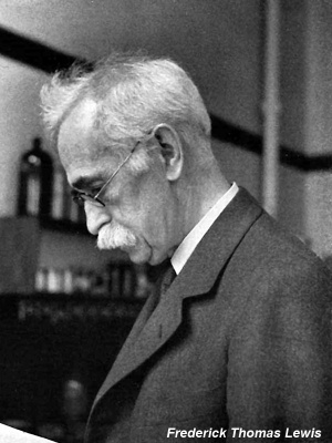