Book - Prenatal Development of the Human with Special Reference to Craniofacial Structure - An Atlas
| Embryology - 16 Apr 2024 |
|---|
| Google Translate - select your language from the list shown below (this will open a new external page) |
|
العربية | català | 中文 | 中國傳統的 | français | Deutsche | עִברִית | हिंदी | bahasa Indonesia | italiano | 日本語 | 한국어 | မြန်မာ | Pilipino | Polskie | português | ਪੰਜਾਬੀ ਦੇ | Română | русский | Español | Swahili | Svensk | ไทย | Türkçe | اردو | ייִדיש | Tiếng Việt These external translations are automated and may not be accurate. (More? About Translations) |
Nishimura H. Semba R. Tanimura T. and Tanaka O. Prenatal development of the human with special reference to craniofacial structures : An atlas. (1977) U.S. Dept. of Health, Education, and Welfare, Public Health Service, National Institutes of Health ; Washington. Bethesda, Md.
Prenatal Development of the Human with Special Reference to Craniofacial Structure - An Atlas

by
Nishimura, H., Semba, R., Tanimura, T., & Tanaka, O.
U.S. Dept. of Health, Education, and Welfare, Public Health Service, National Institutes of Health ; Washington. Bethesda, Md.
Contents
|
|
Foreword
Much progress has been made in techniques to repair congenital craniofacial malformations, particularly in children. As habilitative efforts continue, it is increasingly appreciated that prevention of disfigurements must be the final goal.
To investigate etiologic factors, research proceeds in many directions: human genetic and cytogenetic studies, environmental research, and teratologic experimentation. In recent times the importance of morphologic investigations of human development has received less emphasis. Yet, without thorough knowledge of the basic facts of prenatal human development, erroneous assumptions can be made in more dynamic approaches and lead investigators astray.
Morphologic details may be quite important not only in etiologic research but also in the treatment of congenital malformations. Additions to the knowledge of prenatal development of human oral-facial structures and some of their deviations will therefore be welcomed by many basic scientists and clinicians in the field of facial clefts and other craniofacial malformations such as orbital hypertelorism. This Atlas represents a distinct contribution to our knowledge of craniofacial embryology.
The material contributing to the Atlas was obtained from both induced abortions and stillbirths. While many of the induced abortions were performed for therapeutic reasons, the majority of the specimens were obtained under the Japanese Eugenics and Maternal Protection Law. Subsequently this law has been amended to limit abortion to cases with therapeutic indications. This law made it possible to collect human embryos and fetuses that had developed normally to the late stage. Thus, a unique opportunity was presented for a large-scale study of normal, and sometimes abnormal, human development. Professor Nishimura and his coworkers have made use of these products of legal abortion in the Atlas on Prenatal Development of the Human with Special Reference to Craniofacial Structures. Since induced abortion was not permitted in the last trimester of pregnancy, late stages of development shown in the Atlas are from stillborn fetuses with little postmortem changes.
Other works deal with successive stages of the developing human embryo, but they do not emphasize and illustrate specifically the craniofacial structures. In addition, there are good illustrations of various stages of the face and palate in textbooks of embryology, but they do not follow serially all the changes that lead to the formation of the human face. The present Atlas supplements these publications by serial illustrations of craniofacial development.
The Atlas of Developmental Anatomy of the Face published in 1966 by Kraus, Kitamura and Latham illustrates the craniofacial structures of normal embryos and fetuses from age 35 days to the newborn; it also shows specimens with facial clefts from 41 (lays to full term. The aborted specimens had been gathered from 250 hospitals in various regions of the United States and from several institutions in Japan. One of the aims was to demonstrate natural variations by showing three specimens for each age among the so-called normals or controls. The authors realized that normality was problematic in their material since it consisted of spontaneous abortuses, population that failed for one reason or another to survive." Thus, normality could not be assumed with certainty. In fact, many sections show signs of postmortem changes so that detailed histologic evaluation is at times impossible. Professor Nishimura’s Atlas is therefore a welcomed addition to the existing knowledge because of the excellent state of the undamaged and well-preserved specimens. Embryos were classified according to Streeter's horizons or the Carnegie system; fetuses were presented according to crown-rump length. Some pathologic fetuses with oral-facial or other malformations were added. Roentgenograms and photographs showing the external appearance of the fetuses, of histologic sections, and of cleared specimens were supplemented by carefully drawn and annotated diagrams which interpret the objective presentations. Appendix references to books, reviews and original articles dealing with oral-facial development will further aid the user of the Atlas. The fact that the bibliography is international should be of particular value to the English-speaking reader. This Atlas will remain a standard work for many years to come.
Josef Warkany
Children's Hospital Research Foundation
Cincinnati, Ohio
Preface
An embryological approach with human materials is important for establishing a normal standard of development including individual variabilities as well as clarifying the embryogenesis and etiology of defective development. It has been difficult, however, to obtain a sequential series of specimens in proper condition, and most of the current knowledge in this respect has been derived from a limited number of specimens arising mostly from pathological pregnancy. In view of this, the authors undertook a 12-year systematic study of normal and abnormal development with a large number of embryonic and fetal specimens obtained under the Eugenics and Maternal Protection Law of Japan from normal pregnancies with the assistance of hundreds of obstetricians. The specimens from induced abortions were supplemented with a number of selected specimens of stillborn fetuses and neonatally dead infants for study of the perinatal stage. This long-term experience convinced the authors of the need for as well as the feasibility of preparing a complete pictorial monograph on normal and abnormal development of oral- facial structures. Thus, a proposal for this contract was submitted to and approved by the National Institute of Dental Research. The 2-year study, conducted in collaboration with the Central Institute for Experimental Animals and the Department of Anatomy, Faculty of Medicine, Kyoto University, made possible the completion of this work.
This monograph, presenting the detailed photographs of stages of normal and defective development of the oral-facial complex, will furnish dentists, oral surgeons and other biomedical scientists with reliable standards for embryological and teratological studies.
Contract No. NIH—NIDR~73-2405 (NOI-DE-32405)
Acknowledgement
We are deeply grateful to Dr. K. K. Hisaoka and Dr. R. L. Christiansen of the National Institute of Dental Research, and Dr. R. L. Woolridge of the National Cancer Institute, National Institutes of Health, U.S.A., for their kind advice in planning and conducting the project; to numerous collaborating obstetricians for their cooperation in securing the specimens; to Dr. N. Iwahori, Department of Anatomy, Faculty of Medicine, Kyoto University, and Mr. B. Wongchoawant, medical student at the University, for their aid in preparation of the brain specimens; to Mr. M. Kawabata, Institute for Developmental Research, Aichi Prefectural Colony, for his assistance in X-ray photography; and to Miss Y. Ohta, Central Institute for Experimental Animals, and Miss K. Hirai, Department of Anatomy, Kyoto University, for their secretarial assistance. Also, appreciation is expressed to‘Dr. A. G. Hendrickx, National Center for Primate Biology, University of California, Davis, for his linguistic help during his visiting Professorship at Kyoto University.
Outline of the Work
Specimens
The specimens used for this study were selected cases from the human embryonic collection in the Department of Anatomy, Faculty of Medicine, Kyoto University. This collection, consisting of about 30,000 specimens at stages ranging from about the third week of gestation to term, was established in collaboration with about 800 obstetricians located in the central area of Japan. The Eugenics and Maternal Protection Law in Japan allowed qualified obstetricians to terminate pregnancy at and before the seventh month at the request of both the expectant mother and her spouse for socioeconomic reasons. According to official statistics, in most instances abortion was induced in the second and third month of pregnancy. Cervical dilation and curettage were performed in the second to approximately the fourth month of pregnancy. Occasionally, from about the fifth to the seventh month, delivery was induced by means of bougie or metreurynter. The obstetricians were thus able to provide undamaged embryos (approximately 30 mm in crown-rump length or less) and early fetuses (approximately between 30 mm and 200 mm in crown-rump length). In addition, they furnished a small number of undamaged stillborn fetuses at and after the eighth month of pregnancy, as well as neonatally deceased infants. It should be emphasized that the attempt to collect specimens had no bearing on the determination of abortion by both the expectant mother and the obstetrician. Further, only those fetal and neonatal specimens were acquired whose dissection and preservation were authorized in writing by the mother with no objection of her spouse.
The obstetricians usually fixed the embryos in Bouin’s fluid immediately after acquisition and transferred them to 10% formalin solution the next day. In the case of fetuses, the obstetricians used 10% formalin solution for fixation and storage; occasionally appropriate incisions in the body wall were made prior to fixation. Specimens obtained by obstetricians in and around Kyoto City were brought to the Department of Anatomy, Kyoto University immediately after their recovery. Others were brought in periodically. All specimens were accompanied by a record form completed by the obstetricians. This record form included such items as parental ages, consanguinity, parity, period after previous pregnancy, frequency of past pregnancy wastage and induced abortion, history of the use of contraceptive measures, parental occupation, habits of smoking and drinking, and history of maternal diseases and medical treatments.
Procedures
Appropriate undamaged specimens with no or little postmortem change were selected and their crown-rump length was measured. Careful examination of their external developmental stages was then made under a stereomicroscope or macroscopically. The numerical stage of the embryonic specimens was determined by adopting the Streeter’s horizon system[1][2][3][4] or the Carnegie system proposed by O’Rahilly.[5] The existence of external malformations as well as those in the oral cavity was also checked. The procedures were as follows:
Externally normal specimens
The occurrence of distinctly recognizable developmental features, such as the appearance of each branchial arch, auditory vesicle, buccopharyngeal membrane, medial and lateral nasal swelling, maxillary swelling, hypophyseal pouch, opening of auditory tube, external auditory pore, auricular hillocks, definitive ear auricle and tongue, palatine shelves, and tuberculum labii superior, were examined. During the examinations, appropriate planar cuts and dissections were made on the specimens. Typical developmental stages were then photographed, using the large universal photographic stand Multiphot (Nikon, Tokyo).
Some of the specimens brought to the Department in the fresh state were photographed intact in color.
In selected embryos the whole body was sectioned serially. The sections were stained with hematoxylin and eosin, and photographed at low magnification.
A certain number of cleared specimens at various stages were examined for development of the craniofacial skeleton. The coloration methods used were Van Wijhe’s methylene blue cartilage staining modified by Noback[6] or alizarin red S staining according to Jensh and Brent.[7]
In addition, roentgenograms of a certain number of the fixed specimens at various stages were made with a soft X-ray machine (Softex CSM, Nippon Softex, Tokyo), or a diagnostic X-ray machine (KXO-1000, Toshiba, Tokyo). Occasionally, before roentgenography these specimens were immersed in a 1% aqueous solution of silver nitrate for a period of 2 days to 3 weeks according to O’Rahilly and Meyer.[8]
Specimens with malformations in the oral cavity, face or neck
Careful observations of the malformed region were made for identification of the type of defect, and photographs were taken. Histological examination or dissection was then performed in order to detect internal anomalies. In some cases, other techniques such as X-ray examination were also employed.
References
- ↑ Streeter GL. Developmental horizons in human embryos. Description of age group XI, 13 to 20 somites, and age group XII, 21 to 29 somites. (1942) Contrib. Embryol., Carnegie Inst. Wash. Publ. 541, 30: 211-245.
- ↑ Streeter GL. Developmental horizons in human embryos. Description of age group XIII, embryos about 4 or 5 millimeters long, and age group XIV, period of indentation of the lens vesicle. (1945) Carnegie Instn. Wash. Publ. 557, Contrib. Embryol., Carnegie Inst. Wash., 31: 27-63.
- ↑ Streeter GL. Developmental horizons in human embryos. Description of age groups XV, XVI, XVII, and XVIII, being the third issue of a survey of the Carnegie collection. (1948) Contrib. Embryol., Carnegie Inst. Wash. 575, 32: 133-203.
- ↑ Streeter GL. Developmental horizons in human embryos. Description of age groups XIX, XX, XXI, XXII, and XXIII, being the fifth issue of a survey of the Carnegie Collection (prepared for publication by Heuser CH. and Corner GW.). (1951) Carnegie Instn. Wash. Publ. 592, Contrib. Embryol., 34: 165-196.
- ↑ O'Rahilly R. Developmental Stages in Human Embryos, Including a Survey of the Carnegie Collection. Part A: Embryos of the First Three Weeks (Stages 1 to 9). (1973) Carnegie Instn. Wash. Publ. 631. Washington, D.C.
- ↑ Noback GJ. The use of the Van Wijhe method for the staining of the cartilaginous skeleton. (1916) Anat. Rec. 11: 292-294.
- ↑ Jensh RP & Brent RL. (1966). Rapid schedules for KOH clearing and alizarin red S staining of fetal rat bone. Stain Technol , 41, 179-83. PMID: 4164155
- ↑ MEYER DB & O'RAHILLY R. (1956). Roentgenographic investigation of the human skeleton during early fetal life. Am J Roentgenol Radium Ther Nucl Med , 76, 455-68. PMID: 13354833
Embryonic Stages (Carnegie)
Stage 13

Stage 14

Lateral view of the central nervous system of embryo at Carnegie stage 14. Shown to the level of the myelencephalon (Scale bar is 1 mm).
Stage 15

Stage 16


Stage 21

Stage 23
- Oral Cavity
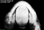
Floor
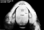
Floor (labeled)

Roof
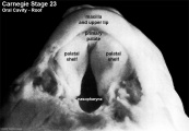
Roof (labeled)


Fetal Stages (Crown-Rump Length)
201 mm to newborn

Human Fetal Brain (CRL 240 mm, scale bar is 1 cm).
Cite this page: Hill, M.A. (2024, April 16) Embryology Book - Prenatal Development of the Human with Special Reference to Craniofacial Structure - An Atlas. Retrieved from https://embryology.med.unsw.edu.au/embryology/index.php/Book_-_Prenatal_Development_of_the_Human_with_Special_Reference_to_Craniofacial_Structure_-_An_Atlas
- © Dr Mark Hill 2024, UNSW Embryology ISBN: 978 0 7334 2609 4 - UNSW CRICOS Provider Code No. 00098G