Book - Manual of Human Embryology 18-9
| Embryology - 19 Apr 2024 |
|---|
| Google Translate - select your language from the list shown below (this will open a new external page) |
|
العربية | català | 中文 | 中國傳統的 | français | Deutsche | עִברִית | हिंदी | bahasa Indonesia | italiano | 日本語 | 한국어 | မြန်မာ | Pilipino | Polskie | português | ਪੰਜਾਬੀ ਦੇ | Română | русский | Español | Swahili | Svensk | ไทย | Türkçe | اردو | ייִדיש | Tiếng Việt These external translations are automated and may not be accurate. (More? About Translations) |
Keibel F. and Mall FP. Manual of Human Embryology II. (1912) J. B. Lippincott Company, Philadelphia.
- XVIII. Development of Blood, Vascular System and Spleen: Introduction | Origin of the Angioblast and Development of the Blood | Development of the Heart | The Development of the Vascular System | General | Special Development of the Blood-vessels | Origin of the Blood-vascular System | Blood-vascular System in Series of Human Embryos | Arteries | Veins | Development of the Lymphatic System | Development of the Spleen
| Historic Disclaimer - information about historic embryology pages |
|---|
| Pages where the terms "Historic" (textbooks, papers, people, recommendations) appear on this site, and sections within pages where this disclaimer appears, indicate that the content and scientific understanding are specific to the time of publication. This means that while some scientific descriptions are still accurate, the terminology and interpretation of the developmental mechanisms reflect the understanding at the time of original publication and those of the preceding periods, these terms, interpretations and recommendations may not reflect our current scientific understanding. (More? Embryology History | Historic Embryology Papers) |
Sabin FR. The Development of the Lymphatic System in Keibel F. and Mall FP. Manual of Human Embryology II. (1912) J. B. Lippincott Company, Philadelphia.
| Online Editor |
|---|
|
Modern Notes - Spleen Development |
V. The Development of the Spleen
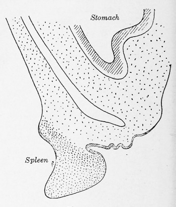
The fundamental problem in connection with the spleen is its general morphology, — that is, to which germ layer does it belong, — and to this question there is a clear, satisfactory answer in human embryology. The spleen is entirely mesodermal in origin. This was first suggested by Muller (1871) from a study of human embryos. He described the splenic anlage as a thickening of the peritoneum which took place early in the life of the embryo. The spleen arises from a thickening of the dorsal mesogastrium, 1 and is readily made out in embryos of the fifth week, 8 to 10 mm. long. Its relations to the stomach and omentum can be seen in Fig. 515 (after Kollmann, from an embryo 10.5 mm. long) and in Fig. 516 (after Tonkoff, from an embryo 20 mm. long).
In 1889 Toldt advanced the idea that an important part of the mesodermal anlage of the spleen came from the deeper layers of the ccelom epithelium. This has been confirmed and illustrated by Kollmann (1900) and by Tonkoff (1900). All three of them find that in human embryos about 10 mm. long the ccelomic epithelium over the splenic anlage is several layers thick, and that from the deepest layers cells are being transformed into mesenchyme cells. Later the epithelium becomes again a single layer and then this transformation ceases.
1 Kolliker (1854). Kollmann (1900), Phisalix (1888), Piper (1902), Toldt (1889), Tonkoff (1900).
The next problem in connection with the spleen is the development of its vascular system. Here it is impossible to give a satisfactory account, for our knowledge is but fragmentary. It will, however, be possible to indicate certain lines of investigation which promise to be fruitful. The study of the vascular system involves the use of the injection method, and hence we shall turn to a study of injected pig embryos and fetuses.
| Fig. 515. Anlage of the spleen in the posterior mesogastrium of a human embryo 10.5 mm. long. X 30. (After Kollmann.) | Fig. 516. Diagram of the spleen showing its relations to the stomach and the omentum in a human embryo 20 mm. long. (After Tonkoff.) |
In making injections of pig embryos through the umbilical artery, it is striking in how few cases any of the injection mass enters the splenic artery. For example, out of 22 specimens of apparently complete injections made into the umbilical artery, the spleen was injected in only four. Since the injections were all made on living embryos, this is probably due to the relative thickness and power of contraction of the muscle in the splenic artery. To get good injections it is best to open the embryo, tie off the aorta above and below the coeliac axis, and then inject the aorta with a hypodermic needle. When this small length of aorta is well filled, the fluid will run into the splenic artery. In a fetal pig 3 em. long, the entire splenic circulation consists of a capillary network which extends throughout the organ. This condition is maintained until the fetus is 7.5 cm. long, as shown in Fig. 517. This represents the tip of the spleen and shows the central artery and vein which run along the hilum of the organ. As is shown in the figure, the branches of both artery and vein are soon lost in a diffuse capillary network. The branches of the artery can be distinguished for a short distance by being narrower than the veins. Thus the spleen confirms the principle that the primitive circulation of any organ is in the form of a capillary network out of which the arteries and veins are formed. The spleen is characterized by a long persistence of the primitive capillary network.
The embryo pig 10 em long marks the transition stage between this primitive condition and the type of circulation peculiar to the adult spleen. This point is shown in Fig. 518, where it will be seen that the branches of the central artery and vein extend much farther toward the border of the spleen, and the arterial branches lead into tufts of capillaries, making the anlage of the vascular unit. These capillaries have a wider calibre than those of the preceding stage.
When the fetus is 12 cm long, as shown in Fig. 519, the transformation of the vascular system has been sufficiently marked to give the key to the adult circulation. By comparison with Fig. 518, which is at the same magnification, it will be seen that between the stages of 10 and 12 cm. there is a rapid increase in size, the spleen more than doubling in width. The position of the central artery and vein allows the comparison. As seen in Fig. 519, the central artery of the hilum gives off a series of branches of the first order which anastomose. These arteries bifurcate into branches of the second and third orders. The branches of the fourth or fifth orders lead into spherules of arterial capillaries which can be seen throughout the spleen, but best at the edge. Most of these spherules have only one artery, but a few receive two ; most of them are isolated, but a few are connected by anastomosing loops. In the upper left-hand corner of the figure can be seen the relation of these spherules of arterial capillaries to the veins. The spherules lead by wide openings into a wide-meshed plexus of venous capillaries, which drain into the still wider venae comites of the arteries of the third order. Thus is illustrated the separation of the artery and vein in the zone of the capillary bed.
At the edge of the organ, it can be seen in total mounts that each spherule of capillaries lies in the centre of a small compartment bounded by trabecular from the capsule. The spherules are the splenic capillaries, characterized by being wider than the usual capillaries. They are at the same time splenic pulp and represent the structural units of Mall (1900). It is the development of these spherules of capillaries that accounts for the rapid increase in the size of the spleen at this time. That they are not accidental is shown by several points,— 1, their constant occurrence at the centre of the lobule at the end of the artery in injections ; 2, their approximately uniform size ; and 3, their definite connection with the veins.
It appears that the number of the structural units of Mall is fixed fairly early; for example, in three fetuses 17 cm. long the number of units along the edge was 150, 204, and 230 respectively, while in three adult spleens the numbers were approximately 190, 180, 260. The size and complexity of these units, however, change greatly; for example, at 17 cm. the average width of a unit is about 0.1 mm. while in the adult it is about 1 mm. Moreover, the embryonic unit consists of one central artery with a single bunch or spherule of capillaries leading to the vein, while the adult unit, as shown in Mali's Fig. 1 (Amer. Jour, of Anat., vol. 2, p. 321, 1902-1903), consists of a central artery with branches which end not in a single spherule of capillaries but in clusters of capillaries like a bunch of grapes. These clusters are the splenic capillaries or pulp. One of the spherules on the edge of the spleen in Fig. 519 shows the bifurcation of the central artery; how this complex unit of the adult is made out of the simple one of the embryo is yet to be determined, but the evidence of embryology is that the capillaries of the spleen are of a wider calibre than the usual capillaries, and that the wider capillary bed is compensated for in the development of the musculature of the spleen by which the capillary bed can be emptied with ease.
Mall (1902 to 1903) proved, by the method of subjecting the living adult spleen to a variety of injection methods, of which the most crucial test was the fixation of the spleen by the injection of formalin into the living animal, that the splenic pulp is the capillary bed of the spleen; that the pulp intervenes between the artery and vein, and, in the normal, living animal, is engorged with blood when the spleen is hypersernic. Hence the capillary circulation of the adult spleen is a cavernous one. The point at which the type of circulation of the early fetal stages — namely that of a primitive system of closed capillaries like the rest of the vascular system — changes over into the secondary type of circulation of the adult, namely a cavernous circulation, is shown in Fig. 519, for the fetal pig.
The work of Mollier (1911) carries us a step farther, by his beautiful specimens of the structure of the wall of the splenic veins. He also, as Mall had done, overthrows the idea of a homogeneous membrane around the endothelium of the adult splenic veins. He shows that the wall of the splenic veins consists of a reticular syncytium of endothelium with denser masses of protoplasm around the nuclei and wide-open meshes between.
It has thus become clear that there is a direct pathway from the terminal artery through the cavernous capillary system into the venous sinuses, and that to investigate the nature of the endothelium of the cavernous capillaries or splenic pulp is a problem which is becoming more and more hopeful. The transition stage between the two types of capillaries is the next point of attack.
| Fig. 517. Piece of a total mount of an injected spleen of an embryo pig 7.5 cm. long, showing the capillary plexus which is characteristic of the circulation at this stage. X 47.
A., artery; C.a., central artery of hilum; C.v., central vein of hilum. |
Fig. 518. Piece of a total mount of an injected spleen of an embryo pig 10 cm. long, showing that the anlage of the splenic unit is a tuft of widened capillaries. X 47.
A.b.c, anlage of capillary spherules or units; C.a., central artery of hilum; C.a.l., central artery of lobule or unit; C.v., central vein of hilum. |
The third problem in relation to the spleen is in connection with its function of the formation of red blood-cells. Kolliker (1854) was the first to suggest the idea of the spleen serving as a place for the formation of erythrocytes and leucocytes, and called attention to the relation of the giant cells to red-blood formation. Luzet (1901) pointed out in human embryos of from 3 to 5 months there were more nucleated red blood-cells than in the heart's blood. Sophie Lifschitz (1906) has shown, in a work which I regard as important, that the active formation of red blood-cells takes place in fetuses between 15 and 30 cm. long. She plotted the curve both of the number of red blood-cells and of the giant cells of the spleen, and found that both increase together to a maximum in fetuses 18 cm. long, while the curve almost at the zero point at 30 cm. the nucleated red blood-cells form cells. It may be noted that this formation corresponds with the period of the formation of the capillary spherules or spleen pulp, which makes the spleen fall in line with the recent work on the bone-marrow by Bunting (1906), and on bone-marrow in the kidney of Maxhnow (1907), that red blood-cell formation goes on within the capillary bed, that it is intravascular. Lifschitz (1906) also called attention to the rapid increase in size of the spleen during the period of red blood-cell formation, which Was also noted in connection with the period of spleen-pulp formation. The injection experiments show that its meaning lies in the rapid increase in the size of the capillaries. The fundamental point that the early spleen is only undifferentiated mesoderm, and that this condition remains until the embryo is 7 cm. long, was noted bv Van der Stricht in 1892. He states that there is a primitive stage in which the structure of the spleen is more or less uniform before there is any differentiation into splenic pulp and Malpighian corpuscles; then he finds a transitional stage characterized by an increase in white corpuscles in certain areas, an increase in erythroblasts and a retardation of the circulation, and finally a secondary stage in which the adult organization of the spleen with pulp and Malpighian corpuscles is established. The fourth problem in the development of the spleen is in connection with the ellipsoids and Malpighian corpuscles. It is definitely known that these structures belong to the latter half of fetal life. In the spleen, as has been shown in the lymph-glands, the lymphocyte first appears in the adventitia of the artery, so that, though red blood-cell formation is within the capillary bed, the lymphocyte is extravascular. The ellipsoids, or capillarhulsen of Schweigger-Seidel, which are on the course of the smallest arteries, appear, as Bannwarth (1891) has shown, before the Malpighian corpuscles, which "are the round follicles along the larger arteries. He found the ellipsoids in a four-months human fetus, whije later — that is, in a seven-months fetus — the ellipsoids had disappeared and follicles were present. The follicle is found only in mammals and some birds, while the ellipsoids occur in fishes as well, as has been shown by Whiting (1895). Thus the ellipsoid is the primitive lymphoid structure of the spleen. In describing their development, Bannwarth shows that in the spleen, as in the lymph-gland, leucocytes appear in the loose adventitia of the artery, and at the same time there is a development of this adventitia, by which the connective-tissue fibrils are laid down in more or less concentric rings around the artery, forming the delicate reticulum characteristic of the lymph-follicle in generaj. Thus, the spleen, hemal glands, and lymph-glands are all vascular structures and are all built on the following simple plan : 1, along the arteries are clumps of lymphocytes in a reticulum called ellipsoids or Malpighian corpuscles or follicles; the ellipsoid is a special name used in the spleen for the oval masses of lymphocytes which lie nearest the capillary bed ; 2, the capillaries, whether they be lymphatic capillaries in lymph-glands or blood-capillaries in the hemal nodes or the spleen, are all wider in calibre than other capillaries. They are densely packed together, and have been termed either sinuses in lymph-glands or pulp spaces in the spleen.
Fig. 519. Piece of a total mount of an injected spleen of an embryo pig 12 cm. long, showing the vascular units. X 47. The arteries are shown darker than the veins. A.b., anastomosis between two capillary balls; c.a., central artery of hilum; c.b., arterial capillary ball; c.b., central artery of hilum; T 1, vein of first order; this vein up to the point marked X was taken from another area of the same slide where the injection was more complete; it is the rule that the veins accompany the arteries in this manner; V.C3., vena comites of the third order; v.c.p., capillary plexus of veins.
Literature
Bannwarth : Die Milz der Katze. Arch, f . mikr. Anat. Bd. 38. 1891. Bezaxpon et Labbe: Trait e d'hematologie. Paris, 1904.
Bunting, C. H. : Experimental Anaemias in the Rabbit. Jour, of Exp. Med. Vol. 8. 1906.
Choronschitzsky, B. J. : Die Entstehung der Milz, Leber. Gallenblase, Bauehspeicheldi*iise und des Pfortadersystems bei den verscbiedenen Abteilungen der Wirbeltiere. Anat. Hefte. Bd. 13. 1900.
Daiber, M. : Zur Frage nacb der Entstebung und Regenerationsf abigkeit der Milz. Jenaisebe Zeitscb. f. Naturwissensehaft. Bd. 42. 1907.
Janosik, A.: Bemerkungen zu der Arbeit, Dr. W. Tonkoff; Die Entwicklung der Milz bei den Amnioten. Arch. f. mikr. Anat. Bd. 57. 1901.
Kolliker, A. : Mikroskopisebe Anatomie. Zweite Halfte. Leipzig, 1S51.
Kollmann, G. : Lebrbuch der Entwicklungsgesckiehte des Menscben. Jena, 1898. Die Entwicklung der Lyinphknotchen in dem Blinddann und in dem processus verrniformis. Die Entwicklung der Tonsillen und die Entwicklung der Milz. Arch. f. Anat. u. Phys. Anat. Abt. 1900.
Lifschitz, S. : Leber die Entwicklung der embryonalen Milz. Med. Diss. Zurich, 1906.
Luzet: Etudes sur les anemies de la premiere enfance etc. These. Paris, 1891.
Mall, F. P. : The Lobule of the Spleen. Johns Hopkins Hospital Bulletin. 1898. The Architecture and Blood-vessels of the Dog's Spleen. Zeitschr. f. Morphol.u. Anthropol. Bd. 2. 1900. The Circulation through the Dog's Spleen. Amer. Jour, of Anat. Vol. 2.1902-1903.
Maximow, A. : Experimented Untersuchungen zur postf etalen Histogenese des myeloiden Gewebes. Beitr. z. path. Anat. u. allgem. Path. Bd. 41. 1907.
Mollier. TV: Leber den Bau der kapillaren Milzvenen (Milzsinus). Arch. f. mikr. Anat. Bd. 76. 1911.
MiiLLER, YV : Milz, in Strieker's Handbuch. Leipzig, 1871.
Neumann, E. : Neue Beitrage zur Kenntnis der Blutbildung. Arch. d. Heilkunde. Bd. 15. 1S74. Ueber Blutregeneration und Blutbildung. Zeitschrift fiir klinische Medizin. Bd. 3. 1881.
Phisalix, C: Etude d'un embryon humain de 10 mm. Arch, de zoolog. experim. et gener. T. 2, p. 6. 1SS8.
Piper, H. Die Entwicklung von Leber, Pancreas und Milz. Inaug.-Diss. Freiburg i. B. 1902.
Sabin FR. On the Development of Lymphatic Nodes in the Pig. (1905) Amer. J Anat. 4:
Sassuchin, P. : Leber die kindliche Milz. Inaug.-Diss. St. Petersburg, 1899.
Schmidt, M. B. : Ueber Blutzellenbildung in der Leber und Milz unter normalen und pathologischen Verhaltnissen. Ziegler's Beitrage. Bd. 11. 1892.
Toldt, C. : Zur Anatomie der Milz. Wiener klin. Wochenschrift. 1S89. Die Darmgekrose und Netze im gesetzmassig und gesetzwidrigen Zustand. Denkschriften d. k. Akad. d. Wissenschaften zu Wien. Bd. 56. 1SS9.
Tonkoff, W. : Die Entwickluns: der Milz bei den Amnioten. Arch, f . mikr. Anat. Bd. 56. 1900.
Van der Stricht, Omer : Xouvelles recherches sur la genese des globules rouges et des globules blanc du sang. Arch, de Biologic T. 12, p. 199. 1892.
Woit. O.: Zur Entwicklung der Milz. Anat. Hefte." Bd. 9. 189S.
Whitixg. A. J.: Comparative Histology and Physiology of the Spleen. Trans. Royal Society of Edinburgh. Vol. 38. Part. 2. No. 8. 1895.


