Book - Human Embryology and Morphology Figures: Difference between revisions
m (→The Limbs) |
|||
| Line 4: | Line 4: | ||
[[Book_-_Human_Embryology_and_Morphology_1|Development or the Face]] | [[Book_-_Human_Embryology_and_Morphology_1|Development or the Face]] | ||
<gallery> | <gallery> | ||
File:Keith1902 fig001.jpg|Fig. 1. Showing the formation of the face by the Nasal, Maxillary, and Mandibular processes in an embryo of the 4th week | File:Keith1902 fig001.jpg|Fig. 1. Showing the formation of the face by the Nasal, Maxillary, and Mandibular processes in an embryo of the 4th week. | ||
File:Keith1902 fig002.jpg|Fig. 2. Showing the parts of the face formed from the Nasal, Maxillary and Mandibular processes. | File:Keith1902 fig002.jpg|Fig. 2. Showing the parts of the face formed from the Nasal, Maxillary and Mandibular processes. | ||
File:Keith1902 fig003.jpg|Fig. 3. Showing the structures formed in the Mesial Nasal Processes. | File:Keith1902 fig003.jpg|Fig. 3. Showing the structures formed in the Mesial Nasal Processes. | ||
| Line 13: | Line 13: | ||
File:Keith1902 fig008.jpg|Fig. 8. Showing the ingrowth of the palatal plates of the two maxillary processes early in the 2nd month. (After Kollmann.) . | File:Keith1902 fig008.jpg|Fig. 8. Showing the ingrowth of the palatal plates of the two maxillary processes early in the 2nd month. (After Kollmann.) . | ||
File:Keith1902 fig009.jpg|Fig. 9. Showing the Hard Palate at birth. The premaxillary part is formed from the Mesial Nasal Processes ; the remainder by the Palatal Plates of the Maxillary Processes. | File:Keith1902 fig009.jpg|Fig. 9. Showing the Hard Palate at birth. The premaxillary part is formed from the Mesial Nasal Processes ; the remainder by the Palatal Plates of the Maxillary Processes. | ||
File:Keith1902 fig010a-c.jpg|Fig. 10, a, b, c. Showing what become of the skeletons of the Mandibular Arch (Meckel's Cartilage) and Maxillary Process (Palato-quadrate Cartilage) | File:Keith1902 fig010a-c.jpg|Fig. 10, a, b, c. Showing what become of the skeletons of the Mandibular Arch (Meckel's Cartilage) and Maxillary Process (Palato-quadrate Cartilage). | ||
File:Keith1902 fig010d.jpg|Fig. 10 D. Illustrating Gadow's view of the origin of the Auditory Ossicles and Tympanic Plate. | File:Keith1902 fig010d.jpg|Fig. 10 D. Illustrating Gadow's view of the origin of the Auditory Ossicles and Tympanic Plate. | ||
File:Keith1902 fig011.jpg|Fig. 11. Showing the manner in which the development of the Maxillary Antrum affects the size of the palate and position of the molar teeth. | File:Keith1902 fig011.jpg|Fig. 11. Showing the manner in which the development of the Maxillary Antrum affects the size of the palate and position of the molar teeth. | ||
File:Keith1902 fig012.jpg|Fig. 12. Showing the Centres of Ossification and age changes in the Lower Jaw. | File:Keith1902 fig012.jpg|Fig. 12. Showing the Centres of Ossification and age changes in the Lower Jaw. | ||
File:Keith1902 fig013.jpg|Fig. 13. The chief types of the Temporo-Maxillary Articulation. A. Carnivorous Type. B. Omnivorous Type. C. Herbivorous Type. | File:Keith1902 fig013.jpg|Fig. 13. The chief types of the Temporo-Maxillary Articulation. '''A.''' Carnivorous Type. '''B.''' Omnivorous Type. '''C.''' Herbivorous Type. | ||
File:Keith1902 fig014.jpg|Fig. 14. Showing the Chief Changes after birth in the form of the TemporoMaxillary Articulation. | File:Keith1902 fig014.jpg|Fig. 14. Showing the Chief Changes after birth in the form of the TemporoMaxillary Articulation. | ||
File:Keith1902 fig015a.jpg|Fig. 15 A. Sagittal Section showing the Stomodaeum and position of the Oral Plate in the 3rd week | File:Keith1902 fig015a.jpg|Fig. 15 A. Sagittal Section showing the Stomodaeum and position of the Oral Plate in the 3rd week. | ||
File:Keith1902 fig015b.jpg|Fig. 15 B. Showing the parts of the Buccal and Nasal Cavities formed from the Stomodaeum. The relative position of the Oral Plate is indicated. | File:Keith1902 fig015b.jpg|Fig. 15 B. Showing the parts of the Buccal and Nasal Cavities formed from the Stomodaeum. The relative position of the Oral Plate is indicated. | ||
</gallery> | </gallery> | ||
Revision as of 09:01, 21 January 2014
| Embryology - 23 Apr 2024 |
|---|
| Google Translate - select your language from the list shown below (this will open a new external page) |
|
العربية | català | 中文 | 中國傳統的 | français | Deutsche | עִברִית | हिंदी | bahasa Indonesia | italiano | 日本語 | 한국어 | မြန်မာ | Pilipino | Polskie | português | ਪੰਜਾਬੀ ਦੇ | Română | русский | Español | Swahili | Svensk | ไทย | Türkçe | اردو | ייִדיש | Tiếng Việt These external translations are automated and may not be accurate. (More? About Translations) |
Keith A. Human Embryology and Morphology. (1902) London: Edward Arnold.
| Historic Disclaimer - information about historic embryology pages |
|---|
 |
Development or the Face
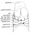
Fig. 1. Showing the formation of the face by the Nasal, Maxillary, and Mandibular processes in an embryo of the 4th week.
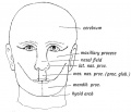
Fig. 2. Showing the parts of the face formed from the Nasal, Maxillary and Mandibular processes.
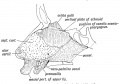
Fig. 3. Showing the structures formed in the Mesial Nasal Processes.
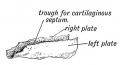
Fig. 4. Showing the trough-shaped Vomer of the newly born.
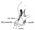
Fig. 5. Showing the suture on the face between the premaxilla and maxilla in the skull of a young orang.
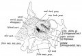
Fig. 6. Showing the structures formed in the Lateral Nasal Processes.
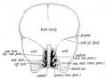
Fig. 7. Coronal section of the skull of a 7th month human foetus to show the cartilages of the Lateral and Mesial Nasal Processes and the bones formed round them.
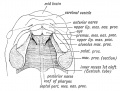
Fig. 8. Showing the ingrowth of the palatal plates of the two maxillary processes early in the 2nd month. (After Kollmann.) .
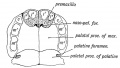
Fig. 9. Showing the Hard Palate at birth. The premaxillary part is formed from the Mesial Nasal Processes ; the remainder by the Palatal Plates of the Maxillary Processes.
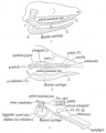
Fig. 10, a, b, c. Showing what become of the skeletons of the Mandibular Arch (Meckel's Cartilage) and Maxillary Process (Palato-quadrate Cartilage).
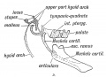
Fig. 10 D. Illustrating Gadow's view of the origin of the Auditory Ossicles and Tympanic Plate.
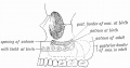
Fig. 11. Showing the manner in which the development of the Maxillary Antrum affects the size of the palate and position of the molar teeth.
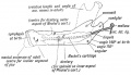
Fig. 12. Showing the Centres of Ossification and age changes in the Lower Jaw.
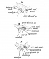
Fig. 13. The chief types of the Temporo-Maxillary Articulation. A. Carnivorous Type. B. Omnivorous Type. C. Herbivorous Type.
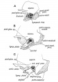
Fig. 14. Showing the Chief Changes after birth in the form of the TemporoMaxillary Articulation.
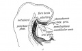
Fig. 15 A. Sagittal Section showing the Stomodaeum and position of the Oral Plate in the 3rd week.
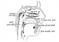
Fig. 15 B. Showing the parts of the Buccal and Nasal Cavities formed from the Stomodaeum. The relative position of the Oral Plate is indicated.


The Nasal Cavities and Olfactory Structures
The Nasal Cavities and Olfactory Structures
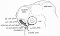
Fig. 16. The Olfactory Pit and Nasal Processes in a 4th week human embryo (After Kollmann.)
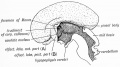
Fig. 17. The Mesial aspect of the Brain of a human foetus, 3J months old, showing the Olfactory Lobe.
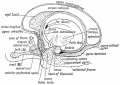
Fig. 18. Showing the parts formed out of the Olfactory Lobe in the brain of an Adult (After BlUot "mitlO olfilctol T Hoots in the Sub-callosal and Uncinate Gyri.' .
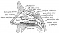
Fig. 19. A diagram of the Lateral Wall of the Nasal Cavity, showing the position of the Air Sinus.
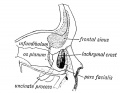
Fig. 20. Showing on the inner wall of the Orbit (1) the position of the Infundibulum, (2) the pars facialis lachrymalis.


- Links: Smell Development
Development of the Pharynx and Neck
Development of the Pharynx and Neck
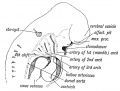
Fig. 21a.
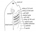
Fig. 21b. Showing the position of the Heart, Visceral and Aortic Arches in a fish. (Diagrammatic — after Gegenbaur.)
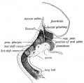
Fig. 22. Showing the Primitive Pharynx of a 3rd week embryo in sagittal section, bounded by the Visceral Arches. (After His.) .
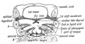
Fig. 23. Showing the Floor of the Pharynx of a 4th week human embryo. (After His.)
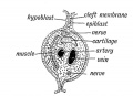
Fig. 24. Schematic Section of a Visceral Arch.
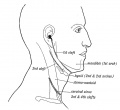
Fig. 25. Showing the position of the External Cleft Depressions in the Adult.
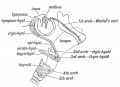
Fig. 26. Showing what become of the Cartilages of the Visceral Arches.
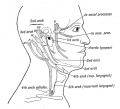
Fig. 27. Showing what become of the Nerves of the Visceral Arches, hyoid arch) is represented by the chorda tympani and great superficial petrosal.
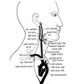
Fig. 28. Showing what become of the Aortic Arches in the adult. Only the shaded parts persist.
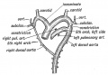
Fig. 29. The condition of the Eight and Left Doral Aortae in a 6th week human foetus.
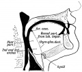
Fig. 30. Showing the Buccal and Pharyngeal parts of the Tongue.
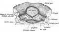
Fig. 31. Showing the origin of the tongue in the floor of the primitive pharynx. (After His.)
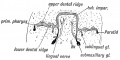
Fig. 32. Showing the origin of the Submaxillary and Sublingual Glands from furrows between the gum and tongue during the flth week. (After His.) .
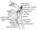
Fig. 33. Showing the position of the Visceral Clefts in the Adult.
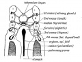
Fig. 34. Showing the origin of the Tonsil, Thymus, and Thyroid from the Internal Cleft Recesses during the 4th week. (After His.).


Development of the Organ of Hearing
Development of the Organ of Hearing
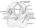
Fig. 35. Diagrammatic Section through the Cephalic region of an embryo, showing the origin of the Auditory System.
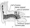
Fig. 36 A. Section of the External Auditory Meatus of the Adult.
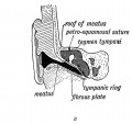
Fig. 36 B. A Section of the External Auditory Meatus at Birth. (After Symington.)
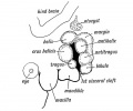
Fig. 37. Showing the Tubercles which arise round the First Visceral Cleft to form the External Ear.
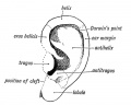
Fig. 38. Showing the part of the Adult Ear formed by each Tubercle.
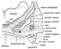
Fig. 39. Showing the condition of the Auditory Organs in a 6th week human foetus. (After Siobenmann.)
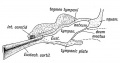
Fig. 40. Showing the Cavities derived from the Inner Recess of the First Cleft.
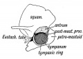
Fig. 41. The temporal bone at birth showing the formation of the Antrum between the Squamosal and Petro-mastoid.
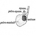
Fig. 42. A transverse section showing how the Walls of the Antrum are formed.
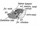
Fig. 43. Showing the outer aspect of the Petro-mastoid at birth after the Squamosal is removed.
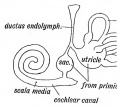
Fig. 44. Diagram of the Membranous Labyrinth.
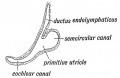
Fig. 45. The Otocyst in an Embryo of five weeks ; it shows a demarcation into the various parts of the Membranous Labyrinth.
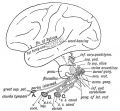
Fig. 46. Showing the Nerve Structures concerned in the Sense of Hearing.


Development and Morphology of the Teeth
Development and Morphology of the Teeth
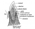
Fig. 47. Showing the parts of an incisor tooth.
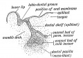
Fig. 48. Section through the lip and mandible of a foetus in the third month, showing the down-growth of the Dental Shelf.
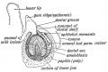
Fig. 49. Showing the stage of development in an incisor tooth of a foetus of six months.
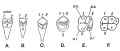
Fig. 50. A. The tritubercular Type of Tooth.


- Links: Tooth Development
The Skin and its Appendages
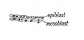
Fig. 51. The strata of the skin during the first month.
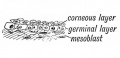
Fig. 52. The strata of the skin during the second month.
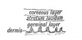
Fig. 53. The strata of the skin from the sixth month onwards.
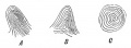
Fig. 54. The more common patterns formed by the dermal papillae on the tips of the fingers.
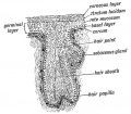
Fig. 55. Diagram of a Developing Hair.
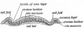
Fig. 56. Diagrammatic Section across a Nail.
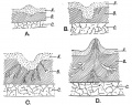
Fig. 57. Showing the various stages in tho development of the Mamma
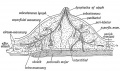
Fig. 58. Diagrammatic Section of the Breast to show the arrangement of its Capsule and Lymphatics.


- Links: Integumentary Development
The Development of the Ovum of the Foetus from the Ovum of the Mother
The Development of the Ovum of the Foetus from the Ovum of the Mother
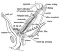
Fig. 59. The position of the Ovary and Fallopian Tube in the 5th month.
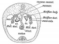
Fig. 60. Diagrammatic section of a foetus at the end of the 2nd month, showing the Attachments of the Ovary and MUllerian duct.
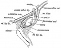
Fig. 61. Showing the position of the Ovary on the lateral wall of tho Pelvis and its relation to the Fallopian Tube.
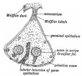
Fig. 62. Diagrammatic Section of an Ovary to show the manner in which the Primitive Ova are carried in by incursions of the Germinal Epithelium.
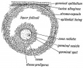
Fig. 63. Diagram of a ripe Graafian Follicle.
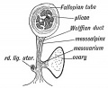
Fig. 64. Diagrammatic section of the Broad Ligament and Fallopian Tube.
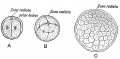
Fig. 65. Showing the production of the Morula from the Ovum. A. The ovum after the first division. B. After the second. C. The Morula .
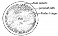
Fig. 66. Diagrammatic section of a Blastodermic Vesicle.
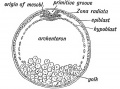
Fig. 67. A diagrammatic section of a Bilaminar Blastoderm made across the primitive streak.
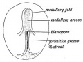
Fig. 68. Diagram of the Embryogenic area of a Bilaminar Blastoderm viewed from above.
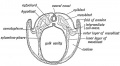
Fig. 69. Diagrammatic section of a Blastodermic Vesicle.
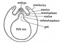
Fig. 70. Diagram of the Blastodermic Vesicle separating into Embryo and Membranes.
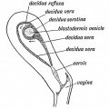
Fig. 71. Section of the Uterus showing the three parts of the Decidua.
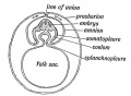
Fig. 72. Showing the folds of the somatopleure uniting over the embryo and becoming demarcated into Amnion and Prechorion.
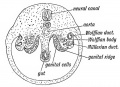
Fig. 73. Diagrammatic section of the abdominal region of the coelom, showing the position of the Genital Ridges from which the Ovary or Testicle is formed.


The Manner in which a Connection is Established between the Foetus and Uterus
The Manner in which a Connection is Established between the Foetus and Uterus
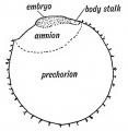
Fig. 74. Showing what becomes of the Somatopleure of the Blastodermic Vesicle.
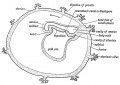
Fig. 75. Showing the growth backwards of the Somatopleuric Head Fold in the human ovum of 15 days to form the Amnion and Prechorion.
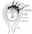
Fig. 76. Showing the arrangement of the Amnion, Chorion, and Deeidua in the 3rd month and the Formation of the Placenta.

Fig. 77. Diagrammatic section to show the Elements which enter into the formation of the Placenta.
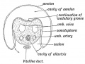
Fig. 78. Diagrammatic section showing the structures which go to form the Umbilical Cord. (After His.)


The Uro-genital System
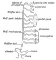
Fig. 79. Scheme of the Wolffian Body of the right side.
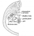
Fig. 80. Diagrammatic section to show the position of the Wolffian and Genital Ridges on the dorsal wall of the abdomen.
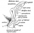
Fig. 81. Remnants of the Wolffian Body in the Female.
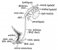
Fig. 82. Remnant of the "Wolffian Body in the Male.
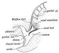
Fig. 83. The Origin of the Renal Bud (diagrammatic).
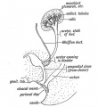
Fig. 84. The Termination of the Ureter in the Bladder and Sub-division of the Renal Bud
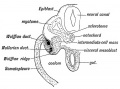
Fig. 85. A transverse section to show the manner in which the Wolffian and Miillerian Ducts arite, and their position in the Wolffian Ridge. (After Kollmann.) .
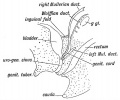
Fig. 86. Diagram of the Genital Ducts at -the commencement of the 3rd month of foetal life. Lateral view.
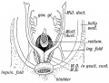
Fig. 87. Diagram of the Miillerian Ducts at the commencement of the 3rd month. Ventral view.
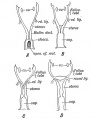
Fig. 88. Evolution of the Human Form of Uterua. A Form seen in lowest mammals, reptiles, amphibians, fishes, and in the 2nd month human foetus. B. Form of Mullerian Ducts in rodents. C. Form in Camivora, etc., and in the 4th month human foetus. D. Form found in man and higher primates.
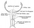
Fig. 89. Showing the manner in which the Mulleriau Ducts fuse to form the Uterus and Vagina.
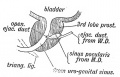
Fig. 90. A section of the Prostate showing the Hemnants of the lower ends of the Mttllerian Ducts in the male.
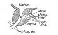
Fig. 91. A section of a Prostate showing an unusually developed Uterus Masculinus. (After Primrose.)

Fig. 92. Section showing the Uro-genital Sinus. A. In the 4th month female human foetus. B. In the 5th month female human foetus.
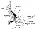
Fig. 93. Section showing the Uro-genital Sinus in the male foetus.
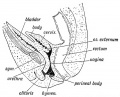
Fig. 94. A section to show the condition of the Vagina and Uterus at the 7th month of foetal life.
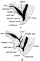
Fig. 95. The Division of the Cloaca into Rectal and Uro-genital Parts.
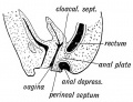
Fig. 96. A case of Imperforate Anus due to a persistence of the Anal Plate.
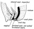
Fig. 97. A case in which the Rectal part of the Anal Plate has persisted and the Cloacal Septum has failed to fuse with the Perineal Septum.
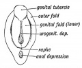
Fig. 98. The Uro-genital Cleft or Depression and the Genital Tubercle and Folds towards the end of the 2nd month.
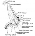
Fig. 99. A section of the male bladder and urethra at birth, showing the structures derived from the intra-abdominal part of the Allantois and from the Cloaca.
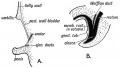
Fig. 100. A A section to show the condition of parts in Ectopia Vesicae.
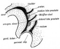
Fig. 101. A diagram to show the position at which the Prostatic Tubules arise.
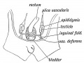
Fig. 102. The Position of the Testis in a foetus of 2£ months .
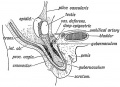
Fig. 103. Showing the Position of the Testis at the 6th month, and the Formation of the Gubernaculum Testis.
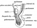
Fig. 104. The manner in which the structures in the wall of the abdomen are carried out so as to form the Inguinal Canal and Coverings of the Testis.
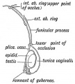
Fig. 105. A diagram of the Processus Vaginalis.


Formation of the Pubo-femoral Region, Pelvic Floor and Fascia
Formation of the Pubo-femoral Region, Pelvic Floor and Fascia
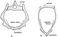
Fig. 106. A. The form of Pelvis and Inguinal Canal in Man.
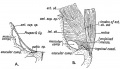
Fig. 107. Poupart's Ligament and the Crural Passage of Man.
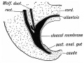
Fig. 108. The caudal end of the body in a human embryo of the 3rd week.
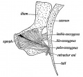
Fig. 109. The pelvic-caudal Muscles of a Monkey.
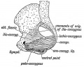
Fig. 110. The corresponding Muscles of Man.
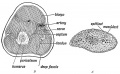
Fig. 111. A. Diagrammatic section of the Arm Bud of an embryo at the commencement of the 4th week. B. Corresponding section of the Adult Arm.
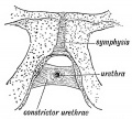
Fig. 112. The Constrictor Urethrae Muscle.


The Spinal Column and Back
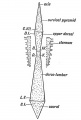
Fig. 113. Diagram of the Pyramids of the Spine.
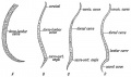
Fig. 114. Diagram of the Curves of the Spinal Column.
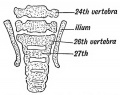
Fig, 115. A section of the Lumbo-sacral Region of the Spine in a Foetus at the end of the 2nd month, showing the 26th vertebra forming the 1st Sacral. (After Rosenberg.)
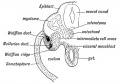
Fig. 116. Diagrammatic transverse section of a human embryo at the end of the 3rd week.
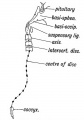
Fig. 117. Where Remnants of the Notochord may occur in the Adult.
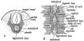
Fig. 118. The development of the Membranous Basis of a Vertebra
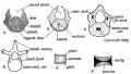
Fig. 119. Showing the Stages in the Development of a Vertebra.
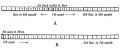
Fig. 120. The Order in which the Centres of Ossification appear in the Bodies (A) and in the Neural Arches (B) of the Spinal Column.
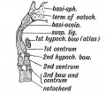
Fig. 121. A diagrammatic section of the Foetal Axis, Atlas, and Basi-occipital.
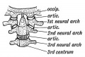
Fig. 122. The nature of the Atlanto-axio-occipital Articulations.
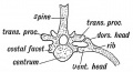
Fig. 123. The Bicipital Rib of a Lower Vertebrate (crocodile).
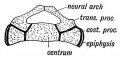
Fig. 124. A section to show the Nature of the Elements composing the Sacrum.


The Segmentation of the Body
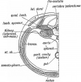
Fig. 125. A transverse section showing the Elements of the 1st Lumbar Segment in the Adult.
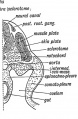
Fig. 126. A corresponding section of an Embryo about the end of the 3rd week (diagrammatic).
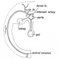
Fig. 127. The distribution of a typical Segmental Artery.
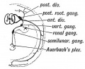
Fig. 128. Diagram of the Nerve System of the 11th Dorsal Segment.
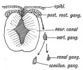
Fig. 129. A diagram showing the derivation of the Parts of the Nerve System of the 11th Segment in the Embryo.


The Cranium
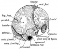
Fig. 130. The Centres of Ossification for the Dermal Bones of the Skull. The Bones which are formed in Cartilage are stippled.
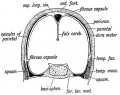
Fig. 131. A coronal section of the Skull of a Foetus, 4£ months old.
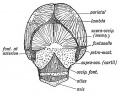
Fig. 132. The Occipital Region in a Foetus of 5 months.
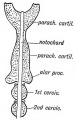
Fig. 133. The Parachordal Cartilages out of which the Cartilaginous Parts of the Occipital Bone are formed.
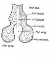
Fig. 134. The expansion backwards of the Parachordal Cartilages to enclose the Foramen Magnum and form the Supra-occipital.
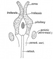
Fig. 135. Diagram of the Trabeculae Cranii, Parachordal Cartilages, and Periotio Capsules.
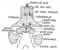
Fig. 136. Diagram of the structures formed from the Trabeculae Cranii.
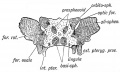
Fig. 137. The Sphenoid in a foetus of 4 months. The Centres of Ossification are deeply shaded. (After Sappey.)
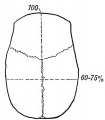
Fig. 138. Diagram of a Long-head (Dolichocephalic).
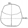
Fig. 139. Diagram of a Short-head (Brachycephalie).
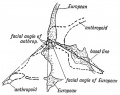
Fig. 140. The Facial Angle of alEuropean contrasted with that of an Anthropoid.


Development of the Structures concerned in the Sense of Sight
Development of the Structures concerned in the Sense of Sight
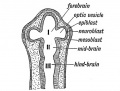
Fig. 141. Diagram of the Elements which form the Eyeball.
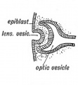
Fig. 142. Invagination of the Epiblast to form the Lena Vesicle.
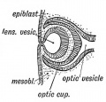
Fig. 143. The manner in which the Lens Vesicle is severed from the Epiblast.
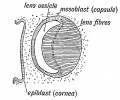
Fig. 144. The Formation of the Lens Fibres from the Epithelium on the posterior Wall of the Vesicle.
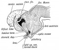
Fig. 145. Diagram showing the condition of the Optic Stalk and Vesicle at the commencement of the 2nd month. (After His.)
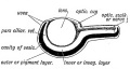
Fig. 146. Diagrammatic Section of the Optic Cup and Lens.
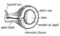
Fig. 147. The Optic Stalk and Cut), viewed on the lower and lateral aspect, showing the closure of the Choroidul Fissure.
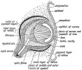
Fig. 148. Diagrammatic Section of the Eye showing the Parts formed from the Mesoblast. (After His' Model of the eye of a 3rd mouth human embryo.)
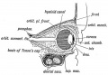
Fig. 149. Section of the Eye and Orbit at birth.
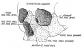
Fig. 150. The Origin of the Bones entering into Formation of the Orbit.
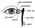
Fig. 151. Diagram of the Plica Semilunaris and Lachrymal Canaliculi.
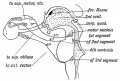
Fig. 152. Diagram of the Motor Nerves of the Muscles of the Eye derived from the 1st, 2nd, and 3rd Cephalic Segments.
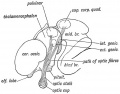
Fig. 153. Diagram of the Foetal Brain at the end of the 2nd month, showing the Position in which the Optic Tracts are developed.
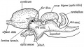
Fig. 154. Mesial Section of the brain of a Lizard showing the resemblance to the human foetal brain (Fig. 153) especially in the development of the Corpora Bigemina.
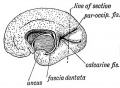
Fig. 155. View of the Mesial Surface of the Brain in the 5th month.
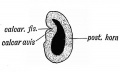
Fig. 156. Section of the Occipital Lobe at the position marked in Fig. 155.
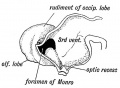
Fig. 157. Mesial Section of the Brain at the 4th week shewing the Rudiment of the Occipital Lobe. (After His.)


- Links: Vision Development
The Brain and Spinal Cord
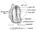
Fig. 158. Medullary Folds uniting to form the Neural Tube in a Human Embryo of about 14 days. (After Graf Spee.)
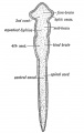
Fig. 159. Diagram of the Four Primary Divisions of the Neural Tube.
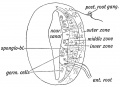
Fig. 160. Diagrammatic Section showing the three Zones of the Spinal Neural Tube at the Gth week.
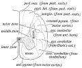
Fig. 161. Diagrammatic Section o£ the Spinal Cord to show the Parts formed in the three Zones of the Embryonic Spinal Cord.
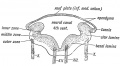
Fig. 162. Section across the Hind Brain of a Human Embryo in the 5th week.
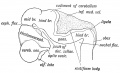
Fig. 163. Lateral view of the Cephalic Part of the Neural Tube in a 5th week human embryo. (After His.)
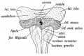
Fig. 164. Diagram of the Attachments of the Inferior Medullary Velum in a 5th month foetus. (After Kollmann.)
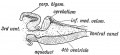
Fig. 165. Median Section of the Cerebellum and 4th Ventricle of a Frog.
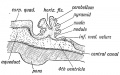
Fig. 166. Diagrammatic Section of the Cerebellum of a 3rd month Human Foetus showing the folding of the Cerebellar Plate.
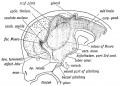
Fig. 167. A schematic figure to show the parts derived from the walls of the fore-brain. (After His.)
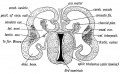
Fig. 168. Transverse Section of the brain of a Human Foetus at the commencement of the 3rd month to show the Cerebral Vesicles overlapping the Thalamencephalon.
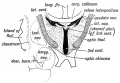
Fig. 169. Diagrammatic Section across the 3rd Ventricle of the Adult to show the Structures formed in its Walls.
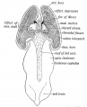
Fig. 170. A dorsal view of the Fore and Mid-brain at the 5th week of development to show the formation of the Velum Interpositum.
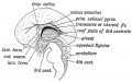
Fig. 171. Mesial Aspect of the human Foetal Brain during the 4th month. (After Minot.)
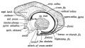
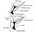
Fig. 172. Diagram to show the structures formed in the Lamina Terminalis and Primitive Callosal Gyrus. (After Elliot Smith.)
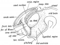
Fig. 173. Showing the Development of the Corpus Striatum in the floor and outer wall of the Cerebral Vesicle.
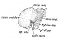
Fig. 174. Lateral Aspect of the Cerebral Hemisphere during the 2nd month.
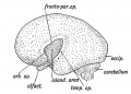
Fig. 175. The same Aspect during the 5th month.
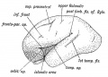
Fig. 176. The same Aspect during the 7th month.
Fiq. 177. Diagram of the Opercula and Fissure of Sylvius. In A the orbital operculum is undivided ; in B it is subdivided. (After Cunningham.)
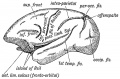
Fig. 178. The Island of Reil and Fissures on the lateral Aspect of the Brain of a dog-like Ape.
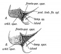
Fig. 179A. The more common Condition of the Island of Reil in Anthropoids. Fig. 179B. The complete isolation of the Island of Reil, the condition seen constantly in the Human Brain and occasionally in the Anthropoid.
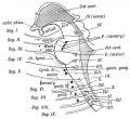
Fig. 180. A Diagram to show the Relationship of the Cranial Nerves to the Primitive Segments of the Head.


Development of the Circulatory System
Development of the Circulatory System
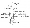
Fig. 181. The Superior Vena Cava of the Adult.
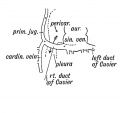
Fig. 182. The Embryonic Venous Trunks out of which the Superior Vena Cava is formed.
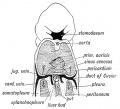
Fig. 183. Diagram to show the manner in which the Ducts of Cuvier encircle the Coelom at the junction of the Pericardial and Pleural Parts.
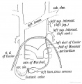
Fig. 184. The Remnants of the Left Superior Vena Cava, derived from the Structures shown in Fig. 185.
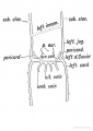
Fig. 185. Diagram of the Sinus Venosus and Ducts of Cuvier of the human embryo about the 3rd week.
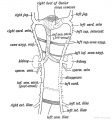
Fig. 186. The Remnants of the Posterior Cardinal Veins in the Adult.
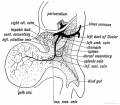
Fig. 187. The Left Vitelline Vein of an Embryo of the 4th week.
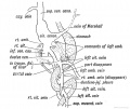
Fig. 188. Diagram showing the Formation of the Ductus Venosus, and the fate of the Umbilical and Vitelline veins.
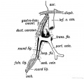
Fig. 189.-Diagram of the Kemnants of the Umbilical Vein in the Adult-viewed from behind.
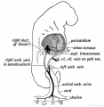
Fig.190. Diagram of the Right Umbilical Vein in an embryo of 3 weeks, before the outgrowth of the Liver Bud
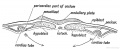
Fig.191. Transverse section of the Blastoderm showing the Right and Left Cardiac Tubes situated in the Splanchnopieure.
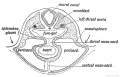
Fig.192. Transverse section at a more advanced stage showiDg the union of the Splanchnopleures to form the Mesocardia and the fusion of the Right and Left Cardiac Tubes.
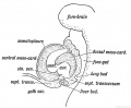
Fig.193. Lateral view of the Heart and Pericardium to show the Attachments of the Dorsal and Ventral Mesocardia.
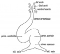
Fig.194. The Primitive Divisions of the Heart.
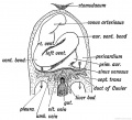
Fig.195. Showing the two chief Bends which occur in the Heart during the 3rd week.
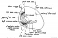
Fig.196. Showing the Structures formed from the Sinus Venosus.
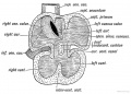
Fig.197. Section of the Heart of a 5th week human foetus showing the Right and Left Venous Valves which guard the entrance of the Sinus Venosus into the Primitive Auricle.
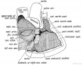
Fig.198. Diagram of the Septa of the Heart viewed on the right side.
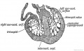
Fig.199. Section of the Ventricles of the Foetal Heart, showing the Muscular Sponge Work within their Cavities.
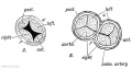
Fig 200. The origin of the semilunar valves.
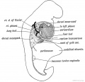
Fig 201. The Form of the Coelom in a 3rd week Embryo as viewed from the right side.
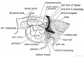
Fig. 202. Diagram to show the manner in which the heart is fixed within the pericardium by the Arterial and Venous Mesocardia in a human embryo of 3 weeks.
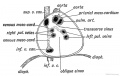
Fig. 203. View of the interior of the Pericardium showing the attachments of the heart to its dorsal aspect by the Arterial or Venous Mesocardia.


The Respiratory System
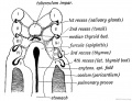
Fig. 204. Floor of the Pharynx and Oesophagus of a human embryo of 3 weeks showing; the Furcula, Pulmonary Groove, and Diverticulum.

Fig. 205. A section of a human embryo to show the Relationships oJ the Pulmonary Buds at the 4th week.
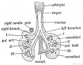
Fig. 206. The condition of the Bight and Left Pulmonary Buds in a 5th week embryo. (After His).
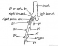
Fig. 207. Scheme of the Bronchial Ramifications in Quadrupedal Mammals.
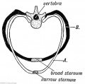
Fig. 208. Diagrammatic Section of the Thorax of a Quadrupedal Mammal (A), contrasted with a corresponding section in Man (B).
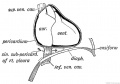
Fig. 209. The Relationship of the Heart to the Diaphragm in Quadrupedal Mammals.
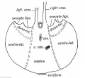
Fig. 210. Diagram of the Diaphragm to show the Parts formed by each of the five Elements.
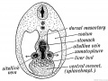
Fig 211. Diagrammatic section behind the Embryonic Heart to show the Part of the primitive Mesentery which forms the mesial Element of the Diaphragm.


The Organs of Digestion
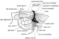
Fig. 212. The Mesentery of the Fore-gut and its Contents, -viewed from the left side (schematic).
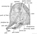
Fig. 213 A. The origin of the Peritoneal Ligaments connected with the Liver.
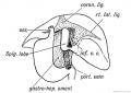
Fig. 213 B. The origin of the Peritoneal Ligaments connected with the Liver.
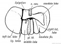
Fig. 214. Diagram of a mammalian Liver viewed from behind and below.

Fig. 215. The lower surface of the Liver of a human foetus during the 3rd month, showing Vestiges of Fissures and Lobes of the typical mammalian Liver.
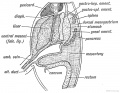
Fig. 216. The Relationship of the Spleen, Pancreas, and Liver to the Mesogastrium in the Embryo.
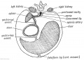
Fig. 217. A diagrammatic transverse Section of the Mesogastrium viewed from behind.
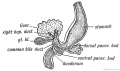
Fig. 218. The Pancreatic and Hepatic Processes of a 4th week human embryo. (After Kollmann.)
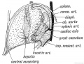
Fig. 219. The Arrangement of Vessels in the Dorsal week (diagrammatic).
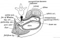
Fig. 220. Diagram to show the Formation of the Lesser Sac of the Peritoneum from the Dorsal Mesogastrium.
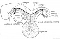
Fig. 221. The Form of the Alimentary Canal in a human embryo of the 3rd week.
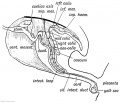
Fig. 222. The Form of the Alimentary Canal during the 5th week.
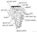
Fig. 223 A. The mesentery of the hind-gut. The Position assumed by the Colon after the rotation of the Gut has taken place.
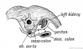
Fig. 223 B. Diagram to show how the descending Meso-colon becomes applied to the parietal Peritoneum of the left Lumbar Region.
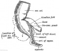
Fig. 224. Diagram of the Apex of the Caecum at the time of birth and the Diverticula which may be produced in the Fundus of the Caecum afterwards.
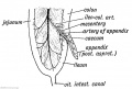
Fig. 225 A.The Appendix and Peritoneal Folds at the end of the 2nd month of foetal life. The Intestinal Loop is viewed from the left side.
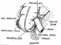
Fig. 225 B. Peritoneal Fossae in the lleo-caecal Region.
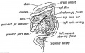
Fig. 226. To show the Kotation of the Intestinal Loop and Formation of the Duodenojejunal Fossa.


The Body Wall, Ribs, and Sternum
The Body Wall, Ribs, and Sternum
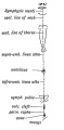
Fig. 227. Diagram of the Structures formed in the Median Ventral Line of the Body.
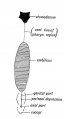
Fig. 228. The Median Ventral Line in an embryo of three weeks, to contrast with the Corresponding Line in the Adult.
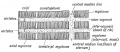
Fig. 229. Scheme of the Manner in which the Somatopleure is segmented.
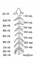
Fig. 230. The Form of Sternum in a Pronograde (quadrupedal) Mammal.
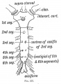
Fig. 231. The Form of Sternum in a Mammal adapted to the orthograde (upright) Posture. The Points of Ossification are also shown.
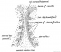
Fig. 232. The Sternal Bars in a human embryo of six weeks (after Paterson).


The Limbs
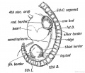
Fig. 233. Lateral view of a human embryo at the 28th day, showing the Limb Buds, Lateral Edges, and Primitive Segments.
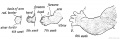
Fig. 234. Development of the Upper Limb.
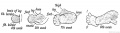
Fig. 235. Development of the Lower Limb.
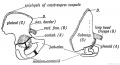
Fig. 236. The Corresponding Points {A, B, C, and D) in the Ilium and Scapula.
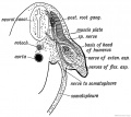
Fig. 237. Section of the Arm Bud of a human embryo at the end of the 4th week.
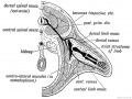
Fig. 238. Schematic Section showing the Origin and Arrangement of the Muscles and Nerves of the Limbs.
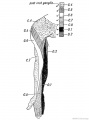
Fig. 239. The Distribution of the Posterior Roots of the Spinal Nerves on the Flexor Aspect of the Arm.
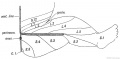
Fig. 240. Diagram to show the typical Manner in which the Posterior Nerve Roots are distributed in the Lower Limb.
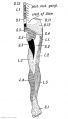
Fig. 241. Flexor Aspect of the Lower Limb showing the Sensory Distribution of the Segmental or Spinal nerves.
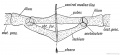
Fig. 242. Diagram of the Pelvic Girdle of a Lizard.
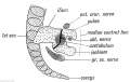
Fig. 243. The Pelvic Girdle of a Human Foetus at the 5th week.
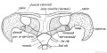
Fig. 244. The Shoulder Girdle of Ornithorynchus.
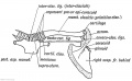
Fig. 245. The Parts in the Shoulder Girdle of a human foetus which correspond with those of Ornithorynehus.
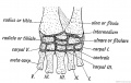
Fig. 246. The Carpal Bones of a Tortoise.
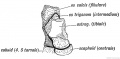
Fig. 247. The Os Trigenum and Bones of the Tarsus
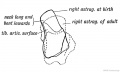
Fig. 248. The Foetal and Adult (in dotted outline) Forma of the Astralagus contrasted.
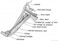
Fig. 249. Latissimo-condyloideus Muscle.
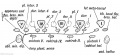
Fig. 250. The Morphology of the Short Muscles of the Digits.
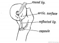
Fig. 251. Showing the Origin of the Ligamentum Teres and Reflected Bundle of the Capsular Ligament.
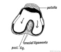
Fig. 252. Showing the Origin of the Crucial Ligaments of the Knee.


| Historic Disclaimer - information about historic embryology pages |
|---|
|
Human Embryology and Morphology (1902): Development or the Face | The Nasal Cavities and Olfactory Structures | Development of the Pharynx and Neck | Development of the Organ of Hearing | Development and Morphology of the Teeth | The Skin and its Appendages | The Development of the Ovum of the Foetus from the Ovum of the Mother | The Manner in which a Connection is Established between the Foetus and Uterus | The Uro-genital System | Formation of the Pubo-femoral Region, Pelvic Floor and Fascia | The Spinal Column and Back | The Segmentation of the Body | The Cranium | Development of the Structures concerned in the Sense of Sight | The Brain and Spinal Cord | Development of the Circulatory System | The Respiratory System | The Organs of Digestion | The Body Wall, Ribs, and Sternum | The Limbs | Figures | Embryology History
Reference
Keith A. Human Embryology and Morphology. (1902) London: Edward Arnold.
Cite this page: Hill, M.A. (2024, April 23) Embryology Book - Human Embryology and Morphology Figures. Retrieved from https://embryology.med.unsw.edu.au/embryology/index.php/Book_-_Human_Embryology_and_Morphology_Figures
- © Dr Mark Hill 2024, UNSW Embryology ISBN: 978 0 7334 2609 4 - UNSW CRICOS Provider Code No. 00098G