Book - Human Embryology and Morphology 7: Difference between revisions
(Created page with "{{Human embryology morphology 1902 header}} {{Human embryology morphology 1902 footer}}") |
mNo edit summary |
||
| (2 intermediate revisions by the same user not shown) | |||
| Line 1: | Line 1: | ||
{{Human embryology morphology 1902 header}} | {{Human embryology morphology 1902 header}} | ||
=Chapter VII. The Development Of The Ovum Of The Foetus Feom The Ovum Of The Mother= | |||
From the Ovum of one generation to the Ovum of the Next. | |||
— The manner in which the face, neck, pharynx and cutaneous sbructures of the body are produced having now been traced, it is necessary, before further progress can be made, to turn to the phenomena which mark the opening stages in the development of the embryo. This may best be done by following the cycle of changes which lead to the production of a new generation of germinal cells from the fertilized ovum of a former generation. Every ovum is the offspring of a fertilized ovum, and the fertilized ovum is the intermediary whereby the characters and properties of a race are handed from one generation to the next. | |||
Descent of the Ovary. — In the female human foetus of the fifth month the ovary has reached the iliac fossa (Fig. 59) in the course of its descent from the lower dorsal region where it originated to its permanent position on the lateral wall of the pelvis. The ovary is then long and narrow, with an upper and lower pole ; it is three-sided in section — the surfaces being inner, outer and inferior or ventral (Fig. 60). The Fallopian tube, derived from the upper part of the Miillerian duct, lies along the outer side of the ovary in the iliac fossa ; its upper fimbriated end terminates at, and is attached to, the upper or cephalic pole of the ovary (Fig. 59). As the parts lie on the iliac fossa, the tube and the ovary are supported each by its own mesentery, the mesosalpinx and meso-ovarium. The two mesenteries have, however, a common origin or attachment to the posterior abdominal wall and to the common attachment the name of common genital mesentery may be given. The upper end of the common mesentery — the plica vascularis (Fig. 59), as it is reflected from the cephalic pole of the ovary and fimbriated extremity of the tube, is continued up towards the diaphragm and in it the ovarian vessels and nerves pass to the ovary and tube. The caudal pole of the ovary is joined to the uterus by its round ligament. The round ligament of the uterus, corresponding to the gubernaculum testis of the male, passes from the brim of the pelvis, where it is attached to the horn of the uterus, almost straight to the internal inguinal opening and assists in the descent of the ovary and tube. | |||
<div id="Fig059"></div> | |||
[[File:Keith1902 fig059.jpg|400px]] | |||
'''Fig. 59.''' The position of the Ovary and Fallopian Tube in the 5th month. | |||
By full time the ovary lies at the brim of the pelvis or partly within it ; after birth the ovary, uterus and rectum come gradually to occupy their adult positions within the pelvis. This is due to a relatively greater growth in the pelvis itself than in its contents. The ovary, as is more frequently the case with the testicle, may be arrested in its descent. | |||
In Fig. 6 an earlier stage is shown ; it represents on section the condition about the end of the second month. The ovary and tube with the remnants of the Wolffian body and duct occupy the position in which they are developed. Both are suspended by mesenteries from the dorsal wall of the peritoneal cavity, at the side of the mesentery of the gut. | |||
<div id="Fig060"></div> | |||
[[File:Keith1902 fig060.jpg|400px]] | |||
'''Fig. 60.''' Diagrammatic section of a foetus at the end of the 2nd month, showing the Attachments of the Ovary and MUllerian duct. | |||
Normal Position of tlie adult Ovary. — When the ovary descends within the pelvis it occupies a definite triangle — the ovarian triangle — on the lateral wall of the pelvic cavity (Fig. 61). The ovarian triangle is bounded above by the upper half of the external iliac artery, below and behind by the internal iliac artery, with the ureter lying on the artery ; in front by the reflection of the posterior layer of the broad ligament on the side of the pelvis. | |||
<div id="Fig061"></div> | |||
[[File:Keith1902 fig061.jpg|400px]] | |||
'''Fig. 61.''' Showing the position of the Ovary on the lateral wall of tho Pelvis and its relation to the Fallopian Tube. | |||
The long axis of the ovary is parallel to the ureter and is vertically placed in the standing posture. The peritoneum covering the triangle forms a depression, or occasionally a pouch, for the ovary. It will be seen that, with the descent of the ovary, the mesosalpinx, the mesovarium, and the common genital mesentery have come to form the major part of the broad ligament. The reflection of" the common genital mesentery from the upper or cephalic pole of the ovary now forms the ovario-pelvic ligament (Figs. 59 and 61). In the ovarian triangle is also situate the internal iliac group of lymphatic glands, into which most of the pelvic lymphatics drain. The ovarian lymphatics end in the glands of the upper lumbar region near to where the ovary was developed. The ovary brings down with it, too, the ovarian vessels and plexus of nerves. The nerves come through the aortic plexus from the 10 th and 11th dorsal segments of the cord. | |||
<div id="Fig062"></div> | |||
[[File:Keith1902 fig062.jpg|400px]] | |||
'''Fig. 62.''' Diagrammatic Section of an Ovary to show the manner in which the Primitive Ova are carried in by incursions of the Germinal Epithelium. | |||
==An Ovum== | |||
As the infantile ovary descends, it is laden with thousands of ova. Each ovum is surrounded by a capsule composed of a layer of columnar epithelium, which is imbedded in, and surrounded by, the stroma of the ovary. The entire capsule — epithelium and stroma — forms a Graafian follicle (Fig. 62). The ovary is covered by a layer of columnar epithelium, which is named the germinal epithelium Amongst the columnar cells of the germ epithelium during foetal life and for sometime after birth larger cells occur. These are the primordial ova from which brood ova arise. The ova are carried within the ovary by tubular ingrowths of germinal epithelium (Fig. 62). These tubular invasions of the ovary become broken up, the isolated masses of the germinal epithelium remaining to form the linings of the Graafian follicles. | |||
Discharge of the Ova. — At puberty, possibly also before it, and for 30 years after it, one egg after another ripens; the ovum enlarges ; so does the Graafian follicle (Fig. 63). The cells of the epithelial lining proliferate and a cavity appears amongst the cells within the follicle, due to a collection of fluid— the liquor folliculi. The ovum remains attached to the wall of the follicle by a group of epithelial cells, the discus proligerus (Fig. 63). As the fluid collects, the follicle works its way to the surface of the ovary ; the tunica albuginea, which forms a capsule for the ovary, and the germinal epithelium, gradually atrophy over it, and at last it bursts and discharges the ovum. | |||
<div id="Fig063"></div> | |||
[[File:Keith1902 fig063.jpg|400px]] | |||
'''Fig. 63.''' Diagram of a ripe Graafian Follicle. | |||
Two opposite opinions exist among gynecologists as to the period of rupture : (1) That it occurs at the onset of menstruation ; (2) that it has no relationship to the menstrual period. The truth is, probably, that both are right. The majority of ova, however, appear to be discharged at the menstrual period. Whether ova are discharged from both ovaries at once, or from only one, and whether one or more than one in a month, are points not yet settled ; but the usual opinion is that one ovum is shed each month, and only from one ovary. | |||
The Graafian follicle, after rupture, fills up with blood ; a cellular tissue is soon developed within its cavity. If pregnancy occurs this tissue forms a true corpus luteum, a large yellow body as big as a pigeon's egg. If pregnancy does not occur, a false corpus luteum is formed, a formation which begins to disappear before the next menstrual period. Both forms lead to a cicatrix, which is seen on the surface of the ovary. The ovary of an old person is commonly covered with such scars. The Graafian follicles may become cystic and give rise to enormous ovarian tumours. | |||
==The Fallopian Tube== | |||
When the ovum drops from the ovary it cannot easily escape the ciliated fimbriae of the Fallopian tube which surround and clutch the ovary. In Fig. 6 1 the relationship of the Fallopian tube to the ovary is shown. The tube may be demarcated into three parts : (a) the isthmus or arm directed outwards to the wall of the pelvis (J to 1 inch) ; (6) the forearm or ampullary part, directed backwards on the lateral pelvic wall above the ovary ; (c) the hand, infundibular, or fimbriated part, folded backwards and grasping the free border and exposed surface of the ovary. The tube is tethered by one of its fimbriae to the cephalic pole of the ovary. | |||
Course of the Ovum in the Tube. — The cilia on the fimbriae work towards the ostium abdominale, the abdominal mouth of the Fallopian tube, which is situated at the bases of the fimbriae, and carry the discharged ovum through the ostium within the tube. The ostium abdominale is shut when the tube is examined after excision ; the closure is probably due to reflex contraction of the tube muscle, caused by handling and cutting. In the infundibular and ampullary segments of the tube, the mucous membrane is thrown into long plicated folds shown in section in Fig. 64. They are covered with ciliated epithelium, which urge the ovum towards the uterus. Within the tube impregnation usually takes place. If an obstruction arrests the fertilized ovum in the tube, perhaps it may be a patch denuded of epithelium and cilia by gonorrheal inflammation, a tubular pregnancy is the result. The growing embryo, commonly about the second month, bursts the tube and falls within the broad ligament or into Douglas's pouch (Fig. 64). | |||
<div id="Fig064"></div> | |||
[[File:Keith1902 fig064.jpg|400px]] | |||
'''Fig. 64.''' Diagrammatic section of .the Broad Ligament and Fallopian Tube. | |||
==The History of the Ovum within the Fallopian Tube== | |||
When the ovum enters the Fallopian tube, it is a cell of very considerable size ('2 mm. = -jhs in.) with a cell wall — the zona radiata (Fig. 63), a nucleus — the germinal vesicle, and a nucleolus — the germinal spot. Then, or before then, the ovum prepares for fertilization by the extrusion from its nucleus of first one, then another polar body, and, with the extrusion, the germinal vesicle becomes the female pronucleus. The polar bodies, which lie outside the protoplasm of the ovum, but within the zona radiata, are parts of the germinal vesicle, which are extruded with all the display of karyokinesis (Fig. 65 A). The meaning of the process has been much guessed at ; the outstanding fact is that two parts of the ovum are segregated and are not involved in the subsequent developmental changes. What become of the polar bodies in the course of development of the fertilized ovum is not known. | |||
<div id="Fig065"></div> | |||
[[File:Keith1902 fig065.jpg|400px]] | |||
'''Fig. 65.''' Showing the production of the Morula from the Ovum. A. The ovum after the first division. B. After the second. C. The Morula . | |||
In the course of fecundation thousands of spermatozoa are lodged in the genital passage ; many stem the adverse current of the uterine cilia, reach and live for days within the interlaminar grooves in the wider parts of the tube. In the course of its descent within one of the grooves the egg may be fertilized. The spermatozoon bursts through the zona radiata, loses its tail, its head enlarges, and forms the male pronucleus. The male and female pronuclei unite, and from their union springs the nucleus of the fertilized ovum. This is the centre from which all future developmental changes start. The ovum may be, but rarely is, fertilized in the ovary, or between the ovary and ostium abdominale, the result being a pelvic gestation. The length of time the fertilized ovum takes to reach the uterus is not known exactly, but probably it spends about ten or twelve days within the Fallopian tube, and during that time it passes through the following stages : | |||
# The Morula. — The ovum, with a full display of karyokinetic changes in the nucleus, divides, subdivides, and grows within the zona radiata. until a rounded mass of cells is formed — the morula or mulberry mass. The cells' are of unequal size and divide at unequal rates (Fig. 65, A, B, C). | |||
# The Blastodermic Vesicle. — This stage is produced from the morula stage by the collection of fluid within the mass of cells. The cells are then seen to be arranged in two sets— (a) a layer lining the zona radiata and forming the wall of the vesicle (Eauber's layer) (Fig. 66), and (6) a group of granular cells within the cavity attached to Eauber's layer at the embryogenic pole of the vesicle (Fig. 66). In Vertebrates with huge stores of yolk in their ova, such as birds have, the vesicle is filled by yolk-bearing cells, continuous with Eauber's cells at the vegetative pole, opposite to the granular cells. | |||
# The Bilaminar Blastoderm. — This stage is produced by the growth of the group of granular germinal cells within Eauber's layer. At first they arrange themselves in two layers — an outer which spreads on the inner aspect of Eauber's layer, and absorbs or mixes with it, to form one layer of more or less columnar cells, the epiblast or ectoderm (Fig. 67); an inner layer also spreads out from the germinal group, and forms a second lining to the blastodermic vesicle, the hypoblast or endoderm (Fig. 67). | |||
<div id="Fig066"></div> | |||
[[File:Keith1902 fig066.jpg|400px]] | |||
'''Fig. 66.''' Diagrammatic section of a Blastodermic Vesicle. | |||
<div id="Fig067"></div> | |||
[[File:Keith1902 fig067.jpg|400px]] | |||
'''Fig. 67.''' A diagrammatic section of a Bilaminar Blastoderm made across the primitive streak. | |||
===The Primitive Streak and Groove=== | |||
In the diagrammatic section of the bilaminar blastoderm, given in figure 67, the hypoblast and epiblast are seen to be fused at one point, and at the point of fusion the epiblast is thickened and somewhat depressed. When the blastodermic vesicle is viewed from the surface, the line along which the fusion of the two layers and thickening of the epiblast take place is marked by the primitive streak and groove (Fig. 68). At the anterior end of the groove is situated the blastopore or neurenteric canal, an opening into the cavity which the hypoblast encloses — the archenteron (see Figs. 67 and 75). Eound the blastopore and along the primitive streak the epiblast is continuous with the hypoblast. | |||
===The Mesoblast=== | |||
The mesoblast or mesoderm, the third of the primitive blastodermic layers, is produced from the margins of the primitive streak (Fig. 67). At the line of junction of the epiblast and hypoblast there is a free proliferation of cells which spreads out between the two primitive layers, and gives rise to a third or intermediate layer — the mesoblast (Fig. 69). In lower vertebrates the mesoblast is entirely produced from the hypoblast. Neural Canal. — It is at this early stage, while the mesoblastic cells swarm in between the epiblast and hypoblast, and while the vesicle has not yet reached the uterus, that a grooved strip of epiblast, the medullary plate, in front of the primitive streak (Fig. 68) is set aside, by a process of infolding, to form the great central nervous system — the brain, spinal cord, and nerves (Fig. 69). | |||
<div id="Fig068"></div> | |||
[[File:Keith1902 fig068.jpg|400px]] | |||
'''Fig. 68.''' Diagram of the Embryogenie area of a Bilaminar Blastoderm viewed from above. | |||
A medullary fold rises up on each side of what is to be the middle line of the back to enclose the medullary plate. The folds rise until their crests meet, and a tube of epiblast is buried by the fusion of their lips. The blastopore is included within the posterior end of the medullary folds (Fig. 68). Thus, for a short space, the cavity of the yolk sac communicates with the neural canal. | |||
===The Notochord=== | |||
Beneath the neural tube a similar infolding of hypoblast takes place, and a tube of cells to form the notochord is detached (Fig. 69). It forms the first basis of the spinal column. | |||
<div id="Fig069"></div> | |||
[[File:Keith1902 fig069.jpg|400px]] | |||
'''Fig. 69.''' Diagrammatic section of a Blastodermic Vesicle showing (1) the origin of the neural canal, (2) the origin of the notochord, (3) the" ingrowth of the mesoblast, and (4) the formation of the coelom. | |||
===The Somatopleure and Splanchnopleure=== | |||
The mesoblast surrounds both the notochord and neural canal as they areformed — perhaps assists in their formation (Figs. 69 and 70). The solid layer which surrounds these structures is known as the paraxial mesoblast. As the mesoblastic cells spread out to cover the outer aspect of the hypoblast and inner aspect of the epiblast, they separate into two layers which enclose between them a cavity — the coelom (Fig. 69). The epiblast, with its accompanying layer of mesoblast, makes up the somatopleure which bounds the coelom without; the hypoblast and overlying layer of mesoblast form the splanchnopleure which bounds the cavity within. | |||
<div id="Fig070"></div> | |||
[[File:Keith1902 fig070.jpg|400px]] | |||
'''Fig. 70.''' Diagram of the Blastodermic Vesicle separating into Embryo and Membranes. | |||
===Embryo and Membranes=== | |||
During the second week, while the blastodermic vesicle is still within the Fallopian tube and some time before it has reached the uterus, changes take place whereby part of the blastodermic vesicle forms the embryo and part is transformed into the enclosing membranes which contain, protect, and nourish it (Fig. 70). Part also forms the yolk sac. | |||
===The Decidua=== | |||
When the uterine cavity is reached, the uterine mucous membrane has become hypertrophied, the decidua being thus formed. In the formation of the decidua, the uterine glands become elongated and enlarged ; the inter-glandular tissue hypertrophied and the mouths of the glands closed, so that the surface layer of the decidua is comparatively solid. The hypertrophy of the mucous membrane is unequal, depressions or pits beingformed on its surface. The deeper layer of the decidua is cavernous, the spaces being formed by the distension of capillary veins into venous spaces, and perhaps also by the enlargement of the gland lumina. | |||
When the blastodermic vesicle reaches the uterus, it commonly adheres to the posterior wall near the fundus (Fig. 71). The decidua forms first a nest for it, and then grows over and buries it, thus forming the outer or decidual membrane of the embryo. The layer which covers the vesicle (see Fig. 71) is the decidua reflexa; the part by which it adheres to the uterus, the decidua serotina; the rest, lining the uterus, the decidua vera. As the vesicle grows, it fills the cavity of the uterus, thus bringing the decidua reflexa in contact with the decidua vera ; the reflexa disappears when it comes in contact with the vera, which remains as the outermost of the foetal membranes. The serotina takes part in the formation of the placenta. | |||
<div id="Fig071"></div> | |||
[[File:Keith1902 fig071.jpg|400px]] | |||
'''Fig. 71.''' Section of the Uterus showing the three parts of the Decidua. | |||
===The Amnion and Chorion=== | |||
Only part of the somatopleure of the blastodermic vesicle takes part in the formation of the embryo ; the remaining parts of the somatopleure are converted into the amnion and chorion in this manner : | |||
As is shown in Fig. 70, the embryonic area appears to sink within the vesicle ; the somatopleure rises up as a wave-like fold right round it, at each side, at the head as well as at the tail end. The folds meet over the embryo and unite, as is shown in Fig. 72. Thus the embryo is wrapped up in a double fold of its own somatopleure. The inner fold or membrane is the Amnion, the outer is the Prechorion (Fig. 72). The prechorion, in turn, is covered by the decidua reflexa, and at its attached part by the decidua serotina. Villi containing vessels grow out from the chorion into the decidua. | |||
<div id="Fig072"></div> | |||
[[File:Keith1902 fig072.jpg|400px]] | |||
'''Fig. 72.''' Showing the folds of the somatopleure uniting over the embryo and becoming demarcated into Amnion and Prechorion. | |||
===Origin of Ova and Spermatozoa=== | |||
The cells which give rise to the ova and spermatozoa, according to sex, are derived from the mesothelial lining of the coelom. They appear with the cleavage of the mesoblast and formation of the coelom. Soon after the coelom is formed (beginning of third week) a ridge — the genital ridge — is seen on the roof of the coelom, situated on each side of the attachment of the mesentery of the gut (Fig. 73). The ridge lies over the intermediate cell mass (Fig. 85 ; p. 1 11), and is covered by a layer of columnar meso-thelium, the germinal epithelium, which is continuous with the mesothelial lining of the coelom. Some of the columnar cells covering the genital ridge become primordial ova, and are set aside to produce ova or spermatozoa, according to the sex. Beard's researches have led him to the conclusion that primordial ova are produced at a very early stage of development, and that they migrate towards the coelom and take up their position amongst the germinal cells covering the genital ridge when that ridge is formed. These cells may stray and give rise in various parts of the body to dermoid tumours (Beard). The manner in which the germ cells and primordial ova grow within the genital ridge to form the ovary or testicle was described at the beginning of this chapter. The cycle of changes which leads to the production of the ova of one generation from the ovum of a former generation have thus been traced. | |||
<div id="Fig073"></div> | |||
[[File:Keith1902 fig073.jpg|400px]] | |||
'''Fig. 73.''' Diagrammatic section of the abdominal region of the coelom, showing the position of the Genital Ridges from which the Ovary or Testicle is formed. | |||
{{Human embryology morphology 1902 footer}} | {{Human embryology morphology 1902 footer}} | ||
Latest revision as of 08:03, 2 January 2014
| Embryology - 19 Apr 2024 |
|---|
| Google Translate - select your language from the list shown below (this will open a new external page) |
|
العربية | català | 中文 | 中國傳統的 | français | Deutsche | עִברִית | हिंदी | bahasa Indonesia | italiano | 日本語 | 한국어 | မြန်မာ | Pilipino | Polskie | português | ਪੰਜਾਬੀ ਦੇ | Română | русский | Español | Swahili | Svensk | ไทย | Türkçe | اردو | ייִדיש | Tiếng Việt These external translations are automated and may not be accurate. (More? About Translations) |
Keith A. Human Embryology and Morphology. (1902) London: Edward Arnold.
| Historic Disclaimer - information about historic embryology pages |
|---|
| Pages where the terms "Historic" (textbooks, papers, people, recommendations) appear on this site, and sections within pages where this disclaimer appears, indicate that the content and scientific understanding are specific to the time of publication. This means that while some scientific descriptions are still accurate, the terminology and interpretation of the developmental mechanisms reflect the understanding at the time of original publication and those of the preceding periods, these terms, interpretations and recommendations may not reflect our current scientific understanding. (More? Embryology History | Historic Embryology Papers) |
Chapter VII. The Development Of The Ovum Of The Foetus Feom The Ovum Of The Mother
From the Ovum of one generation to the Ovum of the Next.
— The manner in which the face, neck, pharynx and cutaneous sbructures of the body are produced having now been traced, it is necessary, before further progress can be made, to turn to the phenomena which mark the opening stages in the development of the embryo. This may best be done by following the cycle of changes which lead to the production of a new generation of germinal cells from the fertilized ovum of a former generation. Every ovum is the offspring of a fertilized ovum, and the fertilized ovum is the intermediary whereby the characters and properties of a race are handed from one generation to the next.
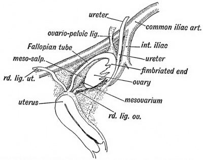
Descent of the Ovary. — In the female human foetus of the fifth month the ovary has reached the iliac fossa (Fig. 59) in the course of its descent from the lower dorsal region where it originated to its permanent position on the lateral wall of the pelvis. The ovary is then long and narrow, with an upper and lower pole ; it is three-sided in section — the surfaces being inner, outer and inferior or ventral (Fig. 60). The Fallopian tube, derived from the upper part of the Miillerian duct, lies along the outer side of the ovary in the iliac fossa ; its upper fimbriated end terminates at, and is attached to, the upper or cephalic pole of the ovary (Fig. 59). As the parts lie on the iliac fossa, the tube and the ovary are supported each by its own mesentery, the mesosalpinx and meso-ovarium. The two mesenteries have, however, a common origin or attachment to the posterior abdominal wall and to the common attachment the name of common genital mesentery may be given. The upper end of the common mesentery — the plica vascularis (Fig. 59), as it is reflected from the cephalic pole of the ovary and fimbriated extremity of the tube, is continued up towards the diaphragm and in it the ovarian vessels and nerves pass to the ovary and tube. The caudal pole of the ovary is joined to the uterus by its round ligament. The round ligament of the uterus, corresponding to the gubernaculum testis of the male, passes from the brim of the pelvis, where it is attached to the horn of the uterus, almost straight to the internal inguinal opening and assists in the descent of the ovary and tube.
Fig. 59. The position of the Ovary and Fallopian Tube in the 5th month.
By full time the ovary lies at the brim of the pelvis or partly within it ; after birth the ovary, uterus and rectum come gradually to occupy their adult positions within the pelvis. This is due to a relatively greater growth in the pelvis itself than in its contents. The ovary, as is more frequently the case with the testicle, may be arrested in its descent.
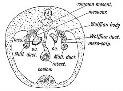
In Fig. 6 an earlier stage is shown ; it represents on section the condition about the end of the second month. The ovary and tube with the remnants of the Wolffian body and duct occupy the position in which they are developed. Both are suspended by mesenteries from the dorsal wall of the peritoneal cavity, at the side of the mesentery of the gut.
Fig. 60. Diagrammatic section of a foetus at the end of the 2nd month, showing the Attachments of the Ovary and MUllerian duct.
Normal Position of tlie adult Ovary. — When the ovary descends within the pelvis it occupies a definite triangle — the ovarian triangle — on the lateral wall of the pelvic cavity (Fig. 61). The ovarian triangle is bounded above by the upper half of the external iliac artery, below and behind by the internal iliac artery, with the ureter lying on the artery ; in front by the reflection of the posterior layer of the broad ligament on the side of the pelvis.
Fig. 61. Showing the position of the Ovary on the lateral wall of tho Pelvis and its relation to the Fallopian Tube.
The long axis of the ovary is parallel to the ureter and is vertically placed in the standing posture. The peritoneum covering the triangle forms a depression, or occasionally a pouch, for the ovary. It will be seen that, with the descent of the ovary, the mesosalpinx, the mesovarium, and the common genital mesentery have come to form the major part of the broad ligament. The reflection of" the common genital mesentery from the upper or cephalic pole of the ovary now forms the ovario-pelvic ligament (Figs. 59 and 61). In the ovarian triangle is also situate the internal iliac group of lymphatic glands, into which most of the pelvic lymphatics drain. The ovarian lymphatics end in the glands of the upper lumbar region near to where the ovary was developed. The ovary brings down with it, too, the ovarian vessels and plexus of nerves. The nerves come through the aortic plexus from the 10 th and 11th dorsal segments of the cord.
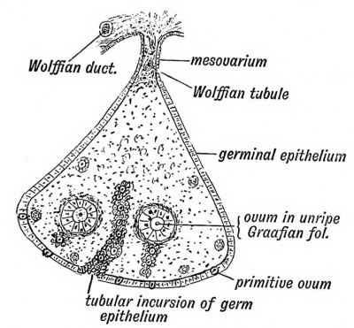
Fig. 62. Diagrammatic Section of an Ovary to show the manner in which the Primitive Ova are carried in by incursions of the Germinal Epithelium.
An Ovum
As the infantile ovary descends, it is laden with thousands of ova. Each ovum is surrounded by a capsule composed of a layer of columnar epithelium, which is imbedded in, and surrounded by, the stroma of the ovary. The entire capsule — epithelium and stroma — forms a Graafian follicle (Fig. 62). The ovary is covered by a layer of columnar epithelium, which is named the germinal epithelium Amongst the columnar cells of the germ epithelium during foetal life and for sometime after birth larger cells occur. These are the primordial ova from which brood ova arise. The ova are carried within the ovary by tubular ingrowths of germinal epithelium (Fig. 62). These tubular invasions of the ovary become broken up, the isolated masses of the germinal epithelium remaining to form the linings of the Graafian follicles.
Discharge of the Ova. — At puberty, possibly also before it, and for 30 years after it, one egg after another ripens; the ovum enlarges ; so does the Graafian follicle (Fig. 63). The cells of the epithelial lining proliferate and a cavity appears amongst the cells within the follicle, due to a collection of fluid— the liquor folliculi. The ovum remains attached to the wall of the follicle by a group of epithelial cells, the discus proligerus (Fig. 63). As the fluid collects, the follicle works its way to the surface of the ovary ; the tunica albuginea, which forms a capsule for the ovary, and the germinal epithelium, gradually atrophy over it, and at last it bursts and discharges the ovum.
Fig. 63. Diagram of a ripe Graafian Follicle.
Two opposite opinions exist among gynecologists as to the period of rupture : (1) That it occurs at the onset of menstruation ; (2) that it has no relationship to the menstrual period. The truth is, probably, that both are right. The majority of ova, however, appear to be discharged at the menstrual period. Whether ova are discharged from both ovaries at once, or from only one, and whether one or more than one in a month, are points not yet settled ; but the usual opinion is that one ovum is shed each month, and only from one ovary.
The Graafian follicle, after rupture, fills up with blood ; a cellular tissue is soon developed within its cavity. If pregnancy occurs this tissue forms a true corpus luteum, a large yellow body as big as a pigeon's egg. If pregnancy does not occur, a false corpus luteum is formed, a formation which begins to disappear before the next menstrual period. Both forms lead to a cicatrix, which is seen on the surface of the ovary. The ovary of an old person is commonly covered with such scars. The Graafian follicles may become cystic and give rise to enormous ovarian tumours.
The Fallopian Tube
When the ovum drops from the ovary it cannot easily escape the ciliated fimbriae of the Fallopian tube which surround and clutch the ovary. In Fig. 6 1 the relationship of the Fallopian tube to the ovary is shown. The tube may be demarcated into three parts : (a) the isthmus or arm directed outwards to the wall of the pelvis (J to 1 inch) ; (6) the forearm or ampullary part, directed backwards on the lateral pelvic wall above the ovary ; (c) the hand, infundibular, or fimbriated part, folded backwards and grasping the free border and exposed surface of the ovary. The tube is tethered by one of its fimbriae to the cephalic pole of the ovary.
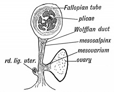
Course of the Ovum in the Tube. — The cilia on the fimbriae work towards the ostium abdominale, the abdominal mouth of the Fallopian tube, which is situated at the bases of the fimbriae, and carry the discharged ovum through the ostium within the tube. The ostium abdominale is shut when the tube is examined after excision ; the closure is probably due to reflex contraction of the tube muscle, caused by handling and cutting. In the infundibular and ampullary segments of the tube, the mucous membrane is thrown into long plicated folds shown in section in Fig. 64. They are covered with ciliated epithelium, which urge the ovum towards the uterus. Within the tube impregnation usually takes place. If an obstruction arrests the fertilized ovum in the tube, perhaps it may be a patch denuded of epithelium and cilia by gonorrheal inflammation, a tubular pregnancy is the result. The growing embryo, commonly about the second month, bursts the tube and falls within the broad ligament or into Douglas's pouch (Fig. 64).
Fig. 64. Diagrammatic section of .the Broad Ligament and Fallopian Tube.
The History of the Ovum within the Fallopian Tube
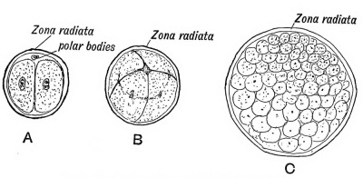
When the ovum enters the Fallopian tube, it is a cell of very considerable size ('2 mm. = -jhs in.) with a cell wall — the zona radiata (Fig. 63), a nucleus — the germinal vesicle, and a nucleolus — the germinal spot. Then, or before then, the ovum prepares for fertilization by the extrusion from its nucleus of first one, then another polar body, and, with the extrusion, the germinal vesicle becomes the female pronucleus. The polar bodies, which lie outside the protoplasm of the ovum, but within the zona radiata, are parts of the germinal vesicle, which are extruded with all the display of karyokinesis (Fig. 65 A). The meaning of the process has been much guessed at ; the outstanding fact is that two parts of the ovum are segregated and are not involved in the subsequent developmental changes. What become of the polar bodies in the course of development of the fertilized ovum is not known.
Fig. 65. Showing the production of the Morula from the Ovum. A. The ovum after the first division. B. After the second. C. The Morula .
In the course of fecundation thousands of spermatozoa are lodged in the genital passage ; many stem the adverse current of the uterine cilia, reach and live for days within the interlaminar grooves in the wider parts of the tube. In the course of its descent within one of the grooves the egg may be fertilized. The spermatozoon bursts through the zona radiata, loses its tail, its head enlarges, and forms the male pronucleus. The male and female pronuclei unite, and from their union springs the nucleus of the fertilized ovum. This is the centre from which all future developmental changes start. The ovum may be, but rarely is, fertilized in the ovary, or between the ovary and ostium abdominale, the result being a pelvic gestation. The length of time the fertilized ovum takes to reach the uterus is not known exactly, but probably it spends about ten or twelve days within the Fallopian tube, and during that time it passes through the following stages :
- The Morula. — The ovum, with a full display of karyokinetic changes in the nucleus, divides, subdivides, and grows within the zona radiata. until a rounded mass of cells is formed — the morula or mulberry mass. The cells' are of unequal size and divide at unequal rates (Fig. 65, A, B, C).
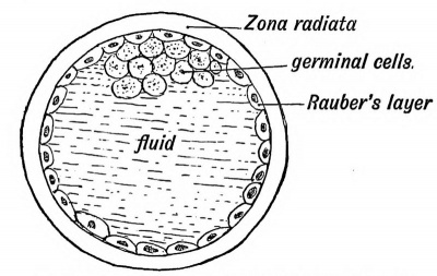
- The Blastodermic Vesicle. — This stage is produced from the morula stage by the collection of fluid within the mass of cells. The cells are then seen to be arranged in two sets— (a) a layer lining the zona radiata and forming the wall of the vesicle (Eauber's layer) (Fig. 66), and (6) a group of granular cells within the cavity attached to Eauber's layer at the embryogenic pole of the vesicle (Fig. 66). In Vertebrates with huge stores of yolk in their ova, such as birds have, the vesicle is filled by yolk-bearing cells, continuous with Eauber's cells at the vegetative pole, opposite to the granular cells.
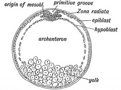
- The Bilaminar Blastoderm. — This stage is produced by the growth of the group of granular germinal cells within Eauber's layer. At first they arrange themselves in two layers — an outer which spreads on the inner aspect of Eauber's layer, and absorbs or mixes with it, to form one layer of more or less columnar cells, the epiblast or ectoderm (Fig. 67); an inner layer also spreads out from the germinal group, and forms a second lining to the blastodermic vesicle, the hypoblast or endoderm (Fig. 67).
Fig. 66. Diagrammatic section of a Blastodermic Vesicle.
Fig. 67. A diagrammatic section of a Bilaminar Blastoderm made across the primitive streak.
The Primitive Streak and Groove
In the diagrammatic section of the bilaminar blastoderm, given in figure 67, the hypoblast and epiblast are seen to be fused at one point, and at the point of fusion the epiblast is thickened and somewhat depressed. When the blastodermic vesicle is viewed from the surface, the line along which the fusion of the two layers and thickening of the epiblast take place is marked by the primitive streak and groove (Fig. 68). At the anterior end of the groove is situated the blastopore or neurenteric canal, an opening into the cavity which the hypoblast encloses — the archenteron (see Figs. 67 and 75). Eound the blastopore and along the primitive streak the epiblast is continuous with the hypoblast.
The Mesoblast
The mesoblast or mesoderm, the third of the primitive blastodermic layers, is produced from the margins of the primitive streak (Fig. 67). At the line of junction of the epiblast and hypoblast there is a free proliferation of cells which spreads out between the two primitive layers, and gives rise to a third or intermediate layer — the mesoblast (Fig. 69). In lower vertebrates the mesoblast is entirely produced from the hypoblast. Neural Canal. — It is at this early stage, while the mesoblastic cells swarm in between the epiblast and hypoblast, and while the vesicle has not yet reached the uterus, that a grooved strip of epiblast, the medullary plate, in front of the primitive streak (Fig. 68) is set aside, by a process of infolding, to form the great central nervous system — the brain, spinal cord, and nerves (Fig. 69).
Fig. 68. Diagram of the Embryogenie area of a Bilaminar Blastoderm viewed from above.
A medullary fold rises up on each side of what is to be the middle line of the back to enclose the medullary plate. The folds rise until their crests meet, and a tube of epiblast is buried by the fusion of their lips. The blastopore is included within the posterior end of the medullary folds (Fig. 68). Thus, for a short space, the cavity of the yolk sac communicates with the neural canal.
The Notochord
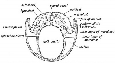
Beneath the neural tube a similar infolding of hypoblast takes place, and a tube of cells to form the notochord is detached (Fig. 69). It forms the first basis of the spinal column.
Fig. 69. Diagrammatic section of a Blastodermic Vesicle showing (1) the origin of the neural canal, (2) the origin of the notochord, (3) the" ingrowth of the mesoblast, and (4) the formation of the coelom.
The Somatopleure and Splanchnopleure
The mesoblast surrounds both the notochord and neural canal as they areformed — perhaps assists in their formation (Figs. 69 and 70). The solid layer which surrounds these structures is known as the paraxial mesoblast. As the mesoblastic cells spread out to cover the outer aspect of the hypoblast and inner aspect of the epiblast, they separate into two layers which enclose between them a cavity — the coelom (Fig. 69). The epiblast, with its accompanying layer of mesoblast, makes up the somatopleure which bounds the coelom without; the hypoblast and overlying layer of mesoblast form the splanchnopleure which bounds the cavity within.
Fig. 70. Diagram of the Blastodermic Vesicle separating into Embryo and Membranes.
Embryo and Membranes
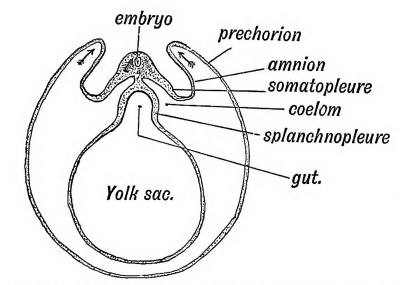
During the second week, while the blastodermic vesicle is still within the Fallopian tube and some time before it has reached the uterus, changes take place whereby part of the blastodermic vesicle forms the embryo and part is transformed into the enclosing membranes which contain, protect, and nourish it (Fig. 70). Part also forms the yolk sac.
The Decidua
When the uterine cavity is reached, the uterine mucous membrane has become hypertrophied, the decidua being thus formed. In the formation of the decidua, the uterine glands become elongated and enlarged ; the inter-glandular tissue hypertrophied and the mouths of the glands closed, so that the surface layer of the decidua is comparatively solid. The hypertrophy of the mucous membrane is unequal, depressions or pits beingformed on its surface. The deeper layer of the decidua is cavernous, the spaces being formed by the distension of capillary veins into venous spaces, and perhaps also by the enlargement of the gland lumina.
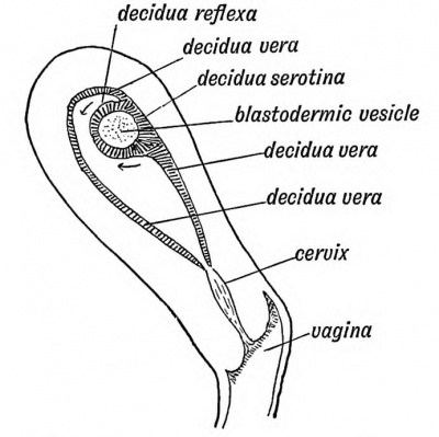
When the blastodermic vesicle reaches the uterus, it commonly adheres to the posterior wall near the fundus (Fig. 71). The decidua forms first a nest for it, and then grows over and buries it, thus forming the outer or decidual membrane of the embryo. The layer which covers the vesicle (see Fig. 71) is the decidua reflexa; the part by which it adheres to the uterus, the decidua serotina; the rest, lining the uterus, the decidua vera. As the vesicle grows, it fills the cavity of the uterus, thus bringing the decidua reflexa in contact with the decidua vera ; the reflexa disappears when it comes in contact with the vera, which remains as the outermost of the foetal membranes. The serotina takes part in the formation of the placenta.
Fig. 71. Section of the Uterus showing the three parts of the Decidua.
The Amnion and Chorion
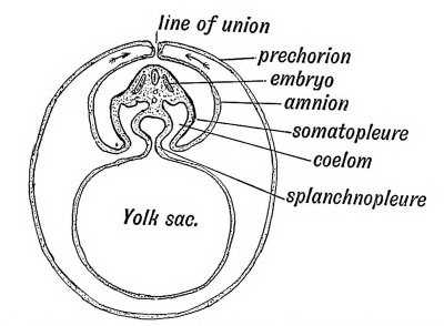
Only part of the somatopleure of the blastodermic vesicle takes part in the formation of the embryo ; the remaining parts of the somatopleure are converted into the amnion and chorion in this manner : As is shown in Fig. 70, the embryonic area appears to sink within the vesicle ; the somatopleure rises up as a wave-like fold right round it, at each side, at the head as well as at the tail end. The folds meet over the embryo and unite, as is shown in Fig. 72. Thus the embryo is wrapped up in a double fold of its own somatopleure. The inner fold or membrane is the Amnion, the outer is the Prechorion (Fig. 72). The prechorion, in turn, is covered by the decidua reflexa, and at its attached part by the decidua serotina. Villi containing vessels grow out from the chorion into the decidua.
Fig. 72. Showing the folds of the somatopleure uniting over the embryo and becoming demarcated into Amnion and Prechorion.
Origin of Ova and Spermatozoa
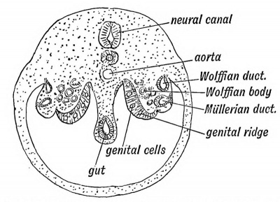
The cells which give rise to the ova and spermatozoa, according to sex, are derived from the mesothelial lining of the coelom. They appear with the cleavage of the mesoblast and formation of the coelom. Soon after the coelom is formed (beginning of third week) a ridge — the genital ridge — is seen on the roof of the coelom, situated on each side of the attachment of the mesentery of the gut (Fig. 73). The ridge lies over the intermediate cell mass (Fig. 85 ; p. 1 11), and is covered by a layer of columnar meso-thelium, the germinal epithelium, which is continuous with the mesothelial lining of the coelom. Some of the columnar cells covering the genital ridge become primordial ova, and are set aside to produce ova or spermatozoa, according to the sex. Beard's researches have led him to the conclusion that primordial ova are produced at a very early stage of development, and that they migrate towards the coelom and take up their position amongst the germinal cells covering the genital ridge when that ridge is formed. These cells may stray and give rise in various parts of the body to dermoid tumours (Beard). The manner in which the germ cells and primordial ova grow within the genital ridge to form the ovary or testicle was described at the beginning of this chapter. The cycle of changes which leads to the production of the ova of one generation from the ovum of a former generation have thus been traced.
Fig. 73. Diagrammatic section of the abdominal region of the coelom, showing the position of the Genital Ridges from which the Ovary or Testicle is formed.
| Historic Disclaimer - information about historic embryology pages |
|---|
| Pages where the terms "Historic" (textbooks, papers, people, recommendations) appear on this site, and sections within pages where this disclaimer appears, indicate that the content and scientific understanding are specific to the time of publication. This means that while some scientific descriptions are still accurate, the terminology and interpretation of the developmental mechanisms reflect the understanding at the time of original publication and those of the preceding periods, these terms, interpretations and recommendations may not reflect our current scientific understanding. (More? Embryology History | Historic Embryology Papers) |
Human Embryology and Morphology (1902): Development or the Face | The Nasal Cavities and Olfactory Structures | Development of the Pharynx and Neck | Development of the Organ of Hearing | Development and Morphology of the Teeth | The Skin and its Appendages | The Development of the Ovum of the Foetus from the Ovum of the Mother | The Manner in which a Connection is Established between the Foetus and Uterus | The Uro-genital System | Formation of the Pubo-femoral Region, Pelvic Floor and Fascia | The Spinal Column and Back | The Segmentation of the Body | The Cranium | Development of the Structures concerned in the Sense of Sight | The Brain and Spinal Cord | Development of the Circulatory System | The Respiratory System | The Organs of Digestion | The Body Wall, Ribs, and Sternum | The Limbs | Figures | Embryology History
Reference
Keith A. Human Embryology and Morphology. (1902) London: Edward Arnold.
Cite this page: Hill, M.A. (2024, April 19) Embryology Book - Human Embryology and Morphology 7. Retrieved from https://embryology.med.unsw.edu.au/embryology/index.php/Book_-_Human_Embryology_and_Morphology_7
- © Dr Mark Hill 2024, UNSW Embryology ISBN: 978 0 7334 2609 4 - UNSW CRICOS Provider Code No. 00098G


