Book - Human Embryology and Morphology 14: Difference between revisions
mNo edit summary |
m (→The Eyeball) |
||
| (2 intermediate revisions by the same user not shown) | |||
| Line 6: | Line 6: | ||
The structures concerned in the sense of sight are : | The structures concerned in the sense of sight are : | ||
# The Eyeball and the Optic Nerve. | |||
# The Eyelids and Lachrymal Apparatus. | |||
# The Orbit, the Muscles, Nerves, and Vessels contained it. | |||
# The Nerve Centres and Tracts. | |||
==The Eyeball== | |||
The condition of the eye in the third week of fetal life is shown diagrammatically in Fig. 141. The three elements which unite to form the eyeball are as yet separate, they are | |||
# Epiblast, which forms (a) the epithelium of the cornea, (b) ie lens, and probably (e) the rods and cones of the retina. | |||
# Neuroblast, which forms (a) the optic nerves, (b) sensitive retina, (c) pars ciliaris retinae, (d) uvea (e) pigmentary layer of retina. | |||
# Mesoblast, which forms (a) outer tunic (sclerotic and fibrous cornea) ; (b) middle tunic (choroid, ciliary- choroid and iris) ; (c) the vitreous humour and its capsule — the hyaloid membrane ; (d) the capsule of the lens. | |||
<div id="Fig141"></div> | |||
[[File:Keith1902 fig141.jpg|400px]] | |||
'''Fig. 141.''' Diagram of the Elements which form the Eyeball. | |||
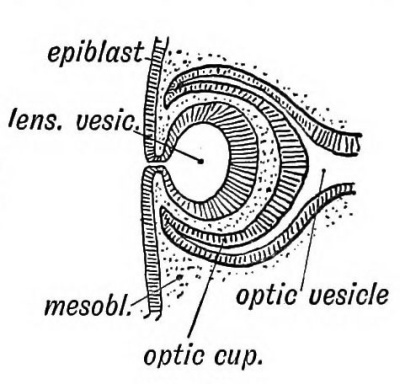
1. Structures derived from the Epiblast. — (a) The lens.— The lens is developed by a saccular invagination of the epiblast situated over the optic vesicle (Kg. 142). It becomes a closed sac by the severance of its connection with the epiblast, its wall being formed by a single layer of epithelial cells. The cavity of the lenticular vesicle is gradually obliterated by the cells of the posterior wall becoming elongated (Fig. 144) until they reach the anterior wall. Each elongated cell is transformed into a is fibre. | 1. Structures derived from the Epiblast. — (a) The lens.— The lens is developed by a saccular invagination of the epiblast situated over the optic vesicle (Kg. 142). It becomes a closed sac by the severance of its connection with the epiblast, its wall being formed by a single layer of epithelial cells. The cavity of the lenticular vesicle is gradually obliterated by the cells of the posterior wall becoming elongated (Fig. 144) until they reach the anterior wall. Each elongated cell is transformed into a is fibre. | ||
<div id="Fig142"></div> | |||
[[File:Keith1902 fig142.jpg|400px]] | |||
'''Fig. 142.''' Invagination of the Epiblast to form the Lena Vesicle. | |||
<div id="Fig143"></div> | |||
[[File:Keith1902 fig143.jpg|400px]] | |||
'''Fig. 143.''' The manner in which the Lens Vesicle is severed from the Epiblast. | |||
<div id="Fig144"></div> | |||
[[File:Keith1902 fig144.jpg|400px]] | |||
'''Fig. 144.''' The Formation of the Lens Fibres from the Epithelium on the posterior Wall of the Vesicle. | |||
Fig. 144. | |||
| Line 70: | Line 56: | ||
<div id="Fig145"></div> | |||
[[File:Keith1902 fig145.jpg|400px]] | |||
Fig. 145. | '''Fig. 145.''' Diagram showing the condition of the Optic Stalk and Vesicle at the commencement of the 2nd month. (After His.) | ||
(5) The pigmentary layer of the retina is formed from the asheathing or outer layer of the optic cup (Fig. 146). At first the wall of the optic vesicle is composed of a single layer of epithelium ; the outer or pigmentary layer of the retina retains lis embryonic form. | |||
<div id="Fig146"></div> | |||
[[File:Keith1902 fig146.jpg|400px]] | |||
'''Fig. 146.''' Diagrammatic Section of the Optic Cup and Lens. | |||
Fig. 146. | |||
| Line 97: | Line 85: | ||
Tie choroidal Fissure. — Occasionally congenital fissures are seen in the lower and outer segment of the iris (coloboma iridis). A white line, due to absence of pigment, may be seen in the corresponding segment of the retina when the interior of the eye is examined. These are due to imperfect closure of the choroidal fissure. The choroidal fissure is the result of the peculiar mode in which the optic vesicle is cupped or invaginated. The lens grows into it from the malar or lower lateral aspect. The lens is lodged in the anterior part of the depression ; the posterior part becomes the choroidal fissure (Fig. 147). The margins of the fissure unite, all traces of it normally disappearing. The margin of the cup left after the union of the lips of the choroidal fissure becomes the boundary of the pupil (Fig. 14*7). | Tie choroidal Fissure. — Occasionally congenital fissures are seen in the lower and outer segment of the iris (coloboma iridis). A white line, due to absence of pigment, may be seen in the corresponding segment of the retina when the interior of the eye is examined. These are due to imperfect closure of the choroidal fissure. The choroidal fissure is the result of the peculiar mode in which the optic vesicle is cupped or invaginated. The lens grows into it from the malar or lower lateral aspect. The lens is lodged in the anterior part of the depression ; the posterior part becomes the choroidal fissure (Fig. 147). The margins of the fissure unite, all traces of it normally disappearing. The margin of the cup left after the union of the lips of the choroidal fissure becomes the boundary of the pupil (Fig. 14*7). | ||
<div id="Fig147"></div> | |||
[[File:Keith1902 fig147.jpg|400px]] | |||
'''Fig. 147.''' The Optic Stalk and Cut), viewed on the lower and lateral aspect, showing the closure of the Choroidul Fissure. | |||
==Binocular Vision== | |||
At first the optic vesicles are directed laterally in the human embryo, and in mammals generally the eyes are so directed, each eye having its own field of vision. In the Primates the eyes swing forwards during the second month; binocular vision is thus made possible. With binocular vision and the combination of images appear in the highest primates : — | |||
# A fovea centralis and macula lutea (L. Johnston) ; | |||
# A partial crossing of the optic fibres at the chiasma ; | |||
# Certain alterations in the attachments of the oblique muscles of the eyeball. | |||
Binocular Vision | |||
| Line 120: | Line 107: | ||
The structures formed from the mesoblast are : ' (1) The Capsule of the lens. — It is developed out of the mesoblast which surrounds the lens. At first the capsule is continuous in front with the basis of the cornea ; behind, it is continuous with the mesoblast of the vitreous humour (Fig. 148). | The structures formed from the mesoblast are : ' (1) The Capsule of the lens. — It is developed out of the mesoblast which surrounds the lens. At first the capsule is continuous in front with the basis of the cornea ; behind, it is continuous with the mesoblast of the vitreous humour (Fig. 148). | ||
<div id="Fig148"></div> | |||
[[File:Keith1902 fig148.jpg|400px]] | |||
Fig. | '''Fig. 148.''' Diagrammatic Section of the Eye showing the Parts formed from the Mesoblast. (After His' Model of the eye of a 3rd mouth human embryo.) | ||
(2) The vitreous humour. — This is formed out of the mesoblast which fills the optic cup behind the lens. The closure of the choroidal fissure cuts the vitreous humour off from the mesoblast which covers the outer layer of the optic cup and becomes transformed into the tunics of the eyeball. The vitreous humour — like Wharton's jelly of the umbilical cord — represents an early form of embryonic tissue. It consists of cells imbedded in a jelly-like matrix. All the connective tissues of the body are originally of this type, and remain as such until the fifth month (Berry Hart). | (2) The vitreous humour. — This is formed out of the mesoblast which fills the optic cup behind the lens. The closure of the choroidal fissure cuts the vitreous humour off from the mesoblast which covers the outer layer of the optic cup and becomes transformed into the tunics of the eyeball. The vitreous humour — like Wharton's jelly of the umbilical cord — represents an early form of embryonic tissue. It consists of cells imbedded in a jelly-like matrix. All the connective tissues of the body are originally of this type, and remain as such until the fifth month (Berry Hart). | ||
| Line 133: | Line 120: | ||
(4) The Aqueous chamber, a space formed in the mesoblast which es between the epiblast of the cornea and the lens (Fig. 148). Part f this mesoblast becomes the anterior capsule of the lens ; part ecomes the connective-tissue basis of the cornea. The aqueous hamber is simply an enlarged lymph space formed between these sro parts. Up to the time of birth the lens lies almost in contact r ith the cornea (Fig. 149). | (4) The Aqueous chamber, a space formed in the mesoblast which es between the epiblast of the cornea and the lens (Fig. 148). Part f this mesoblast becomes the anterior capsule of the lens ; part ecomes the connective-tissue basis of the cornea. The aqueous hamber is simply an enlarged lymph space formed between these sro parts. Up to the time of birth the lens lies almost in contact r ith the cornea (Fig. 149). | ||
<div id="Fig149"></div> | |||
[[File:Keith1902 fig149.jpg|400px]] | |||
'''Fig. 149.''' Section of the Eye and Orbit at birth. | |||
Fig. | |||
| Line 154: | Line 141: | ||
Formation of the Orbit (Fig. 150). — The orbit is formed (1) above by the capsule of the fore-brain in which the frontal bone is developed ; (2) externally and below by the maxillary process (Fig. 1). In the maxillary process the malar bone and superior maxilla (except the ascending nasal process) are developed (Fig. 150). (3) The inner wall is formed by the lateral nasal process, in which the nasals, lachrymals lateral mass of the ethmoid, are formed. The optic nerve enters the orbit between the orbito- and pre-sphenoids- — derivatives of the trabeculae cranii, both of which help to form the orbit. The great wings of the sphenoid are also derived from the trabeculae (p. 168). The orbital plate of the malar cuts the orbit off from the temporal fossa ; it is developed in higher primates only. The nasal duct is formed between the maxillary and nasal processes (Figs. 1 and 150). In lower primates and mammals generally the hamular process of the lachrymal appears on the margin of the orbit ; the pars facialis lachrymalis is sometimes seen in the human skull (Fig 20, p. 26). | Formation of the Orbit (Fig. 150). — The orbit is formed (1) above by the capsule of the fore-brain in which the frontal bone is developed ; (2) externally and below by the maxillary process (Fig. 1). In the maxillary process the malar bone and superior maxilla (except the ascending nasal process) are developed (Fig. 150). (3) The inner wall is formed by the lateral nasal process, in which the nasals, lachrymals lateral mass of the ethmoid, are formed. The optic nerve enters the orbit between the orbito- and pre-sphenoids- — derivatives of the trabeculae cranii, both of which help to form the orbit. The great wings of the sphenoid are also derived from the trabeculae (p. 168). The orbital plate of the malar cuts the orbit off from the temporal fossa ; it is developed in higher primates only. The nasal duct is formed between the maxillary and nasal processes (Figs. 1 and 150). In lower primates and mammals generally the hamular process of the lachrymal appears on the margin of the orbit ; the pars facialis lachrymalis is sometimes seen in the human skull (Fig 20, p. 26). | ||
<div id="Fig150"></div> | |||
[[File:Keith1902 fig150.jpg|400px]] | |||
'''Fig. 150.''' The Origin of the Bones entering into Formation of the Orbit. | |||
Fig. 150. | |||
| Line 170: | Line 155: | ||
The Lachrymal Gland arises as a number of epiblastic buds which spring from the fornix of the conjunctiva beneath the upper lid and grow into the tissue of the outer and upper segment of the orbit. The outer buds form the orbital part of the gland; the more internal buds form the palpebral part. Smaller lachrymal glands may occasionally be found at the •outer angle of the eye. This is the position occupied by the lachrymal glands of birds and reptiles (Wiedershiem). The lachrymal canaliculi and sac and nasal duct are formed out of" solid epithelial cords enclosed between the maxillary and lateral nasal processes (Fig. 151). | The Lachrymal Gland arises as a number of epiblastic buds which spring from the fornix of the conjunctiva beneath the upper lid and grow into the tissue of the outer and upper segment of the orbit. The outer buds form the orbital part of the gland; the more internal buds form the palpebral part. Smaller lachrymal glands may occasionally be found at the •outer angle of the eye. This is the position occupied by the lachrymal glands of birds and reptiles (Wiedershiem). The lachrymal canaliculi and sac and nasal duct are formed out of" solid epithelial cords enclosed between the maxillary and lateral nasal processes (Fig. 151). | ||
<div id="Fig151"></div> | |||
[[File:Keith1902 fig151.jpg|400px]] | |||
'''Fig. 151.''' Diagram of the Plica Semilunaris and Lachrymal Canaliculi. | |||
Fig. 151. | |||
The Orbital Muscles. — We have already seen that the head is composed of nine segments, at least four of these being occipital ; also, that each segment gives rise to a muscle plate. The muscle plate of the first head segment forms the muscles supplied by the third cranial nerve — the motor nerve, of the first cephalic segment. The mesencephalon (crura cerebri) contains the corresponding segment of the neural canal. The ciliary muscle and sphincter of the iris also belong to this segment and are supplied by the 3rd nerve (Fig. 152). The muscle plate of the 2nd head segment produces the superior oblique. In the course of evolution the superior oblique of the right side has shifted to the left and the left to the right (Gaskell), hence the decussation of the 4th nerves (the motor nerves of this segment) on the anterior part of the roof of the hind-brain — the valve of Vieussens. The muscle plate of the third cephalic segment gives rise to the external rectus ; the 6th nerve is the motor nerve of the segment. | The Orbital Muscles. — We have already seen that the head is composed of nine segments, at least four of these being occipital ; also, that each segment gives rise to a muscle plate. The muscle plate of the first head segment forms the muscles supplied by the third cranial nerve — the motor nerve, of the first cephalic segment. The mesencephalon (crura cerebri) contains the corresponding segment of the neural canal. The ciliary muscle and sphincter of the iris also belong to this segment and are supplied by the 3rd nerve (Fig. 152). The muscle plate of the 2nd head segment produces the superior oblique. In the course of evolution the superior oblique of the right side has shifted to the left and the left to the right (Gaskell), hence the decussation of the 4th nerves (the motor nerves of this segment) on the anterior part of the roof of the hind-brain — the valve of Vieussens. The muscle plate of the third cephalic segment gives rise to the external rectus ; the 6th nerve is the motor nerve of the segment. | ||
<div id="Fig152"></div> | |||
[[File:Keith1902 fig152.jpg|400px]] | |||
'''Fig. 152.''' Diagram of the Motor Nerves of the Muscles of the Eye derived from the 1st, 2nd, and 3rd Cephalic Segments. | |||
Fig. 152. | |||
| Line 189: | Line 173: | ||
Development of the Nerve Centres concerned with Sight | ==Development of the Nerve Centres concerned with Sight== | ||
Five parts of the brain are concerned with vision. They are : | |||
# The optic tracts. | |||
# The basal centres surrounding the termination of the aqueduct of Sylvius in the 3rd ventricle. | |||
# The optic radiations. | |||
# The occipital lobes — in part at least. | |||
# The angular gyrus. | |||
bodies and the superior corpora quadrigemina. In these centres the optic fibres end. From some of the cells of these ganglia the efferent fibres of the optic tracts are developed. ~p("2) Tie basal ganglia. — The corpora quadrigemina. — Almost in every structure the human embryonic condition resembles the adult condition of lower vertebrates. A good example is seen in the corpora quadrigemina, The human foetus at the commencement of the third month (Fig. 153) shows the corpora quadrigemina represented by a prominent thickening in the roof of the cavity of the mid-brain, which forms subsequently the aqueduct of Sylvius. The thickening is divided into lateral halves by a median sulcus, each half being nearly as large as the cerebral vesicle of that period. In Fig. 154 is shown the condition in an adult lizard; there is one body on each side — the optic lobes or corpora bigemina. As the human foetus grows older, each lateral lobe becomes divided into an upper and lower part by the formation of a transverse groove, the upper and lower pairs of the corpora quadrigemina being thus formed. The upper pair are connected with sight. In tbe mole they are vestigial, but in compensation the inferior corpora are well developed as they are connected with the sense of hearing, which is very acute in that animal. | (1) The optic tracts are made up of fibres developed from the ganglionic cells of the retina and also in part of efferent fibres developed from cells of the basal ganglia in which the optic tracts are seen to terminate. The fibres grow in by the optic stalk, decussate in the floor of the third ventricle between the origins of the optic vesicles, and thus form the chiasma. The optic fibres grow backwards on the surface of thalamencephalon (see Kg. 153) and on the optic thalamus to reach the nerve centres which afterwards form the pulvinar, geniculate bodies and the superior corpora quadrigemina. In these centres the optic fibres end. From some of the cells of these ganglia the efferent fibres of the optic tracts are developed. ~p("2) Tie basal ganglia. — The corpora quadrigemina. — Almost in every structure the human embryonic condition resembles the adult condition of lower vertebrates. A good example is seen in the corpora quadrigemina, The human foetus at the commencement of the third month (Fig. 153) shows the corpora quadrigemina represented by a prominent thickening in the roof of the cavity of the mid-brain, which forms subsequently the aqueduct of Sylvius. The thickening is divided into lateral halves by a median sulcus, each half being nearly as large as the cerebral vesicle of that period. In Fig. 154 is shown the condition in an adult lizard; there is one body on each side — the optic lobes or corpora bigemina. As the human foetus grows older, each lateral lobe becomes divided into an upper and lower part by the formation of a transverse groove, the upper and lower pairs of the corpora quadrigemina being thus formed. The upper pair are connected with sight. In tbe mole they are vestigial, but in compensation the inferior corpora are well developed as they are connected with the sense of hearing, which is very acute in that animal. | ||
<div id="Fig153"></div> | |||
[[File:Keith1902 fig153.jpg|400px]] | |||
'''Fig. 153.''' Diagram of the Foetal Brain at the end of the 2nd month, showing the Position in which the Optic Tracts are developed. | |||
<div id="Fig154"></div> | |||
[[File:Keith1902 fig154.jpg|400px]] | |||
Fig. 154. | '''Fig. 154.''' Mesial Section of the brain of a Lizard showing the resemblance to the human foetal brain (Fig. 153) especially in the development of the Corpora Bigemina. | ||
| Line 234: | Line 207: | ||
rudiment of occip. lobe . | rudiment of occip. lobe . | ||
<div id="Fig155"></div> | |||
[[File:Keith1902 fig155.jpg|400px]] | |||
'''Fig. 155.''' View of the Mesial Surface of the Brain in the 5th month. | |||
<div id="Fig156"></div> | |||
[[File:Keith1902 fig156.jpg|400px]] | |||
'''Fig. 156.''' Section of the Occipital Lobe at the position marked in Fig. 155. | |||
<div id="Fig157"></div> | |||
[[File:Keith1902 fig157.jpg|400px]] | |||
Fig. 157. | Fig. 157. Mesial Section of the Brain at the 4th week shewing the Rudiment of the Occipital Lobe. (After His.) | ||
culum of the cavity of the vesicle. By the 5th month the occipital lobe has reached far enough back to overlap the cerebellum. | culum of the cavity of the vesicle. By the 5th month the occipital lobe has reached far enough back to overlap the cerebellum. | ||
Latest revision as of 11:42, 13 February 2014
| Embryology - 19 Apr 2024 |
|---|
| Google Translate - select your language from the list shown below (this will open a new external page) |
|
العربية | català | 中文 | 中國傳統的 | français | Deutsche | עִברִית | हिंदी | bahasa Indonesia | italiano | 日本語 | 한국어 | မြန်မာ | Pilipino | Polskie | português | ਪੰਜਾਬੀ ਦੇ | Română | русский | Español | Swahili | Svensk | ไทย | Türkçe | اردو | ייִדיש | Tiếng Việt These external translations are automated and may not be accurate. (More? About Translations) |
Keith A. Human Embryology and Morphology. (1902) London: Edward Arnold.
| Historic Disclaimer - information about historic embryology pages |
|---|
| Pages where the terms "Historic" (textbooks, papers, people, recommendations) appear on this site, and sections within pages where this disclaimer appears, indicate that the content and scientific understanding are specific to the time of publication. This means that while some scientific descriptions are still accurate, the terminology and interpretation of the developmental mechanisms reflect the understanding at the time of original publication and those of the preceding periods, these terms, interpretations and recommendations may not reflect our current scientific understanding. (More? Embryology History | Historic Embryology Papers) |
Chapter XIV. Development of the Structures concerned In the Sense of Sight
The structures concerned in the sense of sight are :
- The Eyeball and the Optic Nerve.
- The Eyelids and Lachrymal Apparatus.
- The Orbit, the Muscles, Nerves, and Vessels contained it.
- The Nerve Centres and Tracts.
The Eyeball
The condition of the eye in the third week of fetal life is shown diagrammatically in Fig. 141. The three elements which unite to form the eyeball are as yet separate, they are
- Epiblast, which forms (a) the epithelium of the cornea, (b) ie lens, and probably (e) the rods and cones of the retina.
- Neuroblast, which forms (a) the optic nerves, (b) sensitive retina, (c) pars ciliaris retinae, (d) uvea (e) pigmentary layer of retina.
- Mesoblast, which forms (a) outer tunic (sclerotic and fibrous cornea) ; (b) middle tunic (choroid, ciliary- choroid and iris) ; (c) the vitreous humour and its capsule — the hyaloid membrane ; (d) the capsule of the lens.
Fig. 141. Diagram of the Elements which form the Eyeball.
1. Structures derived from the Epiblast. — (a) The lens.— The lens is developed by a saccular invagination of the epiblast situated over the optic vesicle (Kg. 142). It becomes a closed sac by the severance of its connection with the epiblast, its wall being formed by a single layer of epithelial cells. The cavity of the lenticular vesicle is gradually obliterated by the cells of the posterior wall becoming elongated (Fig. 144) until they reach the anterior wall. Each elongated cell is transformed into a is fibre.
Fig. 142. Invagination of the Epiblast to form the Lena Vesicle.
Fig. 143. The manner in which the Lens Vesicle is severed from the Epiblast.
Fig. 144. The Formation of the Lens Fibres from the Epithelium on the posterior Wall of the Vesicle.
The cells of the anterior wall retain their primitive form ig. 144). New lens fibres are added by the cells at the margin mator) becoming multiplied and elongated. The lens reaches full size in the 1st year of life and then no more fibres are •med. It will thus be seen that the lens is an area of modified idermis. Like the epidermis, it shows a tendency in the aged be transformed into keratin. The oldest cells (the central or clear fibres) alter first ; . hence the central position of the taract which occurs so frequently in old people.
(b) The cornea. — The epithelial covering of the cornea is ntinuous with the epidermis and in some animals (snakes, etc.) is shed with that structure, rendering the animal blind for the ne being. It becomes transparent. The mesoblast which 3ws in between the lens vesicle and epiblast forms the conctive-tissue basis of the cornea and also the capsule of the lens ig. 148).
(c) It is probable, although not yet verified, that certain cells >m the epiblast grow into the optic vesicle and afterwards form e sensory epithelium of the retina — the rods and cones (G-askell). ifore the incursion of the mesoblast separates them, the optic side and epiblast are in contact. If this is so, then the rods d cones — the sensory cells of the retina, the olfactory cells, the rte cells, the acoustic cells, are all of similar origin — epiblastic.
2. Structures formed from the Optic Vesicles (neuroblastic jment). — (a) The optic nerve is formed out of the stalk of the tic vesicle. The vesicle is well developed at the commencemt of the third week (see Fig. 141) ; even before the medullary ites have quite met to enclose the cavity of the fore-brain the tic vesicles have commenced as evaginations of those plates. iey form a great lateral diverticulum on each side of the ■e-brain — a cavity which becomes the third ventricle in the adult, e condition of the optic nerves at the commencement of the :ond month is shown diagrammatically in Fig. 145. The stalk neck remains constricted while the vesicle enlarges. Invagination of the optic vesicle. — Almost as soon as it begins grow out the optic vesicle becomes invaginated, one half being pushed within the other. It is invaginated by the lens -bud in the same manner as a schoolboy's fist indents a punctured india-rubber ball. The invaginated vesicle is known as the optic cup. The invagination of the vesicle, which takes place in an oblique manner — the pressure being applied from below and behind, leads to the closure not only of the cavity of the vesicle, but also to that of the distal half of the stalk (optic nerve). The mesoblast, surrounding the lens, grows into the invagination and afterwards forms the vitreous humour. The artery, which is folded in with the mesoblast, becomes afterwards the central artery of the retina. Hence the point at which the central artery enters the optic nerve marks the upper limit of the invagination of the optic stalk. By the fourth week the optic vesicle no longer communicates with the cavity of the fore-brain but the recessus opticus, in the floor of the third ventricle, above the chiasma, marks the point at which it entered (Fig. 145). The optic fibres, developed as processes of the neuroblasts of the invaginated layer, grow into the brain from the retina along the optic stalk. They thus form the greater number of the fibres in the optic nerve. The optic fibres also form the chiasma in the floor of the third ventricle and the optic tracts on the wall of the forebrain (Fig. 153). It will thus be seen that the optic nerves and esieles are of the same origin as the cerebral vesicle— both repressing parts of the wall of the fore-brain.
Fig. 145. Diagram showing the condition of the Optic Stalk and Vesicle at the commencement of the 2nd month. (After His.)
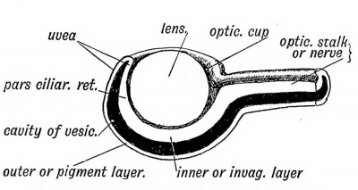
(5) The pigmentary layer of the retina is formed from the asheathing or outer layer of the optic cup (Fig. 146). At first the wall of the optic vesicle is composed of a single layer of epithelium ; the outer or pigmentary layer of the retina retains lis embryonic form.
Fig. 146. Diagrammatic Section of the Optic Cup and Lens.
(c) The uvea is the layer of pigmented epithelium which covers le posterior surface of the iris. It is formed out of both outer id inner layers of the optic cup, and represents the rim of the jp (Fig. 146).
(d) The Pars ciliaris retinae is formed out of that part of the mer or invaginated layer of the optic cup which lies in the ladow of the iris, and is therefore inaccessible to light rays. ; also retains the primitive columnar form of the epithelium. he ora serrata marks the junction of the pars ciliaris retinae id sensitive retina.
Ciliary Processes. — At the commencement of the third month, le pars ciliaris retinae becomes plicated or puckered into 60 or small folds ; mesoblast of the middle tunic (choroid) grows to the puckers and forms the ciliary processes. It should be iserved that the lens lies within the optic cup and the ciliary ■ocesses are formed round the equator or circumference of the
QS.
(«) The Sensitive Retina is formed out of the inner or iniginated layer of the optic cup (Fig. 146). At first the inner wall is composed of a single layer of epithelium. The pars ciliaris retinae retains this form. The cells of the sensitive retina elongate, but the process of formation of rods and cones and other structures in the retina has not been fully followed. If Gaskell is right then the matter is simple. He believes that the epithelial cells of the inner layer of the optic cup become transformed into the fibres of Miiller ; the rods, cones, and ganglionic cells being derived from the cells which grow into the optic vesicle from the epiblast. The ganglionic cells, however, are more probably derivatives of the neuroblasts of the optic vesicle. At any rate an inner layer of ganglionic cells is formed which give off the optic fibres as processes. These fibres converge at the stalk of the vesicle, thus forming the optic disc ; they grow inwards on the stalk, which becomes the optic nerve ; some at least, perhaps all, cross in the floor of the 3rd ventricle forming the chiasma, and pass round, as the optic tracts, to ganglia situated on the mid-brain. There are also efferent fibres in the optic nerve, which end round the ganglion cells of the retina. The inner ganglionic cells of the retina probably correspond to the cells of a posterior root ganglion. According to Gaskell the optic vesicles arose in the ancestry of the vertebrates as diverticula of their alimentary canal ; when the alimentary function of the canal was lost and it became neural, these diverticula became the optic vesicles.
Tie choroidal Fissure. — Occasionally congenital fissures are seen in the lower and outer segment of the iris (coloboma iridis). A white line, due to absence of pigment, may be seen in the corresponding segment of the retina when the interior of the eye is examined. These are due to imperfect closure of the choroidal fissure. The choroidal fissure is the result of the peculiar mode in which the optic vesicle is cupped or invaginated. The lens grows into it from the malar or lower lateral aspect. The lens is lodged in the anterior part of the depression ; the posterior part becomes the choroidal fissure (Fig. 147). The margins of the fissure unite, all traces of it normally disappearing. The margin of the cup left after the union of the lips of the choroidal fissure becomes the boundary of the pupil (Fig. 14*7).
Fig. 147. The Optic Stalk and Cut), viewed on the lower and lateral aspect, showing the closure of the Choroidul Fissure.
Binocular Vision
At first the optic vesicles are directed laterally in the human embryo, and in mammals generally the eyes are so directed, each eye having its own field of vision. In the Primates the eyes swing forwards during the second month; binocular vision is thus made possible. With binocular vision and the combination of images appear in the highest primates : —
- A fovea centralis and macula lutea (L. Johnston) ;
- A partial crossing of the optic fibres at the chiasma ;
- Certain alterations in the attachments of the oblique muscles of the eyeball.
The cavity of the Optic Vesicle (Fig. 146) is of some clinical importance. It is obliterated by the invagination of the vesicle; the rods and cones formed in the inner or invaginated layer grow out across the cavity into the outer or ensheathing pigmented layer of the retina. From accident or disease the retina may be detached; the separation takes place between the pigmented epithelium, which remains in situ, and the rods and cones, which fall inwards with the nerve layer. Fluid then collects in the site of the primitive cavity of the optic vesicle.
3. Parts of the Eyeball formed from the Mesoblast. — As the lens invaginates the optic vesicle and forms the optic cup, it carries in with it the surrounding mesoblast. Thus the lens is surrounded and the cup filled by mesoblast (Fig. 148).
The structures formed from the mesoblast are : ' (1) The Capsule of the lens. — It is developed out of the mesoblast which surrounds the lens. At first the capsule is continuous in front with the basis of the cornea ; behind, it is continuous with the mesoblast of the vitreous humour (Fig. 148).
Fig. 148. Diagrammatic Section of the Eye showing the Parts formed from the Mesoblast. (After His' Model of the eye of a 3rd mouth human embryo.)
(2) The vitreous humour. — This is formed out of the mesoblast which fills the optic cup behind the lens. The closure of the choroidal fissure cuts the vitreous humour off from the mesoblast which covers the outer layer of the optic cup and becomes transformed into the tunics of the eyeball. The vitreous humour — like Wharton's jelly of the umbilical cord — represents an early form of embryonic tissue. It consists of cells imbedded in a jelly-like matrix. All the connective tissues of the body are originally of this type, and remain as such until the fifth month (Berry Hart).
(3) The hyaloid artery. — This is the vessel which supplies the mesoblast of the optic cup ; it terminates in the capsule of the lens (Fig. 148). In the 7th month foetus a trace of the artery can still be seen passing through the vitreous humour from the optic disc to the lens. With the gradual obliteration of the artery, the ipsule of the lens becomes thin and clears up. A foetus born ia le seventh month is blind, because of the vascular and opaque ipsule of the lens. The anterior part of the capsule— filling the upil— is the membrana pupillaris. The part of the hyaloid rtery within the optic nerve persists as the central artery of the 3tina. The canal of the artery within the vitreous humour, from le optic disc to the lens, remains as the hyaloid canal — a lymph ath. The hyaloid artery may persist and cause partial or complete blindness. It disappears some days after birth in cats and rabbits.
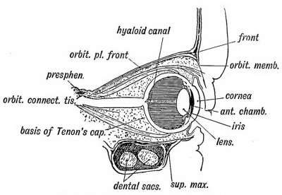
(4) The Aqueous chamber, a space formed in the mesoblast which es between the epiblast of the cornea and the lens (Fig. 148). Part f this mesoblast becomes the anterior capsule of the lens ; part ecomes the connective-tissue basis of the cornea. The aqueous hamber is simply an enlarged lymph space formed between these sro parts. Up to the time of birth the lens lies almost in contact r ith the cornea (Fig. 149).
Fig. 149. Section of the Eye and Orbit at birth.
(5) The choroid, ciliary processes, and iris. — These form the iddle or vascular tunic of the eye, and are developed out of the esoblast which covers the optic cup. They form a vascular and gmented covering through which the optic cup is nourished, le ciliary muscle is formed in this tunic.
(6) The sclerotic. — -This is the outer covering or tunic of niesoblast. It is continuous in front with the cornea ; behind with the optic nerve sheath. In some vertebrates, but not in mammals, plates of bone are developed in the anterior half of the sclerotic.
The Tapetum lucidum is absent in the human and primate eye. It gives the metallic lustre seen on the retinal surface of the eye of the ox, and is formed by a layer of fine fibres which are developed on the retinal surface of the choroid.
(7) The Capsule of Tenon, the bursa or connective-tissue socket of the eyeball, and is developed in the mesoblast surrounding the eyeball. A lymph space separates it from the sclerotic, which is but slightly marked until after birth. The choanoid muscle (retractor bulbi or Midler's muscle) which surrounds the posterior part of the eyeball as a muscular hood in mammals and vertebrates generally, has disappeared in man and the higher primates. Probably its fibrous remnant helps to form the capsule of Tenon.
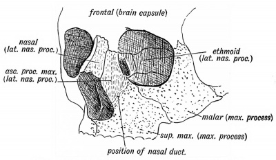
Formation of the Orbit (Fig. 150). — The orbit is formed (1) above by the capsule of the fore-brain in which the frontal bone is developed ; (2) externally and below by the maxillary process (Fig. 1). In the maxillary process the malar bone and superior maxilla (except the ascending nasal process) are developed (Fig. 150). (3) The inner wall is formed by the lateral nasal process, in which the nasals, lachrymals lateral mass of the ethmoid, are formed. The optic nerve enters the orbit between the orbito- and pre-sphenoids- — derivatives of the trabeculae cranii, both of which help to form the orbit. The great wings of the sphenoid are also derived from the trabeculae (p. 168). The orbital plate of the malar cuts the orbit off from the temporal fossa ; it is developed in higher primates only. The nasal duct is formed between the maxillary and nasal processes (Figs. 1 and 150). In lower primates and mammals generally the hamular process of the lachrymal appears on the margin of the orbit ; the pars facialis lachrymalis is sometimes seen in the human skull (Fig 20, p. 26).
Fig. 150. The Origin of the Bones entering into Formation of the Orbit.
The Eyelids are formed by folds or ridges of epiblast which rise above and below the cornea. Mesoblast grows into the folds and forms the tarsal plates. The upper eyelid is formed from the capsule of the fore-brain, the lower from the maxillary process. The epiblast on the deep surface of the folds forms the palpebral conjunctiva. It is continuous with the epiblast of the cornea. The lid-folds meet in front of the cornea during the third month and adhere by their edges. The edges become again separated about the 7th month. From the epiblast between their adherent edges, buds grow into the lids and form the cilia, Meibomian and other glands in the same manner as hairs and sweat glands are developed.
The plica semilunaris (Fig. 151), a fold of conjunctiva in the inner canthus of the eye, is a vestige of the third eyelid (membrana nictitans) which is so fully developed in birds and reptiles. It is well seen in the cat, partially crossing the cornea as the lids are shut. The lachrymal papillae rub in the grooves at the outer and inner margins of the fold.
The Lachrymal Gland arises as a number of epiblastic buds which spring from the fornix of the conjunctiva beneath the upper lid and grow into the tissue of the outer and upper segment of the orbit. The outer buds form the orbital part of the gland; the more internal buds form the palpebral part. Smaller lachrymal glands may occasionally be found at the •outer angle of the eye. This is the position occupied by the lachrymal glands of birds and reptiles (Wiedershiem). The lachrymal canaliculi and sac and nasal duct are formed out of" solid epithelial cords enclosed between the maxillary and lateral nasal processes (Fig. 151).
Fig. 151. Diagram of the Plica Semilunaris and Lachrymal Canaliculi.
The Orbital Muscles. — We have already seen that the head is composed of nine segments, at least four of these being occipital ; also, that each segment gives rise to a muscle plate. The muscle plate of the first head segment forms the muscles supplied by the third cranial nerve — the motor nerve, of the first cephalic segment. The mesencephalon (crura cerebri) contains the corresponding segment of the neural canal. The ciliary muscle and sphincter of the iris also belong to this segment and are supplied by the 3rd nerve (Fig. 152). The muscle plate of the 2nd head segment produces the superior oblique. In the course of evolution the superior oblique of the right side has shifted to the left and the left to the right (Gaskell), hence the decussation of the 4th nerves (the motor nerves of this segment) on the anterior part of the roof of the hind-brain — the valve of Vieussens. The muscle plate of the third cephalic segment gives rise to the external rectus ; the 6th nerve is the motor nerve of the segment.
Fig. 152. Diagram of the Motor Nerves of the Muscles of the Eye derived from the 1st, 2nd, and 3rd Cephalic Segments.
The sensory nerves of these three segments are fused together to form the ophthalmic division of the 5th nerve. The ciliary ganglion is the splanchnic (sympathetic) ganglion of the first segment (see Tig. 180, p. 220). The nerves for the choanoid (Miiller's) muscle, the non-striated muscle of the upper eyelid, and the dilator fibres of the iris, issue from the upper three dorsal segments of the spinal cord, and reach the eye by the cervical sympathetic chain and cavernous plexus. The nerve fibres for the orbicularis palpebrarum pass out with the facial, but they arise from cells in the first segment of the neural canal (oculomotor nucleus). The ophthalmic division of the fifth nerve represents the sensory part of the 1st segment ; hence the reflection of pain along this nerve (frontal headache) in disorders of accommodation, the muscle of accommodation being the ciliary, and its nerve, the oculo-motor, both also derivatives of the first segment.
Development of the Nerve Centres concerned with Sight
Five parts of the brain are concerned with vision. They are :
- The optic tracts.
- The basal centres surrounding the termination of the aqueduct of Sylvius in the 3rd ventricle.
- The optic radiations.
- The occipital lobes — in part at least.
- The angular gyrus.
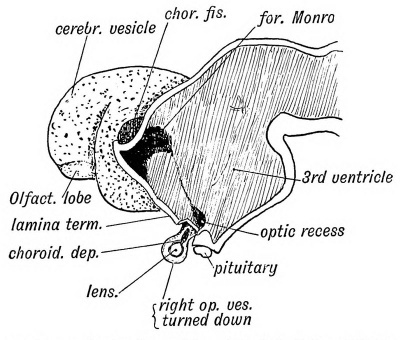
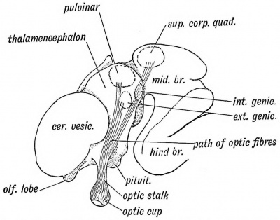
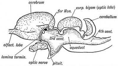
(1) The optic tracts are made up of fibres developed from the ganglionic cells of the retina and also in part of efferent fibres developed from cells of the basal ganglia in which the optic tracts are seen to terminate. The fibres grow in by the optic stalk, decussate in the floor of the third ventricle between the origins of the optic vesicles, and thus form the chiasma. The optic fibres grow backwards on the surface of thalamencephalon (see Kg. 153) and on the optic thalamus to reach the nerve centres which afterwards form the pulvinar, geniculate bodies and the superior corpora quadrigemina. In these centres the optic fibres end. From some of the cells of these ganglia the efferent fibres of the optic tracts are developed. ~p("2) Tie basal ganglia. — The corpora quadrigemina. — Almost in every structure the human embryonic condition resembles the adult condition of lower vertebrates. A good example is seen in the corpora quadrigemina, The human foetus at the commencement of the third month (Fig. 153) shows the corpora quadrigemina represented by a prominent thickening in the roof of the cavity of the mid-brain, which forms subsequently the aqueduct of Sylvius. The thickening is divided into lateral halves by a median sulcus, each half being nearly as large as the cerebral vesicle of that period. In Fig. 154 is shown the condition in an adult lizard; there is one body on each side — the optic lobes or corpora bigemina. As the human foetus grows older, each lateral lobe becomes divided into an upper and lower part by the formation of a transverse groove, the upper and lower pairs of the corpora quadrigemina being thus formed. The upper pair are connected with sight. In tbe mole they are vestigial, but in compensation the inferior corpora are well developed as they are connected with the sense of hearing, which is very acute in that animal.
Fig. 153. Diagram of the Foetal Brain at the end of the 2nd month, showing the Position in which the Optic Tracts are developed.
Fig. 154. Mesial Section of the brain of a Lizard showing the resemblance to the human foetal brain (Fig. 153) especially in the development of the Corpora Bigemina.
The internal geniculate body also belongs to the mid-brain (mesencephalon) ; the pulvinar and external geniculate body, in which the upper division of the optic tract ends, are developed on the wall of the 3rd ventricle (thalamencephalon).
(3) Tbe optic radiations connect the basal optic centres just named with the mesial surface of the occipital lobes. The fibres join the posterior part of the internal capsule and pass under and round the posterior horn of the lateral ventricle to end in the cortex of the calcarine fissure and neighbourhood.
(4) The occipital lobe and calcarine fissure. — A mesial view of the 5th month foetal brain is shown in Fig. 155. The occipital lobe is already well formed ; its inner aspect shows the calcarine and parieto-occipital fissures. A section across the occipital lobe is shown in Fig. 156 ; the posterior horn is large; the calcarine fissure indents its inner wall, giving rise to the calcar avis or hippocampus minor.
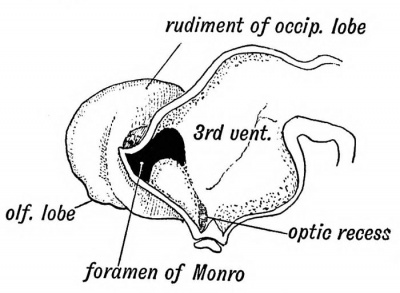
The calcarine is one of the first fissures to appear on the brain ; it appears early in the fifth month. It is present in all primates except the very lowest. The optic radiations end in the cortex of the fissure. In Fig. 157 the condition of the occipital lobe in the 4th week is shown. The cerebral vesicle has arisen as a hollow protrusion from the anterior superior end of the fore-brain (3rd ventricle). The lateral ventricle is as yet undifferentiated into horns and only the rudiment of the occipital lobe is present. The occipital lobe is produced by a backward growth of the cerebral vesicle, the posterior horn being produced as a diverti
rudiment of occip. lobe .
Fig. 155. View of the Mesial Surface of the Brain in the 5th month.
Fig. 156. Section of the Occipital Lobe at the position marked in Fig. 155.
Fig. 157. Mesial Section of the Brain at the 4th week shewing the Rudiment of the Occipital Lobe. (After His.)
culum of the cavity of the vesicle. By the 5th month the occipital lobe has reached far enough back to overlap the cerebellum.
(5) The Angular Gyrus is connected with the calcarine region by association fibres. In it is seated the word-seeing and wordunderstanding centre. It is developed round the posterior end of the 1st temporal or parallel fissure (Fig. 176, p. 214). It is part of the wall of the cerebral vesicle. The first temporal or parallel fissure appears during the sixth month and is one of the primary fissures. It is found in the brains of all primates except the lowest.
Summary. — It will thus be seen that three parts of the neural tube are specialized in connection with sight.
(1) The optic vesicle, an outgrowth from the fore-brain (thalamencephalon).
(2) The occipital end of the cerebral vesicle, which may also be regarded as an outgrowth from the thalamencephalon.
(3) The walls of the 3rd ventricle (thalamencephalon) and mid-brain (mesencephalon), in which the basal optic ganglia are developed.
| Historic Disclaimer - information about historic embryology pages |
|---|
| Pages where the terms "Historic" (textbooks, papers, people, recommendations) appear on this site, and sections within pages where this disclaimer appears, indicate that the content and scientific understanding are specific to the time of publication. This means that while some scientific descriptions are still accurate, the terminology and interpretation of the developmental mechanisms reflect the understanding at the time of original publication and those of the preceding periods, these terms, interpretations and recommendations may not reflect our current scientific understanding. (More? Embryology History | Historic Embryology Papers) |
Human Embryology and Morphology (1902): Development or the Face | The Nasal Cavities and Olfactory Structures | Development of the Pharynx and Neck | Development of the Organ of Hearing | Development and Morphology of the Teeth | The Skin and its Appendages | The Development of the Ovum of the Foetus from the Ovum of the Mother | The Manner in which a Connection is Established between the Foetus and Uterus | The Uro-genital System | Formation of the Pubo-femoral Region, Pelvic Floor and Fascia | The Spinal Column and Back | The Segmentation of the Body | The Cranium | Development of the Structures concerned in the Sense of Sight | The Brain and Spinal Cord | Development of the Circulatory System | The Respiratory System | The Organs of Digestion | The Body Wall, Ribs, and Sternum | The Limbs | Figures | Embryology History
Reference
Keith A. Human Embryology and Morphology. (1902) London: Edward Arnold.
Cite this page: Hill, M.A. (2024, April 19) Embryology Book - Human Embryology and Morphology 14. Retrieved from https://embryology.med.unsw.edu.au/embryology/index.php/Book_-_Human_Embryology_and_Morphology_14
- © Dr Mark Hill 2024, UNSW Embryology ISBN: 978 0 7334 2609 4 - UNSW CRICOS Provider Code No. 00098G


