Book - Human Embryology and Morphology 11: Difference between revisions
(Created page with "{{Human embryology morphology 1902 header}} {{Human embryology morphology 1902 footer}}") |
|||
| (3 intermediate revisions by the same user not shown) | |||
| Line 1: | Line 1: | ||
{{Human embryology morphology 1902 header}} | {{Human embryology morphology 1902 header}} | ||
=Chapter XI. The Spinal Column and Back= | |||
==The Pyramids of the Spine== | |||
The spine, when viewed from the front, is seen to be made up of four pyramids : (1) Cervical ; (2) upper dorsal ; (3) dorso-lumbar ; (4) sacro-coccygeal (Fig. 113). | |||
<div id="Fig113"></div> | |||
[[File:Keith1902 fig113.jpg|400px]] | |||
'''Fig. 113.''' Diagram of the Pyramids of the Spine. | |||
The bases of the two upper pyramids meet at the disc between the 7th cervical and 1st dorsal vertebrae ; the bases of the lower two at the disc between the 5th lumbar and 1st sacral vertebrae. The apices of the two middle pyramids meet at the disc between the 4th and 5 th dorsal vertebrae, which have therefore the narrowest bodies of the vertebral series. The narrowing in the upper dorsal region is due to the fact that the weight of the upper half of the trunk is partly borne by, and transmitted to, the lower dorsal region by the sternum and ribs which thus relieve the spine to some extent (Fig. 113). At the sacrum the weight is transferred to the pelvis and lower limbs, hence the rapid diminution of the sacrum and coccyx. A well-marked thickening or bar in each ilium runs from the auricular surface to the acetabulum and transmits the weight to the femora. | |||
==The Curves of the Spinal Column== | |||
There is only one curve — an anterior concavity — until the 3rd month (Fig. 114 A). About the beginning of the 4th month the sacro-vertebral angle forms between the lumbar and sacral regions (114 £). At birth the cervical and sacral curves have appeared, but the sacral not to a pronounced extent (Fig. 114 C). The lumbar curve appears as the child learns to walk. It is produced to allow the body to be brought vertically over the lower extremities. The sacral and cervical curves also become then more marked (Fig. 114 D). The dorsal curvature and the sacro-vertebral angle are the primitive curves and are present in all mammals. The others are adaptations to the upright posture. The lumbar curve is most pronounced in the highly civilized races. | |||
<div id="Fig114"></div> | |||
[[File:Keith1902 fig114.jpg|400px]] | |||
'''Fig. 114.''' Diagram of the Curves of the Spinal Column. A. At the 6th week of foetal life. B. At the 4th month of foetal life. C. Curves present at Birth. D. Curves present in the Adult. | |||
==Proportion of Cartilage and Bone== | |||
The inter-vertebral discs form one third of the total height of the spine ; the proportion of cartilage is greater in the lumbar than in the dorsal region and greater in the dorsal than in the cervical. The curvatures are due chiefly to the shape of the discs. In the lumbar region, which is convex forwards, only the lower three vertebrae are deeper in front than behind. This is true only for the higher races of mankind, for as Cunningham has shown, in lower races, as in the gorilla, only the lowest lumbar vertebra is deeper in front than behind, and thus helps to maintain the lumbar curvature. | |||
==Unstable Regions of the Spine== | |||
In about 90% of men there are 7 cervical, 1 2 dorsal, 5 lumbar, 5 sacral, and 4 caudal vertebrae, making 33 in all. In the remaining 10% there is some departure from the normal arrangement and these departures affect certain definite regions. | |||
I. The sacro-lumbar. — The 25th vertebra in 95% of people forms the 1st sacral; in 1% the 24th, and 3% the 26th. | |||
<div id="Fig115"></div> | |||
[[File:Keith1902 fig115.jpg|400px]] | |||
'''Fig. 115.''' A section of the Lumbo-sacral Region of the Spine in a Foetus at the end of the 2nd month, showing the 26th vertebra forming the 1st Sacral. (After Rosenberg.) | |||
These percentages are drawn from the observations of Paterson, Eosenberg, and others who have made researches on this subject. The vertebral formula is not fixed. Eosenberg's investigations showed (Fig. 115) that it is the 26th vertebra that forms the first of the sacral series in the early embryo; later the 25th throws out great lateral masses, and thus forms a connection with the ilia. In the lower primates (monkeys) the 27 th forms the 1st sacral; with the evolution of man the 26th, then the 25th underwent sacral modifications, the trunk being correspondingly shortened. It will be seen that the number is not yet definitely fixed. The anterior point of attachment of the ilium fluctuates from the 24th to the 26th vertebra in man. With the sacral transformation of the 25 th and 26 th (lumbar) vertebrae, there was a corresponding movement forwards of the sacral plexus. | |||
II. Sacro-coccygeal. — The 30 th vertebra forms the 1st coccygeal ; not uncommonly this vertebra is sacral in type and forms part of the sacrum. | |||
III. Dorso-lumbar region. — This region is less liable to variation ; the 20 th vertebra instead of forming the 1st lumbar, may simulate the last dorsal in the type of its articular processes, and may bear ribs, probably an atavistic form, or, on the other hand, the 12th dorsal vertebra (19th) may not carry ribs. About 2 °/ of bodies show variations of this kind. | |||
IV. Dorso-cervical. — The 7th vertebra may carry ribs ; rarely the 8th vertebra has no ribs attached to it and is cervical in typeEvolution and Development of the Spinal Column. — -The human spinal column in the process of development passes through three distinct phases : | |||
(1) It is membranous; (2) it becomes cartilaginous; (3) it becomes bony. In the evolution of vertebrates the same three stages are observed. In Amphioxus and Marsipobranchs (excepting the neural arches), the spinal column is membranous ; in Elasmobranchs it is cartilaginous ; in other fishes and in all other vertebrates it is ossified. In the human embryo, as in every other vertebrate, the spinal column is developed from the mesoblast which surrounds the notochord and neural canal. | |||
==The Notochord== | |||
It has already been seen that the notochord is formed at a very early stage (before the 3rd week, Fig. 69, p. ,90) by a tubular invagination of the hypoblast under the neural canal. The notochord, with the mesoblastic tissue round it, represents the most primitive type of spinal support. It is hollow— the canal of the notochord runs from end to end, and into its posterior end the neurenteric canal opens. Afterwards it becomes a solid rod composed of cells of a peculiar type. A sheath is formed round the notochord by cells of the paraxial mesoblast (Fig. 116), which grow inwards and surround it. | |||
<div id="Fig116"></div> | |||
[[File:Keith1902 fig116.jpg|400px]] | |||
'''Fig. 116.''' Diagrammatic transverse section of a human embryo at the end of the 3rd week. | |||
These cells form the sclerotome and spring from the inner parts of the primitive segments (Fig. 116). At the same time the cells of the sclerotome also surround the neural tube. From these cells which grow inwards and surround the notochord and neural canal, the spinal column is formed and also the basioccipital and part of the basi-sphenoid bones of the skull (Fig. 117). | |||
===What becomes of the Notochord=== | |||
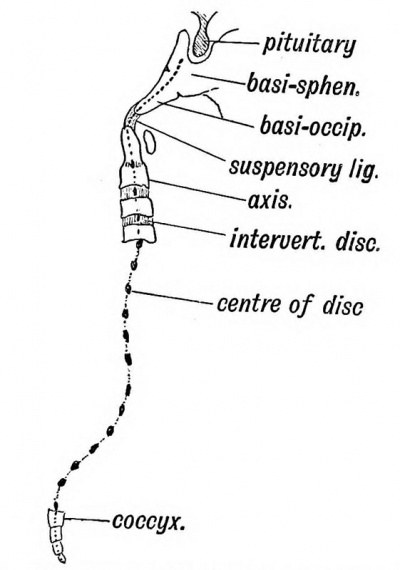
In the second month of foetal life the notochord begins to disappear ; the bodies of the vertebrae and parachordal cartilages form in its sheath and constrict it. The parachordal cartilages form the basi-occipital and basi-sphenoid — the basal part of the skull — behind the i pituitary fossa. As they form, the notochord is obliterated \ between them. Eternod, however, found the anterior part of the notochord on the dorsal wall of the pharynx in the human embryo, so that the parachordal cartilages are evidently developed on its dorsal aspect. The odontoid process represents the body of the atlas and the suspensory ligament the disc between the occipital bone and atlas. A remnant of the notochord is enclosed in the suspensory ligament. The body of each vertebra is formed round the notochord and at first each has an hour-glass canal surrounding the notochord. Within each body the notochord ultimately disappears but in the intervertebral discs it swells out and forms much of the central mucoid core which each disc contains. The notochord with its membranous sheath is the earliest form of spinal column known. The real vertebral column, formed out of its sheath, begins to supplant it even in low vertebrates and in the human foetus of the second month this change is also seen to take place. | |||
<div id="Fig117"></div> | |||
[[File:Keith1902 fig117.jpg|400px]] | |||
'''Fig. 117.''' Where Remnants of the Notochord may occur in the Adult. | |||
==Proto-vertebrae or Primitive Segments== | |||
Proto-vertebrae are not the forerunners of the vertebrae ; they are the primitive segments into which the mass of mesoblast at each side of the neural canal and notochord divides (Figs. 233, p. 289 and 116). The process of division or segmentation begins at the occipital region towards the end of the second week and spreads backwards until 3 5 or more body segments or somites are cut off. Each segment thus separated forms its own muscles (from its muscle plate or myotome), has its own nerve (spinal nerve), its own artery (inter-costal), and the basis for its skeletal tissue (sclerotome). The inter-segmental septum separates one proto-vertebra or segment from another. Eibs, transverse and spinous processes, are formed in the inter-segmental septa. Hence an intercostal space with its muscles, vessels, and nerves, with the corresponding intervertebral structures, represents a differentiated proto-vertebra. In the ventral aspect of the neck and loins, the inter-segmental septa disappear. In the head nine segments are recognised, but their recognition rests on observations made, not on the human embryo, but on the embryoes of lower vertebrates. | |||
Recent work on the segmental arrangements of the nerves gives a practical importance to the number and position of the primitive segments and to the part of the body which each forms. Although the epiblast, hypoblast, and walls of the coelpui never show any trace of segmentation in the embryo, yet clinically there is evidence that each part belongs to, and was formed from, one definite segment. The upper extremity grows out from the 5th cervical to the 2nd dorsal segments (7 segments in all), and the lower from the 2nd lumbar to the 3rd sacral (7 segments in all). In each limb traces of these seven segments are to be found in the nerve distribution. | |||
==Development of a Typical Vertebra, — the 6th Dorsal== | |||
===Membranous Stage=== | |||
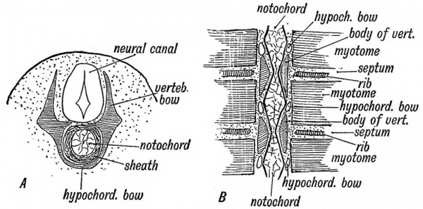
(3rd and 4th weeks). The vertebra then consists of 1st a body surrounding the notochord, formed from the sheath (Fig. 118 A), and 2nd a horse-shoe shaped vertebral bow (Fig. 1 1 8 A and B). The bow consists of a hypo-chordal part, and two lateral limbs, united by the hypo-chordal part ventral to the body. | |||
===Cartilaginous Stage=== | |||
(Fig. 119). It commences in the 4th week. The fibrous basis is transformed into cartilage except the hypochordal part of the bow. It should be noticed (Fig. 118 -B) that the vertebral bodies are formed round the notochord, opposite each inter-segmental septum. Hence each vertebra belongs to two segments. The inter-vertebral disc is situated opposite the middle of a segment. The hypo-chordal part of the bow lies also in the segment, and becomes included in the course of development in the disc in front of the vertebra to which it belongs. The lateral limbs of the cartilaginous bow meet behind (dorsal to) the neural canal in the 4th month, and thus the bow is converted into a ring. The atlas is a permanent representative | |||
<div id="Fig118"></div> | |||
[[File:Keith1902 fig118.jpg|600px]] | |||
'''Fig. 118.''' The development of the Membianous Basis of a Vertebra A. In transverse section. B. In horizontal section showing the relation of th. vertetea to the Pnrmtive Segments. The section is viewed from 'the dorsal of the ring thus formed. In the atlas only does the hypo-chordal part of the bow become cartilaginous and subsequently ossified ; in all the other vertebrae it remains as a fibrous strand in the intervertebral disc. | |||
<div id="Fig119"></div> | |||
[[File:Keith1902 fig119.jpg|600px]] | |||
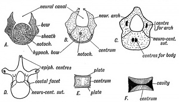
'''Fig. 119.''' Showing the Stages in the Development of a Vertebra. A. In the Membranous Stage. B. In the Cartilaginous Stage. C. The appearance of Ossific Points. D. The appearance of Secondary Ossific Centres, is 1 . The epiphyseal plates of the centra. /•'. Section of an amphicoelous vertebra. | |||
===Bony Stage=== | |||
The body and bow parts of the cartilaginous vertebra fuse and give rise to the condition shown in Fig. 119 C. In the 7th week two centres appear in the body, but quickly fuse ; one appears in each neural arch (8th week) ; at birth the ossific centres of the body and neural arch have met. The central and neural ossifications meet at the neuro-central suture and unite at the 4th or 5th year. The neural ossifications fuse behind (where the spinous process is produced) in the 1st year. The ribs are entirely supported from the neural ossifications (Fig. 119 B). The spinous and transverse processes are formed by outgrowths of cartilage into the septa between the proto- vertebrae or primitive segments. The ribs are also formed in the septa, each at first articulating with a vertebra, which is also inter-segmental in origin. In all the ribs, except the 1st, 11th and 12 th the head is shifted to the inter-vertebral disc in front of its vertebra. Epiphyseal centres for the ossification of the transverse and spinous processes appear about puberty. | |||
The Bodies of Mammalian Vertebrae are peculiar (1) in the development of an upper and lower epiphyseal plate ; (2) in that no trace of the notochord remains within them. In Fishes, as in the early human or mammalian foetus, the bodies are hour-glass shaped (amphicoelous, Fig. 119 F) ; in Amphibians they may retain a concavity in front (procoelous) or behind (opisthocoelous), but in mammals both ends are filled up. | |||
<div id="Fig120"></div> | |||
[[File:Keith1902 fig120.jpg|400px]] | |||
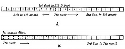
'''Fig. 120.''' The Order in which the Centres of Ossification appear in the Bodies (A) and in the Neural Arches (B) of the Spinal Column. | |||
It will be observed (Fig. 120 B) that the centres of ossification for the neural arches appear first in the anterior end of the spine (1st cervical), the date becoming later the more posterior the vertebra. In the 1st sacral they appear about the 4th month; in the 2nd sacral in the 5th month or later ; in the 3rd they may not appear. In the 4th and 5th sacral and 1st coccygeal vertebrae only vestiges of the neural arches are formed. These vertebrae retain the early foetal type shown in figure 119 B. In the remaining coccygeal vertebrae only the centres for the bodies appear. The centres for ossification of the bodies of the vertebrae appear first in the mid dorsal region (6th dorsal). From that point they spread forwards and backwards, the centres for the odontoid process appearing at the 4th month and that for the 5th sacral at the 5th month, while the last coccygeal does not appear until about the 20th or 25th year (Fig. 120 A). | |||
==The Atlas and Axis== | |||
The atlas represents the completed bow of the 1st cervical vertebra. The body of the vertebra fuses with the body of the 2nd, and forms the odontoid process. A remnant of the disc between the 1st and 2nd vertebrae can sometimes be seen when the odontoid is split open. The suspensory ligament is the representative of the disc between the last occipital segment and the 1st cervical (Fig. 121). | |||
<div id="Fig121"></div> | |||
[[File:Keith1902 fig121.jpg|400px]] | |||
'''Fig. 121.''' A diagrammatic section of the Foetal Axis, Atlas, and Basi-occipital. | |||
===Occipito-atlanto-axial Articulations=== | |||
In the intervertebral discs of the cervical region there is at each side, between the lateral lips of the vertebral bodies, a small articular cavity (Fig. 122). It is situated between the parts of the body formed from the neural arches and in front of (ventral to) the issuing spinal nerves. Between the axis and atlas this articulation is greatly enlarged. At it the rotatory movements of the atlas on the axis take place. The atlanto-occipital joint, which separates the atlas and the last occipital segment is of the same nature. The atlas has neither the upper nor the lower articular processes of the other vertebrae. Hence the 1st and 2nd cervical nerves appear to issue behind the articular processes. At one time the single median occipital condyle seen in birds and reptiles was regarded as very different in nature from the double condyles of mammals. Recently Symington has shown that in the lowest mammals (monotremes), the occipital condyles are fused in the middle line, and that foetal mammals show approximations to this condition. In the human skull a remnant of this median fusion of the condyles is frequently seen on the anterior margin of the foramen magnum ; it is named the third or median occipital condyle. Sometimes the atlas is partly fused with or imperfectly separated from the occipital bone. This seems to be a further manifestation of the process which has led to four segments, which were at one time free body segments, being fused together to form the occipital part of the skull. | |||
<div id="Fig122"></div> | |||
[[File:Keith1902 fig122.jpg|400px]] | |||
'''Fig. 122.''' The nature of the Atlanto-axio-occipital Articulations. | |||
The ribs are developed in the septa between the dorsal primitive segments. At their vertebral ends they come in contact with the vertebral bow (Fig. 118 B). In lower vertebrates (birds, reptiles, etc.) each rib has two heads, a dorsal and ventral (Fig. 123). The tuberosity of the human rib represents the dorsal head ; the ventral head is well developed in man, as in mammals generally. The rib articulates with the neural arches only (Fig. 119 D). | |||
==Vestigial Ribs== | |||
Although the ribs are only fully developed in the dorsal region, yet a representative — a costal element — is present in every vertebra. In the cervical vertebrae the anterior part of the transverse processes represents a costal process, but only in the 6 th (sometimes) and 7 th is the costal process formed by a separate- centre of ossification. The costal process of the 7 th may develop into a rudiment or even a fully formed rib which reaches the sternum. In the lumbar vertebrae only the first shows a separate centre for the formation of the costal process ; it fuses with the tip of the transverse process in the later months of foetal life ; in the other lumbar vertebrae the tips or perhaps the whole of the transverse processes represent costal processes. The 12th dorsal rib varies widely in size ; it may be six inches or ten long or reduced to a mere vestige. | |||
<div id="Fig123"></div> | |||
[[File:Keith1902 fig123.jpg|400px]] | |||
'''Fig. 123.''' The Bicipital Rib of a Lower Vertebrate (crocodile). | |||
In the 1st, 2nd and 3rd sacral vertebrae the costal processes are large and have their own centres of ossification. Their cartilaginous bases fuse early to form the greater part of the lateral masses of the sacrum (Fig. 115). The part of the lateral mass formed by the costal processes is shown in Fig. 124. The costal processes are absent in the 4th and 5th sacral and all the coccygeal vertebrae. The two lateral epiphyseal plates on each •side of the sacrum are new and independent formations. | |||
<div id="Fig124"></div> | |||
[[File:Keith1902 fig124.jpg|400px]] | |||
'''Fig. 124.''' A section to show the Nature of the Elements composing the Sacrum. | |||
The Accessory Processes are found in the lumbar and lowest two dorsal vertebrae. They are developed at the base of the transverse processes and are for the attachment of slips of the longissimus dorsi. The mammillary processes are developed on the articular processes of the lower two or three dorsal and all the lumbar vertebrae. They give attachment to tendons of origin of the multifidus spinae. | |||
The Transverse and Spinous Processes grow out from the vertebral bow (Fig. 119 A) into the septa between the primitive segments. Each transverse process is pierced, while still in the fibrous condition, by a branch of the corresponding segmental (intercostal) artery. In only the cervical region do those perforating arteries and their foramina persist. In that region the perforating arteries anastomose and out of the chain thus formed is developed the vertebral artery. Thus the foramina for the vertebral artery are formed independently of the costal element in each cervical transverse process. The spines are absent on the 1st cervical, 4th and 5 th sacral and coccygeal vertebrae. They are slightly developed and united by ossification of the interspinous ligament in the 2nd and 3rd sacral vertebrae. The 2nd, 3rd, 4th, 5th, and 6th cervical spines are bifid in Europeans;, but in lower races, as in anthropoids, the 5 th and 6 th spines are undivided. | |||
{{Human embryology morphology 1902 footer}} | {{Human embryology morphology 1902 footer}} | ||
Latest revision as of 07:54, 8 January 2014
| Embryology - 19 Apr 2024 |
|---|
| Google Translate - select your language from the list shown below (this will open a new external page) |
|
العربية | català | 中文 | 中國傳統的 | français | Deutsche | עִברִית | हिंदी | bahasa Indonesia | italiano | 日本語 | 한국어 | မြန်မာ | Pilipino | Polskie | português | ਪੰਜਾਬੀ ਦੇ | Română | русский | Español | Swahili | Svensk | ไทย | Türkçe | اردو | ייִדיש | Tiếng Việt These external translations are automated and may not be accurate. (More? About Translations) |
Keith A. Human Embryology and Morphology. (1902) London: Edward Arnold.
| Historic Disclaimer - information about historic embryology pages |
|---|
| Pages where the terms "Historic" (textbooks, papers, people, recommendations) appear on this site, and sections within pages where this disclaimer appears, indicate that the content and scientific understanding are specific to the time of publication. This means that while some scientific descriptions are still accurate, the terminology and interpretation of the developmental mechanisms reflect the understanding at the time of original publication and those of the preceding periods, these terms, interpretations and recommendations may not reflect our current scientific understanding. (More? Embryology History | Historic Embryology Papers) |
Chapter XI. The Spinal Column and Back
The Pyramids of the Spine
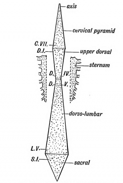
The spine, when viewed from the front, is seen to be made up of four pyramids : (1) Cervical ; (2) upper dorsal ; (3) dorso-lumbar ; (4) sacro-coccygeal (Fig. 113).
Fig. 113. Diagram of the Pyramids of the Spine.
The bases of the two upper pyramids meet at the disc between the 7th cervical and 1st dorsal vertebrae ; the bases of the lower two at the disc between the 5th lumbar and 1st sacral vertebrae. The apices of the two middle pyramids meet at the disc between the 4th and 5 th dorsal vertebrae, which have therefore the narrowest bodies of the vertebral series. The narrowing in the upper dorsal region is due to the fact that the weight of the upper half of the trunk is partly borne by, and transmitted to, the lower dorsal region by the sternum and ribs which thus relieve the spine to some extent (Fig. 113). At the sacrum the weight is transferred to the pelvis and lower limbs, hence the rapid diminution of the sacrum and coccyx. A well-marked thickening or bar in each ilium runs from the auricular surface to the acetabulum and transmits the weight to the femora.
The Curves of the Spinal Column
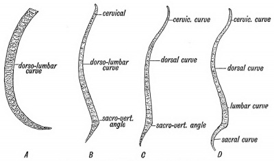
There is only one curve — an anterior concavity — until the 3rd month (Fig. 114 A). About the beginning of the 4th month the sacro-vertebral angle forms between the lumbar and sacral regions (114 £). At birth the cervical and sacral curves have appeared, but the sacral not to a pronounced extent (Fig. 114 C). The lumbar curve appears as the child learns to walk. It is produced to allow the body to be brought vertically over the lower extremities. The sacral and cervical curves also become then more marked (Fig. 114 D). The dorsal curvature and the sacro-vertebral angle are the primitive curves and are present in all mammals. The others are adaptations to the upright posture. The lumbar curve is most pronounced in the highly civilized races.
Fig. 114. Diagram of the Curves of the Spinal Column. A. At the 6th week of foetal life. B. At the 4th month of foetal life. C. Curves present at Birth. D. Curves present in the Adult.
Proportion of Cartilage and Bone
The inter-vertebral discs form one third of the total height of the spine ; the proportion of cartilage is greater in the lumbar than in the dorsal region and greater in the dorsal than in the cervical. The curvatures are due chiefly to the shape of the discs. In the lumbar region, which is convex forwards, only the lower three vertebrae are deeper in front than behind. This is true only for the higher races of mankind, for as Cunningham has shown, in lower races, as in the gorilla, only the lowest lumbar vertebra is deeper in front than behind, and thus helps to maintain the lumbar curvature.
Unstable Regions of the Spine
In about 90% of men there are 7 cervical, 1 2 dorsal, 5 lumbar, 5 sacral, and 4 caudal vertebrae, making 33 in all. In the remaining 10% there is some departure from the normal arrangement and these departures affect certain definite regions.
I. The sacro-lumbar. — The 25th vertebra in 95% of people forms the 1st sacral; in 1% the 24th, and 3% the 26th.
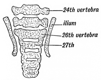
Fig. 115. A section of the Lumbo-sacral Region of the Spine in a Foetus at the end of the 2nd month, showing the 26th vertebra forming the 1st Sacral. (After Rosenberg.)
These percentages are drawn from the observations of Paterson, Eosenberg, and others who have made researches on this subject. The vertebral formula is not fixed. Eosenberg's investigations showed (Fig. 115) that it is the 26th vertebra that forms the first of the sacral series in the early embryo; later the 25th throws out great lateral masses, and thus forms a connection with the ilia. In the lower primates (monkeys) the 27 th forms the 1st sacral; with the evolution of man the 26th, then the 25th underwent sacral modifications, the trunk being correspondingly shortened. It will be seen that the number is not yet definitely fixed. The anterior point of attachment of the ilium fluctuates from the 24th to the 26th vertebra in man. With the sacral transformation of the 25 th and 26 th (lumbar) vertebrae, there was a corresponding movement forwards of the sacral plexus.
II. Sacro-coccygeal. — The 30 th vertebra forms the 1st coccygeal ; not uncommonly this vertebra is sacral in type and forms part of the sacrum.
III. Dorso-lumbar region. — This region is less liable to variation ; the 20 th vertebra instead of forming the 1st lumbar, may simulate the last dorsal in the type of its articular processes, and may bear ribs, probably an atavistic form, or, on the other hand, the 12th dorsal vertebra (19th) may not carry ribs. About 2 °/ of bodies show variations of this kind.
IV. Dorso-cervical. — The 7th vertebra may carry ribs ; rarely the 8th vertebra has no ribs attached to it and is cervical in typeEvolution and Development of the Spinal Column. — -The human spinal column in the process of development passes through three distinct phases :
(1) It is membranous; (2) it becomes cartilaginous; (3) it becomes bony. In the evolution of vertebrates the same three stages are observed. In Amphioxus and Marsipobranchs (excepting the neural arches), the spinal column is membranous ; in Elasmobranchs it is cartilaginous ; in other fishes and in all other vertebrates it is ossified. In the human embryo, as in every other vertebrate, the spinal column is developed from the mesoblast which surrounds the notochord and neural canal.
The Notochord
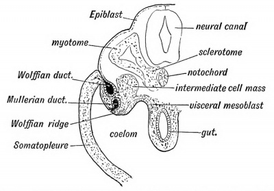
It has already been seen that the notochord is formed at a very early stage (before the 3rd week, Fig. 69, p. ,90) by a tubular invagination of the hypoblast under the neural canal. The notochord, with the mesoblastic tissue round it, represents the most primitive type of spinal support. It is hollow— the canal of the notochord runs from end to end, and into its posterior end the neurenteric canal opens. Afterwards it becomes a solid rod composed of cells of a peculiar type. A sheath is formed round the notochord by cells of the paraxial mesoblast (Fig. 116), which grow inwards and surround it.
Fig. 116. Diagrammatic transverse section of a human embryo at the end of the 3rd week.
These cells form the sclerotome and spring from the inner parts of the primitive segments (Fig. 116). At the same time the cells of the sclerotome also surround the neural tube. From these cells which grow inwards and surround the notochord and neural canal, the spinal column is formed and also the basioccipital and part of the basi-sphenoid bones of the skull (Fig. 117).
What becomes of the Notochord
In the second month of foetal life the notochord begins to disappear ; the bodies of the vertebrae and parachordal cartilages form in its sheath and constrict it. The parachordal cartilages form the basi-occipital and basi-sphenoid — the basal part of the skull — behind the i pituitary fossa. As they form, the notochord is obliterated \ between them. Eternod, however, found the anterior part of the notochord on the dorsal wall of the pharynx in the human embryo, so that the parachordal cartilages are evidently developed on its dorsal aspect. The odontoid process represents the body of the atlas and the suspensory ligament the disc between the occipital bone and atlas. A remnant of the notochord is enclosed in the suspensory ligament. The body of each vertebra is formed round the notochord and at first each has an hour-glass canal surrounding the notochord. Within each body the notochord ultimately disappears but in the intervertebral discs it swells out and forms much of the central mucoid core which each disc contains. The notochord with its membranous sheath is the earliest form of spinal column known. The real vertebral column, formed out of its sheath, begins to supplant it even in low vertebrates and in the human foetus of the second month this change is also seen to take place.
Fig. 117. Where Remnants of the Notochord may occur in the Adult.
Proto-vertebrae or Primitive Segments
Proto-vertebrae are not the forerunners of the vertebrae ; they are the primitive segments into which the mass of mesoblast at each side of the neural canal and notochord divides (Figs. 233, p. 289 and 116). The process of division or segmentation begins at the occipital region towards the end of the second week and spreads backwards until 3 5 or more body segments or somites are cut off. Each segment thus separated forms its own muscles (from its muscle plate or myotome), has its own nerve (spinal nerve), its own artery (inter-costal), and the basis for its skeletal tissue (sclerotome). The inter-segmental septum separates one proto-vertebra or segment from another. Eibs, transverse and spinous processes, are formed in the inter-segmental septa. Hence an intercostal space with its muscles, vessels, and nerves, with the corresponding intervertebral structures, represents a differentiated proto-vertebra. In the ventral aspect of the neck and loins, the inter-segmental septa disappear. In the head nine segments are recognised, but their recognition rests on observations made, not on the human embryo, but on the embryoes of lower vertebrates.
Recent work on the segmental arrangements of the nerves gives a practical importance to the number and position of the primitive segments and to the part of the body which each forms. Although the epiblast, hypoblast, and walls of the coelpui never show any trace of segmentation in the embryo, yet clinically there is evidence that each part belongs to, and was formed from, one definite segment. The upper extremity grows out from the 5th cervical to the 2nd dorsal segments (7 segments in all), and the lower from the 2nd lumbar to the 3rd sacral (7 segments in all). In each limb traces of these seven segments are to be found in the nerve distribution.
Development of a Typical Vertebra, — the 6th Dorsal
Membranous Stage
(3rd and 4th weeks). The vertebra then consists of 1st a body surrounding the notochord, formed from the sheath (Fig. 118 A), and 2nd a horse-shoe shaped vertebral bow (Fig. 1 1 8 A and B). The bow consists of a hypo-chordal part, and two lateral limbs, united by the hypo-chordal part ventral to the body.
Cartilaginous Stage
(Fig. 119). It commences in the 4th week. The fibrous basis is transformed into cartilage except the hypochordal part of the bow. It should be noticed (Fig. 118 -B) that the vertebral bodies are formed round the notochord, opposite each inter-segmental septum. Hence each vertebra belongs to two segments. The inter-vertebral disc is situated opposite the middle of a segment. The hypo-chordal part of the bow lies also in the segment, and becomes included in the course of development in the disc in front of the vertebra to which it belongs. The lateral limbs of the cartilaginous bow meet behind (dorsal to) the neural canal in the 4th month, and thus the bow is converted into a ring. The atlas is a permanent representative
Fig. 118. The development of the Membianous Basis of a Vertebra A. In transverse section. B. In horizontal section showing the relation of th. vertetea to the Pnrmtive Segments. The section is viewed from 'the dorsal of the ring thus formed. In the atlas only does the hypo-chordal part of the bow become cartilaginous and subsequently ossified ; in all the other vertebrae it remains as a fibrous strand in the intervertebral disc.
Fig. 119. Showing the Stages in the Development of a Vertebra. A. In the Membranous Stage. B. In the Cartilaginous Stage. C. The appearance of Ossific Points. D. The appearance of Secondary Ossific Centres, is 1 . The epiphyseal plates of the centra. /•'. Section of an amphicoelous vertebra.
Bony Stage
The body and bow parts of the cartilaginous vertebra fuse and give rise to the condition shown in Fig. 119 C. In the 7th week two centres appear in the body, but quickly fuse ; one appears in each neural arch (8th week) ; at birth the ossific centres of the body and neural arch have met. The central and neural ossifications meet at the neuro-central suture and unite at the 4th or 5th year. The neural ossifications fuse behind (where the spinous process is produced) in the 1st year. The ribs are entirely supported from the neural ossifications (Fig. 119 B). The spinous and transverse processes are formed by outgrowths of cartilage into the septa between the proto- vertebrae or primitive segments. The ribs are also formed in the septa, each at first articulating with a vertebra, which is also inter-segmental in origin. In all the ribs, except the 1st, 11th and 12 th the head is shifted to the inter-vertebral disc in front of its vertebra. Epiphyseal centres for the ossification of the transverse and spinous processes appear about puberty.
The Bodies of Mammalian Vertebrae are peculiar (1) in the development of an upper and lower epiphyseal plate ; (2) in that no trace of the notochord remains within them. In Fishes, as in the early human or mammalian foetus, the bodies are hour-glass shaped (amphicoelous, Fig. 119 F) ; in Amphibians they may retain a concavity in front (procoelous) or behind (opisthocoelous), but in mammals both ends are filled up.
Fig. 120. The Order in which the Centres of Ossification appear in the Bodies (A) and in the Neural Arches (B) of the Spinal Column.
It will be observed (Fig. 120 B) that the centres of ossification for the neural arches appear first in the anterior end of the spine (1st cervical), the date becoming later the more posterior the vertebra. In the 1st sacral they appear about the 4th month; in the 2nd sacral in the 5th month or later ; in the 3rd they may not appear. In the 4th and 5th sacral and 1st coccygeal vertebrae only vestiges of the neural arches are formed. These vertebrae retain the early foetal type shown in figure 119 B. In the remaining coccygeal vertebrae only the centres for the bodies appear. The centres for ossification of the bodies of the vertebrae appear first in the mid dorsal region (6th dorsal). From that point they spread forwards and backwards, the centres for the odontoid process appearing at the 4th month and that for the 5th sacral at the 5th month, while the last coccygeal does not appear until about the 20th or 25th year (Fig. 120 A).
The Atlas and Axis
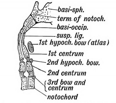
The atlas represents the completed bow of the 1st cervical vertebra. The body of the vertebra fuses with the body of the 2nd, and forms the odontoid process. A remnant of the disc between the 1st and 2nd vertebrae can sometimes be seen when the odontoid is split open. The suspensory ligament is the representative of the disc between the last occipital segment and the 1st cervical (Fig. 121).
Fig. 121. A diagrammatic section of the Foetal Axis, Atlas, and Basi-occipital.
Occipito-atlanto-axial Articulations
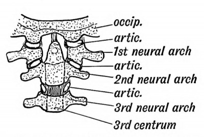
In the intervertebral discs of the cervical region there is at each side, between the lateral lips of the vertebral bodies, a small articular cavity (Fig. 122). It is situated between the parts of the body formed from the neural arches and in front of (ventral to) the issuing spinal nerves. Between the axis and atlas this articulation is greatly enlarged. At it the rotatory movements of the atlas on the axis take place. The atlanto-occipital joint, which separates the atlas and the last occipital segment is of the same nature. The atlas has neither the upper nor the lower articular processes of the other vertebrae. Hence the 1st and 2nd cervical nerves appear to issue behind the articular processes. At one time the single median occipital condyle seen in birds and reptiles was regarded as very different in nature from the double condyles of mammals. Recently Symington has shown that in the lowest mammals (monotremes), the occipital condyles are fused in the middle line, and that foetal mammals show approximations to this condition. In the human skull a remnant of this median fusion of the condyles is frequently seen on the anterior margin of the foramen magnum ; it is named the third or median occipital condyle. Sometimes the atlas is partly fused with or imperfectly separated from the occipital bone. This seems to be a further manifestation of the process which has led to four segments, which were at one time free body segments, being fused together to form the occipital part of the skull.
Fig. 122. The nature of the Atlanto-axio-occipital Articulations.
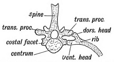
The ribs are developed in the septa between the dorsal primitive segments. At their vertebral ends they come in contact with the vertebral bow (Fig. 118 B). In lower vertebrates (birds, reptiles, etc.) each rib has two heads, a dorsal and ventral (Fig. 123). The tuberosity of the human rib represents the dorsal head ; the ventral head is well developed in man, as in mammals generally. The rib articulates with the neural arches only (Fig. 119 D).
Vestigial Ribs
Although the ribs are only fully developed in the dorsal region, yet a representative — a costal element — is present in every vertebra. In the cervical vertebrae the anterior part of the transverse processes represents a costal process, but only in the 6 th (sometimes) and 7 th is the costal process formed by a separate- centre of ossification. The costal process of the 7 th may develop into a rudiment or even a fully formed rib which reaches the sternum. In the lumbar vertebrae only the first shows a separate centre for the formation of the costal process ; it fuses with the tip of the transverse process in the later months of foetal life ; in the other lumbar vertebrae the tips or perhaps the whole of the transverse processes represent costal processes. The 12th dorsal rib varies widely in size ; it may be six inches or ten long or reduced to a mere vestige.
Fig. 123. The Bicipital Rib of a Lower Vertebrate (crocodile).
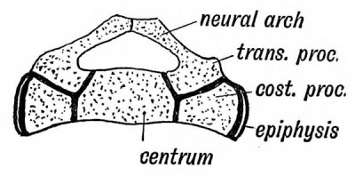
In the 1st, 2nd and 3rd sacral vertebrae the costal processes are large and have their own centres of ossification. Their cartilaginous bases fuse early to form the greater part of the lateral masses of the sacrum (Fig. 115). The part of the lateral mass formed by the costal processes is shown in Fig. 124. The costal processes are absent in the 4th and 5th sacral and all the coccygeal vertebrae. The two lateral epiphyseal plates on each •side of the sacrum are new and independent formations.
Fig. 124. A section to show the Nature of the Elements composing the Sacrum.
The Accessory Processes are found in the lumbar and lowest two dorsal vertebrae. They are developed at the base of the transverse processes and are for the attachment of slips of the longissimus dorsi. The mammillary processes are developed on the articular processes of the lower two or three dorsal and all the lumbar vertebrae. They give attachment to tendons of origin of the multifidus spinae.
The Transverse and Spinous Processes grow out from the vertebral bow (Fig. 119 A) into the septa between the primitive segments. Each transverse process is pierced, while still in the fibrous condition, by a branch of the corresponding segmental (intercostal) artery. In only the cervical region do those perforating arteries and their foramina persist. In that region the perforating arteries anastomose and out of the chain thus formed is developed the vertebral artery. Thus the foramina for the vertebral artery are formed independently of the costal element in each cervical transverse process. The spines are absent on the 1st cervical, 4th and 5 th sacral and coccygeal vertebrae. They are slightly developed and united by ossification of the interspinous ligament in the 2nd and 3rd sacral vertebrae. The 2nd, 3rd, 4th, 5th, and 6th cervical spines are bifid in Europeans;, but in lower races, as in anthropoids, the 5 th and 6 th spines are undivided.
| Historic Disclaimer - information about historic embryology pages |
|---|
| Pages where the terms "Historic" (textbooks, papers, people, recommendations) appear on this site, and sections within pages where this disclaimer appears, indicate that the content and scientific understanding are specific to the time of publication. This means that while some scientific descriptions are still accurate, the terminology and interpretation of the developmental mechanisms reflect the understanding at the time of original publication and those of the preceding periods, these terms, interpretations and recommendations may not reflect our current scientific understanding. (More? Embryology History | Historic Embryology Papers) |
Human Embryology and Morphology (1902): Development or the Face | The Nasal Cavities and Olfactory Structures | Development of the Pharynx and Neck | Development of the Organ of Hearing | Development and Morphology of the Teeth | The Skin and its Appendages | The Development of the Ovum of the Foetus from the Ovum of the Mother | The Manner in which a Connection is Established between the Foetus and Uterus | The Uro-genital System | Formation of the Pubo-femoral Region, Pelvic Floor and Fascia | The Spinal Column and Back | The Segmentation of the Body | The Cranium | Development of the Structures concerned in the Sense of Sight | The Brain and Spinal Cord | Development of the Circulatory System | The Respiratory System | The Organs of Digestion | The Body Wall, Ribs, and Sternum | The Limbs | Figures | Embryology History
Reference
Keith A. Human Embryology and Morphology. (1902) London: Edward Arnold.
Cite this page: Hill, M.A. (2024, April 19) Embryology Book - Human Embryology and Morphology 11. Retrieved from https://embryology.med.unsw.edu.au/embryology/index.php/Book_-_Human_Embryology_and_Morphology_11
- © Dr Mark Hill 2024, UNSW Embryology ISBN: 978 0 7334 2609 4 - UNSW CRICOS Provider Code No. 00098G

