Book - Developmental Anatomy 1924-17: Difference between revisions
| Line 86: | Line 86: | ||
===Vascular System=== | ===Vascular System=== | ||
The heart lies in the pericardial cavity, as seen in Fig. 368. The atrial region (Fig. 370), now includes two lateral sacs, the right and left atria. Similarly, the bulbo-ventricular loop has become differentiated into right and left ventricles, much thicker walled than the atria. The right ventricle is the smaller, and from it the bulbus passes between the atria and is continued as the ventral aorta. Viewed from the caudal and dorsal aspect (Fig. 371), the sinus venosus is seen dorsal to the atria. It opens into the right atrium and receives from the right and left sides the paired common cardinal veins. These veins drain the blood from the body of the embryo. Caudally, the sinus venosus receives the two vitelline veins. Of these, the left is small in the liver and later disappears. The right vitelline vein, now the common hepatic, carries most of the blood to the heart from the umbilical veins, and from the liver sinusoids, gut, and yolk sac. | The heart lies in the pericardial cavity, as seen in Fig. 368. The atrial region (Fig. 370), now includes two lateral sacs, the right and left atria. Similarly, the bulbo-ventricular loop has become differentiated into right and left ventricles, much thicker walled than the atria. The right ventricle is the smaller, and from it the bulbus passes between the atria and is continued as the ventral aorta. Viewed from the caudal and dorsal aspect (Fig. 371), the sinus venosus is seen dorsal to the atria. It opens into the right atrium and receives from the right and left sides the paired common cardinal veins. These veins drain the blood from the body of the embryo. Caudally, the sinus venosus receives the two vitelline veins. Of these, the left is small in the liver and later disappears. The right vitelline vein, now the common hepatic, carries most of the blood to the heart from the umbilical veins, and from the liver sinusoids, gut, and yolk sac. | ||
{| | |||
[[File:Arey1924 fig370.jpg| | | [[File:Arey1924 fig370.jpg|500px]] | ||
| [[File:Arey1924 fig371.jpg|500px]] | |||
'''Fig. 370.''' Ventral and cranial surface of the heart from a 6 mm. pig embryo. X 14. | |- | ||
| '''Fig. 370.''' Ventral and cranial surface of the heart from a 6 mm. pig embryo. X 14. | |||
| '''Fig. 371.''' Dorsal and caudal view of the heart from a 6 mm. pig embryo. X 14. | |||
|} | |||
'''Fig. 371.''' Dorsal and caudal view of the heart from a 6 mm. pig embryo. X 14. | |||
Transverse sections of the embryo through the four chambers of the heart show the atria in communication with the ventricles through the atrio-ventricular foramina, and the sinus venosus opening into the right atrium (Fig. 380). This opening is guarded by the right and left valves of the sinus venosus. Septa partition the two atria and the two ventricles incompletely. In Fig. 380 the atrial septum {septum primum) appears complete, due to the plane of the section, but in Fig. 372, from a slightly smaller embryo, it is seen that the septum primum grows from the dorsal atrial wall of the heart and does not yet meet the endocardial cushions between the atrio-ventricular canals. This opening between the atria is known as the interatrial foramen. Before it closes, another aperture appears in the septum, dorsal in position; this is the foramen ovale which persists during fetal life. In Fig. 372 both openings may be seen, as may also the dorsal and ventral endocardial cushions bounding the atrio-ventricular foramina. The mesothelial layer of the ventricles has become much thicker than that of the atria. It forms the epicardimn and the myocardium; the sponge-like meshes of the latter are now being developed. | Transverse sections of the embryo through the four chambers of the heart show the atria in communication with the ventricles through the atrio-ventricular foramina, and the sinus venosus opening into the right atrium (Fig. 380). This opening is guarded by the right and left valves of the sinus venosus. Septa partition the two atria and the two ventricles incompletely. In Fig. 380 the atrial septum {septum primum) appears complete, due to the plane of the section, but in Fig. 372, from a slightly smaller embryo, it is seen that the septum primum grows from the dorsal atrial wall of the heart and does not yet meet the endocardial cushions between the atrio-ventricular canals. This opening between the atria is known as the interatrial foramen. Before it closes, another aperture appears in the septum, dorsal in position; this is the foramen ovale which persists during fetal life. In Fig. 372 both openings may be seen, as may also the dorsal and ventral endocardial cushions bounding the atrio-ventricular foramina. The mesothelial layer of the ventricles has become much thicker than that of the atria. It forms the epicardimn and the myocardium; the sponge-like meshes of the latter are now being developed. | ||
Revision as of 09:51, 23 October 2016
| Embryology - 25 Apr 2024 |
|---|
| Google Translate - select your language from the list shown below (this will open a new external page) |
|
العربية | català | 中文 | 中國傳統的 | français | Deutsche | עִברִית | हिंदी | bahasa Indonesia | italiano | 日本語 | 한국어 | မြန်မာ | Pilipino | Polskie | português | ਪੰਜਾਬੀ ਦੇ | Română | русский | Español | Swahili | Svensk | ไทย | Türkçe | اردو | ייִדיש | Tiếng Việt These external translations are automated and may not be accurate. (More? About Translations) |
Arey LB. Developmental Anatomy. (1924) W.B. Saunders Company, Philadelphia.
| Historic Disclaimer - information about historic embryology pages |
|---|
| Pages where the terms "Historic" (textbooks, papers, people, recommendations) appear on this site, and sections within pages where this disclaimer appears, indicate that the content and scientific understanding are specific to the time of publication. This means that while some scientific descriptions are still accurate, the terminology and interpretation of the developmental mechanisms reflect the understanding at the time of original publication and those of the preceding periods, these terms, interpretations and recommendations may not reflect our current scientific understanding. (More? Embryology History | Historic Embryology Papers) |
Chapter XVII The Study of Pig Embryos
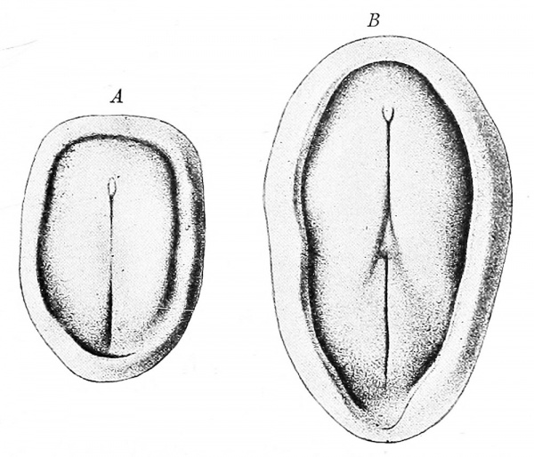
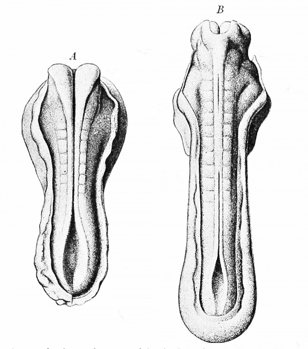
A very young pig embryos of the primitive streak and neural fold stages are shown in Fig. 364. The closure of the neural tube and the progressive appearance of mesodermal segments are likewise illustrated in Fig. 365. The fundamental similarity of these embryos to the early chicks already studied is apparent. For a short time, succeeding stages are complicated by flexion and spiral twisting which make sections difficult for the beginner to interpret. In embryos about 6 mm. long, the twist of .
Fig. 364. Early pig embryos (Keibel). X 20. A, Blastoderm with primitive streak and knot; B, blastoderm with primitive streak and neural groove.
The body has disappeared sufficiently so that its structure may be studied to better advantage. At this time the state of development is generally comparable to that of a four day chick (Fig. 366).
The fetal membranes of the pig stand somewhat intermediate between the chick and man. The amnion, chorion, and allantois develop very much as in the chick (Fig. 349). The yolk sac is small and rudimentary, so its functions are transferred to the allantois which fuses with the chorion ; the two constitute a placenta which is the organ of fetal respiration, nutrition, and excretion (Fig. 39). The development and relations of these extra-embryonic structures are described on pp. 46-49.
In this manual, series of transverse sections are figured and described only. Lateral and sagittal dissections show the longitudinal relations more clearly than serial sections cut in these planes; if, however, sagittal or frontal sections are used, they may be interpreted readily from the corresponding dissections and reconstructions.
Fig. 365. Dorsal views of pig embryos, with the amnion cut away (Keibel). X 20. A, Embryo of seven segments; B, embryo of eleven segments.
The Anatomy of a 6 mm Pig Embryo
The descriptions given here are applicable to the study of embryos between 5 and 8 mm. Due to a shorter term of development, a 6 mm. pig embryo is slightly farther advanced in most respects than a human embryo of the same size (Fig. 61).
External Form
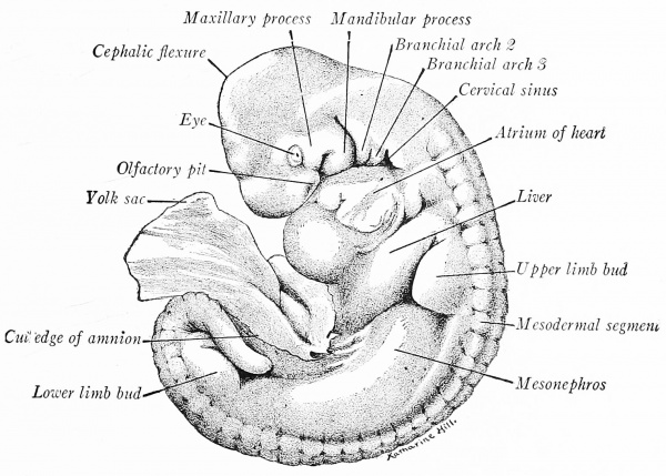
Both head and body are bent in an even curve, convex along the dorsal line, and the tail is recurved sharply (Fig. 366). The cephalic flexure forms an acute angle at the mesencephalon, and there is also a marked cervical flexure. As a result, the head is somewhat triangular in shape. Lateral to the dorsal line may be seen the segments, which become larger and more differentiated toward the head. At the tip of the head, a shallow depression marks the olfactory pit. The lens vesicle of the eye is open to the exterior. Caudal to the eyes, at the sides of the head, are four branchial arches separated by three branchial grooves. The fourth arch is partly concealed in a triangular depression, the cervical sinus, formed by the more rapid growth of the first and second arches (cf. Fig. 369). The first, or mandibular arch, forks ventrally into two parts, a smaller maxillary- and a larger mandibular process; the latter, with^its. fellow, forms the lower jaw. The position of the mouth is indicated by the space between these processes. The furrow from the eye to the mouth is the lacrimal groove. The second, or hyoid arch is separated from the mandibular arch by an ectodermal groove which persists as the external acoustic meatus.
Fig. 366. Pig Embryo of 6 mm with amnion removed X 12
The heart is large, and through the transparent body wall may be seen the dorsal atrium and ventral ventricle. Caudal to the heart, a convexity indicates the position of the liver, dorsal to which is the bud of the upper limb. Extending caudad from the limb bud, a curved convexity indicates the position of the left mesonephros, large and precociously developed in the pig. At its end is the anlage of the lower limb. The amnion has been dissected away along the line of its attachment, ventral to the mesonephros. There is as yet no distinct umbilical cord, and a portion of the body stalk is attached to the embryo.
Nervous System and Sense Organs
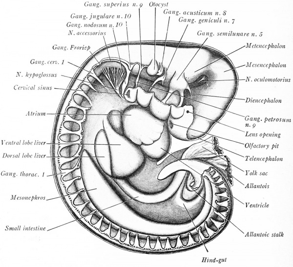
The brain is differentiated into its five regions; telencephalon, diencephalon, mesencephalon, metencephalon, and myelencephalon (Fig. 367). The spinal cord is cylindrical and tapers off gradually to the tail. The anlage of the cranial and spinal ganglia and the main nerve trunks are shown. The olfactory, optic, and trochlear nerves have not yet differentiated, and the oculomotor nerve is just beginning to appear from the ventral wall of the mesencephalon. Ventrodateral to the metencephalon and myelencephalon occur in order: the semilunar ganglion and three branches of the trigeminal nerve; the geniculate ganglion and nerve trunk of the n. facialis; the ganglionic anlage of the 11. acusticus. Caudal to the otocyst, a continuous chain of cells extends lateral to the neural tube into the tail region. Cellular enlargements along this neural crest represent developing cranial and spinal ganglia. They are, in order: the superior, or root ganglion of the glossopharyngeal nerve with its distal petrosal ganglion; the ganglion jugulare and distal ganglion nodosum of the vagus nerve; the ganglionic crest and the proximal portion of the spinal accessory nerve; and the anlage of Froriep - s ganglion, an enlargement on the neural crest just cranial to the first cervical ganglion. Between the vagus and Froriep - s ganglion may be seen the numerous root fascicles of the hypoglossal nerve, which take their origin along the ventro-lateral wall of the myelencephalon and unite to form a single trunk. The posterior roots of the spinal ganglia are very short ; their anterior, or ventral roots are concealed. It should be observed that the fifth, seventh, ninth, and tenth cranial nerves pass to the four branchial arches in the order named. This primitive relation is maintained in the adult when the nerves innervate the derivatives of these arches.
Fig. 367. Lateral dissection of a 5.5 mm Pig Embryo X 18.
The olfactory pits are merely slight depressions in the thickened ectoderm of the ventral head. There are stalked but the resides still open to the exterior. The otocysts are oval, ectodermal vesicles with endolymph ducts just appearing as dorso-mesial outgrowths.
Digestive and Respiratory Systems
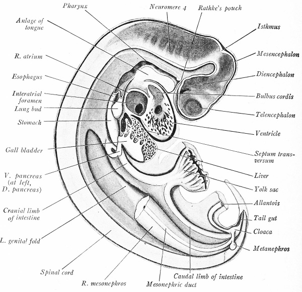
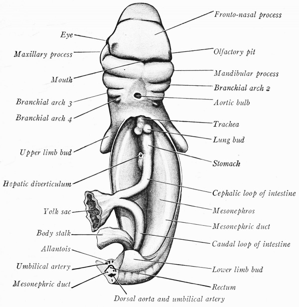
The month lies between the mandible, the median fronto-nasal process of the head, and the maxillary processes at the sides (Fig. 369). The diverticulum of the epithelial hypophysis {Rathke's pouch) extends along the ventral wall of the forebrain (Fig. 368) ; near its distal end, the wall of the brain is thickened, and later the posterior lobe of the hypophysis will develop at this point.
Fig. 368. Median sagittal dissection of a 6 mm pig embryo. X 18.
Fig. 369. Ventral dissection of a 6 mm pig embryo. X 14.
The pharynx is flattened dorso-ventrally and is widest near the mouth. It narrows caudad, and, opposite the third branchial arch, makes an abrupt bend, which corresponds to the cervical flexure of the embryo - s body (Fig. 368). In the roof of the phar^mx, just caudal to Rathke - s pouch, is the somewhat cone-shaped pocket known as Sessell’s pouch, which may be interpreted as the blind, cephalic end of the fore-gut (Fig. 376). The lateral and ventral walls of the pharynx and oral cavity are shown in Fig. S4 A. Of the four arches, the mandibular is the largest, and a groove partly separates the tongue anlages of the two sides. Posterior to this groove and extending in the median plane to the hyoid arch is a triangular elevation, the tuberculmn impar; it later vanishes, apparently contributing nothing to the tongue. At an earlier stage, the median thyroid anlage grew out from the midventral wall of the pharynx just caudal to the tuberculum impar. The ventral ends of the second arches fuse in the midventral plane and form a prominence, the copula (Fig. 84 B). This connects the tuberculum impar with a rounded tubercle derived from the third and fourth pairs of arches, the anlage of the epiglottis. Its cephalic portion forms the root of the tongue. Caudal to the epiglottis are the arytenoid ridges, and a slit between them, the glottis, leads into the trachea.
The branchial arches converge caudad, and the pharynx narrows rapidly before it is differentiated into the trachea and esophagus. Laterally and ventrally, between the arches, are the four paired outpocketings of the pharyngeal pouches (Fig. 375). The pouches have each a dorsal and ventral diverticulum. The dorsal diverticula are large and wing-like; they meet the ectoderm of the branchial grooves and fuse with it to form the closing plates. Between the ventral diverticula of the third pair of pouches lies the median thyroid anlage (Fig. 376). The fourth pouch is smaller than the others; its dorsal diverticulum just meets the ectoderm; its ventral portion is small, tubular in form, and is directed parallel to the esophagus.
The groove on the floor of the pharynx, caudal to the epiglottis, is continuous with the tracheal groove. More caudally, opposite the atrium of the heart, the trachea has separated from the esophagus (Fig. 368). The trachea at once bifurcates to form the primary bronchi and the anlages of the lungs (Fig. 369). The latter consist merely of the dilated ends of the bronchi, surrounded by a layer of splanchnic mesoderm. They bud out laterally on each side of the esophagus near the cardiac end of the stomach, and project into the pleural coelom. The esophagus is short, and widens dorso-ventrally to form the stomach. The long axis of the stomach is nearly straight, but its entodermal walls are compressed and it has revolved on its long axis so that the original dorsal border lies to the left, the ventral border to the right (Fig. 382).
Caudal to the pyloric end of the stomach, and to its right, is given off from the duodenum the hepatic diverticulum (Fig. 368). This is a sac of elongated oval form from which the liver and part of the pancreas take origin, and which later gives rise to the gall bladder, cystic duct, and common bile duct. It is connected by several cords of cells with the trabeculae of the liver; the latter is divided incompletely into four lobes, a small dorsal and a large ventral lobe on each side (Fig. 367).
The pancreas is represented by two outgrowths. The ventral pancreas originates from the hepatic diverticulum near its attachment to the duodenum (Fig. 368). It grows to the right of the duodenum and ventral to the portal vein. The dorsal pancreas takes origin from the dorsal side of the duodenum, caudal to the hepatic diverticulum, and grows dorsally into the substance of the gastric mesentery (Fig. 376). It is larger than the ventral pancreas, and its posterior lobules grow to the right and dorsal to the portal vein, and in later stages anastomose with the lobules of the ventral pancreas.
The intestine of both fore-gut and hind-gut has elongated and curves ventrally into the region of the future umbilical cord wdiere the yolk stalk has nari'ow^ed at its point of attachment to the gut (Fig. 368). The cloaca, a dorso-ventrally expanded portion of the hind-gut, gives off cephalad and ventrad the allantoic stalk. This is at first a narrow tube, but soon expands into a vesicle of large size, a portion of which is seen in Fig. 368. Dorso-laterad, the cloaca receives the mesonephric ducts. The hind-gut is continued into the tail as the transitory tail- gut, or postanal gut, which dilates at its extremity. The midventral wall of the cloaca is fused to the adjacent ectoderm to form the cloacal membrane. In this region the anus will appear.
Coelom and Mesenteries
The coelom communicates throughout, but already consists of a single, large pericardial cavity, paired pleural canals, and a common peritoneal chamber. The septum transversum, which will form most of the diaphragm, is prominent and serves to separate partially the heart cavity from the remainder of the coelom (Fig. 368). The primitive dorsal mesentery is a thick, double layer of splanchnic mesoderm investing the gut and attaching it to the median roof of the peritoneal cavity. As the intestine bends out toward the yolk sac, the dorsal mesentery grows at an equal rate and suspends it (Fig. 367). The liver lies in the ventral mesentery, between the stomach-duodenum and the midventral body wall. Between this level and the yolk stalk, the mesentery is beginning to disappear; the caudal limb of the intestine is already free ventrad (Fig. 376).
Urogenital System
The form of the mesonephroi is seen in Figs. 367 to 369. Each consists of large, vascular glomeruli, associated with coiled tubules which are lined with cuboidal epithelium and open into the mesonephric duct (Fig. 131). The mesonephric {Wolffian) duct, beginning at the anterior end of the mesonephros, curves at first along its ventral, then along its lateral surface. At the caudal end, each duct bends ventrad and to the midplane where it opens into a lateral expansion of the cloaca (Fig. 368). Before this junction takes place, an evagination into the mesenchyme from the dorsal wall of each mesonephric duct gives rise to the anlage of the mctanephros, or permanent kidney. The allantois is a prominent, stalked sac communicating with the ventral part of the cloaca. A slight thickening of the mesothelium along the median and ventral surface of each mesonephros forms a light-colored area, the genital fold (Fig. 368). This ridge is pointed at either end and confined to the middle third of the kidney. It is the anlage of the genital gland.
Vascular System
The heart lies in the pericardial cavity, as seen in Fig. 368. The atrial region (Fig. 370), now includes two lateral sacs, the right and left atria. Similarly, the bulbo-ventricular loop has become differentiated into right and left ventricles, much thicker walled than the atria. The right ventricle is the smaller, and from it the bulbus passes between the atria and is continued as the ventral aorta. Viewed from the caudal and dorsal aspect (Fig. 371), the sinus venosus is seen dorsal to the atria. It opens into the right atrium and receives from the right and left sides the paired common cardinal veins. These veins drain the blood from the body of the embryo. Caudally, the sinus venosus receives the two vitelline veins. Of these, the left is small in the liver and later disappears. The right vitelline vein, now the common hepatic, carries most of the blood to the heart from the umbilical veins, and from the liver sinusoids, gut, and yolk sac.
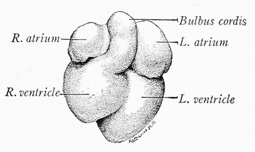
|
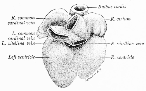
|
| Fig. 370. Ventral and cranial surface of the heart from a 6 mm. pig embryo. X 14. | Fig. 371. Dorsal and caudal view of the heart from a 6 mm. pig embryo. X 14. |
Transverse sections of the embryo through the four chambers of the heart show the atria in communication with the ventricles through the atrio-ventricular foramina, and the sinus venosus opening into the right atrium (Fig. 380). This opening is guarded by the right and left valves of the sinus venosus. Septa partition the two atria and the two ventricles incompletely. In Fig. 380 the atrial septum {septum primum) appears complete, due to the plane of the section, but in Fig. 372, from a slightly smaller embryo, it is seen that the septum primum grows from the dorsal atrial wall of the heart and does not yet meet the endocardial cushions between the atrio-ventricular canals. This opening between the atria is known as the interatrial foramen. Before it closes, another aperture appears in the septum, dorsal in position; this is the foramen ovale which persists during fetal life. In Fig. 372 both openings may be seen, as may also the dorsal and ventral endocardial cushions bounding the atrio-ventricular foramina. The mesothelial layer of the ventricles has become much thicker than that of the atria. It forms the epicardimn and the myocardium; the sponge-like meshes of the latter are now being developed.
The Arteries
Beginning with the ventral aorta, which takes origin from the bulbus cordis, pairs of aortic arches are given off. These run dorsad in the five branchial arches (Figs. 375 and 376) and join the paired descending aorta. The first and second pairs of aortic arches are very small and originate from the small common trunks formed by the bifurcation of the ventral aorta just caudal to the thyroid gland. The fourth aortic arch is the largest. From the apparent fifth arch small pulmonary arteries are developing. There is evidence that this pulmonary arch is really the sixth in the series, the fifth having been suppressed in development (cf. Fig. 186 B). Cranial to the first pair of aortic arches, the descending aortal are continued forward into the maxillary processes as the internal carotids. Caudal to the aortic arches, the descending aortae converge, unite opposite the cardiac end of the stomach, and form the median dorsal aorta (Fig. 376). Both the dorsal and descending aortae give off paired, dorsal intersegmental arteries. From the seventh pair of these arteries (the first set to arise from the medial dorsal aorta) there are developed a pair of lateral branches to the upper limb buds. These vessels are the subclavian arteries. The dorsal aorta also sprouts ventrolateral arteries to the glomeruli of the mesonephros, and median ventral arteries to the gut (Fig. 385). Of the latter, the coeliac artery arises opposite the origin of the hepatic diverticulum. The vitelline artery takes origin by two or three trunks caudal to the dorsal pancreas; of these, the posterior is the larger and persists as the superior mesenteric artery.
Fig. 372. Dissection of the heart from a 5.5 mm. pig embryo, viewed from the left side. X 14.
Opposite the lower limb buds, the dorsal aorta is divided for a short distance. From each division there arise, laterad, three short trunks which unite to form the single umbilical artery on each side. The middle vessel is the largest and apparent^ becomes the common iliac artery. A pair of short caudal arteries, much smaller in size, continue the descending aortae into the tail region.
Fig. 373. Ventral reconstruction of a 6 mm. pig embryo, showing the vitelline and opened umbilical veins (Vehe). X 22. In the small orientation figure (cf. Fig. 376) the various planes are indicated by broken lines .
The Veins
The vitelline veins, originally paired throughout, are now represented distally by a single vessel, which, ramifying in the wall of the yolk sac, enters the embryo and courses cephalad of the intestinal loop (Fig. 373). Crossing to the left side of the intestine and ventral to it, it is joined by the superior mesenteric vein which has developed in the mesentery of the intestinal loop. The trunk formed by the union of these two vessels becomes the portal vein. It passes along the left side of the gut in the mesentery, and, opposite the stem of the dorsal pancreas, gives off a small branch, a rudimentary continuation of the left vitelline vein, which continues cephalad and in earlier stages connects with the sinusoids of the liver. The portal vein then bends sharply to the right, dorsal to the duodenum, and, in the course of the right vitelline vein (passing between the dorsal and ventral pancreas to the right of the duodenum) it soon enters the liver and connects with the liver sinusoids. The portal trunk is thus formed by persisting portions of both vitelline veins, and receives a new vessel, the superior mesenteric vein. The middle portions of the primitive vitelline veins subdivide into the network of liver sinusoids. Their proximal vitelline trunks drain the blood from the liver and open into the sinus venosus of the heart. The right member of this pair is much the larger (Fig. 371) and persists as the proximal portion of the future inferior vena cava.
Fig. 374. Ventral reconstruction of the cardinal and subcardinal veins in a 6 mm. pig embryo, showing the early development of the inferior vena cava ( Vehe). X 22. In the small orientation figure (cf. Fig. 376) the various jdanes are indicated by broken lines.
The umbilical veins, originating in the walls of the chorion and allantoic vesicle, fuse and lie caudal and lateral to the allantoic stalk (cf. Fig. 184). Before the stalk enters the body, they separate again and run lateral to the umbilical arteries. The left vein is much the larger. Both, after receiving branches from the posterior limb buds and from the body wall, pass cephalad' in the somatopleure of each side (Figs. 373 and 375). Their course is first cephalad, then dorsad, until they enter the liver. The left vein enters a wide channel, the ductus venosus, which carries its blood through the liver and thence to the heart by way of the right vitelline trunk. The right vein joins a large sinusoidal continuation of the portal vein in the liver. This common trunk drains into the ductus venosus.
Fig. 375. Reconstruction of a 7.8 mm. pig embryo, showing the veins and aortic arches from the left side (Thyng). X 15. Ph.P. 1, 2, 3, 4, Pharyngeal pouches.
The anterior cardinal veins (Figs. 374 and 375) are formed to drain the plexus of veins on each side of the head. These vessels extend caudad and lie ventro-lateral to the myelencephalon. Each receives branches from the sides of the myelencephalon, then curves ventrad, is joined by the linguo-facial vein from the branchial arches and at once unites with the posterior cardinal of the same side to form the common cardinal vein. This, as already explained, opens into the sinus venosus.
A posterior cardinal vein develops dorso-lateral to each mesonephros (Figs. 374 and 375). Running cephalad, they join the anterior cardinal veins. When the mesonephroi become prominent, as at this stage, the middle third of each posterior cardinal is broken up into sinusoids. Sinusoids extend from the posterior cardinal vein ventrally around both the lateral and medial surfaces of the mesonephros. The median sinusoids anastomose longitudinally and form the subcardinal veins, right and left.
Fig. 376. Median sagittal reconstruction of a 6 mm. pig embryo (Vehe). X 16.5. The numbered heavy lines indicate the levels of the transverse sections shown in Figs. 377-388. The broken lines mark the outline of the left mesonephros and the course of the left umbilical artery and vein ; the latter may be traced from the umbilical cord to the liver where it is sectioned longitudinally.
The subcardinals lie along the median surfaces of the mesonephroi, more ventrad than the posterior cardinals with which they are connected at either end. There is a transverse capillary anastomosis between the two subcardinals, cranial and caudal to the permanent trunk of the vitelline artery (Fig. 374). The right vessel is connected with the liver sinusoids through a small vein (which develops in the mesenchyme of the caval mesentery) located to the right of the mesogastrium (Fig. 383). This vein now carries blood direct to the heart from the right posterior cardinal and right subcardinal, by way of the liver sinusoids and the right vitelline trunk (common hepatic vein). Eventually, the unpaired inferior vena cava forms in the course of these four vessels.
Transverse Sections of a 6 mm Pig Embryo
Having acquainted himself with the anatomy of the embryo from the study of dissections and reconstructions, the student is prepared to examine serial sections. To interpret the structures encountered, there must be constant references to the figures already described which show the positions of the organs. Determine the exact plane of a section with reference to these illustrations, and especially Fig. 376. Representative levels are described on the following pages; the position of each is indicated by the heavy, numbered lines on Fig. 376. These sections are drawn from the cephalic surface, so that the right side of the embryo is at the readers left.
Sections through the Cephalic Flexure
The earliest sections cut the mesencephalon, metencephalon, and thin-roofed myelencephalon. At levels which include two separate portions of the brain, the smaller is the diencephalon, the larger a longitudinal section of the myelencephalon. In the intermediate mass of mesenchyme lie the internal carotid arteries (cf. Fig. 377). Lateral to them are numerous branches of the anterior cardinal veins. Midway along the sides of the hind-brain are the apices of the otocysts.
Section through the Myelencephalon and Otocysts (Fig. 377). - As the head is bent nearly at right angles to the body, the brain is cut twice ; this section passes lengthwise through the myelencephalon and transversely through the diencephalon. The cellular walls of the myelencephalon show a series of six paired constrictions, the neuromeres. Lateral to the fourth pair of neuromeres are the otocysts, which show a median outpocketing at the point of entrance of the endolymph duct. The ganglia of the nn. trigeminus, facialis, acusticus, and the superior ganglion of the glossopharyngeal nerve occur in order on each side. Sections of the anterior cardinal vein appear in several places, and ventral to the diencephalon are the internal carotid arteries.
Passing along down the series into the pharynx region, observe the first, second, and third pharyngeal pouches. Their dorsal diverticula come into contact with the ectoderm , of the branchial grooves and form the closing plates.
Section through the Branchial Arches and the Eyes (Fig. 378). - The section passes lengthwise through the four branchial arches, the fourth sunken in the cervical sinus. The mandibular processes of the first arch have united to form the mandible, or lower jaw; i the maxillary processes of the future upper jaw lie across the stomodeal space in the separate section of the bent head. Dorsad is the spinal cord, with the first pair of cervical ganglia, and laterad the first cervical myotonies. The pharynx is cut across between the third and fourth branchial pouches. In its floor is a prominence, the anlage of the epiglottis. Ventral to the pharynx, the ventral aorta gives off two pairs of vessels. The larger pair are the fourth aortic arches w'hich curve dorsad around the pharynx to enter the descending aortce. The smaller third aortic arches enter the third branchial arches on each side. A few sections higher up in the series, the ventral aorta bifurcates, and the right and left trunks thus formed give off the first and second pair of aortic arches. Cranially, in the angle between their common trunks, lies the median thyroid anlage. The anterior cardinal veins are located dorso-lateral to the descending aortae. The end of the head is cut through the diencephalon and the optic vesicles. On the left side of the figure, the lens vesicle may be seen still connected with the ectoderm. The corresponding optic cup appears asymmetrical because it is cut through the chorioid fissure. The cup is differentiated into a thick inner, and a thin outer layer ; these form the nervous and pigment layers of the retina respectively.
Fig. 378. Transverse section through the branchial arches and eyes of a 6 mm. pig embryo. X 26.5. .Y, aortic arch 4.
Section through the Tracheal Groove, Bulbus Cordis and Olfactory Pits (Fig. 379). - The ventral portion of the figure shows a section through the tip of the head. The telencephalon is not prominent. The ectoderm is thickened and sUghtly invaginated ventrolaterad to form the anlages of the olfactory pits. These deepen in later stages and become the nasal cavities. In the dorsal portion of the section may be seen the cervical spinal cord, the notochord just ventral to it, the descending aortce, and ventro-lateral to them the anterior cardinal veins. The naso-pharynx now is small with a vertical groove in its floor. This is the tracheal groove and more caudad it will become the cavity of the trachea. The bulbus cordis lies in the large pericardial cavity. On either side the section cuts through the cephalic portions of the atria. These will become larger farther caudad in the series.
Fig. 379. Transverse section through the bulbus cordis and olfactory pits of a 6 mm. pig embryo. X 26.5.
Section through the Heart (Fig. 380). - The heart lies in the pericardial cavity. Both the atrium and ventricle are divided incompletely into two chambers. A partial interventricular septum leaves the ventricles in communication dorsad. The septum primum is complete in this section, but cephalad in the series there is an interatrial foramen (cf. Fig. 372). 'T\\q foramen ovale has not formed in this particular embryo. The myocardium of the ventricles is a spongy layer, much thicker than that of the atrial wall. Lateral to the descending aortae are the common cardinal veins. The right common cardinal opens into the sinus venosus which in turn empties into the right atrium, its opening being guarded by the two valves of the sinus venosus. The entrance of the left common cardinal into the sinus venosus is somewhat more caudad in the series. The trachea has now separated from the esophagus and lies ventral to it. Both trachea and esophagus are surrounded by a condensation of mesenchyme which will transform into the muscular and fibrous coats.
Fig. 380. Transverse section through the four chambers of the heart of a 6 mm. pig embryo.X 26.5.
Section through the Lung Buds and Septum Transversum (Fig. 381). - The section passes through the bases of the upper limh buds. A pair of spinal nerves, with ganglia and roots, extends from the spinal cord into these anlages. The tips of the ventricles, lying in the pericardial cavity, still show in this section. Dorsally, the pericardial cavity has given place to the pleuro-pcritoueal cavity. Projecting ventrad into this cavity are the cranial ends of the mesonephric folds in which the posterior cardinal veins partly lie. Into the floor of the pleuro-peritoneal cavities bulge the dorsal lobes of the liver, embedded in splanchnic mesenchyme. This mesenchyme is continuous with that of the somatopleure, and forms a transverse partition between the liver and heart, complete ventrally. This is the septum transversum which takes part in forming the ligaments of the liver and is the chief anlage of the diaphragm. The two proximal trunks of the vitelline veins pass through the septum. Projecting laterally into the pleuro-peritoneal cavities are ridges of mesenchymecovered by splanchnic mesoderm in which the lungs develop as lateral buds from the caudal end of the trachea. The right lung bud is shown in the figure. Between the esophagus and the lung is a crescent-shaped cavity, the cranial end of the omental bursa.
Fig. 381. Transverse section through the right lung bud and septum transversum of a 6 mm.
Fig. 382. Transverse section through the stomach of a 6 mm. pig embryo. X 26.5.
Section through the Stomach (Fig. 382). - The section passes through the upper limb buds and just caudal to the point at which the descending aortas unite to form the median dorsal aorta. As the liver develops in early stages, it comes into relation with the caval mesentery, along the dorsal body wall, at the right side of the dorsal mesogastrium. The space between the liver and plica to the right, and the stomach and its omenta to the left, is a caudal continuation of the omental bursa. The dorsal wall of the stomach is rotated to the embryo - s left, its ventral wall to the right. The liver shows a pair of dorsal lobes, and contains large blood spaces and networks of sinusoids lined with endothelium. Ventral to the liver, the tips of the ventricles still appear.
Section through the Hepatic Diverticulum (Fig. 383). - The upper limb buds are prominent in this section. The mesonephric folds show the tubules and glomeruli of the mesonephroi, and the posterior cardinal veins are connected with the mesonephric sinusoids. From the dorsal attachment of the liver a ridge continues down into this section it lies on the dorsal body wall just to the right (left in figure) of the mesentery. In this ridge is a small vein which connects cranially with the liver sinusoids, caudally with the right subcardinal vein. As it later forms a portion of the inferior vena cava, the ridge in which it lies is termed the caval mesentery. The right dorsal lobe of the liver contains a large blood space into which the portal vein opens. The duodenum is ventral to the position occupied by the stomach in the previous section. There is given off from it, ventrad and to the right, the hepatic diverticulum. In sections cephalad, small ducts from the liver trabeculae may be traced into connection with it. In the left ventral lobe of the liver, a large blood space indicates the position of the left umbilical vein on its way to the ductus venosus. The mesothelial investment of the liver is the tissue of the expanded ventral mesentery into which it grew. Between the stomach and liver, the ventral mesentery is called the lesser omentum; between the liver and ventral body wall, the falciform ligament.
Fig. 383. Transverse section through the hepatic diverticulum of a 6 mm. pig embryo.X 26.5.
Section through the Pancreatic Anlages (Fig. 3S4). - At this level, the upper limb buds still show. The mesonephroi are prominent and marked by their large Bowman - s capsules and glomeruli, located mesad. The right posterior cardinal vein is broken up into mesonephric sinusoids. The vein in the caval mesentary will connect with the right subcardinal vein a few sections lower. The anlage of the dorsal pancreas is seen extending from the duodenum dorsad into the mesenchyme of the mesentery. It soon bifurcates into a dorsal and right lobe, of which the latter is slightly lobulated. Ventro-lateral to the duodenum, the anlage of the ventral pancreas is cut across; it may be traced cephalad in the series to its origin from the hepatic diverticulum. To the right of the ventral pancreas lies the portal vein (at this level the portion contributed by the right vitelline). To the left of the dorsal pancreas is seen the remains of the left vitelline vein. The ventral lobes of the liver are just disappearing at this level. In the mesenchyme that connects the liver with the ventral body wall lie on each side the umbilical veins, the left being the larger. Between the veins is the extremity of the hepatic diverticulum which becomes the gall bladder. The body wall is continued ventrad to form a short umbilical cord.
Fig. 384. Transverse section through the pancreatic anlages of a 6 mm. pig embryo. X 26.5.
Section through the Intestinal Loop and Lower Limb Buds (Fig. 385). - As the posterior half of the embryo is curved in the form of a half circle, sections caudal to the liver, like this one, pass also through the lower end of the body. Two sections of the embryo are thus seen in one, their ventral aspects facing each other and connected by the lateral body wall. In the dorsal part of the section the mesonephroi are prominent, with large posterior cardinal veins lying dorsal to them and mesonephric arteries branching laterally from the aorta and passing to the glomeruli. The trunk of the vitelline artery is the delicate tube taking origin ventrally from the aorta. It may be traced into the mesentery, and through it onto the wall of the yolk sac. On either side of the vitelline artery are the subcardinal veins, the right being the larger. In the mesentery may be seen two sections of the intestinal loop (the small intestine being cut lengthwise, the large intestine transversely), and also sections of the vitelline artery and veins. In the lateral body walls, ventral to the mesonephros, occur the umbilical veins. The left vein is large and cut lengthwise' the right vein is cut obliquely twice.
In the ventral portion of the section, the lower limb buds are prominent laterally. A pair of large arteries, the common iliacs, branch from the aorta and may be traced into connection with the umbilical arteries, d'he colon, supported by a short mesocolon, lies in the colom near the mid plane. On each side are the caudal ends of the mesonephric folds, here small and each showing a section of the mesonephric duct and a single vesicular anlage of the mesonephric tubules. The mesonephric ducts are cut as they curve around from their position in the dorsal portion of the section. The tip of the recurved tail shows.
Fig. 385. Transverse section through the intestinal loop and lower limb buds of a 6 mm. pig embryo. X 26.5.
Section through the Primitive Segments and Spinal Cord (Fig. 386). - In the interval since the level last described are encountered sections which show symmetrical longitudinal views of the body wall, mesonephroi, mesentery, aorta, and notochord. This section is near the end of the series, and, as the body is here curved, it is really a frontal section. At the left side of the spinal cord, the oval cellular masses are the spinal ganglia cut across.
The ectoderm, arching over the segments, indicates their position. Each segment shows an outer dense layer, the dermatome, lying just beneath the ectoderm. This plate curves lateral to the spindle-shaped myotome, which gives rise to the voluntary muscle. Next comes a diffuse mass of mesenchyme, the sclerotome, which, with its fellow of the opposite side, eventually, surrounds the spinal cord and forms the anlage of a vertebra. A pair of spinal nerves and spinal ganglia are developed opposite each somite, and pairs of small vessels are seen between the segments. These are dorsal intersegmental arteries.
Section through the Cloaca and Metanephric Anlages (Fig 387). - Having now studied sections at various levels to near the end of the series, the next step is to examine sections through the caudal region and study the anlages of the urogenital organs. Owing to the curvature of the embryo, it is necessary to go cephalad in the series. The metanephroi appear as dorsal evaginations from the mesonephric ducts, just before their entrance into the cloaca.
Fig. 386. Transverse section through the primitive segments and spinal cord of a 6 mm. pig embryo. X 45.
Fig. 387. Transverse section metanephroi of a 6 mm. pig embryo. X 45 .
Fig. 388. Transverse section through the umbilical vessels, allantois and cloaca of a 6 mm pig embryo. X 45 .
Each consists of an epithelial layer surrounded by a condensation of mesenchyme. Traced a few sections cephalad, the mesonephric ducts open into the lateral diverticula of the cloaca, which, irregular in outline because it is sectioned obliquely, lies ventral to them and receives dorsad the rectum. Caudal to the cloaca, in this embryo, the tail bends abruptly cephalad and to the right. The blind prolongation of the hindgut may be traced out into this portion of the tail until it ends in a sac-like dilatation.
Section through the Umbilical Vessels, Allantois and Cloaca (Fig. 388). - The present section passes through the bases of the limb buds at the level where the allantoic stalk, curving inward from the umbilical cord, opens into the cloaca. At either side of the allantoic stalk may be seen oblique sections of the umbilical arteries, and lateral to these the large left and small right â– umbilical vein. The mesonephric ducts occupy the mesonephric ridges which project into small caudal prolongations of the coelom. Midway between the ducts lies the rectum, dorsal to the cloaca. The tip of the tail is seen in section at the left of the figure.
The Anatomy of 10 to 12 mm Pig Embryos
This is the most instructive single stage of development. The anlages and relations of nearly all the important organs are represented, and yet the embryo is not so complex as to confuse a beginner. Embryos between 8 and 14 mm. long may be used equally well in conjunction with the appended descriptions. A human embryo of 12 mm. is shown in Fig. 64; its internal anatomy is fundamentally the same.
External Form
The head is relatively large on account of the increased size of the brain (Fig. 389). The third branchial arch is still visible in the embryo, but the fourth arch has sunken in the cervical sinus; usually, both disappear at a slightly later stage. The olfactory pits form elongated grooves on the under surface of the head, and the lens of the eye lies beneath the ectoderm, surrounded by the optic cup. The maxillary and mandibular processes of the first branchial arch are large; the former show signs of fusing with the median nasal processes to form the upper jaw, while the mandibular processes have united already. Small tubercles, the anlages of the external ear, bound the first branchial groove, which itself becomes the external acoustic meatus.
At the cervical bend, the head is flexed at right angles with the body, bringing the ventral surface of the head close to the trunk, and it is probably owing to this flexure that the third and fourth branchial arches buckle inward to form the cervical sinus. Dorsad, the trunk forms a long curve, more marked opposite the posterior extremities. The reduction in the trunk flexures results from the increased size of the heart, liver, and mesonephroi. These organs are indicated through the translucent body wall, and the position of the septum transversum may be noted between the heart and the liver (cf. Fig. 390). The limb buds are larger and the umbilical cord is prominent ventrad. Dorsally, the mesodermal segments occur, and, extending in a curve between the bases of the limb buds, is the milk line, a thickened ridge of ectoderm which forms the anlages of the mammary glands. The tail is long and tapering. Between its base and umbilical cord is the genital tubercle (Fig. 390).
Fig. 389. Pig embryo of 10 mm. X 7.
Fig. 390. Lateral dissection of a 10 mm. pig embryo. X 10.5.
Nervous System and Sense Organs - The Brain
Five distinct regions may be distinguished (Figs. 390 to 392): (i) The telencephalon, with its rounded lateral outgrowths, the cerebral hemispheres. Their cavities, the atcral ventricles, communicate by interventricular foramina with the third ventricle. (2) The dience phaion shows a laterally flattened cavity, the third ventricle. Ventro-laterally from the diencephalon pass off the optic stalks, and. an evagination of the midventral wall is the anlage of the posterior hypophyseal lobe. (3) The mesencephalon is undivided, but its cavity becomes the cerebral aqueduct leading caudally into the fourth ventricle. (4) The metencephalon is separated from the mesencephalon by a constriction, the isthmus. Dorso-laterally it becomes the cerebellum, ventrally the pons. (5) The elongated myelencephalon is roofed over by a thin, non-nervous ependymal layer. Its ventro-lateral wall is thickened and still gives internal indication of the neuromeres. The cavity of the metencephalon and myelencephalon is the fourth ventricle.
Fig. 391. Dissection of the postotic cranial nerves and ganglia of a 15 mm. pig embryo, viewed from the right side, X 25.
The Cranial Nerves
Of the twelve cranial nerves, all but the first (olfactory) and sixth (abducens) are represented in Fig. 390; (2) The optic nerve is represented by the optic stalk, cut through in this illustration. (3) The oculomotor, a motor nerve to four of the eye muscles, takes origin from the ventro-lateral wall of the mesencephalon. (4) The trochlear nerve fibers, motor, to the superior oblique muscle of the eye, arise from the ventral wall of the mesencephalon, turn dorsad and cross at the isthmus, thus emerging on the opposite side. From the myelencephalon appear in order: (5) the n. trigeminus, mixed, with its semilunar ganglion and three branches, the ophthalmic, maxillary, and mandibtilar; (6) the n. abducens, motor, from the ventral wmll to the external rectus muscle of the eye; (7) the n. facialis, mixed, with its geniculate ganglion and its chorda tympani, facial, and superficial petrosal branches in the order named; (8) the n. acusticus, sensory, arising cranial to the otocyst, with its acoustic ganglion and sensory fibers to the internal ear; (9) caudal to the otocyst the n. glossopharyngeus, mixed, with its superior and petrosal ganglia; (10) the n. vagus, sensory, with its jugular and nodose ganglia; ( 1 1) the n. accessorius, whose motor fibers take origin from the lateral wall of the spinal cord and myelencephalon between the jugular and sixth cervical ganglia; the internal branch of the n. accessorius accompanies the vagus; the external branch leaves it between the jugular and nodose ganglia and supplies the sterno-mastoid and trapezius muscles; (12) the n. hypoglossus, motor, arising by five or six fascicles from the ventral wall of the myelencephalon ; its trunk passes lateral to the nodose ganglion and supplies the muscles of the tongue.
The orderly innervation of the four branchial arches by the fifth, seventh, ninth, and tenth nerves is not so diagrammatic as in the 6 mm. embryo but these relations continue nevertheless.
A nodular chain of ganglion cells extends caudad from the jugular ganglion of the vagus (Fig. 391). These have been interpreted as accessory vagus ganglia. They may, however, be continuous with Froriep - s ganglion which sends sensory fibers to the n. hypoglossus. In pig embryos of 1 5 mm. this chain is frequently divided into four or five ganglionic masses, of which occasionally two or three (including Froriep - s ganglion) send fibers to the root fascicles of the hypoglossal nerve (Fig. 391).
The Spinal Nerves. - Each of these has its own spinal ganglion, from which the dorsal root fibers are developed (Figs. 390 and 406). The motor fibers take origin from the ventral cells of the neural tube and form the ventral roots which join the dorsal roots in the nerve trunk.
The Sense Organs. - -The olfactory pits are deep foss®, flanked by the nasal processes. The stalked optic cup is prominent and the lens vesicle detached. The otocyst is a compressed oval vesicle with a tubular endolymph duct growing dorsad from its median side.
Digestive and Respiratory Systems
Pharynx. - Dorsally, the anterior lobe of the hypophysis is long and forks at its end (Figs. 392 and 393). In the floor of the pharynx are the anlages of the tongue and epiglottis (Fig. 84 B). From the mandibular arches arise elongated thickenings that will become the body of the tongue. Between, and fused to these thickenings, is the temporary tuberculum impar. The opening of the thyroglossai duct, between the tuberculum impar and the second arch, is obliterated early. A median ridge, or copula, between the second arches represents the root of the tongue and connects the tuberculum impar with the epiglottis, which develops from the bases of the third and fourth branchial arches. On either side of the slit-like glottis are the arytenoid folds of the larynx. The pharyngeal pouches are now larger than in the 6 mm. pig (Fig. 393). The first pouch persists as the auditory tube and middle ear cavity, the - closing plate - beetwen it and the first branchial groove forming the tympanic membrane . The second pouch later largely disappears; about it, develops the palatine tonsil. The third pouch is tubular, directed at right angles to the pharynx, and meets the ectoderm to form a closing plate. The ventral diverticulum of the third pouch is the anlage of the thymus gland; its dorsal diverticulum forms a parathyroid gland. The fourth pouch is smaller and its dorsal diverticulum gives rise to a second parathyroid body ; the ventral diverticulum is a rudimentary thymus anlage. A tubular outgrowth, caudal to the fourth pouch, is regarded as a fifth pharyngeal pouch in human embryos; it^forms the ultimohranchial body on each side. The thyroid gland, composed of branched cellular cords, is located in the midplane between the second and third branchial arches (Fig. 393).
Fig. 392. Median sagittal dissection of a 10 mm. pig embryo. X 10.5.
Fig. 393. Reconstruction of a 10 mm. pig embryo. X 10. The veins are not included; broken lines indicate the outline of the left mesonephros and the positions of the limb buds.
Trachea and Lungs
Caudal to the fourth pharyngeal pouches, the esophagus and trachea separate and form entodermal tubes (Figs. 392 and 393). Cephalad of the point where the trachea bifurcates to form rhe primary bronchi, there appears on its tight side the tracheal bud of the upper lobe of the right lung (Fig. 394). This bronchial bud is developed only on the right side and appears in embryos of 8 to 9 mm. Two secondary bronchial buds arise from the primary bronchus of each lung, and form the anlages of the symmetrical lobes of each lung.
Esophagus and Stomach
The esophagus extends as a narrow tube past the lungs, where it dilates into the stomach. The stomach is wide from its greater to its lesser curvature and shows a cardiac diverticulum. As a whole, it has rotated so the original dorsal border, now the greater curvature, lies to the left, the ventral border (lesser curvature) to the right (Fig. 408).
Fig. 394. Ventral dissection of a 9 mm. pig embryo. X 9. The head is bent dorsad.
Intestine
The pyloric end of the stomach opens into the duodenum, from which the liver and pancreas develop. The liver, with its four lobes, fills in the space between the heart, stomach, and duodenum (Figs. 390 and 392). Extending from the right side of the duodenum along the dorsal and caudal surface of the liver is the hepatic diverticulum. It lies to the right of the midplane and its extremity is the saccular gall bladder. Several ducts connect the diverticulum with the liver cords. One of these persists as the hepatic duct which joins the cystic duct of the gall bladder. The portion of the diverticulum proximal to this union becomes the common bile duct, or ductus choledochus. The ventral pancreas arises from the common bile duct, near its point of origin (Fig. 393). It is directed dorsad and caudad, to the right of the duodenum. The dorsal pancreas arises more caudally from the dorsal wall of the duodenum, and its larger, lobulated body grows dorsally and cranially (Figs. 393, 397 and 410). Between the pancreatic anlages courses the portal vein. In the pig, the duct of the dorsal pancreas persists as the functional duct.
Caudal to the duodenum, the intestinal loop extends well into the umbilical cord (Figs. 392 and 393). At the bend of the intestinal loop is the slender yolk stalk. The cephalic limb of the intestine lies to the right, owing to the rotation of the loop. The small intestine extends as far as a slight enlargement of the caudal limb of the loop, the anlage of the cecum (Fig. 392). This anlage marks the beginning of the large intestine (colon and rectum). The cloaca is now nearly separated into the rectum and urogenital sinus; the cavity of the former is almost occluded by epithelial cells.
Coelom and Mesenteries
The coelom is still a communicating system which includes the single pericardial cavity, the paired pleural canals, and the common peritoneal chamber. Between the heart and liver is a prominent partition, the septum transversum, which will form most of the diaphragm (Fig. 392). The attachment of the liver to it is retained as the coronary and triangular ligaments. The double sheet of splanchnic mesoderm that serves as the dorsal mesentery follows the intestinal loop into the umbilical cord (Fig. 393). The primitive ventral mesentery has mostly disappeared except at the level of the liver where it will persist as the lesser omentum a.ml jalciform ligament.
Urogenital System
The mesonephros is much larger and more highly differentiated than in the 6 mm. embryo (Figs. 390 and 394). Along the middle of its ventro-median surface, the genital j old is now more prominent (Fig. 392). In a ventral dissection (Fig. 394) the course of the mesonephric ducts may be traced. They open into the urogenital sinus, which also receives the allantoic stalk (Fig. 392).
The meianephros, or permanent kidney anlage, lies just mesial to the umbilical arteries where they leave the aorta (Fig. 393). Its epithelial portion, derived from the mesonephric duct, is differentiated into a proximal, slender duct, the ureter, and into a distal, dilated pelvis. From this grow out later the calyces and collecting tubules of the kidney. Surrounding the pelvis is a layer of condensed mesenchyme, or nephrogenic tissue, which is the anlage of the secretory tubules of the kidney.
Vascular System - The Heart
In Fig. 395 the cardiac chambers of the right side are opened. The septum primuni, between the atria, is perforated dorsad and cephalad by the foramen ovale. The inferior vena cava is seen opening into the sinus venosus, which in turn communicates with the right atrium through a sagittal slit guarded by the right and left valves of the sinus venosus. The right valve is the higher and its dorsal half is cut away. The valves were united cephalad as the septum spurium. Between the left valve and the septum primum, the sickle-like fold of the septum secundum is forming; the fusion of these three components gives rise later to the adult atrial septum. The aortic bulb is divided distally into the aorta and the pulmonary artery, the latter connecting with the sixth (apparent fifth) pair of aortic arches. Proximally, the bulb is undivided. The interventricular septum is complete except for the interventricular foramen which leads from the left ventricle into the aortic side of the bulb. Of the bulbar swellings which divide the bulb into aorta and pulmonary trunk, the left joins the interventricular septum, while the right extends to the endocardial cushion. These folds eventually fuse, and the partitioning of the ventricular portion of the heart is completed.
Fig. 395. Heart of a 12 mm. pig embryo, dissected from the right side.
The endocardium, at the atrio-ventricular foramina, is already undermined to form the anlages of the tricuspid and bicuspid valves. From the caudal wall of the left atrium there is given off a single pulmonary vein.
The Arteries
As seen in Fig. 393, the first two aortic arches have disappeared. Cranial to the third arch, the ventral aortae become the external carotids. The third aortic arches and the cephalic portions of the descending aortae constitute the internal carotid arteries. The ventral aortae, between the third and fourth aortic arches, persist as the common carotid arteries. The descending aortae in the same region are slender and eventually atrophy. The fourth aortic arch is largest, and, on the left side, will form the aortic arch of the adult; from the right fourth arch caudad, the right descending aorta is smaller than the left. Opposite the eighth segment, the two aortae unite and continue caudally as the median dorsal aorta. The sixth aortic arches (cf. p. 362) are connected with the pulmonary trunk, and from them arise small pulmonary arteries to the lungs. Intersegmental arteries extend dorsad, six pairs from the descending aortae, others from the dorsal aorta. From the seventh pair, which originate just where the descending aortae fuse, the subclavian arteries pass off to the upper limb buds and the vertebral arteries to the head. The latter are formed by a longitudinal anastomosis between the first seven pairs of intersegmental arteries on each side, after which the stems of the first six pairs atrophy.
Fig. 396. Reconstruction of a 12 mm. pig embryo, to show the veins and heart from the left side (after Lewis). X 9.
Ventro-lateral arteries from the dorsal aorta supply the mesonephros and genital ridge (Fig. 393). Ventral arteries form the cceliac artery to the stomach region, the vitelline, or superior mesenteric artery, to the small intestine, and the inferior mesenteric artery to the large intestine.
The umbilical arteries now arise laterally from secondary trunks which persist as the common iliac arteries.
The Veins
The veins of the head drain into the anterior cardinal veins, which become the internal jugular veins of the adult (Fig. 396). After receiving the newer external jugular veins and the subclavian veins from the ujiper limb buds, the anterior cardinals open into the common cardinal veins (ducts of Cuvier) wduch in turn empty into the right atrium.
The posterior cardinal veins arise in the caudal region, counse dorsal to the mesonephroi, and drain the mesonephric sinusoids. The cardinal veins anastomose just caudal to the origin of the superior mesenteric artery, and the posterior cardinals are interrupted at this level. The proximal portions of the posterior cardinals open into the common cardinal veins, as in the 6 mm. embryo. Of the two subcardinal veins, the right has become very large through its connection with the right posterior cardinal vein and the common hepatic vein, and now forms the middle portion of the inferior vena cava.
Fig. 397. Ventral reconstruction of a 10 mm. pig embryo, to show the umbilical and vitelline veins. X 15. In the small orientation figure (cf. Fig. 393) the various planes are indicated by broken lines .
The umbilical veins (Figs. 396 and 397) anastomose in the umbilical cord, separate on entering the embryo, and course cephalad in the ventrolateral body wall of each side to the ventral lobes of the liver. The left vein is much the larger, and, after entering the liver, its course is to the right and dorsad. After connecting with the portal vein, it continues as the ductus venosus and joins the proximal end of the inferior vena cava. The smaller, right umbilical vein enters the liver and breaks up into sinusoids. It soon atrophies, while the left vein persists until after birth.
The Vitelline Veins
Of these, a distal portion of the left and a proximal portion of the right are persistent. The tw'o fused vessels course from the yolk sac cephalad of the intestinal loop. Near a dorsal anastomosis between the right and left vitelline veins, just caudal to the duct of the dorsal pancreas, the left receives the superior mesenteric vein, a new vessel arising in the mesentery of the intestinal loop. Cranial to this junction, the left vitelline (with the dorsal anatomosis and the proximal portion of the right vitelline vein) forms the portal vein, which gives off branches to the hepatic sinusoids and connects with the left umbilical vein.
Fig. 398. Reconstruction of a 10 mm. pig embryo (cf. Fig. 393). X 8. The numbered lines indicate the levels of transverse sections shown as Figs. 399-413.
Transverse Sections of a Ten mm. Pig Embryo The more important levels, as indicated by guide lines on Fig. 398, are illustrated and described. These are useful for the identification of organs, but the student must interpret his sections with reference to the dissections and reconstructions, and especially Fig. 393. All the sections figured are drawn from the ccuhalic surface; accordingly, the right side of the embryo is at the reader - s left.
Sections through the Cephalic Flexure
Due to the flexed head, the sections first encountered pass through the mesencephalon and metencephalon. Soon, the former becomes continuous with the thin-roofed myelencephalon, and then the mid-brain gives way to the dieucephalon as the brain becomes cut twice. At the latter level, several interesting structures may be identified in the mesenchyme between the two portions of the brain. In the midplane, but nearer the myelencephalon, is the single basilar artery; ventro-lateral to the diencephalon are the paired internal carotids (cf. Fig. 399). A little cephalad in the series, the three vessels unite at the location of the future arterial circle (of Willis). About halfway between the midplane and the lateral wall appear branches of the anterior cardinal veins, and the oculomotor and trochlear nerves. Of the two nerves, the trochlear is smaller and slightly more laterad, but in some series it is inconspicious. The origin and relations of these nerves show plainly in Fig. 390.
Fig. 399. - Transverse section through the eyes and otocysts of a 10 mm. pig embryo. X 22.5.
Section through the Trigeminal Nerve and Apex of the Otocyst. - The general appearance is like Fig. 377 of the 6 mm. stage. The dicucephalon is somewhat oblong in outline; the niyelencepbalon is sectioned lengthwise and its wall still shows the neuromeric notches. At the beginning of the hind-brain is the large scmilimar ganglion of the trigeminal nerve; from its median side nerve fibers join the brain wall. This ganglion, always situated at the angle of the myelencephalon, constitutes one of the most important landmarks of the embryonic head. Midway along each side of the myelencephalon will be seen the apex of an otocyst, and mesial to it the endolyniph duct; the duct opens into the otocyst at a slightly lower level. Sections cut in the plane of this series do not include other structures of importance except those already described as occurring in the mesenchyme between the two portions of the brain.
Fig. 400. - Transverse section through the first and second pharyngeal pouches of a 10 mm. pig embryo. X 22.5.
Section through the Eyes and Otocysts (Fig. 390). - The brain is sectioned twice, lengthwise through the myelencephalon, transversely through the fore-hrain. The brain wall shows differentiation into three layers: (i) an inner ependymal layer, densely cellular; (2) a middle mantle layer of nerve cells and fibers; (3) an outer marginal layer, chiefly fibrous. These same three layers are developed in the spinal cord. A thin, vascular layer, differentiated from the mesenchyme, surrounds the brain wall and is the anlage of the pia mater. The myelencephalon exhibits three neuromeres in this section. The telencephalon is repreSented by the paired cerebral hemispheres, their cavities, the lateral ventricles, connecting through the interventricular joramina with the third ventricle of the diencephalon. Close to the ventral wall of the diencephalon is a section of the epithelial lobe of the hypophysis (Rath he - s pouch), near which are the internal carotid and basilar arteries. Lateral to the diencephalon are the optic cup and lens vesicle of the eye, which are sectioned caudal to the optic stalk. The outer layer of the optic cup forms the thin pigment layer; the inner, thicker layer is the nervous layer of the retina. The lens is now a closed vesicle, distinct from the overlying corneal ectoderm.
The large vascular spaces are the cavernous sinuses, which drain into the internal jugular veins (anterior cardinals). Transverse sections of the maxillary and mandibular branches of the n. trigeminus may be seen; the n. abducens is sectioned longitudinally.
Fig. 401. - Transverse section through the third pharyngeal pouches and olfactory pits of a 10 mm. pig embryo. X 22.5.
In front of the otocyst occur the geniculate and acoustic ganglia of the nn. facialis and acustieus. The wall of the otocyst forms a sharply defined epithelial layer which makes a convenient landmark in identifying ganglia and nerves. Caudal to the otocyst, the n. glossopharyngeus and The jugular ganglion of the vagus are cut transversely while the trunk of the n. accessorius is sectioned lengthwise.
Section through the First and Second Pharyngeal Pouches (Fig. 400). - The end of the head, with parts of the telencephalon and olfactory pits, is now distinct from the rest of the section. Higher in the series, Rathke's pouch opens into the stomodeum between the jaws; this level is at the actual oral opening. The pharynx shows portions of the first and second pharyngeal pouches. Opposite*the first pouch, externally, is the first branchial groove. A shaving from the tubcrcidum impar of the tongue lies the midplane in the pharyngeal cavity. The neural tube is sectioned dorsally at the level of Froriep 's ganglion. Between the neural tube and the pharynx may be seen on each side the several root fascicles of the n. hypoglossus, the fibers of the nn. vagus and accessorius, and the petrosal ganglion of the n. glossopharyngeus. Mesial to the ganglia are the descending aortce, andlateral to the vagus is the internal jugular vein.
Section through the Third Pharyngeal Pouches (Fig. 401). - The tip of the head is now small and includes on either side the deep olfactory pits lined with thickened olfactorv epithelium. Each pit is bordered by a lateral and median nasal process. The first, second, and third branchial arches show; the first pair are fused as the mandible, the third pair are slightly sunken in the cervical sinus. The dorsal diverticula of the third pharyngeal pouches extend toward the ectoderm of the third branchial groove. The ventral diverticula, or thymic anlages, may be traced caudad in the series. The floor of the pharynx is sectioned through the epiglottis. Ventral to the pharyn.x are portions of the third aortic arches and the solid cords of the thyroid gland. Dorsally, the section passes through the spinal cord and first pair of cervical ganglia. Betw'een the cord and pharynx, named in order, are the internal jugular veins, the hypoglossal nerve, and the nodose ganglion of the vagus. Lateral to the ganglion is the external branch of the n. accessorius, and mesial to the ganglia are the small descending aortce.
Fig. 402. Transverse section through the fourth pharyngeal pouches of a 10 mm. pig embryo. X 22.5.
Section through the Fourth Pharyngeal Pouches (Fig. 402). - This region is marked by the disappearance of the head, and the appearance of the heart in the pericardial cavity. The tips of the atria are sectioned as they project on either side of the bulbus cordis. The bulbus is divided into the aorta and pulmonary artery, the latter connected with the right ventricle, which has spongy muscular walls. The crescentic pharynx is continued laterally as the small fourth pharyngeal pouches, and into its midventral wall opens the vertical slit of the trachea. A section of the vagus complex is located between the descending aorta and the internal jugular vein. At this level, the jugular vein receives the linguo-facial vein. The left descending aorta is larger than the right in anticipation of its conversion into the permanent arch of the aorta. The ventral aorta may be traced cranially in the series to the fourth aortic arches. The pulmonary artery, if followed caudad, connects with the sixth aortic arches as in Fig. 403.
Fig. 403. - Transverse section through the sixth pair of aortic arches and bulbus cordis of a 10 mm. pig embryo. X 22.5.
Section through the Sixth Aortic Arches (Fig. 403). - The sixth aortic arch (see p. 362) is complete on the left side of the embryo. From these pulmonary arches small pulmonary arteries may be traced caudad in the series to the lung anlages. The esophagus, now separate from the trachea, forms a curved horizontal slit. All four chambers of the heart are represented, but the aorta and pulmonary artery are divided incompletely by the right and left bulbar swellings, or folds. The vagus nerves are prominent. Ventro-lateral to the spinal cord are diffuse myotome masses.
Section through the Sinus Venosus and the Heart (Fig. 404). - The section is marked by the symmetrically placed atria and ventricles of the heart and by the presence of the upper limb buds. Dorsal to the atria are the common cardinal veins, the right vein forming part of the sinus venosus, the left connecting at a lower level. The sinus venosus drains into the right atrium through a sht-like opening in the dorsal and caudal atrial wall. The opening is guarded by the right and left valves of the sinus venosus, which project into the atrium. The septum primum completely divides the right and left atria at this level, which is caudal to the foramen ovale and the atrio-ventricular openings. The septum joins the fused endocardial cushions. Note that the esophagus and trachea are now tubular and that the left descending aorta is much larger than the right. Around the epithelium of both trachea and esophagus are condensations of mesenchyme, from which their outer layers are differentiated; laterad in this mass lie the vagi.
Section through the Foramen Ovale of the Heart (Fig. 405).- - The level of this section is cranial to that of the previous figure and shows the septum primum interrupted dorsally to form the foramen ovale. Each atrium communicates with the ventricle of the same side through the atrio-ventricular foramen. Between these openings is the endocardial cushion, which in part forms the anlages of the tricuspid and bicuspid valves. The atria are marked off externally from the ventricles by the coronary sulcus. Between the two ventricles is the interventricular septum. The ventricular walls are thick and spongy, forming a network of muscular cords, or trabeculce, surrounded by blood spaces, or sinusoids. The trabeculce are composed of muscle cells, which later become striated and constitute the myocardium. They are surrounded by an endothelial layer, the endocardium. The mammalian heart receives all its nourishment from the blood circulating in the sinusoids, until later, when the coronary vessels of the heart wall are developed. The heart is surrounded by a layer of mesothelium, the epicardium, which is continuous with the pericardial mesothelium lining the body wall form the primary bronchi of the lungs. The limb buds are composed of dense, undifferentiated mesenchyme, surrounded by ectoderm which is thickened at their tips. The seventh pair of cervical ganglia and nerves are cut lengthwise, showing the spindle-shaped ganglia with the dorsal root fibers taking origin from their cells. The ventral root fibers arise from the ventral cells of the mantle layer and join the dorsal root to form the nerve trunk. On the right side, a short dorsal ramus supplies the anlage of the dorsal muscle mass. The much larger ventral ramus unites with those of other nerves at this level to form the brachial plexus.
Fig. 404. - Transverse section through the sinus venosus and heart of a 10 mm. pig embryo.X 22.5.
Fig. 405. - Transverse section through the foramen ovale and heart of a 10 mm. pig embryo.X 22.5.
Fig. 406. - Transverse section through the liver and upper limb buds of a 10 mm. pig embryo, at the level of the tracheal bifurcation. X 22.5.
The descending aorta have now fused, and the seventh pair of dorsal intersegmental arteries arise from the dorsal aorta. From these intersegmental arteries the subclavian arteries are given off two sections caudad in the series. Lateral to the aorta are the posterior cardinal veins. The esophagus, ventral to the aorta, shows a very small lumen, while that of the trachea is continued into the bronchi on either side. Adjacent to the esophagus are the cut vagus nerves. The lung anlages project laterally into the crescentic pleural cavities, of which the left is sepaxated from the peritoneal cavity by the septum transversum. The liver, with its fine network of trabecula and sinusoids, is large and nearly fills the peritoneal cavity. The liver cords are composed of liver cells surrounded by the endothelium of the sinusoids. Red blood cells are developing in the liver at this stage. The large vein, from the liver to the heart, penetrating the septum transversum, is the proximal portion of the inferior vena cava, originally the right vitelline vein. Ventral to the bronchi may be seen sections of the pulmonary veins.
Fig. 407. - Dorsal half of a transverse section through the lung buds of a 10 mm. pig embryo.X 22.5.
Section through the Lung Buds (Fig. 407). - The lungs are sectioned through their caudal ends, and the esophagus is just beginning to dilate into the stomach. On either side of the circular dorsal aorta are the mesonephroi, while dorso-laterally are sympathetic ganglia. The pleural cavities now communicate freely on both sides with the peritoneal cavity. A section of the omental bursa appears as a crescent -shaped slit at the right of the stomach. In the right dorsal lobe of the liver is located the inferior vena cava. Near the median plane, ventral to the omental bursa, is the large ductus venosus.
Section through the Stomach and Mesonephros (Fig. 408). - Prominent in the body cavity are the mesonephroi and liver lobes. The mesonephroi show sections of coiled tubules lined wdth cuboidal epithelium. Glomeruli of the renal corpuscles, median in position, have developed as knots of small arteries which grow into the ends of the tubules. The thickened epithelium along the median and ventral surface of the mesonephros is the anlage of the genital gland. The body wall is thin and lined with mesothelium continuous with that which covers the mesenteries and organs. The mesothelial layer becomes the epithelium of the adult peritoneum, mesenteries, and serous layer of the viscera. The stomach lies on its left side and is attached dorsally by the greater omentum, ventrally to the liver by the lesser omentum. The right dorsal lobe of the liver is attached dorsally to the right of the greater omentum. In the liver, ventral to this fusion, courses the inferior vena cava, and the connection forms the caval mesentery. Between the attachments of the stomach and liver, and to the right of the stomach, is the omental bursa. In the liver, to the left of the midplane, is the ductus vawsus, sectioned just at the point where it receives the left umbilical vein and a branch from the portal vein. The ventral attachment of the liver later becomes the falciform ligament.
Fig. 408. - Transverse section through the stomach and mesonephros of a 10 mm. pig embryo. X 22.5.
Section through the Hepatic Diverticulum (Fig. 409). - The section passes through the pyloric end of the stomach and duodenum, near the attachment of the hepatic diverticulum. The greater omentum of the stomach is larger than in the previous section, and to its right, in the caval mesentery, lies the inferior vena cava. Ventral to the inferior vena cava is a section of the portal vein. The ventral and dorsal lobes of the liver are now separate, and, in the right ventral lobe, is embedded the saccular end of the hepatic diverticulum which forms the gall bladder. To the right of the stomach, the common duct segment of the diverticulum is sectioned just as it enters the duodenum. Ventrally, the left tunbilical vein is entering the left ventral lobe of the liver. It is much larger than the right vein, which still courses in the body wall. On the left side of the embryo the spinal nerve shows, in addition to its dorsal and ventral rami, a sympathetic ramus, the fibers of which passtoa cluster of ganglion cells located dorso-lateral to the aorta. These cells form one of a pair of sympathetic ganglia. On each dorso-lateral surface of the trunk is a thickened ectodermal ridge, representing the milk line from which mammary glands differentiate.
Section through the Pancreatic and Genital Glands (Fig. 410). - The omental bursa, just above the level of this section, has opened into the peritoneal cavity through the epiploic foramen (of Winslow). The mesonephric ducts are now prominent ventrally in the mesonephroi, and along the mesial surfaces are the thickened anlages of the genital glands. The duct of the dorsal pancreas is sectioned tangentially at the point where it takes origin from the d uoden u m. F rom the duct , the lobulated gland may be traced dorsad in the mesentery. To the right of the dorsal pancreatic duct is a section of the ventral pancreas, which may be traced cephalad in the series to its origin from the hepatic diverticulum. Dorsal to the ventral pancreas is a section of the portal vein. The inferior vena cava appears as a vertical slit in the caval mesentery.
Fig. 409. - Transverse section through the hepatic diverticulum of a 10 mm. pig embryo.
Sections through the Lower Body. - Due to the curvature of the lower half of the body, sections transverse to the main axis include two portions of the embryo, facing each other and connected by the lateral body wall. The general appearance is much like Fig. 385 of the 6 mm. pig. In these sections, the intestinal loop may be found in the free mesentery. Besides the gut and allantois, the vitelline artery (superior mesenteric), umbilical arteries, superior mesenteric vein, and umbilical veins may be traced to the umbilical cord. Vcntro-lateral arteries are given off from the aorta to the mesonephros. Tracing the inferior vena cava caudad from the level illustrated in Fig. 410, it becomes continuous with the right subcardinal, now a component of the vena cava (cf. Fig. 385). The left subcardinal is smaller and the two connect by anastomoses.
Fig. 410. - Transverse section through the pancreatic and genital glands of a 10 mm. pig embryo. X 22.5.
Section through the Cloaca. - At a level intermediate between Figs. 410 and 41 1 (cf. Fig. 3 q 8.), the cloaca and cloacal membrane will be encountered in the tail portions of sections. These relations should be compared with Fig. 387 of the 6 mm. pig series.
Fig. 411. - Transverse section through the urogenital sinus and rectum of a 10 mm. pig embryo. .X 22.5.
Section through the Urogenital Sinus and Rectum (Fig. 41 1). - The figure shows only the caudal end of a section, in the dorsal portion of which the mesonephroi were sectioned at the level of the subcardinal anastomosis. A portion of the mesentery is shown with a section of the colon. In the body wall are veins that drain into the umbilical veins, and on each side are the umbilical arteries, just entering the body from the umbilical cord. Between them, in sections cranial to this, the allantoic stalk is located. Here it has opened into the crescentic urogenital sinus. Dorsal to the urogenital sinus (dorsal now being at the bottom of the figure, owing to the curvature of the caudal region), is a section of the rectum, separated from the sinus by a curved prolongation of the coelom. From the ends of the urogenital sinus, as we trace cephalad in the embryo {downward in the series), are given off the mesonephric ducts.
Fig. 412. - Transverse section through the lower limb buds and ureters of a 10 mm. pig embryo. X 22.5.
Section through the Lower Limb Buds and Origin of the Ureter (Fig. 412). - The section cuts through the middle of both lower limb buds. Mesial to their bases are the umbilical arteries, which lie lateral to the mesonephric ducts. From the dorsal wall of the left mesonephric duct is given off the ureter, or duct of the metanephros. Tracing the sections down in the series, both ureters appear as minute tubes in transverse section. They soon dilate to form the pelvis of the kidney, at the level of Fig. 413. Note the undifferentiated mesenchyme of the lower limb buds and their thickened ectodermal tips.
Fig. 413. - Transverse section through the metanephric anlages of a 10 mm. pig embryo. X 22.5.
Section through the Metanephroi and Umbilical Arteries (Fig. 413). - The section passes caudal to the mesonephric ducts, which curve along the ventral surfaces of the mesonephroi (Fig. 393). The umbilical arteries course lateral to the metanephroi. The latter consist merely of the thickened epithelium of the renal pelvis surrounded by a layer of condensed mesenchyme, the nephrogenic tissue, which will differentiate into secretory tubules.
Section through the Vertebral Anlages. - Near the caudal end of the series, the sections pass tangentially through the curved back of the embryo. The appearance is much like Fig. 386. Slightly cephalad of this particular level, the aorta is sectioned lengthwise, and a little higher still the longitudinal notochord appears surrounded by segmental masses of condensed mesenchyme. These are the anlages of vertebvcB differentiating from the union of paired sclerotomes.
The Anatomy of an 18 mm Pig Embryos
The anlages of the important organs are formed in 12 mm. embryos. Older stages are chiefly instructive, therefore, to demonstrate the growth their relative growth, and changes of position. Since the illustrations indicate better than descriptions the several structures and their states of development, only certain features will be mentioned.
Fig. 414. Lateral dissection of an 18 mm. pig embryo. X 8.
External Form
(cf. Fig. 414). - The neck and back are much straighter than before, but the ventral body is exceedingly convex. The head is relatively larger, the umbilical cord smaller. The sense organs are prominent, and the face, with snout and jaws, plain. The branchial grooves and cervical sinus have disappeared from the neck. The limbs show indications of proximal and distal divisions, and the hand and foot are paddlelike. Several mammary gland anlages occur along the milk lines, now located more ventrad. The genital tubercle has become a distinct phallus.
Fig. 415. Median sagittal dissection of an 18 mm. pig embryo. X 8.
Lateral Dissection (Fig. 414). - The cerebral hemispheres are larger and the cerebellum is appearing. Beneath the cerebellum is the pontine flexure of the lirain, ])ointing ventrad. Nerves and ganglia show clearly; the brachial and lumbo-sacral plexuses, opposite the limbs, are noteworthy. The liver and lungs are relatively larger, the heart and mesonephros smaller.
Median Sagittal Dissection (Fig. 415). - The corpus striatum has developed in the floor of the cerebral hemisphere, a chorioid plexus invades the fourth ventricle, and the neural (posterior) lobe of the hypophysis is growing into association with the detached Rathke - s pouch. Sclerotomic anlages of vertebrae condense about the notochord. The viscera show only quantitative changes from the 12 mm. stage, but the urogenital sinus and rectum are now separate. The relation of the intestine and its temporary herniation into the umbilical cord are apparent.
Fig. 416. Ventral dissection of a 15 mm. jug embryo. X 6. The heart and liver have been removed and the lungs are viewed through the transparent pericardium.
Ventral Dissection (Figs. 416, 147 and 148). - In the first two illustrations the lungs appear to lie in the pericardial cavity but in reality they are viewed through its transparent dorsal wall. The three figures show successive stages in the growth of the Mullerian ducts toward the urogenital sinus, and also in the lobation of the lungs.
The Anatomy of a 35 mm Pig Embryo
External Form
(cf. Fig. 417).
The embryo is straighter and its ventral surface less protuberant. The head, with its prominent snout, is shaping like that of a lower mammal, and the neck becomes distinct. Digits have appeared on the elongate extremities.
Lateral Dissection (Fig. 417). - The spinal cord and brain are relatively smaller, but the latter is becoming highly specialized and folded. The cerebral hemispheres are large and olfactory lobes extend forward from the rhinencephalon. The body of the embryo elongates faster than the spinal cord, so that the spinal nerves, at first directed at right angles, course obliquely in the lumbo-sacral region. Note especially how the viscera have receded caudad (cf. Figs. 367 and 390) and how the liver dominates the abdomen. The kidney is exceptional in that it shifts cephalad.
Fig. 417. Lateral dissection of a 35 mm. pig embryo. X 4.
Median Sagittal Section (Fig. 418). - New features of the brain are the olfactory lobes, the chorioid plexus of the third and lateral ventricles, the thalami, the epiphysis, and consolidated hypophysis. The primitive mouth cavity is now divided by the palatine folds into upper nasal passages and lower oral cavity. Of the viscera, the distinct genital and suprarenal glands and the enlarged metanephros command attention, as does the coiling of the intestine. The ureter has acquired a separate opening at the base of the bladder, and the urethra may be traced into the phallus.
Fig. 418. Median sagittal dissection of a 35 mm. embryo. X 4 .
Methods for the Dissection of Pig Embryos
A prominent feature of the laboratory manual prepared by Professor C. W. Prentiss was the emphasizing of dissections as an aid to the study of mammalian embryos. For reference, the methods which he developed so successfully will be appended without material change.
Preparation of Material
Pig embryos, 10 mm. or more in length, may be dissected easily, mounted as opaque objects, and used for several years. Success in dissecting such small embryos depends; (i) on the fixation and hardening of the material employed; (2) on starting the dissection with a clean cut in the right plane; (3) on a knowledge of the anatomy of the parts to be dissected.
Embryos fixed in Zenker's fluid have given the best results. They should then be so hardened in 95 per cent alcohol that the more diffuse mesenchyme will separate readily from the surfaces of the various organs, yet the organs must not be so brittle that they will crumble and break. Embryos well hardened and then kept for two weeks in 80 per cent alcohol dissect well. Old material is usually too brittle ; that just fixed and hardened may prove too soft. As a test, determine whether the mesenchyme separates readily from the cervical ganglia and their roots.
Dissecting instruments include a binocular dissecting microscope, a sharp safety razor blade, large, curved, blunt-pointed dissecting needles, pairs of small, sharp-pointed forceps, and straight dissecting needles, small and large.
In general, it is best to begin the dissection with a clean, smooth cut made by a single stroke with the safety -razor blade, which should be flooded with 80 per cent alcohol. The section is made free hand, holding the embryo, protected by a fold of absorbent cotton, between the thumb and index finger. Having made preliminary cuts in this way, the embryo may be affixed with thin celloidin to a cover glass and immersed in a watch glass containing alcohol. We prefer not to affix the embryo, as the celloidin used for this purpose may interfere with the dissection. Instead, a cut is made parallel to the plane of the dissection so that the embryo, resting in the watch glass upon this flat surface, will be in a fairly stable position. It may thus be held in any convenient position by resting the convex surface of a curved, blunt dissecting needle upon some part not easily injured. The dissection is then carried on under the binocular microscope, using the fine pointed forceps, dissecting needles, and a small pipette to wash away fragments of tissue.
Whole Embryos
For the study of the exterior, whole embryos may be affixed with celloidin to the bottoms of watch glasses which then stack in wide-mouthed jars of 80 per cent alcohol. Such specimens last several years, at a saving of both time and material. Preliminary treatment consists in immersion in 9 5 per cent alcohol one hour, in ether and absolute alcohol at least thirty minutes, in thin celloidin one hour or more. Pour enough thin celloidin into a Syracuse watch glass to cover its bottom, and immerse in this a circle of black mat paper, first wet with ether and absolute alcohol. Pour off any surplus celloidin, mount embryo in desired position, and immerse watch glass in 80 per cent alcohol, in which the specimen may be kept indefinitely. Embryos may also be mounted in gelatin-formalin solution in small, sealed glass jars.
Lateral Dissections of the Viscera
Skill is required to demonstrate most of the cranial nerves, but the central nervous system, cranial and spinal ganglia, and viscera may easily be exposed. Starting dorsally, make a sagittal section of the embryo, sHghtly to one side of the median line and avoiding the umbilical cord ventrally. With the embryo resting on the flat, sectioned surface, begin at the cervical flexure, and with fine forceps grasp the ectoderm and dural anlage at its cut edge, separate it from the neural tube and pia mater, and strip it off ventralwards, exposing the myelencephalon and cervical portion of the cord. As the mesenchyme is pulled away, the ganglia and roots of the cranial nerves will be exposed. The mesenchyme between the ganglia and along the nerves may be removed with the end of a small blunt needle. Care must be exercised in working over the mesencephalon and telencephalon of the brain not to injure the brain wall, which may be Vjrittle. By starting with a clean dissection dorsally, and gradually working ventrad, the more important organs may l>e laid bare without injury. The beginner should compare his specimen with the dissections figured, and also previously study the reconstructions.
Median Sagittal Dissections
Preliminary to the dissection, a cut is made dorsally, as near as possible to the median sagittal plane. Beginning caudally, at the mid-dorsal line, an incision is started which extends in depth through the neural tube and the anlages of the vertebriE. This incision is carried to the cervical flexure, cranial to which point the head and brain are halved as accurately as possible. The blade is then carried ventrally and caudally, cutting through the heart and liver, to the right of the mid plane and umbilical cord, until the starting point is reached. A parasagittal section is next made, well to the left of the median sagittal plane, and the sectioned portion is removed, leaving on the left side of the emlmyo a plane surface. With the embryo resting upon this flat surface, the dissection is begun by removing with forceps the right half of the head. In pulling this away caudalwards, half of the dorsal body wall, the whole of the lateral body wall, and the parts of the heart and liver lying to the right of the midplane will be removed, leaving the other structures intact. If the plane of section were accurate, the brain and spinal cord will be halved in the median sagittal plane. Wash out the cavities of the brain with a pipette, and its internal structure may be seen. Dissect away the mesenchyme between the esophagus and trachea and expose the lung. Remove the right mesonephros, leaving the proximal part of its duct attached to the urogenital sinus. The right dorsal lobe of the liver will overlie the stomach and pancreas. Pick it away with forceps and expose these organs. Dissect away the caudal portion of the liver until the hepatic diverticulum is laid bare. It is whitish in color and may thus be distinguished from the brownish liver. Beginning at the base of the umbilical cord, carefully pull away its right wall with forceps, thus exposing the intestinal loop and its attachment to the yolk stalk. If the umbilical artery is removed in the caudal portion of the umbilical cord, the allantoic stalk may be dissected out. To see the anlage of the genital gland, break through and remove a part of the mesentery, exposing the me,sial surface of the left mesonephros and the genital fold. The dissection of the metanephros and ureter is difficult in small embryos. In lo to 12 mm. embryos, the umbilical artery, just after it leaves the aorta, passes lateral to the metanephros and thus locates it. By working carefully with fine needles, the surface of the metanephros may be laid bare and the delicate ureter may be traced to the base of the mesonephric duct. The extent of the dorsal aorta may also be seen by removing the surrounding mesenchyme. With a few trials, such dissections are made in a short time.
Ventral Dissections
Ventral dissections of the viscera are very easily made. With the safety razor blade, start a cut in a coronal plane through the caudal end of the embryo and the lower limb buds. Extend this cut laterad and cephalad through the body wall and the upper limb bud. The head may be cut away in the same plane of section, and the cut continued through the body wall and upper limb bud of the opposite side back caudally to the starting point. Section the embryo in a coronal plane, parallel with the first section and near the back, so that the embryo will rest upon the flattened surface. With forceps, now remove the ventral body wall. By tearing open the wall of the umbilical cord along one side, it may be removed, leaving the intestinal loop intact. Pull away the heart, noting its external structure. The liver may also be removed, leaving the stomach and intestine uninjured. A portion of the septum transversum covering the lungs may be carefully stripped away and the lungs thus laid bare.
Dissections made in this way show the trachea and lungs, the esophagus, stomach and dorsal attachment of the septum transversum, the course of the intestinal canal, and also the mesonephroi and their ducts. Favorable sections through the caudal end of the body may show the urogenital sinus, rectum, and sections of the umbilical arteries and allantois. In late stages, by removing the digestive organs, the urogenital ducts and glands are beautifully demonstrated (Figs. 147 and 148).
Development of the Face
The heads of pig embryos have long been used for the study of the development of the face. The head should be removed by passing the razor blade between the heart and the adjacent surface of the head, thus severing the neck. Next, cut away the dorsal part of the head by a section parallel to the ventral surface, the razor blade passing dorsal to the branchial clefts and eyes. Mount, ventral side up, three stages from embryos. 6, 12, and 14 mm. long, as shown in Figs. 369 and 67.
Development of the Palate
This may be studied advantageously in pig embryos of two stages; (a) 20 to 25 mm. long; {h) 28 to 35 mm. long. Dissections are made by carrying a shallow incision from the anlage of the mouth back to the external ear on each side (Fig. 73). The incisions are then continued through the neck in a plane parallel to the hard palate. Before mounting the preparation, remove the top of the head by a section cutting through the eyes and nostrils, parallel to the first plane of section. Transverse sections through the snout may also be prepared to show the positions of tongue and palatine folds before and after the fusion of the latter (Fig. 72).
Development of the Tongue
The development of the tongue may be studied from dissections of pig embryos 6, 9, and 13 mm. long. As the pharynx is bent nearly at right angles, it is necessary to cut away its roof by two pairs of sections passing in different planes. The first plane of section cuts through the eye and first two branchial arches just above the cervical sinus (Fig. 419, 1 ). From the surface, the razor blade should be directed obliquely dorsad in cutting toward the median line. Cuts in this plane should be made from either side. In the same way, make sections on each side in a plane forming an obtuse angle with the first section and passing dorsal to the cervical sinus {u). Now sever the remaining portion of the head from the body by a transverse section in a plane parallel to the first {uI). Place the ventral portion of the head in a watch glass of alcohol, and, under the dissecting microscope, remove that part of the preparation cranial to the mandibular arches. Looking down upon the floor of the pharynx, remove any portions of the lateral pharyngeal wall which may still interfere with a clear view of the pharyngeal arches, as seen in Fig. 84. Permanent mounts of the three stages mentioned above may be made and used for study by the student.
Fig. 419. Diagram to illustrate the planes of section made in dissecting the tongue.
| Historic Disclaimer - information about historic embryology pages |
|---|
| Pages where the terms "Historic" (textbooks, papers, people, recommendations) appear on this site, and sections within pages where this disclaimer appears, indicate that the content and scientific understanding are specific to the time of publication. This means that while some scientific descriptions are still accurate, the terminology and interpretation of the developmental mechanisms reflect the understanding at the time of original publication and those of the preceding periods, these terms, interpretations and recommendations may not reflect our current scientific understanding. (More? Embryology History | Historic Embryology Papers) |
Reference
Arey LB. Developmental Anatomy. (1924) W.B. Saunders Company, Philadelphia.
Cite this page: Hill, M.A. (2024, April 25) Embryology Book - Developmental Anatomy 1924-17. Retrieved from https://embryology.med.unsw.edu.au/embryology/index.php/Book_-_Developmental_Anatomy_1924-17
- © Dr Mark Hill 2024, UNSW Embryology ISBN: 978 0 7334 2609 4 - UNSW CRICOS Provider Code No. 00098G


