Book - Contributions to Embryology Carnegie Institution No.56 Figures: Difference between revisions
| (6 intermediate revisions by the same user not shown) | |||
| Line 69: | Line 69: | ||
{{Mall_Meyer1921Plate15}} | {{Mall_Meyer1921Plate15}} | ||
{{Mall_Meyer1921Plate16}} | |||
{{Mall_Meyer1921Plate17}} | |||
{{Mall Meyer1921Plate18}} | {{Mall Meyer1921Plate18}} | ||
| Line 284: | Line 288: | ||
===Plate 14 Figures=== | ===Plate 14 Figures=== | ||
<gallery> | <gallery> | ||
Mall_Meyer1921_fig141.jpg| | Mall_Meyer1921_fig141.jpg|Figs. 141. Hydatiform villi from No. 2250a and b, shown in fig. 134, plate 12, Chapter VIII. X4. | ||
Mall_Meyer1921_fig142.jpg| | Mall_Meyer1921_fig142.jpg|Figs. 142. Hydatiform villi from No. 2250a and b, shown in fig. 134, plate 12, Chapter VIII. X4. | ||
Mall_Meyer1921_fig143.jpg| | Mall_Meyer1921_fig143.jpg|Figs. 143. Twin conceptuses, both with hydatiform chorions. No. 241 a and b. (See Chap. VIII.) X1.35. | ||
Mall_Meyer1921_fig144.jpg| | Mall_Meyer1921_fig144.jpg|Figs. 144. Twin conceptuses, both with hydatiform chorions. No. 241 a and b. (See Chap. VIII.) X1.35. | ||
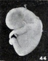
Mall_Meyer1921_fig145.jpg|Fig. 145. | Mall_Meyer1921_fig145.jpg|Fig. 145. Enlarged fetus from same, showing maceration effects. X2. | ||
Mall_Meyer1921_fig146.jpg| | Mall_Meyer1921_fig146.jpg|Figs. 146. Macerated hydatiform villi from the same case. X6. | ||
Mall_Meyer1921_fig147.jpg| | Mall_Meyer1921_fig147.jpg|Figs. 147. Macerated hydatiform villi from the same case. X6. | ||
Mall_Meyer1921_fig148.jpg|Fig. 148. | Mall_Meyer1921_fig148.jpg|Fig. 148. Cyema covered with magma. No. 1771. (See Chapter IX.) X2.67. | ||
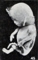
Mall_Meyer1921_fig149.jpg|Fig. 149. | Mall_Meyer1921_fig149.jpg|Fig. 149. Apparent superfetus from a cat. (After Harman.) | ||
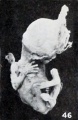
Mall_Meyer1921_fig150.jpg|Fig. 150. | Mall_Meyer1921_fig150.jpg|Fig. 150. Macerated, distorted normal cat fetus 10 mm. long. (After Kunz.) | ||
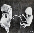
Mall_Meyer1921_fig151.jpg|Fig. 151. | Mall_Meyer1921_fig151.jpg|Fig. 151. Slightly macerated twin fetus. No. 1840a. X 11.35. | ||
Mall_Meyer1921_fig152.jpg|Fig. 152. | Mall_Meyer1921_fig152.jpg|Fig. 152. Pseudosupcrfetus. No. 1840b. X2.67. | ||
Mall_Meyer1921_fig153.jpg|Fig. 153. | Mall_Meyer1921_fig153.jpg|Fig. 153. Opened, everted vesicle of No. 1840a and incised vesicle of 18406. X0.66. | ||
Mall_Meyer1921_fig154.jpg|Fig. 154. | Mall_Meyer1921_fig154.jpg|Fig. 154. Opened chorionic vesicle of 1840b and placcntal area of 1840a. X0.66. | ||
Mall_Meyer1921_fig155.jpg|Fig. 155. | Mall_Meyer1921_fig155.jpg|Fig. 155. Twin fetus. No. 2036b. X0.66. | ||
Mall_Meyer1921_fig156.jpg|Fig. 156. | Mall_Meyer1921_fig156.jpg|Fig. 156. Pseudosuperfetus, No. 2036a, disarticulated by maceration. X2. | ||
</gallery> | </gallery> | ||
===Plate 15 Figures=== | ===Plate 15 Figures=== | ||
<gallery> | <gallery> | ||
| Line 308: | Line 313: | ||
Mall_Meyer1921_fig161.jpg|Fig. 161. Drawing of a portion of the ovary, indicated by small square to the right in fig. 159 (marked fig. 5 in drawing). X70. | Mall_Meyer1921_fig161.jpg|Fig. 161. Drawing of a portion of the ovary, indicated by small square to the right in fig. 159 (marked fig. 5 in drawing). X70. | ||
Mall_Meyer1921_fig162.jpg|Fig. 162. Drawing of a section through free surface of clot, illustrated in section in fig. 158, showing wall of chorion and a villus surrounded by clot. X50. | Mall_Meyer1921_fig162.jpg|Fig. 162. Drawing of a section through free surface of clot, illustrated in section in fig. 158, showing wall of chorion and a villus surrounded by clot. X50. | ||
</gallery> | |||
===Plate 16 Figures=== | |||
<gallery> | |||
Mall_Meyer1921_fig163.jpg|Fig. 163. External appearance of intact specimen. No. 1522. X0.75. | |||
Mall_Meyer1921_fig164.jpg|Fig. 164. External appearance of same specimen, showing where block was removed. X0.75. | |||
Mall_Meyer1921_fig165.jpg|Fig. 165. Appearance of cross section of pregnant ovary and tube, same specimen. X0.75. | |||
Mall_Meyer1921_fig166.jpg|Fig. 166. Photograph of a section taken from the thick portion of the ovarian stroma near the mesovarium, showing a well-developed Graafian follicle. Same specimen. X2.25. | |||
Mall_Meyer1921_fig167.jpg|Fig. 167. Section of part of the same specimen, showing the clot which contains the empty vesicle largely surrounded by ovarian stroma. XI. 9. | |||
Mall_Meyer1921_fig168.jpg|Fig. 168. Marked clubbing of villi of No. 550. X2.25. | |||
Mall_Meyer1921_fig169.jpg|Fig. 169. Cross section of decidua and conceptus. No. 698. (See Chapter XII.) X4.5. | |||
Mall_Meyer1921_fig170.jpg|Fig. 170. Section of conceptus, decidua, and muscularis. No. 970. (See Chapter XII.) X4.5. | |||
Mall_Meyer1921_fig171.jpg|Fig. 171. Section of decidua and conceptus. No. 962. (See Chapter XII.) X4.5. | |||
Mall_Meyer1921_fig172.jpg|Fig. 172. Section of a part of the conceptus, showing chorionic membrane, cyemic (?) rudiment (x) and yolk-sac. No. 1843. (See Chapter XII.) X51.5. | |||
Mall_Meyer1921_fig173.jpg|Fig. 173. External view of No. 2047, showing the distended amnion. (See Chapter XII.) X2.25. | |||
Mall_Meyer1921_fig174.jpg|Fig. 174. Section of the tube and the conceptus. No. 977. (See Chapter XII.) X3.3. | |||
</gallery> | |||
===Plate 17 Figures=== | |||
<gallery> | |||
Mall_Meyer1921_fig175.jpg|Fig. 175. A portion of a chorionic vesicle, showing appearances identical with tissue cultures. No. 545. X135. | |||
Mall_Meyer1921_fig176.jpg|Fig. 176. Fetus showing gluing of hand to face. No. 316. | |||
Mall_Meyer1921_fig177.jpg|Fig. 177. Villi showing obliteration of vessels by maceration and disintegration. No. 640. | |||
Mall_Meyer1921_fig178.jpg|Fig. 178. Villi showing obliteration of vessels by proliferation. No. 317. X97.5. | |||
Mall_Meyer1921_fig179.jpg|Fig. 179. Macerated tubal specimen imbedded in clot and undergoing lysis. No. 1938. X0.75. | |||
Mall_Meyer1921_fig180.jpg|Fig. 180. Fetus showing continuity of epidermis across the mouth, with obliteration of the labial slit. No. 885. X 4.5. | |||
Mall_Meyer1921_fig181.jpg|Fig. 181. Macerated tubal specimen, only the chorionic vesicle remaining. No. 2035. X0.75. | |||
Mall_Meyer1921_fig182.jpg|Fig. 182. Slightly macerated villi. No. 275. X37.5. | |||
Mall_Meyer1921_fig183.jpg|Fig. 183. Greatly macerated villi. No. 7236. | |||
Mall_Meyer1921_fig184.jpg|Fig. 184. Macerated, long-retained villi, with lumina of capillaries plugged with coagulum. No. 286. X37.5. | |||
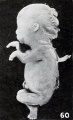
Mall_Meyer1921_fig185.jpg|Fig. 185. Fetus in sagittal section showing maceration, especially of the nervous system. No. 285. X4.5. | |||
Mall_Meyer1921_fig186.jpg|Fig. 186. Cross section of cyema showing homogeneous structure produced by maceration. No. 205. X 11.25. | |||
</gallery> | </gallery> | ||
| Line 316: | Line 355: | ||
File:Mall_Meyer1921_fig189.jpg|Fig. 189. A macerated, disproportional cyema 4 mm. long, showing a development of 5.5 mm. No. 786. X4. | File:Mall_Meyer1921_fig189.jpg|Fig. 189. A macerated, disproportional cyema 4 mm. long, showing a development of 5.5 mm. No. 786. X4. | ||
File:Mall_Meyer1921_fig190.jpg|Fig. 190. A well-preserved cyema 5.5 mm., of the same development as the preceding. No. 1380. X4. | File:Mall_Meyer1921_fig190.jpg|Fig. 190. A well-preserved cyema 5.5 mm., of the same development as the preceding. No. 1380. X4. | ||
File:Mall_Meyer1921_fig191.jpg|Figs. 191. Cyemata illustrating failure of extension of the body upon maceration. | File:Mall_Meyer1921_fig191.jpg|Figs. 191. Cyemata illustrating failure of extension of the body upon maceration. No. 1299 ( X2). | ||
File:Mall_Meyer1921_fig192.jpg|Figs. 192. Cyemata illustrating failure of extension of the body upon maceration. | File:Mall_Meyer1921_fig192.jpg|Figs. 192. Cyemata illustrating failure of extension of the body upon maceration. No. 2216 ( X4). | ||
File:Mall_Meyer1921_fig193.jpg|Figs. 193. Cyemata illustrating changes in form due to maceration. | File:Mall_Meyer1921_fig193.jpg|Figs. 193. Cyemata illustrating changes in form due to maceration. No. 208 (X2). | ||
File:Mall_Meyer1921_fig194.jpg|Figs. 194. Cyemata illustrating changes in form due to maceration. | File:Mall_Meyer1921_fig194.jpg|Figs. 194. Cyemata illustrating changes in form due to maceration. No. 1296 (X2.67). | ||
File:Mall_Meyer1921_fig195.jpg|Figs. 195. Cyemata illustrating changes in form due to maceration. | File:Mall_Meyer1921_fig195.jpg|Figs. 195. Cyemata illustrating changes in form due to maceration. No. 1697 (X2). | ||
File:Mall_Meyer1921_fig196.jpg|Figs. 196. Cyemata illustrating changes in form due to maceration. | File:Mall_Meyer1921_fig196.jpg|Figs. 196. Cyemata illustrating changes in form due to maceration. No. 1477 (X2). | ||
File:Mall_Meyer1921_fig197.jpg|Fig. 197. Illustrating beginning changes in form due to maceration. No. 705. X1.35. | File:Mall_Meyer1921_fig197.jpg|Fig. 197. Illustrating beginning changes in form due to maceration. No. 705. X1.35. | ||
File:Mall_Meyer1921_fig198.jpg|Figs. 198. Similar specimens, showing more pronounced changes. | File:Mall_Meyer1921_fig198.jpg|Figs. 198. Similar specimens, showing more pronounced changes. No. 2244 (X2.67). | ||
File:Mall_Meyer1921_fig199.jpg|Figs. 199. Similar specimens, showing more pronounced changes. | File:Mall_Meyer1921_fig199.jpg|Figs. 199. Similar specimens, showing more pronounced changes. No. 1891 (X2). | ||
File:Mall_Meyer1921_fig200.jpg|Figs. 200. Similar specimens, showing more pronounced changes. | File:Mall_Meyer1921_fig200.jpg|Figs. 200. Similar specimens, showing more pronounced changes. No. 1655 (X2.67). | ||
File:Mall_Meyer1921_fig201.jpg|Figs. 201. Similar specimens, showing more pronounced changes. | File:Mall_Meyer1921_fig201.jpg|Figs. 201. Similar specimens, showing more pronounced changes. No. 1260 (X2.67). | ||
File:Mall_Meyer1921_fig202.jpg|Figs. 202. Similar specimens, showing more pronounced changes. | File:Mall_Meyer1921_fig202.jpg|Figs. 202. Similar specimens, showing more pronounced changes. No. 1333 (X2.67). | ||
File:Mall_Meyer1921_fig203.jpg|Figs. 203. Similar specimens, showing more pronounced changes. | File:Mall_Meyer1921_fig203.jpg|Figs. 203. Similar specimens, showing more pronounced changes. No. 1379 (X2.67). | ||
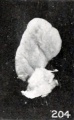
File:Mall_Meyer1921_fig204.jpg|Figs. 204. Similar specimens, showing more pronounced changes. 2244 (X2.67), 1891 (X2), 1655 (X2.67), 1260 (X2.67), 1333 (X2.67), 1379 (X2.67). | File:Mall_Meyer1921_fig204.jpg|Figs. 204. Similar specimens, showing more pronounced changes. 2244 (X2.67), 1891 (X2), 1655 (X2.67), 1260 (X2.67), 1333 (X2.67), 1379 (X2.67). | ||
File:Mall_Meyer1921_fig205.jpg|Figs. 205. Doubtful normally developed cyemata. | File:Mall_Meyer1921_fig205.jpg|Figs. 205. Doubtful normally developed cyemata. No. 1226 (X2.67). | ||
File:Mall_Meyer1921_fig206.jpg|Figs. 206. Doubtful normally developed cyemata. | File:Mall_Meyer1921_fig206.jpg|Figs. 206. Doubtful normally developed cyemata. No. 2361 (X4). | ||
File:Mall_Meyer1921_fig207.jpg|Fig. 207. A cyema illustrating post-partum changes. No. 589. X2.67. | File:Mall_Meyer1921_fig207.jpg|Fig. 207. A cyema illustrating post-partum changes. No. 589. X2.67. | ||
File:Mall_Meyer1921_fig208.jpg|Fig. 208. Cyema showing maceration sulci and ridges, and drooping of the limbs. No. 52 If. X1.66. | File:Mall_Meyer1921_fig208.jpg|Fig. 208. Cyema showing maceration sulci and ridges, and drooping of the limbs. No. 52 If. X1.66. | ||
Latest revision as of 15:50, 15 January 2013
Figures, Plates and Tables

| Embryology - 18 Apr 2024 |
|---|
| Google Translate - select your language from the list shown below (this will open a new external page) |
|
العربية | català | 中文 | 中國傳統的 | français | Deutsche | עִברִית | हिंदी | bahasa Indonesia | italiano | 日本語 | 한국어 | မြန်မာ | Pilipino | Polskie | português | ਪੰਜਾਬੀ ਦੇ | Română | русский | Español | Swahili | Svensk | ไทย | Türkçe | اردو | ייִדיש | Tiếng Việt These external translations are automated and may not be accurate. (More? About Translations) |
Mall FP. and Meyer AW. Studies on abortuses: a survey of pathologic ova in the Carnegie Embryological Collection. (1921) Contrib. Embryol., Carnegie Inst. Wash. Publ. 275, 12: 1-364.
- In this historic 1921 pathology paper, figures and plates of abnormal embryos are not suitable for young students.
1921 Carnegie Collection - Abnormal: Preface | 1 Collection origin | 2 Care and utilization | 3 Classification | 4 Pathologic analysis | 5 Size | 6 Sex incidence | 7 Localized anomalies | 8 Hydatiform uterine | 9 Hydatiform tubal | Chapter 10 Alleged superfetation | 11 Ovarian Pregnancy | 12 Lysis and resorption | 13 Postmortem intrauterine | 14 Hofbauer cells | 15 Villi | 16 Villous nodules | 17 Syphilitic changes | 18 Aspects | Bibliography | Figures | Contribution No.56 | Contributions Series | Embryology History
| Historic Disclaimer - information about historic embryology pages |
|---|
 |
- K12 Note - The historic images in the paper figures and plates of abnormal embryos may not be suitable for young K12 students.

Preface Figure - Terminology
Plates
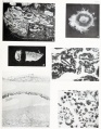
Plate 1
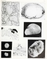
Plate 2
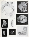
Plate 3
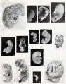
Plate 4
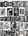
Plate 5
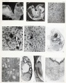
Plate 6
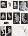
Plate 7
Plate 8
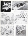
Plate 9
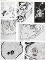
Plate 10

Plate 11
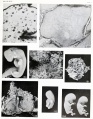
Plate 12
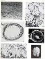
Plate 13
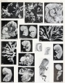
Plate 14
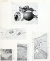
Plate 15
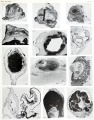
Plate 16
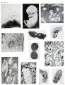
Plate 17
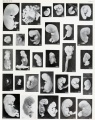
Plate 18
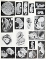
Plate 19
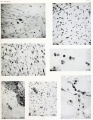
Plate 20
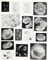
Plate 21
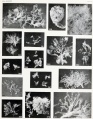
Plate 22
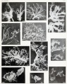
Plate 23
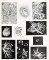
Plate 24


- Plate 2: Fig. 12 | Fig. 13 | Fig. 14 | Fig. 15 | Fig. 16 | Fig. 17 | Fig. 18 | Fig. 19 | Chapter 4 Pathologic analysis
- Plate 3: Fig. 20 | Fig. 21 | Fig. 22 | Fig. 23 | Fig. 24 | Fig. 25 | Fig. 26 | Fig. 27 | Chapter 4 Pathologic analysis
- Plate 4: Fig. 28 | Fig. 29 | Fig. 30 | Fig. 32 | Fig. 33 | Fig. 34 | Fig. 35 | Fig. 36 | Fig. 37 | Fig. 38 | Fig. 39 | Fig. 40 | Fig. 41 | Chapter 4 Pathologic analysis
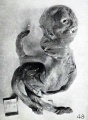
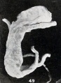
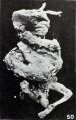
- Plate 5: Fig. 42 | Fig. 43 | Fig. 44 | Fig. 45 | Fig. 46 | Fig. 47 | Fig. 48 | Fig. 49 | Fig. 50 | Fig. 51 | Fig. 52 | | Fig. 53 | Fig. 54 | Fig. 55 | Fig. 56 | Fig. 57 | Fig. 58 | Fig. 59 | Fig. 60 | Fig. 61 | Fig. 62 | Fig. 63 | Fig. 64 | Fig. 65 | Fig. 66 | Fig. 67 | Fig. 68 | Fig. 69 | Fig. 70 | Fig. 71 | Fig. 72 | Chapter 4 Pathologic analysis
- Plate 6: Fig. 73 | Fig. 74 | Fig. 75 | Fig. 76 | Fig. 77 | Fig. 78 | Fig. 79 | Fig. 80 | Fig. 81 | Fig. 82 | Chapter 4 Pathologic analysis
- Plate 7: Fig. 83 | Fig. 84 | Fig. 85 | Fig. 86 | Fig. 87 | Fig. 88 | Fig. 89 | Fig. 90 | Fig. 91 | Fig. 92 | Chapter 7 Localized anomalies
- Plate 8: Fig. 96 | Fig. 97 | Fig. 98 | Fig. 99 | Fig. 100 | Fig. 101 | Fig. 102 | Fig. 103 | Fig. 104 | Fig. 105 | Fig. 106 | Fig. 107 | Chapter 8. Hydatiform Degeneration in Uterine Pregnancy
- Plate 9: Fig. 108 | Fig. 109 | Fig. 110 | Fig. 111 | Fig. 112 | Fig. 113 | Chapter 8. Hydatiform Degeneration in Uterine Pregnancy
- Plate 10: Fig. 114 | Fig. 115 | Fig. 116 | Fig. 117 | Fig. 118 | Fig. 119 | Fig. 120 | Chapter 8. Hydatiform Degeneration in Uterine Pregnancy
- Plate 11: Fig. 120 | Fig. 121 | Fig. 122 | Fig. 123 | Fig. 124 | Fig. 125 | Fig. 126 | Chapter 8. Hydatiform Degeneration in Uterine Pregnancy
- Plate 12: Fig. 127 | Fig. 128 | Fig. 129 | Fig. 130 | Fig. 131 | Fig. 132 | Fig. 133 | Fig. 134 | Chapter 8. Hydatiform Degeneration in Uterine Pregnancy
- Plate 13: Fig. 135 | Fig. 136 | Fig. 137 | Fig. 138 | Fig. 139 | Fig. 140 | Chapter 8. Hydatiform Degeneration in Uterine Pregnancy
- Plate 14: Fig. 141 | Fig. 142 | Fig. 143 | Fig. 144 | Fig. 145 | Fig. 146 | Fig. 147 | Fig. 148 | Fig. 149 | Fig. 150 | Fig. 151 | Fig. 152 | Fig. 153 | Fig. 154 | Fig. 155 | Figs. 156 | Chapter 10 Alleged superfetation
- Plate 16: Figs. 163 | Fig. 164 | Fig. 165 | Fig. 166 | Fig. 167 | Fig. 168 | Fig. 169 | Fig. 170 | Fig. 171 | Fig. 172 | Fig. 173 | Fig. 174 | Chapter 11 Ovarian Pregnancy
- Plate 17: Figs. 175 | Fig. 176 | Fig. 177 | Fig. 178 | Fig. 179 | Fig. 180 | Fig. 181 | Fig. 182 | Fig. 183 | Fig. 184 | Fig. 185 | Fig. 186 | 13 Postmortem intrauterine Changes
- Plate 18: Figs. 187 | Figs. 188 | Fig. 189 | Fig. 190 | Fig. 191 | Fig. 192 | Fig. 193 | Fig. 194 | Fig. 195 | Fig. 196 | Fig. 197 | Fig. 198 | Fig. 199 | Fig. 200 | Fig. 201 | Fig. 202 | Fig. 203 | Fig. 204 | Fig. 205 | Fig. 206 | Fig. 207 | Fig. 208 | Fig. 209 | Fig. 210 | Fig. 211 | Fig. 212 | Fig. 213 | Fig. 214 | Fig. 215 | Fig. 216 | Chapter 13 Post-Mortem Intrauterine Changes
- Plate 19: Figs. 217 | Figs. 218 | Fig. 219 | Fig. 220 | Fig. 221 | Fig. 222 | Fig. 223 | Fig. 224 | Fig. 225 | Fig. 226 | Fig. 227 | Fig. 228 | Fig. 229 | Fig. 230 | Fig. 231 | Fig. 232 | Fig. 233 | Fig. 234 | Chapter 13 Post-Mortem Intrauterine Changes
- Plate 20: Fig. 235 | Fig. 236 | Fig. 237 | Fig. 238 | Fig. 239 | Fig. 240 | Chapter 14 Hofbauer cells
- Plate 21: Fig. 242 | Fig. 243 | Fig. 244 | Fig. 245 | Fig. 246 | Fig. 247 | Fig. 248 | Fig. 249 | Fig. 250 | Fig. 251 | Fig. 252 | Fig. 253 | Fig. 254 | Fig. 255 | Fig. 256 | Chapter 15 Villi
- Plate 22: Fig. 257 | Fig. 258 | Fig. 259 | Fig. 260 | Fig. 261 | Fig. 262 | Fig. 263 | Fig. 264 | Fig. 265 | Fig. 266 | Fig. 267 | Fig. 268 | Fig. 269 | Fig. 270 | Fig. 271 | Chapter 15 Villi
- Plate 23: Fig. 272 | Fig. 273 | Fig. 274 | Fig. 275 | Fig. 276 | Fig. 277 | Fig. 278 | Fig. 279 | Fig. 280 | Fig. 281 | Fig. 282 | Chapter 15 Villi
- Plate 24: Fig. 283 | Fig. 284 | Fig. 285 | Fig. 286 | Fig. 287 | Fig. 288 | Fig. 289 | Fig. 290 | Fig. 291 | Fig. 292 | Chapter 16 Villous Nodules
Figures
In-text Figures
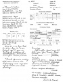
Fig. 1. Specimen page from "Key," showing manner in which records of embryos are entered

Fig. 2. Blank upon which preliminary description of specimen is entered
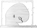
Fig. 3. Method of piling with cardboard outline as guide
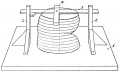
Fig. 4. Method of making guide lines
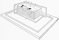
Fig. 5. Method of piling wax mold


Plate 1 Figures
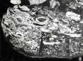
Fig. 6. Old retained hydatiform degeneration. No. 323.
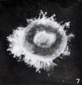
Fig. 7. Abnormal conceptus. No. 1843.
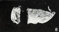
Fig. 8. Decidual cast containing remnants of conceptus. No. 698.
Fig. 9. Festoons of chorionic epithelium. No. 1324.
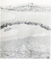
Fig. 10. Detached conceptus, still in utero. No. 1224.
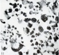
Fig. 11. Almost total lysis or gossamer form. No. 606.


Plate 2 Figures
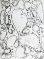
Fig. 12. Villi showing hydatiform degeneration. Same specimen as fig. 10.
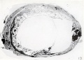
Fig. 13. Hydatiform tubal twins in situ. No. 825.
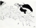
Fig. 14. A fine hydatiform villous tree in section. No. 367.
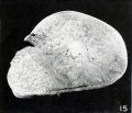
Fig. 15. Full-term nodule accompanying a twin donated by Dr. Slemona. No. 1682.
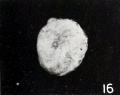
Fig. 16. An unusual nodular form. No. 2288.
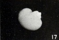
Fig. 17. Reniform nodular cyema. No. 1369.
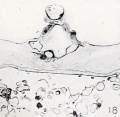
Fig. 18. Cross-section of a nodular cyema, showing simple structure. No. 6510.
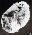
Fig. 19. External appearance of chorion containing small nodular twin. No. 7886.


Plate 3 Figures
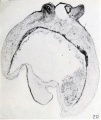
Fig. 20. Cross-section of No. 7886, shown in fig. 19. X45.
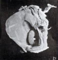
Fig. 21. Macerated normal cyema, No. 788a, companion to 7886. Xl.
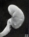
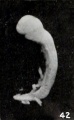
Fig. 22. Divergence in external form of cylindrical cyemata. No. 885. X4.
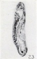
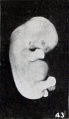
Fig. 23. Illustrating marked changes in form and structure of cylindrical cyemata. No. 446. X6.
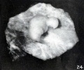
Figs. 24-26. Fig. 24. Fig. 25. Fig. 26. Illustrating variation in form of cylindrical cyemata. Nos. 2236 (X9),2222 (X6),and 1776 (X6).
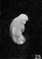
Figs. 24-26. Fig. 24. Fig. 25. Fig. 26. Illustrating variation in form of cylindrical cyemata. Nos. 2236 (X9),2222 (X6),and 1776 (X6).
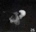
Figs. 24-26. Fig. 24. Fig. 25. Fig. 26. Illustrating variation in form of cylindrical cyemata. Nos. 2236 (X9),2222 (X6),and 1776 (X6).
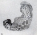
Fig. 27. Longitudinal section showing structure of cylindrical cyema. No. 150. X15.


Plate 4 Figures
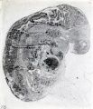
Fig. 28. Longitudinal section of cylindrical embryo. No. 489. X37.5.
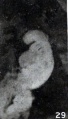
Fig. 29. A cyema in the pre-gossamer stage of lysis. No. 2197. X6.75.
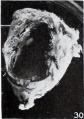
Fig. 30. Twin conceptus with larger chorion opened and location of smaller indicated. No. 587. XU.75.
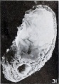
Fig. 31. Part of the same conceptus in section, showing the cavities of both chorionic vesicles. Xl.5.
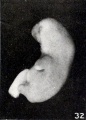
Fig. 32. Side view of cyema, showing so-called stunting. No. 2173. X3.
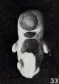
Fig. 33. Front view of same specimen, showing maceration and stunting. X3.
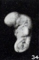
Fig. 34. Illustrating stunting. No. 2233. X4.5.
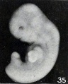
Fig. 35. External view of No. 2473, showing stunting in an older specimen. X3.
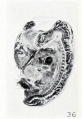
Fig. 36. Section of a similar fetus, to show structure. No. 675. X4.5.
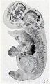
Fig. 37. Sagittal section illustrating fusion of mandibular margin with chest. No. 788a. X4.5.
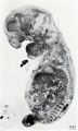
Fig. 38. Sagittal section showing internal maceration changes and gaping of the mouth.
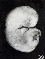
Fig. 39. Macerated, firmly rolled-up, young fetus. No. 921. X2.25.
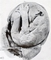
Fig. 40. Macerated, rolled-up, older fetus. No. 1041. X0.75.
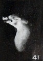
Fig. 41. Cyema classed as fetuses compressus. No. 1245. X3.


Plate 5 Figures
Figs. 42-50. Various forms of cyemata classed as fetus compressus: Nos. 1463 (X2).
Figs. 42-50. Various forms of cyemata classed as fetus compressus: Nos. 1806 (X 1.5).
Figs. 42-50. Various forms of cyemata classed as fetus compressus: Nos. 1301 (X8.5).
Figs. 42-50. Various forms of cyemata classed as fetus compressus: Nos. 1513 (X0.58).
Figs. 42-50. Various forms of cyemata classed as fetus compressus: Nos. 1495c (X0.58).
Figs. 42-50. Various forms of cyemata classed as fetus compressus: Nos. 896 (X.55).
Figs. 42-50. Various forms of cyemata classed as fetus compressus: Nos. 1925 (X.65).
Figs. 42-50. Various forms of cyemata classed as fetus compressus: Nos. 1239 (X0.55).
Figs. 42-50. Various forms of cyemata classed as fetus compressus: Nos. 1515 (X0.58).
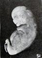
Fig. 51. Distortion due to maceration and retention in utero. No. 1931. XI.
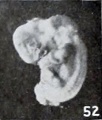
Figs. 52-63. Illustrating the absence of fundamental differences between the group of fetus compressus and grade 3 of the normal specimens. No. 12956 (fig. 52) pathological.
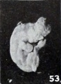
Figs. 52-63. Illustrating the absence of fundamental differences between the group of fetus compressus and grade 3 of the normal specimens. No. 532 (fig. 53) normal. Xl.8.
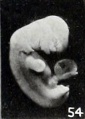
Figs. 52-63. Illustrating the absence of fundamental differences between the group of fetus compressus and grade 3 of the normal specimens. No. 1475 (fig. 54) pathological. X1.5.
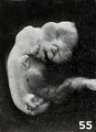
Figs. 52-63. Illustrating the absence of fundamental differences between the group of fetus compressus and grade 3 of the normal specimens. No. 852 (fig. 55) normal. X2.
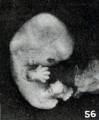
Figs. 52-63. Illustrating the absence of fundamental differences between the group of fetus compressus and grade 3 of the normal specimens. No. 1750 (fig. 56) normal. X1.5. (To be compared with No. 1806, fig. 43.)
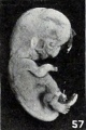
Figs. 52-63. Illustrating the absence of fundamental differences between the group of fetus compressus and grade 3 of the normal specimens. No. 1779 (fig. 57) normal. XI.
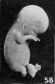
Figs. 52-63. Illustrating the absence of fundamental differences between the group of fetus compressus and grade 3 of the normal specimens. No. 651a (fig. 58) pathological. XI.
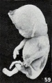
Figs. 52-63. Illustrating the absence of fundamental differences between the group of fetus compressus and grade 3 of the normal specimens. No. 1513 (fig. 59) pathological. X0.58.
Figs. 52-63. Illustrating the absence of fundamental differences between the group of fetus compressus and grade 3 of the normal specimens. No. 1350 (fig. 60) normal. X0.58.
Figs. 52-63. Illustrating the absence of fundamental differences between the group of fetus compressus and grade 3 of the normal specimens. No. 1474<z (fig. 61) normal. X0.58. (To be compared with No. 896, fig. 47 )
Figs. 52-63. Illustrating the absence of fundamental differences between the group of fetus compressus and grade 3 of the normal specimens. No. 1758 (fig. 62) pathological. X0.5. No. 1532 (fig. 63) normal. X0.5.
Figs. 52-63. Illustrating the absence of fundamental differences between the group of fetus compressus and grade 3 of the normal specimens. No. 1532 (fig. 63) normal. X0.5.
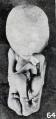
Fig. 64. Fusion of epidermis on lower extremities during maceration. No. 1859. X0.5. Fig.
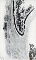
Fig. 65. Epidermal thickenings. No. 316. X7.5.
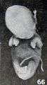
Fig. 66. Early drooping and distortion of extremities. No. 1710. X 1.
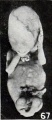
Fig. 67. Showing diversity in position and distortion of extremities. No. 1751. XO 5
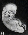
Fig. 68. A similar but younger specimen. No. 1931. XI.
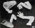
Fig. 69. Distortion of digits. No. 1751. XI.
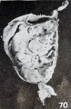
Fig. 70. Unilateral maceration bleb on cord. No. 1523. X0.5.
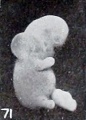
Fig. 71. Bleb formation on neck, same specimen. XI.
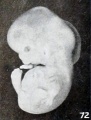
Fig. 72. Bleb formation in whole nuchal region. No. 2261. XI. 5.


Plate 6 Figures
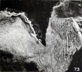
Fig. 73. Nodular fibrosis of the amnion. No. 868. Xl-35.
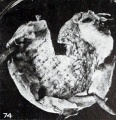
Fig. 74. Nodular fibrosis of the amnion. No. 1048. X0.66.
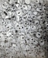
Fig. 75. Large, clear, normal decidual cells. No. 1599. X200.
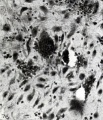
Fig. 76. Maceration, hemorrhage, and lysis, with early transformation of the decidua. No. 872. X200.
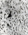
Fig. 77. Further transformation of the polygonal decidual cells into the spindle and fibroblast type of cells. Same specimen as figure 76. X200.
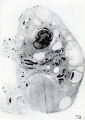
Fig. 78. Longitudinal section of right ovary, showing carcinomatous nodule to the right. No. 865. X2.
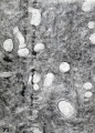
Fig. 79. Same specimen. Portion of carcinomatous nodule. X27.
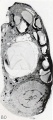
Fig. 80. Left ovary from same case in longitudinal section, showing disk-shaped teratoma in section. X2. Fig.
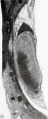
Fig. 81. Teratoma in section, same case. X11.5.
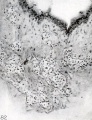
Fig. 82. Remarkable vascular proliferation in the chorionic membrane. No. 309. X 120.


Plate 7 Figures
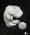
Fig. 83. Normal embryo with cyclopia ; in front of the eye is seen the Cyclopean snout. No. 559. X3.75.
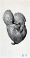
Fig. 84. Normal double monster. No. 249. X0.75.
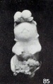
Fig. 85. Specimen with hare-lip and exencephaly. No. 364. X2.25.
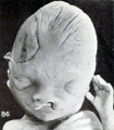
Fig. 86. Specimen with hare-lip. No. 982. XI. 5.
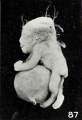
Fig. 87. Stunted fetus with large hernia in umbilical cord. No. 1330. X0.9.
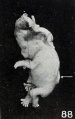
Fig. 88. Normal embryo with exencephaly and spina bifida (the latter opposite the arrow). No. 1315. XI. 5.
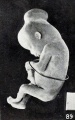
Fig. 89. Normal fetus with hernia of mid-brain. No. 1690. X6.75.
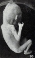
Fig. 90. Anomaly of left hand of No. 306a. Only the thumb and little finger are normal.
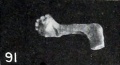
Fig. 91. Left hand, which is club-shaped, from No. 230, a fetus compressus 57 mm. CR. X0.75.
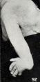
Fig. 92. Deformed wrist with atrophic radius in a normal embryo. No. 789, 50 mm. CR. X3.
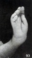
Fig. 93. Right hand with six fingers from macerated specimen. No. 1749. There were six digits on each of the four extremities. X3.
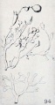
Fig. 94. Hydatiform villi. (After Gierse.) See Chapter VIII.
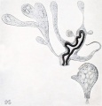
Fig. 95. Hydatiform villi showing vacuolation. (After Gierse.) See Chapter VIII.


Plate 8 Figures
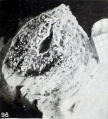
Fig. 96. Young chorionic vesicle in situ, showing hydatiform degeneration of the stroma and syncytial budding. No. 836. X2.6.
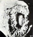
Fig. 97. Same, under greater enlargement, with the cyema in view. X3.75.
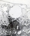
Fig. 98. Hydatiform degeneration. A small portion of No. 1189 still in loco. X4.5.
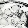
Fig. 99. Another portion of the same specimen. X22.5.
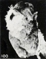
Fig. 100. External appearance of an apparently normal conceptus, partly surrounded by decidua. No. 2077. X0.75.
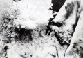
Fig. 101. A portion of the same specimen, clearly showing hydatiform degeneration where in focus. The decidua is reflected. X3.
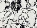
Fig. 102. Portion of villi from No. 690, showing numerous syncytial buds, absence of vessels, and glassy stroma. X37.5.
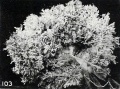
Fig. 103. A chorionic vesicle showing shaggy appearance. No. 2258. XI. 5.
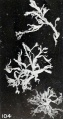
Fig. 104. Hydatiform villi from same specimen. X4.5.
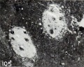
Fig. 105. Macerated normal villi in blood clot. No. 640. X71.
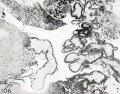
Fig. 106. Well preserved, early hydatiform villi from the same specimen. X33.25.
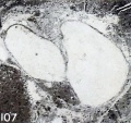
Fig. 107. Hydatiform villi without marked epithelial proliferation. No. 7206.


Plate 9 Figures
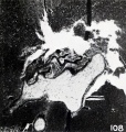
Fig. 108. Hydatiform villi with pronounced epithelial proliferation. No. 7206, also shown in figure 107. X50.
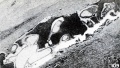
Fig. 109. Hydatiform villi without (at left) and with marked epithelial proliferation (at right). No. 540.
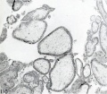
Fig. 110. Fairly late stage in the degeneration of the villous vessels in hydatiform villi. No. 977. X50.
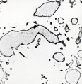
Fig. 111. A slightly later stage in the degeneration of the villous vessels. No. 516.
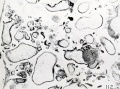
Fig. 112. Almost complete disappearance of the vessels. No. 8746. X50.
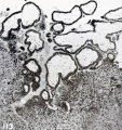
Fig. 113. Non-vascular, early hydatiform implanted villi. No. 396. X50.


Plate 10 Figures
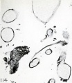
Fig. 114. Portion of No. 435a, showing a small villus in the center with an extremely long syncytial bud. X50.
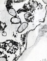
Fig. 115. Framework of syncytium arising from the chorionic membrane and from the villi. No. 962<z. X50.
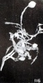
Fig. 116. Apical trophoblastic nodule. No. 516. X6.
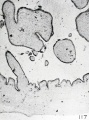
Fig. 117. A portion of the chorionic membrane from No. 714, showing decidedly sinuous epithelium. X50.
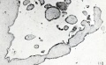
Fig. 118. An early, non- vascular, hydatid villus from the same specimen, showing constrictions. X50.
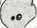
Fig. 119. Epithelial vesicles within the stroma. No. 872. X180.
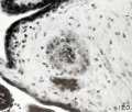
Fig. 120. Extremity of an epithelial vesicle within the stroma, same specimen. X300.


Plate 11 Figures
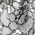
Fig. 121. Slight epithelial proliferation on markedly hydatiform villi. No. 134. X50.
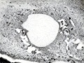
Fig. 122. Extremely marked epithelial proliferation on a small hydatiform villus with glassy stroma. No. 415. X95.
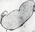
Fig. 123. The disintegrating capillaries are represented merely by large, incomplete curved outlines. No. 749. X50.

Fig. 124. Collapsed capillaries in process of obliteration. No. 712. X95.
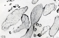
Fig. 125. Fenestrated, macerated, hydatiform villi among others which are quite fibrous. No. 651d. X50.
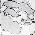
Fig. 126. Villi showing only a trace of the capillaries. No. 651<7- X50.


Plate 12 Figures
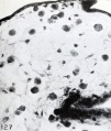
Fig. 127. Section of a villus from No. 510, showing scattered Hofbauer cells. X300.
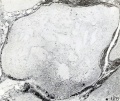
Fig. 128. Section of a villus from No. 749, showing accumulation of Hofbauer cells, especially at the right. X50.
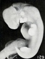
Fig. 129. A somewhat macerated normal embryo from a case of early hydatiform degeneration. No. 2099. X6.
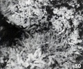
Fig. 130. The chorionic vesicle of the same specimen, showing early hydatiform villi to left where in focus. X4.
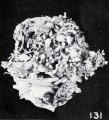
Fig. 131. Basal area of No. 1260, showing pronounced hydatiform degeneration of the villi. XI.
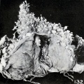
Fig. 132. The same specimen, opened in the capsular region to show the presence of the cyema. Xl.l.
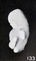
Fig. 133. Marked macerated and deformed fetus from the same case. X4.
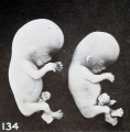
Fig. 134. Twin fetuses, No. 2250a and 6, a case of hydatiform degeneration. XI.


Plate 13 Figures
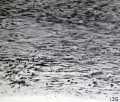
Fig. 135. A portion of a rather fibrous decidua. No. 874o. X300. (See Chap. VIII.)
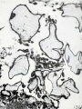
Fig. 136. Hydatiform villi in section. No. 825. X45.
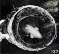
Fig. 137. Cross-section of tube and vesicle. No. 1771. X4.
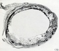
Fig. 138. Cross-section of same, showing hydatiform villi. X2.
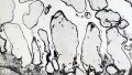
Fig. 139. Hydatiform villi from same, in section. X45.
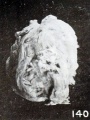
Fig. 140. Hydatiform villi protruding through an incision in the tube wall. No. 2052. X2.


Plate 14 Figures
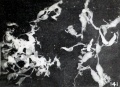
Figs. 141. Hydatiform villi from No. 2250a and b, shown in fig. 134, plate 12, Chapter VIII. X4.
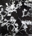
Figs. 142. Hydatiform villi from No. 2250a and b, shown in fig. 134, plate 12, Chapter VIII. X4.
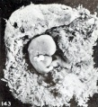
Figs. 143. Twin conceptuses, both with hydatiform chorions. No. 241 a and b. (See Chap. VIII.) X1.35.
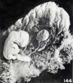
Figs. 144. Twin conceptuses, both with hydatiform chorions. No. 241 a and b. (See Chap. VIII.) X1.35.
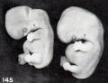
Fig. 145. Enlarged fetus from same, showing maceration effects. X2.
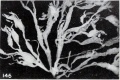
Figs. 146. Macerated hydatiform villi from the same case. X6.
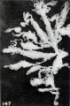
Figs. 147. Macerated hydatiform villi from the same case. X6.
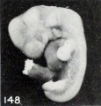
Fig. 148. Cyema covered with magma. No. 1771. (See Chapter IX.) X2.67.
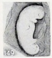
Fig. 149. Apparent superfetus from a cat. (After Harman.)
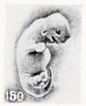
Fig. 150. Macerated, distorted normal cat fetus 10 mm. long. (After Kunz.)
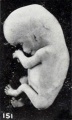
Fig. 151. Slightly macerated twin fetus. No. 1840a. X 11.35.
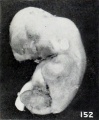
Fig. 152. Pseudosupcrfetus. No. 1840b. X2.67.
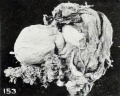
Fig. 153. Opened, everted vesicle of No. 1840a and incised vesicle of 18406. X0.66.
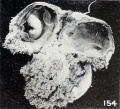
Fig. 154. Opened chorionic vesicle of 1840b and placcntal area of 1840a. X0.66.
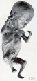
Fig. 155. Twin fetus. No. 2036b. X0.66.
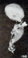
Fig. 156. Pseudosuperfetus, No. 2036a, disarticulated by maceration. X2.


Plate 15 Figures
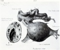
Fig. 157. Drawing of posterior view of pregnant ovary with tube. No. 550. Fig. 158. Transverse section of pregnant ovary cut at point of rupture.
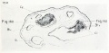
Fig. 159. Outline of transverse section of ovary taken in the region of the letter o in "Ovary" (fig. 157). X2.
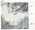
Fig. 160. Drawing of portion of the corpus luteum, indicated by small square to the left in fig. 159 (marked fig. 4 in drawing). X70.
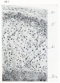
Fig. 161. Drawing of a portion of the ovary, indicated by small square to the right in fig. 159 (marked fig. 5 in drawing). X70.
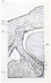
Fig. 162. Drawing of a section through free surface of clot, illustrated in section in fig. 158, showing wall of chorion and a villus surrounded by clot. X50.


Plate 16 Figures
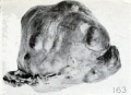
Fig. 163. External appearance of intact specimen. No. 1522. X0.75.
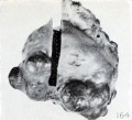
Fig. 164. External appearance of same specimen, showing where block was removed. X0.75.
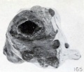
Fig. 165. Appearance of cross section of pregnant ovary and tube, same specimen. X0.75.
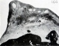
Fig. 166. Photograph of a section taken from the thick portion of the ovarian stroma near the mesovarium, showing a well-developed Graafian follicle. Same specimen. X2.25.
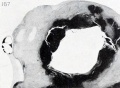
Fig. 167. Section of part of the same specimen, showing the clot which contains the empty vesicle largely surrounded by ovarian stroma. XI. 9.
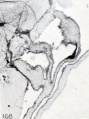
Fig. 168. Marked clubbing of villi of No. 550. X2.25.
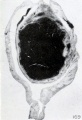
Fig. 169. Cross section of decidua and conceptus. No. 698. (See Chapter XII.) X4.5.
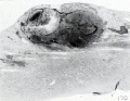
Fig. 170. Section of conceptus, decidua, and muscularis. No. 970. (See Chapter XII.) X4.5.
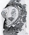
Fig. 171. Section of decidua and conceptus. No. 962. (See Chapter XII.) X4.5.
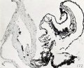
Fig. 172. Section of a part of the conceptus, showing chorionic membrane, cyemic (?) rudiment (x) and yolk-sac. No. 1843. (See Chapter XII.) X51.5.
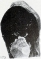
Fig. 173. External view of No. 2047, showing the distended amnion. (See Chapter XII.) X2.25.
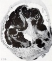
Fig. 174. Section of the tube and the conceptus. No. 977. (See Chapter XII.) X3.3.


Plate 17 Figures
Fig. 175. A portion of a chorionic vesicle, showing appearances identical with tissue cultures. No. 545. X135.
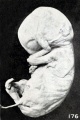
Fig. 176. Fetus showing gluing of hand to face. No. 316.
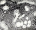
Fig. 177. Villi showing obliteration of vessels by maceration and disintegration. No. 640.
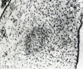
Fig. 178. Villi showing obliteration of vessels by proliferation. No. 317. X97.5.
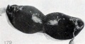
Fig. 179. Macerated tubal specimen imbedded in clot and undergoing lysis. No. 1938. X0.75.
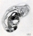
Fig. 180. Fetus showing continuity of epidermis across the mouth, with obliteration of the labial slit. No. 885. X 4.5.
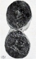
Fig. 181. Macerated tubal specimen, only the chorionic vesicle remaining. No. 2035. X0.75.
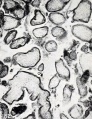
Fig. 182. Slightly macerated villi. No. 275. X37.5.
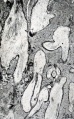
Fig. 183. Greatly macerated villi. No. 7236.
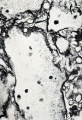
Fig. 184. Macerated, long-retained villi, with lumina of capillaries plugged with coagulum. No. 286. X37.5.
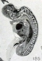
Fig. 185. Fetus in sagittal section showing maceration, especially of the nervous system. No. 285. X4.5.
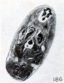
Fig. 186. Cross section of cyema showing homogeneous structure produced by maceration. No. 205. X 11.25.


Plate 18 Figures
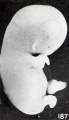
Fig. 187. Slight swelling of fetus from brief maceration. No. 2146. X2.
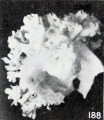
Fig. 188. Matted, slightly macerated villi. No. 1878. X4.
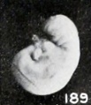
Fig. 189. A macerated, disproportional cyema 4 mm. long, showing a development of 5.5 mm. No. 786. X4.
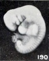
Fig. 190. A well-preserved cyema 5.5 mm., of the same development as the preceding. No. 1380. X4.
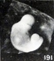
Figs. 191. Cyemata illustrating failure of extension of the body upon maceration. No. 1299 ( X2).
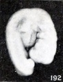
Figs. 192. Cyemata illustrating failure of extension of the body upon maceration. No. 2216 ( X4).
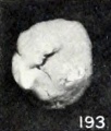
Figs. 193. Cyemata illustrating changes in form due to maceration. No. 208 (X2).
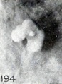
Figs. 194. Cyemata illustrating changes in form due to maceration. No. 1296 (X2.67).
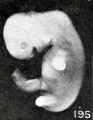
Figs. 195. Cyemata illustrating changes in form due to maceration. No. 1697 (X2).
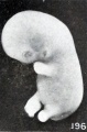
Figs. 196. Cyemata illustrating changes in form due to maceration. No. 1477 (X2).
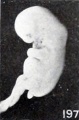
Fig. 197. Illustrating beginning changes in form due to maceration. No. 705. X1.35.
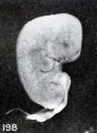
Figs. 198. Similar specimens, showing more pronounced changes. No. 2244 (X2.67).
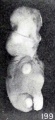
Figs. 199. Similar specimens, showing more pronounced changes. No. 1891 (X2).
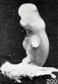
Figs. 200. Similar specimens, showing more pronounced changes. No. 1655 (X2.67).
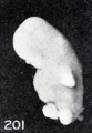
Figs. 201. Similar specimens, showing more pronounced changes. No. 1260 (X2.67).
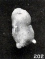
Figs. 202. Similar specimens, showing more pronounced changes. No. 1333 (X2.67).
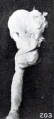
Figs. 203. Similar specimens, showing more pronounced changes. No. 1379 (X2.67).
Figs. 204. Similar specimens, showing more pronounced changes. 2244 (X2.67), 1891 (X2), 1655 (X2.67), 1260 (X2.67), 1333 (X2.67), 1379 (X2.67).
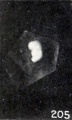
Figs. 205. Doubtful normally developed cyemata. No. 1226 (X2.67).
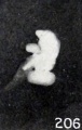
Figs. 206. Doubtful normally developed cyemata. No. 2361 (X4).
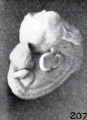
Fig. 207. A cyema illustrating post-partum changes. No. 589. X2.67.
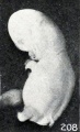
Fig. 208. Cyema showing maceration sulci and ridges, and drooping of the limbs. No. 52 If. X1.66.
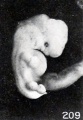
Fig. 209. Cyema showing maceration sulci and ridges, and swelling of the cord. X2.
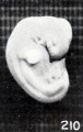
Fig. 210. Normal, well-preserved cat fetus.
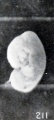
Fig. 211. Normal, poorly preserved cat fetus of approximately the same length.
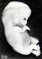
Fig. 212. Appearance of fetus before fixation. No. 1358. X2.
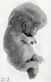
Fig. 213. The same specimen, showing wrinkling due to fixation and staining. X2.
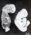
Fig. 214. Macerated, distorted single-ovum twins. No. 2258. X2.67.
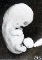
Fig. 215. Minor changes in relief, due to maceration. Swelling and constriction of cord. No. 2014. X2.
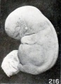
Fig. 216. An intermediate maceration form. No. 1495d. X2.67.


Plate 19 Figures
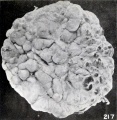
Fig. 217. Interior of the chorionic vesicle showing subchorial hematomata. No. 1495d. X0.77.
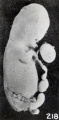
Fig. 218. A fetus and cord showing marked maceration changes. No. 797. X1.35.
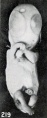
Fig. 219. An older fetus, showing bleb-formation and curvature in extremities, due to maceration and retention. No. 1860. XI- 35.
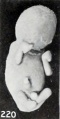
Fig. 220. A fetus showing marked clubbing of the extremities and obliteration of the features. No. 1958. X0.87.
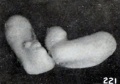
Figs. 221-222. Extremities of same specimen. X2.67.
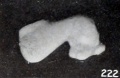
Figs. 221-222. Extremities of same specimen. X2.67.
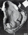
Fig. 223. External appearance of fetus in situ. No. 1751. X0.57.
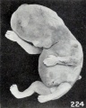
Fig. 224. An older, macerated fetus, with extremities extended instead of folded. No. 1462. X0.77.
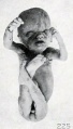
Fig. 225. A similar specimen. No. 1309. X0.23.
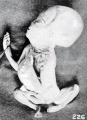
Fig. 226. A long-retained, soft, rolled-up form, photographed in extension. No. 1775. X0.47.
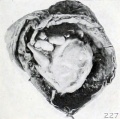
Fig. 227. A similar specimen with markedly flexed head. No. 1525. X0.66.
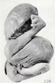
Fig. 228. A similar specimen. No. 1976. X0.47.
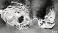
Fig. 229. A similar specimen with mummified chorionic vesicle. No. 1850. X6.
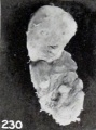
Fig. 230. A decidedly mummified fetus. No. 1295a. X0.77.
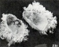
Fig. 231. Unilateral development of villi in a vesicle classed as normal (?). No. 2092. (See Chapter XV.) X1.35.
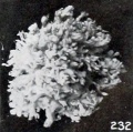
Fig. 232. A somewhat macerated young vesicle with quite uniformly distributed villi which are slightly abnormal in form and structure. No. 1878. (See Chapter XV.) X4.
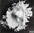
Fig. 233. A portion of same specimen, showing the somewhat more than normally bulbous and rather matted villi. X2.67.
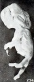
Fig. 234. A very softened, macerated fetus. X0.5.


Plate 20 Figures
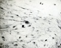
Fig. 235. Transition forms between mesenchyme and Hofbauer cells. No. 645, slide 3. X330.
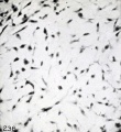
Fig. 236. Transition forms between mesenchyme and Hofbauer cells. No. 592, slide 1. X330.
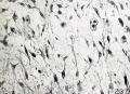
Fig. 237. Transition forms between mesenchyme and Hofbauer cells. No. 645, slide 3b. X330.
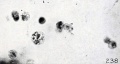
Fig. 238. A phagocytic pseudo-Hofbauer cell. No. 645, slide 2. X650.
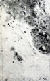
Fig. 239. Fusing Hofbauer cells forming a giant cell. No. 645, slide 2. X300.
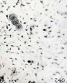
Fig. 240. Fusing Hofbauer cells forming a giant cell. No. 985, slide 1. X300.
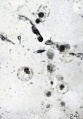
Fig. 241. Pseudo-Hofbauer cells in the ovary. No. 970. X650.


Plate 21 Figures
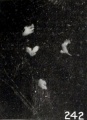
Fig. 242. Isolated villi from No. 1878, shown in figures 232 and 233, plate 19, Chapter XIII. X4.5.
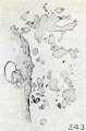
Figs. 243-244. Appearance of villi in section, same specimen. X7.5.
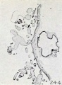
Figs. 243-244. Appearance of villi in section, same specimen. X7.5.
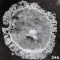
Fig. 245. Cross-section view of an entire normal vesicle with cyema and adnexa. No. 2053. X3.
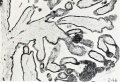
Fig. 246. Section of villi from same. X36.
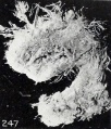
Fig. 247. External appearance of conceptus, showing filiform villi. No. 866. XI. 5.
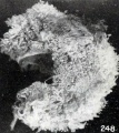
Fig. 248. External appearance of conceptus, showing presence of hydatiform villi. No. 2108. X2.25.
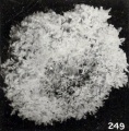
Fig. 249. Sparsely villous area from No. 1892. Xl.5.
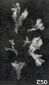
Fig. 250. Isolated villi from same specimen, showing maceration changes. X3.
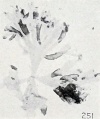
Fig. 251. Isolated shadow or gossamer villi. No. 2197. X9.
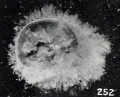
Fig. 252. External appearance of same specimen. Xl.5.
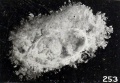
Fig. 253. Cross section of No. 1287. Xl.5.
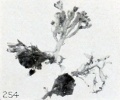
Fig. 254. Villi from No. 1466. X2.25.
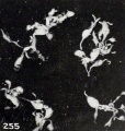
Fig. 255. Hydatiform villi from No. 2233. X4.
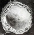
Fig. 256. Section of chorionic vesicle with hydatiform villi in basal area. No. 2361. Xl.5.


Plate 22 Figures
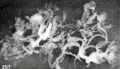
Fig. 257. Isolated villi from No. 2361, showing both maceration and hydatiform degeneration. X6.75.
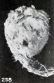
Fig. 258. External appearance of abortus, same case. X0.75.
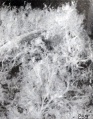
Fig. 259. Changes in external appearance of villi, due to maceration alone. No. 993. X3.
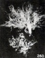
Fig. 260. Isolated villous tree from same vesicle. X3.
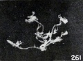
Fig. 261. Isolated filiform villi. No. 1639. X3.
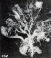
Fig. 262. Isolated villous tree. No. 2336. X6.75.
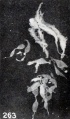
Fig. 263. Fine hydatiform villi. No. 1797. X3.
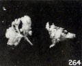
Fig. 264. Normal villi. No. 837. X2.25.
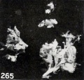
Fig. 265. Transitional villi between normal and hydatid. No. 432. X2.25.
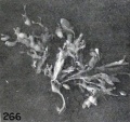
Fig. 266. Transitional hydatiform villi. No. 437. X2.25.
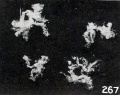
Fig. 267. Normal, knobbed or budded villi. No. 1063. X2.25.
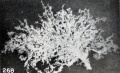
Fig. 268. Villous tree from a normal specimen, illustrating many small knobs. No. 2335. X6.75.
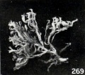
Fig. 269. Villous tree showing few buds or knobs. No. 1840a. X2.25.
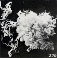
Fig. 270. Filiform villi and a portion of the vesicle from the same specimen. X3.
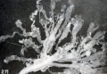
Fig. 271. Villi showing maceration changes. No. 2348. X6.75.


Plate 23 Figures
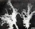
Fig. 272. Villi showing maceration changes. No. 2339. X6.75.
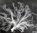
Fig. 273. Villous tree with smooth villi. No. 2253. X6.75.
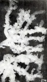
Fig. 274. Villous tree with knobbed villi. No. 2372. X6.75.
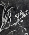
Fig. 275. Filiform villus. No. 2326. X6.75.
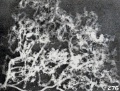
Fig. 276. Fine, frowsy villus. No. 2414. X6.75.
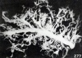
Fig. 277. Massive villous tree. No. 2283. X6.75.
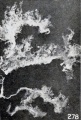
Fig. 278. Knobbed, curly villi. No. 2384. X6.75.
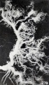
Fig. 279. Mossy villi. No. 2249. X4.5.
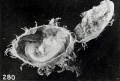
Fig. 280. Tubal conceptus (No. 1151), showing disproportion between the chorionic vesicle and the fetus, sparse development of the villi, and some hydatiform degeneration. Xl.5.
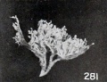
Fig. 281. Villous tree from No. 651ft. X2.25.
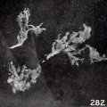
Fig. 282. Villi from No. 1151, shown in figure 280. X3.


Plate 24 Figures
Fig. 283. Cross section of intensely fibrous villi from No. 7656. (See Chapter XV.)
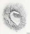
Fig. 284. Conceptus showing numerous trophoblastic nodules. (After Sommering.)
Fig. 285. Portion of chorionic vesicle showing an apical villous nodule to the right and others elsewhere. No. 1495a. X2.4.
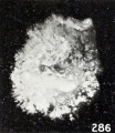
Fig. 286. Chorionic vesicle showing numerous small villous nodules. No. 2197. XI. 6.
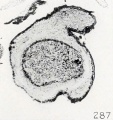
Fig. 287. A villous nodule completely surrounded by stroma, except at its free surface not shown in the figure. No. 556.
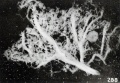
Fig. 288. A villous nodule near the base of a villous tree. No. 2365. X7.2.
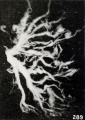
Fig. 289. Villous nodules and lichen-like streamers on a villous tree. No. 2204. X7.2.
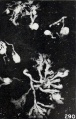
Fig. 290. A group of teased villi bearing numerous trophoblastic nodules. No. 2225. X4.8.
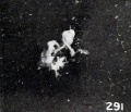
Fig. 291. Several villi ending in a single trophoblastic nodule. No. 308. X4.8.
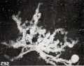
Fig. 292. A macerated villous tree, one branch of which bears a nodule on its extremity. No. 2472. X7.2.


Tables
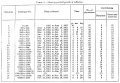
Table 1. Showing gradual growth of collection

Table 2. List of hospitals and laboratories contributing embryological material
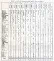
Table 3. Sources of embryological material by states, cities, and countries
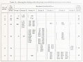
Table 4.
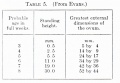
Table 5.
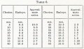
Table 6.
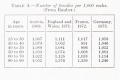
Table A. Number of females per 1,000 males
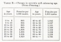
Table B. Changes in sex-ratio with advancing age
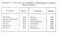
Table C. Sex-ratio of mortality in European countries
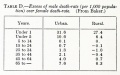
Table D. Excess of male death-rate (per 1,000 population) over female death-rate
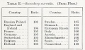
Table E.
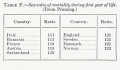
Table F.


Charts
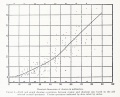
Chart 1. Field and graph showing correlation between cyemic and chorionic size

| Embryology - 18 Apr 2024 |
|---|
| Google Translate - select your language from the list shown below (this will open a new external page) |
|
العربية | català | 中文 | 中國傳統的 | français | Deutsche | עִברִית | हिंदी | bahasa Indonesia | italiano | 日本語 | 한국어 | မြန်မာ | Pilipino | Polskie | português | ਪੰਜਾਬੀ ਦੇ | Română | русский | Español | Swahili | Svensk | ไทย | Türkçe | اردو | ייִדיש | Tiếng Việt These external translations are automated and may not be accurate. (More? About Translations) |
Mall FP. and Meyer AW. Studies on abortuses: a survey of pathologic ova in the Carnegie Embryological Collection. (1921) Contrib. Embryol., Carnegie Inst. Wash. Publ. 275, 12: 1-364.
- In this historic 1921 pathology paper, figures and plates of abnormal embryos are not suitable for young students.
1921 Carnegie Collection - Abnormal: Preface | 1 Collection origin | 2 Care and utilization | 3 Classification | 4 Pathologic analysis | 5 Size | 6 Sex incidence | 7 Localized anomalies | 8 Hydatiform uterine | 9 Hydatiform tubal | Chapter 10 Alleged superfetation | 11 Ovarian Pregnancy | 12 Lysis and resorption | 13 Postmortem intrauterine | 14 Hofbauer cells | 15 Villi | 16 Villous nodules | 17 Syphilitic changes | 18 Aspects | Bibliography | Figures | Contribution No.56 | Contributions Series | Embryology History
| Historic Disclaimer - information about historic embryology pages |
|---|
|