Book - Contributions to Embryology Carnegie Institution No.38
A Morphological Study Of The Tracheal And Bronchial Cartilages
By William Snow Miller,
Professor of Anatomy, University of Wisconsin.
With two plates and eleven text-figures.
| Historic Disclaimer - information about historic embryology pages |
|---|
| Pages where the terms "Historic" (textbooks, papers, people, recommendations) appear on this site, and sections within pages where this disclaimer appears, indicate that the content and scientific understanding are specific to the time of publication. This means that while some scientific descriptions are still accurate, the terminology and interpretation of the developmental mechanisms reflect the understanding at the time of original publication and those of the preceding periods, these terms, interpretations and recommendations may not reflect our current scientific understanding. (More? Embryology History | Historic Embryology Papers) |
Introduction
Horner, writing in 1839, said that "at the orifice of each branch of the bronchia there is a semi-circular cartilage, forming rather more than one-half of its circumference and having its concave edge upwards. The whole arrangement resembles somewhat the pasteboard of an eared bonnet, and is evidently to keep the orifice open." Passing over his reference to the feminine fashions of his day, we find in the above quotation the earliest description of the form of the cartilages found at the place where bronchi divide; moreover, a reason is assigned for the particular form of the cartilage.
In 1840 Jonas Iving published a short article "On the forms of the cartilages which keep open the principal divisions of the bronchial tubes." In a footnote he frankly gives the priority of description to Professor Horner and intimates that his paper was inspired by a demonstration Professor Horner made in Philadelphia to Mr. T. Wilkinson King "some months previous to the last edition of his work." Mr. King must have, on his return to London, imparted the information thus acquired to his namesake, who at once made it the subject of a special study. Iving found, however, that not all the cartilages present at the place where a bronchus
divides had the characteristic saddle shape described by Horner but, "that a considerable number of varieties will be met wdth." His paper is illustrated with several small and unsatisfactory drawings of the cartilages.
An extended search of the literature fails to bring to light any other investigation of the form of the tracheal or bronchial cartilages ; in no place has the author been able to find the cartilages illustrated in plastic form, and such illustrations as are found show the cartilages as irregular plates and are evidently schematic.
Henle's illustrations are poor, but his description is fairly good. He describes the cartilages as having the plates or strips sometimes with short prolongations, arranged, as a rule, transverse to the long axis of the bronchi; they vary also have a longitudinal or oblique direction —
- " Je tiefer hinab, um so mehr reduciren sie sich und um so weiter riicken sie aus einander, bis sie endlich nur noch als platte Ringe oder HalbrLnge um die Miindungen der Seitenzweige und als Stiitzen der die beiden Aeste einer gabligen Theilung trennenden Scheidewand vorkommen."
Illustrations
Miller Links: Plate 1 | Plate 2 | Contribution No.38 | Volume IX | Contributions to Embryology
Fig. 2. —Outline of the section along the line A, in figure 1. Note circular outline of the section. The numbers 2, 3. 4 indicate the cartilages through wliich the plane of the section passes. X6.
Fig. 3. — Outline of the section along the line B in figure 1. Note the outline of the lumen of the trachea; it has notonly adumb-bell shape, but there is also a marked anterior swing of what is to be the right bronchus. 1 B, section of the first right bronchial cartilage; C, C, sections of the carinal cartilage. XO.
Fig. 4. — Reconstruction of the carinal cartilage shown in outline in figure 1. A, anterior view; B, posterior view. Note the extensive fusion of trachea and bronchial elements. This belongs to what is known as the " tracheo-bronchial left" type of carinal cartilage. X6.
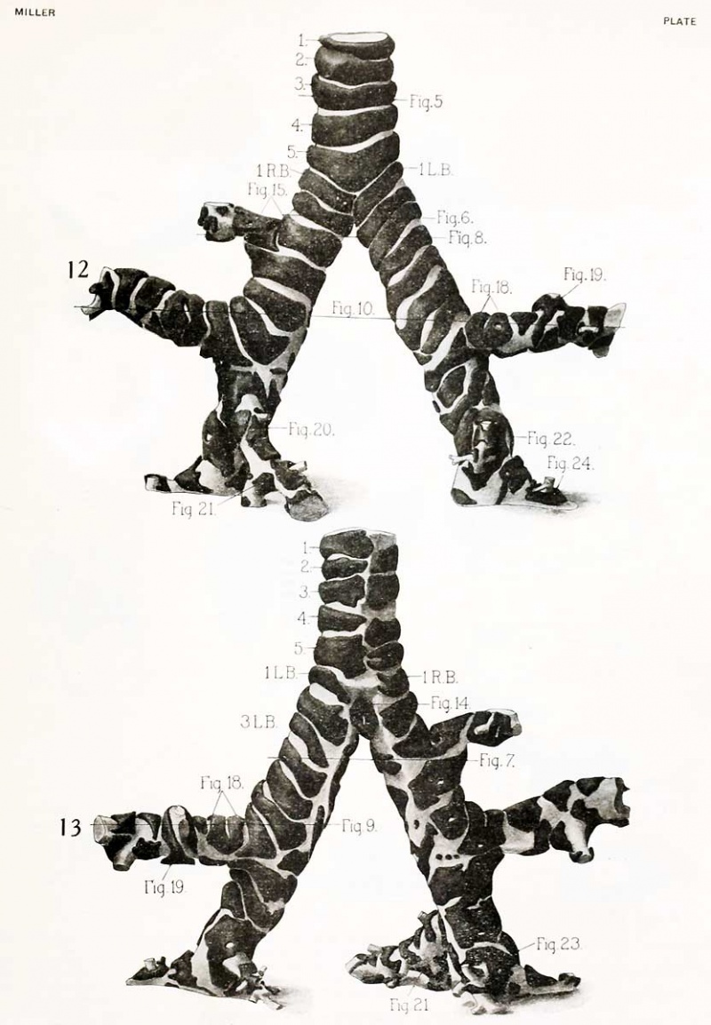
Fig. 5.— Section taken through the center of cartilage No. 3 in figure 12. This is a typical tracheal cartilage without any fission or fusion with an adjoining cartilage. The broken line extending from one end of the cartilage to the other indicates the positions of the muscle. Note the internal attachment of the muscle and that it is not inserted into the ends of the cartilage but some distance lateral to them. X6.
Fig. 6. — Section taken along the plane indicated in figure 12. The bifurcation of the trachea into the right and the left bronchus has taken place. R. B., right bronchus. L. B., left bronchus. 1 L. B., lower portion of the mesial arm of the first left bronchial cartilage which forms the carinal cartilage. 2 L. B., second left bronchial cartilage. 3L.B., third left bronchial cartilage. 2R.B., ventral and dorsal section of the second right bronchial cartilage. XG.
Fig. 7. — Outline of a section taken along the line itdicated on figure 1.3, showing the relation of the cartilages to the right bronchus, R. B., and to the loft bronchus, L. B. X6.
Fig. 8. — Outline of a section taken along the line indicated on figure 12 showing the position of the various pieces of cartilage which enter into the formation of the cartilages shown in figures 12 and 13 along the eparterial bronchus. R. B., right bronchus. L. B., left bronchus. Ep. B., eparterial bronchus. XO.
Fig. 9. — Outline of a section taken along the line indicated in figure 1.3. The plane of the section takes in a portion of the bronchus going to the left lobus anterior and portions of the cartilage (.4) shown in figure 18. L. B., left bronchus. L. I. a., bronchus going to left lobus anterior. X6.
Fig. 10. — Outline of section taken along the line indicated in figure 12. R. B., right bronchus. L. B., left bronchus. R. I. m., bronchus to right lobus medius. L. I. a., bronchus to left lobus anterior. The relation of the various pieces to the complete cartilages can be made out without difficulty. X6.
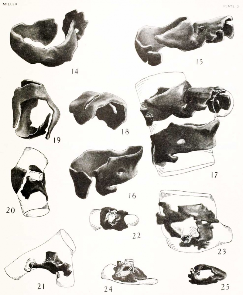
Fig. 11. — A typical saddle-shaped cartilage situated at the first division of the main stem bronchus in the left lobus inferior. A shows the cartilage in situ when seen from the dorsal side; B when seen from the ventral side; C shows the carti- lage alone. X30.
Plate 1
Plate 2
Content to be added----
| Historic Disclaimer - information about historic embryology pages |
|---|
| Pages where the terms "Historic" (textbooks, papers, people, recommendations) appear on this site, and sections within pages where this disclaimer appears, indicate that the content and scientific understanding are specific to the time of publication. This means that while some scientific descriptions are still accurate, the terminology and interpretation of the developmental mechanisms reflect the understanding at the time of original publication and those of the preceding periods, these terms, interpretations and recommendations may not reflect our current scientific understanding. (More? Embryology History | Historic Embryology Papers) |
Glossary Links
- Glossary: A | B | C | D | E | F | G | H | I | J | K | L | M | N | O | P | Q | R | S | T | U | V | W | X | Y | Z | Numbers | Symbols | Term Link
Cite this page: Hill, M.A. (2024, April 18) Embryology Book - Contributions to Embryology Carnegie Institution No.38. Retrieved from https://embryology.med.unsw.edu.au/embryology/index.php/Book_-_Contributions_to_Embryology_Carnegie_Institution_No.38
- © Dr Mark Hill 2024, UNSW Embryology ISBN: 978 0 7334 2609 4 - UNSW CRICOS Provider Code No. 00098G


