Book - Contributions to Embryology: Difference between revisions
mNo edit summary |
mNo edit summary |
||
| (32 intermediate revisions by the same user not shown) | |||
| Line 4: | Line 4: | ||
This historic series of papers published by the Carnegie Institution of Washington in the series "Contributions to Embryology" was published from early in the 20th Century. The papers documented not only early human development, using mainly the [[Carnegie Collection]] of embryos, but also that in animal models of development. | This historic series of papers published by the Carnegie Institution of Washington in the series "Contributions to Embryology" was published from early in the 20th Century. The papers documented not only early human development, using mainly the [[Carnegie Collection]] of embryos, but also that in animal models of development. | ||
<br> | |||
{| width=500px| | {| width=500px| | ||
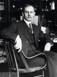
| [[File:Franklin Mall 01.jpg|200px|link=Embryology History - Franklin Mall]] | | [[File:Franklin Mall 01.jpg|200px|link=Embryology History - Franklin Mall]] | ||
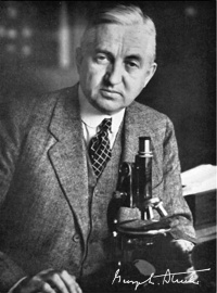
| [[File:George_L._Streeter.jpg|200px|link= | | [[File:George_L._Streeter.jpg|200px|link=Embryology_History_-_George_Streeter]] | ||
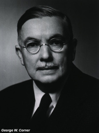
| [[File:George Corner.jpg|200px|link=Embryology_History_-_George Corner]] | |||
|- | |- | ||
| [[Embryology History - Franklin Mall|Franklin Mall]] (1911) the founder and first editor of the series. | | [[Embryology History - Franklin Mall|Franklin Mall]] (1911) the founder and first editor of the series. | ||
| [[Embryology_History_-_George_Streeter|George L. Streeter | | [[Embryology_History_-_George_Streeter|George L. Streeter]] editor from 1917 to 1940. | ||
| [[Embryology_History_-_George Corner|George Corner]] editor from 1940. | |||
|} | |} | ||
<br> | <br> | ||
[[Embryology_History_-_George_Streeter|Dr. George L. Streeter]] was editor of this series, from 1917 to 1940 Volumes VIII to XXIX of the Contributions to Embryology of the Carnegie Institution of Washington. In a letter to Science | [[Embryology_History_-_George_Streeter|Dr. George L. Streeter]] was editor of this series, from 1917 to 1940 Volumes VIII to XXIX of the Contributions to Embryology of the Carnegie Institution of Washington. In a letter to Science{{#pmid:17799310|PMID17799310}} the Carnegie Institute staff noted: | ||
:"The present staff of the department of embryology, with the approval of the president of the institution, has therefore dedicated Volume XXX, which appeared on December 31, 1942, to Dr.Streeter and has placed his portrait at the head of the volume." | :"The present staff of the department of embryology, with the approval of the president of the institution, has therefore dedicated Volume XXX, which appeared on December 31, 1942, to Dr.Streeter and has placed his portrait at the head of the volume." | ||
| Line 26: | Line 29: | ||
'''Carnegie Embryos''' | '''Carnegie Embryos''' | ||
==Other Publications== | |||
{{Ref-LongMark1911}} | |||
{{Ref-Weeds1914}} | |||
{| class="wikitable mw-collapsible mw-collapsed" | |||
! Online Editor | |||
|- | |||
| [[File:Mark_Hill.jpg|90px|left]] This a list of the available Internet Archive volume scans. | |||
<br>https://archive.org/details/carnegieinstitu07washgoog/page/n14/mode/2up Carnegie Trust Deed | |||
[https://archive.org/details/carnegieinstitut222carn Vol 2] | [https://archive.org/details/carnegieinstitut223carn Vol 3] | [https://archive.org/details/carnegieinstitut1916224carn Vol 4] | [https://archive.org/details/carnegieinstitut225carn Vol 5] | [https://archive.org/details/carnegieinstitu33washgoog Vol 6] | [https://archive.org/details/carnegieinstitut227carn Vol 7] | [https://archive.org/details/carnegieinstitut2711918carn Vol 8] | [https://archive.org/details/carnegieinstitut276carn Vol 8] | [https://archive.org/details/carnegieinstitut1921273carn Vol 10] | [https://archive.org/details/carnegieinstitut275carn Vol 12] | [https://archive.org/details/carnegieinstitut2741920carn Vol 13] | [https://archive.org/details/carnegieinstitut277carn Vol 14] | |||
|} | |||
==Volume I== | ==Volume I== | ||
Washington, 1915 | Washington, 1915 | ||
== | {{Ref-Mall1915}} | ||
==Volume II== | |||
Washington, 1915 | |||
{{Ref-Watt1915}} | |||
{{Ref-Clark1915b}} | |||
{{Ref-Meyer1915}} | |||
{{Ref-Corner1915}} | |||
{{Ref- | {{Ref-Essick1915}} | ||
==Volume III== | |||
Washington, 1915 | |||
{{Ref-Sabin1915}} | |||
==Volume IV== | ==Volume IV== | ||
{{Ref-Mall1916}} | {{Ref-Mall1916}} | ||
{{Ref-Cunningham1916}} | {{Ref-Cunningham1916}} | ||
* [[Book_-_Contributions_to_Embryology_Carnegie_Institution_No.11|The structure of chromophile cells of the nervous system]] By E. Y. Cowdry (1 plate) 27-43 | * [[Book_-_Contributions_to_Embryology_Carnegie_Institution_No.11|The structure of chromophile cells of the nervous system]] By E. Y. Cowdry (1 plate) 27-43 | ||
| Line 66: | Line 92: | ||
==Volume VI== | ==Volume VI== | ||
Washington, 1917 | Washington, 1917 | ||
{{Ref-Johnson1917}} | {{Ref-Johnson1917}} | ||
==Volume VII== | ==Volume VII== | ||
Washington, 1918 | Washington, 1918 | ||
{{Ref-Streeter1918}} | {{Ref-Streeter1918}} | ||
| Line 82: | Line 102: | ||
[[Book_-_Contributions_to_Embryology_Carnegie_Institution_No.20|'''Carnegie Institution No.20 Otic Capsule''']]: [[Book_-_Contributions_to_Embryology_Carnegie_Institution_No.20#Introduction|Introduction]] | [[Book_-_Contributions_to_Embryology_Carnegie_Institution_No.20#Terminology|Terminology]] | [[Book_-_Contributions_to_Embryology_Carnegie_Institution_No.20_part_1#Historical|Historical]] | [[Book_-_Contributions_to_Embryology_Carnegie_Institution_No.20_part_2#Material and Methods|Material and Methods]] | [[Book_-_Contributions_to_Embryology_Carnegie_Institution_No.20_part_3#Development_of_the_Cartilaginous_Capsule_of_the_Ear|Development of cartilaginous capsule of ear]] | [[Book_-_Contributions_to_Embryology_Carnegie_Institution_No.20_part_3#Condensation_of_the_Periotic_Mesenchyme|Condensation of periotic mesenchyme]] | [[Book_-_Contributions_to_Embryology_Carnegie_Institution_No.20_part_3#Differentiation_of_Precartilage|Differentiation of precartilage]] | [[Book_-_Contributions_to_Embryology_Carnegie_Institution_No.20_part_3#Differentiation_of_Cartilage|Differentiation of cartilage]] | [[Book_-_Contributions_to_Embryology_Carnegie_Institution_No.20_part_4#Growth_and_Alteration_of_Form_of_the_Cartilaginous_Canals|Growth and alteration of form of cartilaginous canals]] | [[Book_-_Contributions_to_Embryology_Carnegie_Institution_No.20_part_4#Development_of_Periotic_Reticular_Connective_Tissue|Development of the periotic reticular connective tissue]] | [[Book_-_Contributions_to_Embryology_Carnegie_Institution_No.20_part_5#Development_of_the_Perichondrium|Development of the perichondrium]] | [[Book_-_Contributions_to_Embryology_Carnegie_Institution_No.20_part_5#Development_of_Periotic_Tissue_Spaces|Development of the periotic tissue-spaces]] | [[Book_-_Contributions_to_Embryology_Carnegie_Institution_No.20_part_5#Development_of_the_Periotic_Cistern_of_the_Vestibule|Development of the periotic cistern of the vestibule]] | [[Book_-_Contributions_to_Embryology_Carnegie_Institution_No.20_part_5#Development_of_the_Periotic_Spaces_of_the_Semicircular_Ducts|Development of the periotic spaces of the semicircular ducts]] | [[Book_-_Contributions_to_Embryology_Carnegie_Institution_No.20_part_5#Development_of_Scala_Tympani_and_Scala_Vestibuli|Development of the scala tympani and scala vestibuli]] | [[Book_-_Contributions_to_Embryology_Carnegie_Institution_No.20_part_5#Communication_of_Periotic_Spaces_with_Arachnoid_Spaces|Communication with subarachnoid spaces]] | [[Book_-_Contributions_to_Embryology_Carnegie_Institution_No.20_part_6#Summary|Summary]] | [[Book_-_Contributions_to_Embryology_Carnegie_Institution_No.20|'''Carnegie Institution No.20 Otic Capsule''']]: [[Book_-_Contributions_to_Embryology_Carnegie_Institution_No.20#Introduction|Introduction]] | [[Book_-_Contributions_to_Embryology_Carnegie_Institution_No.20#Terminology|Terminology]] | [[Book_-_Contributions_to_Embryology_Carnegie_Institution_No.20_part_1#Historical|Historical]] | [[Book_-_Contributions_to_Embryology_Carnegie_Institution_No.20_part_2#Material and Methods|Material and Methods]] | [[Book_-_Contributions_to_Embryology_Carnegie_Institution_No.20_part_3#Development_of_the_Cartilaginous_Capsule_of_the_Ear|Development of cartilaginous capsule of ear]] | [[Book_-_Contributions_to_Embryology_Carnegie_Institution_No.20_part_3#Condensation_of_the_Periotic_Mesenchyme|Condensation of periotic mesenchyme]] | [[Book_-_Contributions_to_Embryology_Carnegie_Institution_No.20_part_3#Differentiation_of_Precartilage|Differentiation of precartilage]] | [[Book_-_Contributions_to_Embryology_Carnegie_Institution_No.20_part_3#Differentiation_of_Cartilage|Differentiation of cartilage]] | [[Book_-_Contributions_to_Embryology_Carnegie_Institution_No.20_part_4#Growth_and_Alteration_of_Form_of_the_Cartilaginous_Canals|Growth and alteration of form of cartilaginous canals]] | [[Book_-_Contributions_to_Embryology_Carnegie_Institution_No.20_part_4#Development_of_Periotic_Reticular_Connective_Tissue|Development of the periotic reticular connective tissue]] | [[Book_-_Contributions_to_Embryology_Carnegie_Institution_No.20_part_5#Development_of_the_Perichondrium|Development of the perichondrium]] | [[Book_-_Contributions_to_Embryology_Carnegie_Institution_No.20_part_5#Development_of_Periotic_Tissue_Spaces|Development of the periotic tissue-spaces]] | [[Book_-_Contributions_to_Embryology_Carnegie_Institution_No.20_part_5#Development_of_the_Periotic_Cistern_of_the_Vestibule|Development of the periotic cistern of the vestibule]] | [[Book_-_Contributions_to_Embryology_Carnegie_Institution_No.20_part_5#Development_of_the_Periotic_Spaces_of_the_Semicircular_Ducts|Development of the periotic spaces of the semicircular ducts]] | [[Book_-_Contributions_to_Embryology_Carnegie_Institution_No.20_part_5#Development_of_Scala_Tympani_and_Scala_Vestibuli|Development of the scala tympani and scala vestibuli]] | [[Book_-_Contributions_to_Embryology_Carnegie_Institution_No.20_part_5#Communication_of_Periotic_Spaces_with_Arachnoid_Spaces|Communication with subarachnoid spaces]] | [[Book_-_Contributions_to_Embryology_Carnegie_Institution_No.20_part_6#Summary|Summary]] | ||
| [[Book_-_Contributions_to_Embryology_Carnegie_Institution_No.20_part_6#Bibliography|Bibliography]] | [[Book_-_Contributions_to_Embryology_Carnegie_Institution_No.20_part_7|Explanation of plates]] | | [[Book_-_Contributions_to_Embryology_Carnegie_Institution_No.20_part_6#Bibliography|Bibliography]] | [[Book_-_Contributions_to_Embryology_Carnegie_Institution_No.20_part_7|Explanation of plates]] | ||
{{Ref-Stricht1918}} | {{Ref-Stricht1918}} | ||
{{Ref-Wheeler1918}} | {{Ref-Wheeler1918}} | ||
{{Ref-Ingalls1918}} | {{Ref-Ingalls1918}} | ||
==Volume VIII== | |||
{{Ref-Streeter1921}} | {{Ref-Streeter1921}} | ||
E. V. Cowdry. The mitochondrial constituents of protoplasm [[Book_-_Contributions_to_Embryology_Carnegie_Institution_No.25|'''Carnegie Institution No.25 Mitochondria''']] | |||
{{Ref-Kunitomo1920}} | {{Ref-Kunitomo1920}} | ||
==Volume IX== | ==Volume IX== | ||
| Line 123: | Line 126: | ||
--[[User:Z8600021|Mark Hill]] 00:44, 27 March 2012 (EST) Only the introductory text has been added for the papers from Volume IX listed below. | --[[User:Z8600021|Mark Hill]] 00:44, 27 March 2012 (EST) Only the introductory text has been added for the papers from Volume IX listed below. | ||
{{Ref-Bean1919}} | |||
* [[Book_-_Contributions_to_Embryology_Carnegie_Institution_No.27|No.27 The Development And Function Of Macrophages In The Repair Of Experimental Bone- Wounds In Rats Vitally Stained With Trypan-Blue]]. by Charles Clifford Macklin. | * [[Book_-_Contributions_to_Embryology_Carnegie_Institution_No.27|No.27 The Development And Function Of Macrophages In The Repair Of Experimental Bone- Wounds In Rats Vitally Stained With Trypan-Blue]]. by Charles Clifford Macklin. | ||
* [[Book_-_Contributions_to_Embryology_Carnegie_Institution_No.28|No.28 Cytoplasmic Structures In The Seminal Epithelium Of The Opossum]]. by J. Duesberg. | * [[Book_-_Contributions_to_Embryology_Carnegie_Institution_No.28|No.28 Cytoplasmic Structures In The Seminal Epithelium Of The Opossum]]. by J. Duesberg. | ||
* [[Book_-_Contributions_to_Embryology_Carnegie_Institution_No.29|No.29 On The Widespread Occurrence Of Reticular Fibrils Produced By Capillary Endothelium]]. by George W. Corner. | * [[Book_-_Contributions_to_Embryology_Carnegie_Institution_No.29|No.29 On The Widespread Occurrence Of Reticular Fibrils Produced By Capillary Endothelium]]. by George W. Corner. | ||
* | * {{Ref-Wheeler1919}} | ||
* | * {{Ref-Stricht1919}} | ||
* | * {{Ref-Retzer1920}} | ||
* | * {{Ref-Jenkins1920}} | ||
* [[Book_-_Contributions_to_Embryology_Carnegie_Institution_No.34|No.34 The Development Of The External Nose In Whites And Negroes]]. by Adolph H. Schultz. | * [[Book_-_Contributions_to_Embryology_Carnegie_Institution_No.34|No.34 The Development Of The External Nose In Whites And Negroes]]. by Adolph H. Schultz. | ||
* [[Book_-_Contributions_to_Embryology_Carnegie_Institution_No.35|No.35 Muscular Contraction In Tissue-Cultures]]. by Margaret Reed Lewis. | * [[Book_-_Contributions_to_Embryology_Carnegie_Institution_No.35|No.35 Muscular Contraction In Tissue-Cultures]]. by Margaret Reed Lewis. | ||
{{Ref-Sabin1919}} | |||
{{Ref-Bean1919}} | |||
{{Ref-Miller1919}} | |||
{{Ref-Lewis1920}} | {{Ref-Lewis1920}} | ||
{{Ref-Meyer1920}} | {{Ref-Meyer1920}} | ||
| Line 151: | Line 153: | ||
{{Ref-Essick1920}} | {{Ref-Essick1920}} | ||
{{Ref-Streeter1920a}} | {{Ref-Streeter1920a}} | ||
* [[Book_-_Contributions_to_Embryology_Carnegie_Institution_No.44|No.44 The Experimental Production Of An Internal Hydrocephalus]]. by Lewis H. Weed. | * [[Book_-_Contributions_to_Embryology_Carnegie_Institution_No.44|No.44 The Experimental Production Of An Internal Hydrocephalus]]. by Lewis H. Weed. | ||
| Line 165: | Line 162: | ||
Washington, 1921 | Washington, 1921 | ||
Herbert McLean Evans and Katharine J. Scott. On the differential reaction to vital dyes exhibited by the two great groups of connective-tissue cells (11 plates) | |||
[[Book_-_Contributions_to_Embryology_Carnegie_Institution_No.47|'''Carnegie Institution No.47 Two Groups of Connective-Tissue Cells''']] | [[Book_-_Contributions_to_Embryology_Carnegie_Institution_No.47|'''Carnegie Institution No.47 Two Groups of Connective-Tissue Cells''']] | ||
{{Ref-Macklin1921}} | {{Ref-Macklin1921}} | ||
:'''Links:''' [http://www.archive.org/details/contributionstoe10carn Internet Archive - Volume X] | :'''Links:''' [http://www.archive.org/details/contributionstoe10carn Internet Archive - Volume X] | ||
| Line 184: | Line 175: | ||
No. 49. Myeloid metaplasia of the embryonic mesenchyme in relation to cell potentialities and differential factors. By Vera Danchakoff (5 plates) 1-32 | No. 49. Myeloid metaplasia of the embryonic mesenchyme in relation to cell potentialities and differential factors. By Vera Danchakoff (5 plates) 1-32 | ||
{{Ref-Lineback1920}} | {{Ref-Lineback1920}} | ||
{{Ref-Wislocki1920}} | |||
{{Ref-Ingalls1920}} | {{Ref-Ingalls1920}} | ||
{{Ref-Barry1920}} | |||
{{Ref-Corner1920}} | {{Ref-Corner1920}} | ||
{{Ref-Streeter1920a}} | {{Ref-Streeter1920a}} | ||
==Volume XII== | ==Volume XII== | ||
[[File:Contributions_to_Embryology_No.56.jpg|thumb|Volume XII title page]] | [[File:Contributions_to_Embryology_No.56.jpg|thumb|Volume XII title page]] | ||
{{Ref-Mall1921}} | {{Ref-Mall1921}} | ||
{{Carnegie56 TOC2}} | |||
==Volume XIII== | ==Volume XIII== | ||
Washington, 1922 | Washington, 1922 | ||
{{Ref-Cash1921}} | {{Ref-Cash1921}} | ||
Reichert FL. [[Book_-_Contributions_to_Embryology_Carnegie_Institution_No.58|No. 58. On the fate of the primary lymph-sacs in the abdominal region of the pig, and the development of lymph-channels in the abdominal and pelvic regions. ]] (5 text-figures) pp 17-39 | |||
{{Ref-Jenkins1921}} | |||
{{Ref-Corner1922}} | {{Ref-Corner1922}} | ||
{{Ref-Spaulding1921}} | |||
Wislocki GB. Further experimental studies on fetal absorption pp 89-101 | |||
[[Book_-_Contributions_to_Embryology_Carnegie_Institution_No.62|'''Carnegie Institution No.62 Fetal Absorption''']] | [[Book_-_Contributions_to_Embryology_Carnegie_Institution_No.62|'''Carnegie Institution No.62 Fetal Absorption''']] | ||
| Line 279: | Line 219: | ||
* IV. The behavior of the placenta and fetal membranes of the rabbit toward trypan blue injected into the maternal blood-stream. | * IV. The behavior of the placenta and fetal membranes of the rabbit toward trypan blue injected into the maternal blood-stream. | ||
G. B. Wislocki and J. A. Key. The distribution of mitochondria in the placenta pp103-115. | |||
* No. 63. The distribution of mitochondria in the placenta. By G. B. Wislocki and J. A. Key. (1 plate) | |||
By George W. Corner. (4 plates, 2 text-figures) | |||
Corner GW. Cyclic changes in the ovaries and uterus of swine, and their relations to the mechanism of implantation. pp117-146 | |||
By George W. Corner. (4 plates, 2 text-figures) | |||
[[Book_-_Contributions_to_Embryology_Carnegie_Institution_No.64|'''Carnegie Institution No.64 Pig Implantation''']] | [[Book_-_Contributions_to_Embryology_Carnegie_Institution_No.64|'''Carnegie Institution No.64 Pig Implantation''']] | ||
==XIV== | ==Volume XIV== | ||
{{Ref-Sabin1922}} | |||
Buell CE. Origin of the pulmonary vessels in the chick pp11-26 | |||
By Charles Elbert Buell Jr. (2 plates) 11-26 | By Charles Elbert Buell Jr. (2 plates) 11-26 | ||
| Line 302: | Line 241: | ||
[[Book_-_Contributions_to_Embryology_Carnegie_Institution_No.66|'''Carnegie Institution No.66 Chicken Pulmonary Vessels''']] | [[Book_-_Contributions_to_Embryology_Carnegie_Institution_No.66|'''Carnegie Institution No.66 Chicken Pulmonary Vessels''']] | ||
{{Ref-Doan1922}} | |||
{{Ref-Congdon1922}} | |||
{{Ref-Streeter1922}} | |||
{{Ref-Woollard1922}} | {{Ref-Woollard1922}} | ||
{{Ref-Finley1923}} | |||
== | ==Volume XV== | ||
{{Ref-Davis1923}} | {{Ref-Davis1923}} | ||
[[ | [[Carnegie stage 11]] | ||
==Volume XX== | |||
==XX== | |||
{{Ref-Corner1929}} | {{Ref-Corner1929}} | ||
[[ | [[Carnegie stage 10]] | ||
==XXI | ==Volume XXI== | ||
{{Ref-Atwell1930}} | {{Ref-Atwell1930}} | ||
[[ | [[Carnegie stage 11]] | ||
==Volume XXII== | |||
{{Ref-Heuser1930}} | |||
[[Carnegie stage 11]] | |||
{{Ref-Weller1933}} | |||
==Carnegie Year Books== | ==Carnegie Year Books== | ||
| Line 373: | Line 281: | ||
* [[Report_-_Carnegie_Year_Book_37|Year Book No. 38]] (1938) | * [[Report_-_Carnegie_Year_Book_37|Year Book No. 38]] (1938) | ||
* [[Report_-_Carnegie_Year_Book_38|Year Book No. 38]] (1939) | * [[Report_-_Carnegie_Year_Book_38|Year Book No. 38]] (1939) | ||
| Line 382: | Line 289: | ||
{{Historic Disclaimer}} | {{Historic Disclaimer}} | ||
==External Links== | ==External Links== | ||
| Line 388: | Line 296: | ||
* [http://carnegiescience.edu Carnegie Institution of Washington] | [http://www.ciwemb.edu Department of Embryology] | * [http://carnegiescience.edu Carnegie Institution of Washington] | [http://www.ciwemb.edu Department of Embryology] | ||
Latest revision as of 13:55, 11 August 2020
| Embryology - 17 Apr 2024 |
|---|
| Google Translate - select your language from the list shown below (this will open a new external page) |
|
العربية | català | 中文 | 中國傳統的 | français | Deutsche | עִברִית | हिंदी | bahasa Indonesia | italiano | 日本語 | 한국어 | မြန်မာ | Pilipino | Polskie | português | ਪੰਜਾਬੀ ਦੇ | Română | русский | Español | Swahili | Svensk | ไทย | Türkçe | اردو | ייִדיש | Tiếng Việt These external translations are automated and may not be accurate. (More? About Translations) |
Introduction
This historic series of papers published by the Carnegie Institution of Washington in the series "Contributions to Embryology" was published from early in the 20th Century. The papers documented not only early human development, using mainly the Carnegie Collection of embryos, but also that in animal models of development.
|
|
|
| Franklin Mall (1911) the founder and first editor of the series. | George L. Streeter editor from 1917 to 1940. | George Corner editor from 1940. |
Dr. George L. Streeter was editor of this series, from 1917 to 1940 Volumes VIII to XXIX of the Contributions to Embryology of the Carnegie Institution of Washington. In a letter to Science[1] the Carnegie Institute staff noted:
- "The present staff of the department of embryology, with the approval of the president of the institution, has therefore dedicated Volume XXX, which appeared on December 31, 1942, to Dr.Streeter and has placed his portrait at the head of the volume."
| Contributions Links: Carnegie Collection | Franklin Mall | George Streeter | Carnegie Stages | Carnegie Embryos | Carnegie Models | Human Embryo Collections | Embryology History |

Carnegie Embryos
Other Publications
Long JA. and Mark EL. The maturation of the egg of the mouse. (1911) Carnegie Inst. of Washington Pub. No. 147. 77.
Weeds LH. Nuclear masses in the lower portion of the human brain-stem. (1914) Carnegie Inst. Washington Publ. 191. 1: 1-78.
| Online Editor |
|---|

Vol 2 | Vol 3 | Vol 4 | Vol 5 | Vol 6 | Vol 7 | Vol 8 | Vol 8 | Vol 10 | Vol 12 | Vol 13 | Vol 14 |
Volume I
Washington, 1915
Mall FP. On the fate of the human embryo in tubal pregnancy. (1915) Contrib. Embryol., Carnegie Inst. Wash. Publ. 221, 1: 1-104.
Volume II
Washington, 1915
Watt JC. Description of two young twin embryos with 17-19 paired somites. (1915) Contrib. Embryol., Carnegie Inst. Wash. 2: 15-54.
Clark ER. An anomaly of the thoracic duct with a bearing on the embryology of the lymphatic system. (1915) Contrib. Embryol., Carnegie Inst. Wash. 2: 45-54.
Meyer AW. Fields, graphs, and other data on fetal growth. (1915) Contrib. Embryol., Carnegie Inst. Wash. 2: 55-68.
Corner GW. The corpus luteum of pregnancy, as it is in swine. (1915) Contrib. Embryol., Carnegie Inst. Wash. 2: 69-94.
Essick CR. Transitory cavities in the corpus striatum of the human embryo. (1915) Contrib. Embryol., Carnegie Inst. Wash. 2: 95-108.
Volume III
Washington, 1915
Sabin FR. On the fate of the posterior cardinal veins and their relation to the development of the vena cava and azygos in the embryo pig. (1915) Pub. No. 223 Contrib. Embryol., Carnegie Inst. Wash. 3(7): 5-32. PDF
Volume IV
Mall FP. The human magma reticule in normal and in pathological development. (1916) Contrib. Embryol., Carnegie Inst. Wash. Publ. 224, 4:5-26.
Cunningham RS. On the development of the lymphatics of the lungs in the embryo pig. (1916) Contrib. Embryol., Carnegie Inst. Wash. 7:45-68.
- The structure of chromophile cells of the nervous system By E. Y. Cowdry (1 plate) 27-43
- No. 13. Binucleate cells in tissue cultures. By Charles C. Macklin (4 plates, containing 70 figures) 69-106
Volume V
Washington, 1917
Weed LH. The development of the cerebro-spinal spaces in pig and in man. (1917) Contrib. Embryol., Carnegie Inst. Wash., 5, No. 14 .
Mall FP. Cyclopia in the human embryo. (1917) Contrib. Embryol., Carnegie Inst. Wash. Publ. 226, 6:
Quantitative Studies On Mitochondria In Nerve-Cells
Development Of Connective-Tissue Fibers In Tissue Cultures Of Chick Embryos By Margaret Reed Lewis.
Sabin FR. Origin and development of the primitive vessels of the chick and of the pig. (1917) Contrib. Embryol., Carnegie Inst. Wash. 6: 61–124.
Volume VI
Washington, 1917
Johnson FP. A human embryo of twenty-four pairs of somites. (1917) Carnegie Instn. Wash. Publ., Contrib. Embryol., 21: 125-168.
Volume VII
Washington, 1918
Streeter GL. The histogenesis and growth of the otic capsule and its contained periotic tissue-spaces in the human embryo. (1918) Contrib. Embryol., Carnegie Inst. Wash. 8: 5-54.
Carnegie Institution No.20 Otic Capsule: Introduction | Terminology | Historical | Material and Methods | Development of cartilaginous capsule of ear | Condensation of periotic mesenchyme | Differentiation of precartilage | Differentiation of cartilage | Growth and alteration of form of cartilaginous canals | Development of the periotic reticular connective tissue | Development of the perichondrium | Development of the periotic tissue-spaces | Development of the periotic cistern of the vestibule | Development of the periotic spaces of the semicircular ducts | Development of the scala tympani and scala vestibuli | Communication with subarachnoid spaces | Summary | Bibliography | Explanation of plates
van der Stricht O. The genesis and structure of the membrana tectoria and the crista spiralis of the cochlea. (1918) Contrib. Embryol., Carnegie Inst. Wash., 21: 55-86.
Wheeler T. Study of a human spina bifida monster with encephaloceles and other abnormalities. (1918) Contrib. Embryol., Carnegie Inst. Wash., 22: .
Ingalls NW. A human embryo before the appearance of the myotomes. (1918) Contrib. Embryol., Carnegie Inst. Wash. No.23 Publ. 227, 7:111-134.
Volume VIII
Streeter GL. The developmental alterations in the vascular system of the brain of the human embryo. (1921) Contrib. Embryol., Carnegie Inst. Wash. 8:7-38.
E. V. Cowdry. The mitochondrial constituents of protoplasm Carnegie Institution No.25 Mitochondria
Kunitomo K. The development and reduction of the tail and of the caudal end of the spinal cord (1920) Contrib. Embryol., Carnegie Inst. Wash. Publ. 272, 9: 163-198.
Volume IX

"The papers included in this volume have been contributed as a memorial by present and former members of the staff of the late Professor Franklin Paine Mall, in recognition of his inspiring leadership and in response to the strong feeling of affection with which they had come to regard him. A volume of this nature had been under consideration, to commemorate the approaching twenty-fifth anniversary of his occupancy of the chair of anatomy in the Johns Hopkins University. His untimely death, however, just at the close of a quarter century of remarkable producti\ity, interfered with the project as originally planned and left it possible to offer only a belated tribute in the form of the present volume."
Baltimore, August 1, 1919.
--Mark Hill 00:44, 27 March 2012 (EST) Only the introductory text has been added for the papers from Volume IX listed below.
Bean RB. Notes on the postnatal growth of the heart, kidneys, liver, and spleen in man. (1919) No. 37 Contrib. Embryol., Carnegie Inst. Wash.
- No.27 The Development And Function Of Macrophages In The Repair Of Experimental Bone- Wounds In Rats Vitally Stained With Trypan-Blue. by Charles Clifford Macklin.
- No.28 Cytoplasmic Structures In The Seminal Epithelium Of The Opossum. by J. Duesberg.
- No.29 On The Widespread Occurrence Of Reticular Fibrils Produced By Capillary Endothelium. by George W. Corner.
- Wheeler T. Variability In The Spinal Column As Regards Defective Neural Arches (Rudimentary Spina Bifida) (1919) No.30 Contrib. Embryol., Carnegie Inst. Wash.
- Van der Stricht O. The arrangement and structure of sustentacular cells and hair-cells in the developing organ of corti. (1919) No. 31 Contrib. Embryol., Carnegie Inst. Wash.
- Retzer R. The sino-ventricular bundle: A functional interpretation of morphological findings. (1920) Contrib. Embryol., Carnegie Inst. Wash. 9:143–156.
- Jenkins GB. A study of the superior olive. (1920) Contrib. Embryol., Carnegie Inst. Wash. Publ. 272. 33:
- No.34 The Development Of The External Nose In Whites And Negroes. by Adolph H. Schultz.
- No.35 Muscular Contraction In Tissue-Cultures. by Margaret Reed Lewis.
Sabin FR. Studies on the origin of blood-vessels and of red blood-corpuscles as seen in the living blastoderm of chicks during the second day of incubation. (1919) No. 36 Contrib. Embryol., Carnegie Inst. Wash.
Bean RB. Notes on the postnatal growth of the heart, kidneys, liver, and spleen in man. (1919) No. 37 Contrib. Embryol., Carnegie Inst. Wash.
Miller WS. A morphological study of the tracheal and bronchial cartilages. (1919) No. 38 Contrib. Embryol., Carnegie Inst. Wash.
Lewis WH. The cartilaginous skull of a human embryo twenty-one millimeters in length. (1920) Contrib. Embryol., Carnegie Inst. Wash. Publ. 272, 9: 299-324.
Meyer AW. Hydatiform degeneration in tubal and uterine pregnancy. (1920) Carnegie Instn. Wash. Publ., Contrib. Embryol., 40: 327- 364.
Myers BD. A study of the development of certain features of the cerebellum. (1920) Contrib. Embryol., Carnegie Inst. Wash. 41:
Essick CR. Formation of macrophages by the cells lining the subarachnoid cavity in response to the stimulus of particulate matter. (1920) Carnegie Instn. Wash. Publ., Contrib. Embryol., Carnegie Inst. Wash., 42: .
Streeter GL. A human embryo (Mateer) of the pre-somite period. (1920) Contrib. Embryol., Carnegie Inst. Wash. Publ. 272, 9: 389-424.
- No.44 The Experimental Production Of An Internal Hydrocephalus. by Lewis H. Weed.
- No.45 On The Origin And Early Development Of The Lymphatic System Of The Chick. by Eliot R. Clark And Eleanor Linton Clark.
- No.46 The Height-Weight Index Of Build In Relation To Linear And Volumetric Proportions And Surface-Area Of The Body During Post-Natal Development. by C. R. Babdeen.
Volume X
Washington, 1921
Herbert McLean Evans and Katharine J. Scott. On the differential reaction to vital dyes exhibited by the two great groups of connective-tissue cells (11 plates)
Carnegie Institution No.47 Two Groups of Connective-Tissue Cells
Macklin CC. the skull of a human fetus of 43 millimeters greatest length. (1921) Contrib. Embryol., Carnegie Inst. Wash. Publ., 48, 10:59-102.
- Links: Internet Archive - Volume X
Volume XI
Washington, 1920 No. 49-55
No. 49. Myeloid metaplasia of the embryonic mesenchyme in relation to cell potentialities and differential factors. By Vera Danchakoff (5 plates) 1-32
Lineback PE. Studies on the longitudinal muscle of the human colon, with special reference to the development of the taeniae. (1920) Contrib. Embryol., Carnegie Inst. Wash. Publ. 50
Wislocki GB. Experimental studies on fetal absorption. I. The vitally stained fetus. II. The behavior of the fetal membranes and placenta of the cat toward colloidal dyes injected into the maternal blood stream. (1920) Contrib. Embryol., Carnegie Inst. Wash. Publ. 274, 11: 45-60.
Ingalls NW. A human embryo at the beginning of segmentation, with special reference to the vascular system. (1920) Contrib. Embryol., Carnegie Inst. Wash. Publ. 274, 11: 61-90.
Barry LW. The effects of inanition in the pregnant albino rat with special reference to the changes in the relative weights of the various parts, systems, and organs of the offspring. (1920) Contrib. Embryol., Carnegie Inst. Wash. 91-136
Corner GW. A case of true lateral hermaphroditism in a pig with functional ovary. (1920) Contrib. Embryol., Carnegie Inst. Wash. Publ. , : 137-142.
Streeter GL. A human embryo (Mateer) of the pre-somite period. (1920) Contrib. Embryol., Carnegie Inst. Wash. Publ. 272, 9: 389-424.
Volume XII

Mall FP. and Meyer AW. Studies on abortuses: a survey of pathologic ova in the Carnegie Embryological Collection. (1921) Contrib. Embryol., Carnegie Inst. Wash. Publ. 275, 12: 1-364.
1921 Carnegie Collection - Abnormal: Preface | 1 Collection origin | 2 Care and utilization | 3 Classification | 4 Pathologic analysis | 5 Size | 6 Sex incidence | 7 Localized anomalies | 8 Hydatiform uterine | 9 Hydatiform tubal | Chapter 10 Alleged superfetation | 11 Ovarian Pregnancy | 12 Lysis and resorption | 13 Postmortem intrauterine | 14 Hofbauer cells | 15 Villi | 16 Villous nodules | 17 Syphilitic changes | 18 Aspects | Bibliography | Figures | Contribution No.56 | Contributions Series | Embryology History
Volume XIII
Washington, 1922
Cash JR. On the development of the lymphatics in the stomach of the embryo pig. (1921) Contrib. Embryol., Carnegie Inst. Wash. No. 57.
Reichert FL. No. 58. On the fate of the primary lymph-sacs in the abdominal region of the pig, and the development of lymph-channels in the abdominal and pelvic regions. (5 text-figures) pp 17-39
Jenkins GB. Relative weight and volume of the component parts of the brain of the human embryo at different stages of development. (1921) Contrib. Embryol., Carnegie Inst. Wash., 59: 5-54.
Corner GW. Abnormalities of the mammalian embryo occurring before implantation. (1922) Contrib. Embryol., Carnegie Inst. Wash. Publ. 60, : 61-66.
Spaulding MH. The development of the external genitalia in the human embryo. (1921) Contrib. Embryol., Carnegie Inst. Wash. Publ. 81, 13: 69 – 88.
Wislocki GB. Further experimental studies on fetal absorption pp 89-101
Carnegie Institution No.62 Fetal Absorption
- III. The behavior of the fetal membranes and placenta of the guinea-pig toward trypan blue injected into the maternal blood-stream.
- IV. The behavior of the placenta and fetal membranes of the rabbit toward trypan blue injected into the maternal blood-stream.
G. B. Wislocki and J. A. Key. The distribution of mitochondria in the placenta pp103-115.
- No. 63. The distribution of mitochondria in the placenta. By G. B. Wislocki and J. A. Key. (1 plate)
Corner GW. Cyclic changes in the ovaries and uterus of swine, and their relations to the mechanism of implantation. pp117-146
By George W. Corner. (4 plates, 2 text-figures)
Carnegie Institution No.64 Pig Implantation
Volume XIV
Sabin FR. Direct growth of veins by sprouting. (1922) Contrib. Embryol., Carnegie Inst. Wash. No. 65 14: 1–10.
Buell CE. Origin of the pulmonary vessels in the chick pp11-26
By Charles Elbert Buell Jr. (2 plates) 11-26
Carnegie Institution No.66 Chicken Pulmonary Vessels
Doan CA. The circulation of the bone-marrow. (1922) Contrib. Embryol., Carnegie Inst. Wash. Publ. 70 14: 27-45.
Congdon ED. Transformation of the aortic-arch system during the development of the human embryo. (1922) Contrib. Embryol., Carnegie Inst. Wash. Publ 277, 14:47-110.
Streeter GL. Development of the auricle in the human embryo. (1922) Carnegie Instn. Wash. Publ. 277, Contrib. Embryol., 14: 111-138.
Woollard HH. The development of the principal arterial stems in the forelimb of the pig. (1922) Contrib. Embryol., Carnegie Inst. Wash. Publ. 70 14: 139-154.
Finley EB. The development of the subcutaneous vascular plexus in the head of the human embryo. (1923) Contributions to Embryology Carnegie Institution No. 71: 155-161.
Volume XV
Davis CL. Description of a human embryo having twenty paired somites. (1923) Carnegie Instn. Wash. Publ. 332, Contrib. Embryol., 15: 1-51.
Volume XX
Corner GW. A well-preserved human embryo of 10 somites. (1929) Carnegie Instn. Wash. Publ. 394, Contrib. Embryol., Carnegie Inst. Wash. 20: 81-102.
Volume XXI
Atwell WJ. A human embryo with seventeen pairs of somites. (1930) Contrib. Embryol., Carnegie Inst. Wash. Publ. 407, 21: 1-24.
Volume XXII
Heuser CH. A human embryo with 14 pairs of somites. (1930) Carnegie Instn. Wash. Publ. 414, Contrib. Embryol., Carnegie Inst. Wash. 22:135-153.
Weller GL. Development of the thyroid, parathyroid and thymus glands in man. (1933) Contrib. Embryol., Carnegie Inst. Wash. 24: 93-139.
Carnegie Year Books
These are selected Embryology excerpts from the full annual reports.
- Year Book No. 38 (1938)
- Year Book No. 38 (1939)
References
| Historic Disclaimer - information about historic embryology pages |
|---|
|
External Links
External Links Notice - The dynamic nature of the internet may mean that some of these listed links may no longer function. If the link no longer works search the web with the link text or name. Links to any external commercial sites are provided for information purposes only and should never be considered an endorsement. UNSW Embryology is provided as an educational resource with no clinical information or commercial affiliation.
Glossary Links
- Glossary: A | B | C | D | E | F | G | H | I | J | K | L | M | N | O | P | Q | R | S | T | U | V | W | X | Y | Z | Numbers | Symbols | Term Link
Cite this page: Hill, M.A. (2024, April 17) Embryology Book - Contributions to Embryology. Retrieved from https://embryology.med.unsw.edu.au/embryology/index.php/Book_-_Contributions_to_Embryology
- © Dr Mark Hill 2024, UNSW Embryology ISBN: 978 0 7334 2609 4 - UNSW CRICOS Provider Code No. 00098G