Book - Congenital Cardiac Disease 1: Difference between revisions
mNo edit summary |
|||
| (10 intermediate revisions by the same user not shown) | |||
| Line 1: | Line 1: | ||
{{Abbott1915}} | {{Abbott1915}} | ||
=The Development of the Heart= | |||
It is impossible to approach this subject intelligently without a certain preliminary knowledge of the development of the mammalian heart. A brief statement referring especially to the development of the septa, the involution of the bulbus cordis and sinus venosus, and the disappearance of the primitive aortic arches, is therefore necessary here. For fuller details the reader is referred to the fundamental studies of His<ref>This article has been largely rewritten, and curtailed in parts, to permit of the addition of new material, especially under the Development of the Heart, Anomalies of the Pericardium, Dextrocardia, Congenital Rhabdomyoma, Auricular, Ventricular, and Aortic Septal Defects, Deviation of the Aortic Septum, and Patent Ductus Arteriosus. The reader is referred to the earlier edition for the omitted material.</ref> and Born<ref>Beitrdge zur Anatomie des menschlichen Herzens, Leipzig, 1886.</ref> and to the recent contributions of Tandler,<ref>Beitrage zur Entwickelung des Saugethierherzens, Arch, f . miki. anat., 1889, xxxiii.</ref> Monckeberg,<ref>Atlas der Herzmissbildungen, Jena, 1912; also, Verh. d. Deut. Path. GeselL, Marburg, 1913, xvi, p. 228.</ref> and Mall.<ref>Keibel and Mall's Human Embryology, 1912, ii, pp. 534-570. Am. Jour. Anat., 1913, xiv, p. 249. (323)</ref> | |||
The mammalian heart is formed originally of two straight tubes placed | The mammalian heart is formed originally of two straight tubes placed | ||
| Line 31: | Line 20: | ||
is especially interesting in regard to the formation of the bulbus cordis. | is especially interesting in regard to the formation of the bulbus cordis. | ||
The auricle next shifts upward, coming to be above the ventricle, and its auricular appendages develop enormously, pouching forward on either side of the bulbus (Fig. 19). The atrial canal, in which are developing the endocardial cushions which are to separate the two venous ostia, has become elongated and still opens into the common ventricle entirely on the left side. The sinus venosus is now a separate cavity opening into the auricle on its right wall posteriorly through a narrow cleft, the edges of which project into the auricle as the valvulse venosse dextra et sinistra. At its upper border it is elongated laterally into the two sinus horns which receive the two superior venae cavse, while a single short trunk, the inferior cava, enters it below. | |||
The auricle next shifts upward, coming to | |||
auricular appendages develop enormously, pouching forward on either side | |||
of the bulbus (Fig. 19). The atrial canal, in which are developing the endocardial cushions which are to separate the two venous ostia, has | |||
become elongated and still opens into the common ventricle entirely on the | |||
left side. The sinus venosus is now a separate cavity opening into the | |||
auricle on its right wall posteriorly through a narrow cleft, the edges of which project into the auricle as the valvulse venosse dextra et sinistra. | |||
At its upper border it is elongated laterally into the two sinus horns | |||
which receive the two superior venae cavse, while a single short trunk, the | |||
inferior cava, enters it below. | |||
| Line 66: | Line 29: | ||
The separation of the ventricles. (After His.) | The separation of the ventricles. (After His.) | ||
'''Fig. 16. - Embryo 1.9 mm. long.''' The single ventricle gives off the common arterial trunk (B) from | '''Fig. 16. - Embryo 1.9 mm. long.''' The single ventricle gives off the common arterial trunk (B) from its right upper angle, and receives the common auriculoventricular orifice (F.au.c.) entirely on its left side. | ||
its right upper angle, and receives the common auriculoventricular orifice (F.au.c.) entirely on its left side. | |||
'''Fig. 17. - Embryo 3.5 mm. long.''' The common trunk has moved somewhat to the left and is divided | '''Fig. 17. - Embryo 3.5 mm. long.''' The common trunk has moved somewhat to the left and is divided by the aortic septum. The interventricular septum (S.iv.) divides the ventricle into two parts, leaving | ||
by the aortic septum. The interventricular septum (S.iv.) divides the ventricle into two parts, leaving | |||
the interventricular foramen (O.iv.) still open above. The auriculoventricular orifice (F.au.c.) has | the interventricular foramen (O.iv.) still open above. The auriculoventricular orifice (F.au.c.) has | ||
moved to the right, so that part of it lies in each cavity. E.o., upper endocardial cushion; E.u., lower | moved to the right, so that part of it lies in each cavity. E.o., upper endocardial cushion; E.u., lower | ||
| Line 83: | Line 44: | ||
PLATE V | '''PLATE V''' | ||
[[File:Abbott plate 51.jpg|600px]] | [[File:Abbott plate 51.jpg|600px]] | ||
Model of the Heart of a Human Embryo 4.6 mm. | '''Model of the Heart of a Human Embryo 4.6 mm long.''' x 108. F. T. Lewis and M. E. Abbott. (Dr. Begg's Embryo, From the Anatomical Laboratory of the Harvard Medical School.) | ||
C, carotid arch; P. A , pulmonary artery; Per., pericardium; Tr.A., truncus arteriosus; A.d., right auricle; A.s., left auricle; S.v., sinus venosus; Au.c, common auriculoventricular orifice; B.v,, bulboventricular cleft; V., common ventricle. | |||
==The Bulbus Cordis== | ==The Bulbus Cordis== | ||
This name is given to a transitory portion of the | This name is given to a transitory portion of the embryonic heart leading from the right end of the common ventricle to the aortic arches. In the human embryo of 4 to 6 mm. in length the | ||
embryonic heart leading from the right end of the common ventricle to | bulbus is a thick-walled muscular tube passing to the left and upward, lined | ||
the aortic arches. In the human embryo of 4 to 6 mm. in length the | |||
bulbus is a thick- walled muscular tube passing to the left and upward, lined | |||
like the rest of the heart with endothelium, which presents certain endocardial thickenings, spirally arranged (Tandler), the so-called proximal and | like the rest of the heart with endothelium, which presents certain endocardial thickenings, spirally arranged (Tandler), the so-called proximal and | ||
distal bulbar swellings, structures which later form the | distal bulbar swellings, structures which later form the anlagen of the | ||
semilunar cusps as well as of the lower part of the aortopulmonary septum. In later stages the bulbus disappears, its proximal portion being | semilunar cusps as well as of the lower part of the aortopulmonary septum. In later stages the bulbus disappears, its proximal portion being | ||
taken up in the wall of the ventricle, and its distal part, denuded of its | taken up in the wall of the ventricle, and its distal part, denuded of its | ||
musculature and considerably elongated, constituting the primitive aortic | musculature and considerably elongated, constituting the primitive aortic | ||
trunk. The researches of | trunk. The researches of Greil<ref>Morph. Jahrb., 1903, xxxi, p. 123.</ref> on the reptilian heart, and Keith,<ref>Festschrift of the Quatercentenary of Aberdeen University, July, 1906.</ref> and recently of Jane Robertson<ref>Jortr. Path, and Bacterial., 1913, xviii, p. 191.</ref> on the fish, show that the mammalian bulbus represents what was at one time an independent chamber with muscular walls and its own system of multiple valves, which in the "ontogenetic telescoping of phylogenetic stages" has become submerged. | ||
recently of Jane Robertson | |||
represents | |||
walls and its own system of multiple valves, | |||
telescoping of phylogenetic stages" has become submerged. | |||
Robertson correlates her findings in the fish with those of Greil in the | Robertson correlates her findings in the fish with those of Greil in the | ||
| Line 131: | Line 82: | ||
and by Rokitansky of the latter anomaly. | and by Rokitansky of the latter anomaly. | ||
==The Interauricular Septum== | |||
[[File:Abbott_19.jpg|thumb|400px|'''Fig. 19. Transverse section through the heart region of an embryo of 8 mm greatest length.''' A.d., descending | |||
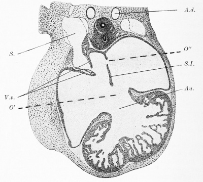
aorta; Au., atrial canal; S., sinus venosus; V.v., valvulse venosse; S.I., septum primum. Note the bifid apex seen at this stage and also the presence of two openings (O' and O") in the primitive auricular septum. In the collection of the I. Anatomical Institute, Vienna. (From Tandler's article in Keibel and Mall's Embryology, vol. ii, p. 549.) ]] | |||
Born showed that the division of the auricles takes place through the development of two different partitions | |||
Born showed that the division of the | |||
auricles takes place through the development of two different partitions | |||
placed in planes parallel with each other developing successively, parts of | placed in planes parallel with each other developing successively, parts of | ||
both of which are temporary, while parts persist to form the permanent | both of which are temporary, while parts persist to form the permanent | ||
| Line 158: | Line 101: | ||
primum is represented by a band of tissue between two orifices, the ostium | primum is represented by a band of tissue between two orifices, the ostium | ||
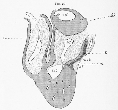
secundum above, and the ostium primum below. (See Figs. 19 and 20.) | secundum above, and the ostium primum below. (See Figs. 19 and 20.) | ||
| Line 176: | Line 110: | ||
in adult life as the aiumdus ovalis, while the valvula foraminis ovalis of the | in adult life as the aiumdus ovalis, while the valvula foraminis ovalis of the | ||
adult left auricle represents the remains of the primary septum, the primary | adult left auricle represents the remains of the primary septum, the primary | ||
and secondary ostia of which have both become obliterated. | and secondary ostia of which have both become obliterated. | ||
==The Interventricular Septum== | ==The Interventricular Septum== | ||
| Line 195: | Line 128: | ||
==The Aortic Septum== | ==The Aortic Septum== | ||
The truncus arteriosus is divided into the two | The truncus arteriosus is divided into the two great efferent vessels of the heart by a septum derived from three sources. Before the fifth week a sharp fold, the aortopulmonary septum proper, appears in the lumen of the truncus at the point of junction of the 4th and 6th arches (which represent respectively the aortic and pulmonary trunks) and grows rapidly downward. Some distance above the heart this aortopulmonary septum proper meets and fuses with a spiral septum derived from fusion of the so-called distal and proximal endocardial hidhar swellings. The bulbus cordis which forms, as stated above, by the involution of its proximal portion the termination of the ventricle, and by the elongation and demuscularization of its distal portion, the first part of the primitive aorta, is supplied internally with a series of endocardial elevations, of which four belong to its distal and two to its proximal part (known respectively as the Distal Bulbar Swellings 1, 2, 3, and 4, and Proximal Bulbar Swellings A and B of Born). These swellings, while symmetrically placed on opposite sides of the tube, have a spiral arrangement from above downward, and the distal swellings 2, and 4, which are much more prominent than the distal swellings 1 and 3, are directly continuous in clock-wise spiral fashion with the proximal swellings A and B. Fusion with each other, first of the more prominent pair of the distal bulbar swellings and later of the proximal ones occurs, the sjnral bulbar septum resulting, uniting at its distal end with the aortopulmonary septum proper, and the two structures being clearly distinguished from each other by their distinctive histological characters. | ||
great efferent vessels of the heart by a septum derived from three sources. | |||
Before the fifth week a sharp fold, the aortopulmonary septum proper, | |||
appears in the lumen of the truncus at the point of junction of the 4th and | |||
6th arches (which represent respectively the aortic and pulmonary trunks) | |||
and grows rapidly downward. Some distance above the heart this aortopulmonary septum proper meets and fuses with a spiral septum derived | |||
from fusion of the so-called distal and proximal endocardial hidhar swellings. | |||
The bulbus cordis which forms, as stated above, by the involution of its | |||
proximal portion the termination of the ventricle, and by the elongation | |||
and demuscularization of its distal portion, the first part of the primitive | |||
aorta, is supplied internally with a series of endocardial elevations, of | |||
which four belong to its distal and two to its proximal part (known respectively as the Distal Bulbar Swellings 1, 2, 3, and 4, and Proximal Bulbar | |||
Swellings A and B of Born). These swellings, while symmetrically placed | |||
on opposite sides of the tube, have a spiral arrangement from above downward, and the distal swellings 2, and 4, which are much more prominent | |||
than the distal swellings 1 and 3, are directly continuous in clock-wise | |||
spiral fashion with the proximal swellings A and B. Fusion with each | |||
other, first of the more prominent pair of the distal bulbar swellings and | |||
later of the proximal ones occurs, the sjnral bulbar septum resulting, uniting at its distal end with the aortopulmonary septum proper, and the two | |||
structures being clearly distinguished from each other by their distinctive | |||
histological characters. | |||
The chambers of the heart and the two great arteries have thus been | |||
completely separated from each other before the eighth week of fetal life. | The chambers of the heart and the two great arteries have thus been completely separated from each other before the eighth week of fetal life. Meantime, the right horn of the sinus venosus has been taken up in the wall of the right auricle, and the valvula venosa sinistra has disappeared, | ||
Meantime, the right horn of the sinus venosus has been taken up in the | a portion of the valvula venosa dextra persisting as the Eustachian valve, and the left sinus horn remaining as the coronary sinus, while the left duct | ||
wall of the right auricle, and the valvula venosa sinistra has disappeared, | of Cuvier becomes obliterated (left superior vena cava) . The pulmonary veins form later, opening at first as a single trunk, which is later taken up | ||
a portion of the valvula venosa dextra persisting as the Eustachian valve, | |||
and the left sinus horn remaining as the coronary sinus, while the left duct | |||
of Cuvier becomes obliterated (left superior vena cava) . The pulmonary | |||
veins form later, opening at first as a single trunk, which is later taken up | |||
in the wall of the left auricle, thereby enlarging it. The semilunar cusps appear to form about the seventh week, from the proximal ends of the four | in the wall of the left auricle, thereby enlarging it. The semilunar cusps appear to form about the seventh week, from the proximal ends of the four | ||
distal bulbar swellings, two of which are subdivided in the descent of the | distal bulbar swellings, two of which are subdivided in the descent of the septum trunci, so that six cusps, three placed in each artery, result. | ||
septum trunci, so that six cusps, three placed in each artery, result. | |||
The Auriculo ventricular Cusps and the Atrioventricular Bundle of His | ==The Auriculo ventricular Cusps and the Atrioventricular Bundle of His== | ||
[[File:Abbott_20.jpg| | [[File:Abbott_20.jpg|thumb|400px|'''Fig. 20. Sagittal section through the heart region of an embryo 8 mm long,''' X 40, No. 113 of Prof Franklin P. Mall's collection. B, bulbus cordis; A.V.B., auriculoventricular bundle; A.F., annulus fibrosis; I.V.C., interventricular canal; F.O'., ostium primum; P.O". ostium secundum; S.I., septum primum; E-, endocardial cushions. Note the extensive development of the endocardial cushions -within the auricle. (From the article by F. P. Mail in the American Journal of Anatomy, July 15, 1912.) ]] | ||
is to be traced to the breaking of the continuity of the atrial with the ventricular musculature by the ingrowth of constricting epicardial connective | The most critical point in the developing heart is undoubtedly the atrial canal. The endocardial cushions, which develop within it, are extensive and vitally important structures, not only as taking the essential part in the formation of the venous ostia, but also as completing the separation | ||
tissue about the external surface of the atrial ring. Thus while in very | of all four chambers by fusion with their respective septa. Moreover, from the observations of Mall on a large series of early human embryos, we learn that the differentiation of the auriculoventricular bundle of His is to be traced to the breaking of the continuity of the atrial with the ventricular musculature by the ingrowth of constricting epicardial connective tissue about the external surface of the atrial ring. Thus while in very early stages the muscle of the auricle is continuous with that of the ventricle at all points, in later stages a single band of atrial tissue passing down posteriorly from the lower border of the sinus venosus to the ventricle, and two minor fasciculi on the anterolateral wall, are the only remaining connection between the chambers. The survival of these isolated portions in the general destruction of the muscular continuity between auricle and ventricle is explained by Mall by the anatomical relations of the posterior part of the interventricular septum, which, growing up toward the posterior endocardial cushion, pushes the epicardial tissue obliquely before it and permits the escape of a small portion of auricular muscle. This surviving atrial tissue, now the only path of conductivity, undergoes differentiation and later, innervation, and becomes readily identified as the auriculoventricular bundle. | ||
early stages the muscle of the auricle is continuous with that of the ventricle at all points, in later stages a single band of atrial tissue passing | |||
down posteriorly from the lower border of the sinus venosus to the ventricle, and two minor fasciculi on the anterolateral wall, are the only | |||
remaining connection between the chambers. The survival of these | |||
isolated portions in the general destruction of the muscular continuity | |||
between auricle and ventricle is explained by Mall by the anatomical relations of the posterior part of the interventricular septum, which, growing up toward the posterior endocardial cushion, pushes the epicardial tissue | |||
muscle. This surviving atrial tissue, now the only path of conductivity, | |||
undergoes differentiation and later, innervation, and becomes readily | |||
identified as the auriculoventricular bundle. | |||
The auriculoventricular cusps are formed from the endocardial cushions | This contention of Mall, that the bundle represents persistent atrial tissue, and that its survival at this point is the result of its anatomical relation with the interventricular septum, has received striking confirmation from the investigations of Monckeberg, and Sato<ref>Professor Adami informs me that this idea of the persistence of the primitive atrial tissue in the Purkinje fibres of the adult heart was suggested by Gaskell in his book on the Evolution of the Vertebrates (1904).</ref> in the distribution of the bundle in various cardiac defects. Thus in a Cor Triloculare Biatriatrmn where, in the entire absence of ventricular septum one might conclude a destruction of auricular tissue in the whole circumference of the atrial ring, this bundle was found to be absent from the normal situation and was represented by a small band of tissue accompanying a small vessel on the anterolateral aspect of the heart. In defects of the interventricular septum at the base on the other hand, in which the posterior part of the septum practically always remains entire, the bundle was seen intact in both ventricles, streaming over the lower border of the defect. | ||
of the atrial canal. These cushions are a series of elevations of the lining | |||
endothelium of the cardiac tube formed by a spongy connective tissue, | |||
and are four in number. Two, the anterior and the posterior, develop very | The auriculoventricular cusps are formed from the endocardial cushions of the atrial canal. These cushions are a series of elevations of the lining endothelium of the cardiac tube formed by a spongy connective tissue, and are four in number. Two, the anterior and the posterior, develop very early, become of large size, and, growing toward each other, fuse to form the wedge-shaped block which separates the venous ostia and completes the cardiac septa. In addition they encroach by their rapid growth on adjacent structures, so that they come to line the lower border of the septum primum in the auricle, while extending also by their apices into the depths of the ventricle. The lateral cushions, of smaller size, develop later, and, with the anteroposterior pair, are converted from endocardial structures into the musculotendinous valves, by the undermining of their substance from without, and by their own invasion of, and fusion with, the spongy musculature of the ventricle. | ||
early, become of large size, and, growing toward each other, fuse to form | |||
the wedge-shaped block which separates the venous ostia and completes | |||
the cardiac septa. In addition they encroach by their rapid growth on | |||
adjacent structures, so that they come to line the lower border of the | |||
septum primum in the auricle, while extending also by their apices into the | |||
depths of the ventricle. The lateral cushions, of smaller size, develop | |||
later, and, with the anteroposterior pair, are converted from endocardial | |||
structures into the musculotendinous valves, by the undermining of their | |||
substance from without, and by their own invasion of, and fusion with, the | |||
spongy musculature of the ventricle. | |||
==Primitive Aortic Arches== | ==Primitive Aortic Arches== | ||
| Line 282: | Line 155: | ||
After considerable discussion, it is now fairly demonstrated that these number six instead of five, as Rathke described, the disputed fifth arch being rudimentary in character. They are more or less evanescent in all animals, except fishes, in which five persist. In birds and mammals the first, third, and fifth disappear on both sides. In man the fourth left arch becomes the aorta, the fourth right, the right subclavian, while the sixth pair become the pulmonary arteries. | After considerable discussion, it is now fairly demonstrated that these number six instead of five, as Rathke described, the disputed fifth arch being rudimentary in character. They are more or less evanescent in all animals, except fishes, in which five persist. In birds and mammals the first, third, and fifth disappear on both sides. In man the fourth left arch becomes the aorta, the fourth right, the right subclavian, while the sixth pair become the pulmonary arteries. | ||
1 Aschoff, Ber. d. Naturfor. Gesell. zu Freiburg, December 3, 191.3, B. xx. | 1 Aschoff, Ber. d. Naturfor. Gesell. zu Freiburg, December 3, 191.3, B. xx. | ||
---- | |||
<references/> | |||
---- | |||
{{Historic Disclaimer}} | {{Historic Disclaimer}} | ||
{{Abbott1915}} | |||
{{Glossary}} | |||
{{Footer}} | |||
Latest revision as of 14:14, 19 February 2017
| Embryology - 18 Apr 2024 |
|---|
| Google Translate - select your language from the list shown below (this will open a new external page) |
|
العربية | català | 中文 | 中國傳統的 | français | Deutsche | עִברִית | हिंदी | bahasa Indonesia | italiano | 日本語 | 한국어 | မြန်မာ | Pilipino | Polskie | português | ਪੰਜਾਬੀ ਦੇ | Română | русский | Español | Swahili | Svensk | ไทย | Türkçe | اردو | ייִדיש | Tiếng Việt These external translations are automated and may not be accurate. (More? About Translations) |
Abbott ME. Congenital Cardiac Disease (1915) Osler & Mccrae's Modern Medicine 6, 2nd Edition.
| Historic Disclaimer - information about historic embryology pages |
|---|
| Pages where the terms "Historic" (textbooks, papers, people, recommendations) appear on this site, and sections within pages where this disclaimer appears, indicate that the content and scientific understanding are specific to the time of publication. This means that while some scientific descriptions are still accurate, the terminology and interpretation of the developmental mechanisms reflect the understanding at the time of original publication and those of the preceding periods, these terms, interpretations and recommendations may not reflect our current scientific understanding. (More? Embryology History | Historic Embryology Papers) |
- 1915 Congenital Cardiac: Congenital Cardiac Disease | Heart Development | Literature | Etiology | Cyanosis | Classification | Pericardium | Heart Displacement | Whole Heart | Anomalous Septa | Interauricular Septum | Interventricular Septum | Absence of Cardiac Septa | Aortic Septum | Pulmonary Stenosis and Atresia | Pulmonary Artery Dilatation | Aortic Stenosis or Atresia | Primary Patency and Ductus Arteriosus | Aorta Coarctation | Aorta Hypoplasia | Diagnosis Prognosis and Treatment | Figures | Embryology History | Historic Disclaimer
The Development of the Heart
It is impossible to approach this subject intelligently without a certain preliminary knowledge of the development of the mammalian heart. A brief statement referring especially to the development of the septa, the involution of the bulbus cordis and sinus venosus, and the disappearance of the primitive aortic arches, is therefore necessary here. For fuller details the reader is referred to the fundamental studies of His[1] and Born[2] and to the recent contributions of Tandler,[3] Monckeberg,[4] and Mall.[5]
The mammalian heart is formed originally of two straight tubes placed
independently on either side of the body, which merge together as the
ventral cleft closes in and finally fuse, the septum thus formed becoming
entirely obliterated before the permanent interventricular septum begins
to appear. Meanwhile a twisting of the heart upon its long axes occurs,
and it becomes no longer symmetrical, but S-shaped, with the ventricular
portion bent forward and downward and the auricular part upward and
backward. It now consists of two chambers, a single ventricle forming
its anterior and lower part with its bulbus cordis passing upward and
to the left and giving off the aortic trunk from its right angle (Figs. 16 and
17), and a single auricle with its sinus venosus lying behind and to the
left. At this stage it resembles the two-chambered heart of the fish, and
is especially interesting in regard to the formation of the bulbus cordis.
The auricle next shifts upward, coming to be above the ventricle, and its auricular appendages develop enormously, pouching forward on either side of the bulbus (Fig. 19). The atrial canal, in which are developing the endocardial cushions which are to separate the two venous ostia, has become elongated and still opens into the common ventricle entirely on the left side. The sinus venosus is now a separate cavity opening into the auricle on its right wall posteriorly through a narrow cleft, the edges of which project into the auricle as the valvulse venosse dextra et sinistra. At its upper border it is elongated laterally into the two sinus horns which receive the two superior venae cavse, while a single short trunk, the inferior cava, enters it below.
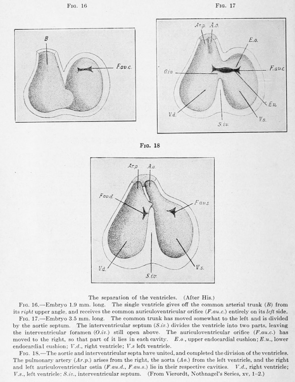
The separation of the ventricles. (After His.)
Fig. 16. - Embryo 1.9 mm. long. The single ventricle gives off the common arterial trunk (B) from its right upper angle, and receives the common auriculoventricular orifice (F.au.c.) entirely on its left side.
Fig. 17. - Embryo 3.5 mm. long. The common trunk has moved somewhat to the left and is divided by the aortic septum. The interventricular septum (S.iv.) divides the ventricle into two parts, leaving the interventricular foramen (O.iv.) still open above. The auriculoventricular orifice (F.au.c.) has moved to the right, so that part of it lies in each cavity. E.o., upper endocardial cushion; E.u., lower endocardial cushion; T'.rf., right ventricle; V.s left ventricle.
Fig. 18. - The aortic and interventricular septa have united, and completed the division of the ventricles. The pulmonary artery (Ar.p.) arises from the right, the aorta (Ao.) from the left ventricle, and the right and left auriculoventricular ostia (F.au.d., F.au.s.) lie in their respective cavities. V.d., right ventricle; V.S., left ventricle; S.iv., interventricular septum. (From Vierordt, Nothnagel's Series, xv, 1-2.)
PLATE V
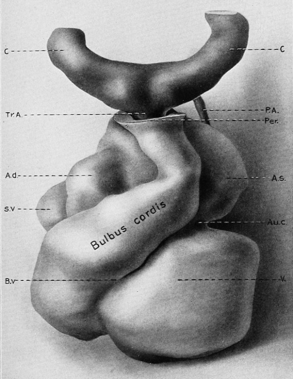
Model of the Heart of a Human Embryo 4.6 mm long. x 108. F. T. Lewis and M. E. Abbott. (Dr. Begg's Embryo, From the Anatomical Laboratory of the Harvard Medical School.)
C, carotid arch; P. A , pulmonary artery; Per., pericardium; Tr.A., truncus arteriosus; A.d., right auricle; A.s., left auricle; S.v., sinus venosus; Au.c, common auriculoventricular orifice; B.v,, bulboventricular cleft; V., common ventricle.
The Bulbus Cordis
This name is given to a transitory portion of the embryonic heart leading from the right end of the common ventricle to the aortic arches. In the human embryo of 4 to 6 mm. in length the bulbus is a thick-walled muscular tube passing to the left and upward, lined like the rest of the heart with endothelium, which presents certain endocardial thickenings, spirally arranged (Tandler), the so-called proximal and distal bulbar swellings, structures which later form the anlagen of the semilunar cusps as well as of the lower part of the aortopulmonary septum. In later stages the bulbus disappears, its proximal portion being taken up in the wall of the ventricle, and its distal part, denuded of its musculature and considerably elongated, constituting the primitive aortic trunk. The researches of Greil[6] on the reptilian heart, and Keith,[7] and recently of Jane Robertson[8] on the fish, show that the mammalian bulbus represents what was at one time an independent chamber with muscular walls and its own system of multiple valves, which in the "ontogenetic telescoping of phylogenetic stages" has become submerged.
Robertson correlates her findings in the fish with those of Greil in the
lizard, and Born in the mammalian embryo, and traces the bulbus of the
latter back through the less blurred stages of the reptile to the simpler
forms seen in the Dipnoan and Elasmobranch fishes. Thus this structure,
represented in the adult mammal, with its fully established double circulation, by the completely separated aortic and pulmonary trunks, is seen
in Lacerta (reptile) to consist of a curved muscular tube divided by a spiral
aortopulmonary septum, wdiich gives way in turn in Lepidosiren (Dipnoan
fish) to a kinked muscular tube with median expansion incompletely
divided by rows of spirally arranged valves, and this again in the Elasmobranch fishes with their purely branchial respiration, is reduced to the
simplest form as a straight channel with muscular walls lined by numerous
rows of longitudinally placed valves. These phylogenetic proofs of an
early bulbar channel with spiral division of its distal portion, are of the
utmost importance in the elucidation of the problems of stenosis of the
pulmonary conus and transposition of the arterial trunks, and yield
striking confirmation of the explanations offered by Keith of the former,
and by Rokitansky of the latter anomaly.
The Interauricular Septum

Born showed that the division of the auricles takes place through the development of two different partitions
placed in planes parallel with each other developing successively, parts of
both of which are temporary, while parts persist to form the permanent
interauricular septum of postnatal life. Of these septa, the one developing
earlier, called by Born the septum primiim, begins about the fourth week
from the upper and posterior wall of the auricle as a sickle-shaped fold
which grows forward and downward toward the ventricular cavity, and for some time an opening exists between the auricles at the lower border of this primitive septum known as the ostium primum. About the beginning of the fifth week a second opening, called by Born the ostium secundiun, forms in the now greatly thinned upper and back part of the
septum primum. This second opening grows larger as the ostium primum
becomes smaller, and finally disappears entirely (end of fifth week),
through the union of the expanded lower margin of the septum primum
with the fused endocardial cushions between the auriculoventricular
ostia. There thus exists a stage in development when the septum
primum is represented by a band of tissue between two orifices, the ostium
secundum above, and the ostium primum below. (See Figs. 19 and 20.)
The septum secundum arises considerably later than the septum primum
in a plane a little to its right, from the upper wall of the right auricle, and
passes downward covering in the upper and anterior portion of the ostium
secundum, thus giving it a valvular character, and transforming it into the
foramen ovale of fetal life. A portion of the septum secundum persists
in adult life as the aiumdus ovalis, while the valvula foraminis ovalis of the
adult left auricle represents the remains of the primary septum, the primary
and secondary ostia of which have both become obliterated.
The Interventricular Septum
This begins about the fourth week, just after the origin of the auricular septum, as a crescentic ridge on the inferior wall of the ventricle. It grows upward and backward, its posterior limb merging with the corresponding walls of the ventricle and with the posterior endocardial cushion, and its anterior limb with the anteroventricular wall along the bulbo-atrial ridges, while its median curved portion unites with a prolongation of the proximal aortic septum (the aortic orifice having moved over from the right to over-ride the ventricular septum), and with the fused endocardial cushions of the auriculo ventricular orifice (which has also come to lie in the median line), so that the ventricles are completely separated from each other and the arterial and venous ostia are placed one in either ventricle (Figs. 17 and 18). The point of union of the aortic with the interventricular septum just below the adjacent ends of the anterior and left posterior aortic cusps, remains transparent and devoid of muscle throughout life, and is known as the pars membranacea, or undefended space.
The Aortic Septum
The truncus arteriosus is divided into the two great efferent vessels of the heart by a septum derived from three sources. Before the fifth week a sharp fold, the aortopulmonary septum proper, appears in the lumen of the truncus at the point of junction of the 4th and 6th arches (which represent respectively the aortic and pulmonary trunks) and grows rapidly downward. Some distance above the heart this aortopulmonary septum proper meets and fuses with a spiral septum derived from fusion of the so-called distal and proximal endocardial hidhar swellings. The bulbus cordis which forms, as stated above, by the involution of its proximal portion the termination of the ventricle, and by the elongation and demuscularization of its distal portion, the first part of the primitive aorta, is supplied internally with a series of endocardial elevations, of which four belong to its distal and two to its proximal part (known respectively as the Distal Bulbar Swellings 1, 2, 3, and 4, and Proximal Bulbar Swellings A and B of Born). These swellings, while symmetrically placed on opposite sides of the tube, have a spiral arrangement from above downward, and the distal swellings 2, and 4, which are much more prominent than the distal swellings 1 and 3, are directly continuous in clock-wise spiral fashion with the proximal swellings A and B. Fusion with each other, first of the more prominent pair of the distal bulbar swellings and later of the proximal ones occurs, the sjnral bulbar septum resulting, uniting at its distal end with the aortopulmonary septum proper, and the two structures being clearly distinguished from each other by their distinctive histological characters.
The chambers of the heart and the two great arteries have thus been completely separated from each other before the eighth week of fetal life. Meantime, the right horn of the sinus venosus has been taken up in the wall of the right auricle, and the valvula venosa sinistra has disappeared,
a portion of the valvula venosa dextra persisting as the Eustachian valve, and the left sinus horn remaining as the coronary sinus, while the left duct
of Cuvier becomes obliterated (left superior vena cava) . The pulmonary veins form later, opening at first as a single trunk, which is later taken up
in the wall of the left auricle, thereby enlarging it. The semilunar cusps appear to form about the seventh week, from the proximal ends of the four
distal bulbar swellings, two of which are subdivided in the descent of the septum trunci, so that six cusps, three placed in each artery, result.
The Auriculo ventricular Cusps and the Atrioventricular Bundle of His

The most critical point in the developing heart is undoubtedly the atrial canal. The endocardial cushions, which develop within it, are extensive and vitally important structures, not only as taking the essential part in the formation of the venous ostia, but also as completing the separation
of all four chambers by fusion with their respective septa. Moreover, from the observations of Mall on a large series of early human embryos, we learn that the differentiation of the auriculoventricular bundle of His is to be traced to the breaking of the continuity of the atrial with the ventricular musculature by the ingrowth of constricting epicardial connective tissue about the external surface of the atrial ring. Thus while in very early stages the muscle of the auricle is continuous with that of the ventricle at all points, in later stages a single band of atrial tissue passing down posteriorly from the lower border of the sinus venosus to the ventricle, and two minor fasciculi on the anterolateral wall, are the only remaining connection between the chambers. The survival of these isolated portions in the general destruction of the muscular continuity between auricle and ventricle is explained by Mall by the anatomical relations of the posterior part of the interventricular septum, which, growing up toward the posterior endocardial cushion, pushes the epicardial tissue obliquely before it and permits the escape of a small portion of auricular muscle. This surviving atrial tissue, now the only path of conductivity, undergoes differentiation and later, innervation, and becomes readily identified as the auriculoventricular bundle.
This contention of Mall, that the bundle represents persistent atrial tissue, and that its survival at this point is the result of its anatomical relation with the interventricular septum, has received striking confirmation from the investigations of Monckeberg, and Sato[9] in the distribution of the bundle in various cardiac defects. Thus in a Cor Triloculare Biatriatrmn where, in the entire absence of ventricular septum one might conclude a destruction of auricular tissue in the whole circumference of the atrial ring, this bundle was found to be absent from the normal situation and was represented by a small band of tissue accompanying a small vessel on the anterolateral aspect of the heart. In defects of the interventricular septum at the base on the other hand, in which the posterior part of the septum practically always remains entire, the bundle was seen intact in both ventricles, streaming over the lower border of the defect.
The auriculoventricular cusps are formed from the endocardial cushions of the atrial canal. These cushions are a series of elevations of the lining endothelium of the cardiac tube formed by a spongy connective tissue, and are four in number. Two, the anterior and the posterior, develop very early, become of large size, and, growing toward each other, fuse to form the wedge-shaped block which separates the venous ostia and completes the cardiac septa. In addition they encroach by their rapid growth on adjacent structures, so that they come to line the lower border of the septum primum in the auricle, while extending also by their apices into the depths of the ventricle. The lateral cushions, of smaller size, develop later, and, with the anteroposterior pair, are converted from endocardial structures into the musculotendinous valves, by the undermining of their substance from without, and by their own invasion of, and fusion with, the spongy musculature of the ventricle.
Primitive Aortic Arches
After considerable discussion, it is now fairly demonstrated that these number six instead of five, as Rathke described, the disputed fifth arch being rudimentary in character. They are more or less evanescent in all animals, except fishes, in which five persist. In birds and mammals the first, third, and fifth disappear on both sides. In man the fourth left arch becomes the aorta, the fourth right, the right subclavian, while the sixth pair become the pulmonary arteries.
1 Aschoff, Ber. d. Naturfor. Gesell. zu Freiburg, December 3, 191.3, B. xx.
- ↑ This article has been largely rewritten, and curtailed in parts, to permit of the addition of new material, especially under the Development of the Heart, Anomalies of the Pericardium, Dextrocardia, Congenital Rhabdomyoma, Auricular, Ventricular, and Aortic Septal Defects, Deviation of the Aortic Septum, and Patent Ductus Arteriosus. The reader is referred to the earlier edition for the omitted material.
- ↑ Beitrdge zur Anatomie des menschlichen Herzens, Leipzig, 1886.
- ↑ Beitrage zur Entwickelung des Saugethierherzens, Arch, f . miki. anat., 1889, xxxiii.
- ↑ Atlas der Herzmissbildungen, Jena, 1912; also, Verh. d. Deut. Path. GeselL, Marburg, 1913, xvi, p. 228.
- ↑ Keibel and Mall's Human Embryology, 1912, ii, pp. 534-570. Am. Jour. Anat., 1913, xiv, p. 249. (323)
- ↑ Morph. Jahrb., 1903, xxxi, p. 123.
- ↑ Festschrift of the Quatercentenary of Aberdeen University, July, 1906.
- ↑ Jortr. Path, and Bacterial., 1913, xviii, p. 191.
- ↑ Professor Adami informs me that this idea of the persistence of the primitive atrial tissue in the Purkinje fibres of the adult heart was suggested by Gaskell in his book on the Evolution of the Vertebrates (1904).
| Historic Disclaimer - information about historic embryology pages |
|---|
| Pages where the terms "Historic" (textbooks, papers, people, recommendations) appear on this site, and sections within pages where this disclaimer appears, indicate that the content and scientific understanding are specific to the time of publication. This means that while some scientific descriptions are still accurate, the terminology and interpretation of the developmental mechanisms reflect the understanding at the time of original publication and those of the preceding periods, these terms, interpretations and recommendations may not reflect our current scientific understanding. (More? Embryology History | Historic Embryology Papers) |
| Embryology - 18 Apr 2024 |
|---|
| Google Translate - select your language from the list shown below (this will open a new external page) |
|
العربية | català | 中文 | 中國傳統的 | français | Deutsche | עִברִית | हिंदी | bahasa Indonesia | italiano | 日本語 | 한국어 | မြန်မာ | Pilipino | Polskie | português | ਪੰਜਾਬੀ ਦੇ | Română | русский | Español | Swahili | Svensk | ไทย | Türkçe | اردو | ייִדיש | Tiếng Việt These external translations are automated and may not be accurate. (More? About Translations) |
Abbott ME. Congenital Cardiac Disease (1915) Osler & Mccrae's Modern Medicine 6, 2nd Edition.
| Historic Disclaimer - information about historic embryology pages |
|---|
| Pages where the terms "Historic" (textbooks, papers, people, recommendations) appear on this site, and sections within pages where this disclaimer appears, indicate that the content and scientific understanding are specific to the time of publication. This means that while some scientific descriptions are still accurate, the terminology and interpretation of the developmental mechanisms reflect the understanding at the time of original publication and those of the preceding periods, these terms, interpretations and recommendations may not reflect our current scientific understanding. (More? Embryology History | Historic Embryology Papers) |
- 1915 Congenital Cardiac: Congenital Cardiac Disease | Heart Development | Literature | Etiology | Cyanosis | Classification | Pericardium | Heart Displacement | Whole Heart | Anomalous Septa | Interauricular Septum | Interventricular Septum | Absence of Cardiac Septa | Aortic Septum | Pulmonary Stenosis and Atresia | Pulmonary Artery Dilatation | Aortic Stenosis or Atresia | Primary Patency and Ductus Arteriosus | Aorta Coarctation | Aorta Hypoplasia | Diagnosis Prognosis and Treatment | Figures | Embryology History | Historic Disclaimer
Glossary Links
- Glossary: A | B | C | D | E | F | G | H | I | J | K | L | M | N | O | P | Q | R | S | T | U | V | W | X | Y | Z | Numbers | Symbols | Term Link
Cite this page: Hill, M.A. (2024, April 18) Embryology Book - Congenital Cardiac Disease 1. Retrieved from https://embryology.med.unsw.edu.au/embryology/index.php/Book_-_Congenital_Cardiac_Disease_1
- © Dr Mark Hill 2024, UNSW Embryology ISBN: 978 0 7334 2609 4 - UNSW CRICOS Provider Code No. 00098G


