|
|
| (18 intermediate revisions by the same user not shown) |
| Line 12: |
Line 12: |
| |} | | |} |
| {{Historic Disclaimer}} | | {{Historic Disclaimer}} |
| | = An Introduction to the Study of Embryology= |
| | [[File:Alfred Cort Haddon.jpg|thumb|alt=Alfred Cort Haddon|Alfred Cort Haddon (1855–1940)]] |
|
| |
|
| | By |
|
| |
|
| | Alfred C. Haddon, M.A. (Cantab.), M.R.I.A. |
|
| |
|
| | Professor Of Zoology In The Royal College Of Science, Dublin. |
|
| |
|
| AN
| |
|
| |
|
| | Philadelphia : P. Blakiston, Son & Co., 1012 Walnut Street. 1887. |
|
| |
|
| INTRODUCTION
| | {| |
| | | valign=middle| |
| | To the memory of |
|
| |
|
| TO THE
| | his beloved master and friend, |
|
| |
|
| STUDY OF EMBRYOLOGY.
| | [[Embryology History - Francis Balfour|'''Francis Maitland Balfour''']] |
|
| |
|
|
| |
|
| BY
| | This Book is dedicated by the Author. |
| | | [[File:Francis Balfour.jpg|alt=Francis Balfour (1851-1882)|thumb|200px|link=Embryology History - Francis Balfour|Francis Balfour (1851-1882)]] |
| | |} |
|
| |
|
| ALFRED C. HADDON, M.A. (Cantab.), M.R.I.A.
| |
|
| |
|
| PROFESSOR OF ZOOLOGY IN THE ROYAL COLLEGE OF
| | ==Preface== |
| SCIENCE, DUBLIN.
| |
|
| |
|
|
| |
|
| With gtumnwtf gltetration#.
| | Although there are at the present time, in addition to the special accounts in various text-books of Human and Comparative Anatomy, two Students - Manuals in the English language solely devoted to the study of Embryology, it has appeared to me that a relatively small work, giving a general review of the subject, might prove of use to students. |
|
| |
|
| | A knowledge of the main facts of Comparative Anatomy and Systematic Zoology has been assumed for the reader, the book being especially designed for Medical Students, or for those who already possess a general acquaintance with the Animal Kingdom. |
|
| |
|
| PHILADELPHIA :
| | It will be noticed that many of the more difficult problems of Ontology and Phylogeny and special modes of development have either been merely alluded to or entirely ignored - as, for instance, the segmentation of the ovum and the formation of the germinal layers in Insecta and Teleostei. This has been of set purpose, as my main object in writing this book has been to give a brief connected account of the principal organs, omitting or barely mentioning structures and phenomena, which may be regarded as of secondary importance. |
|
| |
|
| P. BLAKISTON, SON & CO.,
| | The facts of development have been largely supplemented by hypotheses; and an endeavour has been made so to present the latter, that the student could not mistake them for the former. |
|
| |
|
| 1012 WALNUT STREET.
| | It is inevitable that, in compiling such an introductory textbook as this, many subjects must be treated in a manner similar to that in which they have been dealt with by previous authors ; and therefore I have not hesitated to borrow from them when occasion required. |
|
| |
|
| 1887.
| | In order to facilitate references, very recent, important, or doubtful observations have been associated in many cases with the investigator -s name. It must be distinctly understood that I do not necessarily personally adopt statements or views which have been incorporated in the book; they are merely put forward for what they are worth. |
|
| |
|
| [ All Rights Reserved.']
| | The beginner is advised to pay attention only to the large type in the first reading, as purely theoretical subjects or matters of detail are printed in the smaller type. Most of the figures have been so drawn as to admit of their being coloured ; and the student is recommended to tint each germinal layer and the organs derived from it in a uniform manner throughout the book : thus the epiblast and its derivatives might be coloured pink, and the hypoblast tinted blue. A uniform system of colouration will be found to be of great assistance to the memory. |
|
| |
|
| | The sources from which the figures have been taken are in all cases acknowledged, and in the cases where no source is given the illustrations are original. Figs. 40, 41, 44, 45, 80, 81, and 178* have appeared previously in the Proceedings of the Eoyal Dublin Society. |
|
| |
|
| | The classification adopted will be found in an Appendix. All the genera mentioned in the text have been inserted, in order that their systematic position may be seen at a glance. |
|
| |
|
|
| |
|
| TO
| | A Bibliography has also been appended, which is designed to serve simply as a guide to the more recent literature, and no attempt has been made to render the list exhaustive. It will be noticed that most of the Memoirs cited are of later date than the year 1880. The more important earlier papers are recorded in the late Professor Balfour -s “Treatise of Comparative Embryology.- As any student who seriously studies Embryology must consult that invaluable work, I have considered it superfluous to repeat the Bibliography given by Balfour. The prevalent custom of authors of giving references to the literature of the subject under discussion renders it comparatively easy to discover what has already been written thereon. |
|
| |
|
| IT b c /!!> c m o v p
| | Finally, I would here express my warmest thanks to my friend Professor G. B. Howes, of the Normal School of Science, South Kensington, for his kindness in reading the proofs and in making many valuable suggestions. |
| | |
| OF
| |
| | |
| HIS BELOVED MASTER AND FRIEND,
| |
| | |
| FRANCIS MAITLAND BALFOUR,
| |
| | |
| This Book
| |
| | |
| IS DEDICATED
| |
| | |
| BY
| |
| | |
| | |
| THE AUTHOR.
| |
| | |
| | |
| | |
| PREFACE.
| |
| | |
| | |
| Although there are at the present time, in addition to the special
| |
| accounts in various text-books of Human and Comparative Anatomy, two Students - Manuals in the English language solely devoted
| |
| to the study of Embryology, it has appeared to me that a relatively
| |
| small work, giving a general review of the subject, might prove
| |
| of use to students.
| |
| | |
| A knowledge of the main facts of Comparative Anatomy and
| |
| Systematic Zoology has been assumed for the reader, the book
| |
| being especially designed for Medical Students, or for those
| |
| who already possess a general acquaintance with the Animal
| |
| Kingdom.
| |
| | |
| It will be noticed that many of the more difficult problems of
| |
| Ontology and Phylogeny and special modes of development have
| |
| either been merely alluded to or entirely ignored - as, for instance,
| |
| the segmentation of the ovum and the formation of the germinal
| |
| layers in Insecta and Teleostei. This has been of set purpose,
| |
| as my main object in writing this book has been to give a brief
| |
| connected account of the principal organs, omitting or barely
| |
| mentioning structures and phenomena, which may be regarded as
| |
| of secondary importance.
| |
| | |
| The facts of development have been largely supplemented by hypotheses; and an endeavour has been made so to present the
| |
| latter, that the student could not mistake them for the former.
| |
| | |
| It is inevitable that, in compiling such an introductory textbook as this, many subjects must be treated in a manner similar
| |
| to that in which they have been dealt with by previous authors ;
| |
| and therefore I have not hesitated to borrow from them when
| |
| occasion required.
| |
| | |
| In order to facilitate references, very recent, important, or
| |
| doubtful observations have been associated in many cases with
| |
| the investigator -s name. It must be distinctly understood that I
| |
| do not necessarily personally adopt statements or views which
| |
| have been incorporated in the book; they are merely put forward
| |
| for what they are worth.
| |
| | |
| The beginner is advised to pay attention only to the large
| |
| type in the first reading, as purely theoretical subjects or matters
| |
| of detail are printed in the smaller type. Most of the figures
| |
| have been so drawn as to admit of their being coloured ; and the
| |
| student is recommended to tint each germinal layer and the
| |
| organs derived from it in a uniform manner throughout the
| |
| book : thus the epiblast and its derivatives might be coloured
| |
| pink, and the hypoblast tinted blue. A uniform system of
| |
| colouration will be found to be of great assistance to the
| |
| memory.
| |
| | |
| The sources from which the figures have been taken are in all
| |
| cases acknowledged, and in the cases where no source is given
| |
| the illustrations are original. Figs. 40, 41, 44, 45, 80, 81,
| |
| and 178* have appeared previously in the Proceedings of the
| |
| Eoyal Dublin Society.
| |
| | |
| The classification adopted will be found in an Appendix. All
| |
| the genera mentioned in the text have been inserted, in order
| |
| that their systematic position may be seen at a glance.
| |
| | |
| | |
| A Bibliography has also been appended, which is designed to
| |
| serve simply as a guide to the more recent literature, and no
| |
| attempt has been made to render the list exhaustive. It will be
| |
| noticed that most of the Memoirs cited are of later date than the
| |
| year 1880. The more important earlier papers are recorded in
| |
| the late Professor Balfour -s “Treatise of Comparative Embryology.-
| |
| As any student who seriously studies Embryology must consult
| |
| that invaluable work, I have considered it superfluous to repeat
| |
| the Bibliography given by Balfour. The prevalent custom of
| |
| authors of giving references to the literature of the subject under
| |
| discussion renders it comparatively easy to discover what has
| |
| already been written thereon.
| |
| | |
| Finally, I would here express my warmest thanks to my friend | |
| Professor G. B. Howes, of the Normal School of Science, South | |
| Kensington, for his kindness in reading the proofs and in making | |
| many valuable suggestions. | |
| | |
| | |
| AN INTRODUCTION
| |
| | |
| TO
| |
| | |
| THE STUDY OF EMBRYOLOGY.
| |
| | |
| | |
| CHAPTER I. MATURATION AND FERTILISATION OF THE OVUM.
| |
| | |
| Introduction. - Embryology is the term usually applied to the
| |
| whole cycle of changes undergone by an animal in passing from
| |
| an egg to the adult condition. It is, in other words, the History
| |
| of its Development.
| |
| | |
| The name of embryo (or foetus, in mammalian embryology) is
| |
| restricted to the unborn young. At birth the young may closely
| |
| resemble the parent, or be very dissimilar ; in the latter case, it is
| |
| known as a larva, and undergoes a series of changes or a metamorphosis before it attains the adult state.
| |
| | |
| Even closely allied animals may be “ born - at very different
| |
| stages in their development ; the higher animals are, however,
| |
| generally born at a relatively later stage than those lower in the
| |
| animal scale. They are thus better fitted for the struggle for
| |
| existence, and expend less energy during their development than
| |
| if they had to provide for themselves.
| |
| | |
| In the higher animals the young also have the further advantage
| |
| of the watchful care of their parents, a factor which must have
| |
| materially influenced the evolution of the race.
| |
| | |
| Embryology may be studied under two aspects. The first, or
| |
| Ontogeny, deals solely with the history of the individual, and
| |
| traces the development of the animal as a whole, and of its various
| |
| organs.
| |
| | |
| The second, or comparative aspect, compares the development
| |
| of animals, and taking those phases which are common to all or
| |
| | |
| A
| |
| | |
| | |
| 2
| |
| | |
| | |
| THE STUDY OF EMBRYOLOGY.
| |
| | |
| | |
| | |
| to many, attempts therefrom to deduce or reconstruct the evolution
| |
| of the animal kingdom. This study is known as Phytogeny.
| |
| | |
| The chief result of all embryological inquiry has been to demonstrate that the history of the individual recapitulates in its main
| |
| features the evolution of the race, and thereby to give positive
| |
| evidence in favour of the Theory of Evolution, in the general
| |
| acceptance of the term.
| |
| | |
| It is very important to bear in mind that larval forms, as well
| |
| as adults, have to adapt themselves to external conditions, and
| |
| that they are consequently liable to be variously modified, and,
| |
| within limits, to be highly specialised. These modifications often
| |
| have no relation to the adult structure, and consequently can
| |
| have no phylogenetic significance.
| |
| | |
| Some preliminary knowledge of Zoology and Comparative Anatomy is necessary in order to appreciate fully the phases in the
| |
| development of any one animal, and it is, of course, essential in
| |
| studying the general principles of Embryology, as constant reference must be made to the structure of different forms. Such a
| |
| knowledge will be assumed for the readers of this book.
| |
| | |
| The Animal Kingdom is divided by zoologists into the Protozoa, or unicellular animals, and the Metazoa, or those animals
| |
| composed of a number of cells so united together as to form
| |
| tissues. As the ' latter alone produce ova or eggs, the science of
| |
| Embryology deals solely with the Metazoa. Although there is
| |
| considerable variation in the details of the classification of the
| |
| Metazoa, zoologists are tolerably well agreed upon the main
| |
| divisions, and in this work that classification and terminology
| |
| are adopted which are in most general use in English-speaking
| |
| countries.
| |
| | |
| Reproduction amongst the Protozoa consists in a direct or indirect method of cell- division, each product of such division
| |
| forming a new individual (fig. I, B, c, d). This process may, or
| |
| may not, be preceded by a temporary apposition or permanent
| |
| fusion of two or more individuals. The conjugating individuals
| |
| may either be apparently quite similar (fig. i, e), or may exhibit
| |
| certain differences (fig. I, f) ; but conjugation is always effected
| |
| between forms which are similarly motile - that is, ciliated individuals invariably conjugate with ciliated, and amoeboid with
| |
| other amoeboid, forms. Even among those Elagellate Infusoria
| |
| which pass through a comparatively complicated life-history, an
| |
| individual in the flagellate stage never conjugates with another in
| |
| | |
| | |
| MATURATION AND FERTILISATION OF THE OVUM.
| |
| | |
| | |
| o
| |
| | |
| O
| |
| | |
| | |
| the amoeboid condition. Active reproduction of one kind or other
| |
| usually occurs after conjugation.
| |
| | |
| Some Protozoa form compound masses, but the individuals
| |
| composing the colony are, with rare exceptions (Proterospongia),
| |
| similar to one another, and have a practically independent existence.
| |
| | |
| Although asexual reproduction by various modes of budding
| |
| and fission is known in nearly all the groups of the Metazoa, the
| |
| sexual method is of invariable occurrence. The essential act of
| |
| | |
| | |
| | |
| Fig. i. - Reproduction amongst Protozoa. Not drawn to scale.
| |
| | |
| A-C. Fission in an Amoeba. A. The nucleus has divided into two. B. Two
| |
| contractile vacuoles have also formed and the protoplasm is dividing. C. The
| |
| process is complete. [ After Howes.']
| |
| | |
| D. Fission in Paramsecium bursaria. There are two contractile vacuoles and two
| |
| paranuclei, but the nucleus has not yet completely divided.
| |
| | |
| E. Conjugation of Stylonychia mytilus, illustrating also the fragmentation of the
| |
| nucleus.
| |
| | |
| F. Conjugation of Vorticella microstoma. Two free-swimming microzooids have
| |
| attached themselves to a fixed form. They all possess a curved nucleus and
| |
| a contractile vacuole. [D-F after Stein.]
| |
| | |
| c.v. contractile vacuole ; n. nucleus ; nl. paranucleus.
| |
| | |
| | |
| this form of reproduction consists in the fusion of a flagellate cell
| |
| or spermatozoon with an amoeboid cell, the egg or ovum (figs,
| |
| io and n).
| |
| | |
| In a very few cases the spermatozoa are either amoeboid, as in
| |
| Nematodes, some Arachnids, and Limulus, or often passive and
| |
| rayed, as in most Crustacea; but in the great majority of animals
| |
| the spermatozoa are flagellate and actively motile (fig. 2).
| |
| | |
| The ovum, under very rare and exceptional conditions, may
| |
| develop into a new organism without previous fertilisation by a
| |
| spermatozoon ; this phenomenon is known as 'parthenogenesis.
| |
| | |
| | |
| 4
| |
| | |
| | |
| THE STUDY OF EMBRYOLOGY.
| |
| | |
| | |
| The ovum and spermatozoon unite to form the fertilised ovum
| |
| or oosperm, which then undergoes rapid cell-division ; the cells
| |
| thus produced remain in contact with one another, and though at
| |
| first usually very similar, certain groups of cells soon take upon
| |
| themselves definite characters, and thus initiate the primitive
| |
| tissues.
| |
| | |
| Accepting the view that the Metazoa were derived from colonial Protozoa, it follows
| |
| that every cell of the primitive Metazoa was capable of forming fresh colonies by
| |
| | |
| | |
| | |
| Fig. 2. - Spermatozoa [from various sources]. Not drawn to scale.
| |
| | |
| i. Sponge; 2. Hydroid; 3. Nematode; 4. Crayfish; 5. Snail; 6. Electric
| |
| Ray ; 7. Salamander ; 8. Horse ; 9. Man. In many spermatozoa, as in
| |
| Nos. 7 and 9, an extremely delicate vibratile band is present.
| |
| | |
| cell-division. Many Metazoa possess the power of asexually producing new forms
| |
| by fission or by budding ; but the tissues implicated in this process must be regarded
| |
| as being essentially undifferentiated in character.
| |
| | |
| Owing to the advantage derived from physiological differentiation of labour, the
| |
| reproductive function came to be chiefly retained by certain cells, the remainder
| |
| specialising along other lines. Those cells which pre-eminently retain the reproductive
| |
| function are restricted in their position, and the tissue which they constitute (the
| |
| germinal tissue) is contained within what is known as a generative organ or gland.
| |
| When ripe, the germ-cells become detached, and commence a free existence.
| |
| | |
| | |
| MATURATION AND FERTILISATION OF THE OVUM.
| |
| | |
| | |
| 5
| |
| | |
| | |
| After fertilisation, the ovum, or the embryo into which it
| |
| develops, is in a few cases retained within the oviduct of the
| |
| mother for a longer or a shorter period, and may temporarily
| |
| even be intimately, but very rarely structurally, connected with
| |
| the walls of the oviduct or uterus, as will subsequently be described.
| |
| | |
| The primitive germ-cells of animals are, practically, precisely
| |
| similar to one another (fig. 175), and, when first recognisable as
| |
| germ- cells, it is impossible to tell whether they will develop into
| |
| ova or sperm-cells. In this connection it is suggestive to find
| |
| that both the ovaries and the testes in Sagitta are developed from
| |
| a single primitive germ-cell, which makes its appearance at a very
| |
| early stage of development. The primitive germ-cells may more
| |
| especially be said to correspond to the Protozoon ancestors of the
| |
| Metazoa.
| |
| | |
| Before dealing further with the history of the germ-cells, however, it will be advisable to describe briefly their mode of origin.
| |
| | |
| The Ovum. - The primitive ova usually form part of a definite
| |
| epithelium, of which most of the cells, or it may be only a very
| |
| small number, develop into ripe ova. The germinal epithelium is
| |
| well supplied with nutritive fluid (either blood or the fluid contents of the cavity of the body), which serves for the growth of
| |
| the ova. From the nutriment thus provided the ova generally
| |
| store up a greater or less amount of reserve food-material, which
| |
| is known as “ yolk - or “ food-yolk.-
| |
| | |
| It would be foreign to the purpose of this work to enter into a comparative
| |
| account of the development of ova from primitive germinal-cells. As a general rule,
| |
| certain of the cells of the germinal epithelium are directly converted into ova. In
| |
| Vertebrates, the germinal epithelium is borne upon a distinct germinal ridge ; the
| |
| epithelium increases in thickness, and becomes broken up into cords or trabeculae
| |
| (ovarian tubes of Pfluger), which, by mutual ingrowth, lie in the stroma or mesoblastic core of the germinal ridge. Isolated masses or nests may also be formed (fig. 3).
| |
| | |
| Balfour has shown that in Elasmobranchs and other forms, in addition to the
| |
| foregoing or direct origin of the ova, the protoplasm of the cells forming the nests
| |
| fuses into a single mass containing the nuclei of the previously distinct ova.
| |
| Various changes are undergone, but eventually a few of the nuclei segregate protoplasm round themselves to form the ova, the remainder having broken down to
| |
| pabulum for the permanent ova.
| |
| | |
| Beddard finds that in Protopterus two kinds of ova are developed - (a.) The ovum
| |
| is a mass of granular protoplasm, containing a germinal vesicle limited by a distinct
| |
| membrane, inside of which is a peripheral layer of germinal spots. Later the protoplasm becomes vacuolated, and largely differentiates to form yolk-granules. (6.) The
| |
| ovum arises from the fusion of a nest of germinal cells lying within a follicle ; not
| |
| only is yolk formed within the central mass, but it is also produced within the
| |
| columnar cells of. the follicular epithelium. These cells proliferate and migrate into
| |
| | |
| | |
| 6
| |
| | |
| | |
| THE STUDY OF EMBRYOLOGY.
| |
| | |
| | |
| the interior of the ovum ; eventually they disappear. The yolk of these ova appears
| |
| to be largely derived from the follicular cells.
| |
| | |
| The yolk consists of highly refractive particles, which vary considerably in their
| |
| appearance and structure. As a rule, the yolk elements are small vesicles, which
| |
| usually contain smaller vesicles and other bodies (fig. 28, b). In Birds the whole of
| |
| the yolk at first consists of these white yolk spheres ; but during the development
| |
| of the egg, some of the white yolk spheres become modified to form the yellow yolk
| |
| (fig. 28, A and c). In the ripe unincubated egg the yellow yolk constitutes the great
| |
| mass of the “yolk,- the white yolk being restricted to a peripheral and several concentric layers, and to a central mass which extends in a constricted neck, and again
| |
| widens out to form a bed, upon which the blastoderm rests (fig. 28, A, w, y ).
| |
| | |
| | |
| It not unfrequently happens (many Hydrozoa, Insects, some
| |
| Vertebrates, &c.) that certain of the primitive germ-cells feed
| |
| upon neighbouring germ-cells, so that the growth of the ovum
| |
| | |
| | |
| | |
| Pig. 3. - Section through a Portion of the
| |
| Ovary of a Mammal. Illustrating the mode
| |
| of development of the Graafian follicles. [From.
| |
| Wiedersheim. ]
| |
| | |
| D. discus proligerus ; Ei. ripe ovum ; G. follicular cells of germinal epithelium ; g. bloodvessels ; K. germinal vesicle (nucleus) and germinal spot (nucleolus) ; KE. germinal epithelium ;
| |
| Lf. liquor folliculi ; Mg. membrana- or tunicagranulosa or follicular epithelium ; Mp. zona
| |
| pellucida ; PS. ingrowths from the germinal epithelium, ovarian tubes, by means of which some
| |
| of the nests retain their connection with the epithelium ; S. cavity which appears within the
| |
| Graafian follicle ; So. stroma of ovary ; Tf. theca
| |
| folliculi or capsule ; U. primitive ova. When' an
| |
| ovum with its surrounding cells has become separated from a nest, it- is known as a Graafian follicle.
| |
| | |
| | |
| and its store of food-yolk are made at the expense of its fellow
| |
| germinal cells. In most Platyhelminths that portion of the primitive germinal epithelium which is destined to provide pabulum
| |
| for the ova proper is separated from the ovary as yolk-glands, or
| |
| vitellaria , and their products, yolk-cells or yolk-granules, surround
| |
| the ova after they have left the ovary, and before they are enclosed
| |
| within the egg-capsules. The yolk-cells may be regarded as germinal cells which have lost the power of reproduction, hut retained
| |
| that of forming yolk. Either the ovum or the embryo in due
| |
| course feeds upon this reserve of food.
| |
| | |
| When many ova are deposited within the same egg-capsule as
| |
| in some forms of Prosobranch Gastropods (Buccinum), the more
| |
| | |
| | |
| MATURATION AND FERTILISATION OF THE OVUM.
| |
| | |
| | |
| 7
| |
| | |
| | |
| advanced embryos devour those that are imperfectly developed, so
| |
| that a very limited number, sometimes only a single individual,
| |
| eventually escape from one capsule.
| |
| | |
| The fusion of several germinal cells with one ovum does not
| |
| correspond to the multiple conjugation of some Protozoa, as in the
| |
| | |
| | |
| | |
| Fiq. 4. - Diagrams of Ova [from, various sources after Geddes ]. Not drawn to scale.
| |
| | |
| a. Diagram of a typical ovum with a delicate egg-membrane, granular protoplasm, nucleus (germinal vesicle), and nucleolus (germinal spot), b. Amoeboid
| |
| ovum of Hydra [after Kleinenberg ]. c. Early ovum of a Sea-Urchin (Toxopneustes
| |
| variegatus) with pseudopodia-like processes extending into the gelatinous eggmembrane (vitelline membrane) in order to obtain nutriment from without ;
| |
| afterwards they become much finer and more regular, causing the vitelline
| |
| membrane to have a striated appearauce ; hence it is termed the * • Zona radiata -
| |
| | |
| - the striae are really delicate pores [after Selenka ]. d. Nearly ripe ovum of
| |
| Strongylocentrotus lividus with its zona radiata [after Herticig].
| |
| | |
| | |
| formation of plasmodia ; it is merely the assimilation of several
| |
| cells by one ovum, much as an Amoeba feeds upon its prey.
| |
| | |
| An ovum is a small free cell which is characterised in the
| |
| resting-stage by possessing a large clear nucleus, the germinal
| |
| vesicle, and a well-marked highly refractive nucleolus, the ger
| |
| | |
| Fig. 5. - Ovum of the Cat. Highly magnified ;
| |
| semi-diagrammatic. [From Quain, after Schafer.]
| |
| | |
| gs. germinal spot ; gv. germinal vesicle ; vi. vitellus, or protoplasm of ovum filled with yolk granules, round which a delicate membrane was seen ;
| |
| zp. zona pellucida ( Zona radiata ) ; only a few
| |
| radial pores are drawn.
| |
| | |
| | |
| | |
| minal spot ; in many cases several germinal spots occur. Pigs. 4
| |
| and 5 illustrate various kinds of ova.
| |
| | |
| The protoplasm usually has, as has just been mentioned, the
| |
| power of storing up albuminoid matter as reserve food material by
| |
| a differentiation of its own substance in the form of yolk-granules
| |
| or spheres. The amount of food-yolk varies greatly ; in some few
| |
| | |
| | |
| 8
| |
| | |
| | |
| THE STUDY OF EMBRYOLOGY.
| |
| | |
| | |
| instances none appears to be differentiated ; often only a little is
| |
| formed ; more frequently there is a considerable amount ; and in
| |
| the eggs of Elasmobranchs and of Sauropsida an enormous quantity
| |
| is deposited. The distribution of the yolk within the egg also
| |
| varies, being either chiefly concentrated at one pole ( telolecithal ),
| |
| or towards the centre {centrolecithal), or evenly distributed throughout ( alecithal ).
| |
| | |
| As the amount of protoplasm in an ovum containing much foodyolk is relatively small, the storing of the yolk-granules within its
| |
| substance would naturally cause it to be distended. In those ova
| |
| with a very large amount of yolk, the protoplasmic reticulum
| |
| scarcely more than serves to keep the yolk-granules together
| |
| | |
| | |
| Fig. 5*. - Typicai. Cell and Nucleus
| |
| of the Intestinal Epithelium of a
| |
| Flesh-Maggot (asticot), treated with osmic acid vapour. [From Camay. ]f
| |
| | |
| bn. continuous band of nucleine, contracted to the centre of the nucleus, and
| |
| showing numerous twists ; me. membrane of the cell ; mn. membrane of the
| |
| nucleus ; pc. protoplasm of the cell,
| |
| showing the radiating reticulum and the
| |
| enchylema enclosed in its meshes ; pn.
| |
| plasma of the nucleus, showing a reticulum and a plasmic enchylema, as distinct
| |
| as those of the protoplasm.
| |
| | |
| The structure of an ovum is practically
| |
| identical with that of such a tissue-cell
| |
| as the above.
| |
| | |
| | |
| During development, certain cells of the embryo reconvert the
| |
| food-yolk into active protoplasm.
| |
| | |
| The germinal vesicle of the unripe ovum, as Carnoy points out,
| |
| has the same general structure as the ovum itself; that is, it
| |
| consists of an extremely fine protoplasmic reticulum , the meshes of
| |
| which are filled with a granular fluid {enchylema). The reticulum
| |
| also forms a delicate nuclear membrane. But, in addition to the
| |
| above, the nucleus possesses a distinctive substance, which i&
| |
| variously termed nucleine , nucleoplasm , or, from its being readily
| |
| stained by the action of certain reagents, chromatin. In very
| |
| young ovarian ova, the chromatin occurs in the form of a very
| |
| long, extremely and irregularly contorted thread or nuclear filament.
| |
| | |
| The nuclear filament is condensed in more mature ova into a
| |
| | |
| | |
| | |
| MATURATION AND FERTILISATION OF THE OVUM.
| |
| | |
| | |
| 9
| |
| | |
| | |
| single spherical mass, the germinal spot, or into a few or a large
| |
| number of smaller germinal spots.
| |
| | |
| Ova may be either naked (fig. 4, A and b), or surrounded by
| |
| one or more membranes (fig. 4, c, D, and fig. 5). The primary eggmembranes (vitelline membranes) are usually differentiated from
| |
| the protoplasm of the ovum itself.
| |
| | |
| In Vertebrates two egg-membranes are usually present, an external delicate vitelline membrane, which is probably formed by the
| |
| ovum itself, but in some cases a similar membrane may be secreted
| |
| by the epithelium of the ovarian follicle. This membrane is often
| |
| termed a chorion. Below the vitelline membrane a thicker membrane, perforated by innumerable fine radial pores, is differentiated
| |
| out of the peripheral layer of the ovum. It is known as the. zona
| |
| radiata or zona pellucida. The secondary egg-membranes are either
| |
| | |
| | |
| Fig. 6. - A Fowl -s Egg after about
| |
| Thirty Hours - Incubation. Viewed from
| |
| above, the upper portion of the shell
| |
| being removed. [ From Kolliker after Von
| |
| Baev. J
| |
| | |
| ' a. shell ; 6. shell-membrane ; b'. airchamber at broad end of egg between
| |
| the two layers of the shell-membrane ;
| |
| c. the boundary between the outer and
| |
| middle portion of the albumen ; d. the
| |
| internal layer of more fluid albumen,
| |
| which also extends round the yolk as a
| |
| thin sheath ; e. chalaza ; v. vitellus or
| |
| yolk ; av. area opaca, or that portion of
| |
| the blastoderm which extends over the
| |
| yolk ; the heart-shaped central portion,
| |
| ao, is the vascular area of the area opaca.
| |
| In the centre is the embryo surrounded
| |
| by the area pellucida.
| |
| | |
| | |
| secreted by accessory generative glands, or by the glandular wall of
| |
| the oviduct. When a secondary egg-membrane is impregnated with
| |
| calcareous deposits, it is known as an egg-shell. The secondary
| |
| egg-covering often encloses an albuminous glairy fluid - the white
| |
| of egg - which serves for the protection and further nutriment of
| |
| the embryo (figs. 6, 74, 75). The albumen also is secreted, either
| |
| by special glands (most Invertebrates), or by the wall of the
| |
| oviduct (Vertebrates).
| |
| | |
| Maturation of the Ovum. - Before or after fertilisation, certain
| |
| changes, which are of considerable interest, take place in the ovum.
| |
| The germinal vesicle often becomes amoeboid, and passes to one pole
| |
| of the ovum, and the germinal spot disappears (fig. 7, b-d) ; in fact,
| |
| both the germinal vesicle and spot disappear as such, and pass into
| |
| those karyolitic figures which characterise cell-division (see p. 18).
| |
| | |
| | |
| | |
| 10
| |
| | |
| | |
| THE STUDY OF EMBRYOLOGY.
| |
| | |
| | |
| The resulting nuclear spindle is placed vertically, the peripheral
| |
| nuclear star, or “ aster,- being situated in a small protuberance
| |
| from the surface of the ovum. This process is segmented off from
| |
| the ovum, and a minute cell is formed, containing a portion
| |
| of both the protoplasm and the nucleus of the parent-cell
| |
| (fig. 7, F and l).
| |
| | |
| | |
| | |
| A. Ripe ovum with excentric germinal vesicle and spot ; B-D. Gradual metamorphosis of germinal vesicle and spot, as seen in the living egg, into two
| |
| asters ; F. Formation of first polar cells and withdrawal of remaining part of
| |
| nuclear spindle within the ovum ; G. Surface view of living ovum in the first
| |
| polar cell ; H. Completion of second polar cell ; I. A later stage, showing the
| |
| remaining internal half of the spindle in the form of two clear vesicles ; K. Ovum
| |
| with two polar cells and radial striae round female pronucleus, as seen in the
| |
| living egg. [B, F, H, and I, from picric acid preparations.] L. Expulsion of first
| |
| polar cell.
| |
| | |
| | |
| This phenomenon is repeated, and two cells are budded off from
| |
| the ovum ; these are known as the “ polar cells - (or as polar bodies,
| |
| polar globules, directive bodies, &c.), from the fact that they are
| |
| invariably derived from that pole of the ovum at which the epiblast
| |
| or upper-layer cells will be developed; hence, also, this pole is
| |
| | |
| | |
| | |
| Fig. 8. - Formation op Polar Cells
| |
| in Ovum of Elysia viridis.
| |
| | |
| The upper pole of the ovum becomes
| |
| amoeboid during the formation of the
| |
| polar cells. The second polar cell is in
| |
| process of formation.
| |
| | |
| | |
| usually termed the upper pole of the ovum (see figs. 12 and 17).
| |
| During the production of the polar cells, the ovum, especially at
| |
| its upper pole, may exhibit amoeboid movements ; this is well shown
| |
| in the ovum of Elysia (fig. 8).
| |
| | |
| Although the polar cells may remain attached to the developing
| |
| | |
| | |
| MATURATION AND FERTILISATION OF THE OVUM.
| |
| | |
| | |
| 11
| |
| | |
| | |
| ovum for some time, they take no share in the formation of the
| |
| embryo, and are simply to be regarded as superfluous bodies.
| |
| | |
| What remains of the primitive nucleus passes towards the centre
| |
| of the ovum, usually in an inactive or resting condition, being
| |
| without radial striae. It is known as the female pro-nucleus.
| |
| | |
| The ovum is now in a passive condition, and ready to be
| |
| fertilised. The extrusion of the polar cells, though occasionally
| |
| taking place after fertilisation (ex. Elysia, fig. 8), is really to be
| |
| regarded as the last term in that series of changes which occurs
| |
| before impregnation, and to be, in fact, anticipatory of it.
| |
| | |
| Before following the history of the ovum further, it will be
| |
| necessary to return to the sperm-cells.
| |
| | |
| The Spermatozoon. - Although we find considerable variation
| |
| in certain details of structure, there is a general similarity in the
| |
| appearance of the spermatozoa of animals, a head and vibratile
| |
| tail being of almost universal occurrence : the most important
| |
| exceptions have already been mentioned (p. 3 and fig. 2).
| |
| | |
| The primitive sperm-cells or mother-cells of the spermatozoa arise from a tissue
| |
| corresponding to that which gives origin to the primitive ova (p. 242, fig. 175). The
| |
| exact manner in which the spermatozoa are developed varies in different animals,
| |
| and has been variously described by numerous investigators. This being the case,
| |
| it will be advisable to give simply a sketch of what appear to be the most important
| |
| facts in spermatogenesis , as this process is termed.
| |
| | |
| Those cells of the generative epithelium which develop into male sexual cells
| |
| undergo cell-division in the ordinary manner, and may give rise to a considerable
| |
| number of cells ( spermatoblasts ). Each spermatoblast is converted into a spermatozoon, and, in doing so, gives rise to a small mass of protoplasm, the so-called seminal
| |
| granule , or globule, or accessory corpuscle, which appears to have no further function.
| |
| Fig. 9, a-h, illustrates this process in the Rat.
| |
| | |
| Instead of becoming distinct, the spermatoblasts or incipient spermatozoa may
| |
| remain aggregated together ( spermosphere or sperm-morula), and surround a central
| |
| non-nucleated protoplasmic mass (the sperm-Uastophore ), as in the case of the Snail
| |
| and Earthworm (fig. 9, o-s).
| |
| | |
| In Elasmobranchs (fig. 9, i-n) the nucleus of the sperm-cell (sometimes called the
| |
| spermatocyst) alone divides, forming a number of daughter-nuclei, the remains of the
| |
| parent-nucleus still persisting. The protoplasm of the cell differentiates into the
| |
| tails of the spermatozoa, while the daughter-nuclei constitute the main portion of
| |
| their heads. The ripe spermatozoa are liberated by the rupture of the wall of the
| |
| sperm-cell, leaving behind the parent-nucleus and a small remnant of unused protoplasm. This latter is merely an abbreviated variation of the former process, and the
| |
| residual nucleus and protoplasm clearly correspond to the accessory corpuscle or to
| |
| the sperm-blastophore in the preceding forms.
| |
| | |
| The nucleus of each daughter sperm-cell constitutes the head of a spermatozoon ;
| |
| it is surrounded by an extremely delicate film, which is produced from one end into
| |
| a fine flagellum, and sometimes also into an almost imperceptible undulating membrane ; these are formed by the protoplasm of the spermatoblast. Every spermatozoon is thus a true morphological cell.
| |
| | |
| Kolliker, however, maintains that the entire mammalian spermatozoon is simply
| |
| a free nucleus.
| |
| | |
| | |
| 12
| |
| | |
| | |
| THE STUDY OF EMBRYOLOGY.
| |
| | |
| | |
| Fertilisation of the Ovum. - It is needless to recount the
| |
| various ways by which spermatozoa may reach ova ; suffice it to
| |
| say, that either within the female or in the surrounding water a
| |
| | |
| | |
| | |
| A-H. Isolated sperm-cells of the Rat, showing the development of the spermatozoon, and the gradual transformation of the nucleus into the spermatozoon
| |
| head. In G the seminal granule is being cast off. [ After H. H. Brown.]
| |
| | |
| I-M. Sperm-cells of an Blasmobranch. The nucleus of each cell divides into a
| |
| large number of daughter-nuclei, each one of which is converted into the rodlike head of a spermatozoon.
| |
| | |
| N. Transverse section of a ripe cell, showing the bundle of spermatozoa and the
| |
| passive nucleus. [I-N after Semper .]
| |
| | |
| O-S. Spermatogenesis in the Earthworm : O. young sperm-cell ; P. the same
| |
| divided into four ; Q. spermatosphere with the central sperm-blastophore •
| |
| R. a later stage ; S. nearly mature spermatozoa. [After Blomfield .]
| |
| | |
| | |
| spermatozoon comes into contact with an ovum, and either penetrates any membrane which may surround it, or passes through an
| |
| aperture (micropyle) left in the egg-membrane.
| |
| | |
| When the spermatozoon is approaching the actual surface of an
| |
| | |
| | |
| MATURATION AND FERTILISATION OF THE OVUM.
| |
| | |
| | |
| 13
| |
| | |
| | |
| ovum, a process from the latter sometimes rises up to meet it, and
| |
| a fusion is effected (fig. io, a-d, and fig. n, a). The head of the
| |
| spermatozoon penetrates the ovum, while the tail, after vibrating
| |
| feebly, is absorbed.
| |
| | |
| The head, or rather the nucleus of the spermatozoon, is converted into an aster or star, and is now known as the male pro
| |
| | |
| | |
| Fxo. io Fertilisation of Ovum of a Star-fish (Asterias glacialis). [ From Geddes after FoL]
| |
| | |
| In A-D the spermatozoa are represented as imbedded within the mucilaginous
| |
| coat of the ovum. In A a small prominence is rising from the surface of the
| |
| ovum towards the nearest spermatozoon ; in B they have nearly met, and in G
| |
| they have met. D. The spermatozoon has penetrated the ovum, and a vitelline
| |
| membrane with a crater-like opening has been formed, which prevents the entrance of other spermatozoa. H. ovum showing polar cells and approach of the
| |
| male and female pro-nuclei ; the protoplasm is radially striated round the former.
| |
| | |
| E. F. G. later stages in the coalescence of the two nuclei.
| |
| | |
| nucleus. It travels towards the female pro-nucleus, which, it will
| |
| be remembered, is situated in the centre of the ripe ovum (fig.
| |
| io, h). The female pro-nucleus becomes somewhat amoeboid, and
| |
| fusion occurs between the two elements, thus forming a new
| |
| nucleus (fig. io, e-g). While this is taking place, the ovum itself
| |
| often exhibits amoeboid movements (fig. n, a).
| |
| | |
| | |
| | |
| Fig. ii. - Fertilisation of Ovum of Elysia viridis.
| |
| | |
| A. ovum sending up a protuberance to meet the spermatozoon ; B. approach of
| |
| male pro-nucleus to meet the female pro-nucleus ; F.PN. female pro-nucleus ;
| |
| | |
| M.PN. male pro-nucleus; S. spermatozoon.
| |
| | |
| The fertilised ovum is a very different body from the primitive
| |
| ovum, as it consists of a portion of the original protoplasm and
| |
| nucleus of the latter reinforced by those of another cell, which
| |
| is usually derived from a different animal. The new nucleus is
| |
| called the segmentation nucleus , and it may be well to adopt Balfour -s name of oosperm for the fertilised ovum.
| |
| | |
| | |
| 14
| |
| | |
| | |
| THE STUDY OF EMBRYOLOGY.
| |
| | |
| | |
| There is some doubt whether the male pro-nucleus has the full value of a true
| |
| nucleus, and this has led Flemming to define fertilisation as the union of “the chromatin of a male with that of a female nuclear body.- Yan Beneden has recently
| |
| shown that the essential act of fertilisation consists in the grouping together (or
| |
| probably, more accurately, of the fusion) of the chromatin or germ-plasma of the
| |
| nucleus of the spermatozoon with that of the nucleus of the ovum. During the first
| |
| division of the oosperm, and in all the succeeding phases of segmentation, each new
| |
| cell receives an equal share of the paternal and maternal chromatin. (See chap. ii.
| |
| p. 19, and fig. 13.)
| |
| | |
| The fertilisation of an ovum by a spermatozoon is paralleled
| |
| by the permanent conjugation Cf such Protozoa as Vorticella and
| |
| many Monads. In each case the phenomenon is followed by rapid
| |
| cell-division - the resulting cell-units remaining separate in the
| |
| Protozoa, whereas they group themselves together so as to form an
| |
| aggregate of a higher series in the Metazoa.
| |
| | |
| 00 o o
| |
| | |
| Significance of the Maturation and Fertilisation of the Ovum. - There have
| |
| been numerous speculations concerning the significance of the polar-cells. The
| |
| view now generally accepted is that first propounded by Minot, and subsequently
| |
| (but independently) proposed by Balfour, which suggests that the polar-cells
| |
| represent what may be regarded as the male element of the primitive germinal
| |
| cell, the sexes not being supposed to be differentiated in the latter. The ovum
| |
| is thus preparing itself for the reception of a vigorous element derived from a
| |
| different source. Similarly, the accessory corpuscle, or its equivalent, is regarded as
| |
| the female portion of the primitive sperm-cell, the remaining nuclear matter and
| |
| protoplasm being used up in the manufacture of unisexual (male) cells. The mature
| |
| ovum, being unisexual, is free to conjugate with a male cell. The two are mutually
| |
| complemental, and after union constitute a single perfect unit.
| |
| | |
| The relations between the male and female elements may, according to this view,
| |
| be thus tabulated
| |
| | |
| Indifferent germinal cells, which eventually specialise into
| |
| | |
| ovum (oospore), | sperm-cell (spermospore).
| |
| | |
| Each by cell- division develops into
| |
| | |
| (oosphere), | (sperm-morula) spermosphere, *
| |
| | |
| which is composed of
| |
| A. a passive element,
| |
| | |
| polar-cells, j sperm-blastophore (seminal globules or
| |
| | |
| granules) ;
| |
| | |
| B. an active sexual element,
| |
| | |
| mature ovum, I spermatoblasts, which are directly
| |
| | |
| I converted into spermatozoa.
| |
| | |
| The union of the latter constitutes the fertilised ovum (oosperm).
| |
| | |
| Minot proposed the common term of thelyblast for a mature ovum and for a spermblastophore, and arsenoblasts for the polar-cells and spermatozoa. Sexual reproduction
| |
| would thus consist in the union of a thelyblast from one source with an arsenoblast
| |
| from another source.
| |
| | |
| | |
| * In Mammals the sperm-cell gives rise to spermoblasts, each of which gives off a
| |
| seminal globule, the remainder differentiating into a spermatozoon.
| |
| | |
| | |
| MATURATION AND FERTILISATION OF THE OVUM.
| |
| | |
| | |
| 15
| |
| | |
| | |
| Unfortunately, tlie terms employed in describing the various stages in the development of the generative elements are not used in a synonymous sense by the various
| |
| investigators and writers on the subject ; those in the most general use have been
| |
| here adopted.
| |
| | |
| A more simple view is that the extrusion of the polar-cells prevents the parthenogenetic development of the egg, merely by eliminating a considerable quantity of
| |
| nuclear matter. The researches of Van Beneden on Ascaris have demonstrated in a
| |
| quantitive manner the amount of chromatin thus lost ; the precise amount for Ascaris
| |
| being three-fourths of that present in the nucleus of the ovarian ovum.
| |
| | |
| The spermatozoon supplies a sufficient amount of new chromatin to enable the
| |
| embryo to develop. According to this view, there is no essential distinction between
| |
| the chromatin of the male as opposed to that of the female germ-cell.
| |
| | |
| At the end of this work will be found a summary of Weismann -s and Geddes -
| |
| conclusions respecting the significance of the maturation and fertilisation of the ovum.
| |
| | |
| The next series of changes undergone by the oosperm is that
| |
| known as segmentation. The unicellular oosperm divides, by ordinary cell-division, into a large number of cell-units. The resulting
| |
| mass is a multicellular organism, whose “ life - consists of the
| |
| sum-total of the activities of its component cells. It is thus an
| |
| individual of a higher order than a Protozoon, and one possessing
| |
| an infinitely greater capacity for progressive evolution.
| |
| | |
| | |
| ( 16 )
| |
| | |
| | |
| CHAPTER II.
| |
| | |
| SEGMENTATION AND GASTRULATION.
| |
| | |
| On the cessation of the various phenomena related above, the
| |
| oosperm becomes spherical in contour, and its nucleus reappears
| |
| as a clear rounded vesicle, enclosing a distinct round nucleolus.
| |
| | |
| This nucleus is properly termed the “ segmentation nucleus as
| |
| it differs fundamentally from the original nucleus of the unfertilised
| |
| ovarian ovum. The name “ germinal vesicle,- which is commonly
| |
| applied to the nucleus of ova, is open to the objection that it
| |
| is used indiscriminately for the nucleus both before and after
| |
| fertilisation ; it will here be confined to the former condition.
| |
| | |
| A - Invertebrates. - Typical or Alecithal Segmentation. -
| |
| In order to gain a clear comprehension of the segmentation of the
| |
| oosperm, it will be advisable to take as an example a form in which
| |
| the process is not obscured by secondary details. The early stages
| |
| of segmentation can be readily studied in the eggs of most Freshwater Molluscs. The fSTudibranch Mollusc Elysia viridis also serves
| |
| very well for this purpose ; and the following account refers to the
| |
| segmentation, as seen in the living egg, of that form.
| |
| | |
| After a resting-stage, the nucleus divides into two, the nucleolus
| |
| having immediately before similarly divided (fig. 12, A-c), and each
| |
| new nucleus travels to an opposite pole of the oosperm. Whilst
| |
| this is taking place, the nuclei, as such, disappear ; being apparently
| |
| replaced by two stars, some of the rays of which meet one another
| |
| in the middle line. These polar stars , as they are termed, are
| |
| composed of the radially arranged granular protoplasm of the
| |
| cell. The polar stars then entirely separate, and the oosperm
| |
| usually becomes distinctly amoeboid, especially at its upper pole.
| |
| A shallow groove makes its appearance on the surface of the oosperm
| |
| midway between the two nuclear foci. The groove rapidly deepens
| |
| (fig. 12, D, e), and eventually divides the oosperm into two distinct
| |
| spherical halves, which immediately afterwards become appressed
| |
| together. The nucleus has by this time reappeared as a clear spo'
| |
| | |
| | |
| SEGMENTATION AND GASTEULATION.
| |
| | |
| | |
| 17
| |
| | |
| | |
| in the centre of each polar star ; the rays of the latter disappear
| |
| and the chromatin collects to form a new nucleolus (fig. 12, F, g).
| |
| Each segmentation sphere now passes through a short resting-stage.
| |
| | |
| The application of staining reagents reveals the nature of the
| |
| changes undergone by the nucleus in segmenting oosperms. In all
| |
| cases the nucleus is transformed into a spindle-like arrangement of
| |
| delicate fibres, termed the “nuclear spindle- round the apices of
| |
| which the cell-protoplasm radiates as the above-mentioned polar
| |
| stars. The chromatin aggregates at the centre of each fibre, and
| |
| divides transversly into two, each moiety travelling along its own
| |
| | |
| | |
| | |
| Fig. 12. - Early Stages of Segmentation of Elysia viridis (drawn from the
| |
| living egg).
| |
| | |
| A. oosperm in state of rest after the extrusion of the polar cells ; B. the
| |
| nucleolus alone has divided ; C. the nucleus is dividing ; D. the nucleus, as
| |
| such, has disappeared, first segmentation furrow appears ; E. later stage ;
| |
| F. oosperm divided into two distinct segmentation spheres, the clear nuclear
| |
| space in the centre of the aster of granules is growing larger ; G. resting-stage
| |
| of appressed two spheres ; H. I. similar stages in the production of four
| |
| spheres ; K. formation of eight-celled stage.
| |
| | |
| | |
| fibre towards the nearest apex of the nuclear spindle (fig. 1 3, d -f).
| |
| The fibres between the two receding masses of chromatin thin out,
| |
| and eventually disappear. Finally, the nuclear substance segregates
| |
| into an ordinary resting nucleus and nucleolus.
| |
| | |
| The Behaviour of the Nucleus in Cell-Division. - The behaviour of the nucleus
| |
| during cell- division has received a great deal of attention within the last few years ;
| |
| and as it is a subject of considerable importance, it will be advisable to give a brief
| |
| account of the process.
| |
| | |
| The nucleus of a typical tissue-cell consists of a rounded vesicle containing a
| |
| nuclear matrix, which is termed “ achromatin- as it is only lightly coloured on the
| |
| | |
| B
| |
| | |
| | |
| 18
| |
| | |
| | |
| THE STUDY OF EMBRYOLOGY.
| |
| | |
| | |
| application of staining reagents. The achromatin is permeated by a delicate network or reticulum of a denser substance, the “nucleoplasm- or “chromatin- which
| |
| also forms the delicate wall of the vesicle. This network readily stains deeply, and
| |
| the intersections of the fibres usually give a dotted appearance to the nucleus.
| |
| When the cell is in a resting condition, the chromatin is, as a rule, concentrated
| |
| either into several rounded bodies, or more frequently into a single mass, the
| |
| nucleolus ; but this is usually, if not always, connected with the wall of the nucleus
| |
| by delicate strands of chromatin.
| |
| | |
| During the process of division in such a nucleus as that just described, the con
| |
| | |
| | |
| A-H. karyokinesis of a tissue-cell. A. nuclear reticulum in its ordinary state.
| |
| | |
| B. preparing for division ; the contour is less defined, and the fibres thicker and
| |
| less intricate. C. wreath-stage ; the chromatin is arranged in a complicated
| |
| looping round the equator of the achromatin spindle. D. monaster-stage ; the
| |
| chromatin now appears as centripetal equatorial V -s, each of which should be represented as double. B. a migration of the half of each chromatin loop towards
| |
| opposite poles of the spindle. F. di aster-stage ; the chromatin forms a star
| |
| round each pole of a spindle, each aster being connected by strands of achromatin.
| |
| | |
| G. daughter wreath-stage ; the newly formed nuclei are passing through their
| |
| retrogressive development, which is completed in the resting-stage, H.
| |
| | |
| d-f. karyokinesis of an egg-cell, showing the smaller amount of chromatin than
| |
| in the tissue-cell. The stages d. e. f. correspond to D. E. F. respectively. The
| |
| polar star at the end of the spindle is composed of protoplasm granules of the
| |
| cell itself, and must not be mistaken for the diaster (F). The coarse lines represent the chromatin, the fine lines the achromatin, and the dotted lines cellgranules [chiefly modified from Flemming]. X-Z. direct nuclear division in the
| |
| cells of the embryonic integument of the European Scorpion [ after Blochmann ].
| |
| | |
| tour becomes less defined, owing to the disappearance of its membrane ; the very fine
| |
| close network appears looser in texture and coarser in fibre ; and a contorted looped
| |
| rosette or wreath of chromatin is eventually formed (fig. 13, A-c). The peripheral
| |
| loops fracture, leaving a star-like group of Y-shaped bars of chromatin (aster or
| |
| single star), the angles of which point towards the centre. By this time the
| |
| achromatin has been transformed into a nuclear spindle, and the chromatin wreath
| |
| and single aster lie at right angles to it in its equatorial plane (c, d). Each bent
| |
| chromatin bar next divides longitudinally (the division is not shown in the figure),
| |
| and the loops, instead of pointing inwards, become directed, some towards one pole
| |
| of the long axis of the nucleus, and some towards the other, forming a double star
| |
| | |
| | |
| | |
| | |
| SEGMENTATION AND GASTRULATION.
| |
| | |
| | |
| 19
| |
| | |
| | |
| or diaster. It is important to remember that each half of every longitudinally split
| |
| chromatin bar of the single aster travels towards an opposite pole of the spindle to
| |
| form the daughter-stars (fig. 13, e, f). Thus, the chromatin of every new nucleus
| |
| is not formed by the simple partition of the parent nuclear network, but by an
| |
| actual longitudinal splitting of the chromatin fibre itself, by this means ensuring a
| |
| perfectly equable division, while the preliminary breaking up of the network into
| |
| bent bars facilitates the process. The daughter-stars thus formed gradually pass
| |
| through the reverse process, and each, after becoming a wreath, is transformed into
| |
| a fine reticulum enclosing the achromatin, as in the parent nucleus (fig. 13, G, h).
| |
| | |
| When the daughter-nuclei are in the stellar stage, the protoplasm of the cell itself
| |
| becomes constricted, and the cell is usually quite divided by the time the wreathstage is attained.
| |
| | |
| This mode of cell-division is known as the “ indirect method ,- and the whole process is termed “ Jcaryokinesis. -
| |
| | |
| The following schema of the phases of indirect cell-division is modified from
| |
| Flemming : -
| |
| | |
| | |
| Resting Stage.
| |
| | |
| / Wreath form.
| |
| | |
| |> j Star form.
| |
| | |
| oq ( Transition phase.
| |
| | |
| | |
| Mother-nucleus.
| |
| . 1 . Spira.
| |
| | |
| | |
| Daughter-nucleus.
| |
| | |
| 5. Dispira.
| |
| | |
| | |
| 2. Aster. 1 4. Diaster.
| |
| | |
| -* 3. Metakinesis.
| |
| | |
| | |
| Usually, in segmenting oosperms and in many vegetable cells, the chromatin is less
| |
| abundant, and the achromatin appears to take a larger share in nuclear division
| |
| than in tissue-cells. At the stage when the chromatin is equatorially situated (the
| |
| “equatorial plate,- which is the equivalent of the wreath and aster stage), the
| |
| achromatin forms a well-marked spindle-shaped bundle of fibres, the apices of which
| |
| correspond with the centres of the future nuclei. Later, the chromatin separates
| |
| into two portions, each of which travels along the achromatin fibres to each apex of
| |
| the spindle, the diaster stage. The intervening achromatin threads break across the
| |
| middle and are withdrawn.
| |
| | |
| Van Beneden, who has most carefully studied these phenomena in the oosperm of
| |
| Ascaris, states that the achromatin spindle is probably always present in the ordinary
| |
| tissue-cells though difficult of detection ; but it is readily visible in egg-cells when
| |
| they are properly treated with reagents.
| |
| | |
| During karyokinesis, the granules of the protoplasm of the cell often become
| |
| radially arranged with regard to the foci of the daughter-nuclei. These alone can
| |
| be seen in the living egg (fig. 13, d, e, /), and they should not be mistaken for the
| |
| chromatin fibres, which are only visible after suitable treatment.
| |
| | |
| | |
| The foregoing is a brief summary of the views generally held respecting karyokinesis. Carnoy, however, gives a somewhat different account. As previously
| |
| mentioned, he finds all cells to be composed of a fine protoplasmic reticulum enclosing
| |
| a fluid enchylema , which contains various substances in solution and particles in
| |
| suspension. The nucleus is similarly constituted, but it possesses in addition a
| |
| contorted nuclear filament of chromatin. The nuclear reticulum evidently corresponds to the above-mentioned achromatin. According to Carnoy, the convolutions
| |
| of the nuclear filament very rarely fuse at their intersections so as to constitute an
| |
| actual network. The wall of nucleus is never formed by the chromatin, but solely
| |
| at the expense of its reticulum. The latter also forms the nuclear spindle in dividing
| |
| cells. The polar stars are formed by the reticulum of the cell.
| |
| | |
| It is probable that there is considerable variation in the method of indirect
| |
| nuclear division amongst the Metazoa. Yery rarely, the nucleus simply divides in
| |
| half without forming karyolitic figures. This is known as “ direct nuclear division ,-
| |
| and has been observed by Blochmann in the embryonic integument of the European
| |
| Scorpion, ^and by Ranvier in the division of leucocytes in Axolotl.
| |
| | |
| | |
| 20
| |
| | |
| | |
| THE STUDY OF EMBRYOLOGY.
| |
| | |
| | |
| Our present knowledge appears to warrant the following generalisation concerning
| |
| the evolution of the nucleus. In some of the simplest of all organisms (Protista)
| |
| no nucleus has yet been observed ; probably, however, it will be demonstrated that
| |
| nucleoplasm ( i.e ., chromatin) is present in diffused granules, as Gruber has shown is
| |
| the case in a few Ciliate Infusoria. A concentration of the chromatin occurs in other
| |
| Protozoa, forming either several small nuclei or a single large one. In most Protozoa
| |
| the nucleus divides directly, as it may do, though very rarely, amongst the Metazoa.
| |
| Physiological differentiation has also acted upon the nucleus of Protozoa, and has
| |
| resulted in great variation in structure and behaviour. In some Heliozoa and Ciliata
| |
| the nuclear division bears considerable resemblance to the indirect method characteristic of the tissue -cells of Metazoa.
| |
| | |
| In segmenting oosperms, the process of nuclear division is not so complicated as it
| |
| is in the tissue-cells of adults - partly owing, perhaps, to a paucity of chromatin ;
| |
| | |
| | |
| | |
| Fig. 14. - Early Stages of Segmentation of Elysia viridis (drawn from the living'egg).
| |
| | |
| A. oosperm in state of rest after the extrusion of the polar cells ; B. the nucleolus
| |
| alone has divided; C. the nucleus is dividing; D. the nucleus, as such, has disappeared, first segmentation furrow appears ; E. later stage ; F. oosperm divided
| |
| into two distinct segmentation spheres - the clear nuclear space in the centre of
| |
| the aster of granules is growing larger ; G. resting-stage of two appressed spheres ;
| |
| | |
| H. I. similar stages in/the production of four spheres; K. formation of eightcelled stage.
| |
| | |
| but it is probable that there is an approach in some cases to the direct method of
| |
| nuclear division which is so common amongst the Protozoa ; as, for example, in the
| |
| segmenting oosperm of Elysia (fig. 14, b, c), which may be compared with the nuclear
| |
| division of the Amoeba (fig. 1, a-c). This irresistibly suggests a retention by certain
| |
| segmenting oosperms of the ancestral method of nuclear division.
| |
| | |
| The segmenting typical oosperm was left at a stage in which
| |
| two segmentation spheres had been formed.
| |
| | |
| A second series of changes soon takes place, the long axis of
| |
| the nuclear spindle lying in the same plane as the first, but at
| |
| right angles to it ; four segmentation- spheres are thus formed, all
| |
| lying in the same plane (fig. 14, H, 1).
| |
| | |
| | |
| SEGMENTATION AND GASTRULATION.
| |
| | |
| | |
| 21
| |
| | |
| | |
| After another short resting-stage, each of the four spheres divides
| |
| in a manner essentially identical with the preceding. As the
| |
| nuclear spindle assumes a position at right angles to the two previous directions, the third groove is in a horizontal plane, and a
| |
| mass of eight cells is produced, four above and four below (fig.
| |
| 14, k). The segmentation has thus taken place in the three dimensions of space.
| |
| | |
| In the most regular cases of segmentation, the eight spheres are
| |
| vertically divided to form sixteen spheres, eight above and eight
| |
| below (fig. 15). In the next stage, a furrow is formed on each
| |
| side of the first horizontal or equatorial fissure, and these deepen
| |
| to produce a mass of thirty-two spheres, consisting of four rows of
| |
| eight cells each. A sixty-four-celled stage is next reached (fig.
| |
| 15); but usually, after this, the regular rhythm is lost, and the
| |
| | |
| | |
| 12 4 s
| |
| | |
| | |
| | |
| Fig. 15. - Segmentation of Oosperm of Frog. [After Eclcer .]
| |
| | |
| | |
| The numbers above the figures refer to the number of segments at that stage.
| |
| | |
| The dotted lines represent the position of the next furrows or planes of segmentation. The segmentation, though regular, is somewhat unequal owing to the
| |
| presence of yolk.
| |
| | |
| order of the segmentation becomes obscure. We thus get the
| |
| number of cells in each successive stage as follows : - A. 1 ; B. 2 ; c. 4 ;
| |
| D. 8 ; E. 16; F. 32 ; G. 64 ; n. 00, that is, in geometrical progression.
| |
| It must, however, be definitely understood that this is not the
| |
| invariable rhythm of segmentation, but only a generalised type
| |
| (for example, the Nudibranchs and other Mollusca do not conform
| |
| to it).
| |
| | |
| The result of segmentation is the formation of a multicellular
| |
| body, usually enclosing a central cavity - “ Segmentation cavity - or
| |
| “ Blastocoel - (fig. 16, a). The body itself is variously termed “ Bias tula - or “ Blastosphere.- Excepting in special cases, the wall of the
| |
| Blastula consists of a single layer of cells.
| |
| | |
| Typical Gastrulation. - An oosperm devoid of food-yolk
| |
| (known as alecithal ), or one in which the segmentation is quite
| |
| | |
| | |
| | |
| | |
| 22
| |
| | |
| | |
| THE STUDY OF EMBRYOLOGY.
| |
| | |
| | |
| regular, has been assumed, but this rarely obtains ; more or less
| |
| food-yolk is usually present, and its presence is a disturbing factor
| |
| of great importance. Before, however, discussing the effects of
| |
| food-yolk upon an oosperm, it will be advisable to continue the
| |
| history of the simpler condition ; the ova of Echinodermata being
| |
| particularly suitable for this purpose.
| |
| | |
| On the completion of the Blastula stage, a slight depression
| |
| occurs at the pole opposite to that where the polar cells are situated. This is often preceded by a flattening of that pole of the
| |
| | |
| | |
| | |
| A. blastula ; B. later stage, showing the thickening and flattening of the lower
| |
| pole and appearance of, mesoderm ; C. commencement of gastrulation ; D. later
| |
| stage ; E. early larval stage with commencing oval invagination ; D. and E. from
| |
| living embryos, after Metschnikoff.
| |
| | |
| arc. archenteron ; bl. blastocoel ; bp. blastopore ; ep. epiblast ; hy. hypoblast ;
| |
| m. mesoblast (mesamceboids) ; m.s. mesoblast cell secreting a spicule ; st. stomodseum.
| |
| | |
| blastula (fig. 1 6, b), the cells of the flattened region assuming a
| |
| more columnar form, the first indication of a histological differentiation. The invagination deepens until a cup-like cavity is
| |
| formed (fig. 1 6, d), and eventually there is usually a complete inversion of this pole of the blastula. The growth of the embryo
| |
| is so rapid that the size and general form of the body is at first
| |
| little altered by this process, but soon the absolute size is increased and the embryo becomes oval in shape.
| |
| | |
| These phenomena result in the formation of a two-layered embryo, which has an orifice at one end, the Blastopore or primitive
| |
| | |
| | |
| SEGMENTATION AND GASTRULATION.
| |
| | |
| | |
| 23
| |
| | |
| | |
| mouth, opening into a central sac, the cavity of invagination,
| |
| Archenteron , or primitive stomach. The outer layer is the Epiblast (Ectoderm), the inner layer lining the archenteron is the
| |
| Hypoblast (Endoderm), and between these layers is a larger or
| |
| smaller cavity, which is the more or less reduced segmentation
| |
| cavity. Such an embryo is known as a Gastrula (fig. 1 6, c).
| |
| | |
| A modification of ordinary invagination is sometimes met with which is worthy of
| |
| special notice : - In many Nudibranch Mollusca, the blastula is somewhat quadrate
| |
| in contour and flattened, being notunlike a book in shape (fig. 17). The gastrula
| |
| is formed by a kind of rolling over, combined with a slight amount of invagination.
| |
| An elongated blastopore is the result ; this closes over from behind forwards, the
| |
| anterior extremity (as indicated by the polar cells) appearing to persist as the mouth.
| |
| | |
| An extreme example of the method of gastrula-formation by the rolling round of
| |
| | |
| | |
| | |
| Fig. 17. - Gastrulation of Fiona nobilis.
| |
| | |
| A. oblong flattened blastula (plakula), two embryos in one egg-shell, the lower
| |
| one seen endwise ; B. gastrula in process of formation ; C. gastrula stage - the
| |
| slit-like blastopore (bp.) will be still further reduced from behind forwards.
| |
| | |
| a two-layered flat embryo (the Plakula of Biitschli) is found in the Nematode Worm,
| |
| Cucullanus. Intermediate stages are, however, to be found in other forms. It will
| |
| be noticed that in the plakula stage one surface of the embryo is epiblastic, while
| |
| the other is hypoblastic, and Biitschli compares such an embryo with the problematical organism Trichoplax adhserens [i 7 . E. Schulze ]i
| |
| | |
| The effect of food-yolk upon these changes has now to be considered. Though, as previously mentioned, food-yolk is of only
| |
| secondary significance, yet its presence often greatly influences the
| |
| manner of segmentation and the early development.
| |
| | |
| Effect of Food- Yolk, Telolecithal Segmentation. - In the formation of a gastrula by simple invagination, the pole of the oosperm
| |
| opposite the polar cells ultimately becomes the gastric region of
| |
| | |
| | |
| 24
| |
| | |
| | |
| THE STUDY OF EMBRYOLOGY.
| |
| | |
| | |
| the embryo. As might he expected, the yolk, which is merely
| |
| stored-up nutritive material, is usually almost entirely confined to
| |
| those cells which have a nutritive function, i.e., the hypoblast cells.
| |
| Ova in which the yolk is especially concentrated at one pole are
| |
| termed “ telolecithal As a matter of fact, it is generally possible
| |
| to distinguish between the two layers in the blastula before invagination commences - the epiblast cells being smaller and more
| |
| transparent, while those of the hypoblast are larger, rounder, and
| |
| more opaque. This distinction is often to be observed at still
| |
| earlier stages ; at the stage of eight segmentation-spheres the four
| |
| upper cells may be purely epiblastic, while the four lower may be
| |
| primitive or yolk-hypoblast. According to some investigators,
| |
| even the first furrow may indicate the first epiblastic sphere ; but
| |
| the recent researches of Agassiz and Whitman show this to be
| |
| very doubtful (see p. 268).
| |
| | |
| It is not difficult to conceive that the distension of the hypoblast-cells with inert matter would cause them to segment with
| |
| difficulty, and this would hinder their invagination, while an increase in the amount of yolk would still further retard the process,
| |
| so that a condition might be reached in which it would be impossible for the distended hypoblast cells to be invaginated at all,
| |
| and the inertness of the large quantity of yolk would allow of only
| |
| a very few hypoblast-cells being formed. Though the epiblast has
| |
| been scarcely affected by the increment of yolk in the lower cells,
| |
| its behaviour with regard to them is necessarily modified, and since
| |
| the hypoblast cannot be invaginated, the epiblast is obliged to
| |
| grow round it (fig. 18, a-d).
| |
| | |
| One effect, then, of the addition of food-yolk to the ovum is to
| |
| cause the normal method of gas trula- formation by invagination
| |
| (“ embold-) to be modified into that of overgrowth (“ epiboU -). The
| |
| segmentation cavity is almost obliterated and the blastopore is
| |
| greatly reduced, and occasionally may be entirely absent as a distinct orifice (Cephalopoda).
| |
| | |
| In certain Prosobranch Gastropods, with a large quantity of
| |
| yolk ( e.g ., Ianthina, Fusus), the oosperm divides into two, and
| |
| again into four, large segmentation-spheres (fig. 18, A and a ) ; four
| |
| small cells are next segmented off from the upper poles of these
| |
| spheres. There are then, at this stage, four small clear epiblast
| |
| cells and four large opaque yolk- spheres. The yolk again gives
| |
| rise to four small cells (fig. 18, B and 5), and the first four epiblast
| |
| cells and the four cells just formed themselves divide, so as to
| |
| | |
| | |
| SEGMENTATION AND GASTRULATION.
| |
| | |
| | |
| constitute a group of sixteen cells resting upon four large yolkspheres (fig. 1 8, c). By further cell-division a cap of small
| |
| epiblast cells is formed, which gradually extends round the yolkspheres (fig. 1 8, c, D, and e ), leaving a small uncovered area at the
| |
| ventral pole, which corresponds to the blastopore of other forms
| |
| (fig. 1 8, d and f-bjp). The ventral wall of the archenteron (mesenteron of embryo) appears to be formed, or at least partially, by an
| |
| | |
| | |
| | |
| | |
| Fig. i8. - Segmentation of Two Prosobranchs; to illustrate the effect of the increase of food-yolk.
| |
| | |
| | |
| A-D. Ianthina ; the epiblast cells form a cap which gradually grows round the
| |
| yolk-cells (primitive hypoblast), a-g. Fusus [ after Boibretzky ] ; a-c. surface
| |
| views from above ; d. ventral view ; e-g. sections ; bp. blastopore ; int. commencing intestine ; ms. mesoblast ; oes. stomodseum ; p.k. primitive or larval
| |
| kidney ; sh. shell gland; y, y.liy. yolk-cells or yolk hypoblast.
| |
| | |
| ingrowth of cells at the posterior lip of the blastopore ; the dorsal
| |
| wall is certainly produced by the formation of cells (hypoblast) by
| |
| the yolk-spheres (or primitive hypoblast). The hypoblast cells,
| |
| especially those situated in the gastric region, actively assimilate
| |
| the yolk. The blastopore, in some species at least, persists as the
| |
| mouth, the oesophagus being produced by a further ingrowth of
| |
| epiblast at that orifice. The segmentation in Nassa, as described
| |
| by Bobretzky, is somewhat different from the above.
| |
| | |
| | |
| 26
| |
| | |
| | |
| THE STUDY OF EMBRYOLOGY.
| |
| | |
| | |
| The increase in the amount of food-yolk amongst Invertebrates
| |
| culminates in the Cephalopoda, in which group segmentation
| |
| results in the formation of a cap of small cells resting upon the
| |
| large yolk. This yolk may be regarded as one immense lower-layer
| |
| segmentation-sphere distended with food-yolk, or better, perhaps,
| |
| as several fused together (primitive hypoblast). The cap of cells is
| |
| really the epiblast ; such a cap or layer of cells (one or more cells
| |
| deep) resting on the yolk is termed a “ blastoderm - Soon nuclei
| |
| make their appearance on the circumference of the yolk at its upper
| |
| pole, which nuclei, by aggregating protoplasm round themselves,
| |
| form the future hypoblast cells. This apparently anomalous proceeding is merely a masked form of segmentation, the protoplasm
| |
| of the lower cells is so enfeebled by the mass of yolk that it cannot
| |
| divide; but as segmentation must take place, the nuclei, either
| |
| alone or with a minute portion of protoplasm, travel to the
| |
| periphery, and there, by the assimilation of the yolk, build up their
| |
| cells. A similar phenomenon is common amongst Vertebrates.
| |
| | |
| In two great divisions of the Vermes, the Platyhelminthes and
| |
| the Chsetopods, do we find, as in the Mollusca, that the segmentation may be more or less uniform, resulting in a hollow blastula,
| |
| which further develops into a typical invaginate gastrula ; or, on
| |
| the other hand, sufficient yolk may be present to cause unequal
| |
| segmentation, the partial or total obliteration of the blastocoel and
| |
| the production of an epibolic gastrula.
| |
| | |
| Amongst the Piatyhelminths, Lineus (fig. 49) and Leptoplana
| |
| (fig. 50); and the Earthworm and Ehynchelmis (fig. 53) for the
| |
| Oligochseta illustrate these two types of gastrula formation.
| |
| | |
| Syncytial Segmentation. - Sedgwick has very recently shown
| |
| that, even later than the gastrula-stage in the development of the
| |
| species of Peripatus from the Cape of Good Hope, no definite
| |
| cell-walls are present. The embryo is, in fact, a syncytium (fig.
| |
| 19). What corresponds to segmentation in other forms is here
| |
| effected by the multiplication of nuclei, which aggregate round
| |
| themselves small portions of the continuous vacuolated protoplasm.
| |
| | |
| The gastrula arises by a process of epibole, and is at first solid.
| |
| The archenteron (mesenteron) is simply a large vacuole within a
| |
| multinucleated mass of protoplasm.
| |
| | |
| It remains to be seen whether the segmentation-spheres in other
| |
| developing ova are in all cases separate cells, or whether there may
| |
| not be a direct or indirect protoplasmic continuity between all the
| |
| cells of an embryo.
| |
| | |
| | |
| SEGMENTATION AND GASTRULATION.
| |
| | |
| | |
| 27
| |
| | |
| | |
| There is a considerable resemblance between such an embryo as
| |
| that given in fig. 19, B, and the parenchymula of Obelia, fig. 46, E.
| |
| | |
| Centrolecithal Segmentation. - The food-yolk is not always
| |
| concentrated within the future hypoblast cells, since amongst the
| |
| Arthropoda it is usually equally divided between all the segmentation-spheres, the protoplasm of which is mainly peripherally situated.
| |
| The passiveness of the yolk, generally, not only prevents any entire
| |
| segmentation, but causes a separation to take place between the pro
| |
| | |
| | |
| A. Blastula with about sixteen epiblast cells. B. Early gastrula stage - the
| |
| central large vacuole is the commencing archenteron. C. Completed gastrula.
| |
| | |
| The whole of the protoplasm is directly continuous and largely vacuolated ; the
| |
| hypoblast is at first non-nucleated.
| |
| | |
| toplasm and the yolk ; thus an external continuous single layer of
| |
| cells is formed, within which lies a central mass of yolk, more or
| |
| less free from protoplasm. Such ova are termed “ centrolecithal.-
| |
| | |
| The details of segmentation vary somewhat in the Crustacea.
| |
| In such a comparatively simple case as Callianassa (fig. 20), the
| |
| nucleus divides in the ordinary manner, without affecting the yolk,
| |
| till sixteen nuclei are produced ; by this time they have travelled
| |
| to the periphery of the oosperm, and an external protoplasmic
| |
| layer is formed, which, on the further multiplication of the nuclei,
| |
| | |
| | |
| 28
| |
| | |
| | |
| THE STUDY OF EMBRYOLOGY.
| |
| | |
| | |
| becomes divided into distinct cells, thus constituting a shell of
| |
| cells entirely surrounding a solid core of unsegmented yolk.
| |
| | |
| | |
| / 2 â– 3 4
| |
| | |
| | |
| | |
| 6 e 7 8
| |
| | |
| | |
| | |
| Fig. 20. - Segmentation of Oosperm of Callianassa subterranea. [After Mereschkovjsici.]
| |
| | |
| 1-4. The nucleus divides into 2, 4, 8, 16, and the nuclei travel fi'om the centre
| |
| towards the surface without affecting the oosperm itself. 5-6. 16-cell stage. 5.
| |
| | |
| The oosperm possesses a broad external protoplasmic layer, which passes into the
| |
| central yolk, the former is raised into slight prominences, which correspond to
| |
| the underlying nuclei ; in 6 the different cells are segmented off from one another,
| |
| but not from the central yolk. 7. Further cell-division has occurred, and the
| |
| cells are cut off from the yolk. 8. A single-layered blastoderm of columnar cells
| |
| surrounds the yolk.
| |
| | |
| In the Fresh- water Crayfish, however, the greater portion of the
| |
| yolk itself segments, forming the so-called “ yolk-pyramids - (figs.
| |
| | |
| | |
| | |
| | |
| Fig. 21. - Blastula and Gastrula of Fresh-water Crayfish (Astacus fluviatilis).
| |
| | |
| [After Reichenbach and Huxley .]
| |
| | |
| A. Ovum with the blastoderm, bl., not yet separated from the imperfectly
| |
| segmented yolk, v. B. Ovum in which the epiblast, ep.b., is completely separated
| |
| from the yolk, and the archenteric invagination to form the mid-gut or mesenteron, m.g., has taken place, b.p., blastopore.
| |
| | |
| 21 and 22, a). Subsequently the nucleated peripheral portion of
| |
| each segment breaks away from the yolk-pyramids (fig. 21, b) ;
| |
| | |
| | |
| SEGMENTATION AND GASTRULATION.
| |
| | |
| | |
| 29
| |
| | |
| | |
| and although the latter retain their segmentation for a long time,
| |
| they are not to be regarded as having the value of cells, but are
| |
| merely masses of non-nucleated yolk, with little, if any, active
| |
| protoplasm.
| |
| | |
| Amongst the Crustacea, invagination takes place at one pole of
| |
| the blastula and a gastrula is formed, the residual yolk being of
| |
| necessity contained w T itkin the segmentation-cavity. The yolk is
| |
| gradually absorbed by the hypoblast cells, which emit pseudopodial
| |
| processes for that purpose (fig. 22, e, p). Thus the primitively
| |
| | |
| | |
| | |
| Fig. 22. - Figures Illustrating the Development of Astacus.
| |
| | |
| [ From T. J. Parker after Reichenbach.]
| |
| | |
| A. Section through part of oosperm during segmentation. B and C. Longitudinal
| |
| sections during the gastrula stage. D. Highly magnified view of the anterior lip
| |
| of blastopore to show the origin of the primary mesoblast from the wall of the
| |
| archenteron. E. Two hypoblast-cells to show the intra-cellular digestion of yolk
| |
| spheres. F. Hypoblast-cells giving rise endogenously to the secondary mesoblast.
| |
| | |
| a. Archenteron ; b. blastopore ; c. central yolk mass ; ec. epiblast ; en. hypoblast; n. nuclei; p. pseudopodial process; p.ms. primary mesoblast; s.rns.
| |
| secondary mesoblast ; w.y. white yolk ; y. yolk granules ; y.p. yolk pyramids.
| |
| | |
| | |
| small hypoblast cells become greatly distended with yolk. This
| |
| is what occurs in the Crayfish, and with variations is characteristic
| |
| of the Crustacea.
| |
| | |
| The peculiar segmentation of most Insects may be regarded as
| |
| an extreme modification of the Crustacean type.
| |
| | |
| B. Chordata - Alecithal Segmentation. - The egg of Amphioxus having very little food-yolk, undergoes entire and regular
| |
| segmentation up to the stage of thirty-two spheres. The segmentation then becomes slightly irregular, but results in an almost
| |
| t\
| |
| | |
| | |
| 30
| |
| | |
| | |
| THE STUDY OF EMBRYOLOGY.
| |
| | |
| | |
| spherical blastula, the cells of the lower pole of which are slightly
| |
| larger than the remainder. This pole flattens (fig. 23, a) and
| |
| invaginates to form a wide-mouthed gastrula, in which the segmentation cavity is obliterated (fig. 23, c). The blastopore narrows
| |
| to a small orifice, and the epiblast becomes ciliated. The gastrula
| |
| elongates and its dorsal side becomes flattened. Thus the blastopore comes to have a dorso-terminal position (fig. 57, d). A pair
| |
| of “ hinder-pole mesoderm cells - early make their appearance on
| |
| the future ventral side of the lip of the blastopore ; their further
| |
| history is noticed later (p. 61).
| |
| | |
| Effect of Increase of Food-Yolk. - In the Chordata, as in most
| |
| Invertebrates, the yolk is stored up in the lower portion of the
| |
| oosperm, and it is consequently contained within the segmentationspheres of that pole - in other words, within the hypoblast. These
| |
| | |
| | |
| | |
| Fig. 23. - Blastula and Gastrula of Amphioxus. [ From Claus after Hatschelc.]
| |
| | |
| | |
| A. Blastula with flattened lower pole of larger cells. B. Commencing invagination. C. Gastrulation completed ; the blastopore is still widely open, and one of
| |
| the two hinder-pole mesoderm cells is seen at its ventral lip. The cilia of the
| |
| epiblast cells are not represented.
| |
| | |
| | |
| cells usually have a somewhat complicated history, especially when
| |
| greatly charged with yolk ; as the primitive hypoblast in that case
| |
| is only partially concerned in the formation of the digestive tract
| |
| of the future embryo, it is sometimes termed yolk-hypoblast or
| |
| lower -layer cells, to distinguish it from the hypoblast of the
| |
| adult.
| |
| | |
| The effect of the increase of yolk in the vertebrate oosperm on
| |
| segmentation and gastrulation resembles in the main that which
| |
| occurs in some Molluscs. The segmentation is unequal, and the
| |
| blastocoel is reduced in extent. The epiblast grows round the
| |
| enlarged hypoblast, and consequently the gastrulation is asymmetrical. The invagination of the hypoblast is but partial, and tends
| |
| to be increasingly reduced. The primitive blastopore of the true
| |
| gastrula stage is more and more filled up by yolk- cells (the yolk
| |
| | |
| | |
| | |
| SEGMENTATION AND GASTKULATION.
| |
| | |
| | |
| 31
| |
| | |
| | |
| plug, fig. 24), and it becomes almost if not entirely obliterated as
| |
| an actual orifice.
| |
| | |
| The epiblast usually at first consists of a single layer of cells,
| |
| and its history is simple.
| |
| | |
| During gastrulation a definite ingrowth of hypoblast occurs at
| |
| the dorsal lip of the blastopore. This is most marked in forms
| |
| where there is but a small amount of yolk, and least so in ova
| |
| with a great deal of yolk. This hypoblast is sometimes spoken of
| |
| as invaginated hypoblast - or, better, axial hypoblast, as it extends
| |
| along the median line of the roof of the archenteron. As this
| |
| tissue gives rise to the notochord, it is called by Hertwig and
| |
| others Chorda- ent oblast (see figs. 59-64, ax. hy .)
| |
| | |
| The sides and floor of the archenteron are bounded by the yolkcells in forms which have a relatively small amount of yolk. In
| |
| these ova the yolk cells which immediately bound the archenteron
| |
| are usually directly transformed into the definite hypoblast of the
| |
| digestive portion of the alimentary canal or mesenteron. These
| |
| cells may be called the digestive or gut-hypoblast, or simply
| |
| hypoblast; this is the Darm-entoblast of the Germans (figs. 60-65,
| |
| hy). These cells are distinctly different in character from the axial
| |
| hypoblast. In forms with a great deal of food yolk these cells
| |
| have a slightly different origin, as will be shortly described.
| |
| | |
| The remaining yolk-cells may be termed the yolk-hypoblast;
| |
| and, like the unsegmented yolk, they simply serve as pabulum for
| |
| the developing embryo.
| |
| | |
| In telolecithal ova with a large amount of yolk, only a small
| |
| cap of primitive hypoblast-cells is formed ; in this case these are
| |
| usually termed lower-layer cells. These lower-layer cells more or
| |
| less entirely surround the segmentation cavity, and themselves
| |
| rest upon the large unsegmented yolk (figs. 25, 26, 31).
| |
| | |
| The segmentation cavity or blastocoel in all alecithal ova is bounded on the one
| |
| hand by the epiblast and on the other by the hypoblast (figs. 16, 19, 23, 24). Even
| |
| in such an extreme telolecithal type as the Bird, Duval has shown that the same
| |
| condition obtains in a very early stage (fig. 29). Thus the encroachment of the
| |
| lower-layer cells round the segmentation cavity in the Elasmobranchs (fig. 26) is a
| |
| purely secondary condition of no special import.
| |
| | |
| As will be described in its appropriate place, the primitive
| |
| hypoblast also gives rise to the main mass of the mesoblast. It is
| |
| convenient to restrict the name of archenteron to the cavity of the
| |
| early gastrula stage, and after the formation of the mesoblast to
| |
| term the corresponding cavity the mesenteron (that is, the hypo
| |
| | |
| 32
| |
| | |
| | |
| THE STUDY OF EMBRYOLOGY.
| |
| | |
| | |
| blastic portion of the alimentary canal comprising the pharynx,
| |
| oesophagus, stomach, and intestine). The reasons for this will presently appear sufficiently obvious.
| |
| | |
| The effects of the gradual increase of food-yolk in oosperms will
| |
| now be illustrated in more detail.
| |
| | |
| In the Lamprey (figs. 60, 6i), and slightly more so in the Newt
| |
| (figs. 58, 59), enough yolk is present to cause the cells of the
| |
| primitive hypoblast to be larger than those of the epiblast, and
| |
| to induce an asymmetrical invagination. The axial hypoblast
| |
| | |
| | |
| | |
| Fig. 24. - Blastula and Gastrula Stages of the Frog (Rana temporaria).
| |
| [After Gotte .]
| |
| | |
| | |
| A. early blastula stage ; B. late blastula ; C. commencing gastrula ; D. later
| |
| stage; E. completed gastrula stage, longitudinal section to one side of the
| |
| median line.
| |
| | |
| al. archenteron (mesenteron) ; hi. blastoccel ; blp. blastopore ; ep. deeper, and
| |
| ep'. epidermal layer of epiblast ; Tty. hypoblast ; to. dorsal, and to', ventral mesoblast ; n.p. neural plate of future brain ; y.Tty. yolk hypoblast.
| |
| | |
| is very distinct, and the yolk-cells forming the sides and floor
| |
| of the archenteron are transformed into the hypoblast of the
| |
| mesenteron.
| |
| | |
| More yolk is present in the Frog -s oosperm, but the first stages
| |
| of segmentation are only slightly affected by this increase. The
| |
| third furrow, instead of being equatorial, is nearer to the upper or
| |
| black pole of the egg (figs. 1 5 and 24) : as this pole is less burdened with yolk than the lower pole, it is only to be expected that
| |
| segmentation should be more rapid and complete there. In the
| |
| | |
| | |
| SEGMENTATION AND GASTKULATION.
| |
| | |
| | |
| o o
| |
| | |
| oo
| |
| | |
| final blastula stage, the segmentation-cavity is bounded above by
| |
| two layers of epiblast, an epidermal and an inner nervous layer,,
| |
| the latter eventually becoming three cells thick.
| |
| | |
| The epiblast gradually extends over the surface of the primitive
| |
| hypoblast and the uncovered portion (yolk-plug, anus of Rusconi)
| |
| is reduced to a small round white spot, entirely surrounded by the
| |
| darkly pigmented epiblast (fig. 24, hip). The posterior extremity
| |
| of the future embryo is formed by the dorsal lip of the blastopore.
| |
| At this point an ingrowth of cells occurs (fig. 24, D. hy), which
| |
| constitutes the hypoblastic dorsal wall of the mesenteron. The
| |
| ingrowth of the hypoblast continues, and a slit-like archenteron
| |
| appears between it and the yolk hypoblast. Meanwhile the
| |
| segmentation-cavity has been pushed to one side, and eventually
| |
| disappears. The gastrula in the Frog is thus formed partly by
| |
| invagination ( emhoU ), partly by overgrowth ( epiboU ).
| |
| | |
| In some forms (e.g., Sturgeon) the primitive hypoblast extends
| |
| up the sides of the segmentation-cavity and helps to form its roof.
| |
| | |
| Given more yolk, further complications would arise. Balfour has
| |
| drawn an ideal type (fig. 25), intended to illustrate the passage from
| |
| the Amphibian to the Elasmobranch gastrula. The segmentationcavity is entirely surrounded by lower-layer cells, and below these
| |
| again is the unsegmented yolk penetrated by a protoplasmic
| |
| reticulum. This is merely an exaggeration of the tendency to a
| |
| separation which occurs in the primitive hypoblast between cells
| |
| containing less from those containing more yolk. On reference to
| |
| the Frog -s ovum in fig. 24, c, a mass of smaller primitive hypoblast
| |
| cells (m) will be seen at the lips of the blastopore, which corresponds to the cap of lower-layer cells of fig. 25, A.
| |
| | |
| Asymmetrical invagination is assumed to occur in this ideal
| |
| type, the invaginated hypoblast forming the roof of the archenteron,
| |
| while a portion at least of its floor is derived from cells which form
| |
| round those scattered nuclei (fig. 25, b, n) which appear below the
| |
| archenteron, and which are themselves derived from the nuclei of
| |
| the primitive yolk-cells.
| |
| | |
| Telolecithal Segmentation and Gastrulation. - Owing to the
| |
| immense amount of yolk in the oosperm of an Elasmobranch, segmentation is only very partial. The protoplasm of the oosperm
| |
| mainly segregates to the upper pole, and here also the yolk granulesare of smaller size : this area is termed the germinal disc. A
| |
| delicate protoplasmic network extends throughout the whole of the
| |
| yolk.
| |
| | |
| c
| |
| | |
| | |
| 34
| |
| | |
| | |
| THE STUDY OF EMBRYOLOGY.
| |
| | |
| | |
| Segmentation commences by a groove extending nearly across
| |
| the germinal disc ; this is crossed by a second at right angles to it ;
| |
| subsequently other grooves appear, and horizontal fissures convert
| |
| these into polygonal masses, each of which is provided with a
| |
| nucleus, and is, in fact, a segmentation sphere (compare fig. 27).
| |
| Eventually a circular cap (blastoderm) of minute cells is formed,
| |
| | |
| | |
| | |
| Fig. 25. - Three Diagrammatic Longitudinal Sections through an Ideal
| |
| Type of Vertebrate Embryo, Intermediate in the Mode of Formation
| |
| of its Layers between Amphibia or Lamprey and Elasmobranchii.
| |
| | |
| [ From Balfour .]
| |
| | |
| al. mid-gut ; ep. epiblast ; hf. head-fold ; Tiy. hypoblast ; m. mesoblast ;
| |
| n. nuclei of the yolk ; nc. neural canal ; sg. segmentation-cavity.
| |
| | |
| of which an upper layer is distinctly columnar and constitutes
| |
| the epiblast, while the underlying mass of rounded or polygonal
| |
| cells is the primitive hypoblast or lower-layer cells. A cavity, the
| |
| segmentation-cavity, soon occurs within the latter. Although the
| |
| blastoderm is sharply defined from the underlying yolk, the latter
| |
| must be regarded as essentially homologous with the lower-layer
| |
| cells, the main difference being that the primitive hypoblast
| |
| | |
| | |
| SEGMENTATION AND GASTRULATION.
| |
| | |
| | |
| 35
| |
| | |
| | |
| segments into definite cells in that area where there is sufficient
| |
| protoplasm, whereas in the greater portion of its mass it is unable
| |
| to segment, owing to the preponderance of food-yolk. Nevertheless, the nuclei belonging to the latter divide, and the nuclei thus
| |
| produced (figs. 25, B, c, 26, n) may be seen at the upper surface of
| |
| the so-called yolk. In process of time these free nuclei form cells,
| |
| of which some pass into the blastoderm, and others will constitute
| |
| the floor of the mesenteron.
| |
| | |
| At one region the blastoderm projects slightly from the yolk,
| |
| forming what is termed the embryonic rim. At this spot the
| |
| epiblast, bending round the rim, imperceptibly passes into a columnar layer (hypoblast proper), which is being differentiated from the
| |
| lower-layer cells (fig. 26). This differentiation extends anteriorly,
| |
| | |
| | |
| e.i
| |
| | |
| m
| |
| | |
| %
| |
| | |
| | |
| Fig. 26. - Longitudinal Section through the Blastoderm ok an Elasmobranch
| |
| during Gastrulation. [Modified, from Balfour.]
| |
| | |
| a. archenteron (mesenteron) ; e.r. embryonic rim ; ep. epiblast ; liy. hypoblast
| |
| - the line points to the spot where the invagination occurs at the dorsal rim of
| |
| the blastopore ; 1. 1. lower-layer cells or primitive hypoblast ; m. mesoblast ;
| |
| k. nuclei of the yolk ; s.c. segmentation cavity.
| |
| | |
| In the corner a nucleus of the yolk is shown very highly magnified, and a portion of the protoplasmic network connected with the nucleus.
| |
| | |
| and a space is left between the developing hypoblast and the
| |
| underlying yolk. The embryonic rim is the dorsal lip of the
| |
| blastopore ; the anteriorly progressive differentiation of the lowerlayer cells into true hypoblast corresponds with the gastrula invagination of other types, and the cavity between the hypoblast
| |
| and the yolk is the archenteron or the future mesenteron. Those
| |
| lower-layer cells which do not participate in the hypoblast constitute the mesoblast (see p. 67).
| |
| | |
| The blastopore proper is situated at the posterior end of the
| |
| embryo. The blastoderm gradually extends over the yoke in every
| |
| direction except immediately behind the embryo, which thus comes
| |
| to be situated at the end of a bay or sinus. In process of time
| |
| the yolk is entirely surrounded by the blastoderm, the edges of
| |
| which unite in a linear manner (primitive streak) behind the
| |
| embryo (fig. 35, B, bl).
| |
| | |
| | |
| | |
| - J -
| |
| | |
| | |
| 36
| |
| | |
| | |
| THE STUDY OF EMBRYOLOGY.
| |
| | |
| | |
| The segmentation of the Fowl -s egg corresponds sufficiently
| |
| closely with that of an Elasmobranch to obviate a description.
| |
| Fig. 27 illustrates a superficial view of the segmenting blastoderm,
| |
| and figs. 28-31 show sections at various stages of segmentation.
| |
| Duval states that the segmentation-cavity appears very early
| |
| (fig. 29) ; it is bounded above by a single layer of epiblast-cells,
| |
| and at first below by a single layer of primitive hypoblast cells ;
| |
| | |
| | |
| | |
| Fig. 27. - Surface Views of Six Stages in the Segmentation of a Fowl -s Oosperm.
| |
| [ From K'olliker after Coste. ]
| |
| | |
| | |
| All the eggs were taken from the lower portion of the oviduct. The shading
| |
| outside the germinal disc represents the yolk. Diameter of the germinal disc,
| |
| 3 mm.
| |
| | |
| 1. Earliest stage ; a. the first furrow. 2. Stage of four imperfect cells separated
| |
| by furrows. 3. Stage of nine meridian furrows and cross-furrows have also appeared, which divide the disc into nine large peripheral cells and seven small
| |
| central cells. 4. A later stage nuclei are to be seen in the central clearer cells ; the
| |
| cells are polygonal through mutual pressure. 5. Further stage in segmentation ;
| |
| the cells gradually decrease in size towards the centre. 6. Completion of segmentation ; the blastoderm consists of an upper layer of small cubical cells (epiblast)
| |
| and a lower-layer mass of larger cells.
| |
| | |
| | |
| but the latter soon becomes composed of several layers and the
| |
| segmentation- cavity is obliterated. The blastoderm of a newlylaid egg (figs. 30 and 31) consists of a definite epiblastic layer and
| |
| an inferior irregular mass of rounded cells, the primitive hypoblast
| |
| (lower-layer cells), which lies loosely on the yolk. In the upper
| |
| surface of the yolk free nuclei occur, which have the same significance as those of the Elasmobranch ovum, i.e., they represent
| |
| primitive hypoblast cells whose walls are not limited. After incu
| |
| | |
| | |
| SEGMENTATION AND GASTRULATION.
| |
| | |
| | |
| 37
| |
| | |
| | |
| bating for an hour or two the latter mass is differentiated into a
| |
| lower stratum of flattened cells, the hypoblast proper, and scattered
| |
| mesoblast cells lying between the epiblast and hypoblast. The
| |
| | |
| | |
| Fig. 28. - Semi - Diagrammatic
| |
| Section through a Fowl -s
| |
| Blastoderm corresponding
| |
| to No. 3, Fig. 27. [Modified from Kolliker and Balfour.]
| |
| | |
| B. white yolk spheres ; C. isolated yellow yolk-sphere.
| |
| | |
| bl. blastoderm ; w.y. white
| |
| yolk, the upper finely granular
| |
| layer of which is the seat of cellformation ; y.y. yellow yolk.
| |
| | |
| Fig. A. is cut off just where
| |
| the white yolk is expanding to
| |
| form the central mass.
| |
| | |
| | |
| | |
| imperfect cavity (sub-germinal cavity) between the hypoblast and
| |
| yolk corresponds with the archenteron of other forms.
| |
| | |
| Duval has figured a longitudinal section of the blastoderm of a
| |
| | |
| | |
| Fig. 29. - Section through the Blastoderm of an Unfertilised newlylaid Fowl -s Ovum in the Blastula Stage. [After DuvaL]
| |
| | |
| ep. epiblast ; n. free nuclei in the yolk,
| |
| which aggregate protoplasm round
| |
| themselves to form primitive hypoblast
| |
| cells; s.c. segmentation-cavity (blastocoel). The white yolk is left blank ; it
| |
| rests upon the coarser yellow yolk, represented by dots.
| |
| | |
| | |
| | |
| Canary about this stage (fig. 32). The slit between the blastoderm
| |
| and the yolk is at the posterior end of the future embryo, and corresponds with the slit-like archenteric invagination of the Lamprey
| |
| | |
| | |
| | |
| Fig. 30. - Semi-Diagrammatic Section through the Germinal Disc of a Fowl during
| |
| the Later Stages of Segmentation.
| |
| | |
| | |
| The central cells are the smallest owing to rapid segmentation ; the large peripheral cells of the epiblast rest directly upon the white yolk ; ep. epiblast ; 1 . 1 .
| |
| primitive hypoblast ; n. free nuclei in the yolk ; w.y. white yolk.
| |
| | |
| (fig. 6o) or Frog (fig. 24), or better still, with stage B, or one somewhat earlier, of the diagram of the ideal vertebrate (fig. 25). The
| |
| yolk of the Bird is clearly homologous with the yolk-cells of the
| |
| Frog ; and the surface of the yolk uncovered by the blastoderm
| |
| | |
| | |
| 38
| |
| | |
| | |
| THE STUDY OF EMBRYOLOGY.
| |
| | |
| | |
| of the one corresponds with that area of the yolk-cells not surrounded by the epiblast in the other.
| |
| | |
| It must be remembered that the blastoderm at this stage covers
| |
| only a very small extent of the surface of the ovum, and that figures
| |
| so greatly enlarged as figs. 30-32 rather tend to give an exaggerated
| |
| idea of the relative size of the blastoderm with regard to the
| |
| | |
| | |
| | |
| Fig. 31. - Portion of a Section through an Unincubated Fowl -s Oosperm. [From Klein.}
| |
| | |
| The thin upper layer a. (epiblast) is composed of a single layer of columnar
| |
| cells ; at the edge it rests directly upon the white yolk ; b. irregularly disposed
| |
| lower-layer cells (primitive hypoblast) ; c. the larger so-called formative cells
| |
| resting on the white yolk ; /. archenteron ; the segmentation-cavity is the nearly
| |
| obliterated space between the epiblast and hypoblast.
| |
| | |
| | |
| rest of the oosperm. The blastoderm of this stage is considerably
| |
| smaller than the central pale area in fig. 6.
| |
| | |
| In a surface view of the blastoderm of a newly laid Fowl -s egg,
| |
| a central transparent nearly circular space (area pellucida) is seen
| |
| surrounded by an opaque ring (area opaca). The former appearance is due to the fact that the blastoderm is separated from the
| |
| yolk by a shallow space filled with a fiuid, whereas the area opaca
| |
| | |
| | |
| | |
| Fig. 32. - Section through the
| |
| Blastoderm of an Unfertilised Canary -s Ovum tn the
| |
| Gastrula Stage. [After Duval.}
| |
| | |
| Bp. blastopore ; ep. epiblast, below which is the primitive hypoblast or lower-layer cells ; n. free
| |
| nuclei, which will form primitive
| |
| hypoblast cells.
| |
| | |
| | |
| rests on the yolk itself. The embryo is developed within the area
| |
| pellucida alone (see fig. 6). The area opaca gradually extends
| |
| over the whole surface of the ovum enclosing the yolk, its lowerlayer of primitive hypoblast gradually assimilates the enclosed
| |
| yolk. That portion of the area opaca immediately surrounding
| |
| the area pellucida develops a large number of blood-vessels and
| |
| is known as the area vasculosa. Nutritive matter is transmitted
| |
| | |
| | |
| SEGMENTATION AND GASTEULATION.
| |
| | |
| | |
| to the blood by the hypoblast of the area opaca, and by it conveyed
| |
| to all the regions of the body of the embryo.
| |
| | |
| To anticipate, as the embryo is being formed, an anterior, and
| |
| later a posterior, fold in the blastoderm make their appeareance,
| |
| which mark the anterior and posterior extremities of the embryo ;
| |
| they are known as the head and tail folds. The head-fold travels
| |
| backwards and the tail-fold forwards in such a manner as to constrict the embryo from off the yolk. Less marked lateral folds
| |
| also appear. Eventually the embryo is quite constricted off the
| |
| yolk, so that it is merely connected with the latter (or yolk-sac, as
| |
| | |
| | |
| Fig. 33. - Surface View of the
| |
| Pellucid Area of the Blastoderm of a Fowl of Twenty
| |
| Hours. Magnified 24 diameters. [From Kolliker after
| |
| His.]
| |
| | |
| Ao. area opaca ; Ap. area pellucida ; Pr. primitive streak ;
| |
| vAf. head-fold.
| |
| | |
| | |
| | |
| it is now termed) by a narrow stalk. The development of the
| |
| embryonic structures known as the amnion and the allantois will
| |
| be considered in another section (p. 78).
| |
| | |
| The Primitive Streak. - The first noticeable sign of incubation
| |
| in the blastoderm of the Amniota when viewed from above is the
| |
| appearance of an opaque band which extends some distance forwards from the posterior margin of the area pellucida. This is the
| |
| primitive streak, and its opacity is due to the presence of a greater
| |
| thickness of cells than occurs elsewhere. Shortly after the primitive streak is formed a shallow groove (the primitive groove)
| |
| | |
| | |
| 40
| |
| | |
| | |
| THE STUDY OF EMBRYOLOGY.
| |
| | |
| | |
| extends along its whole length. The area pellucida soon becomes
| |
| oval in outline, and the primitive streak assumes a more central
| |
| position (fig. 33).
| |
| | |
| In a transverse section through the primitive streak on its first
| |
| appearance, the blastoderm is seen to consist of an external layer
| |
| of columnar epiblast ; inferiorly there is a layer of flattened cells
| |
| (hypoblast) which extends from one yolk-wall to the other. Between those two layers is the third germinal layer or mesoblast.
| |
| On each side, especially close to the yolk or germinal wall, the
| |
| mesoblast cells are loosely heaped up, whereas in the centre they
| |
| form a dense mass, which, appearing through the epiblast, gives its
| |
| characteristic appearance to the primitive streak. In the region
| |
| of the primitive streak a fusion of the epiblast with the axial
| |
| | |
| | |
| | |
| Ftg. 34. - Transverse Section through the Anterior End of the Primitive Streak
| |
| of a Fowl -s Blastoderm about the Age of Fig. 34. [From Balfour . ]
| |
| | |
| | |
| Showing the rounded mesoblast cells arising from the primitive streak and the
| |
| stellate cells of hypoblastic origin.
| |
| | |
| ep. epiblast ; liy. hypoblast ; m. mesoblast ; pv. primitive groove ; yh. yolk of
| |
| germinal wall.
| |
| | |
| mesoblast always occurs (figs. 34, 43), and a complete fusion of all
| |
| the layers occurs in a limited area in some forms (fig. 43, c).
| |
| | |
| At a slightly later stage, on the appearance of the primitive
| |
| groove, the epiblast and hypoblast have much the same character
| |
| as before. The axial or primitive-streak mesoblast has, however,
| |
| a greater lateral extension (fig. 34), and is readily distinguishable
| |
| from the other mesoblastic cells, which have now assumed a stellate character.
| |
| | |
| Although, for the sake of convenience, an account of the formation of the mesoblast is relegated to another chapter, it is
| |
| impossible to avoid referring to this germinal layer in this place,
| |
| as its history is so closely connected with that of the primitive
| |
| streak.
| |
| | |
| | |
| SEGMENTATION AND GASTRULATION.
| |
| | |
| | |
| 41
| |
| | |
| | |
| The changes which have occurred are briefly these. The lowerlayer cells or primitive hypoblast have become differentiated into
| |
| an inferior sheet of flattened cells (hypoblast) and an intermediate
| |
| tissue of scattered cells (mesoblast). In the mesial line behind
| |
| the future embryo, the epiblast by rapid cell-division (proliferation) has given rise to a linear mass of axial mesoblast, which later
| |
| widens out into a lateral sheet of cells.
| |
| | |
| Nature of the Primitive Streak. - Very much has been
| |
| written concerning the significance of the primitive streak, but it
| |
| | |
| | |
| | |
| Fig. 35. - Diagrams Illustrating the Position of the Blastopore, and the
| |
| Relation of the Embryo to the Yolk in various Meroblastic Vertebrate Oosperms.
| |
| | |
| A. Type of Frog. B. Elasmobranch type. C. Amniotie Vertebrate. [ From Balfour.']
| |
| | |
| bl. primitive streak, caused by concrescence of the lips of the blastoderm
| |
| behind the embryo ; mg. medullary or neural groove in the centre of the neural
| |
| plate; ne. blastopore; yk. part of the yolk not yet enclosed by the blastoderm.
| |
| | |
| | |
| is now generally admitted that it represents the fusion of the lips
| |
| of the blastoderm, which meet behind the blastopore.
| |
| | |
| The embryo develops subsequently in front of the primitive
| |
| streak, the posterior end of the one coinciding with the anterior
| |
| end of the other (figs. 100, 101). At the anterior end of the
| |
| primitive streak a pit usually occurs, which frequently perforates
| |
| the blastoderm, and corresponds to the blastopore. In the Lizard,
| |
| Weldon finds that the primitive hypoblast first takes on the
| |
| character of the permanent hypoblast at the anterior border of this
| |
| | |
| | |
| | |
| | |
| 42
| |
| | |
| | |
| THE STUDY OF EMBRYOLOGY.
| |
| | |
| | |
| pit (blastopore), in this respect recalling the development of the
| |
| so-called invaginated hypoblast of an Elasmobranch. In the
| |
| primitive streak of a Lizard all the three layers are fused together.
| |
| | |
| Fig. 35 graphically illustrates how Balfour assumes the primitive
| |
| streak to have originated. Fig. A represents a view of a Frog -s
| |
| oosperm at a slightly later stage than Fig. 62 ; the yolk-cells are still
| |
| slightly uncovered. An Elasmobranch -s oosperm is shown at R ;
| |
| owing to the large increase in the yolk the latter is largely uncovered,
| |
| but the blastoderm gradually fuses in the middle line behind the
| |
| | |
| | |
| | |
| Fig. 36. - Portion of the Blastoderm of an Abnormal Fowl -s Ovum of
| |
| Eighteen Hours - Incubation. [After Whitman .]
| |
| | |
| a 0. ai-ea opaca, a small portion of whscli lias alone been shaded ; a.p. area
| |
| pellucida; p.g. primitive groove; p.p.g. posterior prolongation of primitive
| |
| groove; p.s. primitive streak ; m.n. marginal notch.
| |
| | |
| posterior end of the embryo, so that the latter comes to be centrally
| |
| situated in the blastoderm. By an abbreviation of this process in
| |
| the Sauropsida, the primitive streak itself is developed towards the
| |
| centre of the blastoderm (fig. 35, c). This diagram indicates the
| |
| area pellucida with the developing embryo surrounded by the
| |
| area opaca, and beyond this again is the uncovered yolk. The
| |
| edge of the area opaca is often notched immediately opposite to
| |
| the posterior end of the primitive streak ; and Whitman has
| |
| described an abnormal form of a Fowl -s blastoderm (fig. 36) in
| |
| which the primitive streak extended right across the area opaca
| |
| | |
| | |
| SEGMENTATION AND GASTRULATION.
| |
| | |
| | |
| 43
| |
| | |
| | |
| to the marginal notch, which is plainly a reversion to a stage
| |
| analogous to that figured in fig. 35, b.
| |
| | |
| Since the epiblast becomes continuous with the primitive
| |
| hypoblast at the lips of the blastopore of the Frog, it follows that
| |
| on the junction of such lips there would be a fusion of the layers :
| |
| | |
| | |
| Fio. 37. - Section through the YolkBlastopore of Oosperm of Nightingale. [After Duval.]
| |
| ep. epiblast ; hy. hypoblast ; m. mesoblast ; y. yolk, forming a yolk-plug in the
| |
| blastopore.
| |
| | |
| | |
| | |
| this actually occurs in the Lizard. If a differentiation previously
| |
| took place between the mesoblast and permanent hypoblast, the
| |
| fusion of the layers would be less evident. It is then not surprising that, in such an abbreviated development of the primitive
| |
| streak as we find in the Fowl, the hypoblast is already separated
| |
| as a distinct layer (fig. 34). A comparison of a transverse sec
| |
| | |
| | |
| Fig. 38. - First Stages of Segmentation of a Rabbit -s Oosperm : Semi-Diagrammatic.
| |
| [From Quain; drawn by Allen Thomson after E. Van Beneden's description.]
| |
| | |
| a. two-cell stage ; b. four-cell stage ; c. eight-cell stage ; d, e. later stages of
| |
| segmentation, showing the more rapid division of the outer -layer cells and the
| |
| enclosure of the inner -layer cells ; ect. outer-layer cells ; ent. inner-layer cells ;
| |
| pgl. polar cells ; zp. zona pellucida.
| |
| | |
| tion of an uncoalesced primitive streak of a Nightingale (fig. 37)
| |
| with the almost completed blastopore of a Frog (fig. 62, c, and
| |
| d) will further tend to demonstrate the complete homology of the
| |
| two stages. Duval has found traces of a similar condition in some
| |
| Fowls - eggs, and the same may also be seen in a transverse section
| |
| | |
| | |
| 44
| |
| | |
| | |
| THE STUDY OF EMBRYOLOGY.
| |
| | |
| | |
| of the blastopore of a Lizard. Mitsukuri and Ishikawa have very
| |
| recently described a perfectly similar stage in the Turtle (Trionyx).
| |
| | |
| Segmentation of the Mammalian Oosperm - Blastodermic
| |
| Vesicle. - So far as is known, the oosperm of all the higher Mammals (Eutheria) undergo total and, at first, regular segmentation.
| |
| In the Eabbit, according to Van Beneden, the first furrow separates
| |
| what he terms the epiblast from the hypoblast ; but it will be
| |
| better, for the present, to call them, with Heape, the outer and
| |
| | |
| | |
| | |
| | |
| Fig. 39. - Sections through the Oosperm of
| |
| the Rabbit during the Later Stages of
| |
| Segmentation, showing the Formation of
| |
| the Blastodermic Vesicle. [ From Quain,
| |
| after E. Van Beneden .]
| |
| | |
| a. the outer-layer cells have entirely surrounded the inner-layer cells, except at one spot,
| |
| the “blastopore- of Van Beneden; b. the enclosure is complete, and fluid is beginning to accumulate to form the vesicle ; c. and d. later stages ;
| |
| ect. outer layer ; ent. inner layer ; zp. zona pellucida.
| |
| | |
| | |
| inner layer cells (fig. 38). Each sphere divides into two, and
| |
| these into two more spheres.
| |
| | |
| In the eight-celled stage (fig. 38, c) one inner-layer cell is more
| |
| centrally situated. Further segmentation results in a cap of
| |
| smaller, more transparent outer-layer cells surrounding a solid
| |
| mass of granular inner-layer cells (fig. 38, e). Eventually the latter
| |
| are entirely surrounded, except at one spot, the so-called “ blastopore - of Van Beneden (fig. 39, a), but this is also, rapidly closed
| |
| over.
| |
| | |
| The outer layer next enlarges so as to form what is termed the
| |
| | |
| | |
| SEGMENTATION AND GASTRULATION.
| |
| | |
| | |
| 45
| |
| | |
| | |
| blastodermic vesicle, while the inner layer remains attached as an
| |
| irregular mass to that pole of the ovum where Van Beneden -s
| |
| “ blastopore - was situated. Later the blastodermic vesicle increases in size, and is bounded by a single layer of flattened outerlayer cells, and the inner layer forms a small disc of cells attached
| |
| to the upper side of the vesicle (fig. 39, d). In the Bat, however,
| |
| the “blastopore - of Yan Beneden is larger, and persists until there
| |
| is a considerable cavity in the blastodermic vesicle (fig. 40).
| |
| | |
| | |
| Fig. 40. - Section through the Blastodermic Vesicle
| |
| of a Bat. [After Van Beneden and Julin.]
| |
| | |
| The outer-layer cells should be represented with
| |
| large granules. The inner mass consists of finely granular protoplasm with imbedded nuclei, but it is impossible to distinguish the limits of the cells.
| |
| | |
| | |
| | |
| It would appear that this inner mass not only gives rise to a
| |
| layer of flattened hypoblast cells by a differentiation of its inferior
| |
| surface, but that it also gives rise principally, if not entirely, to
| |
| the epiblast of the embryo. As will be immediately shown, the
| |
| inner-layer disc corresponds with the early blastoderm of other
| |
| Vertebrates, the greater portion of the outer-layer cells forming
| |
| the external wall of the blastodermic vesicle ; but they also extend
| |
| | |
| | |
| Fig. 41. - Diagrammatic Section of a Mammalian Blastodermic Vesicle, in which the Primitive Invagination of
| |
| the Blastoderm is Rectified, and the Covering Cells
| |
| have Extended over the Blastoderm.
| |
| | |
| ep. epiblast of future embryo ; ep'. non-embryonic epiblast,
| |
| or the epiblast of the area opaca ; liy. primitive hypoblast ;
| |
| y.s. yolk-sac.
| |
| | |
| | |
| | |
| as a covering layer (Deckenschicht) completely over the blastoderm
| |
| proper. An extension of the hypoblast subsequently forms a
| |
| second layer underlying the epiblast of the blastodermic vesicle.
| |
| | |
| The oosperm appears at this stage (fig. 41) as a vesicle, of which
| |
| the upper half is three-layered, the layers being the covering layer,
| |
| the epiblast, and the now differentiated hypoblast (fig. 41, hy),
| |
| while the lower half consists for some time of a single layer of
| |
| | |
| | |
| 46
| |
| | |
| | |
| THE STUDY OF EMBRYOLOGY.
| |
| | |
| | |
| epiblast. The covering cells, however, soon disappear, either
| |
| entering into the formation of the embryonic epiblast or become
| |
| attached to the decidua (see p. 92) ; in the latter case they would
| |
| not form any portion of the embryo proper.
| |
| | |
| A translucent circular patch next appears at what corresponds
| |
| with the upper pole of other oosperms (fig. 42), this embryonic
| |
| area soon becomes ovoid and is homologous with the area pellucida
| |
| of the Fowl. A primitive streak with its groove makes its appearance at the posterior end of the area. In the Mole, according
| |
| to Heape, the blastoderm is perforated immediately in front of
| |
| where the primitive streak is commencing to form (fig. 43, a) ;
| |
| later this spot is marked by a small down-growth of the epiblast,
| |
| which really corresponds with the anterior border of the blastopore.
| |
| Somewhat more posteriorly a complete fusion takes place between
| |
| the epiblast and incipient mesoblast (fig. 43, b), while at the pos
| |
| | |
| Fig. 42. - Rabbit -s Oosperm Seven Days after
| |
| Impregnation. 3.47 mm. in length. Side
| |
| view deprived of its envelopes. Magnified
| |
| about 10 diameters. [ From KdlliJcer.]
| |
| | |
| ag. Area pellucida, or embryonic area ; ge. inferior limit of the hypoblast ; below this line the
| |
| blastoderm consists solely of a single layer of
| |
| epiblast.
| |
| | |
| | |
| terior end of the streak a complete fusion of all the layers occurs
| |
| (fig. 43, c) ; but the three layers are distinct beyond the streak'itself.
| |
| | |
| The similarity of a Mammalian blastoderm at this stage with
| |
| that of a Bird, or especially of a Lizard, is very striking, and it led
| |
| Balfour to propose the view that the Mammalian ovum originally
| |
| possessed a large quantity of yolk, since the blastodermic vesicle
| |
| is clearly homologous with the yolk-sac and contains a coagulable
| |
| fluid comparable to some extent with the yolk. The primitive
| |
| streak is the same structure in both Sauropsids and Mammals,
| |
| that is, it represents a vanished blastopore.
| |
| | |
| It has since been proved by Haacke and Caldwell that the previously known but discredited fact was true that the Monotremata
| |
| are oviparous, and that the eggs are in all essential points perfectly comparable with those of Reptiles. Thus Balfour -s deduction
| |
| from purely embryological data has been verified.
| |
| | |
| | |
| | |
| | |
| SEGMENTATION AND GASTKULATION.
| |
| | |
| | |
| 47
| |
| | |
| | |
| The primitive possession and the subsequent loss of food-yolk must
| |
| be taken into consideration when dealing with the early stages of
| |
| the development of the higher Mammalia. It has already been
| |
| demonstrated how the presence of a large quantity of yolk is a
| |
| | |
| | |
| | |
| Fig. 43. - Sections through the Blastoderm of a Mole (Talpa). [After Heape.]
| |
| | |
| A. Longitudinal section through the middle line of part of an embryonic area
| |
| in which the primitive streak has commenced to form ; the blastoderm is perforated in front of the primitive streak. B. Transverse section through the
| |
| middle of a well-developed primitive streak ; the epiblast and mesoblast are
| |
| fused, but the hypoblast is distinct ; the mesoblast here extends beyond the
| |
| embryonic area. C. Same as B, but through the hind-knob of the primitive
| |
| streak. All the layers are fused in the embryonic area, but are distinct beyond.
| |
| | |
| bp. blastopore; ep. epiblast; hy. hypoblast; m. mesoblast; p.sk. primitive
| |
| streak.
| |
| | |
| disturbing factor; the subsequent loss of this would necessarily still
| |
| further complicate matters.
| |
| | |
| Suggested Explanation of Mammalian Segmentation. - Tlie following suggestions, previously published by the author, may perhaps tend to elucidate the apparent
| |
| anomaly of the process of segmentation in a Mammalian oosperm. A somewhat
| |
| similar hypothesis was independently arrived at by Minot.
| |
| | |
| | |
| Fig. 44. - Diagrammatic Transverse Section
| |
| | |
| THROUGH THE BLASTODERM AND YOLK. OF
| |
| | |
| the Oosperm of a Hypothetical Primitive Mammal.
| |
| | |
| ep. epiblast of future embryo; ep'. non-embryonic epiblast, which is surrounding the yolk ;
| |
| hy. primitive hypoblast ; y. yolk.
| |
| | |
| | |
| | |
| The oosperm of a hypothetical primitive mammal (the Monotreme -s oosperm is doubtless very similar to this) in which the yolk is still present is represented in fig. 44.
| |
| The blastoderm, which rests upon the yolk, consists of an epiblastic layer and a mass of
| |
| lower-layer cells ; the yolk is being surrounded by the non-embryonic epiblast [ep').
| |
| | |
| | |
| 48
| |
| | |
| | |
| THE STUDY OF EMBRYOLOGY.
| |
| | |
| | |
| An oosperm in which the yolk is supposed to have been lost is shown in fig. 45, a \
| |
| and, owing to its absence, the yolk blastoderm or non-embryonic epiblast has precociously completed the blastodermic vesicle, and the blastoderm has sunk into the
| |
| cavity of the now empty yolk-sac. This figure practically corresponds with the
| |
| oosperm of the Bat figured above (fig. 40).
| |
| | |
| The inner mass is thus composed from the first of epiblast and primitive hypoblast, and the break in the outer layer (“blastopore- of Yan Beneden) merely
| |
| indicates the passage from the yolk blastoderm or area opaca to the embryonie
| |
| blastoderm or area pellucida.
| |
| | |
| The increase of yolk during the evolution of a meroblastic from a primitively
| |
| lioloblastic oosperm results in a growth of the epiblast over the yolk. This also
| |
| occurs in the Monotremata ; but even after the yolk was lost this long-inherited
| |
| tendency would persist ; and since the yolk is absent, the completion of the overgrowth would necessarily be very precocious ; so it comes about that in the Rabbit
| |
| it is completed in about seventy hours (fig. 39).
| |
| | |
| In fig. 45, b, the epiblast has grown over the embryonic area, forming the covering
| |
| cells (Deckenzellen). Lastly, the invagination of the embryonic area is rectified
| |
| | |
| | |
| | |
| Fig. 45. - Diagrammatic Transverse Sections through a Hypothetical Mammal Oosperm.
| |
| | |
| A. Stage corresponding to figs. 40, a, and 41. The yolk of the primitive mammalian oosperm is now lost. B. Later stage, corresponding to fig. 39, c and d.
| |
| | |
| The non-embryonic epiblast has grown over the embryonic area to form the
| |
| covering cells.
| |
| | |
| ep. epiblast of embryo ; ep'. epiblast of yolk-sac ; hy. primitive hypoblast ;
| |
| y.s. yolk-sac or blastodermic vesicle.
| |
| | |
| (fig. 41), and there is a double -layered oosperm, the covering cells forming the spurious third layer, which misled Yan Beneden into describing the oosperm at this
| |
| stage as consisting of the three primitive germinal layers.
| |
| | |
| The completion of gastrulation, which in Vertebrates with meroblastic (telolecithal) oosperms is indicated hy the appearance of the
| |
| primitive streak, marks the close of the last stage of development
| |
| which is common to all the Metazoa.
| |
| | |
| C. Gastrulation by Immigration and Delamination - All
| |
| the above-mentioned cases of gastrula formation may be reduced
| |
| to one common type - invagination. There is, however, another
| |
| series of phenomena which equally result in the formation of a
| |
| double-layered from a single-layered embryo, which only occurs
| |
| amongst the Hydromedusae, and possibly in some Sponges.
| |
| | |
| The development of Obelia (fig. 46), which has been recently
| |
| | |
| | |
| SEGMENTATION AND GASTRULATION.
| |
| | |
| | |
| 49
| |
| | |
| | |
| studied by Merejkowsky, will serve as a type. The segmentation
| |
| is regular, and results in a large oval blastula, the cells of which
| |
| are equal in size and ciliated ; the wall is also stated to be perforated by small pores. The embryo next becomes somewhat
| |
| narrowed at the posterior end.* One by one the cells at the extreme hinder end of the embryo become amoeboid and pass into
| |
| the segmentation-cavity and wander about, congregating at first
| |
| chiefly at the hinder extremity ; eventually the entire segmentationcavity is filled up by a cellular network formed by the fusion of
| |
| the pseudopodia of these endoderm cells. Metschnikoff proposes
| |
| the name “ parenchymula- for such an embryo, which is formed of
| |
| an ectodermal layer and a central solid mass of endodermal cells,
| |
| | |
| | |
| | |
| Fig. 46. - Formation of the Planula of Obelia. [After Merejkowsky .]
| |
| | |
| A. Longitudinal section of a blastula with a few scattered endoderm cells,
| |
| chiefly at the hind-end. B. Posterior extremity of a slightly earlier stage, showing the proliferation of the terminal cell ; the resulting endoderm cells immigrate into the segmentation-cavii y. C. Surface view of a small area of a blastula
| |
| with two pores. D. Section through a pore. E. Planula in which the segmentation-cavity is filled up with branched endoderm cells. F. Two-layered ciliated
| |
| planula, with a definite archentric cavity, but no mouth. After a short free life
| |
| the planula becomes fixed.
| |
| | |
| but without a mouth. The term “ planula - is usually applied to
| |
| this and the succeeding stage. The endoderm now applies itself
| |
| to the ectoderm as a definite layer, leaving a central cavity ; the
| |
| archenteron and the free-swimming planula is a ciliated elongated two-layered embryo, also destitute of a mouth. After a
| |
| short free existence, the planula attaches itself by its anterior end,
| |
| the ectoderm secretes a perisarc, a mouth and tentacles appear,
| |
| and the hydroid stage commences.
| |
| | |
| In this type the endoderm is formed by immigration, which is
| |
| positively stated to occur only at one pole of the blastula.
| |
| | |
| W. K. Brooks describes the planula of the Hydromedusoid
| |
| | |
| * The terms u anterior- and. “posterior- have reference merely to the direction
| |
| of progression of the larva.
| |
| | |
| D
| |
| | |
| | |
| 50
| |
| | |
| | |
| THE STUDY OF EMBEYOLOGY.
| |
| | |
| | |
| Eutima as transparent and pear-shaped ; he actually witnessed the
| |
| inner ends of some of the ectoderm cells splitting off (delaminating) to form the endoderm ; this takes place most rapidly at the
| |
| small end, but endoderm cells are formed over the whole inner
| |
| surface, and they arrange themselves in a single layer one cell
| |
| thick around a central digestive cavity.
| |
| | |
| In the specialised Hydromedusa Geryonia (fig. 47), Eol describes
| |
| the formation of the endoderm by delamination from all the
| |
| primitive cells of the blastula ; a mouth subsequently opens into
| |
| the gastric cavity thus formed.
| |
| | |
| These three types appear to form a series, of which the first can scarcely be
| |
| doubted to be the most primitive ; and the formation of the endoderm by delamination may be regarded as derived secondarily from immigration.
| |
| | |
| | |
| | |
| Fig. 47. - Sections through Three Stages in the Segmentation of Geryonia. [After Fol . ]
| |
| | |
| A. Stage of thirty-two cells ; each cell is divided into an external, finely granular
| |
| layer (indicated in the figure by shading) and an inner layer of clearer protoplasm. B. Later stage, in which the outer portion of the cells has given rise to
| |
| a second cell, and the inner portions exhibit a protoplasmic reticulum. C. The
| |
| endoderm (hypoblast) has been formed by a delamination of the inner portion
| |
| of the cells ; it now encloses the alimentary cavity (archenteron). The outer
| |
| cells constitute the ectoderm.
| |
| | |
| In some Hydrozoa segmentation is stated to result in a solid
| |
| mass of cells (Morula), the outer layer (ectoderm) of which is next
| |
| split off from the internal solid mass. A central cavity appears
| |
| in the latter ; the cells bounding it are ultimately arranged as a
| |
| single layer of endoderm.
| |
| | |
| Although there is still difference of opinion on the subject, the present evidence
| |
| points to the view that immigration is closely allied to invagination, of which, indeed, it may be regarded a special form. Delamination has probably arisen through
| |
| precociousness in the formation of the endoderm.
| |
| | |
| D. Segmentation and Gastrulation of Sponges. - There is so much diversity in
| |
| the development of Sponges that it is at present impossible to reduce the variations
| |
| to one common type, as can be done in other groups of animals.
| |
| | |
| Segmentation, which is fairly regular, results in the formation of a hollow blastula,
| |
| the further development of which varies accordingly as a planula or an ampliiblastula
| |
| is formed.
| |
| | |
| The Planula . - The planula is a solid embryo consisting of an external columnar
| |
| | |
| | |
| SEGMENTATION AND GASTRULATION.
| |
| | |
| | |
| 51
| |
| | |
| | |
| flagellate ectoderm and a central gelatinous substance containing amoeboid cells. On
| |
| becoming fixed the ectodermal cells are greatly flattened and lose their flagella, and
| |
| a central cavity appears lined by a distinct endodermal epithelium, which in their
| |
| turn become flagellate. The intermediate tissue persists as the mesoderm.
| |
| | |
| The walls of the central cavity bud off flagellate chambers into the mesoderm, and
| |
| all the endoderm, excepting that which lines the chambers, is converted into a platelike epithelium.
| |
| | |
| By perforations in its walls oscula and pores arise, and by various foldings of
| |
| different parts the adult stage is reached. The structure of Sponges is, as a rule,
| |
| greatly complicated by accelerated and retarded growth combined with concrescence
| |
| and imperfect gemmation.
| |
| | |
| The Amphiblastula . - The amphiblastula is a hollow larva, one hemisphere being
| |
| formed of granular amoeboid cells, the other of columnar flagellate cells. The latter
| |
| (endoderm) eventually are invaginated within the former (ectoderm).
| |
| | |
| The hitherto free-swimming gastrula becomes attached by its blastopore. A
| |
| middle layer (mesoderm) is now developed, apparently from the ectodermal cells
| |
| [Metschnikoff], but this requires confirmation. The complications which succeed
| |
| differ according to the group to which the embryo belongs.
| |
| | |
| Other methods of embryo-formation have been described, but the two above
| |
| mentioned may be taken as fairly representative ; the second appears to be almost
| |
| confined to the Calcispongise.
| |
| | |
| In all cases the spicules are of mesodermal origin. Nerve-cells and sense-cells
| |
| have quite recently been described in a few forms by Stewart, Yon Lendenfeld, and
| |
| Sollas (p. 165), these are stated by Yon Lendenfeld to be of mesodermal origin, as
| |
| are also the unicellular glands and the muscle cells.
| |
| | |
| The Porifera form such a distinct and divergent group of the Metazoa that their
| |
| development appears to have no direct bearing upon that of other Metazoa.
| |
| | |
| | |
| ( 52 )
| |
| | |
| | |
| CHAPTEE III.
| |
| | |
| FORMATION OF THE MESOBLAST.
| |
| | |
| As has been previously mentioned, a middle or third germinal
| |
| layer early makes its appearance in ova between the epiblast and
| |
| the hypoblast, which is known as the mesoblast or mesoderm.
| |
| | |
| Although the mesoblast is probably phylogenetically younger
| |
| (that is, arose later in the evolution of the primitive Metazoa) than
| |
| the gastrula stage, it not unfrequently, so to speak, is developed
| |
| precociously ; and throughout the animal kingdom the mesoblast may often be recognised very early in development. This is
| |
| why it has been unavoidable to entirely omit any reference to the
| |
| mesoblast when dealing with segmentation and gastrulation. In
| |
| reading this chapter, it must be remembered that the formation of
| |
| the mesoblast is synchronous with the phenomena previously dealt
| |
| with.
| |
| | |
| There has been considerable difficulty in comprehending the
| |
| nature of the mesoblast, owing to the fact of its diverse origin in
| |
| the embryos of various animals ; but, thanks to numerous recent
| |
| researches, it is now possible to arrive at a more definite conclusion.
| |
| | |
| It is necessary to bear in mind that two entirely distinct
| |
| structures are included under the single name of mesoblast or
| |
| mesoderm; these have been termed ‘‘mesenchyme- [Hertwig] and
| |
| “ mesothelium - [Minot]. For the sake of clearness these will be
| |
| considered apart.
| |
| | |
| i. Origin of the Mesenchyme. - In the embryos of a number
| |
| of forms, amoeboid cells are budded off during the blastula stage,
| |
| either from the epiblast or the hypoblast, or from both layers.
| |
| Minot has proposed the term “ mesamoeboids - for such wandering
| |
| cells, instead of the more cumbersome titles of “mesenchyme
| |
| germs - or “ primitive mesenchyme cells - of Hertwig.
| |
| | |
| Mesenchyme alone is present in Sponges; the mesoderm consisting in this group of mesamoeboids derived in the adult from the
| |
| | |
| | |
| FORMATION OF THE MESOBLAST.
| |
| | |
| | |
| 53
| |
| | |
| | |
| endoderm cells, although it is stated to arise from the ectoderm in
| |
| the embryo.
| |
| | |
| In the Coelenterata the mesoderm may be represented only by
| |
| the structureless lamella, as in Hydra ; or by gelatinous tissue in
| |
| which are scattered stellate cells (mesamoeboids) mostly of hypoblastic origin in the Scyphomedusae, and mainly of epiblastic
| |
| origin in the Ctenophora.
| |
| | |
| | |
| | |
| Fig. 48. - Blastula of Echinus; drawn from the living embryo. [After Metschnikoff.]
| |
| | |
| A. Mesamoeboids arising from the hypoblastic pole of the blastula. B. Later
| |
| stage ; the blastula is ciliated.
| |
| bl. blastoccel ; m. mesamoeboids.
| |
| | |
| During, or even anterior to, the invagination of the archenteron
| |
| in Echinoderms (fig. 16), mesamoeboids are budded off from the
| |
| incipient hypoblast (fig. 48). These cells wander throughout the
| |
| segmentation-cavity and adhere to all the organs as they are
| |
| formed, thus forming a mesoblastic investment.
| |
| | |
| In the Platyhelminth Lineus obscurus, Hubrecht has recently
| |
| shown that the mesamoeboids arise during the gastrula stage from
| |
| | |
| | |
| 54
| |
| | |
| | |
| THE STUDY OF EMBRYOLOGY.
| |
| | |
| | |
| the epiblast and hypoblast (fig. 49), but mainly from the latter ;
| |
| and it is probable that the truly mesoblastic organs are derived
| |
| solely from the latter (see p. 165). In Leptoplana, four primitive
| |
| mesoblast cells are segmented from the four yolk-hypoblast cells,
| |
| and very soon they come to be situated at the lips of the blastopore.
| |
| As the epiblast grows over the yolk-hypoblast (fig. 50), the meso
| |
| | |
| | |
| Fio. 49. - Origin of Mesenchyme in Lineus. [After Hubrecht.]
| |
| | |
| A. Gastrula stage ; mesamoeboids are seen arising from the hypoblast. B.
| |
| Later stage, in which the epiblast is giving origin to mesamoeboids.
| |
| a. archenteron ; ep. epiblast ; hy. hypoblast ; m. mesoderm.
| |
| | |
| | |
| blasts, which now appear as four bands, pass to the upper pole
| |
| and obliterate the segmentation-cavity. The large amount of
| |
| yolk present in the hypoblast has clearly exerted a disturbing
| |
| influence upon the origin of the mesoblast.
| |
| | |
| In the Discophora the mesoblast early forms two bands, which
| |
| arise from cells which must be regarded as yolk-hypoblast.
| |
| | |
| | |
| | |
| | |
| | |
| Fig. 50. - Gastrulatton and Origin of Mesoblast in Leptoplana Tremellaris. [After
| |
| Hallez. ]
| |
| | |
| hi. blastopore ; ep. epiblast, ciliated in C ; hy. yolk cells (primitive hypoblast) ; m. mesoblast.
| |
| | |
| | |
| Stellate mesoblast cells, which may be considered as mesencliymatous, traverse the space between the epiblast and archenteron
| |
| in the free-swimming larvse of some Polychsete Worms (Serpula)
| |
| before the true coelom is developed. Similar cells are also to be
| |
| found in the pre-oral lobe of embryo Oligochsetes.
| |
| | |
| | |
| | |
| | |
| FORMATION OF THE MESOBLAST.
| |
| | |
| | |
| 55
| |
| | |
| | |
| In the Mollusca, as a whole, the mesoblast is derived from cells
| |
| intermediate in position between the epiblast and the hypoblast
| |
| (fig. 1 8), but which may be considered as belonging to the latter
| |
| rather than to the former.
| |
| | |
| The presence of mesenchyme in any of the higher Metazoa
| |
| must for the present be regarded as an open question.
| |
| | |
| 2. Origin of the Mesothelium. - Paired outgrowths from the
| |
| archenteron, which ultimately become constricted off as closed
| |
| sacs, make their appearance on the completion of the gastrula
| |
| stage in such diverse groups of the Metazoa as the following : -
| |
| All the Echinodermata ; the Chaetognatha (Sagitta) ; Bracliiopoda
| |
| (fig. 51); Peripatus (fig. 69); Balanoglossus ; and Amphioxus
| |
| | |
| | |
| Fig. 51. - Four Stages in the
| |
| Development of Argiope.
| |
| [After Koiccilevsky.]
| |
| | |
| A. early gastrula stage; B, C.
| |
| illustrating the development of
| |
| the archenteric diverticula ; C.
| |
| stage after the larva has become
| |
| divided into three segments.
| |
| | |
| bl. blastopore ; cce. archenteric
| |
| diverticula ; me. mescnteron ; f.
| |
| provisional setae.
| |
| | |
| | |
| | |
| | |
| (fig. 56). The cavity of these sacs will form the body-cavity or
| |
| coelom of the adult, and the walls constitute such mesothelial
| |
| tissues as the peritoneum, mesentery, muscles, and the excretory
| |
| and generative organs.
| |
| | |
| Amongst the Echinodermata a pair of such diverticula usually
| |
| arise from the blind end of the archenteron; sometimes only a
| |
| single vesicle is constricted off, which immediately divides into
| |
| two. The former is probably the more primitive mode. These
| |
| two sacs enlarge and lie one on each side of the archenteron
| |
| (fig. 5 2) ; the left further gives rise to the third vesicle, which by
| |
| radial prolongations develops into the ambulacral system of these
| |
| animals. The two remaining sacs eventually increase in size, so as
| |
| to fill up the whole of the segmentation-cavity. The alimentary
| |
| canal thus comes to be surrounded by the two vesicles : when
| |
| these meet each other in the middle line, their applied walls
| |
| | |
| | |
| 56
| |
| | |
| | |
| THE STUDY OF EMBRYOLOGY.
| |
| | |
| | |
| become more or less absorbed, the remains forming the mesentery
| |
| of the adult, and the conjoint cavities constitute the coelom or
| |
| body-cavity proper. It will be remembered that there exists a
| |
| layer of mesenchyme between the epithelium of the body-cavity
| |
| (mesothelium) on the one hand, and the epiblast of the body-wall
| |
| and the hypoblast of the alimentary canal on the other.
| |
| | |
| With the exception of not giving rise to an ambulacral system,
| |
| and a possible absence of mesenchyme in some, the formation of
| |
| the coelom is practically identical in the above-mentioned forms
| |
| with that of the Echinoderms.
| |
| | |
| Conn has recently stated that in Serpula, which appears to have
| |
| | |
| | |
| | |
| Fig. 52. - Three Larval Stages of the Star-Fish (Asterias).
| |
| | |
| A. Late gastrula stage, with commencing archenteric diverticula. B. Coelomic
| |
| pouches constricted off. C. Early larval stage, with stomodaeum not yet opening
| |
| into the mesenteron : the left coelom has formed the rudiment of the ambulacral
| |
| system.
| |
| | |
| a. anus (persistent blastopore, bp .) ; amb. primitive ambulacral vesicle ;
| |
| a.v. anal ring of cilia ; arc. archenteron (mesenteron in C.) ; b.c. right, and b'.c'.
| |
| left coelomic sac ; int. intestine ; m. mouth (stomodaeum) ; ms. mesamoeboids ;
| |
| p.o. pre-oral ciliated band.
| |
| | |
| a more simple development than most other Chsetopods, the
| |
| mesoblast arises at the posterior end of the elongated blastopore.
| |
| At first stellate mesenchyme cells are formed which stretch across
| |
| the segmentation-cavity, and some of which enclose a small
| |
| posterior vesicle (anal vesicle). The remaining mesoblast cells
| |
| rapidly give rise to two bands of cells, one on either side of the
| |
| alimentary canal, and extending forwards to the mouth: these
| |
| ‘‘mesodermal bands- segment and become hollow, thus forming
| |
| the many-chambered body-cavity, and giving rise to the usual
| |
| mesoblastic structures. In one Earthworm (Lumbricus trapezoides) the mesoblast is partly derived from “ mesoblasts - which
| |
| are distinguishable before the segmentation-spheres are arranged
| |
| | |
| | |
| FORMATION OF THE MESOBLAST.
| |
| | |
| | |
| 57
| |
| | |
| | |
| into distinct layers ; but Kleinenberg inclines to the view that they
| |
| are epiblastic in origin. The mesoblasts by cell-division form a
| |
| pair of latero-ventral mesoblastic bands, which further develop as
| |
| in Serpula. As the development of the Oligochoeta is undoubtedly
| |
| abbreviated, the origin of the mesoblast is consequently liable to
| |
| be modified.
| |
| | |
| In the Fresh- water Oligochsete Bhvnchelmis (Euaxes), as a considerable amount of yolk is present, the gastrulation is epibolic.
| |
| The chief portion of the mesoderm arises very early from two
| |
| mesoblasts, which are derived from the primitive hypoblast cells.
| |
| The two mesoblastic bands occur at the junction of the epiblast
| |
| with the hypoblast (fig. 53).
| |
| | |
| There is considerable uniformity in the accounts of the origin of
| |
| the mesoblast amongst the Crustacea. It may be formed by paired
| |
| | |
| | |
| | |
| Fig. 53. - Gastrulation and Formation of Mesoblast in Khynchelmis (Euaxes).
| |
| [After Kowalevs/ci/.]
| |
| | |
| A. Section through blastula stage of twenty cells. B. Late blastula stage, with
| |
| commencing mesoblast. C. Epibolic gastrula with paired mesoblastic bands.
| |
| ep. epiblast ; hy. yolk, or primitive hypoblast ; to . mesoblast.
| |
| | |
| | |
| proliferations from the hypoblast cells of the neck of the archenteron
| |
| during gastrulation (fig. 54), or from one or a pair of cells which,
| |
| in the blastula stage, occupy a position between the future epiblast
| |
| and hypoblast, and which sink into the segmentation-cavity. It is
| |
| probable that the latter case is merely a precocious abbreviation of
| |
| the former. The presence of mesenchyme in this group is not yet
| |
| satisfactorily established, though Beichenbach has described the
| |
| development of what he terms “ secondary mesoblast - from the
| |
| hypoblast cells of the Crayfish (fig. 54, f) on the completion of the
| |
| gastrula stage.
| |
| | |
| The origin of the mesoblast in the Tracheate Arthropoda is
| |
| still obscure. In Insects it is partly derived from a ventral groove
| |
| of the epiblast, and in Spiders from an analogous solid keel. The
| |
| latter is probably a modification of the former process, and Balfour has homologised the mesoblastic groove of Insects with the blastopore of a vanished gastrnla stage. In both groups the mesoblast
| |
| | |
| | |
| | |
| A. Section through part of oosperm during segmentation. B and C. Longitudinal sections during the gastrula stage. I). Highly magnified viuw of the
| |
| anterior lip of blastopore, to show the origin of the primary mesoblast from the
| |
| wall of the archenteron. B. Two hypoblast-cells to show the intra-cellular digestion of yolk-spheres. F. Hypoblast-cells giving rise endogenously to the secondary
| |
| mesoblast.
| |
| | |
| a. archenteron ; b. blastopore ; c. central yolk mass ; ec. epiblast ; en. hypoblast ; 7i. nuclei ; p. pseudopodial process; p.ms. primary mesoblast ; s. ms.
| |
| secondary mesoblast ; w.y. white yolk ; y. yolk spheres ; y.p. yolk pyramids.
| |
| | |
| | |
| appears to be added to by cells arising from the yolk-hypoblast.
| |
| A pair of mesoblastic bands soon appear, much as in the Chaeto
| |
| | |
| | |
| Fig. 55. - Diagrammatic Representation of an
| |
| ideal Gastrula Stage of an Insect at the
| |
| Time when the Archenteric Diverticula
| |
| are Formed. [After 0. and R. Hertwig.]
| |
| | |
| bl. blastopore ; ep. epiblast ; hy. mesenteric
| |
| hypoblast ; m. parietal or somatic layer of mesoblast - between this and the hypoblast is the
| |
| visceral or splanchnic layer of mesoblast ; 71 . nerve
| |
| cord ; y. yolk-cells or primitive hypoblast.
| |
| | |
| | |
| pods, which similarly segment, each segment containing a portion
| |
| of the coelom.
| |
| | |
| In all the invertebrate groups the mesoblast mainly arises from cells which grow
| |
| inwardly from the lip of the blastopore. In closely allied forms the primitive cells
| |
| | |
| | |
| FORMATION OF THE MESOBLAST.
| |
| | |
| | |
| 59
| |
| | |
| | |
| vary from an apparently epiblastic to an apparently hypoblastie, or to an intermediate place of origin. The extreme variations may be neglected, as being in all
| |
| probability of only secondary significance.
| |
| | |
| Since this was in type Sedgwick has shown that the somites in Peripatus (fig. 69)
| |
| do not directly arise as archenteric diverticula, but are separated from a pair of
| |
| mesoblastic bands as in Chsetopoda. The somites are at first ventro-lateral in
| |
| position, but soon acquire a dorsal extension and divide into two parts. The dorsal
| |
| parts come into contact above the enteron, but do not unite with their fellows ;
| |
| anteriorly they are early obliterated, but persist posteriorly as the generative glands.
| |
| The ventral moieties remain distinct, and consist of a small vesicle situated in the
| |
| base of the appendages, leading from which is a small coiled tube (nephridium),
| |
| which acquires an external opening. The Hertwigs have interpreted the formation
| |
| of the mesoblast in Insects in terms of archenteric diverticula (fig. 55), but the
| |
| undoubtedly primitive character of Peripatus renders its development especially
| |
| important. Although the cavities of mesoblastic bands and archenteric diverticula
| |
| are homologous, their exact relation to one another is somewhat obscure.
| |
| | |
| Whatever views may be held as to the precise position of the Chaetognatha,
| |
| Bracliiopoda, and Balanoglossus, the presence of archenteric diverticula in these
| |
| | |
| | |
| | |
| Fig. 56. - Transverse Sections of Embryos of Amphioxus. [After Hatsche/c.]
| |
| | |
| A. Section through the first somite or primitive segment of an embryo in
| |
| which the fifth somite is being formed. B. Section through the same region of
| |
| an embryo with eight somites. C. Section through the centre of the body of an
| |
| embryo with eleven somites.
| |
| | |
| al. mesenteron ; be. coelom ; m. muscle fibres ; n. neural plate and canal ;
| |
| nch. notochord.
| |
| | |
| | |
| forms proves that it occurred in several of the primitive Worms ; so it maybe safely
| |
| concluded that the mesoblast (for the most part, at all events) of the Gepliyrea,
| |
| Polyzoa, and Nematoda belongs to this category.
| |
| | |
| It will probably be shown that mesothelial mesoblast occurs also in all Mollusca.
| |
| It is probable that the pericardium of this group represents the true body-cavity of
| |
| other orders ; but even if this is the case, there would be a marked preponderance of
| |
| mesenchyme over mesothelium in the mesoblast.
| |
| | |
| There are not sufficient data to come to a definite conclusion concerning the exact
| |
| nature of the mesoblast of the Platy helminths.
| |
| | |
| | |
| Origin of the Mesoblast in the Chordata. - There appears
| |
| to be no valid reason for refusing to accept Bateson -s conclusion
| |
| that Balanoglossus is a persistent representative of an early stage
| |
| | |
| | |
| | |
| 60
| |
| | |
| | |
| THE STUDY OF EMBRYOLOGY.
| |
| | |
| | |
| in the evolution of the Chordata from the unsegmented Worms.
| |
| He has extended the observation of others that the mesoblast in
| |
| this remarkable form is derived from archenteric diverticula in
| |
| a manner very similar to that which is characteristic of the
| |
| Echinodermata. But the details of mesoderm formation in this
| |
| form and in the Tunicata must be passed by.
| |
| | |
| In Amphioxus the formation of the mesoblast is of remarkable
| |
| simplicity. The development of this form (p. 29) was traced to
| |
| | |
| | |
| | |
| Fig. 57. - Three Larval Stages of Amphioxus.
| |
| | |
| [From Claus, after Hatschek.]
| |
| | |
| D. Stage with two somites (primitive segments), seen in optical longitudinal section.
| |
| | |
| E. Stage with nine somites, seen from
| |
| above, showing the asymmetry of the segments. F. Living larva with mouth and
| |
| first gill-slit, seen from the left side; the
| |
| second, fourth, and sixth bent lines represent respectively the posterior boundary
| |
| of the first, second, and third somite of the
| |
| opposite (right) side.
| |
| | |
| Bl. ventral blood-vessel ; Ch. notochord ;
| |
| | |
| D. intestine ; K. gill-slit ; MF. unsegmented mesoderm fold ; N. neural canal ;
| |
| | |
| 0. mouth ; Oe. anterior orifice of neural
| |
| canal ; Us. somites.
| |
| | |
| | |
| an elongated gastrula stage with a dorso-posterior blastopore.
| |
| Two small pouches soon arise from the archenteron (fig. 56, a)
| |
| near the anterior end of the embryo, one on each side of the
| |
| median dorsal line. These are followed by others, which are
| |
| successively developed from before backwards (fig. 57, D, e). These
| |
| extend laterally along the dorsal side of the embryo ; but, as seen
| |
| in fig. 57, E and F, they are not placed symmetrically opposite one
| |
| another.
| |
| | |
| | |
| FORMATION OF THE MESOBLAST.
| |
| | |
| | |
| 61
| |
| | |
| | |
| The archenteric diverticula very shortly become constricted off
| |
| from the archenteron, or mesenteron, as it should now be termed
| |
| (fig. 56, b), and form a series of closed sacs (mesoblastic somites or
| |
| primitive segments). Each somite encloses a distinct cavity or
| |
| coelom. The somites gradually extend in a ventral direction,
| |
| enclosing the alimentary canal (mesenteron) (fig. 56, c) ; and by
| |
| the subsequent fusion of their cavities form the small coelom or
| |
| body-cavity of the adult.
| |
| | |
| The outer layer of the somites is known as the somatic or
| |
| peripheral mesoblast, the inner layer being termed the splanchnic
| |
| or visceral mesoblast. The dorsal moities of the somites lose their
| |
| cavities, and become transformed into the great lateral muscle of
| |
| the larva and adult ; but the primitive segmentation is permanently
| |
| retained.
| |
| | |
| It is readily apparent (fig. 56, a) that the mesoblast is derived
| |
| from two regions of the hypoblast. The ventral layer is continuous
| |
| with the digestive portion of the hypoblast ; while the dorsal half
| |
| is derived from the axial hypoblast. The remainder of this latter
| |
| is converted into the notochord (fig. 56, B, nch). The separation of
| |
| the somites and the notochord from the archenteron appears to be
| |
| due to the dorsal growth and coalescence of the digestive hypoblast
| |
| below these structures. The cavity of the archenteron equals that
| |
| of the mesenteron + the coeloms of the mesoblastic somites.
| |
| | |
| There would seem to be no mesenchymatous elements in the
| |
| mesoblast of Amphioxus, unless the pair of “hinder-pole mesoderm
| |
| cells- (fig. 23) are to be regarded as such. They arise from the
| |
| hypoblast at the ventral lip of the completed gastrula, and are
| |
| stated by Hatschek to give rise solely to the caudal mesoderm.
| |
| | |
| The origin of the mesoblast in the Newt (Triton) is very instructive, as it serves to elucidate the formation of the mesoblast in
| |
| Eeptilia and to reduce the latter to the type of Amphioxus. On
| |
| the completion of the gastrula stage the mesoblast is only to be
| |
| found close to the blastopore (fig. 58). The main portion grows
| |
| out as a pair of lateral sheets dorsal to and at each of the blastopore (fig. 58, B, m). The brothers Hertwig at first described
| |
| the mesoblast as composed from the commencement of two
| |
| distinct layers, the outer growing from the epiblast of the lips
| |
| of the blastopore, and the inner from the primitive hypoblast.
| |
| Each lateral sheet is, however, at first a solid mass of cells, which
| |
| gradually extends forwards and downwards, i.e., anteriorly and
| |
| ventrally. According to Scott and Osborn, the lateral meso
| |
| | |
| 62
| |
| | |
| | |
| THE STUDY OF EMBRYOLOGY.
| |
| | |
| | |
| blast also increases at the expense of the yolk-hypoblast. The
| |
| mesoblastic sheets very early split into two layers, an external
| |
| somatic and an internal splanchnic. The cavity between the two
| |
| layers extends ventralwards, and forms the body-cavity or coelom.
| |
| The anterior extension of the paired or dorsal mesoblast appears
| |
| | |
| | |
| | |
| Fig. 58. - Late Gastrula Stage of the Newt (Triton). [After 0. Hertioig.]
| |
| | |
| A. Median vertical longitudinal section. B. Horizontal section through the
| |
| same.
| |
| | |
| a. archenteron ; bp. blastopore ; d.l. dorsal lip of blastopore ; ep. epiblast ; hy.
| |
| dorsal or axial hypoblast ; l.l. lateral lip of blastopore ; m. dorsal mesoblast ; v l.
| |
| ventral lip of blastopore ; v.m. unpaired ventral mesoblast ; y.hy. yolk-hypoblast.
| |
| | |
| to occur at the expense of the hypoblast, in a similar manner to
| |
| that described for Amphioxus (fig. 59). Hertwig describes the
| |
| dorsal layer as arising from the “ Chorda-entoblast - (axial or
| |
| notochordal hypoblast), and the ventral from the “Darm-entoblast - (digestive or gut hypoblast).
| |
| | |
| Fig. 59. - Transverse Section of the Dorsal
| |
| Portion of an Embryo
| |
| Newt (Triton). [After
| |
| 0. Hertwig .]
| |
| | |
| a. mesenteron; ax.hy.
| |
| axial hypoblast in process
| |
| of forming the notochord ;
| |
| b.c. coelom (body -cavity) ;
| |
| ep. epiblast ; hy. digestive
| |
| hypoblast ; n.p. neural
| |
| plate ; so.m. somatic mesoblast ; sp.m. splanchnic
| |
| mesoblast.
| |
| | |
| | |
| In the Newt, and all the higher Chordata, as in Amphioxus,
| |
| the axial hypoblast or notochord is in direct contact with the
| |
| neural epiblast, consequently the dorsal mesoblast is distinctly
| |
| paired. There is a ventral growth of unpaired mesoblast from the
| |
| lower lip of the blastopore (fig. 58, A, v, m). This occurs at the
| |
| | |
| | |
| | |
| FORMATION OF THE MESOBLAST.
| |
| | |
| | |
| 63
| |
| | |
| | |
| spot where the epiblast and hypoblast pass into each other, and it
| |
| is difficult to say which layer has the larger share in its formation ;
| |
| if either, it is perhaps the epiblast.
| |
| | |
| The formation of the mesoblast in the Lamprey is, according to
| |
| Calberla, practically identical with that in the Newt; in some
| |
| | |
| | |
| | |
| Fig. 6o. - Late Gastrula Stage of Lamprey. [After Scott.]
| |
| | |
| A. Median longitudinal vertical section. B. Section to one side of A.
| |
| | |
| The relation of the hypoblast to the mesoblast is more clearly seen in fig. 61.
| |
| a. archenteron (mesenteron) ; bp. blastopore ; ep. epiblast ; hy. hypoblast of
| |
| mesenteron, axial hypoblast in A. ; m. paired mesoblast ; m' ventral unpaired
| |
| mesoblast ; y.liy. yolk-hypoblast.
| |
| | |
| | |
| respects it is simpler than in the latter, owing to less food- yolk
| |
| being present in the ovum. The position of the paired mesoblast
| |
| is clearly shown in figs. 60 and 6 1. The two latero-dorsal sheets
| |
| extend from the lip of the blastopore some distance forwards, but
| |
| | |
| | |
| | |
| Fig. 6i. - Transverse Sections through the Upper Portions of Two Embryo Lampreys
| |
| (Petromyzon planeri). [After Calberla .]
| |
| | |
| | |
| A. Same stage as fig. 63. B. Later stage.
| |
| | |
| a. archenteron ; ax.hy. axial (notochordal) hypoblast ; ep. epiblast ; hy. (digestive) hypoblast ; m. mesoblast ; n.k. neural keel ; n.p. neural plate ; y.h. yolkhypoblast.
| |
| | |
| | |
| they have not yet acquired any lateral or rather ventral extension ;
| |
| dorsally they are separated from one another by the axial hypoblast (figs. 60, A, and 6 1, a).
| |
| | |
| According to Scott, a single layer of mesoblast (fig. 60, m')
| |
| surrounds the lateral and ventral surface of the yolk-hypoblast
| |
| | |
| | |
| , N
| |
| | |
| | |
| 64
| |
| | |
| | |
| THE STUDY OF EMBRYOLOGY.
| |
| | |
| | |
| from which it is derived ; he also states that the paired mesoblast
| |
| grows forward from the blastopore, and that it does not exhibit
| |
| any intimate relation with the axial hypoblast.
| |
| | |
| In his recently published paper, Shipley states that in Petromyzon fluviatilis the first formation of the mesoblastic plates
| |
| appears to take place by a differentiation of the hypoblastic yolkcells in situ , and not from invaginated cells. The subsequent
| |
| downward growth is brought about by the cells proliferating along
| |
| the free ventral edge of the mesoblast ; these cells then growing
| |
| | |
| | |
| | |
| Fig. 62. - Origin of Mesoblast in the Frog. [After 0. Hertwig .]
| |
| | |
| | |
| A. Median longitudinal vertical (sagittal) section through a gastrula with a
| |
| wide blastopore. B. Enlarged view of a portion of the same. C. Horizontal
| |
| (frontal) section through a stage similar to that of A. D. Lateral lip of a
| |
| corresponding stage. E. Horizontal section through a nearly closed blastopore.
| |
| | |
| F. Section through the anterior lip of a closed blastopore.
| |
| | |
| a. archenteron ; ax.liy. axial hypoblast; be. blastocoel ; bl. blastopore; d.l.
| |
| dor~al, l.l. lateral, v.l. ventral lip of blastopore; ep. epiblast ; hy. hypoblast;
| |
| m. lateral mesoblast ; v.m. ventral mesoblast; y. Ivy. yolk hypoblast.
| |
| | |
| ventralwards push their way between the yolk-cells and the epiblast.
| |
| | |
| In the Frog the mesoblast has a fundamentally similar origin to
| |
| that above described, but the invagination of the mesoblast is less
| |
| marked. The greater portion of the mesoblast is apparently derived by the metamorphosis of the small cells of the yolk-hypoblast in situ (figs. 24 and 62) ; the result being that there is very
| |
| early a sheath of mesoblast, one or more cells thick, below the
| |
| epiblast. The mesoblast is only interrupted along the median
| |
| dorsal line. The explanation of figs. 24 and 62 sufficiently illus
| |
| | |
| | |
| | |
| | |
| FORMATION OF THE MESOBLAST.
| |
| | |
| | |
| 65
| |
| | |
| | |
| trate tlie character of the mesoblast of the Frog on its first
| |
| appearance.
| |
| | |
| Mitsukuri and Ishikawa have very recently shown that in
| |
| the Turtle (Trionyx) the formation of the mesoblast closely recalls
| |
| the same process in the Newt. Fig. 63, which represents a transverse section through the hind-portion of the head, demonstrates
| |
| the paired mesoblast as arising by proliferation from the hypoblast
| |
| at the spot where the digestive hypoblast is contiguous with the
| |
| | |
| | |
| | |
| Fig. 63. - Mesoblast of Trtonyx. [ After Mitsukuri and Ishikawa .]
| |
| | |
| A. Transverse section through the head region before the closure of the neural
| |
| groove. B-D. Portions of successive sections of the same embryo.
| |
| | |
| am. amnion; ax.hy. axial hypoblast; Ip. a. epiblastic and hy.a. hypoblastic
| |
| layer of amnion ; liy. hypoblast ; m. mesoblast ; nc. neural canal ; ncli. notochord.
| |
| | |
| axial or notochordal hypoblast. In this case, as in so many other
| |
| instances, the proliferation may be regarded as a degenerate form
| |
| of invagination.
| |
| | |
| Behind the blastopore the mesoblast arises, as in Amphibia, as
| |
| an unpaired mass, and in this region there is a fusion of the
| |
| three germinal layers, thus forming a primitive streak.
| |
| | |
| The formation of the mesoblast in the Lizard (fig. 64) is intermediate between that which occurs in the Turtle and the Fowl.
| |
| The paired mesoblast has much the same origin as that to be
| |
| | |
| E
| |
| | |
| | |
| 66
| |
| | |
| | |
| THE STUDY OF EMBRYOLOGY.
| |
| | |
| | |
| shortly described for the Fowl. It arises posteriorly from the
| |
| walls of the blastopore as a pair of lateral sheets, which are free
| |
| for the greater portion of their extent, but are fused in the median
| |
| line of the posterior region of the embryo with the axial hypoblast. Anteriorly the mesoblast is derived from branched cells,
| |
| | |
| | |
| | |
| Fig. 64. - Transverse Section through a Portion of the Blastoderm of a Lizard
| |
| (Lacerta Muralis). [Ajter Weldon.]
| |
| | |
| The section illustrates the double origin of the mesoblast in the embryonic
| |
| region, i.e., in front of the primitive s' reak.
| |
| | |
| a. mesenteron ; ax.hy. axial hypoblast, which is about to develop into the notochord ; ep. epiblast ; hy. hypoblast ; m. mesoblast, partly derived from the axial,
| |
| and partly from the permanent.hypoblast ; n.f. neural fold ; n.g. neural groove.
| |
| | |
| which are budded off partly from the axial, and partly from the
| |
| lateral hypoblast (fig. 64, m).
| |
| | |
| The origin of the mesoblast has been very carefully studied in
| |
| Birds. One portion of the mesoblast arises as a pair of lateral
| |
| plates by the proliferation of the epiblast along the line of the
| |
| | |
| | |
| | |
| Fig. 65. - Transverse Section through the Anterior End of the Primitive Streak
| |
| of a Fowl -s Blastoderm about the Age of Fig. 34. [ From Balfour.]
| |
| | |
| Showing the rounded mesoblast cells arising from the primitive streak and the
| |
| stellate cells of hypoblastic origin.
| |
| | |
| ep. epiblast ; hy. hypoblast ; m. mesoblast ; p.v. primitive groove ; yh. yolk of
| |
| germinal wall.
| |
| | |
| primitive streak (fig. 65). Balfour even says that during this
| |
| period many sections through the primitive streak give an
| |
| impression of the mesoblast being involuted along the lips of a
| |
| groove. A second portion of the mesoblast is that which gives
| |
| rise to the lateral plates of mesoblast in the head and trunk of the
| |
| | |
| | |
| FORMATION OF THE MESOBLAST.
| |
| | |
| | |
| 67
| |
| | |
| | |
| embryo. This is formed of stellate cells, which are at first readily
| |
| distinguishable from the rounded cells of the former class ; they
| |
| arise from the hypoblast mainly on each side of the median line,
| |
| and especially in the region in front of the primitive streak ; in
| |
| other words, in the embryonic region. They are continuous behind
| |
| with the lateral wings of mesoblast which grow out from the
| |
| primitive streak, and on their inner side are also at first continuous
| |
| with the cells which form the notochord.
| |
| | |
| The third portion of the mesoblast is derived partly from those
| |
| cells of the lower-layer cells which do not form the permanent
| |
| hypoblast, and which are scattered between that layer and the
| |
| epiblast (figs. 30-34), and partly from the germinal wall, or that
| |
| ridge of cells, nuclei, and yolk-granules which in the early stages
| |
| | |
| | |
| | |
| Fig. 66. - Section through the Germinal Ridge of a Fowl -s Blastoderm. [After Kcllmann.]
| |
| | |
| a. archenteron ; ep. epiblast ; hy. hypoblast ; m. mesoblast cells (mesamceboids
| |
| or “ Poreuten -) which have been derived from the primitive hypoblast cells of the
| |
| germinal ridge ; y. yolk ; y . yolk-spheres ingested by the primitive hypoblast.
| |
| | |
| of incubation forms the marginal boundary of the lower -layer
| |
| cells or primitive hypoblast (figs. 65, 66). The large primitive
| |
| hypoblast cells of the germinal wall are undoubtedly nutritive
| |
| in function, and ingest the underlying yolk. By cell-division
| |
| they give origin to amoeboid wandering cells (fig. 66, m), which
| |
| are stated by Kollmann to form the primitive vascular system, the
| |
| blood, and also the connective tissue. In either case, the cells have
| |
| the same morphological value since they are derived from lowerlayer cells before the hypoblast proper is differentiated.
| |
| | |
| While the paired mesoblast referred to above is clearly mesothelial in character, the mesoblast which arises from the lowerlayer cells and the germinal wall appears to be mesenchymatous
| |
| in nature.
| |
| | |
| The development of the mesoblast in the Mole (Talpa) (fig. 67)
| |
| has been shown by Heape to agree very closely with that de
| |
| | |
| 68
| |
| | |
| | |
| THE STUDY OF EMBRYOLOGY.
| |
| | |
| | |
| scribed above for Birds. Posteriorly the mesoblast arises where
| |
| the epiblast and hypoblast are fused at the primitive streak, and
| |
| clearly owes its existence to both. In the region in front of the
| |
| primitive streak the mesoblast is proliferated from the hypoblast
| |
| as two lateral masses which posteriorly unite with the abovementioned mesoblast. There also appears to be an actual continuity between the developing notochord and the dorsal portion
| |
| of the paired mesoblast.
| |
| | |
| There is some diversity of opinion amongst other investigators concerning the
| |
| origin of the mesoblast amongst Mammals. It may be concluded that the Mole,
| |
| being an Insectivore, would probably not have a very specialised development for
| |
| | |
| | |
| | |
| Fig. 67. - Sections through the Blastoderm of a Mole (Talpa). [After Heape.\
| |
| | |
| A. Longitudinal section through the middle line of part of an embryonic area
| |
| in which the primitive streak has commenced to form ; the blastoderm is perforated in front of the primitive streak. B. Transverse section through the
| |
| middle of a well-developed primitive streak; the epiblast and mesoblast are
| |
| fused, but the hypoblast is distinct; the mesoblast here extends beyond the
| |
| embryonic area. C. Same as B, but through the hind-knob of the primitive
| |
| streak. All the layers are fused in the embryonic area, but are distinct beyond.
| |
| | |
| bp. blastopore ; ep. epiblast ; hy. hypoblast ; m. mesoblast ; p.s/c. primitive
| |
| streak.
| |
| | |
| a Mammal, and, for the present, the above statement may be regarded as holding
| |
| good for Mammalia generally.
| |
| | |
| When the embryology of the Prototheria (Ornithodelphia) is investigated, it will
| |
| doubtless be found to resemble that of the Lizard in many points, and will demon*
| |
| strate that any peculiarities in the development of Mammals is due first to the
| |
| presence, and secondly to the subsequent loss, of food-yolk.
| |
| | |
| Although in most Vertebrates the mesothelial mesoblast is at first solid, it very
| |
| shortly splits into two layers, a peripheral or somatopleur, and a visceral or splanchnopleur (figs. 59, 71). The pleuro-peritoneal cavity or coelom thus produced is
| |
| strictly homologous with the persistent body-cavity of such forms as have hollow
| |
| archenteric diverticula.
| |
| | |
| It is evident from the foregoing summary that the derivation of the true bodycavity or coelom from archenteric diverticula occurs in one or more examples of
| |
| | |
| | |
| FORMATION OF THE MESOBLAST.
| |
| | |
| | |
| 69
| |
| | |
| | |
| nearly all the main groups of the animal kingdom. In the majority of cases it occurs
| |
| in generalised, or, geologically speaking, in ancient types. It may then be safely
| |
| concluded that this is the primitive method of the formation of the coelom. This
| |
| statement does not preclude the possibility of interstitial spaces or cavities occurring,
| |
| as in Platyhelmintlies, Arthropoda and Mollusca ; but these, not being lined by an
| |
| epithelium derived from the archenteron, should always be distinguished as pseudoccelous or archiccelous cavities, as opposed to a true body-cavity. It is known that
| |
| mesodermal (mesenchymatous) cells bounding a pseudoccel (archiccel), or cavities
| |
| derived therefrom, may sometimes become flattened and form an endothelium,
| |
| t - There can be no doubt that the lateral sheets of mesoblast of Vertebrates with
| |
| telolecithal ova are identical with the mesoblastic somites of Amphioxus, and the
| |
| latter again with the archenteric diverticula of many Invertebrates.
| |
| | |
| A very instructive series can be traced from such an alecithal ovum as that of
| |
| Amphioxus through the Lamprey, Newt, Frog, Turtle, and Lizard, to the extreme
| |
| telolecithal type of the Bird. The lateral proliferation of the hypoblast in the
| |
| Lizard and Fowl (figs. 64, 65) to form the mesoblast is possibly a secondary process.
| |
| Throughout this series the axial hypoblast takes its share in formation of the paired
| |
| mesoblast along with what has been spoken of as the digestive hypoblast. %
| |
| | |
| The primitive-streak mesoblast, as it is termed, is the equivalent of the mesoblast
| |
| which arises from the lips of the blastopore ; as, for example, in the Newt. A
| |
| reference to the section dealing with telolecithal gastrulation and the nature of the
| |
| primitive streak will render further comment needless.
| |
| | |
| Allusion has previously been made to the origin of certain indifferent mesoblast
| |
| cells from the primitive hypoblast, which appear to differ in character from the
| |
| former, and which have been regarded as being mesenchymatous in nature.
| |
| | |
| Summary. - The following is a brief resume of the mesoblastic
| |
| elements of the Metazoa.
| |
| | |
| Mesamoeboids arise, apparently indiscriminately, from the endoderm (hypoblast) of larval and adult Sponges, and from the same
| |
| layer in Coelenterates. The cells which migrate from the ectoderm
| |
| into the gelatinous tissue in the latter group are practically epiblastic
| |
| mesoderm. In most of the Coelenterates arch enteric diverticula are
| |
| found, which never become separated from the alimentary canal.
| |
| | |
| In the Echinoderms, mesamoeboids arise in the blastula stage,
| |
| chiefly, if not entirely, from the incipient hypoblast ; and after the
| |
| formation of the gastrula, archenteric diverticula arise, which become completely shut off to form the body- cavity of the adult.
| |
| | |
| The mesamoeboids of the Platyhelminths are derived, in some
| |
| cases, at all events, from both layers of the gastrula.
| |
| | |
| The exact nature of the mesoblast of Molluscs has not yet been
| |
| satisfactorily demonstrated.
| |
| | |
| The Arthropods and their ancestors, the Segmented Worms,
| |
| possess an enterocoelous body-cavity, although, in the great
| |
| majority of cases, its method of development masks its real
| |
| nature. The presence of mesenchymatous mesoblast in these
| |
| groups has been questioned.
| |
| | |
| | |
| 70
| |
| | |
| | |
| THE STUDY OF EMBRYOLOGY.
| |
| | |
| | |
| Lastly, in Vertebrates the mesothelial mesoblast is extremely
| |
| well developed ; according to some investigators, mesenchyme is
| |
| also present.
| |
| | |
| It is worthy of note that mesenchyme is certainly phylogenetically older than mesothelinm, and that those requirements which
| |
| caused it to first develop may have continually recurred ; so that
| |
| whereas all the tissues or organs derived from mesothelium are,
| |
| to a certain extent, homologous, those composed of mesenchyme
| |
| may not necessarily be so.
| |
| | |
| In this connection it is interesting to note that, according to
| |
| Sedgwick, at an early stage in the development of Vertebrates,
| |
| most of the connective tissues of the wall of the body and gut are
| |
| derived by a process of growth outwards of cells from the epithelium of the body-cavity. The same, he believes, holds good for
| |
| the connective tissue and blood-vessels of the Wolffian body.
| |
| | |
| Ccelomic cavities. -Sedgwick has very recently drawn attention
| |
| to the history of the cavities enclosed by mesothelial mesoblast.
| |
| He finds from his researches on Peripatus that it is probable that
| |
| throughout the Arthropoda the cavity of the body and all the
| |
| vascular spaces are pseudoccelous. The lumen of the generative
| |
| organs is in all cases coelomic, as is also the nephridial apparatus of
| |
| Peripatus. The excretory organs of other Arthropoda require
| |
| re-investigation.
| |
| | |
| In Mollusca the pericardium, nephridia and possibly the ducts
| |
| of the generative organs are coelomic. The vascular system and all
| |
| the lacunae in the body are pseudoccelous.
| |
| | |
| In the Chaetopoda and Chordata the cavity of the body is entirely
| |
| coelomic, and from its walls are derived the nephridia and the
| |
| generative organs. The pseudocoel (archicoel) is only represented
| |
| in the adult by the complicated system of vascular channels.
| |
| | |
| | |
| ( 71 )
| |
| | |
| | |
| CHAPTER IV.
| |
| | |
| GENERAL FORMATION OF THE BODY AND DEVELOPMENT OF THE
| |
| EMBRYONIC APPENDAGES.
| |
| | |
| The three germinal layers, the development of which lias now
| |
| been traced, constitute the rough material, so to speak, for the
| |
| further building up of the embryo. No new formative tissue will
| |
| make its appearance, and it now remains to follow the further
| |
| history of these layers. Before this can be done in detail, it is
| |
| necessary to gain some idea concerning the formation of the
| |
| embryo as a whole, and of some of the various secondary structures which are often associated with larval or foetal life.
| |
| | |
| In all those forms possessing a small amount of food-yolk, such
| |
| as the Ccelenterata, Echinodermata, most Vermes, a few Mollusca,
| |
| Amphioxus, Lamprey, and Amphibia, the embryo lias been carried
| |
| to a stage which may roughly be stated to consist of an oval
| |
| or rounded body with usually a single layer of epiblast. The
| |
| primitive stomach or archenteron is lined with a single layer of
| |
| hypoblast, and opens to the exterior by the usually posteriorly situated blastopore. The archenteron is more or less surrounded by
| |
| mesoblast, which, as has just been shown, may have a single or a
| |
| multiple origin.
| |
| | |
| Ccelenterata - Radial Symmetry. - The Hydroids may, in
| |
| general terms, be said never to advance beyond this stage. In
| |
| the fixed forms, which may be regarded as tentaculate gastrulse,
| |
| the mesoblast is merely represented by the inconspicuous structureless lamina, the gelatinous tissue of the medusoid forms with
| |
| its stellate cells, clearly having relation to their mode of life. The
| |
| development of many Hydroids is obscured by abbreviation.
| |
| | |
| The Actinozoa can also be briefly dismissed, but they arrive at a
| |
| higher stage of evolution than the Hydrozoa. A further ingrowth
| |
| of epiblast takes place at the blastopore, so that a mouth and
| |
| oesophagus lined by epiblast are formed. Such an epiblastic ingrowth is known as a stomodseum. The walls of the body are
| |
| | |
| | |
| 72
| |
| | |
| | |
| THE STUDY OF EMBRYOLOGY.
| |
| | |
| | |
| further symmetrically and bilaterally infolded. The cavity of the
| |
| body (archenteron) is thus divided into a number of diverticula or
| |
| pouches, separated by mesenteries, which primarily extend to the
| |
| wall of the depending oesophagus. The epiblast (ectoderm) does
| |
| not enter into the mesenteries.
| |
| | |
| The Actinozoa have advanced beyond the purely gastrula stage
| |
| by acquiring a stomodseum and persistent archenteric diverticula.
| |
| The compound and skeleton-producing forms exhibit no real
| |
| advance upon this plan. Hseckel maintains that the Scyphomedusse and Actinozoa are offshoots from a primitive branch
| |
| (Scvphopolypi) of the Coelenterata.
| |
| | |
| | |
| | |
| ig. 68. - Ideal Section through the Long Axis of a Sea-Anemone.
| |
| | |
| The sides of the mouth and oesophagus are supposed to be appressed together,
| |
| leaving only the two extremities open, which, in this case, form two channels of
| |
| communication with the temporary stomach.
| |
| | |
| b.c. inter-mesenterial chambers or body-cavity: m. edge of mesentery; ces.
| |
| oesophagus or stomodteum ; st. temporary stomach, formed by the contiguous
| |
| upper digestive edges of the mesenteries ; t, t. axial tentacles in longitudinal
| |
| section. The mesoderm is merely represented by the line between the ectoderm
| |
| and endoderm.
| |
| | |
| It is a very significant fact that, so far as is known at present, digestion takes
| |
| place in the Actinozoa only by means of the enlarged edges of the mesenteries.
| |
| When food is introduced into the body, the edges of the mesenteries close round it
| |
| and thus form a temporary stomach, which, for the time being, is cut off from the
| |
| archenteric diverticula. This “stomach- communicates with the exterior by the
| |
| elongated mouth. The latter is often temporarily constricted at the sides, merely
| |
| leaving an orifice at each end, which simulates a mouth and anus, as shown in fig. 68.
| |
| Wilson and others, appreciating these facts, have speculated upon the possible origin
| |
| of the higher Metazoa from such a primitive form.
| |
| | |
| Formation of Body-Cavity. - In the Echinodermata a distinct
| |
| advance in structure is made consequent upon a free as opposed
| |
| to a sessile existence. Owing, probably, to the hypoblast actually
| |
| lining the wall of the body in the Actinozoa, the gastric pouches
| |
| | |
| | |
| | |
| GENERAL FORMATION OF THE BODY, ETC. 73
| |
| | |
| can only be formed by ingrowths of the hypoblast and mesoblast
| |
| into the archenteron. There is, however, in the Echinodermata
| |
| (fig. 52) a large space, the segmentation-cavity, between the
| |
| archenteron and the body-wall (epiblast now being lined by
| |
| mesamoeboids). Thus archenteric diverticula can oe directly
| |
| formed, and, being developed, can surround the archenteron. This
| |
| method of forming a true body-cavity is also characteristic of all
| |
| the ccelomatous Metazoa, although it may be greatly modified
| |
| and abbreviated. The actual formation of the body-cavity in
| |
| representative examples of the Metazoa has already been briefly
| |
| described.
| |
| | |
| Metamerism. - When only a single pair of archenteric diverticula are formed, the animal is, in the true sense of the term,
| |
| unsegmented. But usually a considerable number of diverticula
| |
| appear, either directly from the archenteron, as in Amphioxus
| |
| (fig. 57), or indirectly from the lateral mesoblastic bands, the
| |
| abbreviated but usual method (p. 56). These forms are termed
| |
| segmented, and the segments may remain more or less distinct
| |
| (Chaetopod Worms) or become almost obliterated (Vertebrates).
| |
| | |
| The question of metameric segmentation is too intricate a one to be
| |
| here discussed. It must suffice to point out that, while externally
| |
| unsegmented, many Platyhelminth Worms have a repetition of
| |
| their internal organs, especially in the case of the gastric diverticula and the generative glands. In the Chsetopoda the body is
| |
| divided into a large number of mesoblastic somites, and more or
| |
| fewer of the organs may be implicated in this metamerism. In
| |
| the great majority of Arthropods the segmentation tends to become obscured - it only affecting the exoskeleton, the appendages,
| |
| the muscular system, and the nervous system.
| |
| | |
| The metamerism of the Chordata has many peculiar features, as
| |
| several important organs are unaffected by it, and others only
| |
| partially so. The neural plate and notochord always appear very
| |
| early, and are from the first unsegmented. Whether primitively
| |
| segmental or not, the nerves would necessarily acquire a serial
| |
| position if the muscles were segmented. The segmentation of the
| |
| vertebral column is unquestionably a secondary phenomenon. As
| |
| Bateson points out in dealing with the ancestry of the Chordata,
| |
| the segmentation of the gill-slits has been acquired within the
| |
| group of the Chordata, as nothing resembling them occurs outside
| |
| it. The liver is from the first a single structure (e.g., Amphioxus
| |
| upwards), and never shows any indication of having a paired
| |
| | |
| | |
| 74
| |
| | |
| | |
| THE STUDY OF EMBRYOLOGY.
| |
| | |
| | |
| or multiple origin. Although the mesodermal segmentation of
| |
| Amphioxus is so marked (figs. 5 6, 57), the metamerism of the
| |
| Chordata is really very partial, and there is insufficient evidence in
| |
| support of the view that the Chordata were derived from segmented ancestors ; the converse proposition is perhaps more in
| |
| accordance with the facts. Hubrecht has brought forward numerous arguments in favour of his belief that the Nemertean Worms
| |
| and the Chordata arose from a common stock. Dohrn and
| |
| Semper are the leading advocates of the Annelidan ancestry of
| |
| the Chordata.
| |
| | |
| Bilateral Symmetry. - Metamerism and bilateral symmetry
| |
| are the results of the progression of the animal in a determinate
| |
| direction, and this also induces the development of paired ambulatory appendages and the specialisation of an anterior region or
| |
| head, and conversely of a passively following region or tail.
| |
| | |
| It is as a result of the different impressions made upon them,
| |
| and of their response to these stimuli, that the different regions of
| |
| the body possess such marked and constant characters.
| |
| | |
| | |
| When an animal is sessile, external influences may act upon it equally in every
| |
| direction, and in response to these the animal acquires a radial symmetry ; but when
| |
| an animal progresses in a definite direction, the two sides of the body will be subject
| |
| to somewhat different conditions from those affecting the extreme anterior extremity;
| |
| On the development of distinct muscles to assist in progression, the stress of the
| |
| muscles would probably make the bilateral symmetry more marked. It is also evident that there would accrue a distinct advantage to the organism if the muscles
| |
| were symmetrically situated and were of comparatively short length, as by this means
| |
| they could act in concert or in opposition and give considerable power of motion to
| |
| the animal. This is exactly the condition of the muscular somites (muscle-plates),
| |
| which have already been described for numerous embryos.
| |
| | |
| Those Echinodermata which can move in any direction, such as
| |
| the Starfish, have a radial symmetry, which almost completely
| |
| masks their fundamental bilateral symmetry as exhibited in the
| |
| embryo. Almost without exception the remaining Metazoa are
| |
| entirely bilaterally symmetrical.
| |
| | |
| A far greater degree of specialisation can be reached in segmented animals, as the serial multiplication of organs gives the
| |
| necessary material for concentration, as a consideration of the
| |
| anterior segments of the higher Worms and the concentration and
| |
| adaption which has taken place in the head and anterior region of
| |
| Arthropoda will fully demonstrate.
| |
| | |
| The region in front of the stomodseum usually projects forward
| |
| as the pre-oral lobe. This? portions of the body, and that immedi
| |
| | |
| GENERAL FORMATION OF THE BODY, ETC.
| |
| | |
| | |
| atelv surrounding the mouth, are collectively known as the head.
| |
| From its position, the head is the seat of most of the sense-organs,
| |
| and of the most specialised portion of the nervous system.
| |
| | |
| A post-anal extension of the body constitutes the tail; this very
| |
| rarely exhibits any features of special interest apart from the
| |
| mechanical function of propulsion, which it sometimes performs.
| |
| | |
| Paired lobes from the head or sides of the body are usually
| |
| developed, which are jointed only in Arthropods. The dorsal processes on the head are usually sensory in function ; when ventral
| |
| cephalic appendages are present they are modified to form masticatory organs (jaws). The paired lateral appendages of the body
| |
| variously serve for progresssion, prehension, or respiration.
| |
| | |
| It must be taken as granted that the form of any given embryo
| |
| is determined by two causes, first by inheritance, and secondly by
| |
| the special conditions in which it is placed. It is one of the most
| |
| difficult of embryological problems to distinguish between these
| |
| two, and to discover whether the larval form has any special
| |
| phylogenetic significance.
| |
| | |
| The characteristic larval forms of most groups of animals are
| |
| now recognised to be of such great importance that they are
| |
| described in most zoological text-books, and therefore need not
| |
| be here dealt with.
| |
| | |
| Fate of the Blastopore. - The fate of the blastopore is so varied
| |
| as to have led to very different conceptions concerning its real
| |
| nature, since the blastopore may persist as the mouth or the anus,
| |
| or as both, or it may form neither.
| |
| | |
| A. Invertebrates. - Without entering deeply into controversial
| |
| questions, it may be regarded as a generally received opinion that
| |
| the blastopore was primitively elongated (see fig. 17). In Peripatus
| |
| (fig. 69), which is admittedly an unspecialised form, the elongated
| |
| blastopore becomes constricted in the middle, thus leaving an orifice
| |
| at each end, one of which persists as the mouth and the other as
| |
| the anus. Both these orifices communicate with the archenteron,
| |
| and as the body elongates the apertures become widely separate,
| |
| and form the terminal openings of the alimentary canal. The
| |
| ventral or neural aspect of the body thus corresponds with the
| |
| surface on which the blastopore occurs, the fused lips of the
| |
| blastopore coinciding with the median ventral line.
| |
| | |
| As an ingrowth of epiblast usually occurs round the lips of the
| |
| blastopore, the cavities of the mouth and anus are lined with
| |
| epiblast. As has been previously mentioned, the oral invagination
| |
| | |
| | |
| 76
| |
| | |
| | |
| THE STUDY OF EMBKYOLOGY.
| |
| | |
| | |
| is termed the stomodseum, and the anal is called the proctodseum.
| |
| As a rule, the stomodaeum and proctodseum constitute a very small
| |
| portion of the alimentary canal as compared with that which is
| |
| formed by the archenteron (mesenteron). In Crustacea, however,
| |
| the hypoblastic portion of the alimentary canal is, as a rule, relatively very minute (fig. 140).
| |
| | |
| In Nudibranchs the elongated blastopore closes over from behind
| |
| forwards, so that only the oral aperture persists (fig. 17). In the
| |
| Pulmonate Mollusc Paludina it is the anus which remains unclosed, and in most cases when the blastopore persists it does so
| |
| as the anus.
| |
| | |
| | |
| | |
| Fig. 69. - Embryos of Peripatus Capensis. [After Balfour.]
| |
| | |
| A. Surface view of gastrula with, elongated somewhat constricted blastopore.
| |
| B. Later embryo in which the sides of the still more elongated blastopore have
| |
| grown together ; five mesoblastic somites are present. C. Transverse section
| |
| through the blastopore of the last.
| |
| | |
| a. anus (proctodseum) ; b. c. body-cavity (coelom) ; bp. blastopore ; ep. epiblast ;
| |
| hy. hypoblast ; m. mouth in B, mesoblastic somite in C ; me. mesenteron.
| |
| | |
| | |
| Chordata. - The relation of the mouth and the anus of the
| |
| Chordata to the blastopore is a problem which is at the present
| |
| time receiving considerable attention.
| |
| | |
| The belief, however, is gaining ground that the neural aspect of
| |
| the body in Vertebrates is identical with that of Invertebrates ; in
| |
| other words, the terms dorsal and ventral have opposite meanings
| |
| as ordinarily applied in these two groups..
| |
| | |
| An ancestral form, of the Chordata may be conceived as having been an elongated
| |
| animal with a mouth and anus which were the persistent terminal orifices of the
| |
| elongated blastopore. The body was produced in front of the mouth into a pre-oral
| |
| lobe, but the anus was situated at the extreme hinder end of the animal. The
| |
| segmented body-cavity was derived from archenteric diverticula, as is now the case
| |
| | |
| | |
| GENERAL FORMATION OF THE BODY, ETC.
| |
| | |
| | |
| 77
| |
| | |
| | |
| in Amphioxus. The nervous system was differentiated from the external skin, and,
| |
| being derived from a nervous ring round the primitive blastopore, consisted of a
| |
| ventral plate mainly situated between the mouth and the anus ; the symmetrical
| |
| halves of which it is composed would result from the junction of the lips of the blastopore. In front of the mouth the neural plate was greatly enlarged in connection
| |
| with the specialisation of the pre-oral lobe to form the brain, on which the pit-like
| |
| eyes were situated (fig. 139).
| |
| | |
| The folding over of the neural plate to form a neural tube greatly diminished the
| |
| facility of the communication of the archenteron with the exterior. In the larval
| |
| Amphioxus the archenteron for a long time opens into the posterior end of the
| |
| neural canal, through what is known as the neurenteric canal (fig. 57), the neural
| |
| canal itself opening to the exterior by an anterior pore. But the anterior region of
| |
| the archenteron (pharynx) communicated with the exterior by means of the developing gill-slits ; and it is assumed by Dohrn and others that an anterior pair of gillslits gradually became modified to form the vertebrate mouth. Sedgwick, however,
| |
| believes that the mouth is homologous all through the Metazoa, and that it always
| |
| retains its original position at the anterior end of the true primitive blastopore.
| |
| | |
| Most embryologists consider the anus of Vertebrates to be a new structure, but
| |
| Sedgwick regards it as the posterior extremity of the primitive blastopore. In the
| |
| Lamprey, and several Amphibia, the blastopore is stated to remain permanently
| |
| open, and to persist as the anus. Weldon describes the proctodaeum in the Lizard
| |
| as arising within the region of the primitive streak. If the second view be established, it follows that, as in many Invertebrates, the anus of the Chordata assumes
| |
| a secondary position on the opposite, abneural, side of the body to its place of origin,
| |
| owing to the elongation of the body. This prolongation constitutes the tail of the
| |
| Chordata, see figs. 98, 99, which illustrate this for the Frog.
| |
| | |
| It has been further supposed by Cunningham that the neural plate of the primitive Chordata was folded along the median line, so as to form a groove into which
| |
| the primitive mouth and anus opened. By this time the anterior region of the
| |
| archenteron was perforated by paired slits, forming the characteristic respiratory
| |
| pharynx of the group.
| |
| | |
| The primitive mouth opened into the archenteron near the anterior extremity of
| |
| the neural plate. The folding over of the latter to form the neural canal would
| |
| render the former useless, and a pair of gill-slits are supposed to have assumed its
| |
| function. Cunningham suggests that the infundibulum (see p. 1 10) is the remnant
| |
| of the primitive mouth, a view which he maintains is supported by the relations of
| |
| that diverticulum.
| |
| | |
| The invagination of the neural plate caused the eyes, which appear to have been
| |
| simple pit-like depressions of the pre-oral lobe, to develop as outgrowths from the
| |
| anterior region of the brain (fig. 139) ; the relative position of the ganglionic to the
| |
| retinal layer of the optic vesicle entirely supports this conclusion. An account of
| |
| the development of the eye will be given later (p. 157). Other sense-organs were
| |
| developed according to the requirements of the animal.
| |
| | |
| The limbs of Vertebrates are now usually considered to be specialisations of a
| |
| primitively continuous lateral ridge or fin.
| |
| | |
| Accepting the interpretation given above of the homology of the Vertebrate embryo,
| |
| the following fusions of the embryonic layers must be supposed to occur (see fig. 62).
| |
| | |
| 1. The fusion of the lips of the primitive blastopore, extending from the primitive
| |
| mouth to the primitive anus, a region which roughly corresponds with the neural
| |
| plate. Miss Johnson has described a primitive groove and a primitive streak with
| |
| the fusion of the layers in this region in the Newt.
| |
| | |
| 2. The union of the lips of the blastoderm behind the embryo in telolecithal ova,
| |
| forming the “primitive streak- of most authors.
| |
| | |
| 3. The junction of the edges of the blastoderm as they unite after extending over
| |
| the yolk.
| |
| | |
| | |
| 78
| |
| | |
| | |
| THE STUDY OF EMBRYOLOGY.
| |
| | |
| | |
| Free Larvae. - Embryos may commence a free existence in practically any stage of development, though the age at which an embryo
| |
| is hatched or born is definite for the species, if not for the group.
| |
| | |
| Those forms which commence their free existence at an early
| |
| stage of development possess many larval structures and organs
| |
| to enable them to hold their own in the struggle for existence.
| |
| During their further life-history they pass through regular stages
| |
| of development, which are usually attained by gradual growth ; but
| |
| in some cases (e.g., Arthropoda) the changes are hurried over during
| |
| moults of the skin. Speaking generally, alecithal ova are soonest
| |
| hatched.
| |
| | |
| The acquirement of food-yolk is associated with a prolongation
| |
| of pre-natal existence, but the tendency to undergo a metamorphosis still persists. Consequently rudimentary organs occur during
| |
| development which receive their explanation in the loss of a free
| |
| larval life, and even moultings of the skin may occur.
| |
| | |
| In Vertebrates higher than the Amphibia (Amniota) certain
| |
| foetal appendages are developed, which must now be considered.
| |
| | |
| Foetal Membranes of Birds. - The following account of the
| |
| embryonic appendages refers to the Fowl, but doubtless it is equally
| |
| applicable to other Birds.
| |
| | |
| Owing to the large amount of yolk present in the ova of Birds
| |
| the embryonic area is relatively small. At first the germinal disc
| |
| is flat, but soon the anterior extremity of the embryo is limited
| |
| by a fold in the area pellucida, which is known as the head-fold,
| |
| and, as was described on p. 39 (fig. 72), the embryo is gradually
| |
| constricted off from the yolk, which is henceforth known as the
| |
| yolk-sac (umbilical vesicle of Mammals).
| |
| | |
| The middle germinal layer (mesoblast) early splits into two layers ;
| |
| the outer layer unites with the epiblast to form the somatopleur
| |
| or body-wall and the inner unites with the hypoblast and constitutes the somatopleur. The space thus produced, and which is
| |
| surrounded by the mesoblast, is the future body-cavity (coelom),
| |
| but it is often termed the pleuro-peritoneal cavity, as being the
| |
| cavity which encloses the lungs and abdominal viscera ; as will
| |
| be subsequently described, the lungs come to be enclosed in a
| |
| special portion of the coelom. The splitting of the mesoblast
| |
| first occurs in the embryonic area, but as the mesoblast extends
| |
| farther and farther round the yolk, it continues to split, as will be
| |
| seen in figs. 70-75. Thus when the mesoblast entirely surrounds
| |
| the yolk-sac (fig. 72, F and G; 7 5, d), the latter really lies within
| |
| | |
| | |
| GENERAL FORMATION OF THE BODY, ETC.
| |
| | |
| | |
| 79
| |
| | |
| | |
| the body- cavity (pleuro-peritoneal cavity) of the embryo. By this
| |
| time the yolk-sac is greatly reduced in size owing to the absorption of the yolk by the hypoblast and blood-vessels of the area
| |
| vasculosa, and ultimately it dwindles away.
| |
| | |
| | |
| Fig. 70. - Transverse Section of an Embryo
| |
| Fowl of Three Days -
| |
| Incubation. The size
| |
| of the embryo is exaggerated. [Prom KolUlcer.]
| |
| | |
| am. amniotic cavity; blh.
| |
| extension of the pleuroperitoneal cavity outside
| |
| the embryo ; d. vitelline
| |
| membrane ; dr. cavity of
| |
| the mesenteron ; ect. epiblast ; ent. hypoblast ; g.
| |
| yolk ; mes. border of the
| |
| splanchnic mesoblast (area
| |
| vasculosa) ; r. edge of the
| |
| blastoderm, here consisting
| |
| only of epiblast and hypoblast ; s. serous or subzonal
| |
| membrane.
| |
| | |
| | |
| S
| |
| | |
| | |
| | |
| Amnion. - About the twentieth hour of incubation of a Fowl -s
| |
| egg a semilunar fold of the blastoderm appears in front of the
| |
| future anterior extremity of the embryo (fig. 33). This fold, which
| |
| | |
| | |
| Fig. 71. - -Details of the Edge of the Mesoblast of a Fowl -s
| |
| Ovum about the Stage of Fig. 70. [After Duval.]
| |
| | |
| ep. epiblast ; hy.n. free nuclei in the yolk, which will give rise to
| |
| the hypoblast of the yolk-sac ; pp. pleuro-peritoneal cavity or coelom ;
| |
| so.m. somatic mesoblast; sp.m. splanchnic mesoblast ; y. yolk. J4
| |
| | |
| | |
| | |
| is a reduplicature of the somatopleur, is the anterior fold of the
| |
| amnion. Somewhat later a second fold makes its appearance
| |
| behind the posterior extremity of the embryo ; this unites with the
| |
| | |
| | |
| 80
| |
| | |
| | |
| THE STUDY OF EMBEYOLOGY,
| |
| | |
| | |
| anterior fold through the production of lateral folds, and the
| |
| embryo lies in a shallow depression bounded by the amniotic fold.
| |
| The folds now increase in size (fig. 72, A, D, b), and soon unite in
| |
| | |
| | |
| | |
| Fig. 72. - Diagrams to Illustrate the Development oe the Amnion and Allantois.
| |
| | |
| [From Bell, after Foster and Balfour.']
| |
| | |
| In A the embryo ( e ) is being constricted off from the yolk-sac, and the folds of
| |
| the amnion are to be seen rising up at either end of the embryo, the anterior fold
| |
| (at) being, the larger ; in B the amniotic folds nearly meet, and in C they have
| |
| entirely coalesced. In D, which is a litt'e later stage than A, the allantois (al) is
| |
| budding out from the intestine ; in E, which is a stage corresponding with C, the
| |
| allantois is seen extending round the embryo. In F the yolk-sac (y) is reduced
| |
| in size, and in G it is being withdrawn into the body of the embryo. The allantois
| |
| in F and G is omitted for the sake of sim plicity.
| |
| | |
| These diagrams only very roughly indicate the relations of the parts. In all
| |
| the embryo is represented by horizontal shading, the pleuro-peritoneal cavity is
| |
| dotted, and the yolk-sac has concentric lines. The dotted ^line indicates the
| |
| vitelline membrane.
| |
| | |
| | |
| the median line above the embryo ; their walls coalesce, and finally
| |
| break down at the points of apposition, so that the enclosed cavity
| |
| becomes continuous (figs. 72, c, E, and 79, 2, 3, 4).
| |
| | |
| | |
| GENERAL FORMATION OF THE BODY, ETC.
| |
| | |
| | |
| 81
| |
| | |
| | |
| The closure of the amniotic orifice by the fusion of the folds
| |
| takes place from before backwards, till, at the commencement of
| |
| the third day, a small opening is left over the tail, which then
| |
| closes over.
| |
| | |
| The inner membrane of the amnion (amnion proper) thus forms
| |
| a complete sac round the embryo (figs. 70-83), and the enclosed space is the cavity of the amnion containing the liquor
| |
| amnii. The outer amniotic membrane (false amnion or serous
| |
| membrane) lies immediately below the vitelline membrane.
| |
| | |
| The space between the true and the false amnion, as will be
| |
| clearly seen on reference to figs. 70-79, is merely an extension
| |
| of the body-cavity or coelom (pleuro-peritoneal cavity). It is
| |
| everywhere bounded externally by the somatopleur, and internally by the splanchnopleur, which invests the yolk (fig. 72, B
| |
| | |
| | |
| Fio. 73 . - Formation of the
| |
| Allantois. Longitudinal section of the posterior
| |
| extremity of an embryo
| |
| Fowl of the third day.
| |
| Osmic acid preparation
| |
| strongly contracted by
| |
| the reagent. [From KoLlikerJ] Magnified 150.
| |
| | |
| al. rudiment of the allantois ; am. amnion ; cl. cloaca ;
| |
| d. posterior border of the intestino-umbilical orifice ; d'
| |
| rectum ; dg. splanchnopleur,
| |
| where the intestinal wall
| |
| passes round the yolk, thus
| |
| forming the anterior border
| |
| of the tail-fold ; s. posterior
| |
| extremity of embryo.
| |
| | |
| | |
| and e). The body-cavity is thus gradually extending below the
| |
| yolk-sac at F (fig. 72), the two sides have met, and have quite
| |
| coalesced in G.
| |
| | |
| Allantois. - During the formation of the folds of the amnion a
| |
| sac projects from the splanchnopleur of the hind-gut into the
| |
| body-cavity. This is the allantois ; it is lined internally with
| |
| hypoblast (figs. 72-75). The allantois grows rapidly, extending
| |
| all round the embryo in the space enclosed by the false amnion.
| |
| | |
| The further history of the allantois in Birds has recently been
| |
| carefully studied by Duval. He finds that the outer membrane
| |
| of the allantois fuses with the serous membrane, or, as it is preferable to call it, the subzonal membrane. (The compound tissue
| |
| thus formed consists of an outer epiblastic epithelium, a middle
| |
| layer produced by the fusion of the mesoblast of the subzonal
| |
| membrane (somatic mesoblast) with that of the allantois (splaneh
| |
| | |
| | |
| | |
| 82 '
| |
| | |
| | |
| THE STUDY OF EMBRYOLOGY.
| |
| | |
| | |
| nic mesoblast), and an inner epithelium, the hypoblastic lining of
| |
| the allantois, fig. 74, b).
| |
| | |
| Instead of remaining, as it were, within the confines of the
| |
| body-cavity of the embryo, the allantois protrudes beyond the inferior margin of the yolk-sac, of course carrying the subzonal
| |
| membrane with it (fig. 75, A, b).
| |
| | |
| The inferior folds of the allantois enclose the albumen and
| |
| meet one another below the embryo (fig. 75, c). They next considerably overlap each other, and eventually fuse together (fig. 75, d).
| |
| | |
| | |
| A B.
| |
| | |
| | |
| | |
| Fig. 74. - A. Diagrammatic Loncitudinal Section through the Egg of a Fowl.
| |
| B. Detail of a Portion of the same at a Time when the Allantois
| |
| reached the Spot marked x in A. [After Duval . ]
| |
| | |
| al. cavity of allantois ; alb. albumen ; ali. mesenteron ; al.hy. hypoblastic
| |
| epithelium of allantois; al.m. mesoblast of allantois; am. cavity of amnion;
| |
| 6. blood-vessel; emb. embryo; ep. epiblastof outer layer of amnion (serous membrane) ; ep.am. epiblastic epithelium of inner layer of amnion (amnion proper);
| |
| m.am. mesoblastic layer of latter; sh. egg-shell; sorn. somatic mesoblast of outer
| |
| layer of amnion ; v.m. vitelline membrahe; •}• point where the mesoblastic tissue
| |
| of the allantois fuses with that of the serous membrane.
| |
| | |
| | |
| The remaining albumen of the egg is thus enclosed, in a space
| |
| bounded above by the ventral wall of the yolk-sac, and below by
| |
| the folds of the allantois. This space is termed by Duval the
| |
| placental sac. Simple villi grow out from the epiblast lining the
| |
| placental sac to absorb the contained albumen, the nutriment
| |
| being conveyed to the embryo by the blood-vessels of both the,
| |
| yolk-sac and the allantois.
| |
| | |
| It is interesting to note that at first villi arise from the epiblast
| |
| of the inferior pole of the yolk-sac (fig. 75, a, b). Later they are
| |
| developed from that portion of the non-embryonic epiblast which
| |
| | |
| | |
| GENERAL FORMATION OF THE BODY, ETC.
| |
| | |
| | |
| 83
| |
| | |
| | |
| is lined by the allantois ; in other words, from a true chorion (see
| |
| p. 90).
| |
| | |
| The cavity of the amnion gradually extends all round the em
| |
| | |
| | |
| Fig. 75. - Diagrams Illustrating the Formation of the Placental Sac in Birds.
| |
| | |
| [After Duval.]
| |
| | |
| A. Section of an egg of a Warbler (“ Fauvette -) corresponding to that of a Fowl
| |
| from the eighth to the tenth day. B. Detail of a portion of the above. C. Ventral portion of an egg of the same, corresponding to that of a Fowl about the
| |
| fifteenth day. The two allantoic culs-de-sae have come into contact, forming a
| |
| placental sac with internal villi. D. Diminished placental sac of the same,
| |
| shortly before hatching.
| |
| | |
| al. cavity of allantois, the thick line in B-D indicates its hypoblastic epithelium ; al.e., al.i. outer and inner layers of the allantois ; am. amnion ; ep. epiblast
| |
| of serous or sub-zonal membrane, - the dotted line between the epiblast and the
| |
| hypoblast of the allantois indicates diagrammatically the distinction between the
| |
| mesoblast of the serous membrane and that of the allantois ; hy. hypoblast surrounding the yolk, - the folds of the hypoblast enclose biood-vessels which have
| |
| been developed from the splanchnic mesoblast ; hy.n. free nuclei which will form
| |
| the vitelline hypoblast ; m. unsplit mesoblast ; p.p. extra-embryonic body-cavity
| |
| (pleuro-peritoneal cavity); p.s. placental sac ; sh. egg-shell ; sp.m. splanchnic
| |
| mesoblast ; v. epiblastic villi of placental sac ; v.m. vitelline membrane ; y. yolk.
| |
| | |
| | |
| bryo, but for some time leaves a narrow pedicel surrounding the
| |
| stalks of the yolk-sac and allantois (fig. 72, G, the latter is omitted
| |
| in this fig., and figs. 79, 5 ; 83). This pedicel is known in Mammals
| |
| | |
| | |
| 84
| |
| | |
| | |
| THE STUDY OF EMBRYOLOGY.
| |
| | |
| | |
| as the umbilical cord. In Birds it ruptures just before hatching
| |
| after the withdrawal of the yolk-sac into the body-cavity of the
| |
| embryo. In Mammals it is only severed after birth.
| |
| | |
| Foetal Membranes of Reptiles. - Our knowledge of the foetal membranes of Reptiles is still very imperfect.
| |
| | |
| The amnion first appears as a hood covering that anterior portion of the embryo
| |
| which very early sinks into the yolk-sac. The anterior fold of the amnion consists of
| |
| both epiblast and somatic mesoblast, and it gradually extends backwardly in conjunction with lateral folds which arise along the sides of the neural plate. The posterior
| |
| fold of the amnion does not appear to be present.
| |
| | |
| The allantois probably resembles that of Birds.
| |
| | |
| Haacke has shown that in the Lizard Trachydosaurus asper the egg-shell is absent
| |
| except for a small disc-shaped rudiment which lies between the yolk-sac and the
| |
| uterus ; thus the embryo is readily seen through the thin walls of the uterus and the
| |
| transparent embryonic membranes. This Lizard is viviparous, and the vascular wall
| |
| of the yolk-sac is only separated from the special capillary network of the uterine
| |
| vessels, which is concerned in the nutrition of the embryo, by the porous and friable
| |
| rudiment of the egg-shell.
| |
| | |
| Foetal Membranes of Mammals. - The early stages in the development of the embryo in Mammals closely resemble those of
| |
| Birds ; but there are a few important differences in the nature of
| |
| the foetal membranes. The differences are mainly due in Mammals higher than the Monotremes to the absence of an egg-shell
| |
| with its membranes, and of the albumen and yolk. The ovum is
| |
| merely protected by the zona radiata (zona pellucida), within
| |
| which a delicate membrane has been observed (fig. 5).
| |
| | |
| The hollow yolk-sac or blastodermic vesicle grows rapidly ;
| |
| being distended by a contained fluid, the zona becomes very thin
| |
| and early disappears. As has previously been mentioned (p. 45),
| |
| the germinal area alone of the oosperm possesses the three germinal
| |
| layers ; the remaining portion of the upper half of the oosperm is
| |
| lined with epiblast and primitive hypoblast, whereas the lower half
| |
| of the blastodermic vesicle is composed solely of epiblast (fig. 42).
| |
| | |
| Simple non- vascular villi, which serve to attach the embryo to
| |
| the walls of the uterus, usually project from the epiblast of the
| |
| blastodermic vesicle (subzonal membrane). In the Babbit they
| |
| only occur on that area of the epiblast under which the mesoblast
| |
| will not extend (figs. 77, 78), with the exception of a horse-shoe
| |
| shaped patch which early makes its appearance in the region of
| |
| the future placenta, and with which it shortly becomes identified
| |
| (eg. 76.pi).
| |
| | |
| The following account of the development of the amnion is
| |
| taken from Van Beneden and Julin -s recent researches on the
| |
| development of the Babbit.
| |
| | |
| | |
| GENERAL FORMATION OF THE BODY, ETC.
| |
| | |
| | |
| 85
| |
| | |
| | |
| Pro-amnion. - The mesoblast (fig. 76, a.v) extends for some
| |
| distance from the embryo in every direction, except immediately
| |
| around the head ; but the two limbs of mesoblast which bound
| |
| this emargination gradually extend round some distance in front
| |
| of the head and eventually unite (fig. 76). Thus it comes about
| |
| that there is a nearly circular area in front of the head in which
| |
| the blastoderm consists of epiblast and hypoblast only.
| |
| | |
| This area early sinks into the cavity of the blastodermic vesicle,
| |
| and the anterior extremity of the embryo projects into this depression, which Yan Beneden and Julin term the pro-amnion (figs.
| |
| 76-78, p.am).
| |
| | |
| Amnion. - Very slightly later the true amnion is developed,
| |
| but only over the posterior end of the embryo (figs. 76, 77). It
| |
| | |
| | |
| Fig. 76. - Diagrammatic Dorsal View
| |
| of an Embryo Rabbit with its Membranes at thf. Stage of Nine Somites. \ Modified from Van Beneden
| |
| and Julin.]
| |
| | |
| al. allantois, showing from behind the
| |
| tail fold of the embryo ; am. anterior
| |
| border of true amnion ; a.v. area vasculosa, the outer border of which indicates
| |
| the farthest extension of the mesoblast ;
| |
| bl. blastoderm, here consisting only of
| |
| epiblast and hypoblast ; o.m.v. omphalomesenteric or vitelline veins ; p.am. proamnion ; pi. non-vascular epiblastic villi
| |
| of the future placenta; s.t. sinus terminalis.
| |
| | |
| | |
| | |
| rapidly grows forwards until it comes in contact with the raised
| |
| anterior rim or fold of the pro-amnion, with which it fuses. The
| |
| cavity of the amnion coalesces with the space (extra-embryonic
| |
| pleuro-peritoneal cavity) resulting from the splitting of the mesoblast, which now extends in front of the embryo and the proamnion.
| |
| | |
| In process of time the pro-amnion gradually atrophies, and the
| |
| true amnion correspondingly advances forwards.
| |
| | |
| It is now generally admitted that the amnion was primitively caused by the embryo sinking into the yolk-sac by its own weight. The protection to the embryo
| |
| by the formation round it of what is virtually a water-sac resulted in the precocious
| |
| development of the amnion before the embryo in its ontogeny had any appreciable
| |
| weight.
| |
| | |
| The pro-amnion probably originated from a similar bearing-down of the heaviest
| |
| | |
| | |
| 86
| |
| | |
| | |
| THE STUDY OF EMBRYOLOGY.
| |
| | |
| | |
| (anterior) end of the embryo, when .the blastoderm of that region was still diploblastic (two-layered). The pro-amnion is, in fact, an exaggeration of the head-fold.
| |
| | |
| | |
| | |
| Fig. 77. - Diagrammatic Median Vertical Longitudinal Sections through the Embryo
| |
| Rabbit. [ After Van Beneden and Julin .]
| |
| | |
| A. Section through embryo of fig. 76. B. Section through embryo of eleven days.
| |
| al. allantois; am. amnion; a.ms. anterior median plate of mesoblast, formed
| |
| by the junction of the anterior horns of the area opaca ; a.pl. area placentalis ;
| |
| a.v. area vasculosa ; ch. chorion; ece. coelom of embryo; cce f . extra embryonic
| |
| portion of the body-cavity ; ep. epiblast ; hy. hypoblast ; m. unsplit mesoblast ;
| |
| o.a. orifice of amnion; pi. placenta; pro.a. proamnion; s.t. sinus terminalis ;
| |
| v. epiblastic villi of blastodermic vesicle.
| |
| | |
| Van Beneden and Julin affirm that it not only occurs in Rodents, but also in Bats
| |
| and the Dog, and that it probably exists for a short period in the Fowl and in Lizards.
| |
| | |
| | |
| | |
| Fig. 78. - Fietal Envelopes of a
| |
| Rabbit Embryo. [ From Minot,
| |
| | |
| after Van Beneden and Julin.]
| |
| Later stage than fig. 77, B. The amnion has become fused with the blastoderm in front of the embryo, and its
| |
| cavity is therefore continuous with the
| |
| extra-embryonic portion of the bodycavity in front of the embryo.
| |
| | |
| Al. allantois ; am. amnion ; am'.
| |
| portion of the amnion united with the
| |
| walls of the allantois ; A.pl, area
| |
| placentalis; Av. area vasculosa; Ch.
| |
| chorion; Coe. coelom or body-cavity;
| |
| coe". extra-embryonic portion of the
| |
| body-cavity ; Coel. anterior portion of
| |
| the same, produced by the fusion of the
| |
| cavity of the amnion with that of the
| |
| anterior portion of the area opaca ; Ec.
| |
| epiblast ; En. alimentary canal of the
| |
| embryo; Ent. hypoblast; PI. placenta;
| |
| pro. A. proamnion ; T. sinus terminalis-;
| |
| V. villi of blastodermic vesicle ; Y.
| |
| cavity of blastodermic vesicle.
| |
| | |
| | |
| It would appear, therefore, that the pro-amnion is a structure which is comnlon to a
| |
| greater or less extent to the Sauropsida and Mammalia.
| |
| | |
| | |
| GENERAL FORMATION OF THE BODY, ETC.
| |
| | |
| | |
| 87
| |
| | |
| | |
| The anterior fold of the true amnion is certainly absent in the
| |
| Babbit, and this may prove to be the case for Mammals generally,
| |
| now attention has been drawn to the question. At all events, the
| |
| posterior fold of the amnion is always well developed.
| |
| | |
| By this time the partially vascular yolk-sac has gradually
| |
| diminished in size; and the vascular allantois is greatly increasing
| |
| in size and importance, and is functionally replacing the yolk-sac.
| |
| | |
| Allantois. - The Mammalian allantois has a similar origin to
| |
| that in Birds (figs. 77, 79). It extends to a greater or less extent
| |
| between the amnion and the serous membrane or subzonal
| |
| membrane.
| |
| | |
| The outer membrane of the highly vascular allantois fuses, as
| |
| in Birds, with the subzonal membrane, the villi of which become
| |
| vascular and usually grow more complex. The compound membrane thus formed is known as the chorion. That portion of the
| |
| chorion which enters into immediate connection with the uterus of
| |
| the mother constitutes the foetal portion of the placenta.
| |
| | |
| As will be shown later (p. 259), the proximal portion of the stalk
| |
| (urachus) of the allantois persists as the urinary bladder, and it is
| |
| generally admitted that the urinary bladder (urocyst) of Amphibia
| |
| is a homologous organ with that of the Amniota. It is thus a
| |
| fair assumption to make that the allantois is merely the precociously
| |
| developed urinary bladder.
| |
| | |
| In the lower Yertebrates the egg is usually laid in water, and the larva is, as a rule,
| |
| early hatched, respiration being effected by gills situated on the gill-arches.
| |
| | |
| In Alytes and Notodelphis ovipara and some other Anura, large external gills are
| |
| developed while the embryo is still within the egg-covering, their function apparently
| |
| being to give increased facility for respiration to the unhatched young. A similar
| |
| condition also occurs in some Elasmobranchs.
| |
| | |
| Certain Anura, however, have such an abbreviated larval existence that the young
| |
| are hatched as small Frogs, and in some of these the external gills atrophy early
| |
| (Pipa americana), or are said to be entirely absent (Rhinoderma darwinii, Nototrema
| |
| marsupiatum). In Pipa the long tail of the tadpole functions as a respiratory organ
| |
| [Peters], and the same holds good for Hylodes. Boulenger finds that the abdomen
| |
| of the just-hatched Rana opisthodon is provided with a lateral series of symmetrical
| |
| folds, which probably have a respiratory function.
| |
| | |
| The abo vo facts tend to show that some Frogs are losing their ancestral larval
| |
| breathing organs, and are utilising other organs for respiratory purposes ; and it is
| |
| very significant that this occurs amongst those Frogs which do not deposit their eggs
| |
| in water. It is then not difficult to imagine that some primitive Amphibian which
| |
| had acquired an increase of food-yolk (as a few recent Anura have done) would find
| |
| in the urinary bladder an organ which could be pressed into the service of aerial
| |
| respiration.
| |
| | |
| If we may assume that some such Amphibian was the ancestor of the Amniota, we
| |
| have a clue to the significance of the total absence of even rudimentary gill-filaments
| |
| on the gill arches of even the youngest embryos of the less specialised Amniota in
| |
| | |
| | |
| 88
| |
| | |
| | |
| THE STUDY OF EMBRYOLOGY.
| |
| | |
| | |
| / / *
| |
| | |
| | |
| | |
| Fig. 79. - Five Diagrammatic Figures Illustrating the Formation of the Fcetal
| |
| Membranes of a Mammal. [From Kolliker. J
| |
| | |
| In 1, 2, 3, 4, the embryo is represented in longitudinal section.
| |
| | |
| 1. Oosperm with zona pellucida, blastodermic vesicle, and embryonic area. 2.
| |
| Oosperm with commencing formation of umbilical vesicle and amnion. 3. Oosperm
| |
| with amnion about to close and commencing allantois. 4. Oosperm with villous
| |
| subzonal membrane, larger allantois, and mouth and anus. 5. Oosperm in which
| |
| the vascular mesoblast of the allantois has extended round the inner surface of
| |
| the subzonal membrane, and united with it to form the chorion ; the cavity of
| |
| the allantois is aborted. The yolk-sac (umbilical vesicle) has greatly diminished.
| |
| The large amniotic cavity surrounds the umbilical cord. This fig. represents an
| |
| early human ovum.
| |
| | |
| a. epiblast of embryo ; a'. epiblast of non-embryonic part of the blastodermic
| |
| vesicle ; ah. cavity of the amnion ; al. allantois ; am. amnion ; as. amniotic
| |
| sheath round the umbilical cord ; ch. chorion ; ch.z. villi of chorion ; d. zona
| |
| pellucida (radiata) ; d'. processes of zona ; dd. embryonic hypoblast ; df. area
| |
| vasculosa ; dg. stalk of yolk-sac; ds. yolk-sac (umbilical vesicle); e. embryo ; hh.
| |
| pericardial cavity ; i. non-embryonic hypoblast ; kh. cavity of the blastodermic
| |
| vesicle, which practically is equivalent to the yolk-sac ; ks. head-fold of amnion ;
| |
| m. embryonic, m' non-embryonic, mesoblast ; r. space between chorion and
| |
| amnion containing albuminous fluid ; sh. subzonal (serous) membrane ; st. sinus
| |
| terminalis ; sz. subzonal villi ; vl. ventral body-wall in the region of the heart.
| |
| | |
| | |
| GENERAL FORMATION OF THE BODY, ETC.
| |
| | |
| | |
| 89
| |
| | |
| | |
| the supposition that the loss of larval gills was a pre-amniote character. This was
| |
| rendered possible before the lungs were functional in ontology by the acquisition of
| |
| an accessory respiratory organ ; in this case it was the thin-walled vascular urinary
| |
| bladder. The adoption of this organ for respiratory purposes causes it to grow
| |
| enormously in size, and at the same time to appear earlier. Hence the great
| |
| development it now attains.
| |
| | |
| It has just been shown that in Birds the epiblast which underlies the yolk-sac is produced into villi (fig. 75, b, v), which absorb
| |
| the nutritive albumen before the allantoic villi are developed.
| |
| The same also occurs in the lower Mammalia.
| |
| | |
| In the Virginian Opossum (Didelphys), according to Osborn,
| |
| when the allantois is still very small, the yolk-sac is provided
| |
| with simple vascular villi (fig. 80, v), which, in addition to serving
| |
| to attach the embryo to the uterine wall, are undoubtedly nutritive
| |
| in function. In these Mammalia there is no albumen to feed
| |
| | |
| | |
| Fig. 80. - Diagram of the Fcetal Membranes of
| |
| the Virginian Opossum. \_After Osborn .J
| |
| | |
| Two villi are shown greatly enlarged. The processes of the cells, which have been exaggerated,
| |
| doubtless correspond to the pseudopodia described
| |
| by Caldwell.
| |
| | |
| al. allantois ; am. amnion ; st. sinus terminalis ;
| |
| sz. subzonal membrane ; v. villi on the subzonal
| |
| membrane in the region of the yolk-sac ; ys. yolksac. The vascular splanchnopleur (hypoblast and
| |
| mesoblast) is indicated by the black line.
| |
| | |
| | |
| upon, but better nutriment can be directly obtained by osmosis
| |
| from the mother.
| |
| | |
| Caldwell has shown that in the Native Bear (Phascolarctos
| |
| cinereus) (fig. 81) the inferior non- vascular moiety of the yolk-sac
| |
| is, even up to a comparatively late period, surrounded only by
| |
| hypoblast and the non-embryonic epiblast (subzonal membrane).
| |
| The cells of the latter send out pseudopodia (fig. 81, amb), which
| |
| fit in between the cells of the uterine epithelium. Although the
| |
| allantois is larger than in the preceding form, and comes into
| |
| contact with the subzonal membrane, no villi are formed by it ;
| |
| in other words, in the Marsupials the true chorion, if present, is
| |
| rudimentary, and, so far as is known, never develops villi. The
| |
| previous researches of Owen point to the same conclusion.
| |
| | |
| Prom the nature of the case, no adhesion occurs between the
| |
| embryo and the parent in the Prototheria, any more than in
| |
| Sauropsida. In the Metatheria a very slight connection does
| |
| | |
| | |
| | |
| 90
| |
| | |
| | |
| THE STUDY OF EMBRYOLOGY.
| |
| | |
| | |
| occur, but in this union the subzonal membrane surrounding the
| |
| yolk-sac alone takes part. As the latter was the sole nutritive
| |
| organ of the embryos of the earlier Mammals, it would probably
| |
| but slowly part with this function.
| |
| | |
| Ryder has suggested that the degeneracy of the yolk in the Mammalian oosperm
| |
| -may be due to the development of the so-called uterine milk from the uterine
| |
| glands, and it subsequently completely disappeared in consequence of the perfectly
| |
| parasitic connection temporarily subsisting between the mother and the embryo. (The
| |
| latter supposition was first put forward by Balfour.) At this stage of evolution the
| |
| allantois was respiratory, -as it practically is in the Sauropsida, Monotremes, and
| |
| , Marsupials, and the yolk-sac was becoming less nutritive in function.
| |
| | |
| As the allantois is used in Birds to absorb the albumen, so in
| |
| the higher Mammals (Eutheria) it develops villi where it is fused
| |
| with the subzonal membrane, and forms the chorion.
| |
| | |
| | |
| | |
| Fig. 8i. - Diagram of the Fcetal Membranes of the
| |
| Native Bear. \After Caldwell .]
| |
| | |
| al: allantois; am. amnion ; amb. amoeboid processes
| |
| of the subzonal epiblast in the non-vascular region of
| |
| the yolk-sac ; hy. hypoblast of the non-vascular region
| |
| of the yolk-sac; s.t. sinus terminalis; s.z. subzonal
| |
| membrane ; y.s. yolk-sac. The black line indicates the
| |
| vascular splanchnopleur (hypoblast and mesoblast).
| |
| A greatly magnified portion of the ventral wall of the
| |
| yolk-sac is also given.
| |
| | |
| | |
| The term chorion is now limited to those areas of the subzonal
| |
| ^membrane to which the yolk-sac or the allantois are attached.
| |
| Balfour distinguished the former of these as the false and the
| |
| latter as the true chorion. In the Babbit (fig. 82) the false chorion
| |
| is very large, and the true (or placental) chorion relatively small ;
| |
| but in most Mammals the true chorion has a much greater ex^
| |
| tension.
| |
| | |
| | |
| It is possibly owing to the large size of the yolk-sac that the
| |
| allantois, forms such a small chorion in the Babbit. There is a remarkably close resemblance between the general disposition and
| |
| structure of the foetal membranes in the Babbit (%s. 78, 82) and
| |
| some Marsupials (figs. 80, 81). In both, the epiblast (subzonal
| |
| | |
| | |
| GENERAL FORMATION OF THE BODY, ETC.
| |
| | |
| | |
| 91
| |
| | |
| | |
| membrane) of the yolk-sac (blastodermic vesicle) gives rise to non' vascular villi only in the region where the mesoblast has not
| |
| extended. The allantois also unites with the subzonal membrane
| |
| above the embryo to a small extent ; but in the Rabbit vascular
| |
| villi are developed at this spot, which thus form a true placenta.
| |
| | |
| The epiblast of the blastodermic vesicle appears to give rise to
| |
| villi in other Mammals, but more precise information is required
| |
| on this point.
| |
| | |
| The nature and position of the villi of the chorion vary considerably. The villi fit into depressions of crypts of the uterine
| |
| wall, the conjoint structure being known as the placenta.
| |
| | |
| The placenta of the Rodentia, Insectivora, and Chiroptera is
| |
| usually dorsally situated and discoidal, as in the Rabbit, and ife
| |
| | |
| | |
| Fig. 82. - Diagrammatic Longitudinal
| |
| Section of Oosperm of Rabbit at
| |
| an Advanced Stage of Pregnancy.
| |
| [From Kolliker after B ischoff.]
| |
| | |
| ' a. amnion ; al. allantois with its bloodvessels ; c. embryo; ds. yolk-sac; ed,
| |
| ed', ed". hypoblastic epithelium of the
| |
| .yolk-sac ana its stalk (umbilical vesicle
| |
| and cord) ; fd. vascular mesoblastic
| |
| membrane of the umbilical cord and
| |
| vesicle; pi. placental villi formed by
| |
| the allantoig and subzonal membrane;*r. space filled with fluid between the
| |
| amnion, the allantois,- and the yolk-sac ;
| |
| st. sinus terminalis (marginal vitelline
| |
| blood-vessel) ; u. urachus or stalk of the
| |
| allantois.
| |
| | |
| | |
| ^co-extensive with the area of contact between the allantois and the
| |
| ^subzonal membrane. In these forms the yolk-sac is in contact
| |
| with the larger portion of the subzonal membrane.
| |
| | |
| In Edentata the placenta may be discoidal (Loricata), or domeshaped (Pilosa), or zonary (Tubulidentata), that is, occupying a
| |
| •broad band round the chorion, leaving the ends free from villi, or
| |
| diffuse (Squamata).
| |
| | |
| In the Dog the large vascular yolk-sac does not fuse with the
| |
| subzonal membrane. The allantois first grows out on the dorsal
| |
| side of the embryo, where, coalescing with the subzonal membrane,
| |
| it forms an at first discoidal placenta. The villi soon extend, so
| |
| as to form, a zonary placenta. The zonary placenta is found in
| |
| the Carnivora, llyrax, and Elephas.
| |
| | |
| | |
| t i -r
| |
| | |
| | |
| | |
| 92
| |
| | |
| | |
| THE STUDY OF EMBRYOLOGY.
| |
| | |
| | |
| The extension of the placenta over the whole of the chorion
| |
| results in what is termed a diffused placenta ; this is characteristic
| |
| of the Perissodactyla, the Suina, the Tragulina, the Tylopoda, the
| |
| Sirenia, the Cetacea, the Lemuroidea.
| |
| | |
| The collection of the villi into groups constitutes what is known
| |
| as a cotyledonary placenta. This variety is confined to the Pecora.
| |
| In the Giraffe, the placenta is partly diffused and partly cotyledonary. Weldon finds that in the Pour-horned Antelope (Tetraceros) the whole surface of the chorion is thrown into vascular
| |
| ridges, exactly as in the Pig, and the cotyledons are very few in
| |
| number (twenty to thirty), other Antelopes having sixty or more.
| |
| The Bovidse possess a large number of cotyledons, while the
| |
| Cervidse have only a very few. In Moschus, however, the placenta
| |
| is finely folded, cotyledons being absent.
| |
| | |
| In the Anthropoidea, the villi are at first diffuse, but ultimately
| |
| they are restricted to the ventral surface, forming a secondary
| |
| discoidal placenta (metadiscoidal).
| |
| | |
| The simplest kind of placenta is one in which the papilla-like
| |
| villi of the chorion fit into corresponding depressions in the uterus.
| |
| The villi are ranged in irregular ridges in the Pig. In such forms
| |
| the chorion can be withdrawn at birth from the placenta; in other
| |
| words, the placenta is non-deciduate.
| |
| | |
| The following animals have a non-deciduate placenta : - Artiodactyla, Perissodactyla, Sirenia, Cetacea, Lemuroidea, and some
| |
| Edentata (Squamata). But in some of these the villi are more or
| |
| less branched and complicated ; and in many of the Pecora this
| |
| interlocking is so close that the parts of the epithelium of the
| |
| maternal cotyledons may be carried away at birth.
| |
| | |
| In all the other Eutheria the foetal villi are so intimately connected with the uterine wall, that at birth a greater or less portion
| |
| is brought away with the allantois (after-birth). This form of
| |
| placenta is known as the deciduate.
| |
| | |
| The uterus merely develops short tubular crypts to surround
| |
| the foetal villi in the case of those Mammals with a simple nondeciduate placenta. But in those with a deciduate placenta the
| |
| wall of the uterus undergoes varied structural modifications, which
| |
| reach their extreme form in the Anthropoidea, where the foetal
| |
| villi are immersed in large uterine blood-sinuses.
| |
| | |
| Very shortly after the human ovum has entered the uterus, the
| |
| walls of the latter grow round and incapsulate it (fig. 83). The
| |
| reflected portion of the uterus is called the decidua reflexa. That
| |
| | |
| | |
| GENERAL FORMATION OF THE BODY, ETC.
| |
| | |
| | |
| 93
| |
| | |
| | |
| portion of the wall to which the embryo is attached is known as
| |
| the decidua serotina, the decidua vera being the remaining surface of the uterus. All these structures are cast off in the act of
| |
| birth.
| |
| | |
| The decidua reflexa is more or less developed in a few other
| |
| Mammals, e.g., Seals, and some Insectivora.
| |
| | |
| | |
| Inversion of Germinal Layers in Rodents. - A peculiar inversion of the germinal
| |
| layers in the blastoderm of the Guinea-pig was first described by Bischoff, and later
| |
| confirmed by Hensen and Schaffer. Four papers were simultaneously published at
| |
| the end of the year 1882, in each of which there was practically an identical solution
| |
| | |
| | |
| | |
| Fig. 83. - Diagrammatic Section of Pregnant Homan Uterus, with
| |
| Contained Fcetus. {From Huxley after Longet .]
| |
| | |
| al. allantoic stalk (urachus); am. amnion; c. cervix uteri ; ch. chorion; dr.
| |
| decidua reflexa ; ds. decidua serotina ; du. decidua vera ; l. Fallopian tube
| |
| (oviduct) ; nb. umbilical vesicle or yolk-sac ; 2. foetal villi of the true placenta ;
| |
| 2'. villi of the non-placental part of the chorion.
| |
| | |
| The portion of the uterine wall to which the embryo is attached is the decidua
| |
| serotina ; that portion which grows round the embryo is the decidua reflexa,
| |
| while the general wall of the uterus, not related to the embryo, is the decidua
| |
| vera.
| |
| | |
| | |
| of this difficult problem. The forms studied were the Field-vole (Arvicola arvalis)
| |
| by Kupffer, the House-mouse (Mus musculus) by Selenka, the Guinea-pig (Cavia
| |
| cobaya) by Hensen, and the House-mouse, the Rat (Mus decumanus), and the Guineapig by Fraser. Slightly later, fresh light was thrown on the subject by Spee, and
| |
| lastly Heape -s researches on the Mole (Talpa europea) have supplied additional
| |
| information.
| |
| | |
| The explanation of the phenomenon is briefly as follows. As has already been
| |
| described, the solid mass of inner-layer cells, attached to one pole of the blastodermic
| |
| vesicle in the Rabbit (fig. 39, b, c ), flattens out to form the germinal disc (fig. 39, d).
| |
| | |
| In the Mole the primitive inversion of the blastoderm is retained slightly longer,
| |
| the embryonic epiblast forming a cup-like depression at one pole of the blastodermic
| |
| vesicle ; the secondary cavity thus formed being filled with loose cells of epiblastic
| |
| origin. The whole is roofed over by a layer of covering cells, which is continuous
| |
| | |
| | |
| 94
| |
| | |
| | |
| THE STUDY OF EMBRYOLOGY.
| |
| | |
| | |
| with the outer wall of the blastodermic vesicle (compare fig. 45, b). Later, in this
| |
| Insectivore the blastoderm becomes flattened out, and development proceeds much
| |
| as in the Rabbit.
| |
| | |
| In the Field- vole the ovum forms a normal blastodermic vesicle, with a blastoderm
| |
| consisting of epiblast and primitive hypoblast (fig. 41). The layer of covering cells
| |
| which overlies the embryonic epiblast is the seat of an early and rapid proliferation
| |
| (fig. 83V a), thus forming a mass of cells which pushes the blastoderm before it
| |
| (fig. 83*, b). The embryo is developed from the centre of the germinal area, the folds
| |
| of the amnion arising between the embryo and the covering cells (fig. 83*, c).
| |
| | |
| In the House-mouse and Rat the blastoderm is pushed for a considerable distance
| |
| within the blastodermic vesicle by the proliferating epiblast (fig. 83*, e). Subsequently
| |
| an elongated cavity appears within the latter, extending along the whole length of
| |
| | |
| | |
| | |
| Fig. 83*. - Diagrams Illustrating the Inversion of the Germinal Layers
| |
| in the Blastodermic Vesicle of Rodents.
| |
| | |
| A-C. Field-vole [after Kupffer ]. D. House-mouse [ after SelenJca ]. E-H. Rat
| |
| [after Fraser ]. None of the figures are drawn to scale.
| |
| | |
| a. commencement of the folds of the amnion ; all. allantois ; b.v. blood-vessel
| |
| of uterine wall ; c.c. covering cells which primitively overlie the blastoderm, and
| |
| which serve to connect the future placental- pole of the blastodermic vesicle with
| |
| the wall of the uterus ; e.a. embryonic area of blastoderm ; emb. embryo ; e.p. embryonic epiblast ; ep'. non-embryonic epiblast, or epiblast of blastodermic vesicle ;
| |
| f.a. false amnion or serous membrane; hy. hypoblast; m. mesoblast ; n.a. neuramniotic cavity (amni otic cavity); s.c. secondary cavity; y.s. cavity of yolk-sac
| |
| or blastodermic vesicle. In the Rat (E-H) the wall of the blastodermic vesicle
| |
| consists of two layers, epiblast and hypoblast ; only the former is shown in the
| |
| diagrams. The notch above the line pointing from m in H indicates the neurenteric canal, and marks the posterior end of the embryo.
| |
| | |
| | |
| the previously solid plug of epiblast cells. This cavity clearly corresponds to the
| |
| hollow simple cup-like invagination of the blastoderm of the Mole and Field-vole.
| |
| The germinal disc occupies the bottom of the depression, and the embryo develops
| |
| on the upper surface of the secondary cavity (fig. 83*, h, emb) ; thus, to borrow an
| |
| illustration, at a certain stage (fig. 83*, c, f) the embryo bears the same relation to
| |
| the secondary cavity that the embryo Fowl does to the cavity of the amnion at an
| |
| early stage in the formation of the amniotic folds (fig. 72, a, b). Whether it is
| |
| rectified (Talpa) or not (Arvicola, Mus), the body of the embryo always lies morphologically outside the blastodermic vesicle.
| |
| | |
| The primitive elongated secondary cavity of the House-mouse and Rat soon becomes
| |
| constricted into two vesicles, one of which occupies the fundus of the involution, while
| |
| the other lies in its stalk (fig. 83*, f, g). The former has been named by Fraser the
| |
| neur-amniotic cavity, as it is from the walls of this vesicle alone that the embryo is
| |
| | |
| | |
| GENERAL FORMATION OF THE BODY, ETC.
| |
| | |
| | |
| 95
| |
| | |
| | |
| formed. This vesicle is merely the isolated extremity of the primitive secondary
| |
| cavity ; its wall is composed of an inner layer of epiblast and an outer layer of
| |
| primitive hypoblast, and the dorsal surface of the embryo consequently projects into
| |
| its central cavity. In other words, it is the cavity of the amnion of more normal
| |
| embryos (fig. 79, 3-5, ah).
| |
| | |
| The second vesicle encloses what Fraser terms the false amnion cavity. The
| |
| epiblastic epithelium lining it is the exact equivalent of the false amnion or serous
| |
| membrane of other Amniotes.
| |
| | |
| During further development these two vesicles become separated by a considerable
| |
| space from one another. The mesoblast, which has by this time made its appearance
| |
| in the embryonic area, extends into this “interamniotic space,- and the allantois also
| |
| penetrates into it as an, at first, solid bud of cells (fig. 83*, H, all). The interamniotic
| |
| space into which the mesoblast and allantois immigrate is simply the extra-embryonic
| |
| body-cavity (pleuro-peritoneal cavity) of other forms (figs. 72, 77, 78, coe).
| |
| | |
| The last term of the series is found in the Guinea-pig, in which Rodent the neuramniotic cavity, with its embryonic area, appears to be precociously separated from
| |
| the upper pole of the blastodermic vesicle, so as to form a vesicle at the opposite pole.
| |
| The neur-amniotic vesicle is thus a hollow ball composed of two layers of cells, the
| |
| outer layer being the primitive hypoblast and the inner layer the epiblast. There is
| |
| a thick ingrowth or plug (“ Trager -) of epiblast cells at the upper pole, as in the
| |
| House-mouse (fig. 83*, d).
| |
| | |
| Summary of Evolution of Foetal Membranes. - Food-yolk is
| |
| stored up in the primitive hypoblast of most Vertebrates, sometimes to an enormous extent. In the latter case the embryo is, as
| |
| it were, pinched off from the large yolk-sac.
| |
| | |
| During its development the embryo digests and absorbs the
| |
| yolk by means of the surrounding hypoblast and the vascular
| |
| splanchnopleur. In the case of a few Elasmobranchs the vascular yolk also obtains nutriment directly from the blood-vessels
| |
| of the enlarged oviduct (uterus) of the mother, prominences from
| |
| the yolk-sac fitting into depressions of the oviduct. In the Teleost
| |
| Anableps the vascular yolk-sac is provided with villi, which
| |
| absorb nutriment from the fluid secreted by the walls of the
| |
| dilated ovarian chamber, within which the embryos are developed
| |
| [Wyman].
| |
| | |
| T. J. Parker finds in Mustelus antarcticus that the pregnant oviduct was subdivided
| |
| into five to eight compartments, each containing one embryo. The wall of each compartment can be resolved into two *â– layers : an outer highly vascular membrane
| |
| (pseudo-chorion), derived, from the oviduct ; and an inner cuticular non-vascular
| |
| layer, secreted by the former. As the enclosed cavity is tense with a fluid giving the
| |
| reactions of the amniotic fluid, as generally understood, he proposes to call the latter
| |
| membrane the pseudamnion.
| |
| | |
| In Birds, simple villi develop from the yolk-sac for the purpose
| |
| of absorbing the albumen.
| |
| | |
| When the ancestors of the Metatheria (Didelphia) and Eutheria
| |
| (Monodelphia) were ceasing to deposit their eggs, and were retain
| |
| | |
| 96
| |
| | |
| | |
| THE STUDY OF EMBRYOLOGY.
| |
| | |
| | |
| in g the by-this-time shell-less ova within the oviduct, the ova
| |
| were placed in a most favourable condition for obtaining supplemental nutriment. The vascular yolk-sac would readily become
| |
| slightly attached to the wall of the oviduct, as in some Elasmobranchs and Lizards (Trachydosaurus and Cyclodus [Haacke]).
| |
| | |
| The nutriment (blood of the oviduct or uterus, and probably
| |
| the secretion of the uterine glands) thus at the disposal of the
| |
| ovum was more easily assimilated than the yolk; and it is not
| |
| surprising that the yolk-sac gradually lost its yolk, and that the
| |
| embryo became entirely dependent upon the maternal bloodvessels. The yolkless yolk-sac of Mammals is known as the
| |
| blastodermic vesicle.
| |
| | |
| The blastodermic vesicle was primitively the only means of
| |
| connection between the embryo and the parent, as it still is in the
| |
| Metatheria, and at first is in the embryos of the Eutheria.
| |
| | |
| By this time the allantois, from being an almost purely respiratory organ, became attached to the serous or subzonal membrane,
| |
| and assumed a nutritive function. In Birds (and probably in Reptiles), the allantoic villi also absorb the albumen which lies within
| |
| the egg-shell. In the Eutheria, the egg-shell being absent, the
| |
| villi enter into direct union with the uterine wall. As the allantois
| |
| became more closely attached to the uterus, it gradually usurped
| |
| the functions of the yolk-sac, and eventually entirely superseded
| |
| it.
| |
| | |
| The allantoic villi are collectively termed the placenta, and
| |
| distinct lines of specialisation in the disposition of the villi and
| |
| structure of the placenta can be traced in the Eutheria, the
| |
| main object to be gained being the increase in the facility for
| |
| transfusion between the maternal and foetal fluids. The result is
| |
| that in the higher forms the villi become more complex, and instead of being readily withdrawn from the uterine crypts at birth,
| |
| they fuse with the uterine wall, and thus form a deciduate, as
| |
| opposed to a non-deciduate placenta.
| |
| | |
| The complex foetal membranes of the higher Eutheria are
| |
| evidently the result of the gradual differentiation of pre-existing
| |
| structures.
| |
| | |
| Amnion of Insects. - The Insects are characterised by possessing an embryonic protective membrane, which is termed the
| |
| amnion. It consists of a reduplicature of the epiblast, which extends over the ventral (neural) aspect of the body and encloses all
| |
| the appendages.
| |
| | |
| | |
| GENERAL FORMATION OF THE BODY, ETC.
| |
| | |
| | |
| 97
| |
| | |
| | |
| The two amniotic folds unite and fuse in the median ventral
| |
| line below the developing embryo, and the two membranes thus
| |
| formed separate and constitute a double covering for the embryo,
| |
| as in the case of the Amniota.
| |
| | |
| In the Insects, the two folds of the amnion are purely epiblastic
| |
| in origin, but they may conveniently receive the same relative
| |
| names as those of the Amniota, the outer one being called the
| |
| serous membrane, and the membrane next to the embryo is termed
| |
| the amnion proper.
| |
| | |
| If they have not previously disappeared, the amniotic membranes are either absorbed or cast off at hatching.
| |
| | |
| | |
| ( 93 )
| |
| | |
| | |
| CHAPTER Y.
| |
| | |
| ORGANS DERIVED FROM THE EPIBLAST.
| |
| | |
| As the epiblast constitutes the external skin of the embryo, it
| |
| naturally has a protective function ; and it gives rise in the adult
| |
| to the epidermis, together with those portions of the organs of active
| |
| or passive defence which arise from the epidermis. It further
| |
| gives origin to numerous glands, and also to the nervous system
| |
| and to the sensory portion of the sense-organs. The functions of
| |
| this layer may be summed up as protective, secretory, respiratory,
| |
| and sensory.
| |
| | |
| Protective Structures - Invertebrates. - When the outer skin
| |
| (epidermis) of an animal consists of a single layer of cells, it is
| |
| usually protected by a more or less continuous and structureless
| |
| membrane or cuticle.
| |
| | |
| The cuticle may become cornified, as in most compound
| |
| Hydrozoa and some Polyzoa; or calcified, as in the Hydrocorallinae and other Polyzoa; or chitinised, as in the majority of
| |
| Arthropoda. In most Crustacea the cuticle is both calcified and
| |
| chitinised. Such an indurated cuticle (exoskeleton) may be produced into spines and other weapons of attack or defence.
| |
| | |
| The horny axial skeleton (coenenchyma) of the Antipathidse
| |
| (and possibly of the Gorgoniidse) has been shown by Yon Koch to
| |
| be the secretion of an invaginated ectodermal epithelium. It has
| |
| been recently stated by Klaatsch that in some Hydrozoa (Clytia)
| |
| the perisarc is produced by the outer layer of the ectoderm itself
| |
| becoming chitinised. According to Yon Koch and Fowler, the
| |
| hard parts of the Hexacoralla are also probably secreted by the
| |
| ectoderm. The cells which secrete the spicules of Alcyonaria are
| |
| also of epiblastic origin.
| |
| | |
| Iu many cases the outer layer (ectosarc) of the protoplasm of
| |
| the epiblast cells gives rise to one or numerous delicate contractile
| |
| protoplasmic hair-like processes (flagella or cilia), which penetrate
| |
| the cuticle, when present, and have a lashing movement. They
| |
| serve for the progression of the embryo or adult, or to set up a
| |
| | |
| | |
| ORGANS DERIVED FROM THE EPIBLAST.
| |
| | |
| | |
| 99
| |
| | |
| | |
| current in the surrounding medium for the procuring of food,
| |
| aeration of the tissues, discharge of waste matter, and other
| |
| purposes.
| |
| | |
| The epiblast cells (usually termed ectoderm) may in the Coelenterata develop within themselves, by a modification of their own
| |
| protoplasm, sacs containing a long coiled thread, - the thread -cells
| |
| or nematocysts, - which can be suddenly projected and form
| |
| powerful stinging organs. In the Turbellarian worms analogous
| |
| short rods often occur.
| |
| | |
| The shells of Brachiopods are secreted by the outer surface of
| |
| the delicate pallial membrane, and therefore may be regarded as a
| |
| | |
| | |
| | |
| Fig. 84. - Veliger Larvae of Mollusca.
| |
| | |
| A. Side view of veliger of Purple-snail (Ianthina). B. Longitudinal vertical
| |
| optical section of early veliger of the Pond-snail (Lymnseus stagnalis), [After
| |
| Hoices .] C. Optical section of primitive kidney of embryo Murex. D. Section of
| |
| shell-gland of the same.
| |
| | |
| a. archenteron ; an. ciliated patch in position of future anus; bl. blastocoel
| |
| (archicoel) ; blp. blastopore ; c. tuft of cilia above thickened epiblast at the apex
| |
| of the head ; ep. epiblast ; /. foot ; hy. hypoblast ; mn. mantle-fold ; ms. mesoblast ; pig. spot of violet pigment ; p.k. primitive kidney ; r. invagination to
| |
| form the sac of the radula ; sh. shell; sh.g. shell-gland; st. stomodseum ; v.
| |
| velum ; y. yolk-cells, forming the liver in B.
| |
| | |
| special form of cuticle. The shells of adult Mollusca are composed
| |
| of three layers, of which the cuticle or epiostracum (“ epidermis -)
| |
| and the prismatic layer are secreted by the thickened edge of the
| |
| mantle, while the general upper surface of the mantle secretes the
| |
| nacreous layer.
| |
| | |
| In all Molluscan embryos an invagination of columnar epiblast
| |
| (figs. 84 and 18) takes place on the dorsal side behind the velum.
| |
| This is known as the shell-gland, and is of invariable occurrence :
| |
| later it flattens out; the surface thus formed, the mantle, secretes
| |
| the larval shell. In the Lamellibranchs the axial line of the
| |
| shell-area remains uncalcified, and persists as the ligament and
| |
| | |
| | |
| | |
| 100
| |
| | |
| | |
| THE STUDY OF EMBRYOLOGY.
| |
| | |
| | |
| hinge-line of the adult. The primitive shell of Mollusca at first
| |
| forms the apex of the permanent one, but it usually disappears
| |
| in time.
| |
| | |
| A pit-like depression of the mantle occurs in all embryo Cephalopoda, which may
| |
| be termed the shell-sac. This soon atrophies in Octopus, while that of the Squid
| |
| and Cuttlefish secretes the 11 £ pen - and “ cuttle-bone - respectively. The shell-sac is
| |
| often regarded as the equivalent of the shell-gland of other Mollusca, but Lancaster
| |
| has shown that it cannot have the simple significance which it appears to possess :
| |
| the student is referred to his paper for a statement of the argument. The conclusion
| |
| arrived at is, in brief, that the shell-sac of embryo Cephalopoda is not only equivalent
| |
| to the shell-gland of other Mollusca, but in addition corresponds with an upgrowth
| |
| of mantle-folds over the original external shell, much in the same manner as the shell
| |
| of Aplysia is concealed.
| |
| | |
| In all cases the shells of the Mollusca are entirely epiblastic
| |
| in origin, and are consequently, morphologically speaking, always
| |
| external, or exoskeletal, structures.
| |
| | |
| | |
| | |
| Fig. 85. Sections of Skin of Embryo Birds. [After Jeffries.']
| |
| | |
| A. Section of epidermis of hi hours - Fowl embryo. B. Of 134 hours - Fowl.
| |
| C. Of 17 days - Duckling.
| |
| | |
| e. epitrichial layer ; m. mucous layer ; t. transitional cells.
| |
| | |
| | |
| Chordata. - In the majority of Chordata embryos the epiblast
| |
| consists at first of a single layer (figs. 23, 26, 29-32, 43); but in
| |
| the Anura (figs. 24 and 62) two layers are present. Of these two
| |
| layers, the lower alone is the active layer, and from it are developed
| |
| the glandular and nervous structures. In the Urodela the primitively single layer (figs. 58, 59) early becomes double, the lower
| |
| one of which behaving as in the Anura.
| |
| | |
| The epidermis of Amphioxus permanently remains as a single
| |
| layer.
| |
| | |
| In all other embryo Vertebrates, the epiblast, from being single,
| |
| becomes double layered, owing to the primitive epiblast giving rise
| |
| to a layer of flattened epithelial cells, the epitrichial layer (fig. 85).
| |
| This may be regarded as the primitive horny or protective layer of
| |
| the epidermis. The lower layer is the mucous or Malpighian
| |
| | |
| | |
| ORGANS DERIVED FROM THE EPIBLAST.
| |
| | |
| | |
| 101
| |
| | |
| | |
| epithelium, and persists throughout life as the active and regenerative layer of the epidermis. Later the mucous epithelium
| |
| gives rise to cells of irregular shape which eventually become
| |
| more or less spindle-shaped (transitional cells of Jeffries). The
| |
| epitrichial layer is always shed, and the oldest transitional cells,
| |
| by a process of drying and consequent shrinkage, become the
| |
| horn-cells of the adult.
| |
| | |
| The horny layer is present in all the purely terrestrial Vertebrates ( e.g ., Mammalia, Aves, and Eeptilia), but not in other
| |
| | |
| | |
| A B
| |
| | |
| | |
| | |
| Fig. 86. - Six Stages in the Development of Haik.
| |
| | |
| [From Wiedersheim .]
| |
| | |
| C. derma ; CZ. central zone of hairgerm, which forms the hair-shaft with its
| |
| medulla or pith and its sheaths ; Dr. sebaceous gland; F. mesoblastic sheath or
| |
| follicle ; HK. hair-knob ; P. commencement of formation of hair-papilla ; P\ the
| |
| same at a later stage when it has become
| |
| vascular; PZ. peripheral zone of hairgerm, later giving rise to the outer rootsheath ; Sc. stratum comeum of epidermis ;
| |
| SM. stratum Malpighii.
| |
| | |
| | |
| | |
| forms. In these latter there are parenchymatous cells very
| |
| similar to an early stage in the development of the horn-cells.
| |
| | |
| The horn-cells are doubtless an adaptation to, or result of, an aerial life, and consequent drying of the surface of the body. Such protected surfaces as cavities of
| |
| the ear and nostrils do not develop horn-cells, although the evanescent embryonic
| |
| epitrichial layer is present. It is thus the effete epiblastic cells themselves which
| |
| constitute the protective layer.
| |
| | |
| A transverse section of the epidermis of Man, which may be taken as being typical
| |
| of Mammalia generally, shows a superficial horny layer ( stratum comeum ), and a
| |
| deeper-seated Malpighian layer or rete viucosum. The latter has a basal layer of
| |
| columnar cells, from which the whole epidermis is derived, and there is a complete
| |
| transition between this layer and the flattened scales, which are thrown off the surface by desquamation. Anatomists usually distinguish several layers in the epider
| |
| | |
| | |
| 102
| |
| | |
| | |
| THE STUDY OF EMBRYOLOGY.
| |
| | |
| | |
| mis, but the three layers already referred to, the mucous epithelium, the transitional
| |
| cells ( = mucous layer), and the horny layer, are alone of morphological importance.
| |
| | |
| Nails, claws, hoofs, horny beaks, the horny sheaths of the horns
| |
| of the Bovidse, are merely local condensations of the horny layer
| |
| of the epidermis, while hairs are similar linear extensions.
| |
| | |
| A hair commences as a minute solid ingrowth of the columnar
| |
| layer of the epidermis into the derma (fig. 86). A small bulb, the
| |
| hair papilla, containing nutritive capillaries, grows up from below
| |
| | |
| | |
| | |
| Fig. 87. Development of Feathers. [After Jeffries.]
| |
| | |
| A. Transverse section of a feather papilla near the tip, twenty days - Duckling.
| |
| | |
| B. The same lower down of an eighteen days - Duckling. C. Longitudinal section
| |
| of the same as A ; there is a capillary within the pulp full of blood-corpuscles.
| |
| | |
| D. Transverse section of a pin-feather of an embryo Robin.
| |
| | |
| b. primitive barbs ; bl. incipient barbules ; c. capillary ; e. epitrichial layer :
| |
| h. horn cells; m. mucous or Malpighian layer; m.b. mucous layer of barb;
| |
| p. pulp; p.s. pith of shaft; s. shaft; t. transitional cells; v. vane; * point of
| |
| division between the two vanes.'
| |
| | |
| into the hair follicle. The outer cells of this papilla elongate, become cornified, and thus form a hair, which soon forces its way to
| |
| the exterior through the follicle.
| |
| | |
| Down-feathers arise from large papillae, which contain a central vascular mesohlastic pulp ; as the papillae grow in length, they tend to sink below the surface,
| |
| more especially at the posterior side, thus producing the backward slant of most
| |
| feathers ; the depressions are known as feather-follicles. Two thickenings of the
| |
| epidermis appear on the upper surface of the papilla and encroach on the pulp, starting from the top, and slowly extending downwards (fig. 87, A, b ). Whilst these first
| |
| | |
| | |
| | |
| ORGANS DERIVED FROM THE EPIBLAST.
| |
| | |
| | |
| 103
| |
| | |
| | |
| two barbs are growing, the epitrichial layer becomes more compact and the transitional cells horny, thus forming a protective case for the incipient feather. As the
| |
| papilla grows, more barb folds appear (fig. 87, b, b). The barbs are formed by the
| |
| cells at the angle of the thickenings, as seen in section, while the cells on the sides
| |
| arrange themselves in columns (fig. 87, c, bl), which bend slightly towards the tip of
| |
| the papilla, and ultimately form the barbules. The walls of the cells of the barbs
| |
| and barbules finally become converted into a kind of horn, and the protoplasmic
| |
| contents dry up.
| |
| | |
| The contour feathers of adult Birds are developed upon the same plan as the
| |
| down-feathers, by a renewed growth of the primitive papilla. Two primary barbfolds appear as before, and are very shortly followed by numerous others. The two
| |
| primaries unite to form the two halves of the shaft, and are joined later by those on
| |
| the sides. At the side of the papilla, opposite to that where the shaft is formed, is
| |
| a slight inversion of the mucous epithelium ; it is here that the separation will
| |
| occur which results in the two vanes of the feather. As the barbs are set at an angle
| |
| of about forty-five degrees, the portions farthest from the shaft in a transverse section are sections of the tips of lower barbs. In feathers with a hollow shaft, the two
| |
| sides bend in and enclose a column of the pulp (fig. 87, d), which subsequently
| |
| dries up ; in solid shafted feathers the sides are simply flattened together. When
| |
| the feather is matured, the covering falls off and the pulp withers away, and the
| |
| barbs separate into the two vanes. Thus it comes about that the upper surface of
| |
| the shaft and barbs of a feather (together with the whole of the barbules) is formed
| |
| from horn cells or modified transitional cells, whereas the lower surface is composed
| |
| of degraded mucous epithelium. The quill is produced by a cornification of the
| |
| walls of the lower portion of the papilla.
| |
| | |
| As hairs consist of the horny layer of the epidermis only, it is
| |
| evident that they can scarcely he regarded as strictly homologous
| |
| with feathers; the latter are never found out of the group of Birds,
| |
| and the former are equally peculiar to Mammals.
| |
| | |
| The scutse which occur on the legs of Birds are mere folds of
| |
| skin with a horny layer, a mucous epithelium, and a mesodermal
| |
| core. They occasionally bear feathers.
| |
| | |
| The scales of Snakes, of Chelonia (tortoise-shell), and also of
| |
| some Lizards are purely epidermal structures ; but those of other
| |
| Lizards (Anguis, Cyclodus, Scincus) and the scutes of Crocodiles,
| |
| and of the Armadillos amongst Mammals, are partly derived from
| |
| the epidermis, but chiefly from the corium; in other words, they
| |
| are mainly of mesoblastic origin (see p. 193). The scales of the
| |
| Manis, like the horn of the Rhinoceros, are formed of hairs agglutinated together.
| |
| | |
| Teeth are not purely epiblastic organs, hut they may be conveniently dealt with here. There is little doubt that teeth were
| |
| primitively structures similar to the placoid scales of Elasmobranchs
| |
| which have been retained and emphasised in the jaws.
| |
| | |
| A placoid scale arises as an ingrowth from the derma into the
| |
| epidermis, the basal columnar cells of the latter are pushed up and
| |
| | |
| | |
| 104
| |
| | |
| | |
| THE STUDY OF EMBRYOLOGY.
| |
| | |
| | |
| thus form a kind of sheath to the papilla. The basement membrane,
| |
| which is a product of the epidermis, becomes thickened and calcified
| |
| at the apex of the papilla, and constitutes an enamel cap, the
| |
| papilla becoming converted into dentine, bone, and pulp. The
| |
| point of the scale eventually forces its way to the exterior.
| |
| | |
| In the development of a milk (deciduous) tooth a prolongation
| |
| from the epidermis arises which passes into the derma (fig. 88) ;
| |
| the inferior end becomes dome-shaped, forming the “ enamelorgan - (fig. 89). A papilla of the derma projects into the hollow
| |
| of the dome, and soon becomes vascular ; the papilla produces the
| |
| dentine and cement of the tooth, while the columnar layer of the
| |
| | |
| | |
| b
| |
| | |
| | |
| Fig. 88. Fig. 89.
| |
| | |
| Early Stages in the Development of Milk-Teeth.
| |
| | |
| [From Landois and Stirling.']
| |
| | |
| Fig. 88. - a. dental ridge; b. commencement of the enamel organic, dentine
| |
| germ, first trace of the pulp ; d. first indication of the mesoblastic investment or
| |
| tooth-sac.
| |
| | |
| Fig. 89. - a. dental ridge; 1 upper, 3 lower or secreting layer of the enamel
| |
| organ (b), 2 intermediate epiblast cells ; c. dental papilla, with capillary ; d. commencement of dental sac; e. enamel germ of the corresponding permanent
| |
| tooth.
| |
| | |
| enamel-organ or germ, which overlies the papilla, is stated to
| |
| secrete the enamel layer (fig. 90). The permanent teeth are similarly developed, but the enamel germ arises as a bud (fig. 89, e )
| |
| from that of the deciduous tooth. Huxley, and after him Miss
| |
| Nunn, asserts that the enamel, like the dentine of teeth and
| |
| scales, owes its origin to odontoblasts, and is therefore mesoblastic ;
| |
| and that the cuticula dentis is formed by the metamorphosis, either
| |
| in whole or in part, of the enamel cells, which have nothing whatever to do directly with the formation of the enamel. However
| |
| this may be, the large size and the invariable presence of the
| |
| enamel organ prove that it has, or has had, an important function
| |
| in the production of teeth.
| |
| | |
| | |
| | |
| | |
| ORGANS DERIVED FROM THE EPIBLAST.
| |
| | |
| | |
| 105
| |
| | |
| | |
| In Elasmobranchs all the teeth of each jaw are developed from a common rod of
| |
| tissue, which is derived from a ridge-like proliferation of the mesoblast into the epidermis of its jaw. The enamel cap of each tooth is formed in the same manner as
| |
| in the placoid scales.
| |
| | |
| Scott finds that the horny teeth of the metamorphosing Lamprey are developed
| |
| from the deeper layer of the epiblast which rises in a cap-like manner over a mesoblastic papilla : this appears to be the representative of the enamel organ. A second
| |
| tooth is developed vertically below the first. The original papilla and enamel organ
| |
| are functional throughout the life of the animal.
| |
| | |
| Horny teeth and a horny sheath to the jaws occur in larval Anura.
| |
| | |
| Epiblastic Glands. - The epidermis is the seat of origin of
| |
| many and varied glands. The simplest cases are where certain cells
| |
| become enlarged and secretive, forming unicellular glands. These
| |
| | |
| | |
| | |
| *</
| |
| | |
| | |
| Fig. 90. - Later Stage in the Development op a Tooth. [From Wiedersheim.]
| |
| | |
| Bg. connective tissue which forms the dental sac: D.S. dentine; E.M. epithelium
| |
| of mouth; Ma. membrana adamantina (cuticula dentis); O. odontoblasts; SK.
| |
| enamel germ ; ZK. tooth germ.
| |
| | |
| alone occur in the Ccelenterata ; in higher forms they often coexist with multicellular glands.
| |
| | |
| Multicellular glands may be simple (figs. 18, 84) or compound
| |
| (figs. 91, C-E ; 93, A, m.gl. ; 141, f). The development of such a very
| |
| complex gland as a salivary or mammary gland is as follows : - A
| |
| simple solid process from the epidermis sinks into the derma;
| |
| branches sprout out from its blind end ; these acquire a central
| |
| cavity, elongate, and greatly increase in number, until a muchbranching tubular organ is developed. The ultimate ramifications
| |
| in the above-mentioned glands expand into secretory pouches or
| |
| alveoli.
| |
| | |
| | |
| 106
| |
| | |
| | |
| THE STUDY OF EMBRYOLOGY.
| |
| | |
| | |
| Although there is a solid ingrowth of the epidermis, it is the
| |
| Malpighian layer alone which forms the secretory tissue of the
| |
| gland; the central epidermal cells eventually disappear. The
| |
| solid ingrowth of the incipient gland is clearly a secondary
| |
| process.
| |
| | |
| Other glands may always remain simple tubes, or at most
| |
| become slightly branched.
| |
| | |
| Thus the most complex type of gland reproduces in its own
| |
| development those simpler conditions which it must have passed
| |
| through in the course of its evolution, and which are severally the
| |
| permanent states of other glands. In the simplest glands all the
| |
| cells are secretory, but as complication arises the stem and main
| |
| | |
| | |
| | |
| Fig. 91. - Diagrams to Illustrate the Evolution oe
| |
| Complex Glands. [ After
| |
| | |
| Huxley. ]
| |
| | |
| A. Section of an ideally simple
| |
| skin, showing the mucous and
| |
| horny layer of the epidermis (ep),
| |
| and a capillary (c) within the
| |
| derma or cutis ( d ). B. A simple
| |
| gland, with its capillary network.
| |
| C, D, E. Glands of increased complexity. The vascular supply is
| |
| omitted in these figures. .
| |
| | |
| | |
| branches lose this function and constitute ducts to convey the fluid
| |
| secreted by the terminal portions.
| |
| | |
| All the glands opening on the general surface of the body are of
| |
| epibiastic origin ; such are the sweat, scent, anal, poison, adhesive,
| |
| byssus, slime, spinning and mammary glands. The salivary glands
| |
| of Insects develop as paired invaginations from the ventral plate of
| |
| the mouth, behind the stomodaeum, and on the inner side of the
| |
| mandibles.
| |
| | |
| According to Klaatsch, the mammary glands develop from a shallow depression,
| |
| the glandular area or areolar epithelium, the margin of which is slightly raised.
| |
| This condition is permanent in Monotremes (fig. 92). In adult Man, the glandular
| |
| area is raised to form the nipple ; the same occurs in the Mouse, but the glands
| |
| have a single duct. The nipple of Carnivores, Pigs, Horses, and especially that of
| |
| Ruminants, is formed by the upward growth of the raised margin in such a manner
| |
| that the glaudular area forms a pit, at the bottom of which the glands open.
| |
| | |
| | |
| ORGANS DERIVED FROM THE ETCBLAST.
| |
| | |
| | |
| 107
| |
| | |
| | |
| It seems probable that the mammary glands are greatly enlarged and modified
| |
| sebaceous glands, the hairs to which they belong having disappeared in course of
| |
| | |
| | |
| | |
| Fig. 92. - Diagrams of the Arrangements of the Ducts of the Mammary
| |
| Glands in Various Mammals. [ From Bell after KLaatsch.]
| |
| | |
| Tlie glandular area of the epidermis is indicated by the thicker line.
| |
| | |
| A. A dult Echidna. B. Human embryo. C. Human adult. D. Adult Mouse.
| |
| | |
| E. Embryo Cow. F. Adult Cow.
| |
| | |
| time (see figs. 86, e and f, Dr; 93, b, h) ; but Rein denies this, and believes that they
| |
| are organs sui generis. Gegenbaur has lately shown that the so-called mammary
| |
| | |
| | |
| | |
| Fig. 93. - Development of Mammary Glands of Marsupials.
| |
| | |
| A. Embryo of Phalangista vulpina (9.5 cm.) B. The same of Perameles gunii
| |
| (8.6 cm.) Vertical section through rudiment of the mammary depression. [After
| |
| Klaatsch.~\
| |
| | |
| d. derma; ep. horny layer of epidermis; h. hair; m. Malpighian layer of
| |
| epidermis ; rn.gl. milk-glands ; p. processes of Malpighian layer, indistinguishable
| |
| from hair or gland rudiments ; s.gl. sebaceous gland.
| |
| | |
| glands of the Monotremes are phylogenetically distinct from those of other Mammals.
| |
| They consist of tubular glands, modified derivatives of the sudoriferous type.
| |
| | |
| | |
| 108
| |
| | |
| | |
| THE STUDY OF EMBRYOLOGY.
| |
| | |
| | |
| Those glands derived from the stomodaeum (p. no) and proctodaeum are also epiblastic in origin.
| |
| | |
| In the “Veliger- larval stage of marine Prosobranch Gasteropoda
| |
| a group of epiblast cells, on each side of the body behind the
| |
| velum enlarge, become vacuolate, and constitute what are generally regarded as provisional renal organs (fig. 84, A, p.k, c). The
| |
| red or violet pigment spots occurring in the Veligers of Opisthobranchs and a few other Molluscs may be of a similar nature
| |
| (fig. 84, A, pgt).
| |
| | |
| De Meuron describes the primitive renal organs of Helix as
| |
| arising from epiblastic invaginations, and not as being mesoblastic
| |
| in origin, as are, according to Eabl, the kidneys of the aquatic
| |
| Pulmonata. This organ is a tube with a ciliated internal orifice as
| |
| in other Pulmonata. Pol had previously described the provisional
| |
| excretory organs of the terrestrial Pulmonates as a pair of nonciliated epiblastic pits, with no internal orifice. The permanent
| |
| kidney appears to be formed as an epiblastic invagination supplemented by mesoblastic tissue.
| |
| | |
| Muscular Elements. - The root-like prolongations of the large
| |
| ectodermal cells of Coelenterates are contractile, and practically
| |
| form an external muscular sheath to the body. The brothers
| |
| Hertwig have demonstrated distinct ectodermal muscle-cells in
| |
| the Actiniae, and Hubrecht has found similar cells in the Nemertea.
| |
| | |
| The non-striated muscle-fibres which surround some sweat glands
| |
| are stated by Eanvier to be derived from the epidermis in Man.
| |
| | |
| Respiratory Organs.- - Prom the nature of the case, the external skin of the body must always act as a respiratory surface,
| |
| except when it is surrounded with an impervious cuticle or exoskeleton ; but certain areas are usually more especially devoted to
| |
| the interchange of those gases which constitute what is known as
| |
| respiration.
| |
| | |
| Invertebrates. - The external organs for aquatic respiration
| |
| are mostly delicate filaments or plates (branchiae or gills), within
| |
| which the blood freely circulates.
| |
| | |
| The manner in which such organs are developed is so selfevident as to need no special comment. In certain cases, as, for
| |
| example, in the gills of some Lamellibranchs (e.g. } Anodonta, Dreissena), the primitively simple bent gill-filaments form the perforated
| |
| plate-like gills of the adult by concresence between their two
| |
| limbs, and by the union of the filaments with each other.
| |
| | |
| It may be here noted that the respiratory plumes of Serpulaceae
| |
| | |
| | |
| ORGANS DERIVED FROM THE EPIBLAST.
| |
| | |
| | |
| 109
| |
| | |
| | |
| amongst the Cbtetopoda are supported by (probably mesoblastic)
| |
| cartilaginous bars.
| |
| | |
| Hartog and others have shown that anal respiration occurs in
| |
| probably all larval Crustacea, and also in some adults. Leaf-like
| |
| respiratory organs occur in the rectum of larval Dragon-flies. It
| |
| will be shown that in the Arthropoda the rectum is derived from
| |
| the proctodseum (see p. 1 1 1).
| |
| | |
| Aerial respiration has supplemented branchial respiration in
| |
| some Mollusca and Crustacea by the upper portion of the branchial
| |
| chamber becoming vascular and functioning as a lung ( e.g ., Ampullaria, Birgus). In the Pulmonate Mollusca the gills have entirely
| |
| disappeared. Lankester has suggested the probable evolution of
| |
| the pulmonary sacs of the aerobranchiate Arachnida (Scorpions
| |
| and Spiders) from the lamellate gills of their Limulus-like ancestors (Haematobranchiata).
| |
| | |
| The trachese of the tracheate Arthropoda appear to have been
| |
| derived from simple diffused cutaneous glands, which have evolved
| |
| into delicate branching respiratory tubes.
| |
| | |
| Hubrecht has shown that the ciliated pits which penetrate the posterior brainlobes of the higher Nemertean worms arise solely from the epiblast, and not partially
| |
| from the oesophagus, as previous observers had stated. It is probable that these pits
| |
| have a sensory as well as a respiratory function.
| |
| | |
| Chordata. - The epidermis is undoubtedly respiratory in some
| |
| Chordata, especially amongst the Amphibia.
| |
| | |
| The characteristic respiratory organs of the Chordata are of
| |
| hypoblastic origin (see p. 177).
| |
| | |
| External or epiblastic gills are, however, developed in a few
| |
| forms. These are the larval external gills of the Ganoid
| |
| Polypterus, the Teleost Cobitis, and the permanent external gills
| |
| of the Dipnoid Protopterus, and of some Urodeles. In other
| |
| Amphibia they are purely larval organs. The so-called external
| |
| gills of embryo Elasmobranchs are merely the extremely long
| |
| filaments of the internal gills of the posterior lamellae, only, of each
| |
| arch, which protrude beyond the clefts.
| |
| | |
| In certain Teleosts {e.g., Anabas, some Siluroids) accessory respiratory organs,
| |
| which are supported by very delicate contorted bony plates, may grow out from the
| |
| upper portion of the gill-arches into the branchial chamber (p. 179). They are thus
| |
| necessarily invested by the epidermis, and constitute organs for aerial respiration.
| |
| In Saccobranchus and a few other Fishes, air is respired by means of membranous
| |
| sacs which evaginate from the branchial chamber and push their way along the
| |
| lateral muscles of the body. These Teleosts have thus acquired new epiblastic
| |
| organs for breathing air direct ; but the problem of aerial respiration has been more
| |
| satisfactorily solved by the utilisation of the air-bladder of Fishes and the development of lungs in the ancestral Amniota.
| |
| | |
| | |
| 110
| |
| | |
| | |
| THE STUDY OF EMBRYOLOGY.
| |
| | |
| | |
| Stomodaeum. - The invagination of the epiblast, which in most
| |
| animals forms the month of the adult, is known as the stomodseum.
| |
| There is reason to believe, as has already been stated (p. 75), that
| |
| in the Invertebrates the stomodseum corresponds with the anterior
| |
| extremity of the primitive mouth (blastopore). In the Chordata
| |
| the same interpretation is held by some embryologists, but Dohrn
| |
| and his school believe that the stomodseum is a new formation
| |
| possibly corresponding to a pair of fused gill-slits.
| |
| | |
| Whatever theory may be held concerning its nature, the fact
| |
| remains that in a large number of animals the mouth arises as an
| |
| epiblastic invagination which subsequently unites with the blind
| |
| anterior end of the mesenteron (archenteron). The following types
| |
| will serve as examples: - Asterias (fig. 52), Lymnseus (fig. 84, b),
| |
| Astacus (fig. 140), and Petromyzon and Bombinator (fig. 94).
| |
| | |
| Usually the stomodseum forms but an insignificant portion of
| |
| the alimentary canal; but in the Arthropoda, especially in the
| |
| Crustacea (fig. 140, f.g), it is of considerable size. In the latter
| |
| group the stomodseum forms the large crop or masticatory
| |
| “ stomach,- which in the Decapoda is complicated by the development of the gastric mill and the filtering apparatus. In Insects
| |
| it forms the oesophagus, crop, and proventriculus or gizzard, when
| |
| such are present. The mouth, oesophagus, and masticatory apparatus of Botifers are also derived from the stomodseum.
| |
| | |
| All the structures and glands developed from the stomodseal
| |
| epithelium are necessarily of epiblastic origin, amongst which
| |
| may be mentioned the radula sac of Mollusca (fig. 84 B, r ), with
| |
| its contained odontophore, the enamel organ of teeth, and various
| |
| mucous and salivary glands, but not the salivary glands of
| |
| Insects.
| |
| | |
| The Pituitary Body (Hypophysis Cerebri). - The pituitary
| |
| body arises in most Vertebrates as a tubular invagination of the
| |
| roof of the mouth (stomodseum) approaching the infundibulum
| |
| (see p. 127). The upper end becomes swollen, and the stem
| |
| gradually atrophies. The enlarged distal portion, which is surrounded by vascular tissue, becomes lobed, and the central lumen
| |
| may or may not persist; it ultimately enters into close union with
| |
| the infundibulum (figs. 107, hph ; 108, pit, 109, H; 112, hp; n 6,
| |
| hph ) ; but it is only in the Mammalia that the two structures fuse
| |
| with one another.
| |
| | |
| According to Scott, the pituitary body arises in the Lamprey as an epiblastic invagination between the olfactory epithelium and the stomodseum (fig. 94, a). In the
| |
| Frog it appears before the invagination of the stomodseum; but owing to the large
| |
| | |
| | |
| ORGANS DERIVED FROM THE EPIBLAST.
| |
| | |
| | |
| Ill
| |
| | |
| | |
| size of the latter, and the rapid growth of the cerebral hemispheres, the pituitary
| |
| body is carried into the mouth (fig. 94, c). In other forms the early development of
| |
| the cerebral lobes, probably combined with a later appearance of this now function less organ, causes it to be apparently derived from the roof of the stomodaeum itself.
| |
| | |
| Miss Johnson and Miss Sheldon state that in the Frog and Newt the stomodaeum
| |
| is at first a solid ingrowth of the deeper layer of the epiblast ; the lower part of the
| |
| ingrowth fuses with the fore-gut, while the upper part projects freely and forms the
| |
| pituitary body.
| |
| | |
| In Teleosts, according to Dohrn, the hypophysis arises as a pair of hypoblastic
| |
| e vagin ations in front of the mouth, and Hoffmann finds that the earliest rudiment
| |
| of the hypophysis is developed in the common Snake from the anterior end of the
| |
| archenteron ; the same apparently occurs in the human embryo (fig. 143, H.p).
| |
| | |
| The organ has probably a pre -vertebrate, and possibly a pre-chordate, significance.
| |
| | |
| Proctodaeum. - The arguments in favour of the stomodaeum
| |
| corresponding to the anterior end of the primitive blastopore of
| |
| | |
| | |
| | |
| Fig. 94. - Diagrams to Illustrate the Relation of the Pituitary Invagination
| |
| to the Stomodaeum.
| |
| | |
| A. Longitudinal vertical section through the head of an embryo Lamprey just
| |
| before hatching \from Scott], B, C. Similar sections through the head of an embryo, and of a young Tadpole, respectively, of a Toad (Bombinator) [ from Scott,
| |
| after Gotte],
| |
| | |
| ep. epiblast ; hy. hypoblast ; inf. infundibulum ; ncli. notochord ; olf. olfactory
| |
| epithelium ; ph. pharynx ; pt. pituitary invagination ; st. stomodseum ; th. thyroid invagination.
| |
| | |
| Invertebrates apply to the proctodaeum with regard to its posterior
| |
| extremity. The blastopore, or a portion of it, however, often persists as the anus, or the anus shortly appears at the spot where it
| |
| has closed up.
| |
| | |
| Any invagination of epiblast at the anus constitutes a proctodaeum. In most Invertebrates the proctodaeum is small, but the
| |
| long rectum of Crustacea (fig. 140) is derived from this invagination ;
| |
| it is also large in other Arthropods.
| |
| | |
| The Malpighian tubules of the Arachnida and Insecta arise as a
| |
| single pair of evaginations from the anterior portion of the proctodaeum ; but these usually increase in number.
| |
| | |
| The proctodaeum forms the cloaca of many of the lower Yerte
| |
| | |
| 112
| |
| | |
| | |
| THE STUDY OF EMBRYOLOGY.
| |
| | |
| | |
| brates, or at all events its outer portion, the anterior section being
| |
| formed by the dilated end of the alimentary canal, into which the
| |
| urogenital organs open (figs. 73, 143, c). The epiblastic section of
| |
| the cloaca is sometimes marked off from the hypoblastic portion
| |
| by a small fold.
| |
| | |
| Cloaca of Amniota. - In a recent paper Gadow states that “ the cloaca of the Amniota consists originally, either permanently or in the embryo only, of three successive
| |
| chambers. I. The Proctodceum [Lankester]. The outermost anal chamber of epiblastic origin, with its derivatives : (i.) bursa Fabricii in Birds, (2.) various hedonic
| |
| glands in most Amniota, (3.) the copulatory organs, the, at least partly, epiblastic
| |
| nature of which is indicated by the frequently developed horny armament of the
| |
| glans, by the various sebaceous glands, and by development. II. The Urodceum
| |
| [Gadow]. Hypoblastic, this is the middle chamber or primitive cloaca, into which
| |
| open the urinogenital ducts and through which pass the faeces. With its differentiations : (i.) urinary bladder, ventral ; (2.) anal sacs in Tortoises, dorsal. III. The
| |
| Coprodceum. This is the innermost cloacal chamber.
| |
| | |
| “ The urodaeum is the oldest portion of the whole cloaca, then follows the proctodaeum, and, lastly, the coprodaeum has secondarily assumed cloacal functions.-
| |
| | |
| Nervous System. - The nervous system and the sensory surfaces
| |
| of the sense-organs are, as has been stated, derived from the epiblast.
| |
| In scarcely any other section of Embryology is more light thrown
| |
| upon the significance of the facts of development by a comparative
| |
| study of the adult condition of these structures in the lower
| |
| animals. For the sake of convenience the development of the
| |
| central nervous system will be first considered, and afterwards that
| |
| of the sense-organs.
| |
| | |
| Invertebrates. - In the majority of Invertebrates the central
| |
| nervous system originates from certain areas of the epiblast. The
| |
| cells of these areas are usually more or less columnar, and undergo
| |
| rapid cell-division (proliferation). The mass of cells thus formed
| |
| sinks into the underlying mesoblast, and eventually differentiates
| |
| into nerve-cells or ganglion-cells, and into nerve-fibres. Outgrowths from the incipient nerve-centres (ganglia) form nerves
| |
| and commissures.
| |
| | |
| Nerve-cells and nerve-fibres occur in all the higher or more
| |
| active Coelenterata. They are undoubtedly modified ectodermal
| |
| cells which have assumed a deeper position, and in the case of
| |
| nerve-fibres have become greatly elongated. As all the ectodermal
| |
| cells are connected with one another by means of their basal rootlike processes, the nervous system is from the first connected with
| |
| the superficial ectoderm cells on the one hand, and the deeper
| |
| seated muscle-cells on the other, that is, of course, when the latter
| |
| are present.
| |
| | |
| | |
| ORGANS DERIVED FROM THE EPIBLAST.
| |
| | |
| | |
| 113
| |
| | |
| | |
| This undifferentiated nervous system is generally diffused over
| |
| certain areas, chiefly the oral surface, or it may be restricted to a
| |
| circum-oral ring, as in certain Hydroids.
| |
| | |
| Even in adult Starfish the nervous system is scarcely separated
| |
| from the epidermis ; and it has recently been shown that in most
| |
| Echinoderms a nervous network surrounds the whole animal.
| |
| There is, however, a more concentrated nervous tract round the
| |
| mouth and along the ambulacral areas in all Echinoderms. The
| |
| Carpenters and Marshall have proved the existence of an additional
| |
| aboral nervous system in Crinoids.
| |
| | |
| A diffused nervous system lying immediately below the epidermis occurs, according to Hubrecht, in the lower Neinertean
| |
| worms, in addition to the lateral cords of the higher forms (see
| |
| also p. 165).
| |
| | |
| | |
| | |
| Fig. 95. - Sections to Illustrate the Development of the Nervous System in an
| |
| Earthworm (Lumbricus trapezoid.es). [After Kleinenberg.
| |
| | |
| A. Through the head. c.c. cephalic portion of the body-cavity ; c.g. cephalic
| |
| ganglion ; ce. oesophagus.
| |
| | |
| B. Through the ventral wall of the trunk, c. body-cavity ; ep. epiblast ; hy.
| |
| hypoblast ; m. longitudinal muscles ; n. ventral nerve cord ; so. somatic mesoblast ; sp. splanchnic mesoblast ; v.g. ventral groove ; v.v. ventral blood-vessel.
| |
| | |
| Hatschek describes the nervous system of Criodrilus as first
| |
| arising as an anterior ectodermal thickening which extends backwards as a cord on either side of the mouth forming the oesophageal
| |
| commissures. The process of thickening continually extends backwards, resulting in the formation of the double ventral nerve-cord.
| |
| The nervous system of the Earthworm (Lumbricus), according to
| |
| Kleinenberg, develops from the epiblast as two long cords on
| |
| each side of a shallow ciliated median ventral groove (fig. 95).
| |
| The two cords early unite, and segmental ganglionic enlargements
| |
| are soon indicated.- The cephalic ganglion is apparently at first
| |
| quite independent of the ventral cords. Hatschek states that the
| |
| ventral groove invaginates, and takes part in the formation of the
| |
| nerve-cord.
| |
| | |
| In the Mollusca the nervous system is usually developed in the
| |
| | |
| H
| |
| | |
| | |
| | |
| 114
| |
| | |
| | |
| THE STUDY OF EMBRYOLOGY.
| |
| | |
| | |
| ordinary manner by proliferation of the epiblast. This occurs in
| |
| two regions. In the early Veliger larva of Gasteropods, or at the
| |
| corresponding stage of other Molluscs, a pair of cephalic plates is
| |
| formed on the pre-oral lobe within the velum by the rapid celldivision of the locally thickened epiblast. These give rise to the
| |
| cephalic ganglia. The pedal ganglia arise from a pair of similar
| |
| areas in the foot. Tig. 96, A, shows the proliferating areas which
| |
| are giving rise to the cephalic and pedal ganglia in a Prosobranch
| |
| Gasteropod (Purpura) ; these are seen in section in fig. 96, B.
| |
| | |
| The nerve-cords of Chitons have been shown by Kowalevsky to arise throughout
| |
| their whole length from the epiblast in the region corresponding to that which they
| |
| occupy in the adult. They form, in fact, a double nervous ring surrounding the
| |
| latero-ventral aspect of the bodjn On recalling the relationships of the mouth and
| |
| anus witli the primitive blastopore (p. 76), it will be found that the nervous system
| |
| | |
| | |
| | |
| A. Side view of early veliger. B. Transverse section of the same.
| |
| c.g. cephalic ganglion; /. foot; m. mesoblast ; mtl. tuantleedgejp.gr. pedal
| |
| ganglion ; s. shell ; v. velum ; y. yolk.
| |
| | |
| of these primitive Mollusca constitutes a double circum-oral ring, in other words, a
| |
| nervous system comparable with that of many Coelenterates.
| |
| | |
| Kowalevsky also finds that in Dentalium the cephalic ganglia are derived from pitlike invaginations of the cephalic plates. The depressions soon lose their connection
| |
| with the external epiblast and later their central cavity disappears. The pedal
| |
| ganglia at first arise from an unpaired area ; this divides, and each ganglion increases
| |
| at the expense of the epiblast of the foot.
| |
| | |
| Lankester states that in the Cephalopoda, the white body
| |
| originates from the epiblast of the head in the same manner and
| |
| in the same position as the cephalic ganglia in other Mollusca, but
| |
| that the true ganglia are of mesoblastic origin, the white body
| |
| becoming an apparently functionless structure.
| |
| | |
| Bobretzky also derives the nerve-ganglia of Cephalopoda and of the Prosobranch
| |
| Gasteropod Fusus from the mesoblast. Two explanations suggest themselves.
| |
| 1. That the earliest stage of these structures has not yet been observed. 2. That
| |
| if the observations are correct, it is a secondary phenomenon due to precocious
| |
| segregation (see p. 165).
| |
| | |
| | |
| ORGANS DERIVED FROM THE EPIBLAST.
| |
| | |
| | |
| 115
| |
| | |
| | |
| The formation of the nervous system in the Lamellibranchiata
| |
| is, so far as is known, quite normal.
| |
| | |
| The ventral nerve-cord in the Crayfish arises in the median
| |
| ventral line on each side of a central groove ; this thickened epiblast is continued along anteriorly round the stomodaeum, and
| |
| passes into the incipient cerebral ganglia, which are formed in the
| |
| centre of the pro-cephalic lobes.
| |
| | |
| According to Eeichenbach, the development of the nervous
| |
| system is somewhat more complicated, but the above account is
| |
| probably substantially correct.
| |
| | |
| The development of the nervous system is very uniform throughout the Arthropoda ; the ventral cord may arise as a single or a
| |
| double thickening of the ventral epiblast ; the median groove may
| |
| be shallow, deep, or absent : it is stated to sometimes take part in
| |
| the formation of the nerve-cord. The cerebral ganglia are apparently always continuous with the ventral cord.
| |
| | |
| The series of ganglia and the commissures connecting them,
| |
| which together constitute the central nervous system of Invertebrates, is thus developed directly from the epiblast. These commissures are usually composed of nerve-cells as well as of nervefibres ; in fact, the ganglia are merely local thickenings of the
| |
| commissures with a preponderance of the nerve-cells.
| |
| | |
| The nerves proper develop as prolongations from the central
| |
| nervous system, and may give rise to other ganglionic enlargements.
| |
| | |
| Nature of the Invertebrate Brain. - The portion of the central nervous system
| |
| situated in front of the mouth (pre-oral) is always associated with the eyes, and constitutes the primitive brain. Lankester has appropriately termed this the archicerebrum. All the nerves which originate from it supply the pre-oral region of the
| |
| head.
| |
| | |
| The brain of most, if not of all, Worms is an archi- cerebrum, as is also the preoral nervous system of the Amphineurous Mollusca (Neomenia, Chiton).
| |
| | |
| There is a tendency in the Arthropoda for the anterior appendages with their
| |
| ganglia to shift forwards. In this manner a composite brain (syn-cerebrum) is
| |
| formed. As the nervous system is composed of two lateral halves, there is no antecedent improbability in the migration forward of the ganglia.
| |
| | |
| All the appendages of the Nauplius larva of Crustacea are post-oral; and Pelseneer
| |
| has recently shown that in Apus the ganglia of the first pair of antennae have
| |
| migrated to the brain, although their nerves apparently arise from the (esophageal
| |
| commissure. The concentration is still greater in other Crustacea ; thus in this
| |
| group the brain is always a syn-cerebrum.
| |
| | |
| Balfour has shown that in the Spider the ganglia of the Chelieerae are post-oral,
| |
| but they soon become fused with the pre-oral ganglia.
| |
| | |
| The antennae of Insects and Myriapods develop from the pro-cephalic lobes, and
| |
| are always innervated by the pre-oral ganglia. Therefore the antennae of these forms
| |
| | |
| | |
| 116
| |
| | |
| | |
| THE STUDY OF EMBRYOLOGY.
| |
| | |
| | |
| are probably not homologous with those of the Crustacea, and their brain is an archicerebrum. Hatschek states that the ganglia of the mandibular segment disappears
| |
| in the oesophageal commissures, and that the sub-cesophageal ganglion is formed
| |
| by the ganglia of the two maxillary segments.
| |
| | |
| An analogous concentration occurs in the brain of the higher Mollusca.
| |
| | |
| There is in the embryos of Arthropoda a pair of ganglia for each segment of the
| |
| body, but a fusion of ganglia often occurs in the thoracic region of the body, notably
| |
| in the case of the Brachyura and Spiders ; in the former case the concentration occurs
| |
| around the sternal artery.
| |
| | |
| Central Nervous System of the Chordata. - Throughout the
| |
| Chordata the central nervous system appears very early, usually as
| |
| a more or less well-defined plate of columnar epiblast (neural or
| |
| medullary plate) in the median dorsal line of the embryo (figs. 59,
| |
| 61, 9 7, 100). A central shallow longitudinal groove (neural or
| |
| medullary groove) appears in this plate ; it is often widely open at
| |
| both ends. The neural plate extends from the dorsal rim of the
| |
| blastopore to what will he the anterior extremity of the embryo.
| |
| | |
| | |
| Fig. 97. - Embryo of Frog, with Split-like
| |
| Blastopore and well-developed Neural Folds. [After 0. Her twig. ~\
| |
| | |
| bl. blastopore ; d.f. dorsal furrow : n. neural
| |
| folds.
| |
| | |
| | |
| The walls of the neural groove bend over, and, fusing in the
| |
| median line, convert the groove into a canal, the neural or medullary canal (figs. 63, 64, 102). The enlarged anterior portion of the
| |
| neural tube is the incipient brain; the remainder will develop into
| |
| the spinal cord. Before closing over the canal becomes ciliated
| |
| in Amphioxus and the Fowl.
| |
| | |
| Neurenteric Canal. - In those Vertebrate embryos which have but little food-yolk,
| |
| the blastopore occurs as an opening from the archenteron to the exterior, and the
| |
| neural groove arises immediately dorsal and anterior to it ; the neural folds, as a
| |
| matter of fact, extend round each side of the blastopore (fig. 97).
| |
| | |
| The supposed relation of the blastopore of such embryos to the primitive blastopore, and the position of the latter with regard to the nervous system, has already
| |
| been briefly mentioned (p. 76).
| |
| | |
| When the neural folds unite in the median line to form the neural canal, their
| |
| posterior portion which surrounds the blastopore may also close over. ;By this
| |
| means the blastopore would be shut off from the exterior by this overgrowth, and
| |
| would necessarily open into the posterior extremity of the neural canal. The short
| |
| | |
| | |
| | |
| ORGANS DERIVED FROM THE EPIBLAST.
| |
| | |
| | |
| 117
| |
| | |
| | |
| tube connecting the cavities of the nervous system and archenteron is known as the
| |
| neurenteric canal (fig. 99, ne). The ventral portion of the canal is also termed the
| |
| post-anal gut.
| |
| | |
| It was till quite recently supposed that this occurred in the Cyclostomi and
| |
| Amphibia ; but in these groups it appears that the blastopore persists as the anus,
| |
| consequently what was termed the post-anal gut (solid in the Newt), which was
| |
| imagined to extend between the closed-over blastopore and the new anus, is merely a
| |
| ventral extension of the neural canal, owing to the growth of the tail taking place
| |
| above the blastopore (figs. 98, 99).
| |
| | |
| | |
| Fig. 98. - Diagrammatic Longitudinal Section THROUGH THE EMBRYO OF A FROG.
| |
| [From Balfour after Gotte.]
| |
| | |
| al. alimentary canal (archenteron) ; m.
| |
| mesoblast ; nc. neural canal ; yk. yolk-cells ;
| |
| x. point of junction of epiblast and hypoblast
| |
| at the dorsal lip of the blastopore. For the
| |
| sake of simplicity the epiblast is represented
| |
| as if composed of a single row instead of two
| |
| layers of cells.
| |
| | |
| | |
| | |
| In those forms in which the blastopore, as such, is obsolete, being partially represented by the primitive streak (see p. 41), the neurenteric canal may be lost, but
| |
| in many ( e.g ., Lizard, Goose, Duck, Parrot, Mole) it still occurs and occupies the
| |
| same relative position. In the Fowl and other Amniota the canal is lost, but
| |
| traces of it may occur.
| |
| | |
| The closure of the neural groove takes place from behind forwards in Tunicates and Amnhioxus. but usuallv in Vertebrates it
| |
| | |
| | |
| Fig. 99. - Longitudinal Section
| |
| | |
| THROUGH AN ADVANCED EMBRYO of a Toad (Bombinator). [ From Balfour after
| |
| Gotte.]
| |
| | |
| an. anus, this should be represented as an opening into the
| |
| alimentary canal ; ch. notochord ;
| |
| l. liver ; m. mouth (stomodseum) ;
| |
| me. neural (medullary) canal ;
| |
| ne. neurenteric canal, - between
| |
| this and an is the so-called postanal gut ; pn. pineal gland.
| |
| | |
| | |
| | |
| first closes in the region of the neck or hind-brain (fig. 100). The
| |
| closure in some cases takes place more rapidly backwards, but in
| |
| others the brain is the first to close over (fig. 10 1).
| |
| | |
| It is important to note that in the Tunicates and Amphioxus an anterior pore
| |
| (neural pore) persists for some time after the rest of the canal is completed. At this
| |
| stage (fig. 57, oe) the cavity of the archenteron can only communicate with the exterior through this pore. For suggestions concerning a possible significance of this
| |
| arrangement, the reader is referred to papers by Sedgwick and Van Wijhe.
| |
| | |
| | |
| 118
| |
| | |
| | |
| THE STUDY OF EMBRYOLOGY.
| |
| | |
| | |
| In the Teleostei, Lepidosteus, and Lamprey (fig. 61, b), the
| |
| central nervous system arises as a solid axis of epiblast cells ;
| |
| the epidermal layer may, however, be carried down into this keel
| |
| to line the subsequently acquired central lumen ; but Shipley denies
| |
| that this occurs in the Lamprey. This variation has only a
| |
| secondary significance.
| |
| | |
| | |
| | |
| Fig. ioo. - Embryo Fowl, 3 mm. long, of about
| |
| twenty-four hours, seen from above, magnified
| |
| thirty-nine diameters. [From Kolliker].
| |
| | |
| Mn. union of the medullary folds in the region
| |
| of the hind-brain ; Pr. primitive streak ; Pz. parietal zone ; Bf. posterior portion of widely open
| |
| neural groove ; Rf'. anterior part of neural groove ;
| |
| Rw. neural ridge ; Stz. trunk zone ; vAf. anterior
| |
| amniotic fold ; vD. anterior umbilical sinus showing through the blastoderm.
| |
| | |
| His divides the embryonic rudiment into a
| |
| central trunk zone, and a pair of lateral or parietal
| |
| zones.
| |
| | |
| | |
| In those forms in which the epiblast is early separable into an
| |
| epidermic and nervous or mucous layer (some Ganoids and Anura)
| |
| (fig. 24, e), the nervous tract is entirely formed at the expense of
| |
| the latter, while the epidermal layer of the medullary plate persists
| |
| as the epithelium of the central canal of the nervous system.
| |
| | |
| | |
| ORGANS DERIVED FROM THE EPIBLAST.
| |
| | |
| | |
| 119
| |
| | |
| | |
| It will be convenient first to trace the further history of the
| |
| spinal cord and its nerves, and afterwards that of the brain and
| |
| the cranial nerves. The nervous system at this stage consists of a
| |
| tube of epiblast several cells thick, with an anterior enlargement
| |
| (fig. 98). This is practically the adult condition in Amphioxus,
| |
| except that in this form there is no increase in size of the neural
| |
| canal anteriorly.
| |
| | |
| | |
| | |
| Fig. ioi. - Embryo Fowl, 4.2 mm. long, of the second day, seen from above, magnified a little
| |
| over fifty diameters. [From Kolliker.]
| |
| | |
| Ao. area opaca or vasculosa, bounded by tbe rudiment of the terminal vessel ;
| |
| the more external portion of this area has not been shaded, and the blood-vessels
| |
| are not represented ; Ap. area pellucida ; Hh. hind-brain ; Mh. mid-brain ; Vh.
| |
| fore-brain ; om. rudiments of omphalo-mesenteric veins ; omr. point where the
| |
| closure of the neural groove is travelling backwards ; Uw. muscle-plates ; other
| |
| lettering as in fig. 100.
| |
| | |
| Spinal Nerves. - Immediately after the neural tube has become
| |
| quite disconnected from the epidermis, paired outgrowths from
| |
| the dorsal portion of the nervous wall arise at definite intervals
| |
| (fig. 102). These grow ventral-wards, and are the dorsal (afferent,
| |
| sensory, or posterior) roots of the spinal nerves. An enlargement,
| |
| which is apparent very early, is the rudiment of the ganglion. A
| |
| short time after the appearance of the dorsal roots, the ventral
| |
| | |
| | |
| 120
| |
| | |
| | |
| THE STUDY OF EMBRYOLOGY.
| |
| | |
| | |
| (efferent, motor, or anterior) roots sprout from the inferior angle of
| |
| the spinal cord ; eventually they fuse with the former.
| |
| | |
| In Amphioxus there are large nerves with dorsal roots, and the ventral roots are
| |
| epresented by a few loose nerve-fibres which do not unite with the former. The
| |
| ventral roots form distinct nerves in the Marsipobranchs, but in Myxine alone are
| |
| they united with the dorsal into a common trunk.
| |
| | |
| In a fully developed spinal nerve (fig. 103) a dorsal branch
| |
| (ramus dorsalis) passes off to the dorsal region immediately below
| |
| the ganglion ; below the latter a branch (ramus intestinalis) passes
| |
| to the sympathetic system, and finally the main trunk (ramus
| |
| ventralis) divides into its peripheral branches.
| |
| | |
| The dorsal roots of the spinal nerves are generally stated to arise from a median
| |
| dorsal ridge of cells, termed by Marshall the “neural crest.- Later, they emerge
| |
| more from the sides of the spinal cord ; and, in some forms, all or some of the
| |
| | |
| | |
| Fig. 102. - Transverse Section through the Trunk of an
| |
| Embryo Dog-Fish (Pristiurus). [ From Baljour.']
| |
| | |
| al. alimentary canal ; ao. aorta ; mp. muscle-plate ; mp'. portion of muscle-plate converted into muscle ; nc. neural canal ;
| |
| pr. dorsal root of spinal nerve arising from the neural crest ; sc.
| |
| somatic mesoblast ; sp. splanchnic mesoblast ; Vv. portion of the
| |
| vertebral plate which will give rise to the vertebral bodies ; x.
| |
| subnotochordal rod.
| |
| | |
| | |
| roots on each side are temporarily connected together by a longitudinal commissure (fig. 104). It is possible that the lateral attachment is not, as some
| |
| investigators believe, an entirely new formation, but that it is due to the upward
| |
| growth of the dorsal portion of the spinal cord, and the commissures may be each
| |
| lateral half of the neural crest.
| |
| | |
| It is, however, conceivable that while the apparent shifting of the attachment of
| |
| the dorsal roots may primitively be due to the dorsal growth of the spinal cord
| |
| itself, in some cases, at all events, a second connection due to concrescence may have
| |
| originated lower down on the sides of the spinal cord.
| |
| | |
| Sympathetic Nervous System - The sympathetic ganglia arise,
| |
| according to Balfour, as enlargements of the main branches of the
| |
| spinal nerves. Later they are removed from their nerves, but are
| |
| still connected by short nerves (fig. 103).
| |
| | |
| Schenck and Birdsell state that in Mammals the main portion of the sympathetic
| |
| system arises from the lower portion of the spinal ganglia, and that especially in
| |
| the neck the sympathetic cords arise as a continuous ganglionated chain.
| |
| | |
| | |
| | |
| ORGANS DERIVED FROM THE EPIBLAST.
| |
| | |
| | |
| 121
| |
| | |
| | |
| Histogenesis of the Spinal Cord. - When the neural canal is completed, its walls
| |
| are several cells deep ; the thickness increases, and gradually differentiation occurs.
| |
| | |
| | |
| | |
| Fig. 103. - Transverse Section through the Anterior Part of the Trunk of an
| |
| Embryo Dog-Fish (Scyllium). [ From Balfour. J
| |
| | |
| As a matter of fact, the ventral nerve roots do not arise immediately below the
| |
| dorsal but half-way between two dorsal roots.
| |
| | |
| ao. aorta; ar. ventral root ; ca.v. cardinal vein ; ch. notochord; du. duodenum ;
| |
| dn. ramus dorsalis ; hp.d. point of junction of hepatic duct with duodenum ; mp.
| |
| muscle-plate ; mp'. part of muscle-plate already converted into muscles ; mp. 1. part
| |
| of muscle-plate which gives rise to the muscles of the limbs ; nl. nervus lateralis ;
| |
| pan. pancreas ; sd. segmental duct; sp.c. spinal cord; sp.g. ganglion of dorsal
| |
| root ; sp.n. spinal nerve ; st. segmental tube ; sp.g. sympathetic ganglion ;
| |
| umc. umbilical canal.
| |
| | |
| The peripheral cells lose their cellular appearance, become much elongated in a
| |
| longitudinal direction forming nerve-fibres. The nerve-fibres are at first non
| |
| | |
| Fig. 104. - Vertical Longitudinal Section through
| |
| Part of the Trunk of a Young Scyllium Embryo. [From Balfour.]
| |
| | |
| ar. ventral (anterior) roots of spinal nerves ; com.
| |
| commissure uniting the dorsal ends of the dorsal nerveroots ; ge. epithelium lining the body-cavity in the region
| |
| of the future germinal epithelium ; pr. ganglia of the
| |
| dorsal (posterior) roots ; sd. segmental duct ; st. segmental tubes.
| |
| | |
| | |
| | |
| medullated, and occur in greatest profusion in certain definite tracts (white matter),
| |
| usually ventral or lateral, but soon extending all round the cord. The remaining
| |
| | |
| | |
| 122
| |
| | |
| | |
| THE STUDY OF EMBRYOLOGY.
| |
| | |
| | |
| primitive cells metamorphose into the nerve-cells of the grey matter, with the exception of those cells which line the central canal, and which always retain their
| |
| epithelial character.
| |
| | |
| The nerve-cells are at first rounded and apolar. His states that in the human
| |
| embryo radial processes arise very early, and that the majority of the cells are at
| |
| first bipolar.
| |
| | |
| The central canal retains its primitive slit-like appearance in transverse sections
| |
| for a long time, but the exact form of the canal in section varies according to the
| |
| region of the body and age of the embryo. Ultimately it becomes reduced by
| |
| closure from above downwards to the minute round canal of the adult, which therefore represents the ventral portion of the primitive canal.
| |
| | |
| The ventral (anterior) fissure is produced by lateral downgrowths of the cord,
| |
| while the dorsal (posterior) fissure has in the Pig, according to Barnes, the following
| |
| origin. After the dorsal (posterior) columns of white matter nearly meet one
| |
| another in the median dorsal line, they grow downwards as two horns (Burdach -s
| |
| tract) ; in the narrow space between them are wedged two masses of cells (Goll -s tract),
| |
| which are either derived from the cord, or more probably are of mixed origin, i.e .,
| |
| partly mesoblastic (fig. 105). They are separated below by “horn fibres,- derived
| |
| | |
| | |
| | |
| | |
| Fig. 105. - Diagrams Illustrating the Formation of the Anterior and
| |
| Posterior Fissures of the Lumbar Region of the Sfinal Cord in a
| |
| Pig. [After Barnes.]
| |
| | |
| A. From an embryo 43 mm. in length.
| |
| | |
| B. ,, ,, 65 „ ,,
| |
| | |
| C. ,, „ 97 >>
| |
| | |
| af anterior (ventral) fissure ; b. Burdach -s column ; c.c. central canal ; o. Goll -s
| |
| column ; g.m. grey matter ; p.h.f. posterior horn fibres ; w.m. white matter.
| |
| | |
| from the degraded epithelial cells of the retreating central canal. The dorsal fissure
| |
| is thus produced by ingrowths of the dorsal columns of white matter, and the
| |
| atrophy of the tissue lying between them. The downgrowth appears to be independent of the reduction of the canal, as the latter may be reduced to nearly its minimum
| |
| length before the former commences (fig. 105, c).
| |
| | |
| Development of the Vertebrate Brain. - The enlarged anterior portion of the neural canal early exhibits definite dilatations ;
| |
| of these, three primary brain vesicles are usually recognised, the
| |
| fore-, mid-, and hind- vesicles (fig. 106, Vh, Mh, Eh), but these must
| |
| not be regarded as having equal morphological value.
| |
| | |
| The middle-brain vesicle is apparently simple in character, but
| |
| the last is undoubtedly compound, being formed of several imperfect dilatations, each of which is comparable with the mid-vesicle.
| |
| The anterior one of these (fig. 106, Hh) is always well marked, and
| |
| dorsally gives rise to the cerebellum.
| |
| | |
| | |
| ORGANS DERIVED FROM THE EPIBLAST.
| |
| | |
| | |
| 123
| |
| | |
| | |
| A noticeable feature in the embryonic brain is the downward
| |
| curvature of its anterior portion. The flexure is slight in those
| |
| forms which have small cerebral hemispheres (Cyclostomi, Ganoidei, Teleostei, Amphibia), but well marked in the remaining
| |
| groups. The “ cranial flexure,- as it is termed, is apparently rectified as development proceeds, but this is merely due to the increased
| |
| | |
| | |
| size of the cerebral hemispheres,
| |
| thing, becomes more pronounced.
| |
| | |
| | |
| Fig. 106. - Dorsal View or Anterior Portion
| |
| of Embryo Fowl at the End of the
| |
| Second Day, 4.27 mm. long. Magnified 40
| |
| diameters. [From Kolliker .]
| |
| | |
| Abl. optic vesicle ; H. heart ; Hh. cerebellar
| |
| dilatation of the primitive brain ; Mil. mid-brain ;
| |
| Mr. neural canal ; Mr', wall of mid-brain ; Uw.
| |
| muscle-plates ; Vh. anterior primary brain vesicle ;
| |
| Venn, omphalo-mcsenteric vein.
| |
| | |
| | |
| The primitive flexure, if anyYh
| |
| | |
| | |
| | |
| Before describing the development of the brain, it will be advisable to give a brief
| |
| account of the structure of such an unspecialised type of brain as that of the Frog.
| |
| | |
| The posterior region of the Frog -s brain, the medulla oblongata, gradually passes
| |
| behind into the spinal cord or myelon. It is triangular in shape, with thick sidewalls and floor, but the roof is very thin, and richly supplied with blood-vessels
| |
| forming the choroid plexus. The central canal of the spinal cord expands in the
| |
| medulla to form the fourth ventricle.
| |
| | |
| The dorsal anterior wall of this region of the brain is thickened and dorsally produced (fig. 107, cbl), and is known as the cerebellum.
| |
| | |
| The roof of the brain in front of the cerebellum is produced into two thick-walled
| |
| hollow vesicles, the optic lobes. The cavity of the region of the brain, into which
| |
| the optic lobes open, is the iter a tertio ad quartum ventriculum (or passage between
| |
| the third and fourth ventricle), or more shortly, the iter. The anterior end of the
| |
| | |
| | |
| 124
| |
| | |
| | |
| THE STUDY OF EMBRYOLOGY.
| |
| | |
| | |
| iter is narrowed ; in the dorsal wall of this neck lies a transverse bundle of nervefibres, the posterior commissure.
| |
| | |
| The cavity of the brain again expands to form the third ventricle ; this brain
| |
| region is the thalamencephalon. The anterior portion of its roof is prolonged to form
| |
| the pineal gland, and the posterior portion of its floor forms the sac-like infundibulum, to the extremity of which the pituitary body is attached. A fan- shaped
| |
| bundle of nerve-fibres passes down the side walls of the thalamencephalon, and
| |
| decussating on its ventral wall, forms the optic-chiasma (fig. 107, o.ch). The median
| |
| anterior wall of the thalamencephalon is called the lamina terminalis ; about
| |
| half-way up is situated the “ anterior commissure - of authors, but this latter is
| |
| really composed of a separated upper and lower bundle. Osborn has recently shown
| |
| that the upper bundle (which occurs in all Amphibia and Reptiles) is a rudimentary
| |
| corpus callosum, as it contains the fibres of the dorso -medial moiety of the hemi
| |
| | |
| A B C D
| |
| | |
| | |
| | |
| A. Dorsal view. B. Ventral view. C. Horizontal section. D. Side view [ after
| |
| Howes]. E. Longitudinal section [after Osborn].
| |
| | |
| a.c'‘". anterior commissure (pars olfactoria and pars temporalis); chi cerebellum ; cc. corpus callosum ; c.h. cerebral hemisphere ; c.pl. 3 and 4. choroid plexus
| |
| of the third and fourth ventricles respectively ; f.m. foramen of Munro ; hph.
| |
| hypophysis (pituitary body); inf. infundibulum; iter, aqueduct of Sylvius;
| |
| l.tm. lamina terminalis; my. myelon ; op. optic lobe; op.ch. optic chiasma;
| |
| op.th. optic thalamus ; s.c. superior commissure ; 1. olfactory nerve ; 11. optic
| |
| nerve ; iv. fourth cranial nerve ; 3 and 4. third and fourth ventricles.
| |
| | |
| spheres. The lower bundle (Reptiles, Amphibia, Fishes) represents the anterior
| |
| commissure of Mammals (fig. 109, Ca). Two regions are discernible in the lower
| |
| bundle, the pars olfactoria and the pars temporalis ; the latter, feebly developed in
| |
| the Amphibia, increases with the progressive development of the temporal lobe.
| |
| | |
| The antero-lateral angles of the thalamencephalon are produced into a pair of
| |
| elongated lobes, the cerebral hemispheres. They gradually narrow in front, but
| |
| again slightly enlarge to form the olfactory lobes ; from their anterior extremities
| |
| the olfactory nerves (fig. 107, 1) pass off to the nose. The olfactory lobes are fused
| |
| | |
| | |
| ORGANS DERIVED FROM THE EPIBLAST.
| |
| | |
| | |
| 125
| |
| | |
| | |
| together in the middle line. The common cavity, lateral ventricle, of each hemisphere and olfactory lobe communicates with the third ventricle through the foramen
| |
| of Munro.
| |
| | |
| A diagram of a section of the brain of an embryo Fowl (fig. 108) may be
| |
| advantageously compared with the Frog -s brain. It will be at once noticed that the
| |
| | |
| Fig. 108. - Diagrammatic Outline
| |
| of a Longitudinal Section
| |
| THROUGH THE BRAIN OF A FOWL
| |
| | |
| Embryo of Ten Days. [From
| |
| Quain after Mihalkovics .]
| |
| | |
| ac. anterior commissure ; amv. anterior medullary velum ; below this are
| |
| the aqueduct of Sylvius and the crura
| |
| cerebri ; ba. basilar artery ; bg. corpora
| |
| bigemina ; cai. internal carotid artery ;
| |
| cbl. cerebellum ; ch$, ch i . choroid plexus
| |
| of the third and fourth ventricles respectively ; h. cerebral hemisphere ; inf. infundibulum ; It. lamina terminalis ; Iv.
| |
| lateral ventricle ; obi. medulla oblongata ; olf. olfactory lobe and nerve ; opc.
| |
| optic commissure ; pin. pineal gland ;
| |
| pit. pituitary body; ps. pons Varolii ;
| |
| r. roof of fourth ventricle; st. corpus
| |
| striatum ; v$. third ventricle ; v*. fourth
| |
| ventricle.
| |
| | |
| thalamencephalon with the hemispheres and the cerebellum are in this case relatively
| |
| much larger, and the optic lobes smaller. This is increasingly the case as development proceeds.
| |
| | |
| A figure of a vertical section through the human brain is given (fig. 109) to
| |
| illustrate the disproportionate increase in size of the cerebral hemispheres over the
| |
| rest of the brain, and other Mammalian characteristics.
| |
| | |
| | |
| | |
| | |
| Fig. 109. - Longitudinal Section of an Adult Human Brain.
| |
| | |
| [ From Wiedersheim after Reichert .]
| |
| | |
| Aq. aqueduct of Sylvius ; B. corpus callosum ; Ca. anterior commissure ; Cm. middle
| |
| commissure ; Col. lamina terminalis ; Cp. posterior commissure ; FM. foramen of
| |
| Munro ; G. fornix ; H. pituitary body ; HH. cerebellum ; MH. corpora quadrigemina ;
| |
| | |
| NH. medulla oblongata ; P. pons Yarolii ; R. spinal cord ; Sp. septum lucidum ; T.
| |
| infundibulum ; Tch. tela choroidea ; To. optic thalamus ; VH. cerebrum ; Z. pineal
| |
| gland ; I. olfactory lobe and nerve ; II. optic nerve.
| |
| | |
| The Posterior Primary Brain Vesicle. - At first the walls of
| |
| the hind- vesicle have a fairly uniform thickness (figs. 159, 160),
| |
| hut a noticeable change occurs when the above-mentioned anterior
| |
| thickening (cerebellum) increases in size. The side walls of the
| |
| posterior multiple division, medulla oblongata, become much
| |
| | |
| | |
| 126
| |
| | |
| | |
| THE STUDY OF EMBRYOLOGY.
| |
| | |
| | |
| thickened and grow away from each other dorsally, leaving a very
| |
| thin roof which possesses but little nervous tissue (figs. 125, 126).
| |
| In transverse sections the medulla at this stage has a very
| |
| characteristic triangular outline (figs. 1 12, 126).
| |
| | |
| The side walls and floor of the medulla become greatly thickened, and local enlargements form the olivary bodies and pyramids.
| |
| The thin roof of the cavity of the medulla, fourth ventricle, soon
| |
| becomes very vascular, and is known as the choroid plexus of
| |
| the fourth ventricle.
| |
| | |
| The minor enlargements of this region of the brain alluded to
| |
| above disappear very early and leave no trace.
| |
| | |
| The Cerebellum at first appears as a thickened anterior dorsal
| |
| border to the medulla ; in many types this undifferentiated condition is practically retained throughout life (Marsipobranchs, some
| |
| Ganoids. Dipnoi, Amphibia, and some Eeptiles). In other forms the
| |
| roof becomes greatly enlarged ; in Elasmobranchs the cerebellum is
| |
| relatively very large, and at an early stage appears to be composed
| |
| of two lateral halves. In Birds a central lobe appears and grows
| |
| to a very large size ; the walls being much folded, constitute what
| |
| is termed an abor- vitae ; there are two small lateral lobes or flocculi.
| |
| In the development of the higher Mammals the central lobe
| |
| (vermis) is the first to appear, and remains relatively large for
| |
| some time, but the lateral lobes (hemispheres) usually eventually
| |
| dwarf the former. In connection with this it is interesting to note
| |
| that the cerebellum in the Monotremes consists almost entirely
| |
| of the median lobe, and that in the Marsupials the lateral lobes
| |
| are still small. The cerebellar fissures at first appear on the
| |
| vermis and then extend to the hemispheres.
| |
| | |
| The Pons Yarolii, being the ventral commissure connecting the
| |
| two hemispheres of the cerebellum, has a proportionate development with them, and appears rather late. In the Monotremes it
| |
| is scarcely more developed than in many Sauropsida.
| |
| | |
| The Middle Primary Brain Vesicle. - The mid- vesicle, or, as
| |
| it is usually termed, the mid-brain, has a much simpler history
| |
| than the other regions of the brain. The cavity always remains
| |
| small, and is known as the Aqueductus Sylvii or iter a tertio ad
| |
| quartum ventriculum. In most of the lower Vertebrates the roof
| |
| is produced into two vesicles, the optic lobes or corpora bigemina
| |
| (fig. 107, ojj). In Birds these assume a lateral position, and the
| |
| roof of the mid-brain is thin. In Mammals the roof gives rise to
| |
| the solid corpora quadrigemina (fig. 109).
| |
| | |
| | |
| ORGANS DERIVED FROM THE EPIBLAST.
| |
| | |
| | |
| 127
| |
| | |
| | |
| In an early stage of their development in Mammals the corpora quadrigemina are
| |
| said to appear as an indistinct pair of lobes, a phase comparable with the optic lobes
| |
| (corpora bigemina) of the lower Vertebrates. But Kolliker states that the anterior
| |
| pair are at first separated from one another by a short longitudinal groove and only
| |
| partially from the posterior undivided mass. Later the posterior bodies are completed
| |
| by a meeting of the lateral grooves and a posterior extension of the median groove.
| |
| In the Monotremes the anterior bodies are well marked, the posterior being inconspicuous, and, according to Owen, not separated by a median groove.
| |
| | |
| The floor of the mid- vesicle is greatly thickened, and forms the
| |
| crura cerebri. The relative size of this section of the brain is very
| |
| much greater in the embryo than in the adult.
| |
| | |
| The Anterior Primary Brain Vesicle. - The primitively single
| |
| cavity of the fore-vesicle is very early produced into a pair of lateral
| |
| vesicles, the optic vesicles (figs. 106, 1 10), the further history of
| |
| | |
| | |
| Fig. no. - Horizontal Section of the
| |
| Brain of a Rabbit of Ten Days. Magnified 40 diameters. [From Kolliker.']
| |
| ab. mesoderm ; as. peduncle of optic vesicle
| |
| (83 fj. diam.); ch. notochord ; g. thickening of
| |
| the epiblast in the region of the future olfactory pits ; i. infundibulum ; m. mid-brain ;
| |
| mes. optic vesicle (26 mm. high) ; v. anterior
| |
| brain vesicle ; v. veins.
| |
| | |
| | |
| which is connected with the development of the eye (pp. 1 57-167).
| |
| The fore vesicle grows anteriorly, and a small downgrowth from
| |
| the roof indicates the distinction between the anterior and posterior
| |
| divisions of the fore-brain. The posterior division is the thalamencephalon (figs, m-115); the anterior will give rise to the
| |
| cerebral hemispheres and olfactory lobes.
| |
| | |
| The anterior portion of the floor of the thalamencephalon thickens
| |
| to form the optic chiasma, while the posterior part is produced
| |
| into a blind backwardly directed pouch, the infundibulum (figs,
| |
| no, 115, 135).
| |
| | |
| In the lower Vertebrates the infundibulum is usually relatively large, but in the
| |
| higher forms it is much reduced. In Teleostei ventral-lateral swellings of the infundibulum constitute the lobi inferiores ; the single tuber cinereum of Mammals
| |
| occupies a similar position. The corpus albicans, which is single in the lower
| |
| Mammals, but double in Man and the higher Apes, though single when first developed, arises behind the infundibulum.
| |
| | |
| | |
| | |
| 128
| |
| | |
| | |
| THE STUDY OF EMBRYOLOGY.
| |
| | |
| | |
| The pituitary body (figs. 107, hph; 109, h; 112, hp; 116, hph) (hypophysis cerebri)
| |
| becomes more or less intimately connected with the fundus of the infundibulum, but
| |
| it is in nowise a nervous structure (see p. 100).
| |
| | |
| The walls of the thalamencephalon greatly increase in thickness,
| |
| and form the optic thalami (fig. 111). The middle or soft com
| |
| | |
| | |
| Fig. hi. - Horizontal Section of Anterior Portion of the
| |
| Brain of an Embryo Sheep, 15 mm. long. Magnified 5
| |
| diameters. [From Kolliker.]
| |
| | |
| h. cerebral hemispheres ; m. position of the future foramen of
| |
| Munro ; 0. recess which, deeper down, passes into the optic nerve ;
| |
| t. third ventricle ; t'. central portion of thalamencephalon, in front
| |
| is the lamina terminalis ; th. optic thalamus.
| |
| | |
| | |
| missure of Mammals unites these structures anteriorly across the
| |
| cavity of this region of the brain (third ventricle). It is probably homologous with a commissure described by Balfour in Elasmobranchs, and by Osborn in Amphibia (supra-commissura) (fig.
| |
| | |
| | |
| Fig. 112. - Horizontal Section of the
| |
| Head of an Embryo Sheep, 15 mm.
| |
| long. Magnified 50 diameters. [From
| |
| Kdlliker.]
| |
| | |
| d. thin roof of fourth ventricle q ; g.
| |
| Gasserian ganglion ; gr. nerve-cells in floor
| |
| of fourth ventricle; h. cerebral hemisphere ; hp. hypophysis (pituitary body) ;
| |
| l. lateral ventricle ; to. position of future
| |
| foramen of Munro ; ms. axial portion of
| |
| skull ; 0. cavity of optic stalk ; p. nervefibres of pyramid ; s. lamina terminalis ;
| |
| t. posterior and deeper portion of third
| |
| ventricle ; t'. anterior portion of the same.
| |
| | |
| | |
| 107, s.c.), which crosses the roof of the third ventricle immediately
| |
| in front of the pineal gland.
| |
| | |
| The pineal gland, or epiphysis cerebri, develops as a diverticulum from the roof of the third ventricle (figs. 107-109, 116). It
| |
| usually becomes a long narrow tube, the lumen of which may persist throughout life, but usually the proximal end atrophies to a
| |
| | |
| | |
| | |
| ORGANS DERIVED FROM THE EPIBLAST.
| |
| | |
| | |
| 129
| |
| | |
| | |
| thread-like stalk, while the distal portion is enlarged, and becomes
| |
| lobular or branched. The enlarged termination may remain outside the cranium (Raja and Anura) or become imbedded within
| |
| it (Acantliias and some Lizards), but in most cases it lies beneath
| |
| the roof of the skull. In Elasmobranchs and some Urodela the
| |
| pineal gland retains its sac-like character (fig. 138*, b).
| |
| | |
| Ahlborn regards the pineal gland as the rudiment of a primitive unpaired eye,
| |
| from its position, origin, and mode of development, and compares it with the unpaired eye of Amphioxus and larval Ascidians. This view has since been confirmed
| |
| by De Graaf, who has shown that in Anguis the epiphysis has the structui'e of an eye
| |
| constructed on the invertebrate plan. Spencer has still more recently extended this
| |
| discovery to Hatteria and other Lizards (fig. 138*, c-e). This organ is lodged within
| |
| the parietal foramen. A similar foramen is found in the skulls of Labyrinthodonta
| |
| and certain extinct Reptilia, and also, as Osborn has pointed out, in the Mesozoic
| |
| Mammal Tritylodon (see also p. 162).
| |
| | |
| Behind the pineal gland the optic thalami are further connected
| |
| across the roof of the brain in the Elasmobranchii, Amphibia (fig.
| |
| 107, p.c), Sauropsida, and Mammalia (figs. 109, Cp; 11 6, p.com)
| |
| by a transverse commissure, the posterior commissure. This is
| |
| always situated at the base of the posterior peduncle of the pineal
| |
| gland.
| |
| | |
| In front of the pineal gland the greatly thinned roof of the
| |
| third ventricle, velum interpositum, becomes very vascular, and
| |
| forms the choroid plexus of the third ventricle or tela choroidea
| |
| (figs. 107-109, 1 16, ch.p 3).
| |
| | |
| The cerebral hemispheres usually arise as a pair of lobes from
| |
| the roof of the anterior or cerebral portion of the fore-brain, each
| |
| containing a cavity, lateral ventricle, which is continuous with that
| |
| of the central nervous system (figs. 107, 108, hi, 112).
| |
| | |
| That portion of the fore-brain lying in the median line between
| |
| the cerebral hemispheres is the lamina terminalis (figs. 108, 116,
| |
| l.t ), and it extends from the roof of the thalamencephalon to the
| |
| optic chiasma.
| |
| | |
| The Y-shaped passage connecting the lateral ventricles with the
| |
| third ventricle is the primitive foramen of Munro. Though at
| |
| first wide (fig. in, m), it is ultimately greatly narrowed (fig.
| |
| 109, F, m).
| |
| | |
| There is throughout the Vertebrate series considerable diversity
| |
| in the size and structure of the cerebral hemispheres. Their
| |
| condition in the Amphibia has already been described.
| |
| | |
| The cerebral hemispheres show a marked increase in size in the
| |
| Sauropsida, and reach their culminating point in the Birds ; but
| |
| | |
| 1
| |
| | |
| | |
| 130
| |
| | |
| | |
| THE STUDY OF EMBRYOLOGY.
| |
| | |
| | |
| even here they attain a low stage of evolution as compared with
| |
| the hemispheres of the Mammalia.
| |
| | |
| Not only do the cerebral hemispheres in Mammals grow forward,
| |
| but they extend backward so as to hide the thalamencephalon
| |
| and the mesencephalon in a dorsal view, and even project beyond
| |
| the cerebellum in Man (fig. 109) and the higher Apes. The com
| |
| Fig. 113. - Lateral View of the Brain of an
| |
| Embryo Calf of 5 cm. [From Balfour
| |
| after Mihalkovics.\
| |
| | |
| The outer wall of the hemisphere is removed,
| |
| so as to give a view of the interior of the left
| |
| lateral ventricle.
| |
| | |
| am. hippocampus major (cornu ammonis) ;
| |
| cb. cerebellum ; d. choroid plexus of lateral
| |
| ventricle ; fm. foramen of Munro ; hs. cut wall
| |
| of cerebral hemisphere ; in. infundibulum ; mb.
| |
| mid-brain ; op. optic tract ; ps. pons Varolii,
| |
| close to which is the fifth nerve with the
| |
| Gasserian ganglion ; st. corpus striatum ; iv. v.
| |
| roof of fourth ventricle.
| |
| | |
| plexity of this region of the adult brain is due to local thickening,
| |
| reduction, infolding, and fusion.
| |
| | |
| The external walls of the primitively simple cerebral hemispheres become greatly thickened, while the inner walls - i.e., those
| |
| in contact with one another in the median line - are extremely thin.
| |
| | |
| The mesoblastic sheath surrounding the developing brain grows
| |
| downwards as a lamina into the longitudinal fissure between the
| |
| | |
| | |
| Fig. i 14. -Brain of a Human Embryo of Six
| |
| Months. Natural Size. [From Kolliker.]
| |
| | |
| c. cerebellum ; /. flocculus ; fs. fossa Sylvii ; 0.
| |
| y s \ olivary body ; ol. olfactory bulb ; p. pons Varolii.
| |
| | |
| o'l
| |
| " ' F
| |
| | |
| S
| |
| | |
| | |
| hemispheres. From this will be derived the falx cerebri and the
| |
| choroid plexus (fig. 1 1 5, / and pi).
| |
| | |
| The floors of the hemispheres become much thickened and
| |
| constitute the corpora striata. These protrude so much into the
| |
| lateral ventricles as to cause them to assume a curved appearance
| |
| in a longitudinal vertical section (fig. 1 1 3, st), thus constituting the
| |
| anterior and posterior cornua of the lateral ventricles.
| |
| | |
| | |
| | |
| | |
| ORGANS DERIVED FROM THE EPIBLAST.
| |
| | |
| | |
| 131
| |
| | |
| | |
| The position of the corpus striatum is indicated in an external
| |
| side view of a cerebral hemisphere by the fossa Sylvii (fig. 1 14,/s),
| |
| which demarcates the frontal and temporal lobes.
| |
| | |
| Owing to their backward extension, the corpora striata become
| |
| increasingly connected with the optic thalami (fig. 1 1 5, st } th ),
| |
| with which they ultimateh fuse so completely that the line of
| |
| separation cannot be recognised.
| |
| | |
| The corpora striata are connected together by the anterior
| |
| commissure which traverses the anterior wall of the third ven
| |
| | |
| | |
| Fig. 115. - Transverse Section of'The Brain of an Embryo Sheep, 2.7 cm. long.
| |
| | |
| Magnified 10 diameters. [From Kolliker.]
| |
| | |
| a. cartilage of orbito-sphenoid ; c. peduncular fibres ; ch. optic chiasma ; /.
| |
| median cerebral fissure ; h. cerebral hemispheres, with a convolution upon their
| |
| inner wall projecting into the lateral ventricles, l ; m. foramen of Munro ; 0.
| |
| optic nerve ; p. pharynx ; pi. lateral plexus ; s. termination of the median fissure
| |
| which forms the root of the third ventricle ; sa. body of the anterior sphenoid ;
| |
| st. corpus striatum ; t. third ventricle ; th. anterior deep portion of the optic
| |
| thalamus.
| |
| | |
| tricle. This is the earliest developed commissure which connects
| |
| the cerebral hemispheres, and is found, though of smaller size, in
| |
| the Sauropsida and Ichthyopsida. It lies in the substance of the
| |
| lamina terminalis (figs. 107- 109, 116 a.c).
| |
| | |
| The inner wall of each hemisphere projects into its lateral
| |
| ventricle as two longitudinal ridges extending from the foramen
| |
| of Munro to nearly the posterior end of the descending cornua.
| |
| The upper one, hippocampus major or cornu ammonis (figs. 1 13, am;
| |
| 1 1 5, h), is a solid nervous structure, while the lower ridge is very
| |
| thin and folded, and by the ingrowth into it of a large number of
| |
| | |
| | |
| 132
| |
| | |
| | |
| THE STUDY OF EMBRYOLOGY.
| |
| | |
| | |
| blood-vessels from the falx forms the choroid plexus of the lateral
| |
| ventricles (figs. 1 1 3, d ; 1 1 5, pi).
| |
| | |
| The cerebral hemispheres of Mammals unite with one another
| |
| in front of and above the lamina terminalis ; the fused internal
| |
| walls being very thin, are termed the septum lucidum or septum
| |
| pellucidum (figs. 109, Sp; 116, s.l). In Man the two walls
| |
| enclose a slit-like cavity, the so-called fifth ventricle. As this
| |
| space is really only a portion of the longitudinal fissure between
| |
| the hemispheres enclosed by overgrowth, it, morphologically speaking, lies outside the brain, and consequently is not lined by an
| |
| epithelium, like the true ventricles.
| |
| | |
| The fornix (fig. 1 1 6, fx) is a band of nerve-fibres which unites
| |
| the hemispheres along the inferior border of the septum. In front
| |
| it divides into two anterior pillars or columns, each of which,
| |
| passing in front of the foramen of Munro and behind the anterior
| |
| | |
| Fig. 116. - Longitudinal Vertical Section
| |
| | |
| THROUGH THE ANTERIOR PART OF THE
| |
| | |
| Brain of an Embryo Babbit of 4 cm.
| |
| [After Mihalkovics.]
| |
| | |
| a.com. anterior commissure ; c.h. cerebral
| |
| hemisphere'; c.p. cerebral peduncles ; cal.
| |
| corpus callosum; ch.p. 3. chloroid plexus
| |
| of the third ventricle ; f.m. foramen of
| |
| Munro ; fx. fornix ; hph. hypophysis (pituitary body) ; inf. infundibulum ; iter, acqueductus; l.t. lamina terminalis; to . b. midbrain ; olf. olfactory lobe ; op. ch. optic
| |
| chiasma; p.com. posterior commissure ; pin.
| |
| pineal gland ; p. V. pons Varolii ; s.l. septum
| |
| lucidum ; F.3. third ventricle.
| |
| | |
| commissure, terminates in the corpus albicans (or in each of the
| |
| two corpora in Man). Behind, the fornix also divides into two
| |
| posterior pillars or crura, each of which eventually passes into the
| |
| hippocampus major in the descending cornu of the lateral ventricle
| |
| of its side.
| |
| | |
| The characteristic commissure of the Mammalia, the corpus
| |
| callosum, arises last of all in the upper portion of the septum
| |
| lucidum, and serves to directly connect the two cerebral hemispheres. The curved anterior section (genu) is the first portion to
| |
| develop, and this alone occurs in the Monotremata and Marsupials ;
| |
| in these groups the anterior commissure is relatively very large.
| |
| The corpus callosum keeps pace with the hemispheres as they
| |
| increase in size and extend backwards. As was stated on p. 124
| |
| a rudiment of the corpus callosum is found in Amphibia and
| |
| Beptiles.
| |
| | |
| | |
| | |
| ORGANS DERIVED FROM THE EPIBLAST.
| |
| | |
| | |
| 133
| |
| | |
| | |
| In the lower Vertebrates the cerebral hemispheres are smooth
| |
| throughout life, but in the higher Mammals the surface of the
| |
| hemispheres is thrown into a number of folds (convolutions) with
| |
| deep grooves, or sulci between them.
| |
| | |
| Kolliker was the first to distinguish two kinds of cerebral convolutions and sulci, which he now terms primitive and secondary.
| |
| The former appear early, and all but disappear long before birth.
| |
| The sulci are the expression of actual infoldings of the walls of
| |
| the hemispheres, and correspond with those local thickenings
| |
| which constitute such structures as the corpus striatum, hippocampus major, &c. The sulcus of the first of these (fig. 114,/s)
| |
| is the only one which markedly persists throughout life.
| |
| | |
| The secondary convolutions begin to appear about the middle
| |
| of foetal life in Man. They affect only the more superficial portion
| |
| of the cerebral walls, and probably originate by arrest of growth
| |
| in the sulci, accompanied by active growth in the convolutions ;
| |
| the arrest of growth may be partly induced by the pressure of the
| |
| main blood-vessels of the hemispheres.
| |
| | |
| In many of the lower Mammals the cerebral hemispheres are
| |
| smooth, i.e., free, or nearly so, from the secondary convolutions.
| |
| The order of the appearance of the convolutions is too special a
| |
| subject to be dealt with here ; but, speaking in general terms, the
| |
| cerebral convolutions of the brains of certain adult Lemurs and
| |
| Monkeys correspond with stages observed in the development of
| |
| the human brain.
| |
| | |
| The olfactory lobes (Bhinencephala) usually arise as hollow
| |
| prolongations from the antero-ventral end of the cerebral hemispheres (figs. 107, 108, 1 16). According to Marshall, they arise in
| |
| Elasmobranchs (fig. 120, ol.v ) and Birds after the appearance of
| |
| the olfactory nerves. They are relatively large in the adults of
| |
| low forms, and in the embryos of the higher Mammals.
| |
| | |
| In all Mammals the olfactory lobes are at first hollow, the
| |
| cavities being prolongations of the lateral ventricles ; in Man the
| |
| lobes become solid and quite small (figs. 109, 1; 114, ol). In the
| |
| lower Mammals they constitute the anterior extremity of the
| |
| brain ; but owing to the forward growth of the cerebral hemispheres in the higher Mammals, they eventually occupy a ventral
| |
| position.
| |
| | |
| The envelopes of the brain are entirely of mesoblastic origin.
| |
| | |
| | |
| 134
| |
| | |
| | |
| THE STUDY OF EMBRYOLOGY.
| |
| | |
| | |
| Summary of the History of the Mammalian Brain. - The primitive neural
| |
| tube dilates to form certain vesicles, all of which have not the same morphological
| |
| value. They may be thus tabulated : -
| |
| | |
| | |
| Primary Vesicles.
| |
| | |
| Secondary Vesicles.
| |
| | |
| Huxley,
| |
| | |
| Wilder, “Quain.-
| |
| | |
| | |
| / Fore -brain*
| |
| | |
| Prosencephalon
| |
| | |
| Prosencephalon
| |
| | |
| Anterior or
| |
| Fore- Vesicle
| |
| | |
| < Inter-brain or )
| |
| | |
| Thalamencephalon
| |
| | |
| ( Thalamencephalon or
| |
| | |
| | |
| 1 -Tween-brain )
| |
| | |
| ( Diencephalon
| |
| | |
| Middle or
| |
| Mid- Vesicle
| |
| | |
| j- Mid-brain
| |
| | |
| Mesencephalon
| |
| | |
| Mesencephalon
| |
| | |
| Posterior or
| |
| | |
| (Hind-brain
| |
| | |
| Metencephalon
| |
| | |
| Epencephalon
| |
| | |
| Hind- Vesicle
| |
| | |
| (After-brain
| |
| | |
| Myelencephalon
| |
| | |
| Metencephalon
| |
| | |
| | |
| The greater portion of the walls of these primitive vesicles become enormously
| |
| thickened, thus the anterior portion of the roof of the hind-vesicle (hind-brain)
| |
| forms the cerebellum, and the floor and sides develop the olivary bodies, pyramids,
| |
| &c., and anteriorly the pons Yarolii.
| |
| | |
| The corpora bigemina (or quadrigemina) are developed from the roof of the middle
| |
| vesicle, and the crura cerebri from the floor.
| |
| | |
| In the anterior vesicle, the floor of the thalamencephalon develops the corpus
| |
| albicans and optic chiasma, and the walls of the optic thalami. The floor of each
| |
| half of the prosencephalon (cerebral hemispheres) develops the corpora striata, and
| |
| the inner walls the hippocampus major ; the external walls are greatly thickened.
| |
| | |
| But portions of the primitive vesicles remain thin and develop vascular plexi ;
| |
| these are : - The roofs of the myelencephalon (medulla) and thalamencephalon, and
| |
| part of the inner walls of the prosencephalon.
| |
| | |
| The cerebral hemispheres grow backward, and their lateral vesicles are considerably altered in shape and their cavities reduced by the ingrowth of the walls and
| |
| floor ; as, for example, the hippocampi and corpora striata.
| |
| | |
| The lateral elements of the brain are co-ordinated by the development of transverse
| |
| commissures, of which the following are the- most important : - Pons Yarolii for the
| |
| cerebellum, posterior commissure, anterior portion of the roof of the mesencephalon,
| |
| middle commissure across the third ventricle, and the anterior commissure in its
| |
| front wall. This, with the fornix at the base of the septum lucidum and the corpus callosum above it, serve to directly connect the cerebral hemispheres with each
| |
| other. The decussation of the fibres of the optic chiasma, strictly speaking, come
| |
| under this head.
| |
| | |
| The Cranial Nerves. - The dorsal roots of the cranial nerves,
| |
| like those of the spinal nerves, arise from the dorsal portion of the
| |
| cerebro-spinal axis. A neural crest, continuous with that of the
| |
| spinal cord, is probably always present.
| |
| | |
| Most of the cranial nerves are usually regarded as homologous
| |
| with the spinal nerves, and as having a segmental significance, but
| |
| | |
| * Corresponding to the German Yorderhirn, Zwischenhirn, Mittelhirn, Hinterhirn,
| |
| and Nachhirn.
| |
| | |
| | |
| ORGANS DERIVED FROM THE EPIBLAST.
| |
| | |
| | |
| 135
| |
| | |
| | |
| considerably modified, owing to the great changes which have taken
| |
| place in the cephalic region.
| |
| | |
| The following is a brief summary of what is known concerning
| |
| the development of the cranial nerves. The numeration and
| |
| terminology is that which is usually adopted by anatomists.
| |
| | |
| XII. and XI. The Twelfth or Hypoglossal, and the Eleventh
| |
| or Spinal Accessory Nerves. - Neither of these nerves is constant as a cranial nerve throughout the vertebrate series. For the
| |
| present they may be dismissed, as they are regarded by some as
| |
| belonging to the spinal series (see p. 14 1). Their development is
| |
| not well known.
| |
| | |
| X. The Tenth or Vagus Nerve. - The tenth nerve arises from
| |
| the neural ridge in the myelencephalon (medulla) behind the audi
| |
| | |
| | |
| Fig. 117. - Diagram Illustrating the General Distribution of the Cranial Nerves.
| |
| | |
| [Modified from Beard,.]
| |
| | |
| A-C. The three anterior head-cavities. I.-X. The cranial nerves (ordinary numeration).
| |
| | |
| au. auditory vesicle; bi -by. seven branchial clefts; ci. ciliary ganglion;
| |
| h. hyoid cleft; int. intestinal branch of vagus nerve; m. mouth; olf. olfactory
| |
| pit ; oph. v. and vn. ophthalmic division of the trigeminal and facial nerves
| |
| respectively ; oph. prof, ophthalmicus profundus ; pal. palatine branch of the facial
| |
| nerve. The radix longa unites the ciliary with the Gasserian ganglion (v.).
| |
| | |
| tory involution ; it soon develops a large ganglion, beyond which it
| |
| is produced as the intestinal branch. Later several anterior roots
| |
| arise from the ventral surface of the brain and join the vagus.
| |
| This nerve sends a pair of branches to supply the two sides of the
| |
| posterior branchial (visceral) clefts (see p. 177). In the Marsipobranchii, and in Notidanus, the last six of the seven branchial clefts
| |
| are supplied by thir nerve; in other Vertebrates the number is
| |
| less. Thus the tenth nerve is usually regarded as equivalent
| |
| to at least six segmental nerves, the single origin of the tenth
| |
| nerve being supposed to be of secondary significance. For several
| |
| reasons Amphioxus cannot be utilised for comparison, one being
| |
| that there is no correspondence between the number of the body
| |
| segments and branchial clefts in that form.
| |
| | |
| | |
| 13 G
| |
| | |
| | |
| THE STUDY OF EMBRYOLOGY.
| |
| | |
| | |
| IX. The Ninth or Glosso-Pharyngeal Nerve. - The ninth
| |
| nerve usually has a common origin with the tenth nerve, but it
| |
| very soon becomes distinct, and, like the latter, it acquires numerous
| |
| roots. This nerve passes immediately behind the auditory capsule
| |
| and expands above the first branchial cleft into a ganglion. From
| |
| the latter a thick posterior branch is distributed to the anterior
| |
| border of the first branchial arch, and a thinner branch to the
| |
| posterior border of the hyoid arch.
| |
| | |
| VIII. The Eighth or Auditory Nerve. - The eighth nerve
| |
| (fig. 126, E, viii) arises in such close contiguity with the seventh
| |
| that it is usually stated to be a branch of it ; but Beard maintains
| |
| that it is a true segmental nerve. It is a short thick nerve with
| |
| a large ganglion, and is solely the sensory nerve of the ear.
| |
| | |
| VII. The Seventh or Facial Nerve - The seventh nerve
| |
| early develops as an outgrowth from the neural crest on the dorsal
| |
| surface of the myelencephalon just in front of the auditory capsule.
| |
| At an early stage it acquires a new or secondary attachment to
| |
| the side of the brain ; but, unlike any other nerve, cranial or spinal,
| |
| the original or primary root is retained as well as the secondary
| |
| [Marshall]. The main branch of this nerve passes down the
| |
| anterior side of the hyoid arch (p. 178); a smaller branch (praespiracular) forks over the hyomandibular cleft (spiracle) ; in
| |
| Mammals it joins the mandibular division of the fifth nerve, and
| |
| is known as the chorda tympani. The seventh nerve also gives
| |
| rise very early to two anterior branches, the upper (portio facialis
| |
| of the ophthalmicus superficialis) passes to the front end of the
| |
| head along with the ophthalmic division of the fifth nerve. The
| |
| lower or palatine (superficial petrosal of Mammals) runs superficially to the superior maxillary division of the fifth.
| |
| | |
| VI. The Sixth or Abducent Nerve. - The sixth nerve arises
| |
| from the median ventral line of the brain below the seventh nerve,
| |
| and never develops ganglion cells. It is an exclusively motor
| |
| nerve, which supplies the rectus externus muscle of the eyeball,
| |
| and also in some forms the retractor muscle of the bulb of the
| |
| eye and the nictitating membrane.
| |
| | |
| V. The Fifth or Trigeminal Nerve. - The fifth nerve develops
| |
| from the neural ridge in front of the seventh nerve. After expanding into a large ganglion (Gasserian ganglion), it arches over the
| |
| mouth, the main trunk (mandibular or inferior maxillary) beingdistributed over the lower jawq and the smaller (superior maxillary)
| |
| over the upper jaw. The dorsal division of the fifth nerve emerges
| |
| | |
| | |
| ORGANS DERIVED FROM THE EPIBLAST.
| |
| | |
| | |
| 137
| |
| | |
| | |
| anteriorly from the Gasserian ganglion, and follows the ophthalmic
| |
| division of the seventh nerve to its distribution at the anterior end
| |
| of the head ; it is known as the portio profunda or minor of the
| |
| ophthalmicus superficialis. A nerve connecting the Gasserian
| |
| with the ciliary ganglion is usually termed the ophthalmic division
| |
| of the fifth nerve ; it appears not to be a branch of that nerve.
| |
| | |
| IY. The Fourth, Pathetic or Trochlear Nerve. - In its
| |
| earliest recognised condition the fourth nerve has the same
| |
| position that it occupies in the adult, viz., the dorsal surface of
| |
| the extreme hinder border of the mid-brain. It invariably innervates the superior oblique eye-muscle, and in many Vertebrates
| |
| sends sensory branches to the conjunctiva and the skin of the
| |
| upper eyelid.
| |
| | |
| III. The Third or Oculomotor Nerve. - Marshall thinks it
| |
| is probable that the third nerve grows from the neural crest on
| |
| the top of the mid-brain ; but as in the adult it arises very near
| |
| the mid-ventral line, it must undergo the maximum amount of
| |
| change of position. But Beard states that the nerve described by
| |
| Marshall is really the radix longa, and believes, though he has no
| |
| direct evidence to give, that the oculomotor does not arise from
| |
| the neural crest. This nerve is associated with the ciliary or
| |
| ophthalmic ganglion, and is distributed to all the muscles of the
| |
| eyeball except those supplied by the fourth and sixth nerves, as
| |
| well as to the levator palpebrge superioris and the circular muscles
| |
| of the iris.
| |
| | |
| II. The Second or Optic Nerve. - The second nerve is merely
| |
| a degenerate portion of the brain itself, being the stalk of the optic
| |
| vesicle (p. 160).
| |
| | |
| I. The First or Olfactory Nerve. - The first nerve arises from
| |
| the dorsal part of the sides of the anterior cerebral vesicle before
| |
| the cerebral hemispheres have commenced to develop. Owing to
| |
| the enormous development of the latter in the higher Vertebrates,
| |
| the nerve comes to occupy a ventral position. It is exclusively
| |
| distributed to the nasal fossse (figs. 117, 120, 1).
| |
| | |
| Hypotheses concerning the Segmental Value of the Cranial Nerves. - Recently
| |
| both Spencer and Beard have shown that after the (dorsal) roots of the cranial
| |
| nerves arise from the neural ridge, they fuse with the epiblast at the level of the
| |
| notochord. The epiblast cells at these spots proliferate the masses of cells thus
| |
| developed, forming the cranial ganglia ; and at the same time a rudimentary structure is formed, termed by Beard the branchial sense organ, and by Spencer the
| |
| sense organs of the lateral line in the head. As these organs at first only appear in
| |
| the gill-bearing region of the body, the former term is perhaps the preferable.
| |
| | |
| | |
| 138
| |
| | |
| | |
| THE STUDY OF EMBRYOLOGY.
| |
| | |
| | |
| Miss Johnson and Miss Sheldon, who have still more recently investigated the
| |
| development of the cranial nerves in the Newt, admit the fusion of the cranial nerves
| |
| with the incipient serial sense organs (mucous canals or lateral line organs of the
| |
| head). They deny that the ganglion is derived from this fusion, but state that it
| |
| takes its origin from the original outgrowth from the neural ridge, as Redot has
| |
| also shown for the spinal nerves in Triton.
| |
| | |
| Beard states that the dorsal root of a cranial nerve develops in the following
| |
| manner. The nerve grows downwards from the neural ridge below, hut unconnected
| |
| with the epiblast. About the level of the notochord it fuses with the epiblast, but
| |
| part of the nerve passes on to the lateral muscle-plates of the segment (fig. 118);
| |
| this main or posterior branch (post-branchial nerve) of Beard, chiefly innervates the
| |
| gill-muscles. Proliferation at the junction of the nerve with the epiblast gives rise to
| |
| the ganglion of the dorsal root, and externally to the rudiment of the primitive branchial sense organ of that root. As the ganglion separates from the skin a nervous
| |
| tract is left, the so-called dorsal branch (supra-branchial nerve). The anterior or
| |
| praebranchial nerve, and probably the pharyngeal branch, are also derived from
| |
| this proliferation.
| |
| | |
| Following up this discovery, Beard has attempted a re-enumeration of the seg
| |
| | |
| Fig. i 18. - Diagrammatic Transverse Section THROUGH THE GILL-BEARING REGION
| |
| OF AN ELASMOBRANCH OR OTHER ICHTHY
| |
| opsid. [After Beard. ]
| |
| | |
| Neural canal not yet closed over. On the
| |
| left side the gill muscle-plate is shown, and
| |
| on the right tue gill cleft.
| |
| | |
| br.g. branchial ganglion ; br.o. branchial
| |
| sense-organ; cl. visceral cleft; d.n. dorsal
| |
| (posterior) root of segmental nerve ; h.c.
| |
| head-cavity; l.m.p. lateral muscle-plate; n.c.
| |
| neural canal ; nch. notochord ; ph. pharynx ;
| |
| p.n. post-branchial nerve.
| |
| | |
| | |
| ments of the head, and a review of the nature of the nerves themselves. The
| |
| following is briefly his position.
| |
| | |
| The ganglia known to human anatomists as the olfactory bulbs, ciliary, Gasserian,
| |
| geniculate, auditory, petrous, and pneumogastric ganglia, all belong to the same
| |
| series, and are associated with primitive sense organs. The table given on p. 140,
| |
| when compared with fig. 117, will elucidate their relationships, and but few remarks
| |
| will be necessary.
| |
| | |
| On this hypothesis the nerves arising from the cranial neural crest and uniting
| |
| with the primitive sense organ of its segment correspond to some extent with the
| |
| dorsal branches of the spinal nerves. The nerves and their ganglia are: (1) the
| |
| olfactory nerve and ganglion ; (2) the radix longa (or the nerve uniting the ciliary
| |
| with the Gasserian ganglion) and the ciliary ganglion ^ (3) the trigeminal nerve
| |
| and the Gasserian ganglion ; (4) the facial nerve with its ganglion ; (5) the auditory
| |
| nerve and ganglion ; (6) the glosso-pharyngeal nerve and its ganglion ; (7+ ) the vagus
| |
| nerve with its segmental branches and their associated ganglia.
| |
| | |
| The association of the dorsal root of the ciliary nerve (radix longa) with the
| |
| Gasserian ganglion, instead of its directly arising from the brain, is explained by
| |
| Beard as being due to the primitive outgrowths being very close together.
| |
| | |
| | |
| | |
| ORGANS DERIVED FROM THE EP1BLAST.
| |
| | |
| | |
| 139
| |
| | |
| | |
| The oculomotor (III.), trochlear (IV.), and abducent (VI) nerves are regarded as
| |
| the anterior roots of the radix longa (ciliary), trigeminal (V.), and facial (VII.) nerves
| |
| respectively. They all supply the eye-muscles, the latter being developed from the
| |
| first two (? three) head cavities.
| |
| | |
| The fact that there are two anterior branches (ophthalmic and palatine) of the
| |
| seventh nerve, is one reason for supposing that there may be a missing head segment
| |
| between the third and fourth of the above enumeration. Independently of this,
| |
| there are two pre-oral segments ; and counting the auditory as a true segment, there
| |
| are nine post-oral in the Fish, with the greatest number of gill-clefts (Notidanus).
| |
| This makes a total of at least twelve segments in the Vertebrate head.
| |
| | |
| Little need be added concerning the segmental sense organs, as they usually at
| |
| first appear as patches of columnar cells lining a slight depression of the epidermis.
| |
| | |
| Serial Cranial Sense Organs. - The organs of the lateral line consist of a series of
| |
| mucous canals containing groups of sense-cells which are segmentally disposed in
| |
| the trunk (see p. 148). The canals are variously distributed in the head, but in the
| |
| body they almost invariably extend along the middle line of each side, as far as the
| |
| tail. This system of sense organs is only found in Fishes, Urodele Amphibia, and
| |
| the larvae of the Anura.
| |
| | |
| In the head the canals are innervated by cranial nerves, the lateral line proper
| |
| being supplied by the lateral branch of the vagus.
| |
| | |
| The lateral line itself is developed from a backward growth of the epiblastic
| |
| proliferation, which gives rise to the sense organ of the vagus. This ploughs its way
| |
| along the superficial epiblast and the indifferent epiblast cells, which are thus thrust
| |
| aside are probably lost [Beard] (fig. 103).
| |
| | |
| As in other cases, the nerve of the sense-organ is formed from the deeper layer
| |
| of the sensory thickening.
| |
| | |
| The extension of these (primitively branchial) sense organs to the hinder end of
| |
| the body is supposed by Beard to be of only secondary significance. Some authors,
| |
| however, believe that the connection of the (segmental) organs of the lateral line
| |
| with the vagus is itself secondary.
| |
| | |
| Of the primitive segmental sense organs, the first has become retained and modified
| |
| as the olfactory organ. In most Ichthyopsida the organs of the lateral line of the
| |
| head are still innervated by certain cranial nerves (ciliary, trigeminal, facial, and
| |
| glosso-pharyngeal). The auditory organ may possibly be a highly specialised segmental sense organ, its histological structure also lending support to this view.
| |
| The posterior organs persist as the organs of the lateral line of the body in the
| |
| Ichthyopsida.
| |
| | |
| The presence of primitive branchial sense organs is not confined to the Ichthyopsida. Froriep has discovered rudiments of them for the facial, glosso-pharyngeal,
| |
| and vagus segments in Cow and Sheep embryos ; and Beard finds them in the Fowl
| |
| for the ciliary and trigeminal, in addition to the above segments. In all cases they
| |
| disappear very soon.
| |
| | |
| Thymus Gland. - The paired serial rudiments of the thymus gland arise in a
| |
| manner which is very suggestive of their having possessed a primitive branchial
| |
| sensory function. For the sake of convenience the development of this composite
| |
| gland will be described in another section (p. 184).
| |
| | |
| The primitive branchial clefts suffer great reduction. The more or less rudimentary hyoidean cleft (spiracle) is lost in the Teleosts. Most Fishes have but five true
| |
| branchial clefts. The absolute extinction of the branchial clefts is well exhibited in
| |
| the higher adult Urodele Amphibia ; but in these and in all higher animals the
| |
| hypoblastic evagination concerned in the hyoid cleft more or less persists as the
| |
| Eustachian tube or recess (p. 180).
| |
| | |
| | |
| Table of Cranial Segments and their Nerves and Sense-Organs.
| |
| | |
| | |
| 140
| |
| | |
| | |
| THE STUDY OF EMBRYOLOGY.
| |
| | |
| | |
| £Ph
| |
| C 3 ©
| |
| | |
| <D >
| |
| > %
| |
| A
| |
| | |
| | |
| T3
| |
| | |
| 03 P
| |
| | |
| g g 8
| |
| | |
| . _ »>-> CD
| |
| | |
| ^ Pp PZ2
| |
| | |
| | |
| | |
| | |
| P
| |
| | |
| o
| |
| | |
| S ©
| |
| | |
| ss >
| |
| | |
| pa «
| |
| | |
| i*
| |
| | |
| p
| |
| | |
| P
| |
| | |
| 02
| |
| | |
| | |
| £P Pin
| |
| | |
| o o
| |
| | |
| | |
| CO ®
| |
| | |
| PO Ph
| |
| P p
| |
| | |
| | |
| <2 ad
| |
| | |
| | |
| Oh
| |
| | |
| P
| |
| | |
| m
| |
| | |
| | |
| Pa
| |
| | |
| p
| |
| | |
| 02
| |
| | |
| | |
| 03
| |
| | |
| | |
| Ph
| |
| | |
| 6
| |
| | |
| CO
| |
| | |
| O
| |
| | |
| O
| |
| | |
| | |
| VO
| |
| | |
| Cl
| |
| | |
| of
| |
| | |
| I
| |
| | |
| >
| |
| | |
| | |
| 8 ®
| |
| | |
| 02 O
| |
| | |
| O o
| |
| | |
| | |
| P
| |
| | |
| .P
| |
| | |
| .P
| |
| | |
| p
| |
| | |
| | |
| p
| |
| | |
| p
| |
| | |
| P)
| |
| | |
| O
| |
| | |
| P
| |
| | |
| P
| |
| | |
| | |
| £
| |
| | |
| | |
| 3
| |
| | |
| | |
| ^p
| |
| | |
| R
| |
| | |
| P
| |
| | |
| R
| |
| | |
| P
| |
| | |
| R
| |
| | |
| Ctf
| |
| | |
| 35
| |
| | |
| p
| |
| | |
| 1
| |
| | |
| w
| |
| | |
| Pq
| |
| | |
| m
| |
| | |
| m
| |
| | |
| 4
| |
| | |
| m
| |
| | |
| pq
| |
| | |
| | |
| | |
| | |
| >»
| |
| | |
| w
| |
| | |
| | |
| too -3
| |
| | |
| p s
| |
| | |
| | |
| c2 'P
| |
| dd P
| |
| | |
| O P3
| |
| | |
| | |
| | |
| C3
| |
| | |
| rC
| |
| | |
| P,
| |
| | |
| M
| |
| | |
| 1 S
| |
| | |
| ci P
| |
| | |
| _o
| |
| | |
| 6
| |
| | |
| oT
| |
| | |
| of §<
| |
| | |
| 35
| |
| | |
| GO
| |
| | |
| VI
| |
| | |
| P
| |
| | |
| bC
| |
| | |
| gp*
| |
| | |
| p
| |
| | |
| 43
| |
| | |
| c3
| |
| | |
| c3 ' - "
| |
| | |
| | |
| o
| |
| | |
| | |
| | |
| | |
| S' S *
| |
| | |
| ► £ M
| |
| | |
| | |
| ORGANS DERIVED FROM THE EPIBLAST.
| |
| | |
| | |
| 141
| |
| | |
| | |
| This table will be found to differ from fig. 117 in having two hyoid segments, and
| |
| consequently in accounting for a total of thirteen segments. The first segment
| |
| corresponds with the fore-brain vesicle, the second with the mid-vesicle, and the
| |
| remainder with the hind region of the brain.
| |
| | |
| Froriep divides the Mammalian head into three regions: - (1) prepituitary or
| |
| trabecular, with the nose and eyes ; (2) pseudo-vertebral, with the trigeminal, facial,
| |
| glosso-pharyngeal, and composite vagus nerves, which supply the pharyngeal clefts ;
| |
| (3! vertebral, consisting of the occipital bone and hypoglossal nerve. He has found
| |
| that in the embryos of Ruminants there are rudiments of three distinct protovertebrae
| |
| in front of the first cervical (spinal) nerve and behind the vagus. In front of each
| |
| of these rudiments ventral nerve roots arise, which all unite in a single trunk, the
| |
| hypoglossus. A single dorsal ganglionated root unites with this composite nerve.
| |
| Thus the hypoglossus is a fusion of at least three segmental nerves, and the occipital
| |
| region corresponds to as many vertebrae. (This view has been independently arrived
| |
| at by M c Murrich on purely anatomical grounds.) It must further be admitted that
| |
| the occipital region of the cranium is not identical throughout the vertebrate series.
| |
| | |
| Ahlborn, from his studies on Petromyzon and Anura, has also arrived at the view
| |
| that the hinder portion of the skull and the anterior cervical vertebrae may not
| |
| respectively be homologous in different Craniates. He has come to the conclusion,
| |
| mainly from a consideration of the cephalic mesodermic segments, that there were
| |
| primitively nine pairs of spinal nerves in the hind-brain, of which Nerves III., IV.,
| |
| and VI. , had only motor roots ; but as neural segmentation (neuro-merism) is
| |
| secondary, the spinal-like cerebral nerves of the craniota cannot be compared with
| |
| the segmental spinal nerves.
| |
| | |
| An endeavour has been made to give a brief account of some of the views which
| |
| are held respecting the significance of the cranial nerves, and of a few of the attempts
| |
| which have been made to utilise the nerves in solving the problem of the segmentation of the Vertebrate head. It must, however, be borne in mind that there are
| |
| very good reasons for regarding the apparent segmentation of the cephalic region as
| |
| an arrangement perfectly distinct from the metamerisation of the trunk.
| |
| | |
| Sense Organs. - The simplest organs of sense are epiblastic
| |
| cells, which, haying a stiff hair-like process, are excited by vibrations in the external medium (fig. 119). These sense-cells are
| |
| usually collected into groups or series, and constitute definite
| |
| sense organs.
| |
| | |
| Sense organs may be roughly grouped into those which appreciate
| |
| vibrations of air or matter, and those which are stimulated by
| |
| light.
| |
| | |
| It is usually possible to distinguish between sense organs which
| |
| have a tactile, olfactory, gustatory, and auditory function; but in
| |
| the lower animals it is probable that other kinds of vibrations may
| |
| be appreciable which give rise to sensations of a less distinct, or
| |
| even of a different character. These various senses are doubtless
| |
| differentiations of a primitive tactile sense ; this is rendered more
| |
| probable from the similarity in their development and their fundamental similarity of structure.
| |
| | |
| Tactile Organs. - Tactile organs are direct modifications of
| |
| epidermal cells ; they may either be the simplest of sense-cells, or
| |
| | |
| | |
| 142
| |
| | |
| | |
| THE STUDY OF EMBRYOLOGY.
| |
| | |
| | |
| may be more or less differentiated. Numerous kinds of tactile
| |
| organs are described in works on comparative anatomy and histology. They may be generally diffused or restricted to certain
| |
| prolongations of the body, more especially of the anterior end,
| |
| such as tentacles, palpi, and antennae.
| |
| | |
| Olfactory Organs. - The higher invertebrate Metazoa alone
| |
| possess any organs which can be recognised as olfactory. In the
| |
| | |
| | |
| | |
| | |
| Fig. i 19. - Sense-Cells of Ccelenterates.
| |
| | |
| A. Isolated sense-cells from dorsal nerve-ring in connection with two multipolar ganglion cells (from iEginura myosura). [After Haeckel.']
| |
| | |
| B-E. Isolated elements from the upper nerve-ring of Carmarina hastata. [After
| |
| 0. and R. Hertwig. ]
| |
| | |
| B. Ordinary small sense-cell. C. Large sense-cell. D. Large ganglion cell. E.
| |
| Ordinary ganglion cells and nerve-fibrills.
| |
| | |
| F. Three supporting cells and one sense-cell from tentacle of Anthea cereus.
| |
| | |
| G. Isolated sense-cell from the same. [After 0. and R. Hertwig.]
| |
| | |
| Arthropoda these are minute bristles which are connected with
| |
| nerve-fibrils. The olfactory organ of Mollusca (osphradium of
| |
| Lankester) consists of a patch of sense-cells which is situated
| |
| over each gill.
| |
| | |
| A pit or papilla behind or above each eye is stated to be the
| |
| olfactory organ of the Cephalopoda.
| |
| | |
| In Amphioxus a single ciliated pit, situated on the left side at
| |
| the anterior end of the neural canal, is usually spoken of as an
| |
| | |
| | |
| ORGANS DERIVED FROM THE EPIBLAST.
| |
| | |
| | |
| 143
| |
| | |
| | |
| olfactory organ ; but Hatscliek has shown that it is of hypoblastic
| |
| origin (p. 185).
| |
| | |
| An undoubted olfactory organ is present in all higher Chordata.
| |
| It first appears as a pair of tracts of columnar epiblast at the
| |
| anterior end of the body, immediately in front of the stomodaeum
| |
| (fig. 94, A, olf). The sensory epithelium invaginates as two shallow
| |
| pits (fig. 1 1 7, 120), which soon deepen. Although the internal
| |
| epithelium (Schneiderian membrane) is thrown into folds to
| |
| increase the sensory surface, or the surface may be further increased
| |
| by the projection of coiled, and sometimes very complicated, cartilages and bones (turbinal bones), yet the sac-like character and
| |
| the primitive opening of the nasal pits are always retained.
| |
| | |
| The single nasal sac of the Cyclostomi has probably no phylo
| |
| | |
| | |
| Fig. 120. - Sections through Two Stages in the Development of the Olfactory
| |
| Organ of an Embryo Dog-Fish (Scyllium). [After A. M. Marshall.']
| |
| | |
| A. Early, B. Later stage.
| |
| | |
| c.h. cerebral hemisphere ; f.b. fore-brain ; olf. olfactory pit ; ol.v. olfactory vesicle
| |
| or lobe ; jpn. pineal gland ; sch. Schneiderian folds ; i. olfactory nerve.
| |
| | |
| genetic significance, as in the younger stages there are distinct
| |
| evidences of a double nature. In all other Vertebrates the nose
| |
| is paired from the first.
| |
| | |
| In Elasmobranchs the orifice of the olfactory pit is ventrally
| |
| situated. In the Ganoids and Teleosts a distinct and often wide
| |
| bridge of tissue divides the orifice of the nasal sac into an afferent
| |
| and an efferent orifice, which always come to be situated on the
| |
| dorsal aspect of the snout.
| |
| | |
| A groove extends in many Elasmobranchs from each nasal sac
| |
| to the mouth ; the central flap of skin between the grooves is the
| |
| nasal valve or fronto-nasal process (fig. 121), The lateral folds of
| |
| the fronto-nasal process sometimes fuse with the cephalic integument across the nasal groove, in this way forming two apertures
| |
| to the nasal sac.
| |
| | |
| | |
| | |
| | |
| 144
| |
| | |
| | |
| THE STUDY OF EMBRYOLOGY.
| |
| | |
| | |
| The walls of this groove grow over and coalesce in the middle
| |
| in Dipnoi and all higher animals, thus forming a canal which
| |
| opens in front by the anterior nares or nostrils, and behind as the
| |
| posterior nares. The latter are situated just behind the upper lip
| |
| in Dipnoi and Urodela. In Anura and higher forms they lie
| |
| somewhat farther back, but they are, in all, morphologically in
| |
| | |
| | |
| | |
| Fig. i 2 i. - U nder Surface of Head of Dog-Fish.
| |
| | |
| /. nasal flap, reflected on the left side of the fig. ; g. nasal groove ;
| |
| to. mouth ; na. opening of olfactory organ.
| |
| | |
| | |
| front of the palatine bones. With the formation of the palate,
| |
| the mouth cavity becomes subdivided into two, a lower buccal
| |
| cavity and an upper nasal passage. The secondary posterior
| |
| nares thus established may be carried back, as in the Crocodilia,
| |
| Myrmecophaga, and in some Cetacea, even to the extreme hinder
| |
| end of the mouth.
| |
| | |
| The development of the nasal passage in the Fowl is briefly as follows. The
| |
| edge of the nasal pit develops a thickened border, except towards the mouth, thus
| |
| | |
| | |
| | |
| | |
| | |
| Fig. i22 . - Ventral Views of the Heads of Embryo Fowls, (i) At the end
| |
| of the fourth day of incubation. (2) At the commencement of the fifth
| |
| day. [From Kolliker. ]
| |
| | |
| an. outer nasal process ; in. inner nasal process ; V. second visceral arch
| |
| (hyoid) ; to. mouth ; n. nasal or olfactory pit ; nf. nasal groove ; o. superior, and
| |
| u. inferior, maxillary process of the first (mandibular) visceral arch ; s. cavity of
| |
| pharynx ; sp. choroidal fissure of the eye ; st. fronto-nasal process.
| |
| | |
| | |
| leaving a shallow groove, the nasal groove. The central portion of this groove is
| |
| converted into a canal by the lower angle of the fronto-nasal process overlapping,
| |
| and ultimately fusing, with the superior maxillary process. The nasal canal thus
| |
| formed opens well within the mouth by the posterior nares.
| |
| | |
| The adult condition of the nasal groove in some Elasmobranchs
| |
| (fig. 1 21) corresponds with a transient stage (fig. 122) in the embryos of those Vertebrates which have posterior nares.
| |
| | |
| | |
| ORGANS DERIVED FROM THE EPIBLAST.
| |
| | |
| | |
| 145
| |
| | |
| | |
| Tlie organ of Jacobson is primitively developed as a pair of:
| |
| diverticula from the nasal sac. These are at first large, but their
| |
| subsequent development is less rapid than that of the olfactory
| |
| sacs. Eventually they give rise to comparatively small organs,
| |
| which usually open directly into the mouth independently of the
| |
| posterior nares.
| |
| | |
| A shallow depression, which extends from the eye to the nasal
| |
| pit while the nasal groove is still open, separating the outer nasal
| |
| process (as the outer raised border of the nasal pit is termed)
| |
| from the superior maxillary process, is known as the lachrymal
| |
| groove.
| |
| | |
| The lachrymal duct is formed from a solid cord of epiblast cells
| |
| which separates from the floor of the groove. It subsequently becomes hollow, and places the orbit in communication with the
| |
| nasal chamber.
| |
| | |
| Gustatory Organs. - The gustatory organs always retain so
| |
| simple a condition that they require no special mention.
| |
| | |
| Auditory Organs. - The so-called auditory organs of the invertebrate Metazoa are very varied in origin and position, but, except in the case of a few Medusae, they are all epiblastic structures.
| |
| | |
| Some of these organs appear to possess a truly auditor} 7- function. Balfour has
| |
| suggested that in some cases their function may be to enable the animals provided
| |
| with them to detect the presence of other animals in their neighbourhood, through
| |
| the undulatory movements in the water caused by the swimming of the latter. In
| |
| the case of the Medusae, however, the vibrations of waves reflected from the shore
| |
| and rocks would affect these organs, and may possibly warn the Medusae of danger.
| |
| | |
| Two forms of auditory organ are found amongst the Medusae,
| |
| tlie first alone being purely epiblastic, and consisting of an open
| |
| sac, which may be converted into a complete cup. These occur
| |
| along the base of the velum in the Vesiculate Hydromedusae. Some
| |
| of the cells form a concretion (otolith) within their walls, and
| |
| others are sense-cells with auditory hairs, which lie close to the
| |
| former (fig. 123, A, b).
| |
| | |
| The second form is found in the Trachymedusae and Acraspeda,
| |
| and consists of a modified tentacle, the terminal endodermal cells
| |
| of which secrete otoliths, but the auditory hairs are solely ectodermal. The whole structure is usually more or less enclosed
| |
| within a reduplicature of the ectoderm, sometimes forming a
| |
| vesicle which entirely surrounds the auditory tentacle. In all
| |
| cases the auditory cells of the Medusae are connected with the
| |
| peripheral nerve-ring (fig. 123, u.n.r).
| |
| | |
| K
| |
| | |
| | |
| 146
| |
| | |
| | |
| THE STUDY OF EMBRYOLOGY.
| |
| | |
| | |
| Paired otocysts containing several otoliths, rarely one, occur in some Nemerteans,
| |
| Nematodes, and a few Annelids. Practically nothing is known of their structure,
| |
| and their origin is also unknown; this also applies to the unpaired otocyst of
| |
| Planarians.
| |
| | |
| The otocysts of Mollusca develop as epiblastic pits (fig. 124)
| |
| close to the proliferating areas which form the pedal ganglia.
| |
| Very rarely they are at first solid. The pits are converted into
| |
| rounded vesicles, from which a small ciliated canal (ductus Kollikeri)
| |
| | |
| | |
| | |
| Fig. 123. - Auditory Organs of Various Medusae. [After 0. and R. Hertwig.']
| |
| | |
| A. Open auditory pit of Mitrocoma annae. B. Closed auditory sac of iEquorea
| |
| forskalea. C. Endodermal otoliths in a modified tentacle of Cunina lativentris.
| |
| | |
| a.h. auditory hairs; c.c. circular canal; ec. ectoderm; en. endoderm ; l.n.r.
| |
| lower nerve-ring ; m. muscle-fibres ; m.v. muscle of velum ; ot. otolith; s.c. sensecells ; s.l.v. supporting lamina of velum ; u.n.r. upper nerve-ring.
| |
| | |
| often projects, this being the remnant of the tube which for a time
| |
| connects the vesicle with the orifice of the primitive invagination.
| |
| At first a single small concretion is secreted by one of the cells of
| |
| the vesicle; this may increase in size, and persist as a single otolith ;
| |
| in other cases it remains small, and a large number of minute concretions are added (Pteropods, Dentalium, Nautilus, most Gasteropods). Earely the numerous otoliths fuse to form a single large one
| |
| | |
| | |
| cg pr^o :
| |
| | |
| | |
| | |
| Fig. 124. - Two Stages in the Development of the Otocyst in Murex.
| |
| | |
| A. Open pib. B. Closed vesicle, with
| |
| small otolith.
| |
| | |
| ep. epiblast ; m. mesoblast.
| |
| | |
| | |
| (Paludina, Decapods). The interior of the vesicle is clothed with
| |
| cilia; but in the specialised otocysts of Heteropods there is a
| |
| patch of definite auditory cells (macula acustica), and a similar
| |
| ridge (crista acustica) occurs in Decapods. The otocyst often
| |
| shifts its position anteriorly, and usually comes to be innervated
| |
| from the cephalic ganglion.
| |
| | |
| The Arthropoda never possess otocysts comparable with those
| |
| of other Invertebrates. Unicellular hairs, or setse on various parts
| |
| | |
| | |
| ORGANS DERIVED FROM THE EPIBLAST.
| |
| | |
| | |
| 147
| |
| | |
| | |
| of the body, especially on the antennae of Crustacea, are generally
| |
| regarded as auditory; they are usually lodged within cuticular
| |
| depressions.
| |
| | |
| In the Candida! (Shrimps and Prawns) the auditory hairs usually occur on the basal
| |
| joints of the antennules and on the tail; auditory pits may occur at both ends of the
| |
| body. In the Schizopods a large otolith is present, which is secreted by the walls
| |
| of the sac, and is renewed after moult. The auditory sac is situated in the caudal
| |
| endopodite. The auditory hairs are restricted in Decapods to the basal joint of the
| |
| antennules ; they are usually feathered, and often bent. The otocvst in these forms
| |
| may be widely open (Palinurus), but the opening is usually reduced to a narrow fissure
| |
| In Hippolyte the sac is completely closed. Only in the Crabs does the otocyst become
| |
| | |
| | |
| | |
| Fig. 125. - Transverse Section through the Auditory Involutions of an
| |
| Embryo Fowl of the Second Half of the Second Day. Magnified 84
| |
| diameters. [From Kolliker.]
| |
| | |
| a. descending aortse ; am. amnion, with, its two layers ; am', amniotic suture,
| |
| situated on the right side and not drawn in its whole extent ; c. root of the
| |
| inferior cerebral vein ; dfp. splanchnic mesoblast (fibro-intestinal layer) of the
| |
| pharynx, continuous with the external envelope of the heart and forming
| |
| an inferior cardiac mesentery; H. heart; hp. somatopleur passing into the
| |
| amnion ; ihh. endothelium of the heart ; ph. pharynx ; va. widely open auditory
| |
| sacs.
| |
| | |
| at all complicated. The otoliths are entirely foreign particles, and appear to be introduced by the animal itself.
| |
| | |
| A remarkable sense organ, usually stated to be acoustic, is found in certain
| |
| Hexapoda, and is situated either on the thorax or at the base of the legs. It consists
| |
| essentially of a series of nerve-fibres, each of which passes into a nerve- cell, from
| |
| which arises a multicellular elongated structure, usually containing a stiff rod. The
| |
| multicellular fibre is usually attached to a tympanum, supported by a chitinous ring.
| |
| The whole structure is always situated over an air sac.
| |
| | |
| In Appendicularia there is a single otocyst on the left side of the ganglion, consisting of a spherical sac enclosing a spherical otolith which is supported by delicate
| |
| isolated hairs. In other pelagic Tunicates there are two symmetrically placed otocysts ;
| |
| their development is not known. In fixed Ascidians an otolith is developed from a
| |
| single cell on the dorsal and right side of the brain. This cell projects into the
| |
| cavity of the brain, and its free end is pigmented. Eventually the cell becomes
| |
| | |
| | |
| 148
| |
| | |
| | |
| THE STUDY OF EMBRYOLOGY.
| |
| | |
| | |
| stalked, and travels round the right side of the brain until it reaches the summit
| |
| of a patch of cylindrical sense-cells, the crista acustica. The adult organ thus
| |
| consists of a crista acustica on the floor of the anterior region of the brain and projecting into its cavity, upon which is perched an oval otolith, the lower part of
| |
| which is clear and refractive, while the upper half is pigmented. This is the only
| |
| known example of a cerebral auditory organ.
| |
| | |
| The Organs of the Lateral Line -In Teleostei the sense
| |
| organs of the lateral line appear in segmental patches of simple
| |
| sense-cells ; each area is then invaginated to form a short groove,
| |
| which partially closes over. The fusion of these channels forms the
| |
| canal of the lateral line, but numerous external openings are left.
| |
| The lateral line of Elasmobranchs is at first a solid cord of cells,
| |
| | |
| | |
| | |
| Fig. 126.- - Early Stages in the Development of the Vertebrate Ear.
| |
| | |
| A-D. Four stages in the development of the labyrinth of a Fowl. [After
| |
| Meissner.'] E. Transverse section through the auditory pit of a Fowl -s embryo of
| |
| fifty hours. [After Marshall.] F. Transverse section through the head of a foetal
| |
| Sheep (16 mm. in length) in the region of the hind-brain. [ After Bottcher.]
| |
| a.c.v. anterior cardinal (jugular) vein ; am. amnion ; ao. aortic arch ; c.g. cochlear
| |
| ganglion ; d.c. ductus cochlearis; h.b. hind-brain ; nch. notochord ; ph. pharynx ;
| |
| r.v. recessus (aqueductus) vestibuli; v. vestibulum ; v.c. vertical semicircular
| |
| canal ; vm. auditory nerve.
| |
| | |
| but this is probably an abbreviated process. In Chimaera the lateral
| |
| line persists in the adult as an open groove. (See also p. 139.)
| |
| | |
| The Vertebrate Ear. - The auditory organ of Vertebrates may
| |
| possibly prove to be a highly specialised organ of the lateral line
| |
| series. The auditory sac first appears as a shallow depression of
| |
| the epiblast in the region of the posterior brain vesicle above
| |
| the first (hyoid) visceral cleft (figs. 125, 126). It soon becomes a
| |
| flask- shaped vesicle which is separated from the skin, although in
| |
| some Elasmobranchs the primitive opening to the exterior is retained throughout life.
| |
| | |
| The stalk of invagination persists as the aqueductus vestibuli,
| |
| and its blind swollen distal extremity is the saccus endo-lymphaticus or recessus vestibuli (figs. 126 and 127, r.v).
| |
| | |
| The swollen portion of the primary auditory vesicle is modified
| |
| | |
| | |
| ORGANS DERIVED FROM THE EPIBLAST.
| |
| | |
| | |
| 149
| |
| | |
| | |
| to form the utriculus and the semicircular canals, while a Ventral
| |
| diverticulum gives rise to the cochlea and the sacculus hernisphericus.
| |
| | |
| The rudiments of the anterior and posterior semicircular canals
| |
| grow out from the lateral wall of the vesicle as two flattened processes. Their central walls become applied together, obliterating
| |
| the cavity, except at the circumference, and eventually the centre
| |
| is absorbed, leaving two ring-like canals. The horizontal semicircular canal is developed somewhat later in a similar manner.
| |
| | |
| The Cylostomi possess two imperfect vertical canals, which,
| |
| with the utriculus, form a ring-shaped membranous labyrinth. All
| |
| other Vertebrates have the three semicircular canals.
| |
| | |
| The body of the primitive vesicle persists as the vestibule or
| |
| utriculus.
| |
| | |
| | |
| Fig. 127. - Transverse Section of Auditory Labyrinth of an Embryo Cow, lines in length.
| |
| Magnified 30 diameters. [From Kolliker .]
| |
| | |
| a. boundary of the cavity in the cranial wall containing the epithelial labyrinth (6), which does not
| |
| everywhere fill up the cavity ; c. mouth of cochlea ;
| |
| c'. lagena of cochlea ; ch. notochord ; rv. recessus
| |
| vestibuli ; se. horizontal (external) semicircular canal ;
| |
| sh. cranial cavity ; sr. mouth of sacculus hemisphericus (?) ; ss. vertical semicircular canal ; v. vestibulum.
| |
| | |
| | |
| | |
| The cochlea of Mammals higher than the Monotremes consists
| |
| of a helicoid spiral tube, connected with the utriculus by a narrow
| |
| canalis reuniens. It develops as a simple process from the inferior
| |
| end of the auditory vesicle. The various stages in its development
| |
| in the higher forms are permanently retained in the adults of
| |
| various lower animals.
| |
| | |
| The sacculus hemisphericus is a round vesicle which is evaginated
| |
| from the base of the cochlea shortly after the appearance of the
| |
| horizontal canal. A constriction opposite the mouth of the
| |
| aqueductus causes the passage between the utriculus and the
| |
| sacculus to diverge slightly up the aqueductus instead of pursuing
| |
| a straight course (fig. 128).
| |
| | |
| The simple epiblastic aural invagination becomes in this manner
| |
| a complicated labyrinth. The sense-cells are restricted to certain
| |
| tracts, and, with the exception of the organ of Corti, they retain a
| |
| very simple character. The auditory hairs project into the fluid
| |
| (endolymph) contained within the labyrinth. The otoliths or
| |
| | |
| | |
| 150
| |
| | |
| | |
| THE STUDY OF EMBRYOLOGY.
| |
| | |
| | |
| otoconia are masses of carbonate of lime secreted by the lining
| |
| epithelium.
| |
| | |
| The neighbouring mesoblast enters into relation with the auditory apparatus, the cells immediately surrounding the labyrinth
| |
| being converted into a connective tissue investment (the membranous labyrinth). The whole being protected by a cartilaginous, and, in most animals, a subsequently osseous capsule, which
| |
| is known as the osseous labyrinth. The latter is undeveloped at
| |
| one spot, the fenestra ovalis in Elasmobranchs, Amphibia, and
| |
| higher animals. A second foramen occurs in Mammalia, the
| |
| fenestra rotunda.
| |
| | |
| Between the membranous and osseous labyrinths imperfect lymph spaces are
| |
| found in the Sauropsida ; these are well developed in the Mammalia.
| |
| | |
| In the cochlea of the latter two longitudinal lymph-spaces are formed, the dorsal of
| |
| | |
| | |
| Fig. 128. - Diagram of the Auditory Labyrinth : A. of a Fish ;
| |
| B. of a Bird ; C. of a Mammal.
| |
| [From Bell after Waldeyer .]
| |
| | |
| 6. lagena ; c. cochlea ; cr. canalis
| |
| reuniens; k. coil (helix) of the
| |
| cochlea ; r. recessus vestibuli ;
| |
| s. sacculas ; u. utriculus or vestibulum with the three semicircular
| |
| canals ; v. csecal sac.
| |
| | |
| | |
| which (scala vestibuli) communicates with the cavity round the membranous labyrinth, and at the apex of the cochlea is continuous with the ventral space (scala tympani). The latter terminates blindly at the fenestra rotunda. The fluid contained
| |
| within these lymph spaces is the perilymph.
| |
| | |
| It must not be forgotten that the cavity (scala media or canalis cochleae) lying
| |
| between the two scalae is the sensory portion of the cochlea, and is alone lined by
| |
| epiblast. The scalae and the bony labyrinth are protective structures.
| |
| | |
| In most Fish the labyrinth or internal ear is more or less enclosed within the ear
| |
| capsule, and is quite cut off from the outer world, the sound vibrations passing
| |
| through the skull to the ear. But in some Teleosts the fenestra ovalis or its equivalent is in connection with the air-bladder through the intervention of a chain of
| |
| ossicles (e.g., Cyprinoids and Siluroids). (See p. 181.)
| |
| | |
| Howes calls attention to a fenestra in the roof of the chondrocranium of many
| |
| Elasmobranchs situated behind the orifice of the aqueductus vestibuli, the covering
| |
| of which evidently functions as a tympanic membrane.
| |
| | |
| The hypoblastic diverticulum of the pharynx, which forms the
| |
| hyoid cleft of Eishes (see p. 178), may acquire an external opening
| |
| | |
| | |
| | |
| ORGANS DERIVED FROM THE EPIBLAST.
| |
| | |
| | |
| 151
| |
| | |
| | |
| in some Amphibia which soon closes over. In all higher Vertebrates it persists as a blind recess, the Eustachian tube, dilating
| |
| distally into a chamber (tympanic cavity) which partially surrounds the utriculus.
| |
| | |
| The external auditory meatus corresponds to the lower section of the outer or
| |
| epiblastic portion of the original hyoid cleft. The meatus is formed principally, if
| |
| not entirely, by the growth of the surrounding tissue in such a manner as to leave
| |
| a deep tube. A pit (Hunt -s depression), corresponding to the upper section of the
| |
| cleft, soon disappears. The external ear, concha or auricle, appears early (in the Pig)
| |
| as a small triangular flap arising from the anterior border of the hyoid arch opposite
| |
| the meatus ; it corresponds in position with the operculum of Fishes.
| |
| | |
| The tympanum in Mammals is at first a vertical thick wall of tissue separating
| |
| the Eustachian tube from the shallow external depression, much as in Amphibia.
| |
| By the subsequent extension of the two tubes the tympanum is reduced to a thin
| |
| membrane, and is situated in a plane perpendicular (instead of parallel) to the
| |
| surface of the head. The outer epithelium of the tympanum is clearly of epiblastic
| |
| origin, while the inner epithelium is hypoblastic.
| |
| | |
| There is in Amphibia and Sauropsida a bony rod, the columella
| |
| auris, extending from the fenestra ovalis to the tympanum. The
| |
| greater portion, according to Parker, is a dismembered section of J
| |
| the hyoid arch ; the base (stapes) being a plug of cartilage severed j
| |
| from the auditory capsule.
| |
| | |
| A chain of three ossicles, the stapes, incus and malleus, connects
| |
| the tympanum with the fenestra ovalis in Mammals ; the first of
| |
| these is homologous with the Reptilian stapes, but there has been
| |
| a good deal of discussion concerning the nature of the last two
| |
| bones. Huxley and Parker -s original view was, that the incus is
| |
| the proximal portion of the hyoid arch and the malleus is the
| |
| arrested quadrate ; the processus gracilis of the malleus representing the primitive continuation into Meckel -s cartilage. The current
| |
| view in Germany is that both the incus and the malleus belong to
| |
| the mandibular arch (in which case the former might represent
| |
| the quadrate and the latter the articular element of the lower 1
| |
| jaw). This homology, which was independently arrived at by
| |
| Salensky and Eraser, now receives Parker -s unqualified support.
| |
| According to Reichert, the stapes is part of the hyoid arch, but
| |
| Salensky and Fraser hold that it arises from a mesoblastic blastema |j
| |
| which surrounds the mandibular artery, hence the perforation of
| |
| the stapes.
| |
| | |
| Albrecht maintains, however, that the quadrate cannot form part of the chain of
| |
| auditory ossicles of Mammalia, and that the zygomatic portion of the squamosal is
| |
| the homologue of the quadrate of Sauropsida. Dollo supports this conclusion, and adds
| |
| that he has found an element in Lacertilia which he homologises with the malleus of
| |
| Mammalia. He slightly modifies Albrecht -s series of homologies in the following
| |
| | |
| | |
| 152
| |
| | |
| | |
| THE STUDY OF EMBRYOLOGY.
| |
| | |
| | |
| } manner. The symplectic + hyomandibular of Teleosts or the suspensorium of Fishes
| |
| ! generally equals the columella of Urodeles and the four ossicles of Anura. These,
| |
| again, are equivalent to the malleus + columella of Sauropsida and the malleus +
| |
| | |
| ' incus + os lenticulare + stapes of Mammalia.
| |
| | |
| Visual Organs. - The more or less definite appreciation of those
| |
| vibrations of ether which result in the sensation of sight is a
| |
| faculty which is readily acquired by the outer cells of the body,
| |
| hence what are termed eyes have appeared perfectly independently
| |
| in numerous groups of the animal kingdom. Even in the same
| |
| order of animals eyes of quite dissimilar morphological value may
| |
| occur, as, for example, the eyes in the shells of certain Chitons
| |
| [Moseley], on the back of Onchidium [Semper], on the edge of
| |
| the mantle, and on the siphon of numerous Lamellibranchs ; but
| |
| it is almost certain that the cephalic eyes of the Odontophora,
| |
| when present, including even the transient eyes of larval Chitons,
| |
| are homologous all through the group.
| |
| | |
| It is probable that the power of distinguishing light from darkness is a primary characteristic of protoplasm ; if this be so, it
| |
| would necessarily be readily retained by epiblastic cells, especially
| |
| if pigment is present. Semper has suggested that a simple rounded
| |
| tubercle covered with a transparent cuticle, or a mere local thickening of the cuticle, would serve to concentrate rays of radiant
| |
| energy and would stimulate the adjacent cells ; but eyes appear
| |
| to have been derived from the much more elementary condition of
| |
| a small patch of pigmented epithelium. From such a simple
| |
| beginning almost any kind of eye can be derived without special
| |
| difficulty.
| |
| | |
| Eyes of Invertebrates. - Eyes consisting of but slightly modified epithelial cells covered by a thickened cuticle occur in nearly
| |
| all the lower Metazoa. It is characteristic of the eyes of the
| |
| Invertebrates that the light falls directly on the sensory (retinal)
| |
| cells, their inferior extremities being connected with nerve-fibrils
| |
| which transmit the stimulus to the nerve centres. The dorsal eyes
| |
| of Onchidium and the pallial eyes of Pecten and Spondylus offer
| |
| a remarkable exception to this rule, as in these Molluscs the rays
| |
| of light, after passing through the cornea and lens, have to penetrate a layer of nerve-fibres before impinging upon the sense-cells.
| |
| Patten has shown that in Pecten this is due to the primitive optic
| |
| cup being converted into a vesicle, of which the lower (inner) wall
| |
| becomes aborted, the retina being formed of the upper (outer) wall.
| |
| The sensory surface of the latter would necessarily be internal to
| |
| | |
| | |
| ORGANS DERIVED FROM THE EPIBLAST.
| |
| | |
| | |
| 153
| |
| | |
| the cup, and the nerve layer external. The same general arrangement also occurs in the eye of the Chordata.
| |
| | |
| The simplest eyes in the Arthropoda are those of the larvae of
| |
| certain Insects ; in these the hypodermis forms a slight depression
| |
| (fig. 129), the lowermost cells of which form the retina, and are
| |
| connected with the fibres of the optic nerve ; a biconvex thickening of the cuticle forms the lens.
| |
| | |
| Lankester, working on the lines of Grenacher, has suggested the following stages
| |
| of evolution as occurring in the Arthropod eye : -
| |
| | |
| Instead of remaining distinct (non-retinulate), the retinal cells may he aggregated
| |
| together to form what is termed a retinula, as in the lateral eyes of Scorpions and
| |
| Limulus, and the eyes of Myriapoda.
| |
| | |
| A higher stage of differentiation consists in the division of the retinal cells into an
| |
| outer vitreus and an inner retinal layer. These double-layered eyes (diplostichous, as
| |
| opposed to the above-mentioned single-layered or monostichous eyes) may either be
| |
| composed of separate cells (non-retinulate), as in the dorsal eyes of Spiders and the
| |
| simple eyes of adult Insects, or the sensory cells may be grouped into retinulse.
| |
| | |
| The retinulate diplostichous eyes may either be provided with a single lens
| |
| | |
| | |
| Fig. 129. - Section of Eye of Larva
| |
| of a Water- Beetle (Dytiscus). [From
| |
| Bell after Grenacher .]
| |
| | |
| An example of a non-retinulate, monostichous, monomeniscus eye.
| |
| | |
| g-p. optic cup ; h. hypodermis (epidermis) ; l. lens ; 0. optic nerve ; r. retina.
| |
| | |
| | |
| (monomeniscous), as in the central eyes of Scorpions and Limulus, or the cornea may
| |
| become divided into a number of lenses or facets (polymeniseous), as in the compound
| |
| eye of Insects and Crustacea.
| |
| | |
| It seems that a non-retinulate eye cannot be polymeniseous, since the segregation of
| |
| retinulse is the developmental antecedent of the segregation of the lens. Hence we
| |
| may have monostichous polymeniseous eyes (lateral eyes of Limulus) as well as diplostichous polymeniseous eyes, but all non-retinulate eyes are monomeniscus. The compound (poly meniscus) eye is formed, not by the gradual concrescence of a number of
| |
| simple eyes, but by the segregation of the elements of a simple eye, which affects first
| |
| the retina and then the lens.
| |
| | |
| All these structures are modifications of the epiblast.
| |
| | |
| It is stated that in Astacus the corneal lenses and the crystalline cones are directly
| |
| developed from the epiblast of the optic pit which very early makes its appearance on
| |
| the procephalic lobes of the embryo ; while the retinulse with their rhabdoms,
| |
| together with the optic ganglion and nerve, are developed from the cephalic ganglion.
| |
| But, it will be remembered, the latter also arises from a proliferation of the epiblast
| |
| of the same area. The pigment is stated to be derived from neighbouring mesoblast
| |
| cells, but the visual pigment is probably epiblastic.
| |
| | |
| Patten believes the development of the Decapod eye to be as follows : - The cephalic
| |
| epithelium (hypodermis) gives rise, by cell proliferation, to two layers - an inner one,
| |
| the brain ; and an outer one, the permanent epidermis. That part of the brain arising
| |
| from the seat of the future eye gives rise to the optic ganglion, which is never entirely
| |
| | |
| | |
| | |
| 154
| |
| | |
| | |
| THE STUDY OF EMBRYOLOGY.
| |
| | |
| | |
| separated from the seat of its origin. That part of the epidermis from which the
| |
| optic ganglion originated again thickens and divides into two layers, an outer
| |
| corneal hypodermis and an inner ommateal layer, consisting of retinophorae surrounded by their circles of retinulae (see p. 156).
| |
| | |
| Kingsley has very recently found that in Crangon, the cephalic pits, which Reichenbach formerly believed to be concerned in the development of the cephalic ganglia,
| |
| | |
| | |
| | |
| Fig. 130. - Ocellus of Larval Insect. [After Patten.']
| |
| | |
| ax.n. axial nerve; c.c. corneal cuticula; c.liy. corneal
| |
| epidermis (hypodermis ) ; rtf. retinophorae. Each retinophora (retinal cell of Grenacher) consists of a group of
| |
| four cells round an axial nerve. The cuticular portion or
| |
| rod of each retinophora is provided witli a plexus of
| |
| nerve-fibrils (not shown in fig.), and projects into the
| |
| optic vesicle ; rtn. retinulae or pigmented cells ; v. b.
| |
| vitreous body.
| |
| | |
| A section of a retinophora showing the peripheral and
| |
| axial nerves is placed by the side of the figure.
| |
| | |
| | |
| are the rudiments of the eyes. Each optic pit is converted into a vesicle which sinks
| |
| below the epidermis. The outer portion of the optic vesicle develops into the retina,
| |
| while the inner portion forms the ganglionic layer. Later mesoblastic cells migrate
| |
| between the retina and the ganglionic layer ; these subsequently become pigmented.
| |
| Nerves grow from the ganglionic layers into the retinal elements. The eyes are only
| |
| connected with the cephalic ganglia at about the time of hatching.
| |
| | |
| | |
| | |
| A-B. Gasteropod (Murex). C-D. Cephalopod (Loligo). [The latter after LanJcester .]
| |
| | |
| c.g. proliferation to form cephalic ganglion ; m. mesoblast ; op. optic pit ; p.
| |
| pigment ; r. retina.
| |
| | |
| According to Patten, the primitive optic pit (fig. 1 29) is converted
| |
| into an optic vesicle (fig. 130), the anterior wall of which atrophies,
| |
| while the posterior is greatly thickened to form the retina. This
| |
| view differs fundamentally from Grenacher -s.
| |
| | |
| The cephalic eyes of the Mollusca arise as a single pit of the
| |
| epiblast from the area from which the cephalic ganglia proliferate,
| |
| and at the base of the tentacles (fig. 131, a).
| |
| | |
| | |
| ORGANS DERIVED FROM THE EPIBLAST.
| |
| | |
| | |
| 155
| |
| | |
| | |
| Fraisse first demonstrated that the eyes of the Limpet (Patella)
| |
| never advance beyond this stage of development (fig. 132), and
| |
| that Haliotis is intermediate between this larval eye and the eyes
| |
| of such Gasteropods as Fissurella (fig. 132, c) or Helix (fig • 133 . B).
| |
| | |
| In the last two forms, as in most other Odontophora, the embryonic pit is converted into a vesicle, the inner wall of which
| |
| constitutes the retina. The lens is a cuticular deposit. The
| |
| outer wall of the vesicle, together with the overlying epidermis,
| |
| form the cornea. The eyes of Chaetopoda and Peripatus are very
| |
| similar to this.
| |
| | |
| The stalked eyes of the Nautilus (fig. 133, a) always persist as
| |
| | |
| | |
| A B C
| |
| | |
| | |
| | |
| Fig. 132 - Diagrams Illustrating Three Stages in the Evolution of Eye of
| |
| Gasteropods. [A and C. after Fraisse ; B. after Patten.} A. Patella. B.
| |
| Haliotis. C. Fissurella.
| |
| | |
| | |
| In A. the eye persists as a simple optic cup. In B. the lower or retinulate layer
| |
| of the cuticle is converted into the retinal rods ; the corneal layer is divided into
| |
| a semi-fluid inner portion ( v.b ) and a harder outer portion (i). In C. the optic
| |
| cup is converted into a vesicle, and the epidermis is continued under the
| |
| cornea.
| |
| | |
| c. cornea; c.c. corneal cuticula; ep. epidermis; l. lens; op.n. optic nerve;
| |
| r. retina; r.r. rods of retinophorse ; v.b. vitreous body.
| |
| | |
| a simple optic pit, although considerable differentiation occurs in
| |
| the retinal cells.
| |
| | |
| The most complex type of eye occurring amongst the Invertehrata is found
| |
| in the Dibran chiate Cephalopoda. In these forms the two stages just mentioned are
| |
| passed through, hut a second smaller lens is secreted by the corneal epiblast
| |
| immediately in front of the former, and an annular pigmented fold of skin (fig. 133)
| |
| which develops round the front of the eyeball functions as an iris. Later a circular
| |
| fold surrounds the eye ; it may either grow completely over, or leave a smaller or
| |
| larger central aperture. This fold becomes transparent and forms a secondary cornea ;
| |
| the space between it and the lens is known as the anterior optic chamber. An eyelid is usually superadded. The secondary cornea passes below into a tough mesoblastic sheath or “ sclerotic,- which is further protected externally by a cartilaginous
| |
| capsule. The optic cavity is bounded behind by the several layered retina, and in
| |
| front by the lens ; a ciliary body is developed where the retina joins the lens. The
| |
| outer wall of the eyeball contained within the anterior optic chamber is sometimes
| |
| termed the choroid.
| |
| | |
| | |
| 156
| |
| | |
| | |
| THE STUDY OF EMBRYOLOGY.
| |
| | |
| | |
| The complexity of this type of eye is “merely the result of secondary folds of the
| |
| external skin (iris, cornea, eyelid), more or less enclosing the typical Molluscan
| |
| eye. The white body is a problematical structure which is situated at the side of
| |
| the optic ganglion (see p. 114). Although the eye of these Cephalopoda strangely
| |
| simulates that of Vertebrates, there is a profound morphological dissimilarity, which
| |
| is readily apparent when their development is compared together.
| |
| | |
| | |
| | |
| Fig. 133. - Three Diagrammatic Sections of the Eyes of Mollusca. [After Grenacher.]
| |
| | |
| | |
| A. Nautilus. B. Gasteropod (Limax or Helix). C. Dibranchiate Cephalopod.
| |
| ci. epithelium of ciliary body; co. cornea; e.l. eyelid; ep. epidermis; i.l.r.
| |
| inner layer of retina; ir. iris; l. lens; l'. outer segment of lens; n.s.r. nervous
| |
| stratum of retina ; op.g. optic ganglion ; op. n. optic nerve ; R. retina.
| |
| | |
| The nature and evolution of eyes of certain Invertebrates has most recently been
| |
| studied by Patten ; his views briefly are that the structural element (ommatidium)
| |
| of all eyes consists of from two to four colourless cells (retinophorse) surrounded by a
| |
| circle of pigmented ones (retinulse). The external cuticle consists of two layers, an
| |
| outer structureless one (corneal cuticula), and an inner layer (retinidial cuticula),
| |
| | |
| Fig. 134. - Diagram Representing the
| |
| Transformation of Epidermal,
| |
| Cells into Sense- and NerveCells in Mollusca. [After Patten .]
| |
| | |
| a. neuro-epithelial cell with its nervous
| |
| prolongation, transformed in c to a bipolar and in d to a multipolar nerve-cell
| |
| (g ) ; d. a myo-epithelial cell with its
| |
| radiating fibres forming a basal membrane, two hypodermic nerves (n) are
| |
| shown, the fibrils of which form a network ( nt . retia terminalia) on the upper
| |
| portion of the cell and in the lower layer
| |
| of the cornea ; at e the essential portion
| |
| (ommatidium) of an invaginate eye is
| |
| figured : the central retinophora (rtf) is
| |
| composed of two cells, whose nuclei persist, enclosing an axial nerve (ax. n) which
| |
| supplies its retinal rod ; the two lateral
| |
| pigment cells, retinulse (rtn), have also
| |
| retinal rods (rtn), which, however, disappear in more specialised eyes ; their
| |
| nerves (n) form a network on the rods ;
| |
| c.c. corneal cuticula ; r.c. retinidial cuticula.
| |
| | |
| filled with the retia terminalia or ultimate ramifications of the hypodermic nerves.
| |
| The cuticular secretion of each cell forms a rod containing a specialised part of the
| |
| retia terminalia (retinidium).
| |
| | |
| In the more specialised ommatidia the rods of the retinulse disappear, leaving the
| |
| double (ex. Molluscs, Worms) (fig. 134) or quadruple (crystalline cone of Arthropoda)
| |
| (fig. 130) rods of the retinophorse.
| |
| | |
| | |
| a. 1. c. d. e
| |
| | |
| | |
| | |
| ORGANS DERIVED FROM THE EPIBLAST.
| |
| | |
| | |
| 157
| |
| | |
| | |
| The apposed walls of the retinopliorse disappear to a greater or less extent, so that
| |
| the nerve-fibres between the cells come to lie in the centre of the group, and constitute the axial nerve (fig. 134, ax.n)
| |
| According to Patten, the epidermis of Molluscs consists mainly of columnar cells,
| |
| the inferior expansions of which form the basal membrane. The cuticle secreted by
| |
| these cells consists of two layers, an outer corneal layer (fig. 134, c.c) and an inner
| |
| retinidial layer ( r.c ). The nerve-fibres of the skin ramify into an extremely delicate
| |
| fibrillar network on the upper portion of these cells, and into the lower (retinidial)
| |
| layer of their corresponding euticular areas or rods (fig. 134, n.t). An eye is initiated
| |
| by the appearance of (red) pigment in one or more of these cells, the red pigment
| |
| (ommerythrine) being peculiarly sensitive to light vibrations. An optic element or
| |
| ommatidium consists of a group of such pigmented cells (retinulse) round one or
| |
| more colourless nervous cells (retinophorse). Although at first all the cells of an
| |
| ommatidium are sensitive, the retinophorse persists as the truly sensitive cells, while
| |
| the retinulse take on secondary functions. It must be distinctly understood that
| |
| Patten alone is responsible for the above conclusions.
| |
| | |
| Lankester draws attention to the fact that “ it is difficult to make out what precisely is the situation and the limit of the pigment in all Arthropod eyes.- Pigment
| |
| granules are often very freely developed in the protoplasm of the ordinary hypodermis
| |
| (epidermis) cells and of the indifferent cells (both perineural and interneural) of the
| |
| ommateum. Should the nerve-end cells be pigmented, the pigment granules are confined to the surface of the cell, leaving the axis transparent.
| |
| | |
| “ The relation of pigment to the optical apparatus cannot be said to be at present
| |
| properly understood. It is perfectly certain that in some eyes, and possibly in all,
| |
| pigment does not play a primary part in the physiological process set going by light.
| |
| Light acts with full effect upon transparent protoplasm, and no pigment is necessary,
| |
| converting the energy of light into the energy of heat, in order that the protoplasm
| |
| of cells may constitute an apparatus sensitive to light. The function of pigment in
| |
| an eye is a secondary one, as we learn from the sight of albino varieties. What precisely the significance of pigment may be in relation to the cells in which the optic
| |
| nerve ends, is not yet agreed upon by physiologists.-
| |
| | |
| Eyes of Vertebrates. - The eyes of the Vertehrata are of a compound nature, part being developed from the brain and part
| |
| from the outer skin of the head ; both these elements are therefore of epiblastic origin, and they are protected by mesodermal
| |
| structures.
| |
| | |
| The first rudiment of the eye to appear is a pair of diverticula,
| |
| which bud out from the sides of the anterior cerebral vesicle
| |
| (figs. 106, abl, and no, mes), and which are known as the primary,
| |
| optic vesicles. They usually arise as soon as the primitive brain
| |
| shows traces of serial dilatations (cerebral vesicles) ; but in some
| |
| Mammals, at all events, the optic vesicles are recognisable before
| |
| the cerebral neural groove is converted into a canal.
| |
| | |
| The optic vesicles at first have a wide opening into the brain,
| |
| but they are soon partially constricted off, and their narrowing
| |
| stalks will develop into the optic nerve. The constriction which
| |
| separates the optic vesicle from the brain extends from above and
| |
| from the front, so that the stalk of the vesicle is situated at the
| |
| | |
| | |
| 158
| |
| | |
| | |
| THE STUDY OF EMBRYOLOGY.
| |
| | |
| | |
| base of the brain, and arises from the posterior region (thalamencephalon) of the anterior cerebral vesicle.
| |
| | |
| The external wall of the optic vesicle invaginates until it is
| |
| completely inverted (fig. 135), recalling the manner in which a
| |
| blastula is typically converted into a gastrula.
| |
| | |
| The epiblast of the head, which lies immediately external to the
| |
| optic vesicles, becomes columnar, and invaginates as a rounded
| |
| vesicle at the same time that the optic vesicle is introverted. The
| |
| sac thus formed is the rudiment of the lens (fig. 1 1 2). As this
| |
| becomes constricted off, the outer skin again becomes continuous,
| |
| and is eventually transformed into the cornea.
| |
| | |
| | |
| | |
| Fig. 135. - Horizontal Section through the Head of an Embryo Fowl, Illustrating
| |
| the Development of the Eye.
| |
| | |
| A. Embryo of fifty-four hours - incubation. [ After Marshall .] The section is
| |
| oblique ; on one side it passes through the optic stalk.
| |
| | |
| B. Section of about the same age, thi-ough another plane. C. Later stage.
| |
| | |
| a. a. aortic arches ; a.c.v. anterior cardinal (jugular) vein ; au. auditory vesicle ;
| |
| c.h. cerebral hemispheres ; /. 6. fore-brain ; h.b. hind-brain; inf. infundibulum ;
| |
| l. lens ; l.t. lamina terminalis ; nch. notochord ; o.c. optic cup ; p. pigment layer
| |
| of the retina ; ph. pharynx ; pit. pituitary body ; r. retina; v.c. visceral clefts.
| |
| | |
| The eye at this stage consists of a stalked double-layered cup,
| |
| containing a hollow sphere, and bounded externally by the skin
| |
| (figs. 1 1 2, 1 35 a). The cavity within which the lens lies is known
| |
| as the secondary optic vesicle, or, more correctly, as the optic cup.
| |
| The lens does not grow so rapidly as the optic cup, and consequently is soon relatively much smaller, and comes to be
| |
| embraced by the rim of the mouth of the cup (figs. 135, c, 136).
| |
| | |
| The various elements of the eye will now be described separately,
| |
| but previously certain points concerning the mode of the invagination of the optic vesicle require consideration.
| |
| | |
| The invagination does not occur solely on the outer face of the
| |
| | |
| | |
| ORGANS DERIVED FROM THE EPIBLAST.
| |
| | |
| | |
| 159
| |
| | |
| | |
| optic vesicle, but also, in a linear manner, along its ventral line.
| |
| The cup thus has a wide mouth, plugged by the rudiment of the
| |
| lens, and a ventral slit (choroidal fissure) which opens into the
| |
| cavity of the eyeball (fig. 136, ch.f).
| |
| | |
| To again borrow a simile, the orifice of invagination of the optic
| |
| cup may be said to resemble a linear blastopore with an anterior
| |
| enlargement. The latter persists, but the former ultimately becomes closed.
| |
| | |
| It is at present an open question how far the invagination to
| |
| form the optic cup is primitively the result of the pressure of the
| |
| lens.
| |
| | |
| | |
| | |
| Fig. 136. - Diagram Illustrating the Position of the Choroidal Fissure.
| |
| | |
| A. Surface view, from the side. B. Skeletal view, the greater portion
| |
| of the optic cup being supposed to be cut away.
| |
| | |
| ch.f. choroidal fissure ; l. lens ; o.n. optic nerve ; p. pigment layer ; r. retina.
| |
| | |
| From the first, the inner or anterior layer of the optic cup is
| |
| thicker than the outer or posterior, and it becomes increasingly
| |
| so. The former is the rudiment of the retina, while the latter
| |
| persists as the pigment layer within which the retinal rods are
| |
| imbedded (the so-called pigmented epithelium of the choroid)
| |
| (figs. 13 7 ,p.ch; 138,3)).
| |
| | |
| The retina soon becomes several cells deep, but it is probable
| |
| that for some time, at least, each cell extends throughout its whole
| |
| thickness. The histogenesis of the retina is still obscure. It however appears to be unquestionable that the layer of rods and cones
| |
| is developed from the epithelial layer of the central nervous system
| |
| (fig. 139) ; and that the main portion of the retina, with its nerve
| |
| | |
| 160
| |
| | |
| | |
| THE STUDY OF EMBRYOLOGY.
| |
| | |
| | |
| fibres and nuclear layers, together with the inner (anterior) layer
| |
| of nerve-fibres and nerve-cells, is formed from the more specially
| |
| nervous portion of the cerebral epiblast.
| |
| | |
| The optic nerve is, as has already been stated, derived from the
| |
| stalk of evagination of the optic vesicle. The reduction of the
| |
| cavity of the canal by the thickening of its internal walls takes
| |
| place centripetally, i.e., from the eye to the brain.
| |
| | |
| | |
| | |
| Fig. 137. -Section of the Eye of a Fowl at the Fourth Day.
| |
| | |
| [From Balfour .]
| |
| | |
| e.p. superficial epiblast of the side of the head ; R. true retina, anterior wall of
| |
| the optic cup; p.Ch. pigment epithelium of the choroid, posterior wall of the optic
| |
| cup ; b. is placed at the extreme lip of the optic cup, at what will become the margin
| |
| of the iris ; l. lens, - the hind-wall, the nuclei of whose elongated cells are shown at n.l,
| |
| now forms nearly the whole mass of the lens, the front wall being reduced to a layer
| |
| of flattened cells, el ; m. the mesoblast surrounding the optic cup, and about to form
| |
| the choroid and sclerotic, - it is seen to pass forward between the lip of the optic cup
| |
| and the superficial epiblast.
| |
| | |
| Filling up a large part of the hollow of the optic cup is seen a hyaline mass, the
| |
| rudiment of the hyaloid membrane and of the coagulum of the vitreous humour (»/).
| |
| | |
| In the neighbourhood of the lens it seems to be continuous, as at cl, with the tissue a,
| |
| which appears to be the rudiment of the capsule of the lens and suspensory ligament.
| |
| | |
| While the optic stalk is still partially open, fibres appear in its
| |
| proximal portion, some of which pass over into the root of the
| |
| other, and thus initiate the optic chiasma. The nerve-fibres later
| |
| extend along the optic stalk.
| |
| | |
| The optic nerve is at first continuous with both layers of the
| |
| optic cup (fig. 136), but in process of time the connection with
| |
| the outer or pigment layer is lost.
| |
| | |
| | |
| ORGANS DERIVED FROM THE EPIBLAST.
| |
| | |
| | |
| 161
| |
| | |
| | |
| In Mammals the distal portion of the optic stalk is flattened and its cavity
| |
| obliterated whilst the optic cup is forming ; and since the stalk itself partakes in
| |
| the invagination, the choroidal fissure may be said to extend for some distance along
| |
| the nerve. The central blood-vessels of the retina (fig. 138) enter this groove, and
| |
| are subsequently surrounded by the overgrowth of the nerve. The retinal circulation
| |
| is entirely confined to these vessels and their capillaries. Kolliker suggests that the
| |
| invagination of the optic stalk is due to the pressure of the mesoblast, which develops
| |
| into the blood-vessels.
| |
| | |
| The retina is unprovided with true retinal blood-vessels in animals lower than the
| |
| | |
| | |
| | |
| Fig. 138. - Horizontal Section of the Eye of a Rabbit of Eighteen Bays.
| |
| | |
| Magnified 30 diameters. [ From Kolliker .]
| |
| | |
| ap. orbito-sphenoid (lesser wings of the sphenoid) ; c. cornea, with its anterior
| |
| epithelium, e ; f. rudiment of the choroid ; g. vitreous body detached from the retina
| |
| by shrinkage, except behind, where the central artery of the retina passes into it ;
| |
| i. iris ; l. crystalline lens ; l'. epithelium on the anterior face of the lens ; m, m. rectus
| |
| superior, and r. inferior muscles ; mp. membrana pupillaris ; o. optic nerve ; p. outer
| |
| pigmented layer of the retina ; p'. anterior border of secondary optic cup, where the
| |
| retina proper passes into the pigmented layer ; pa. upper eyelid ; pp. lower eyelid ;
| |
| r. retina ; re. pars ciliaris retinae.
| |
| | |
| | |
| Mammals, but their place is possibly to some extent taken by the vascular structures
| |
| which penetrate the cavity of the eyeball through the choroidal fissure. These
| |
| are known as th e processus falciformis in Ichthyopsida, and the pecten in Sauropsida.
| |
| | |
| The lens was left as an oval vesicle, with uniformly thick walls.
| |
| Very soon the cells of the front wall become thinner and flattened,
| |
| while those of the inner wall elongate and entirely obliterate
| |
| the cavity of the vesicle (fig. 137). The latter cells early become
| |
| | |
| L
| |
| | |
| | |
| 162
| |
| | |
| | |
| THE STUDY OF EMBRYOLOGY.
| |
| | |
| | |
| strap-shaped and acquire their final disposition (fig. 138). At no
| |
| time is the wall of the lens more than one cell deep.
| |
| | |
| The lens capsule is a cuticular membrane probably secreted by
| |
| the epithelial cells of the lens.
| |
| | |
| The vitreous humour appears to be derived from a fluid transudation from the vascular ingrowth, which enters the retinal chamber through the choroidal fissure. In some cases a few embryonic
| |
| mesoblast cells occur.
| |
| | |
| The anterior epithelium of the cornea is formed by the growing
| |
| together of the epiblast after the formation of the lens. Its
| |
| deeper or proper substance is of mesoblastic origin, and is derived
| |
| from an ingrowth of the neighbouring mesoblast. A similar but
| |
| shorter inferior fold constitutes the iris. The mesoblast cells of
| |
| the incipient cornea occupy a space which lies between the epithelium of the cornea and a flattened epithelium (membrane of
| |
| Descemet), which is also of mesoblastic origin.
| |
| | |
| The aqueous humour is a watery fluid which occupies the cavity
| |
| between the lens and the cornea.
| |
| | |
| Eyelids are developed as simple folds of the skin ; their inner
| |
| surface is lined by a mucous membrane, the conjunctiva, which
| |
| also covers part of the sclerotic and the exposed surface of the
| |
| cornea. There may be three eyelids, a dorsal, a ventral, and an
| |
| anterior, the nictitating membrane, arising from the inner angle of
| |
| the eye.
| |
| | |
| The eyelids are rudimentary or absent in Eishes, except in some
| |
| Elasmobranchs. All three eyelids are present in most Amphibia
| |
| and Sauropsida, but the nictitating membrane is rudimentary in
| |
| Mammals.
| |
| | |
| In many Mammals the two eyelids meet together and unite
| |
| during a period of embryonic life. A similar condition is permanent throughout life in Snakes and some Lizards, the lachrymal
| |
| ducts opening into the space thus formed between the fused lids
| |
| and the cornea.
| |
| | |
| Lachrymal glands occur in the Amniota. Their character varies
| |
| greatly in the different groups, but they always arise as solid
| |
| ingrowths of the conjunctiva.
| |
| | |
| The sclerotic and choroid coats of the eye are protective envelopes developed from the mesoblast.
| |
| | |
| Epiphysial Eye. - The possession of a rudimentary median eye,
| |
| lodged in the parietal foramen and developed from the pineal gland,
| |
| in several Lizards has already been alluded to (p. 129). The lens
| |
| | |
| | |
| ORGANS DERIVED FROM THE EPIBLAST.
| |
| | |
| | |
| 163
| |
| | |
| | |
| of this eye is a direct derivative of the optic cup, and what light
| |
| reaches it impinges directly on the retina without first penetratingthrough the retinal layer of the fibres of the optic nerve (fig.
| |
| 138*, d). Thus, as in the Pectinidae and Onehidium, certain
| |
| Invertebrates have accessory eyes constructed on the vertebrate
| |
| plan, so some Lacertilia amongst the Vertebrates possess a typically
| |
| invertebrate unpaired eye. A radical distinction between the
| |
| pineal eye of Lizards and the eyes of Invertebrates consists in the
| |
| fact that the essential constituents (retina and lens) of the former
| |
| | |
| | |
| | |
| Fig. 138*. - Diagrams Illustrating the Evolution of the Epiphysis
| |
| (Pineal Gland). [After Spencer .]
| |
| | |
| A. Early stage of epiphysis in Bufo cinerea ; this corresponds with the early stage
| |
| in larval Tunicates and the probable condition in the ancestral Chordate. B. Early
| |
| stage in all higher Chordata ; permanent in Elasmobranchs and Cyclodus. C. Later
| |
| stage in Anura and Sauropsida ; permanent in Chameleo. D. Adult stage in certain
| |
| living Lacertilia, e.g., Hatteria, Varanus ; probable condition in Labyrinthodonta,
| |
| and in ancestors of Reptilia and Aves. Final stage in many Lacertilia, e.g., Calotes,
| |
| Seps, Leiodera. F. Anura, adult. G. Aves, adult. H. Mammalia, adult.
| |
| | |
| It will be seen from the above figure that the epiphysial or pineal eye of certain
| |
| living Lizards is differentiated from the distal vesicular portion of the pineal gland.
| |
| The central section being converted into an optic nerve, the proximal practically
| |
| forms an optic lobe. A-D. illustrate the development of the organ to its most
| |
| specialised condition. - E-H. indicate various phases of degeneration. The shaded
| |
| portion indicates the parietal bone. In D. the anterior portion of the vesicle is modified to form a lens, the posterior wall differentiating into an inner pigmented retinal
| |
| layer and an outer layer of nerve-cells.
| |
| | |
| | |
| are entirely differentiated from a diverticulum of the brain (fig.
| |
| 138*, c-e), -whereas in the latter they are invariably epidermal
| |
| structures.
| |
| | |
| | |
| Hypothetical Evolution of the Vertebrate Eye. - The fact that the optic cup is
| |
| developed from the anterior brain vesicle is at first sight very anomalous. The
| |
| following considerations, however, may tend to throw some light upon it.
| |
| | |
| It will be remembered that an ancestral form of the Chordata was assumed (p. 76)
| |
| to possess a nervous system but little differentiated from the epiblast extending
| |
| along the primitive oral aspect of the body, and expanding in front of the mouth.
| |
| Upon this region a pair of cuplike eyes was supposed to be situated, the eyes having
| |
| | |
| | |
| | |
| 164
| |
| | |
| | |
| THE STUDY OF EMBBYOLOGY.
| |
| | |
| | |
| essentially the same structure as in Patella (fig. 132). This condition is diagrammatically represented in fig. 139, a, b, the latter being a supposed transverse section
| |
| through the pre-oral region of a. It will be seen that the eye-pits are connected
| |
| with the pre-oral neural epiblast, much in the same manner as the eye-pits of
| |
| Mollusca (fig. 1 31) are developed in connection with the proliferations which form
| |
| the cephalic ganglia (fig. 96, c.g).
| |
| | |
| The involution of the nervous area to form the neural canal also implicated the
| |
| optic pits (fig. 139, c). Since this figure was drawn, Heape has shown that in the
| |
| Mole the optic vesicles appear as depressions of the cephalic neural plate even before
| |
| the neural groove is established. Heape figures a section which very closely resembles
| |
| the diagram given in c, fig. 139. On the closure of the neural tube the pits would
| |
| appear as vesicles (optic vesicles) opening into the anterior cephalic enlargement.
| |
| | |
| A local thickening of the overlying lateral epiblast to form a lens might be a
| |
| mechanical cause for the invagination of the optic vesicle to form the optic or retinal cup. Every subsequent stage of evolution, being an optical improvement,
| |
| could be accounted for once the retinal cup was established.
| |
| | |
| Fig. 139 also illustrates that the visual sense-cells (rods and cones) are derived
| |
| from the epithelial layer of the central nervous system, in other words, from the
| |
| | |
| | |
| | |
| Fig. 139. - Diagrams Illustrating a Hypothetical Evolution of the
| |
| Vertebrate Eye.
| |
| | |
| A. Surface view of head of a hypothetical type. B. Vertical section of same across
| |
| the optic pits. C. Invagination of the pre-oral neural plate and optic pits. D. The
| |
| process completed. E. Formation of lens and optic cup.
| |
| | |
| ep. epidermal layer of epiblast of head; ep.b. epithelium of brain; l. lens; m.
| |
| mouth ; n. nervous layer of epiblast of head ; n.b. nervous layer of brain ; n.p. neural
| |
| plate ; n.r. nervous layer of retina ; o.p. optic pit ; op.v. optic vesicle; p.b.v. primary
| |
| brain vesicle ; p.o. pre-oral neural plate ; r.r. layer of rods of retina.
| |
| | |
| external epiblastic epithelium ; that is to say, from precisely the same layer which
| |
| gives rise to the similar elements in Invertebrates. The deeper or nervous layer of
| |
| the epiblast is concerned in the formation of the layer of nerve-fibres and nerve-cells
| |
| of the retina.
| |
| | |
| The transparency of the body of the primitive Chordata, assumed by Lankester,
| |
| would enable light to reach the optic pits, although the latter were situated within
| |
| the brain. But as the animal became more opaque, it may be assumed that the
| |
| visual apparatus (optic vesicles) would grow out towards the sides of the head through
| |
| which most light would penetrate. The lens is clearly a secondary structure. On
| |
| this hypothesis the eye could be functional whilst it was undergoing this unique
| |
| metamorphosis.
| |
| | |
| Observations on the Evolution of the Nervous System and Sense Organs. -
| |
| | |
| The origin of the nervous system and sense organs from the epiblast is one of
| |
| the best attested of embryological discoveries, and from the foregoing brief account
| |
| they would appear to be universally so derived. The only general statement, however, that can be made is that nerve and sense cells have arisen in response to a
| |
| stimulus, or, more correctly, as the result of a stimulus.
| |
| | |
| As a matter of fact, such a stimulus would most readily and frequently act upon
| |
| | |
| | |
| ORGANS DERIVED FROM THE EPIBLAST.
| |
| | |
| | |
| 165
| |
| | |
| | |
| the exterior of the body, and therefore upon epiblastic tissue ; hence the almost
| |
| universal origin of these structures from that layer; but there are a few exceptions which are of considerable interest.
| |
| | |
| The brothers Hertwig have demonstrated that in addition to the diffused ectodermal nervous system present in the Actinise, there is a distinct layer of nerve fibres
| |
| and cells, and in some cases of sense-cells, which can only be derived from the
| |
| endoderm. The occurrence of the latter may possibly receive an explanation from
| |
| the fact that the mesenteric chambers open widely into the digestive cavity of the
| |
| body in these animals. As the wide mouth and oesophagus are so generally open, there
| |
| is really considerable facility for stimuli, such as vibrations in the external medium,
| |
| to act upon the internal tissues. In both cases, therefore, the differentiation occurs
| |
| in tissues directly exposed to the surrounding medium.
| |
| | |
| Quite recently Hubrecht has discovered that the nervous system, i.e., the brain
| |
| and lateral nerve cords of the Nemertean Worm Lineus obscurus are derived from the
| |
| mesenchyme. Certain of these wandering cells (mesamoeboids) apply themselves to
| |
| the interior of the body- wall in definite areas, and there differentiate into the nervous
| |
| system of the adult. The mesenchyme has a double origin, being partly derived
| |
| from the epiblast and partly from the hypoblast (fig. 49). Although direct proof is
| |
| not attainable, it is fair to assume that the nervous system is developed out of the
| |
| epiblastic rather than from the hypoblastic mesenchyme. If this be the case, it is
| |
| probably another example of “ precocious segregation.-
| |
| | |
| As has been already mentioned (p. 114), Bobretzky states that the nervous system
| |
| of the Prosobranch Gasteropod Fusus is derived from the mesoblast, and that the
| |
| wandering cells apply themselves to certain areas of the epiblast, as in the case of Lineus,
| |
| but in all the other Gasteropods which have been examined, and even in the allied
| |
| forms of Purpura (fig. 96) and Murex, the nerve centres have an epiblastic origin.
| |
| Bobretzky -s statement must therefore be received with caution. The same applies
| |
| to Fol -s account of the origin of the pedal ganglia from the mesoblast of the foot of
| |
| Limax, while the cephalic ganglia are developed from the epiblast of the velum.
| |
| | |
| Lastly, the origin of the sense-cells and nerve-cells of Sponges, which have been
| |
| described by Stewart, Yon Lendenfeld, and Sollas, is still somewhat uncertain.
| |
| They have been stated to be mesodermal (mesenchyme) elements, from the fact that
| |
| the ectoderm of Sponges always occurs as a delicate flattened epithelium and never
| |
| exhibits any transitional stages into sense-cells, in this respect offering a marked
| |
| contrast to that of Coelenterates. Whereas the position and appearance of the nerve
| |
| and sense-cells irresistibly suggest a mesodermal origin.
| |
| | |
| One important point should not be lost sight of in these considerations. It is
| |
| that protoplasm from its very nature is what has been termed “ irritable,- that is to
| |
| say, it responds to stimuli. This irritability is inherent to all cells, and probably is
| |
| never lost while the cell lives ; certain cells have this function greatly developed,
| |
| while in others it is more or less diminished. It is probable that stimuli may readily
| |
| pass from one cell to another in most tissues, as animal cells are usually in close
| |
| contiguity when not in actual continuity. In many adult animals, and usually in
| |
| embryos, different tissues may be connected together by branched mesoblastic cells
| |
| (indifferent connective tissue), which may also be amoeboid. If these latter cells
| |
| retain their irritability, there is probably nothing to prevent their transmitting as
| |
| well as receiving stimuli. They may thus serve as incipient nerve-fibres ; and it is
| |
| further possible that this function may be sufficiently pronounced to cause the formation of a definite nervous tissue which is purely mesoblastic in origin. This
| |
| secondary nervous system might be developed in adults as well as in embryos. The
| |
| observations of Yon Lendenfeld on Sponges tend to support this hypothesis.
| |
| | |
| From numerous researches on the nervous system of the lower Metazoa, it is not
| |
| difficult to trace the stages by which ectodermic (epiblastic) cells are gradually
| |
| modified into nerve-cells.
| |
| | |
| | |
| 166
| |
| | |
| | |
| THE STUDY OF EMBRYOLOGY.
| |
| | |
| | |
| In the primitive Metazoon most of the external cells of the body were probably
| |
| ciliated, and had very similar functions. In process of time certain cells would
| |
| gradually acquire a greater degree of sensitiveness, while others would become more
| |
| protective in function. If, for instance, a cilium-like prolongation of a cell lost its
| |
| power of contractility and became rigid, it would then, as a mechanical necessity,
| |
| vibrate in response to the vibrations of the surrounding medium. These induced
| |
| vibrations would act as stimuli to the cell and excite a manifestation of irritability,
| |
| which might expend itself in various ways. Most sense-cells are constructed on
| |
| this plan ; they are, in fact, epidermal cells with a stiff projecting hair or rod-like
| |
| process, and are interiorly continuous with other cells.
| |
| | |
| Chatin has recently found that all intermediate stages can be found between the
| |
| auditory rods and ciliated cells of the auditory epithelium of the labyrinth in
| |
| Batrachia.
| |
| | |
| It is now demonstrated that the cells of the tissues of the Coelenterata are connected with each other by means of very delicate, usually branching, root-like processes, which serve for the contraction and general co-ordination of the parts or
| |
| whole of the organism or colony. The sense-cells form no exception, and in some of
| |
| them the upper sensory portion appears to be gradually becoming smaller, while the
| |
| lower portion, which contains the nucleus, is swollen (fig. 119, c, g). As the nucleus
| |
| is mainly the centre of the activity of the cell, it may be assumed that in these cells
| |
| general irritability is preponderating over special sensibility, and that it only needs
| |
| a slight further specialisation to constitute a cell wholly given over to irritability ;
| |
| in other words, a nerve-cell. The same process also occurs in the skin of Molluscs.
| |
| In fig. 134, a, b, c, d, diagrammatically represent the gradual transformation of a
| |
| sense-cell, a, into a multipolar nerve-cell, g.
| |
| | |
| The nerve-cell retains connection with the neighbouring cells by its root-like
| |
| processes, and thus may be united with a sense-cell on the one hand, and with a
| |
| glandular or muscular cell on the other. By this double connection the nerve-cell
| |
| may receive a stimulus from a sense-cell, and by the excitation of its own irritability
| |
| may transmit the stimulus in an intensified form to the distal cell, and the latter
| |
| will be stimulated to perform its special function.
| |
| | |
| The foundation of a distinct nervous system will thus be laid, and the multiplication and localisation of sense-cells and nerve-cells has probably been effected to a
| |
| large extent independently in the different groups.
| |
| | |
| This suggestion concerning the evolution of the nervous system seems to be
| |
| warranted from a consideration of the histology of adult Coelenterates (fig. 119) and
| |
| Molluscs (fig. 134) ; but even if it be a correct interpretation of the facts in these
| |
| groups, it is possible that in other forms the history may be somewhat different.
| |
| For example, nerve-cells may originate by the division of certain epidermal cells into
| |
| an outer protective portion and an inner more irritable or nervous moiety, the
| |
| latter always retaining connection with the former by means of protoplasmic
| |
| threads.
| |
| | |
| In the embryos of the lower Chordata the epiblast primitively consists of a single
| |
| layer ; in Amphioxus alone is this condition retained in the adult. In the Urodele
| |
| Amphibia the epiblast is single layered till the completion of the gastrula stage; but
| |
| in the Anura the epiblast is several layers thick in the blastula stage.
| |
| | |
| In all cases the distinctly nervous elements of the central nervous system and
| |
| sense organs is formed entirely from the deeper layer of the epiblast. Thus there is
| |
| in the Anura and some other groups, Ganoids and Teleosts, an early separation of the
| |
| epiblast into the epithelial and the mucous or nervous layer.
| |
| | |
| Spencer has recently stated that the segmental nerves and ganglia in the Frog
| |
| arise in situ by a local persistence of this deeper layer ; thus there is, as he points
| |
| out, in Amphibia a primitive nervous sheath to the body, the nervous tracts being
| |
| local retentions of this diffused nervous system. Later still Misses Johnson and
| |
| | |
| | |
| ORGANS DERIVED FROM THE EPIBLAST.
| |
| | |
| | |
| 167
| |
| | |
| | |
| Sheldon, from their studies on the Newt and Frog, support the generally received
| |
| view of the outgrowth of the nerves from the neural ridge.
| |
| | |
| In this connection it is interesting to notice that Bateson has shown that in
| |
| Balanoglossus (the lowest known member of the Chordate series) the central nervous
| |
| system arises as a delamination of a solid cord of epiblast in the dorsal middle line of
| |
| the middle third of the body of the embryo ; this, by invagination of its two ends, is
| |
| afterwards extended as a tube in both directions. Other collections of nerve-fibres are
| |
| afterwards deposited in various parts of the body, and finally a general network of
| |
| nerve-fibres occurs on the under surface of the skin of the body, especially in the
| |
| line of the gill-slits. The tail-like processes of the epiblast cells run into the different
| |
| superficial nervous tissue, and many fibres pass into the subjacent mesoblastic tissues.
| |
| The fibres entering this nerve-substance on its outer side are plainly sensory, or at
| |
| all events afferent, and the fibres passing from it on its inner side are presumably
| |
| motor, or at least efferent, seeing that they innervate the mesoblast.
| |
| | |
| li It is clear, then (as Bateson points out), that on the separation from the skin of
| |
| a cord thus composed the relations of the efferent fibres will not be changed, as they
| |
| still remain in contact with the mesoblast. But, on the other hand, if this nervecord be entirely separated from the skin, the supply of outer or afferent fibres is cut
| |
| off from it, unless cords of epiblast remain to connect it with the skin. Applying
| |
| this reasoning to the particular case of the separation of the dorsal cord, we see that
| |
| the afferent fibres are entering it on its dorsal side, and that the efferent fibres are
| |
| leaving it on its ventral side. If the nervous system arose in this way, the dorsal
| |
| roots were from the first sensory, and did not arise as differentiations of roots of
| |
| mixed function, as has often been supposed.-
| |
| | |
| The epithelium lining the cavity of the central nervous system and the sensory
| |
| epithelium of the sense organs are derived from, or from what corresponds to, the
| |
| external layer of the epiblast. Exceptions occur in the auditory sacs of Ganoids and
| |
| Teleosts, which are solely developed from the deeper layer of the epiblast, and in
| |
| the optic vesicles of Teleosts, which are formed as solid buds from the solid nervous
| |
| keel which will develop into the brain. In this, as in many other respects, the
| |
| development of the Teleosts is extremely modified.
| |
| | |
| | |
| (168 )
| |
| | |
| | |
| CHAPTER VL
| |
| | |
| ORGANS DERIVED FROM THE HYPOBLAST.
| |
| | |
| In a previous section the archenteron was left as a simple sac
| |
| or tube, opening to the exterior anteriorly by the stomodseum, and
| |
| posteriorly by the proctodaeum.
| |
| | |
| From what was said concerning the effects of the presence of a
| |
| large amount of food-yolk, it will be obvious that there will be a
| |
| discrepancy in the relative time of the development of various
| |
| hypoblastic structures ; for example, in telolecithal ova the ventral
| |
| wall of a considerable portion of the alimentary canal must of
| |
| necessity be completed very late.
| |
| | |
| The primitive function of the hypoblast is undoubtedly alimentation, but in the course of evolution it has acquired several other
| |
| functions. The digestive organs will now be first considered, and
| |
| subsequently other hypoblastic derivatives will be described.
| |
| | |
| Digestive Organs. - The simple sac-like archenteron of the
| |
| gastrula, as has already been described, is produced into pouches
| |
| in a large number of animals.
| |
| | |
| When this occurs in Sponges the characteristic hypoblast cells
| |
| (choano-flagellate cells) become restricted to the extremities (ciliated
| |
| chambers) of the often complicated diverticula. All the exhalent
| |
| canals are lined with flattened hypoblast cells.
| |
| | |
| The gastric diverticula of Coelenterates appear to be chiefly concerned with the circulation or distribution of the nutritive fluid, the
| |
| actual process of digestion being probably confined to the stomach
| |
| of the Hydromedusse, and the edges of the mesenteries in the
| |
| Actinozoa (fig. 68).
| |
| | |
| In the Coelomata, or those animals provided with a true body
| |
| cavity, these diverticula are cut off from the gastric cavity, and
| |
| are henceforth spoken of as mesodermal structures.
| |
| | |
| The gastric diverticula of the Turbellarians, of certain Nemerteans, and of the Leeches, cannot be regarded as coelomic diverticula which have never severed their connection with the
| |
| archenteron.
| |
| | |
| | |
| ORGANS DEBITED FROM THE HYPOBLAST.
| |
| | |
| | |
| 169
| |
| | |
| | |
| It has been shown (p. 29) that in most centrolecithal ova (e.g.,
| |
| Crustacea) some of the hypoblast cells engulf the food-yolk which
| |
| lies within the segmentation-cavity (fig. 22). In other ova the
| |
| yolk is originally located within the primitive hypoblast. In both
| |
| cases it is digested by those cells.
| |
| | |
| The actual conversion of the primitive hypoblast into special
| |
| digestive cells has not been fully investigated, but it must be
| |
| readily effected, as digestion and assimilation are primary properties
| |
| of protoplasm.
| |
| | |
| The hypoblastic portion (mesenteron) of the alimentary canal is
| |
| always divisible into definite regions, and, with the exception of
| |
| most of the Arthropoda, it forms by far the largest section of the
| |
| tract.
| |
| | |
| The various regions of the alimentary canal of different animals
| |
| which appear to be similar had received corresponding names
| |
| before their development was known, consequently many apparent
| |
| morphological anomalies must be expected.
| |
| | |
| Usually among the , Invertebrates the stomodseum is prolonged
| |
| as the oesophagus ; the mesenteron includes the stomach and intestine and their associated glands, while the proctodeum is small.
| |
| The Arthropoda, as a whole, are an exception to this rule, for in
| |
| Insects the mesenteron is that portion of the alimentary canal
| |
| lying between the crop or proventriculus, when that is present, and
| |
| the point of origin of the Malpighian tubes. The mesenteron may
| |
| be a simple tube, or divided into regions, of which the anterior may
| |
| possess numerous small caeca (some Beetles) or eight large ones
| |
| (Cockroach). In low forms, such as the Myriapoda and Peripatus,
| |
| the mesenteron is long and simple.
| |
| | |
| In the lower Crustacea the mesenteron is relatively long. There
| |
| are in Amphipods, in addition to the two or four digestive caeca,
| |
| which are so commonly present throughout the Crustacea, two
| |
| long narrow tubes which open into the extreme hinder end of the
| |
| mesenteron. These tubes are undoubtedly excretory, but, as
| |
| Spencer has shown, they are hypoblastic and not epiblastic, they
| |
| cannot be regarded as homologous with the Malpighian tubules of
| |
| the Tracheata (p. 111).
| |
| | |
| The mesenteron of the Decapod Crustacea is restricted to the
| |
| usually minute chamber between the so-called pyloric chamber
| |
| (fig. 140) and the commencement of the intestine (proctodaeum) ;
| |
| it is separated from the former by valves. It is to this that the
| |
| term stomach should be restricted. The digestive gland or so
| |
| | |
| 170
| |
| | |
| | |
| THE STUDY OF EMBRYOLOGY.
| |
| | |
| | |
| called “liver- opens by a wide aperture on each side into the
| |
| mesenteron. The latter is the only portion of the alimentary canal
| |
| of these animals which is not lined by cuticle.
| |
| | |
| In the Mollusca (figs. 1 8 and 84) only the buccal cavity is lined
| |
| by epiblast, the stomach and intestine being archenteric derivatives.
| |
| The stomodaeum gives rise to the buccal cavity and its organs
| |
| (radula or odontophore, salivary glands), and to the oesophagus.
| |
| The proctodaeum is very small. In the Cephalopoda the ink sac
| |
| | |
| | |
| | |
| Fig. 140. - Diagrammatic Sections of Embryos of the Cray-Fish (Astacus
| |
| Fluviatilis). [From Huxley after Reichenbach.]
| |
| | |
| C. Longitudinal section of an ovum in which the rudiments of the abdomen, of the
| |
| hind-gut, and of the fore-gut have appeared. D. Later stage of similar embryo. E.
| |
| Longitudinal section of newly-hatched embryo.
| |
| | |
| a. anus ; e. eye ; ep.b. epiblast : f.g. fore-gut (stomodaeum) ; f.g\ its oesophageal, and
| |
| f.g 2 . its gastric portion ; h. heart; li.g. hind-gut (proctodaeum) ; m. mouth; m.g. midgut, mesenteron, or archenteron ; v. yolk. The dotted portions in D and E represent
| |
| the nervous system.
| |
| | |
| early grows out as a simple diverticulum from the ventral wall of
| |
| the hinder end of the intestine.,
| |
| | |
| Invertebrate Digestive Gland or “Liver.- - The large digestive gland associated with the mesenteron in the higher Invertebrates (Molluscs and Arthropods) is usually spoken of as a “liver.-
| |
| As a matter of fact, it is now known to be a more universal digestive gland than its name would apply, and that it more closely
| |
| corresponds in function with the Vertebrate pancreas, combining,
| |
| | |
| | |
| ORGANS DERIVED FROM THE HYPOBLAST.
| |
| | |
| | |
| 171
| |
| | |
| | |
| as it does, the function of liver and pancreas, it has been appropriately termed the hepato-pancreas. It is a complex gland which
| |
| typically develops from the wall of the mesenteron (fig. 140) in
| |
| the usual manner, but, in some forms, the liver appears to be
| |
| formed by a metamorphosis of the remnant of the yolk-cells which
| |
| remain after the formation of the mesenteron (fig. 84, B, y).
| |
| | |
| Mesenteron of Chordata. - The hypoblastic portion of the
| |
| alimentary canal of the Chordata is divisible into the following
| |
| regions: pharynx, oesophagus, stomach, and intestine (figs. 14 1,
| |
| | |
| 143).
| |
| | |
| The egg being yolkless in Amphioxus, the archenteron (fig. 57)
| |
| is directly converted into the alimentary canal of the adult.
| |
| | |
| The effect on the formation of the mesenteron by the presence
| |
| | |
| | |
| Fig. 141. - Isolated Alimentary Canal of Embryo Dog of
| |
| Twenty-Five Days. Multiplied 5 diameters. [ From Kolliker
| |
| after Bischoff .]
| |
| | |
| a. pharyngeal or branchial pouches ; 6. rudiment of laryngeal portion
| |
| of the pharynx ; c. lungs ; d. stomach ;•/. liver; g. dorsal wall of the
| |
| vitelline sac, with which the intestine still communicates by a large
| |
| orifice (the umbilicus) ; h. rectum.
| |
| | |
| The inner white line indicates the hypoblast ; the surrounding dark
| |
| border representing the splanchnic or visceral (mesoblastic) sheath of
| |
| the alimentary tract. Compare with A, fig. 143.
| |
| | |
| | |
| A
| |
| | |
| at first of a small, and then of a gradually increasing amount of
| |
| food-yolk, has already been described (p. 30). The constriction
| |
| off of the digestive tract from the yolk-sac in telolecithal ova takes
| |
| a comparatively long time, and not a few Fish are hatched with
| |
| the yolk-sac still depending from their bodies. In fig. 141, which
| |
| illustrates the isolated alimentary canal of an embryo Dog, viewed
| |
| from the ventral surface, it will be seen that all the main organs
| |
| have made their appearance while the umbilicus is still widely
| |
| open (see also fig. 143). The neck of the yolk-sac gradually narrows to form the vitelline duct, and the first fold of the intestine
| |
| (figs. 144, I; 143, c) occurs at the spot where the vitelline duct
| |
| joins it. A diverticulum which occasionally occurs in Man in the
| |
| lower part of the ileum is the persistent base of the vitelline duct ;
| |
| and not unfrequently the proximal portion of the vitelline duct
| |
| | |
| | |
| | |
| 172
| |
| | |
| | |
| THE STUDY OF EMBRYOLOGY.
| |
| | |
| | |
| may persist in Birds as a short tube connected with the small
| |
| intestine.
| |
| | |
| Pharynx. - The pharynx probably extended along a considerable
| |
| length of the body in the primitive Chordata, as is still the case
| |
| in Amphioxus and Lampreys. The lateral walls were devoted to
| |
| respiratory purposes, as will be described subsequently.
| |
| | |
| A deep ciliated groove, the endostyle, extends along the median
| |
| ventral line of the pharynx (branchial sac) in Ascidians. The
| |
| cilia work from before backwards and thus carry the mucus, which
| |
| is secreted by the glandular cells of the endostyle, along with entangled food particles into the oesophagus.
| |
| | |
| The hypopharyngeal ridge of Amphioxus, with its glandular
| |
| cells, has a similar function.
| |
| | |
| This region corresponds to the non-respiratory ventral portion
| |
| of the pharynx of Balanoglossus.
| |
| | |
| Fig. 142. - Diagrammatic Longitudinal Section through
| |
| the Head of a Larval
| |
| Lamprey (Pefcromyzon. [From
| |
| Claus after Balfour .]
| |
| | |
| Ab. optic vesicle; C. heart; cb.
| |
| cerebellum ; c.h. cerebral hemisphere ;
| |
| Chd. notochord; H. hypophysial
| |
| (thyroid) involution ; inf. infundibulum ; ks. branchial pouches ; m.b.
| |
| mid-brain ; md . medulla ; N. nervous
| |
| system; O. stomodseum; 01. olfactory
| |
| pit ; ot. auditory vesicle, represented
| |
| as visible; pn. pineal gland (below
| |
| which the optic thalamus is shown) ;
| |
| v.cuo. ventral aorta; ve. velum. The
| |
| oblique line between the velum and
| |
| the first branchial pouch represents
| |
| the left of a pair of ciliated grooves
| |
| which converge on the median ventral
| |
| line to meet the orifice of the thyroid.
| |
| | |
| | |
| A considerable groove is developed in the front portion of the
| |
| floor of the pharynx in the larval Lamprey (fig. 142), and to a
| |
| decreasing extent in higher forms.
| |
| | |
| We may therefore conclude that the ventral portion of the primitive pharynx
| |
| was concerned in the transmission of food. The special mechanism by which this
| |
| was effected afterwards degraded into the median element of the gland known
| |
| as the thyroid body (see p. 183). It is possible that this change of function was
| |
| correlated with the increase in size of the primitive Chordata and the consequent
| |
| ability to eat larger prey. The latter, from their size, would not have the tendency
| |
| to escape through the gill-slits, which minute organisms could easily do, and would
| |
| further pass into the oesophagus without requiring the assistance of the ventral
| |
| groove. The latter, owing to disuse, would naturally degenerate.
| |
| | |
| Throughout the Xchthyopsida the pharynx gradually becomes
| |
| greatly shortened, as is also the case in Amphibia and Amniota.
| |
| | |
| (Esophagus. - The oesophagus calls for no special mention. It
| |
| is a simple tube of variable length, which in some forms (Crocodilia
| |
| and many Birds) has a ventral saccular dilatation or crop.
| |
| | |
| | |
| | |
| ORGANS DERIVED FROM THE HYPOBLAST.
| |
| | |
| | |
| 173
| |
| | |
| | |
| Stomach. - The oesophagus may pass imperceptibly or abruptly
| |
| into the stomach. The stomach is usually a simple dilatation of the
| |
| alimentary canal (figs. 141-144). Its exact form varies considerably, but it only becomes at all complicated in a few Mammals
| |
| (e.g., Sloths, Cetacea, Buminants, some Marsupials and Bodents).
| |
| | |
| There is an instructive modification in the stomach of Buminants
| |
| during growth. In the early foetus the relative size of the compartments and general form of the stomach are almost exactly
| |
| those of the adult. After birth, owing to the milk-diet, the
| |
| growth of the peptic stomach or abomasus is greatly in excess of
| |
| that of the others; but as a herbivorous diet is acquired, the
| |
| characteristic form of the adult stomach is re-acquired.
| |
| | |
| To secure increase of secreting surface without proportionate
| |
| extent of superficies, crypts or pockets of digestive cells were
| |
| developed forming simple glands. In time these became more
| |
| complex, as was previously described for epiblastic glands (p. 106),
| |
| the cells which actually secrete the digestive fluid being restricted
| |
| to the blind extremities or alveoli of the gland.
| |
| | |
| Three types of such glands are found in Mammals ; the simple
| |
| tubular crypts of Lieberkuhn in the small intestine. A gland
| |
| with a non-glandular duct and a few simple tubules is illustrated
| |
| by the peptic and pyloric glands of the stomach, and the glands
| |
| of Brunner in the pylorus, while the liver and pancreas represent
| |
| the most specialised form of gland.
| |
| | |
| Liver. - The “ liver - in Amphioxus, alone of all Chordata, retains
| |
| its primitive tubular form. It is the earliest hypoblastic gland
| |
| to be developed, and it is relatively very large in foetal life. It
| |
| appears to be entirely absent in Balanoglossus.
| |
| | |
| In some of the lower Vertebrata (Elasmobranchs and Amphibia)
| |
| (fig. 99) the liver arises from a single ventral diverticulum from
| |
| the intestine, which soon becomes bilobed. In Birds and Mammals (fig. 1 41) the liver appears to be bilobed from the first.
| |
| | |
| The incipient liver buds out into a local thickening of the splanchnic mesoblast,
| |
| which thus becomes penetrated by a number of rod-like prolongations (hepatic cylinders) of the primitive diverticula. As a rule the hepatic cylinders appear to be solid,
| |
| but in Elasmobranchs Balfour found that they are hollow, as they are also stated to
| |
| be in Amphibia. A system of ducts appears in due course. The hepatic cylinders
| |
| have the peculiarity, which is unique among glands, of uniting with one another at
| |
| numerous points, thus forming a network within the meshes of which the enveloping
| |
| mesoblast develops into blood-vessels.
| |
| | |
| The gall-bladder is simply an enlargement of, or a diverticulum
| |
| from, the main duct of the liver. Its presence is very variable ;
| |
| | |
| | |
| 174
| |
| | |
| | |
| THE STUDY OF EMBKYOLOGY.
| |
| | |
| | |
| the number and position of the ducts of the liver opening into the
| |
| intestine are also inconstant in various animals.
| |
| | |
| Pancreas. - The pancreas occurs very constantly among the
| |
| Vertebrates. It is absent in the Cyclostomi and Perennibranchiate
| |
| Amphibia, and rudimentary or absent in many Teleosts. The pancreas may be partially imbedded in the liver in Ganoids, and completely so in Siluroids. It first appears as a tubular outgrowth
| |
| from the dorsal wall of the intestine, opposite to, but slightly
| |
| behind, the diverticulum, which forms the rudiment of the liver.
| |
| According to His, the pancreatic rudiment at first appears in front
| |
| | |
| | |
| | |
| Fig. 143. - Four Stages in the Development of the Human Alimentary Canal,
| |
| | |
| AS SEEN FROM THE LEFT SIDE AND ISOLATED. \After His .]
| |
| | |
| all. stalk of allantois; b.p. bursa pelvis; c. caecum; ep. epiglottis; g.e. genital eminence; k. kidney; l. liver; la. larynx ; l.d. duct of liver; Ig. lung; l.j. lower jaw; p.
| |
| pancreas ; pr. proctodaeum ; R.p. Rathke -s pouch (hypophysial evagination), behind
| |
| it in A and B is Seesfeel -s pouch ; st. stomach ; t. tongue ; tky. median rudiment of
| |
| thymus- gland; tr. trachea; u. ureter; umb. umbilical vesicle; v.d. vitelline duct;
| |
| W.d. Wolffian duct.
| |
| | |
| | |
| of the liver in the human embryo, and later shifts its position to
| |
| behind that viscus (fig. 143, b-d). Hollow diverticula arise from
| |
| the main duct, which continually subdivide. The surrounding
| |
| mesoblast develops as usual into blood-vessels and connective
| |
| tissue. In some cases two pancreatic diverticula have been observed.
| |
| | |
| Intestine. - The intestine is the post-gastric portion of the
| |
| mesenteron. It is always a straight tube in epabryos, and persists
| |
| as such in many of the lower Chordata. In other forms it becomes
| |
| variously looped, owing to its length exceeding that of the body
| |
| ; cavity within which it lies.
| |
| | |
| | |
| ORGANS DERIVED FROM THE HYPOBLAST.
| |
| | |
| | |
| 175
| |
| | |
| | |
| The posterior portion of the intestine in the adult, but not in
| |
| the embryo, is usually of markedly greater diameter than the
| |
| anterior portion or small intestine ; it is known as the large
| |
| intestine.
| |
| | |
| The secreting and absorbing surface of the alimentary canal is
| |
| increased in the lowest Vertebrates by the development of a longitudinal fold projecting into the cavity of the intestine, which is
| |
| known as the spiral valve.
| |
| | |
| The fold is slightly developed in the Cyclostomi, and reaches its
| |
| highest state of development in some Elasmobranchs. It becomes
| |
| less marked in the Ganoids, and traces of it may be found in the
| |
| intestine of a few Teleosts. In no higher Vertebrate has it been
| |
| definitely recognised. A similar fold is found in the intestine of
| |
| some Ascidians ; such a fold may be compared with the typhlosole
| |
| of certain Invertebrates (ex. Earthworm and Fresh-water Mussel).
| |
| | |
| | |
| Fig. 144. -Rough Diagrams Illustrating the
| |
| Change in Relative Position undergone by
| |
| the Digestive Tract in Mammals. [ From
| |
| Landois and Stirling.]
| |
| | |
| 6. colon ; o. vitelline duct ; r. rectum ; t. small intestine ; v. stomach.
| |
| | |
| | |
| | |
| Concomitantly, according to Wiedersheim, with the disappearance of the spiral valve in Fishes a number of hollow diverticula
| |
| (pyloric caeca) make their appearance from the anterior region of
| |
| the small intestine (duodenum). These are found in some Ganoids,
| |
| in which group their development is not always inversely proportional to that of the spiral valve, and in most Teleosts, but in
| |
| no other animals. Their function appears to be, in some forms,
| |
| to increase the absorbing surface of the intestine, as a digestive
| |
| function may be present or absent [Stirling, Macullum]. In a
| |
| few Teleosts they occur side by side with the pancreas.
| |
| | |
| Those animals which possess a spiral valve have, in the main, an
| |
| alimentary canal which pursues a straight course through the body
| |
| cavity. In other forms (excepting Teleosts) the greater length of
| |
| the intestine probably renders a spiral valve superfluous.
| |
| | |
| The relative length of the alimentary canal is largely dependent
| |
| | |
| | |
| | |
| 176
| |
| | |
| | |
| THE STUDY OF EMBRYOLOGY.
| |
| | |
| | |
| upon the nature of the food of the animal. This is well illustrated
| |
| in the case of the Frog -s tadpole. When still subsisting upon
| |
| its stored-up food-yolk, the alimentary tract retains its primitive
| |
| straight course (figs. 98, 99). After the tadpole is hatched it commences to feed upon decaying vegetable matter, and the intestine
| |
| grows to a great length, and is coiled up like a watch-spring.
| |
| Later on the young Frog takes to an animal diet, and the intestine
| |
| is relatively very much shorter, and is only slightly looped.
| |
| | |
| The valvulae conniventes of Man, and similar folds in other
| |
| animals, also serve to increase the absorbing surface of the small
| |
| intestine. The development of all these structures is too obvious
| |
| to require description.
| |
| | |
| In Mammals the end of the large intestine, where it passes into
| |
| the small intestine, is usually enlarged to form the csecum. In
| |
| Man there is at first no csecum (fig. 143, A-c), then a simple conical
| |
| projection appears (fig. d) ; later the csecum lengthens, but the
| |
| terminal portion does not keep pace with the growth of the base,
| |
| and consequently becomes much narrower in calibre. The basal
| |
| portion eventually grows so large that it is commonly called the
| |
| csecum, while the true csecum is designated as the vermiform
| |
| appendix. Several of the stages in the development of the human
| |
| csecum are permanently retained in the adult stage in certain
| |
| Mammals. It is not known whether the so-called vermiform
| |
| appendix of the Wombat is, as in the higher Primates, a remnant
| |
| of an originally elongated apex of the true csecum.
| |
| | |
| In some Armadillos the csecum is distinctly bilobed, and in
| |
| Cyclothurus didactylus there are two distinct cseca. In addition
| |
| to a capacious true csecum, Hyrax possesses a pair of simple conical
| |
| Cseca in the large intestine.
| |
| | |
| In most Birds there are two cseca of variable length at the
| |
| commencement of the large intestine.
| |
| | |
| A csecum is usually stated to first appear in Eeptiles, where it
| |
| never attains a large size ; but Huxley has described and Howes
| |
| has figured a representative of it in the Frog.
| |
| | |
| A simple rectal gland is found in Elasmobranchs.
| |
| | |
| Endodermal Muscles. - Muscular processes arising from the
| |
| endodermal cells have been demonstrated by Jickeli in Hydra;
| |
| these run transversely round the body, as opposed to the longitudinal direction of the similar fibres of the ectodermal cells. Endodermal muscular fibres have been demonstrated in the Actiniae
| |
| by the brothers Hertwig.
| |
| | |
| ORGANS DERIVED FROM THE HYPOBLAST. 177
| |
| | |
| Respiratory Organs of Invertebrates. - In but few Invertebrates does the alimentary tract function directly in respiration.
| |
| The endoderm lining the general cavity of the body in Actinozoa
| |
| is, however, probably largely concerned in respiration, especially in
| |
| such forms as Edwardsia, Cerianthus, and Peachia, which live imbedded in the sand.
| |
| | |
| Respiration probably occurs all along the intestine in Proneomenia, and along the rectum in ETeomenia.
| |
| | |
| The anal respiration of many Crustacea is, as has already been
| |
| stated (p. 109), really proctodseal.
| |
| | |
| The respiratory trees of most Holothuroidea are probably of
| |
| hypoblastic origin. In other Echinoderms the ambulacral system
| |
| is partially respiratory.
| |
| | |
| Chordata. - The anterior portion of the chordate mesenteron is
| |
| mainly devoted to respiration ; this may appropriately be termed
| |
| the branchial region, or, more shortly, the pharynx.
| |
| | |
| In most Chordata several pairs of wide lateral pouches arise
| |
| from the sides of the pharynx and come into close contact with
| |
| the external skin. There is apparently a slight invagination of
| |
| the latter to meet the former ; an absorption of the applied membranes results in the formation of lateral slits (branchial or visceral
| |
| clefts), by means of which the cavity of the pharynx is put into
| |
| direct communication with the exterior.
| |
| | |
| Delicate processes of the hypoblastic epithelium covering the
| |
| intermediate bars (branchial or visceral arches) constitute the gills
| |
| or branchiae. These are richly supplied with blood by the branchial vessels (p. 226). True gills, however, are never developed in
| |
| the Amniota at any period of life.
| |
| | |
| Almost invariably the anterior (hyomandibular) visceral cleft
| |
| is the first to appear, the remainder appearing in order from before
| |
| backwards.
| |
| | |
| | |
| The worm-like Balanoglossus has pharyngeal gill-slits which arise in the same
| |
| manner as those of Vertebrates ; for a long time there is only one pair, hut subsequently they are repeated in pairs, increasing in number with the increase in the
| |
| size of the body [Bateson]. The collar at the base of the proboscis grows backward as
| |
| an opercular fold to a variable extent in different species of Balanoglossus, but it
| |
| never extends beyond three gill-slits. The enclosed cavity is termed the atrial
| |
| cavity by Bateson.
| |
| | |
| Van Beneden and Julin have shown that all Ascidians have but a single pair of
| |
| visceral clefts, which arise as a pair of pharyngeal pouches met by corresponding
| |
| epiblastic depressions. This condition is permanently retained by the interesting
| |
| tailed form Appendicularia. In all other Ascidians the gill-clefts fuse together to
| |
| form a single chamber (peribranchial cavity or atrium), which almost entirely sur
| |
| M
| |
| | |
| | |
| 178
| |
| | |
| | |
| THE STUDY OF EMBRYOLOGY.
| |
| | |
| | |
| rounds the pharynx (branchial sac). It is probable that the atrial pore is the persistent opening of the fused gill-slits. The atrium may be formed more especially
| |
| from the hypoblastic or the epiblastic portion of these clefts. The numerous and
| |
| usually irregular orifices (stigmata) in the pharynx clearly do not correspond with
| |
| the gill-slits of higher forms, but are merely secondary perforations. We may say,
| |
| with these authors, “ the Tunicata are Chordata with a single pair of branchial clefts,
| |
| while the Yertebrata are furnished with several, and the Cephalochorda (Amphioxus)
| |
| with a great number.-
| |
| | |
| In Amphioxus also a single pair of gill-slits first makes its
| |
| appearance. This is subsequently followed by a large number
| |
| (70-100), which slant obliquely from before backward. In the
| |
| young form the gill-slits open directly to the exterior, but they
| |
| are eventually covered by a pair of dorsal folds of skin which
| |
| grow downwards, leaving a space between themselves and the gillslits (the branchial chamber or atrium). The two flaps of skin
| |
| meet below the body and fuse throughout their whole extent except at one spot, the branchial or atrial pore. It will be readily
| |
| apparent that the branchial chamber of Amphioxus is by no means
| |
| homologous with that of Ascidians.
| |
| | |
| The number of gill-clefts never exceeds eight pairs in the
| |
| Yertebrata. There are seven in the Cyclostomi and in Hexanchus,
| |
| eight in Notidanus (Heptanchus), but six in all other Elasmobranchii ; amongst the Teleostei a further reduction in the number
| |
| of clefts occurs, owing to the suppression of the hyoid pair.
| |
| | |
| The first cleft succeeding the mouth is termed the hyomandibular
| |
| or hyoid cleft (spiracle), as it lies between the mandibular and
| |
| hyoid arches. The second is correspondingly the hyobranchial or
| |
| first branchial, and is bounded by the hyoid and the first branchial
| |
| arches. The remaining slits are the branchial clefts.
| |
| | |
| Dohrn finds that the pair of ciliated grooves which lie in front
| |
| of the gill-pouches in the Lamprey (fig. 142) is developed in the
| |
| same manner as the branchial pouches, but an external opening
| |
| is never acquired. This supposed lost pair of visceral clefts is
| |
| termed by Dohrn spiracular or thyroidean.
| |
| | |
| Primitively all the visceral clefts were undoubtedly respiratory
| |
| in function, and in many Eiasmobranchs the mandibular border
| |
| of the spiracle bears a rudimentary gill. In Chimaera, some Ganoids,
| |
| and many Teleosts, the hyoid border of the second cleft possesses
| |
| only a rudimentary gill (opercular pseudobranch), which undergoes
| |
| all stages of degeneration amongst the Teleosts, all the anterior
| |
| gill-filaments having atrophied. The posterior gills have a tendency to disappear in Teleosts, the greatest reduction occurring in
| |
| | |
| | |
| ORGANS DERIVED FROM THE HYPOBLAST.
| |
| | |
| | |
| 179
| |
| | |
| | |
| Amphipnous cuchia, in which one branchial arch alone bears
| |
| branchial filaments.
| |
| | |
| The gill-clefts in the Cyclostomes and Elasmobranchs are left
| |
| quite exposed on the surface of the neck, but in Chimsera, Ganoids,
| |
| Teleosts, and Dipnoi they are protected by a fold of skin (operculum), supported by skeletal elements ; the branchiostegal
| |
| membrane and its supporting skeleton are derivatives of the
| |
| hyoid arch. In some forms the border of the operculum fuses
| |
| with the skin of the body, merely leaving a small orifice on each
| |
| side leading from the branchial chamber.
| |
| | |
| In Amphibia the hyoid pharyngeal pouch never communicates
| |
| with the exterior, but persists as the Eustachian recess. In larval
| |
| life four, or rarely three (some Urodela), branchial clefts appear.
| |
| The first, second, and third branchial arches develop external gills
| |
| which may be covered by epiblast. These usually atrophy, and
| |
| internal, probably hypoblastic, gills are developed on each side of
| |
| the three branchial clefts. The internal gills are always lost, but
| |
| in some Urodeles the external gills are retained throughout life.
| |
| Cope has recently stated that the Siren loses and then re-acquires
| |
| its external gills. Other Urodeles, which normally lose their gills
| |
| when adult, may, however, become oexually mature while still retaining their gills (Axolotl).
| |
| | |
| An opercular fold grows back from each hyoid arch in Anura,
| |
| and fusing above and below with the skin of the body, envelops
| |
| the gills within a branchial chamber. At first the branchial
| |
| chambers open widely to the exterior by an orifice on each side ;
| |
| these persist in Dactylethra, according to Huxley. In Bombinator
| |
| and certain other forms the openings of the branchial chambers
| |
| unite to form a single ventral orifice. In the majority of Anura
| |
| (Rana, Bufo), the two branchial chambers communicate by a ventral canal, and the opening of the right chamber is closed up,
| |
| leaving a single asymmetrical pore on the left side.
| |
| | |
| External gills are present in some Ichthyopsida, but they have
| |
| already been alluded to (p. 109).
| |
| | |
| The external gill filaments of Elasmobranch embryos arise as
| |
| simple elongations of the posterior lamellae of each arch, the
| |
| anterior not elongating at all. Dohrn finds that yolk is present in
| |
| these filaments and in their veins, and also in the posterior branchial vein and the efferent arteries, but never in the heart or in
| |
| the branchial artery. It would thus appear that these elongated
| |
| filaments serve also to absorb the yolk.
| |
| | |
| | |
| 180
| |
| | |
| | |
| THE STUDY OF EMBKYOLOGY.
| |
| | |
| | |
| In none of the Amniota do the visceral clefts bear gills at
| |
| any period of life. In all forms there are four pairs of clefts,
| |
| the last two being very small in Mammals. The visceral arches
| |
| between the clefts are well marked (fig. 145, k", k"'), each possessing a central artery ; hut in Mammals the last cleft is not bounded
| |
| by a posterior arch. In Man, at least, none of the visceral clefts
| |
| are actually perforated [His], and the fourth and fifth external
| |
| visceral furrows are withdrawn into a fold or sinus of the neck
| |
| (sinus prsecervicalis), (figs. 146, 147).
| |
| | |
| The visceral clefts close up and entirely disappear, with the exception of the first (hyoid or hyomandibular), which, as has already
| |
| been described (151), persists as the Eustachian tube and tympanic
| |
| cavity.
| |
| | |
| | |
| Fig. 145. - Head of Embryo Rabbit of Ten Days.
| |
| | |
| Magnified 12 diameters. [ From Kolliker.]
| |
| | |
| a. eye ; at. atrium or primitive auricle of the heart ;
| |
| b. aortic bulb ; k\ k", k" -. first (mandibular), second (hyoid),
| |
| third (xst branchial) visceral arch ; m. mouth ; 0. superior
| |
| maxillary process, and u. inferior maxillary (mandibular)
| |
| process of the right side ; s. mid-brain, which forms the
| |
| interior extremity of the body ; v. anterior portion of head
| |
| and fore-brain ; v. ventricle of the heart.
| |
| | |
| | |
| Intestinal Respiration. - Many Teleosts swallow atmospheric
| |
| air, which passes along the alimentary canal and is ejected by the
| |
| anus. There can be no doubt that this is a method of supplementary respiration. In these forms the hypoblast of the intestine
| |
| is a respiratory tissue. Gage finds that the papillate mucous
| |
| membrane of the pharynx of the American fresh-water Turtle,
| |
| Aspidonectes spinifer, is distinctly respiratory in function, but this
| |
| does not appear to hold good for other forms [Haswell].
| |
| | |
| Air-Bladder. - A tubular diverticulum grows out from the
| |
| dorsal side of the oesophagus or stomach in most Ganoids (fig.
| |
| 152, A, a.b) and Teleosts. In the Salmon and Carp [Yon Baer] it
| |
| arises just in front of the liver, and slightly to the right side. It
| |
| grows backwards, and in some cases forwards as well. Excepting
| |
| in some Teleosts this structure persists as the air-bladder.
| |
| | |
| It is possible that the primitive diverticulum from the mesenteron, which afterwards developed into the air-bladder, was originally connected with secretion. A
| |
| | |
| | |
| | |
| ORGANS DERIVED FROM THE HYPOBLAST.
| |
| | |
| | |
| 181
| |
| | |
| | |
| small sac of unknown function occurs on the dorsal wall of the gullet in some
| |
| Elasmobranchs.
| |
| | |
| The gases contained in the alimentary canal, and also, perhaps, air swallowed for
| |
| respiratory purposes, would naturally tend to collect in a dorsal diverticulum. A
| |
| hydrostatic apparatus would thus be formed, the muscular walls of the sac (airbladder) enabling the quantity of the contained gases to be regulated.
| |
| | |
| In some Fish (Physoclysti) the duct (pneumatic duct) by means of which the airbladder communicates with the alimentary canal becomes closed ; and in others, as
| |
| in the Pleuronectidse, the air-bladder may entirely disappear.
| |
| | |
| In the Physoclysti the amount of gas in the air-bladder is regulated by diffusion
| |
| through a network of blood-vessels. Under some conditions the fish may respire with
| |
| the air secreted in the air-bladder by its own blood-vessels ; but this is a purely
| |
| accessory and temporary mode of respiration.
| |
| | |
| The air-bladder in some Ganoids and Teleosts, and notably in the Dipnoids, is
| |
| cellular and very vascular, and atmospheric air is in some of them known to be
| |
| sucked in through the mouth, so that the air-bladder functions like a true lung.
| |
| | |
| In Gurnards and other Teleosts the air-bladder is used in making grunting sounds.
| |
| In many Teleosts the air-bladder functions as an accessory auditory organ, either by
| |
| impinging directly on the vestibulum of the internal ear, or by being indirectly
| |
| connected with it by means of a chain of ossicles. The auditory function is most
| |
| highly developed in the Siluroids, in which group the air-bladder becomes strangely
| |
| modified, and may come anteriorly into close contact with the body-wall immediately behind the shoulder-girdle. The body- wall may become extremely thin at this
| |
| spot, so as to form a regular tympanum. It is interesting to note that this tympanic
| |
| membrane, like the tympanum of the ear, is lined externally by epiblast and internally by hypoblast.
| |
| | |
| In no organ of Vertebrates is there so varied a change of function as there is in
| |
| this enteric diverticulum of Fishes.
| |
| | |
| Lungs. - The lungs are developed from the ventral wall of
| |
| the oesophagus immediately behind the pharynx as an elongated
| |
| groove, which abruptly terminates posteriorly (fig. 143, A, Ig).
| |
| This ventral groove becomes constricted off from the oesophagus,
| |
| except at its anterior end (glottis), where it still retains its connection with the pharynx (fig. 143, la). The blind slightly swollen
| |
| extremity of the newly formed tube is the rudiment of the lung,
| |
| and the duct is the trachea.
| |
| | |
| The lung very early exhibits a bilobed character (figs. 14 1, c;
| |
| 146, c, T). Some observers state that it is from the first distinctly
| |
| paired.
| |
| | |
| In most Amniota the surrounding splanchnic mesoblast becomes
| |
| greatly thickened, and the hypoblastic sac-like lungs burrow into
| |
| the stroma, dividing and subdividing as they advance. Eventually
| |
| an extremely ramified system of tubes is formed in Mammals,
| |
| each ultimate branch of which being terminally distended into a
| |
| sacculated ampulla (infundibulum).
| |
| | |
| The primitive sac-like character of the lungs (fig. 143, b) is
| |
| retained in the Amphibia and most Reptilia, the walls being
| |
| merely infolded to give increased respiratory surface.
| |
| | |
| | |
| 182
| |
| | |
| | |
| THE STUDY OF EMBRYOLOGY.
| |
| | |
| | |
| In the Chameleons variable branched prolongations of the lungs project freely into
| |
| the body cavity. Analogous diverticula appear in the embryos of Birds, and ultimately form the air-sacs. Prolongations from the latter pass into many of the bones
| |
| in most Birds, the penetration of these delicate sacs into the bones being due to
| |
| bone-absorption consequent on pressure.
| |
| | |
| The cartilaginous rings of the bronchi and trachea and the
| |
| cartilages of the larynx are of mesoblastic origin.
| |
| | |
| The air-bladder of the Dipnoids is clearly homologous with the same organ of
| |
| other Fishes, but in this remarkable group of animals the air-bladder is distinctly
| |
| double ; its walls are greatly infolded (“spongy - or “ cellular -) and very vascular ;
| |
| the blood supply is taken directly from the last aortic arch, and not from the caeliac
| |
| artery, the blood being returned directly to the heart, and not to the liver, as in other
| |
| Fishes ; lastly, the wide pneumatic duct opens on the ventral wall of the throat
| |
| (the same also occurs in the Ganoid Polypterus). In all these points the air-bladder
| |
| of the Dipnoi resembles the lungs of Amphibia. From these facts it is usually
| |
| concluded that lungs are directly derived from the air-bladder of Fishes.
| |
| | |
| Minot, however, has suggested that the lungs have been evolved by the modification of a pair of gill-pouches, which do not break through in the neck, but grow down
| |
| into the thorax (figs. 14 1, c, 146, c, l).
| |
| | |
| Albrecht considers it erroneous to homologise dorsal with ventral organs, and
| |
| points out the difficulty of the migration of the dorsal air-bladder to a sub -oesophageal
| |
| position. In the Gymnodont Teleosts, in addition to the dorsal air-bladder, there
| |
| are ventral air-sacs proceeding from the oesophagus, by means of which these fishes
| |
| can inflate themselves. These sacs are considered by him as homologous with lungs,
| |
| and heterologous to the dorsal air-bladder. The air-bladder of Polypterus would
| |
| therefore be the homologue of the lungs. Dorsal diverticula from the oesophagus
| |
| opposite the larynx may normally (Pig) or abnormally (Man) be present.
| |
| | |
| | |
| Tongue. - Born finds that in the Pig the tongue is developed
| |
| from the anterior portion of the ventral floor of the pharynx. The
| |
| space between the ventral ends of the first and second visceral
| |
| arches is at first depressed; but later a longitudinal ridge grows
| |
| up, separated from the arches on each side by a groove. The
| |
| anterior portion of this ridge grows forward and becomes the free
| |
| part of the tongue. The tongue does not extend back beyond the
| |
| second arch, but the posterior portion of the ridge projects between
| |
| the third and fourth arches and develops the epiglottis. As Minot
| |
| points out, the epithelium covering of the tongue is thus hypoblastic in origin.
| |
| | |
| If the above statement is correct, the taste-buds on the papillae are hypoblastic
| |
| sense organs. The gustatory goblet-cells on the tongue of Amphibia possibly have a
| |
| similar origin. The goblet-shaped organs in the mouth and pharynx of Fishes may
| |
| have a similar function, but those of the mouth appear to be homologous with
| |
| similar organs situated in the skin. Macullum has very recently recorded the
| |
| occurrence of taste-buds in the oesophagus of the Sturgeon ; he also states that he
| |
| has found them in Ampliioxus as far back as the opening of the hepatic caecum.
| |
| | |
| | |
| ORGANS DERIVED FROM THE HYPOBLAST.
| |
| | |
| | |
| 183
| |
| | |
| | |
| According to His, the tongue in Man has a double origin. From
| |
| the anterior region of the ventral space (mesobranchial area),
| |
| between the visceral arches of an early embryo, a small round
| |
| projection (tuberculum impar) is formed ; behind this are a pair of
| |
| folds (furcula), which eventually will form the epiglottis. The
| |
| ventral portions of the second and third arches grow towards their
| |
| fellows of the opposite side between the tuberculum and the
| |
| furcula. The basal growths of the arches form the roots of the
| |
| future tongue, and unite together behind the tuberculum impar;
| |
| | |
| | |
| | |
| Fig. 146. - Diagrams Illustrating the Visceral Arches and Development op the
| |
| Tongue in the Human Embryo. [After His.]
| |
| | |
| Seen from above, the dorsal (posterior) wall of the pharynx being supposed to be
| |
| cut away. In B. the branchial blood-vessels are indicated.
| |
| | |
| F. furcula ; l. lungs ; R. roots of the tongue ; s.pc. sinus praecervicalis ; T. tuberculum
| |
| impar, body of the tongue in D ; 1-5. visceral arches.
| |
| | |
| the median pit between these structures is the rudiment of the
| |
| median lobe of the thyroid body. The tongue is formed by the
| |
| fusion of the two roots with the tuberculum.
| |
| | |
| Thyroid Body. - The generally received view of the significance of the thyroid
| |
| body has already been mentioned (p. 172).
| |
| | |
| In the Lamprey (fig. 142) the thyroid body arises as a wide diverticulum from the
| |
| floor of the anterior portion of the pharynx. The orifice becomes restricted to a pore
| |
| and eventually disappears. During larval life it consists of a median ciliated portion
| |
| communicating with a pair of complicated lateral glandular sacs.
| |
| | |
| In some higher forms the thyroid is stated to develop as a tubular diverticulum
| |
| or solid down-growth from the anterior region of the pharynx, which later becomes
| |
| bilobed. Subsequently it is quite detached from the pharynx, and is produced into
| |
| | |
| | |
| 184
| |
| | |
| | |
| THE STUDY OF EMBRYOLOGY.
| |
| | |
| | |
| a number of hollow or solid processes, between which connective tissue septa and
| |
| blood-vessels enter.
| |
| | |
| Born reconciles various conflicting observations regarding the origin of the thyroid
| |
| body in Mammals by finding that, according to his investigations, the organ has a
| |
| double origin. An unpaired portion arises as an invagination from the floor of the
| |
| pharynx opposite the front edge of the second visceral cleft. It separates from the
| |
| pharyngeal epithelium, expands laterally, and migrates backwards. The other
| |
| portion of the thyroid is derived from the paired remnants of the fourth visceral
| |
| clefts. These are at first somewhat pear-shaped hollow sacs, but on becoming connected with the central portion they acquire a spongy interior. Pischelis confirms
| |
| Born -s statements from his researches on the Pig, Rabbit, and Birds. His finds, in
| |
| the human embryos, that the median thyroid rudiment arises as a hollow diverticulum
| |
| between the third visceral arches, and that the lateral portions are evaginated from
| |
| the posterior end of the pharynx near the glottis (fig. 147, l.thyr). The several parts
| |
| become separated from their parent tissues and sink into the deeper portion of the
| |
| neck. The duct of invagination of the median portion persists for some time as the
| |
| ductus thyreoglossus (fig. 147, d.thyr). The foramen csecum, cornu medium, and the
| |
| various bursse which may be present iq the adult are rudiments of this duct.
| |
| | |
| The last investigation on the thyroid body is that of De Meuron, who finds
| |
| that the median element is always (Elasmobranch, Frog, Lizard, Fowl, Sheep,
| |
| and Man) developed from a median pit in the pharynx at the level of the second
| |
| visceral arch. He homologises the supra-pericardial bodies of Elasmobranchs [Van
| |
| Bemmelen] and Amphibia [De Meuron] with the accessory or lateral thyroid bodies
| |
| of the Amniota (the left alone occurs in Acanthias and Lacerta). The structure of
| |
| both resembles that of the median element. These bodies arise as a pair of diverticula
| |
| behind the sixth branchial cleft (seventh visceral), which is imperfectly developed in
| |
| many Elasmobranchs. It may be concluded that these represent a degenerate pair
| |
| of gills as in Heptanchus, in which there are seven branchial clefts, the supra-pericardial bodies are absent. In the higher Fishes and larval Amphibia the lateral
| |
| rudiments of the thyroid develop directly from the pharynx behind the last (fifth
| |
| branchial cleft. Owing to a further reduction of the clefts, which also disappear
| |
| without leaving a trace and a consequent shortening of the pharynx, the lateral
| |
| thyroids appear to develop from the fourth branchial cleft ; this is most marked in
| |
| Mammals. The similarity in structure of the fully-developed lateral thyroids with
| |
| the median element and their close connection in adult Mammals rather tend to
| |
| support Dohrn -s hypothesis concerning the primitive condition of the median thyroid,
| |
| i.e., that it represents a pair of degraded hyoid clefts.
| |
| | |
| Thymus Gland. - Maurer finds that in the embryo Trout the thymus takes its
| |
| origin from four thickenings of the epithelium of the visceral clefts on each side
| |
| of the body, a rudiment being situated in the dorsal angle of each of the four clefts.
| |
| A proliferation of the epithelium takes place, and the four rudiments on each side
| |
| become fused together. Each lateral thymus gland sinks into the underlying mesoblast and takes on the character of a lymphatic gland.
| |
| | |
| Dohrn states that there is primitively one thymus rudiment for each branchial
| |
| cleft in Elasmobranchs, but the fifth disappears in the Sharks. The separation of
| |
| these rudiments from the epithelium is due to the shortening of the clefts and the
| |
| bending of the visceral arches. The parts thus isolated gave rise to a new organ, the
| |
| thymus, which was afterwards transmitted by heredity to higher Vertebrates.
| |
| | |
| In the Pig, according to Born, the thymus arises as a pair of ventral evaginations
| |
| from near the inner openings of the third pair of visceral clefts, the outer portions
| |
| of which atrophy. The end of the thyroid rests against the pericardium at the spot
| |
| where the aorta leaves it. The central cavity disappears, and many branches grow
| |
| out from the solid cord, mainly in the direction of the heart.
| |
| | |
| His finds in the human embryo that the primary (epithelial) rudiment of the
| |
| | |
| | |
| ORGANS DERIVED FROM THE HYPOBLAST.
| |
| | |
| | |
| 185
| |
| | |
| | |
| thymus arises from the epithelium of the inner portion of the fourth, third, and
| |
| partly also of the second visceral clefts. These parts become massed together and
| |
| separated from the outer skin (fig. 147, thm). He asserts that it is developed from
| |
| the epidermis and cannot be regarded as a hypoblastic structure, since in Man none
| |
| of the visceral clefts become perforated.
| |
| | |
| It is evident that the function of the gland in the Amniota is secondarily acquired,
| |
| and that it is a degraded epithelial organ, "which, from its relation to the gill-clefts
| |
| in Fishes, may possibly have been some form of sense organ.
| |
| | |
| De Meuron has also studied the development of the thymus. In Ichthyopsida and
| |
| Sauropsida it arises as solid thickenings of the epithelium of the dorsal side of the
| |
| branchial clefts. In Fishes it arises from the first four branchial clefts, in the Lizard
| |
| from the second, third, and fourth, and in Birds from the third and fourth visceral
| |
| | |
| | |
| A
| |
| | |
| | |
| | |
| Fig. 147. - Development of the Thymus Gland and Thyroid Body in the
| |
| Human Embryo. [After His.]
| |
| | |
| | |
| A. Transverse section through the hinder portion of the head. B. Transverse
| |
| section through the larynx of an older embryo. C. Profile reconstruction of the
| |
| thyroid and thymus glands, seen from below. D. The same seen from the side.
| |
| | |
| ao.a, ao.d. ascending and descending aorta ; c. carotid artery ; d.thyr. ductus thyreoglossus ; ep. epiglottis ;/.c. foramen caecum ;j.v. jugular vein ; l. larynx; l.thyr. lateral
| |
| thyroid rudiment; m.thyr. median thyroid rudiment; ce. oesophagus; p.a. plicae
| |
| aryepiglotticse ; ph. pharynx ; r.t. roots of the tongue ; t. tongue ; thm. rudiment of the
| |
| thymus gland ; tr. trachea ; ix. glossopharyngeal ; x. vagus ; xi. hypoglossal ; xn.
| |
| spinal accessory nerves ; 1-5. visceral arches.
| |
| | |
| | |
| clefts. In the last three groups the thickening of the third cleft is the largest. In
| |
| Anura the second visceral cleft alone develops a thymus. The history of the thymus
| |
| is very different in Mammals ; dorsal rudiments are developed, as in Birds, from the
| |
| third and fourth visceral clefts, but nearly the whole of the adult organ is derived
| |
| from a ventral caecum from the third branchial cleft.
| |
| | |
| Gustatory Organ of Amphioxus. - The organ usually known as the olfactory organ
| |
| of Amphioxus consists of an outer ciliated sac opening to the exterior and also into
| |
| an inner sense-organ, which again communicates with the mouth. Hatschek finds
| |
| that the whole organ is developed from the left of a pair of archenteric diverticula in
| |
| front of the mouth, and that it is therefore of purely hypoblastic origin. It probably
| |
| is an organ of taste. Hatschek and Dohrn are inclined to homologise it with the
| |
| hypophysis.
| |
| | |
| | |
| 186
| |
| | |
| | |
| THE STUDY OF EMBRYOLOGY.
| |
| | |
| | |
| Excretory Organs. - The only excretory organs which appear
| |
| to be of hypoblastic origin are the paired urinary tubes which occur
| |
| in Amphipods. They arise from the extreme hind-end of the
| |
| mesenteron, there being a distinct break of continuity when the
| |
| latter ceases and the hind-gut (rectum) begins [Spencer]. Their
| |
| development is unknown.
| |
| | |
| Skeletal Structures. - Notochord. - The primitive axial supporting rod or skeleton (notochord), or chorda dorsalis, which is
| |
| peculiar to the Chordata, and from which they derive their name,
| |
| is of hypoblastic origin.
| |
| | |
| Hemichordata. - Bateson has shown that in the larval Balanoglossus (B. kowalevskii) the median dorsal wall of the pharynx is
| |
| constricted off and grows forward as a short hollow diverticulum
| |
| of hypoblast, which afterwards becomes solid except posteriorly,
| |
| where its lumen opens throughout life into the pharynx. The cells
| |
| scon become vacuolated as in the notochord of higher forms.
| |
| | |
| Urochordata. - In Ascidians the notochord is developed solely
| |
| in the tail, it being derived from the dorsal wall of the caudal
| |
| arclienteron.
| |
| | |
| Cephalochordata. - In Amphioxus the notochord is, as it were,
| |
| pinched off from the median dorsal wall of the archenteron (fig.
| |
| 56 nch). Ultimately its folded appearance and its connection
| |
| with the archenteron are lost. It is constricted off from before
| |
| backwards.
| |
| | |
| Vertebrata. - In the lower Vertebrates the notochord is distinctly derived from the dorsal wall of the mesenteron (archenteron). Hertwig -s researches on the development of the Newt
| |
| (Triton) show that the dorsal hypoblast (usually referred to in this
| |
| book as invaginated hypoblast and the chorda entoblast of Hertwig
| |
| and others) not only is distinctly columnar, as opposed to the
| |
| rounded ordinary hypoblast cells, but it also lines a distinct
| |
| groove (fig. 148). The two sides of the notochordal groove, as it
| |
| may be termed, come together and form a solid rod of cells, the
| |
| arrangement of which gives no indication of their origin. The
| |
| notochordal groove is scarcely apparent in the Frog.
| |
| | |
| Mitsukuri and Ishikawa have demonstrated that the notochord
| |
| in the Snapping Turtle (Trionvx japonicus) (fig. 149) is developed
| |
| in a manner perfectly comparable with that of the Newt. Indications of a similar origin of the notochord are found in Lizards, and
| |
| notably in Mammals.
| |
| | |
| In Birds the axial hypoblast very early becomes converted into
| |
| | |
| | |
| ORGANS DERIVED FROM THE HYPOBLAST.
| |
| | |
| | |
| 187
| |
| | |
| | |
| the rudiment of the notochord, and this may occur almost before
| |
| the permanent hypoblast can be recognised as such; hence the
| |
| supposition of some authors that the notochord was derived from
| |
| the mesoblast.
| |
| | |
| | |
| Fig. 148. - Transverse Section of the Dorsal
| |
| Portion oe an Embryo
| |
| Newt (Triton). [ 4 /f«‘
| |
| O. Hertivig.]
| |
| | |
| a. mesenteron ; ax.hy. axial
| |
| hypoblast in process of forming the notochord; b.c. coelom
| |
| (body-cavity) ; ep. epiblast ;
| |
| hy. digestive hypoblast ; n.p.
| |
| neural plate ; so.m. somatic
| |
| mesoblast; sp.m. splanchnic
| |
| mesoblast.
| |
| | |
| | |
| | |
| The rudiment of the notochord consists of a solid rod of cells
| |
| lying between the neural tube and the mesenteron. Posteriorly
| |
| it is connected with the fusion of the layers which occurs at the
| |
| | |
| | |
| | |
| (Fig. 149. - Formation of Notochord in Trionyx. [ After Mitsukuri and Ishikawa.]
| |
| | |
| A. Transverse section through the head region before the closure of the neural groove
| |
| B. D. Portions of successive sections of the same embryo. The shading of the epiblast
| |
| is purely diagrammatic.
| |
| | |
| am. amnion ; ax.hy. axial hypoblast ; ep.a. epiblastic, and hy.a. hypoblastic layer of
| |
| amnion; hy. hypoblast; to. mesoblast; n.c. neural canal: nch. notochord.
| |
| | |
| dorsal lip of the blastopore (fig. 62), or, when there is no distinct
| |
| blastoporic passage, as in the Fowl, it passes into the primitive
| |
| streak. At a later stage the notochord terminates anteriorly
| |
| | |
| | |
| 188
| |
| | |
| | |
| THE STUDY OF EMBRYOLOGY.
| |
| | |
| | |
| behind the infundibulum, its extremity being often recurved.
| |
| Posteriorly the notochord terminates at the end of the tail.
| |
| | |
| A definite sheath (elastica limitans interna) is soon formed as a
| |
| secretion from the peripheral cells of the notochord. The cells of
| |
| the notochord become vacuolated, so that the notochord has a
| |
| spongy appearance ; a few nuclei surrounded by a little protoplasm remain attached to some of the meshes of the network (figs.
| |
| 150, 152, 173, 175, ch).
| |
| | |
| The notochord and its sheath are replaced in most Vertebrates,
| |
| leaving only a small rudiment, as will be mentioned in the description of the development of the vertebral column (pp. 196-199).
| |
| | |
| Sub-Notochordal Rod. - A solid rod of cells is developed from
| |
| the dorsal wall of the alimentary canal in Ichthyopsida after the
| |
| formation of the notochord (figs. 150, 173, 175, x).
| |
| | |
| This sub-notochordal rod, as it is termed, has about the same
| |
| extension as the notochord. Its function or homology is unknown,
| |
| but it appears to persist as the sub-vertebral ligament in the
| |
| Sturgeon.
| |
| | |
| Significance of the Notochord. - Few embryological problems are more obscure
| |
| than the probable phylogenetic significance of the notochord. The embryological
| |
| evidence points to its hvpoblastic origin. We are justified in assuming the primitive, or at all events the archaic, nature of its development in the Amphioxus
| |
| (fig. 56) and the Newt (fig. 148). The variations which are met with in other
| |
| Vertebrates can be reduced to the type of the Newt, as is proved by the Chelonia
| |
| (fig. 149).
| |
| | |
| The development of the urochord in the Ascidians is manifestly a degraded process.
| |
| | |
| The restricted notochord of Balanoglossus develops in an essentially similar manner
| |
| to that of Amphioxus, but the central lumen is retained for a much longer period.
| |
| It is interesting to note that in some Amniota a transient canal occurs at the
| |
| posterior end of the notochord.
| |
| | |
| Upon an examination of the figures given by authors illustrating the development
| |
| of the notochord in Balanoglossus, Amphioxus, the Newt, Chelonia, Lizards, and
| |
| Mammals, the conclusion seems to be almost inevitable that we must regard the
| |
| notochord as a secondary structure. It may be that the ancestor of the Chordata
| |
| possessed a longitudinal groove along the neural aspect of its alimentary tract, which
| |
| may have had some special secretory (? mucous) function. The extremely early
| |
| acquisition of distinctive histological characters may be recalled in this connection.
| |
| | |
| The closure of the notochordal groove in ontology at the time of the constriction
| |
| off of the archenteric diverticula from the mesenteron is suggestive of phylogenetic
| |
| synchrony.
| |
| | |
| It is not difficult to imagine that a rod of cells, even though containing at first a
| |
| small lumen, might form a mechanical support to the body which would prove of considerable value, and, being internal, it would grow with the growth and requirements of the animal.
| |
| | |
| Urinary Bladder. - The urinary bladder is properly speaking a hypoblastic organ,
| |
| but it is more convenient to deal with it at the same time as the uro-genital ducts
| |
| (P- 2 59)
| |
| | |
| ( 189 )
| |
| | |
| | |
| CHAPTER VII.
| |
| | |
| ORGANS DERIVED FROM THE MESOBLAST.
| |
| | |
| However it arises, the mesoblast gives rise to the deeper layer
| |
| of the skin, i.e., the derma or cutis.; to the whole of the muscular
| |
| system in animals higher than the Ccelenterata ; to nearly all the
| |
| internal supporting structures of the body ; to the lining membrane
| |
| of the body-cavity, peritoneum, in the broadest sense of the term :
| |
| to the whole of the vascular system ; to the excretory organs ; and
| |
| to the generative glands.
| |
| | |
| Indifferent Mesoblast. - Under the term indifferent mesoblast
| |
| may be classed the general parenchyma of the body of the lowest
| |
| Metazoa.
| |
| | |
| In the Porifera, between the two primitive epithelia of the body
| |
| irregular amoeboid cells occur in greater or less abundance, imbedded in a jelly-like matrix. Sollas suggests the appropriate
| |
| term of archseocytes for such cells. The origin of these mesamoeboids has been described ; they function in various ways, probably
| |
| mainly in nutrition, by carrying food-products to various parts of
| |
| the organism, and in the transportation of waste matter, in this
| |
| respect resembling the leucocytes of higher animals. Many of
| |
| the mesamoeboids secrete spicules; some develop into m uscle- cells ;
| |
| others constitute germ-cells, and some are stated to act as nervecells.
| |
| | |
| The oval or anastomosing stellate cells in the gelatinous tissue
| |
| of Scyphomedusse arise mostly from the hypoblast, and the
| |
| muscular stellate cells of Ctenophora from the epiblast, though
| |
| some are stated by Chun to be of hypoblastic origin. There may
| |
| be connective-tissue cells in the fibrillar lamina of Actinozoa.
| |
| | |
| The mesamoeboids enclosed within the spacious segmentationcavity of larval Echinoderms have many functions to perform ; as
| |
| Metschnikoff has shown, they devour degenerate tissues (see p. 274),
| |
| and they also secrete the larval skeleton (fig. 16, m.s.).
| |
| | |
| The spongy parenchyma which fills up the space between the
| |
| | |
| | |
| 190
| |
| | |
| | |
| THE STUDY OF EMBRYOLOGY.
| |
| | |
| | |
| epiblast of the skin and the hypoblast of the meseateron in
| |
| Platyhelminths appears to be of mesenchymatous origin. These
| |
| cells are essentially “ indifferent - in character, and Lankester has
| |
| shown how that in the Leech this tissue, which he terms skeletotrophic, may insensibly pass into blood-vessels and blood-cells on
| |
| the one hand, or into connective tissues generally on the other.
| |
| A good deal of the intermediate parenchymatous tissue of Molluscs
| |
| might be placed in this category.
| |
| | |
| In higher forms the wandering cells of the body (colourless blood
| |
| corpuscles, leucocytes), retain their amoeboid nature, and probably
| |
| have diverse functions. The generative or germ-cells may be considered as the least specialised cells in the body.
| |
| | |
| Dermal Mesoblast. - That mesoblastic tissue which immediately
| |
| underlies the embryonic epiblast, and which constitutes the derma
| |
| or cutis of the adult, may be termed dermal or peripheral mesoblast.
| |
| | |
| Such, for instance, are those mesamceboids which in Echinoderms
| |
| are enclosed between the lining membrane of the body-cavity and
| |
| the epiblast. They constitute the main thickness of the body-wall,
| |
| and are productive of muscles, ligaments, and the calcareous
| |
| spicules, plates, and spines.
| |
| | |
| It would be superfluous to enumerate the various aspects which
| |
| the dermal mesoblast assumes.
| |
| | |
| The derma of Vertebrates typically consists of - (i.) Connective
| |
| tissue fibres and elastic fibres. The fibres of the derma in Ichthyopsida are usually arranged in more or less regular vertical and
| |
| horizontal bundles, whereas those of the Amniota are irregularly
| |
| felted together. (2.) Pigment cells and wandering leucocytes.
| |
| (3.) Often a deeper layer of fat cells. (4.) Non-striated muscular
| |
| fibres; and, lastly, it is penetrated by blood-vessels and nerves
| |
| from the one side, and by glands and hair-bulbs on the other.
| |
| | |
| Muscular System. - There is considerable uncertainty with regard to the exact origin of the muscular system of many Invertebrates. In some cases it is wholly or partially mesenchymatous
| |
| (Echinodermata, Platyhelminths). In the Echinoderms the epithelial cells of the archenteric diverticula are stated by Metschnikoff to possess muscular processes, but it is not known whether
| |
| these furnish all the muscular elements of the body-wall. The
| |
| external muscle fibres, which cause the movements of the spines of
| |
| the Echinoids, are almost certainly not so derived. The muscles
| |
| are known to be mesothelial in origin in the Earthworm ; but even
| |
| | |
| | |
| ORGANS DERIVED FROM THE MESOBLAST.
| |
| | |
| | |
| 191
| |
| | |
| | |
| in the Chaetopoda and Arthropoda (?) mesenchymatous elements
| |
| are stated by some observers to be present, and these may possibly
| |
| form muscle-cells.
| |
| | |
| In the Chordata the muscular system is entirely of mesothelial
| |
| origin, being derived from the somatic and splanchnic layers.
| |
| | |
| The first muscles to make their appearance in Amphioxus
| |
| | |
| | |
| Fig. 150. - Transverse Section trough the Trunk oe an
| |
| Elasmobranch Embryo (Pristiurus). [From Balfour.]
| |
| | |
| al. mesenteron ; ao. aorta ; mp. muscle-plate ; uip'. portion of
| |
| muscle-plate converted into muscle ; nc. neural canal ; pr. dorsal root
| |
| of spinal nerve arising from the neural crest ; sc. somatic mesoblast ;
| |
| sp. splanchnic mesoblast; V.v. portion of the vertebral plate which
| |
| will give rise to the vertebral bodies ; x. subnotochordal rod.
| |
| | |
| The intermediate cell mass connects the dorsal and ventral mesoblast ; it is seen on the left-hand side of the figure, between the lines
| |
| pointing to x and ao.
| |
| | |
| | |
| | |
| (fig- 56, c, m) are the longitudinal muscles which lie on each side
| |
| of the notochord ; they arise as differentiations of the basal portion
| |
| of the splanchnic cells of that region.
| |
| | |
| If fig. 56 is compared with figs. 150 and 175, it will be seen that
| |
| the great lateral muscles of Elasmobranchs are developed from
| |
| similar splanchnic cells, and the same may be traced in an early
| |
| stage in the muscle-plates of the Amniota. In the embryo Bird
| |
| | |
| | |
| Fig. 151. - Horizontal Section through the Trun
| |
| of an Embryo Fowl.
| |
| | |
| The section passes through the notochord and shows the
| |
| separation of the cells to form the vertebral bodies from the
| |
| muscle-plates.
| |
| | |
| ep. epiblast ; l.m. longitudinal muscles differentiated from
| |
| the splanchnic portion of the muscle-plate, m.p ; nch. notochord ; v.r. vertebral rudiment.
| |
| | |
| | |
| | |
| the first-formed muscles have a longitudinal direction, and are
| |
| divided into segments.
| |
| | |
| A horizontal section through a portion of the body of an embryo
| |
| Fowl (fig. 1 51) on the level of the notochord clearly exhibits the
| |
| segmented character of the dorsal mesoblast. The section is taken
| |
| at a stage when the splanchnopleur has differentiated into an inner
| |
| vertebral rudiment (p. 199) and an outer layer of longitudinal
| |
| muscles, while the somatopleur is unmodified. A comparison of
| |
| | |
| | |
| 192
| |
| | |
| | |
| THE STUDY OF EMBRYOLOGY.
| |
| | |
| | |
| this figure with that of Amphioxus brings out the fact that the
| |
| dorsal portion of the body is characterised by a series of mesoblastic pouches, each of which contains an isolated portion of the
| |
| body-cavity. This primitive character is masked in most other
| |
| forms, but in all the Chordata the great lateral muscles are developed therefrom.
| |
| | |
| Balfour terms each mesoblastic pouch a somite, which is the
| |
| equivalent of a protovertebra of many authors, reserving the
| |
| name of muscle-plate to the somite after it has given rise to the
| |
| vertebral rudiment, as it is then entirely metamorphosed into the
| |
| voluntary muscular system.
| |
| | |
| The muscle-plates increase in size and extend into the ventrolateral wall of the embryo. The splanchnopleur is first converted
| |
| into muscle-cells, the somatopleur becomes implicated later.
| |
| | |
| The musculature of the limbs early appears as dorsal and ventral bands, which originate from processes from the muscle-plates
| |
| (fig. 103, mp.l). These become segmented off from the muscleplates, which then pass into the ventral wall of the body.
| |
| | |
| We may conclude that the primitive continuous lateral fin was put in motion by
| |
| muscular processes from each muscle-plate ; and that when the limbs were differentiated from the fin, some, at least, of the segmental muscles were so grouped as to
| |
| form the muscles of the limbs.
| |
| | |
| The muscles of the head, including the eye-muscles, arise from
| |
| the walls of the cephalic somites (p. 140), in the same manner
| |
| as those of the body.
| |
| | |
| The transformation of an epithelial -cell into a muscle-cell occurs by the differentiation of the protoplasm into the contractile fibrils either at one side or peripherally ;
| |
| in the former case the original nucleus is lateral, in the latter it is situated in the
| |
| centre of each cell.
| |
| | |
| Dermal Skeletal Structures. - Invertebrates. - Mesodermal
| |
| exo-skeletal structures scarcely occur amongst the Invertebrates.
| |
| The Holothuroidea have thin perforated calcareous plates or
| |
| spicules imbedded in their skin; all the other Echinoderms are
| |
| characterised by an extensive development of solid calcareous
| |
| plates and spines.
| |
| | |
| Chordata. - The dermal skeletal elements of the Chordata may
| |
| be conveniently reduced to one type, namely, to a placoid scale, the
| |
| development of which has already been noticed (p. 103). Minute
| |
| placoid scales or denticles scattered over the skin constitute the
| |
| shagreen of Elasmobranchs. Each denticle has a basal plate
| |
| formed of bone.
| |
| | |
| | |
| ORGANS DERIVED FROM THE MESOBLAST.
| |
| | |
| | |
| 193
| |
| | |
| | |
| The large dermal plates of Ganoids and some Teleosts are by
| |
| some regarded as formed by the fusion of the basal plates of numerous denticles, the polished surface of the plate being due to a
| |
| deposit of enamel.
| |
| | |
| The thin scales of Amia, most Teleosts, (and Dipnoi) are undoubtedly the somewhat degraded representatives of the bony plates of their Ganoid ancestors. In
| |
| many cases the supposed epiblastic portion (enamel) of the scales and dermal plates
| |
| atrophies or is undeveloped.
| |
| | |
| The dermal plates, which have a purely mesoblastic origin,
| |
| form the group of bones known as membrane bones (see also p.
| |
| 2 io). To this category belong the parosteal elements of the skull,
| |
| and the “ clavicles - of Teleosts.
| |
| | |
| Recent Amphibia are peculiarly deficient in a dermal exo-skeleton. Bony plates
| |
| occur in skin of the back in Ceratophrys dorsata and Ephippifer aurantiacus, and
| |
| scutes in the Ctecilians.
| |
| | |
| The scutes (often called scales) of Lacertilia and Crocodilia are
| |
| formed as ossifications in the derma. The scale-papilla may be
| |
| best compared to an extremely flattened feather-papilla, which,
| |
| like the latter, is set at an angle within a follicle. The mesoblastic
| |
| core ossifies, and the overlying Malpighian layer of the epiblast
| |
| possibly in some cases deposits a layer of enamel.
| |
| | |
| Among recent Reptilia the Chelonia have by far the most developed dermal exo-skeleton, which forms a dorsal carapace and
| |
| a ventral plastron. Parosteal riblike bones (splints) occur in the
| |
| ventral wall of the abdomen of Hatteria and Crocodilia. Similar
| |
| ossifications are occasionally present in the intermuscular septa of
| |
| Teleosts.
| |
| | |
| The bony plates which occur in the sclerotic in Birds, Reptiles,
| |
| and many Pishes belong to this category.
| |
| | |
| No dermal skeletal structures occur in the trunk of Birds, and
| |
| but few in Mammals, the most noticeable being the extensive
| |
| scutes of the Armadillos.
| |
| | |
| Mesoblastic Endo- Skeletal Structures. -Invertebrates. -
| |
| | |
| The supporting or endo-skeletal structures of the Invertebrates
| |
| are almost universally of epiblastic origin. The following are the
| |
| chief mesoblastic formations.
| |
| | |
| The spicules of Sponges arise from a single mesamoeboid ; when bundles of delicate
| |
| spicules (trichites) occur, the whole mass is developed from a single cell.
| |
| | |
| The exact origin of the gelatinous supporting tissue of Coelenterates (mesogloea)
| |
| has not been fully made out. The calcareous skeleton of the Hexacoralla and the
| |
| calcareous spicules of the Octocoralla are secreted by cells derived from the ectoderm.
| |
| | |
| N
| |
| | |
| | |
| 194
| |
| | |
| | |
| THE STUDY OF EMBRYOLOGY.
| |
| | |
| | |
| The horny axial 'skeleton of the Gorgon iidse, the alternate horny and calcareous
| |
| axis of the Isidinse, and the calcareous stem of Corallium, may prove to be epiblastic, like the horny axis of the Antipatliidse.
| |
| | |
| In the free-swimming larvae (Plutei, &c.) of the Echinoidea, Ophiuroidea, and
| |
| Crinoidea, a calcareous spicular skeleton is secreted by the mesamceboids (fig. 16, m.s).
| |
| | |
| A cartilaginous axis supports the branchial plume of the Serpulae.
| |
| | |
| True cartilage occurs in the Cephalopods and in connection with the odontophore
| |
| in Gasteropods ; the former are the only Invertebrates in which the brain is protected by a cartilaginous brain-case.
| |
| | |
| Chordata. - An endo-skeleton which supports the body and
| |
| grows with its growth is one of the principal characteristics of the
| |
| Chordata as a whole. It would perhaps be hardly too much to
| |
| say that the possession of this and the adaptive axial skeleton was
| |
| probably the main factor in the evolution of the group. The endoskeleton of the Chordata includes an axial and appendicular
| |
| elements. The former consists primitively of the notochord with
| |
| its skeletogenous sheath, and secondarily of the vertebral column
| |
| and the cranium.
| |
| | |
| The appendicular skeleton is derived from the primitive supports
| |
| of the locomotory organs (fins). These at first were entirely independent of the axial skeleton, but a more or less intimate connection has subsequently been acquired with the latter.
| |
| | |
| Other structures have appeared in the walls of the body which
| |
| have all come to be connected with the axial skeleton; for example,
| |
| the ribs in the somatopleur of the trunk, the internal branchial
| |
| visceral bars in the splanchnopleur of the pharynx, and the labial
| |
| cartilages of the face.
| |
| | |
| Vertebral Column. - The notochord with its sheath persists
| |
| as the axial skeleton in Amphioxus, the Cyclostomes, Dipnoi, and
| |
| Selachian Ganoids. In all the higher Vertebrates a skeletogenous
| |
| sheath is developed round the notochord.
| |
| | |
| Skeletogenous Sheath of Notochord. - The skeletogenous or
| |
| cartilaginous sheath of the notochord is developed from a layer of
| |
| mesoblast cells which range themselves round the elastica limitans
| |
| interna (fig. 152, b). The layer increases in thickness, and forms
| |
| a continuous unsegmented tube of fibrous tissue with flattened
| |
| concentrically arranged nuclei. Outside this layer another sheath
| |
| is developed, variously known as the elastica limitans externa or
| |
| outer sheath of the notochord.
| |
| | |
| This unconstricted condition of the notochord is retained by the
| |
| adult Cartilaginous Ganoids and Dipnoi (fig. 153, A). In Chimsera
| |
| there are added thin calcareous rings, which bear no relation to
| |
| | |
| | |
| ORGANS DERIVED FROM THE MESOBLAST.
| |
| | |
| | |
| 195
| |
| | |
| | |
| the neural arches, and are more numerous. In some Elasmobranchs
| |
| true vertebrae are imperfectly developed.
| |
| | |
| In all other forms the notochord is serially constricted by the
| |
| development of true vertebral centra, and is eventually partially
| |
| or entirely replaced by the mesoblastic vertebral column.
| |
| | |
| Vertebral Arches and Vertebral Bodies. - In Amphioxus the
| |
| neural canal is merely protected by a sheath of connective tissue ;
| |
| but in the true Vertebrates a series of cartilaginous bars, neural
| |
| | |
| | |
| | |
| Fro. 152 . - Notochord of Lepidosteus. [ After Balfour and Parker .]
| |
| | |
| A. Transverse section through the anterior part of the trunk of an embryo about a
| |
| month after hatching, showing the connection of the air-bladder with the throat. B.
| |
| | |
| Portion of transverse section through the vertebral column of a larva of 5.5 cm.
| |
| | |
| (through the vertebral region).
| |
| | |
| a.b. air-bladder; c.sh. cuticular sheath of notochord; h.a. haemal arch; i.s. interspinous bone; l.l. ligamentum longitudinale superius; m.e. membrana elastica externa ; n.a. neural arch ; n.a' . dorsal element of same ; n.c. neural canal ; nch. notochord; ces. oesophagus \ pc. pericardium; p.g. pectoral girdle; pr.n. pronephros; sh.
| |
| sheath of notochord (elastica limitans interna) ; v. ventricle.
| |
| | |
| The cartilage is dotted ; its bony sheath is left blank in B.
| |
| | |
| arches, are developed, which at first laterally, and then dorsally as
| |
| well, protect the neural canal.
| |
| | |
| In the lowest true Vertebrates, the Cyclostomi, the neural
| |
| arches are irregularly arranged bars of cartilage which do not meet
| |
| over the neural canal.
| |
| | |
| Fishes. - In other forms the neural arches first appear as a pair
| |
| of continuous cellular ridges resting on the skeletogenous sheath
| |
| of the notochord. A similar ventral ridge, which is better developed
| |
| in the caudal region, is known as the haemal ridge (fig. 1 52).
| |
| | |
| | |
| 196
| |
| | |
| | |
| THE STUDY OF EMBRYOLOGY.
| |
| | |
| | |
| The neural ridges become enlarged at each intermuscular
| |
| septum in Fishes. These enlargements are converted into cartilage and form the neural arches. The haemal arches develop in
| |
| a similar manner ; but it is only in the region of the tail that the
| |
| haemal bars unite in the median ventral line to form a true haemal
| |
| arch.
| |
| | |
| | |
| In developing and young, and a few adult Elasmobranchs, in certain young and
| |
| adult Ganoids (Sturgeon, Polyodon, Amia), and in Chimaera, intervertebral or intercalary neural arches are developed. Interhaemal arches are developed in some cases.
| |
| | |
| The neural arches always make their appearance between the spinal nerves. The
| |
| interneural arches, when present, usually arise between the dorsal and ventral roots
| |
| of the nerves.
| |
| | |
| In the adult Scyllium the dorsal root of a spinal nerve passes through the intercalated cartilage, and the ventral root traverses the neural arch immediately in front.
| |
| | |
| | |
| | |
| Fig. 153. - Diagram Representing the Various Types of Vertebral Columns
| |
| in Longitudinal Section. [ From. Gegenbaur .]
| |
| | |
| A. Primitive type, with no vertebral segmentation. B. Type of Fishes, with
| |
| vertebral constrictions of the notoehord. C. Amphibian type, intervertebral constrictions of the notochord by intervertebral rings of the cellular sheath. D. Intervertebral constriction of the notochord as in Sauropsida. E. Vertebral constriction
| |
| of the notochord of Mammals, the intervertebral regions of the cartilaginous sheath
| |
| being converted into intervertebral ligaments.
| |
| | |
| c. notochord; cs. cuticular sheath of notochord; g. intervertebral articulations;
| |
| iv. intervertebral regions; s. cellular or cartilaginous sheath ; v. vertebral regions or
| |
| bodies of the vertebrae.
| |
| | |
| | |
| The skeletogenous sheath of the notochord also undergoes segmentation, and an annular thickening occurs in the vertebral
| |
| region (fig. 153, b). This ring becomes converted into hyaline
| |
| cartilage and encroaches on the notochord, which becomes considerably constricted at these points, but not in the intervals. In
| |
| the intervertebral regions the sheath of the notochord assumes a
| |
| fibrous character.
| |
| | |
| From their mode of formation the vertebrae of Fislies are biconcave (amphicoelous).
| |
| The gelatinous intervertebral spheres are the degraded remnants of the unconstricted
| |
| portions of the notochord. Lepidosteus is the only Fish in which the centra of the
| |
| vertebrae directly articulate with one another, the faces of the bodies or centra of the
| |
| vertebrae being convex in front and concave behind (opisthocoelous). In this form
| |
| the bases of the neural and haemal arches extend into the intervertebral regions*
| |
| | |
| | |
| ORGANS DERIVED FROM THE MESOBLAST.
| |
| | |
| | |
| 197
| |
| | |
| | |
| forming cartilaginous rings. Each intervertebral ring becomes divided into two
| |
| parts, which will respectively form the anterior face of a given vertebral centrum
| |
| and the posterior face of that in front of it. There is thus in this Ganoid a secondary
| |
| intervertebral constriction of the notochord ; the latter entirely disappears, except in
| |
| the tail.
| |
| | |
| The greater part of the bodies of the vertebrae and of the arches are ossified in
| |
| Lepidosteus and Teleosts from the membranous perichondrium.
| |
| | |
| The neural arches rarely unite with their fellows in Fishes, the
| |
| neural arch being completed above by accessory cartilages and a
| |
| longitudinal elastic band.
| |
| | |
| The various forms of Fishes - tails are described later (p. 203).
| |
| | |
| | |
| | |
| Fig. 154. - Longitudinal Section through the Vertebral Column oe Various
| |
| Urodeles. [After Wiedersheim.]
| |
| | |
| A. Ranodon sibericus; JB. Amblystoma tigrinum; C. Gyrinophilus porphyriticus
| |
| (Vertebrae, i, 2, 3). D. Salamandrina perspicillata.
| |
| | |
| a.h. articular head, and a.s. articular socket of vertebral body ; b. peripheral bony
| |
| covering of centrum ; cli. notochord; i.c.s. intervertebral thickening of cartilaginous
| |
| sheath; ligt. intervertebral ligament ; m. c. marrow cavity ; t. transverse processes and
| |
| ribs; v.c. vertebral cartilage and fat cells; x. vertebral constriction of notochord in
| |
| Amblystoma tigrinum without cartilage and fat cells.
| |
| | |
| The cartilage is dotted and the bone is left white.
| |
| | |
| Amphibia. - The Amphibia present us with an interesting series
| |
| of phases in the development of the vertebral column.
| |
| | |
| At first, in IJrodele larvae, as in most Fishes, the notochord is vertebrally constricted,
| |
| and the cellular sheath, which is the equivalent of the skeletogenous sheath of Fishes,
| |
| is early surrounded by a delicate layer of bone which is formed in the investing connective tissue. This biconcave character of the vertebrae is retained by the Csecilians
| |
| and the gilled Urodeles.
| |
| | |
| Later, in the intervertebral regions the sheath becomes greatly thickened, forming
| |
| deep cartilaginous rings, which constrict and ultimately obliterate the notochord
| |
| (fig. 154, c).
| |
| | |
| | |
| 198
| |
| | |
| | |
| THE STUDY OF EMBEYOLOGY.
| |
| | |
| | |
| Finally, an articular cavity is produced by absorption in each intervertebral region,
| |
| in such a manner that the convex cartilaginous anterior extremity of one vertebra
| |
| articulates with a corresponding concavity in the preceding vertebra. Thus the
| |
| caducibranch Urodeles have opisthocoelous vertebrae.
| |
| | |
| Three stages can be distinguished in the development of the
| |
| vertebral column of Urodeles - (i) a connection of the vertebrae by
| |
| means of the intervertebrally expanded notochord, as in Fishes
| |
| generally ; (2) a union of the centra by means of intervertebral
| |
| masses of cartilage; (3) an articular condition. An ossification of
| |
| the articular surfaces of the centra of the vertebrae occurs in Lepidosteus, Anura, and most Amniota.
| |
| | |
| It may be noted that the articular facets appear to be the only cartilaginous
| |
| portions of the vertebrae of Urodeles, their vertebrae being ossified from membrane
| |
| (connective tissue), as in Lepidosteus and Teleosts. In the Anura the vertebrae ossify
| |
| from cartilage as in the Amniota. The notochord persists in a cartilaginous form
| |
| within the centra of the vertebrae for a long time, and may even be found in adult
| |
| Frogs. The articular facets of the vertebral bodies are mostly concave in front and
| |
| convex behind (procoelous) in Anura.
| |
| | |
| Haemal arches are present in the tail of Urodeles, as are also
| |
| transverse processes which may bear ribs. In the Anura the urostyle is formed by the fusion of the two anterior caudal vertebrae
| |
| with the cellular sheath of the notochord.
| |
| | |
| Sauropsida. - -The cellular sheath of the notochord and the neural
| |
| arches from the first form a continuous structure.
| |
| | |
| In Hatteria and the Geckos, alone of living Eeptiles, are the
| |
| vertebrae biconcave, owing to the vertebral constriction of the
| |
| notochord. This condition was common amongst the extinct
| |
| forms. All the other Sauropsida agree in the sheath encroaching
| |
| on the notochord in the intervertebral regions (fig. 153, d). A
| |
| split occurs in the centre of each intervertebral enlargement, as in
| |
| Amphibia, which forms the interarticular cavity. In Eeptiles the
| |
| articular facets of the centra are usually procoelous ; they are saddleshaped in at least the cervical region of Birds. Intervertebral
| |
| discs or menisci occur between the vertebrae of Crocodiles, and,
| |
| except in the cervical region, of Birds also.
| |
| | |
| Mammalia. - The view that Mammals have arisen from some
| |
| group of unspecialised Eeptiles receives additional support from
| |
| the mode of origin of the vertebrae, as the notochord is from the
| |
| first vertebrally constricted. The intervertebral regions become
| |
| wholly converted into the fibro-cartilaginous menisci, intervertebral ligaments, in the centre of which the notochord persists
| |
| in a degraded form as the nucleus pulposus or gelatinous pulp of
| |
| | |
| | |
| ORGANS DERIVED FROM THE MESOBLAST.
| |
| | |
| | |
| 199
| |
| | |
| | |
| the intervertebral disc. Articular surfaces are never developed
| |
| between the bodies of the vertebrae, although they occur on the
| |
| neural arches. Vertebral epiphyses are peculiar to Mammals ;
| |
| they are found amongst Monotremes only in the caudal region, but
| |
| are universally present in other Mammals, except the Sirenia.
| |
| | |
| Evolution of the Vertebral Column - It is interesting to
| |
| note that at its first appearance the foundation tissue of the
| |
| skeletogenous sheath is segmented (fig. 1 5 1, v.r), the segments
| |
| corresponding with the muscle-plates ; but this segmentation is
| |
| soon lost.
| |
| | |
| The final segmentation of the vertebral column is alternate to that of the muscleplates, so that the centre of each vertebra is opposite to the intermuscular septa.
| |
| | |
| As Balfour says, “The explanation of this character in the segmentation is not
| |
| difficult to find. The primary segmentation of the body is that of the muscle-plates,
| |
| which were present in the primitive forms in which vertebrae had not appeared. As
| |
| soon, however, as the notochordal sheath was required to be strong as well as flexible,
| |
| it necessarily became divided into a series of segments.
| |
| | |
| “ The condition under which the lateral muscles can best cause the flexure of the
| |
| vertebral column is clearly that each myotome shall be capable of acting on two
| |
| vertebrae, and this condition can only be fulfilled when the myotomes are opposite
| |
| the intervals between the vertebrae. For this reason, when the vertebrae became
| |
| formed, their centres were opposite, not the middle of the myotomes, but the intermuscular septa.-
| |
| | |
| The stages of evolution were thus - (1) the formation of axial
| |
| skeletal mesoblast round the notochord by the segmented muscleplates ; (2) the fusion of these elements to form a flexible continuous sheath round the notochord and nervous axis; (3) the
| |
| secondary segmentation of the vertebral column above described.
| |
| The last stage consists of two phases - (a) cartilaginous, ( b ) osseous.
| |
| | |
| Ribs. - In most Ganoids and Teleosts the ribs arise as the cutoff extremities of the haemal processes; in the caudal region, where
| |
| the haemal processes approach one another, the key of the arch is
| |
| formed by the fused ribs. The same probably occurs in the
| |
| Dipnoi.
| |
| | |
| The differentiation of the ribs is independent of that of the
| |
| haemal processes in Elasmobranchs, in which group they arise as
| |
| cartilaginous bars in the connective tissue of the intermuscular
| |
| septa, eventually they become connected with the haemal processes.
| |
| | |
| The ribs appear to develop in Amphibia and Amniota much in
| |
| the same way as in the Elasmobranchs, but in these groups they
| |
| are attached to the neural arches or to the transverse processes.
| |
| | |
| Ribs are present in the embryos of all Amniotes throughout the
| |
| vertebral column except in the tail. In the Amniota the cervical
| |
| | |
| | |
| 200
| |
| | |
| | |
| • THE STUDY OF EMBRYOLOGY.
| |
| | |
| | |
| ribs usually fuse with the transverse processes, but one or more
| |
| (rarely all) may remain free. Several ribs unite to form the sternum ; their ventral moities are often incompletely or entirely
| |
| unossified, and constitute the sternal ribs. Behind these “ true -
| |
| ribs there are usually others, often termed “ false,- which do not
| |
| reach the sternum.
| |
| | |
| In all Vertebrates the pelvis is always supported by sacral ribs ;
| |
| these may remain distinct, as in Urodeles, or may fuse with the
| |
| transverse processes of their sacral vertebrae.
| |
| | |
| The occasional presence of abdominal parosteal splints has
| |
| already been noticed (p. 193). They have been erroneously termed
| |
| “ abdominal ribs - by some authors.
| |
| | |
| As a matter of fact, but little is really known concerning the
| |
| development of ribs, and our knowledge must be increased before
| |
| it is possible to satisfactorily determine the homologies of these
| |
| structures.
| |
| | |
| Sternum. - The sternum is derived from a fusion from before
| |
| backwards of the ventral extremities of the ribs. The pair of
| |
| cartilaginous bars thus formed fuse together to form a central
| |
| plate which is later segmented off from the ribs. In Mammals
| |
| especially the sternum ossifies from a series of paired centres. It
| |
| is doubtful how far the so-called sternum of Amphibia is strictly
| |
| homologous with the sternum of the Amniota.
| |
| | |
| Miss Lindsay has come to the conclusion that the sternum of Birds has undergone
| |
| an anterior shortening, consequent upon the lengthening of the neck and the shortening of the trunk in the Avian as compared with the Reptilian type, owing to which
| |
| the sternum has been severed from the ribs that formed it. The “ manubrium - or
| |
| “ rostrum - of the Avian sternum has nothing in common with the manubrium sterni
| |
| of Mammals ; it is a secondary outgrowth for the attachment of the sterno-clavicular
| |
| ligaments. Miss Lindsay gives the following classification of the parts of the sternum. A. Part common to Sauropsida and Mammalia : Costal sternum arising in two
| |
| bands ; connected with sternal ribs in the adult, but often losing its connection with
| |
| the ribs which took part in its early formation. B. Part common to Ratitae and
| |
| Carinatae, but wanting in early embryos of the former, but never of the latter :
| |
| Metasternum. C. Part apparently common to both Ratitse and Carinatae, but really
| |
| of different origin : Anterior lateral process ; added to costal sternum in the Ostrich,
| |
| formed by atrophy of anterior ribs in the Fowl and Gannet. D. Part absent in
| |
| Ratitae, but common to all Carinatae : lceel ; the median ventral outgrowth of B. The
| |
| posterior lateral process is common to some Ratitae and to most Carinatae. The accessory processes of metasternum, the rostrum, and the xiphoid ends of posterior processes
| |
| are variable in Carinatae.
| |
| | |
| Pectoral Girdle. - Two distinct elements occur in the pectoral
| |
| girdle, the one being the primitive cartilaginous element, the
| |
| other consisting of superadded dermal bones (clavicles).
| |
| | |
| | |
| ORGANS DERIVED FROM THE MESOBLAST.
| |
| | |
| | |
| 201
| |
| | |
| | |
| Without entering upon disputed details, it may be asserted in
| |
| general terms that the primitive girdle consisted of a pair of
| |
| laterally placed cartilaginous bars, each of which supported a
| |
| pectoral fin, and which possibly arose by the fusion or extension
| |
| of the basal elements of the fin itself.
| |
| | |
| In most Vertebrates the girdle is developed from such a pair
| |
| of plates, which subsequently are segmented into certain pieces.
| |
| Taking the articulation of the fore-limb as a starting-point, the
| |
| dorsal portion is known as the scapula, and the ventral as the
| |
| coracoid element. The latter is usually divisible into an anterior
| |
| bar or pre-coracoid, and into a posterior coracoid proper. The
| |
| girdle always becomes connected with the sternum.
| |
| | |
| Balfour found that in Elasmobranclis the girdle developed external to the muscleplate.
| |
| | |
| The clavicles first appear in the Ganoids as large dermal scutes which have become applied to the cartilaginous girdle. In the Sturgeon there are three pairs of
| |
| these scutes, the dorsal or supraclavicles, which are connected with the otic capsules
| |
| of the cranium by the intervention of the post-temporal bones ; the lateral elements
| |
| are the clavicles, while the infraclavicles (interclavicles) meet each other in the
| |
| median ventral line.
| |
| | |
| In Teleosts the dermal scutes have become subdermal bones ; the interclavicles
| |
| are replaced by a single median element, and postclavicles may be added. In* these
| |
| fishes the clavicles have, so to speak, usurped the place of the original girdle, so that
| |
| while the limb is borne by the scapular and coracoid, the latter are supported by the
| |
| enormously developed clavicles.
| |
| | |
| According to Gotte, the interclavicles are segmented oft' from the ventral ends of
| |
| the clavicles in Birds, and, extending between the inner edges of the two halves of
| |
| the sternum, give rise when the latter unite to the keel (crista sterni). It is most
| |
| probable that the keel is a new structure, secondarily acquired in response to the
| |
| need of increased surface of attachment for the pectoral muscles. It may ossify from
| |
| a single or a pair of centres. The clavicles fuse in the middle line to form the
| |
| furculum.
| |
| | |
| In a recent paper Howes homologises the two small coracoid ossifications so constantly present in the Eutheria with the coracoid and epicoracoid of Prototheria
| |
| (Monotremes), the former being the “ coracoid epiphysis - and the latter the “ coracoid - of human anatomists. These two elements are readily seen in the young
| |
| Rabbit, which is in this respect in an intermediate condition between the Prototherian and the Eutherian type of shoulder girdle. The Mammalian “ clavicles -
| |
| may now be definitely regarded as ossifications around pre-existing bars of cartilage
| |
| which are at first continuous with the scapulae. The “ clavicles - thus correspond
| |
| with the precoracoid of Anura.
| |
| | |
| Gotte has shown that the cartilaginous predecessor of the Mammalian clavicle early
| |
| unites with its fellow in the median line ; the tract resulting from this coalescence
| |
| eventually segments into five pieces, viz., paired clavicular bars, two small nodules
| |
| which represent the “ lateral episterna- of Gegenbaur or the “omosterna- of Parker
| |
| and a median episternum. The lateral episterna are stated by him to become
| |
| attached to the clavicle or converted into the sterno-clavicular ligament. The middle
| |
| piece enters into connection with the omosternum, and either becomes confluent therewith (Mole) or undergoes a retrogressive metamorphosis within its perichondrium
| |
| (Lepus). Thus if the lateral bars represent, as unquestionably they appear to do, the
| |
| | |
| | |
| 202
| |
| | |
| | |
| THE STUDY OF EMBKYOLOGY.
| |
| | |
| | |
| primary predecessors of the clavicles, this median episternum can only represent that
| |
| of the interclavicle. This being so, all the elements of the Prototherian shoulder
| |
| girdle are represented in that of the Eutheria.
| |
| | |
| There is so much contradiction in the accounts of the development of the clavicular elements in the Amniota that it is at present
| |
| difficult to determine their precise homology.
| |
| | |
| It is possible that the “ clavicles - of the Ganoids and Teleosts
| |
| form a series by themselves, and that the “ clavicle - of Amphibia
| |
| and Amniota is merely an ossified precoracoid.
| |
| | |
| Pelvic Girdle. - The pelvic girdle arises as a pair of cartilaginous
| |
| bars much in the same way as the pectoral girdle develops.
| |
| | |
| Dorsal to the articulation for the hind-limb is a single element,
| |
| the ilium ; but ventrally there are two elements, an anterior pubis
| |
| and a posterior ischium. The space between them is known as
| |
| the obturator foramen.
| |
| | |
| Locomotory Appendages. - Throughout the animal kingdom,
| |
| when distinct organs for locomotion occur, apart from ciliated
| |
| areas, they always develop as folds of the epiblast supported by
| |
| an axial layer of mesoblast.
| |
| | |
| The epidermal surface may not be specially modified, but the
| |
| mesoblast is differentiated into muscles, often numerous and
| |
| complex in their action, which serve to put the appendage or
| |
| limb in motion. Nerves are always, and sense-cells usually,
| |
| present.
| |
| | |
| The appendages of the Craniata are always productions from
| |
| the body-wall, and, being solid, never contain a diverticulum
| |
| from the body-cavity; the reverse is the rule in Invertebrates.
| |
| The skeletal elements of the appendages are axial in the Craniata,
| |
| and, as a rule, external in Invertebrates.
| |
| | |
| Invertebrates. - The “ arms - of the Starfishes are mere prolongations of the body ;
| |
| but owing to the reduction of the body-cavity in them, the arms of the Ophiuroidea
| |
| and Crinoidea have a superficial resemblance to mere appendages. In all, the calcareous axial skeleton is of mesoblastic origin.
| |
| | |
| The parapodia of the Chsetopoda are segmentally paired lateral prolongations of
| |
| the body-wall, the cavity of which communicates with the body-cavity. The walls
| |
| are usually greatly thickened, and normally bear setae, and often scales, cirri, and
| |
| gills. The setae are of epiblastic origin ; often a pair are immersed so deeply within
| |
| each parapodium that they must serve to give a certain amount of rigidity to the
| |
| structure, and thus function as a skeletal element. The parapodia may be rudimentary, and even absent (Earthworm).
| |
| | |
| The appendages of the Arthropoda are jointed tubular paired processes from
| |
| the ventro-lateral aspect of each segment. The mainly chitinous exoskeleton is
| |
| secreted by the epiblast ; the muscles are entirely internal. The limbs at first develop
| |
| as hollow buds, their cavity freely communicating with the body-cavity; but in
| |
| | |
| | |
| ORGANS DERIVED FROM THE MESOBLAST.
| |
| | |
| | |
| 203
| |
| | |
| | |
| most cases the limb subsequently becomes solid. In several Arachnoids the alimentary canal sends prolongations into the limbs.
| |
| | |
| The larval velum and the adult locomotory organs of the Mollusca call for no
| |
| special mention.
| |
| | |
| Chordata. - The locomotory appendages of the Chordata fall
| |
| into two classes, the median and the paired.
| |
| | |
| Unpaired Limbs. - A median unpaired fin is characteristic of
| |
| all the Ichthyopsida ; in its fullest development it extends along
| |
| the dorsal side of the body, commencing behind the head, passing
| |
| round the tail, and, running forward along the ventral aspect of
| |
| the tail, it terminates just behind the anus. The median ventral
| |
| fin, however, extends in front of the anus in the adult Amphioxus
| |
| and in embryo Teleosts.
| |
| | |
| Median Fin. - Usually the median fin is interrupted, above and
| |
| below, in front of the end of the tail, so that definite regions are
| |
| established which are known as the dorsal, caudal, and anal fins.
| |
| The dorsal fin is frequently further subdivided.
| |
| | |
| Its development is very simple, since the fin arises as a lamellar
| |
| fold of the epiblast, within which the mesoblast is modified to
| |
| form muscles ; and, later, fine supporting rods or fin-rays are developed, which are quite independent of the axial skeleton, although
| |
| they may subsequently be closely connected with the neural and
| |
| hsemal spines. The fin-rays never occur in the unpaired fin of
| |
| Amphioxus or Amphibia.
| |
| | |
| The median fin is found in all larval Amphibians, and it is more
| |
| or less developed in those adult Urodeles which retain an aquatic
| |
| mode of life. The males of the ISTewt have it largely developed
| |
| during the breeding season.
| |
| | |
| A dorsal fin occurs in many Cetacea. Here it is a fold of the
| |
| skin which is supported by fibrous and fatty tissue, but without
| |
| any skeletal elements. It, of course, has no connection with the
| |
| dorsal fin of Fishes, but has been independently acquired.
| |
| | |
| Caudal Fin. - There can be no doubt that primitively the notochord extended as
| |
| a straight tapering rod to the extreme posterior end of the animal, and that the
| |
| caudal fin passed symmetrically round it. Such a protocercal or diphycercal tail
| |
| is found in Amphioxus, Cyclostomi, Dipnoi, and larval Elasmobranchs, Ganoids,
| |
| Teleosts, and Amphibia.
| |
| | |
| The next stage in the development of the tail in Teleosts is characterised by the
| |
| greatly increased size of the ventral lobe, resulting in the dorsal flexion of the
| |
| notochord. This is the permanent condition in most Elasmobranchs, and is known
| |
| as a heterocercal tail.
| |
| | |
| The ventral lobe projects still farther, and the dorsal portion which contains the
| |
| notochord dwindles away, merely forming a kind of dorsal border to the permanent
| |
| caudal fin, a condition which is characteristic of Ganoids.
| |
| | |
| | |
| 204
| |
| | |
| | |
| THE STUDY OF EMBRYOLOGY.
| |
| | |
| | |
| Finally the tail becomes symmetrical externally ; the fin-rays are supported by one
| |
| or two greatly developed haemal arches (hypural bones). The now ossified vertebral
| |
| column apparently ends abruptly, but a rodlike bone, the urostyle, can usually be
| |
| detected, which extends obliquely into the upper part of the base of the fin. The
| |
| urostyle is the unsegmented ossified sheath of the upturned posterior extremity of the
| |
| notochord. This is usually, but not invariably, the condition which obtains in the
| |
| tails of Teleosts. The tail of the ordinary adult Teleost is, strictly speaking, as heterocereal as that of Elasmobranchs or Ganoids ; but having a superficial symmetry,
| |
| it is usually termed homocercal.
| |
| | |
| The protocercal nature of the larval tail is retained in Urodele Amphibia, but the
| |
| notochord is replaced by the segmented vertebral column.
| |
| | |
| Paired Limbs. - Paired limbs are developed in all Craniata
| |
| higher than the Cyclostomi, except in a few groups in which they
| |
| have become lost.
| |
| | |
| Dohrn believes that he has found a rudiment of the pelvic fins of the Lamprey in
| |
| the longitudinal folds bordering the anus and rudiments of muscles in the Ammocoete-stage.
| |
| | |
| In the Elasmobranchs, and to a less extent in Birds, the paired
| |
| limbs are developed from a larval lateral ridge, which extends
| |
| from behind the gill-clefts to the anus.
| |
| | |
| The ridge consists of a fold of epiblast with a core of mesoblast.
| |
| It is rapidly produced into an anterior and posterior process ; the
| |
| intervening portion (Wolffian ridge) disappears, leaving the fore
| |
| and hind pair of limbs.
| |
| | |
| In most animals the lateral ridge is not visible, each pair of
| |
| limbs being apparently independent of the other. It is now
| |
| generally held that the paired limbs are to be regarded as special
| |
| developments of a pair of posteriorly converging lateral fins, which
| |
| had essentially the same structure as the median fin.
| |
| | |
| The axial mesoblast of the limbs differentiates into cartilage, and
| |
| forms the skeleton of the appendages.
| |
| | |
| Two main types of limb occur in the Craniata : the one found
| |
| in Eishes is known as the ichthyopterygium; the other, peculiar to
| |
| Amphibia and Amniota, is termed the cheiropterygium.
| |
| | |
| There is much controversy respecting the nature of the ichthyopterygium, based largely on speculation, but with very little positive
| |
| embryological evidence ; the subject is therefore quite beside the
| |
| scope of this book.
| |
| | |
| The relation of the ichthyopterygium to the cheiropterygium is
| |
| also at present obscure ; the structure of the latter is fundamentally
| |
| identical in all those animals in which it occurs. The main differences are attributable to modifications in accordance with the
| |
| | |
| | |
| ORGANS DERIVED FROM THE MESOBLAST.
| |
| | |
| | |
| 205
| |
| | |
| | |
| habits of the animal, to the loss of certain elements, and to the
| |
| fusion of parts primarily distinct.
| |
| | |
| Skull. - The skull is a composite structure, and in order to gain
| |
| a clear conception of it as a whole it is necessary to bear in
| |
| mind the distinctness of the parts involved. The morphology of
| |
| the skull is one of the most intricate of zoological problems,
| |
| consequently only the main points can be touched upon here, and
| |
| these but lightly.
| |
| | |
| The old view of the segmentation of the skull, which regarded it
| |
| as composed of four modified vertebrae, is now entirely abandoned,
| |
| thanks to the labours of Huxley, Parker, Gegenbaur, and others.
| |
| In that view the radical distinction between membrane bone and
| |
| cartilage, with the bones ossified- from it, was entirely overlooked,
| |
| and no recourse had been made to embryology.
| |
| | |
| According to the now generally received opinion, without itself
| |
| being distinctly segmented, the head corresponds to some dozen or
| |
| so of the anterior segments of the body, excluding an unsegmented
| |
| portion in front of the mouth, the pre-oral lobe.
| |
| | |
| The skull is essentially composed of an axial brain-box or
| |
| cranium, and of three pairs of sense-capsules, and various bars
| |
| which surround the mouth and visceral clefts, and which collectively form what are termed the visceral arches. To the primitive
| |
| cartilaginous cranium, and the bones which may develop within
| |
| it, are usually added a large number of dermal bones. Por the
| |
| sake of simplicity, the cranium, the visceral arches, and the dermal
| |
| bones will be considered more or less separately.
| |
| | |
| In Amphioxus (Cephalochordata) the notochord extends in
| |
| front of the neural tube ; in all the Craniata the notochord terminates anteriorly immediately behind the infundibulum (fig. 94) ; its
| |
| extremity being usually bent downwards, being probably acted
| |
| upon by the cranial flexure or by the down-growth of the infundibulum.
| |
| | |
| Cranium. - A layer of mesoblast at first surrounds the brain
| |
| and constitutes what is known as the membranous cranium, the
| |
| notochord extending along its floor as far as the infundibulum.
| |
| | |
| A continuous tract of cartilage is next developed on each
| |
| side of the notochord, hence termed parachordal ; and a separate
| |
| pair of bowed rods appears in front, the trabeculae cranii. The
| |
| posterior extremities of the trabeculae embrace the apex of the
| |
| notochord (fig. 155, a). The curved trabeculae enclose a space
| |
| known as the primitive pituitary space; in front they usually fuse
| |
| | |
| | |
| 206
| |
| | |
| | |
| THE STUDY OF EMBRYOLOGY.
| |
| | |
| | |
| together below the nasal capsules. A median rod of cartilage,
| |
| the prenasal rostrum, is often present between the anterior ends
| |
| of the trabeculse.
| |
| | |
| The parachordals early fuse with each other, and entirely enclose
| |
| the notochord, with its skeletogenous sheath, to form the basilar
| |
| plate.
| |
| | |
| The cartilaginous auditory capsule also unites with the basilar
| |
| plate, which forms a ventral support for the posterior half of the
| |
| brain. The notochord gradually atrophies, and, as a rule, entirely
| |
| disappears.
| |
| | |
| The trabeculse enlarge in size and fuse with the basilar plate ; in
| |
| the nasal region a considerable amount of cartilage is formed, and
| |
| the pituitary space is reduced (fig. 155, b).
| |
| | |
| | |
| | |
| A. Early stage, with the trabeculse and parachordals as simple bars and membranous sense capsules. B. Later stage, in which a fusion of the above elements has
| |
| occurred and the cartilaginous nasal and auditory capsules are incorporated in the
| |
| cranium. C. Side view of about same stage as B.
| |
| | |
| a.o. antorbital process ; au. auditory capsule ; hr. branchial arches ; c. cornua
| |
| trabeculse ; hy. hyoid arch ; mn. Meckel -s cartilage, mandibular arch ; na. nasal
| |
| capsule ; n.ar. neural arch ; nch. notochord ; op. optic capsule ; p. ch. parachordal :
| |
| pl.pt. palato-pterygoid arch; p.o. post-orbital process ; p.t.s. pituitary space ; q. quadrate ; r. rostrum ; tr. trabeculse.
| |
| | |
| The nasal capsule is supported anteriorly by the outwardly
| |
| curved extremities of the trabeculse, the cornua trabeculse; and
| |
| posteriorly by a spur of cartilage, the preorbital process. Thus,
| |
| like the auditory capsule, the nasal capsule is early engrafted into
| |
| the cranium. The optic capsules or eyeballs always remain free.
| |
| | |
| The floor of the cranium being thus laid, the walls are raised by
| |
| vertical upgrowths from the sides of the basal cartilage. Between
| |
| the auditory capsules the walls usually meet above the brain and
| |
| form the posterior cranial roof ; and a solid upgrowth of cartilage
| |
| often occurs anteriorly between the nasal capsules.
| |
| | |
| The primitive cartilaginous cranium (chondro-cranium) thus consists of a ventral plate and lateral walls of cartilage, which enclose
| |
| the auditory and olfactory capsules, and a posterior roof. The
| |
| | |
| | |
| ORGANS DERIVED FROM THE MESOBLAST.
| |
| | |
| | |
| 207
| |
| | |
| | |
| floor is perforated by the pituitary space, through which also the
| |
| internal carotid artery at first passes. The cranial nerves pass
| |
| through apertures (foramina) left during the extension of the
| |
| cartilage.
| |
| | |
| Definite regions can be made out in the cliondro-cranium at this
| |
| stage, which may now be enumerated.
| |
| | |
| The posterior roofed extremity of the skull, occipital region,
| |
| articulates with the anterior vertebra (except in Cyclostomes,
| |
| some Elasmobranchs, Ganoids (except Lepidosteus), and Dipnoids,
| |
| in which forms the persistent notochord is continued into the skull,
| |
| or the occipital region is fused with more or fewer of the anterior
| |
| vertebrae. In front of the occipital is the auditory region, and
| |
| between them is the aperture (foramen lacerum posterius) for the
| |
| glosso-pharyngeal (ix.) and vagus (x.) nerves (fig. 156).
| |
| | |
| The sphenoidal region extends from the auditory to the nasal
| |
| capsule ; an anterior and posterior pair of cartilages usually grow
| |
| up from the basi-sphenoidal cartilage, which are respectively known
| |
| as the orbito- and ali-sphenoid plates. A large slit-like orifice
| |
| (foramen lacerum medius) is left between the auditory capsule and
| |
| the ali-sphenoid; in it is lodged the Gasserian ganglion, and through
| |
| it emerges the trigeminal (v.) nerve. Between the ali- and orbitosphenoid is a cleft (foramen lacerum anterius or sphenoidal fissure)
| |
| through which the optic nerve (11.) and the motor nerves of the
| |
| eyeball (in., iv., vi.) pass ; the fourth nerve sometimes passes out
| |
| independently above the optic foramen. The cartilage at the base
| |
| of the ali-sphenoids (basi-sphenoid) is continuous with that below
| |
| the orbito-sphenoids (pre-sphenoid).
| |
| | |
| Between the nasal capsules is the ethmoid region.
| |
| | |
| Such a chondro-cranium as that described above is practically
| |
| the permanent condition of the crania of all Fishes, except the
| |
| Teleosts and bony Ganoids. In the Ganoids ossification commences in more or fewer of these cartilaginous areas, and, with
| |
| some variation, the bones which result from these centres of
| |
| ossification occur all through the Vertebrate series.
| |
| | |
| It not unfrequently happens that cartilage extends beyond its
| |
| primitive area and encroaches on other regions or surrounds certain
| |
| nerves or blood-vessels, or two or more ossifying tracts may fuse
| |
| to form a compound bone. On the other hand, portions of the
| |
| chondro-cranium may atrophy, or even not be developed at all.
| |
| | |
| Visceral Arches. - Cartilaginous bars are early developed in
| |
| the lateral walls of the pharynx between the visceral clefts. These
| |
| | |
| | |
| 208
| |
| | |
| | |
| THE STUDY OF EMBRYOLOGY.
| |
| | |
| | |
| visceral arches, as they are termed, are primitively very similar,
| |
| and each consists of a simple bar of cartilage, which later may
| |
| become segmented, and usually more or less ossified. The greatest
| |
| number occurring in any animal are found in the Cyclostomi and
| |
| Notidanus, where there are nine in all : as a rule, there are seven
| |
| in the Ichthyopsida and fewer in the Amniota.
| |
| | |
| The first is the mandibular arch, the second is the hyoid, and
| |
| the remainder are known as branchials.
| |
| | |
| | |
| It was formerly thought that the branchial basket-work of the Cyclostomi belonged
| |
| t o a different series of cartilages from the visceral arches of Gnathostomatous Crani
| |
| | |
| | |
| Fig. 156. - The Chondro-Cranium and Visceral Skeleton with the Anterior Part of the Vertebral Column of a Dog-Fish (Scyllium
| |
| canicula). Seen from the right side; the labial cartilages are omitted. [ Ajttr
| |
| A. M. Marshall .]
| |
| | |
| A. auditory capsule ; B. post-orbital groove ; c. inter-orbital canal ; D. pre-spiracular
| |
| (meta-pterygoid) ligament, with the pre-spiracular cartilage ; e. upper jaw (pterygoquadrate arcade) ; f. lower jaw (Meckel -s cartilage) ; G. hyo-mandibular cartilage ;
| |
| h. cerato-hyal; 1. pharyngo-branchial ; K. epi-branchial ; L. cerato-branchial ; M.
| |
| extra-branchial ; n. vertebral neural arch ; no. olfactory capsule ; 0. centrum of
| |
| vertebra ; p. intervertebral neural arch ; R. neural spine ; s. foramen for the ventral
| |
| root of a spinal nerve ; t. foramen for the dorsal root of the preceding nerve ; u.
| |
| orbital grooves, lodging the ophthalmic branches of the fifth and seventh nerves ; w.
| |
| aperture at end of orbital groove through which the above-mentioned branches leave
| |
| the orbit ; z. ethmo-palatine (palato-trabecular) ligament. 11. optic foramen ; in.
| |
| foramen for third nerve ; iv. foramen for fourth nerve ; V. foramen for the main
| |
| branches of the fifth and seventh nerves and for the sixth nerve ; va. foramen for the
| |
| ophthalmic branch of the fifth nerve ; vna. foramen for the ophthalmic branch of the
| |
| seventh nerve ; ix. foramen for the ninth or glosso-pharyngeal nerve.
| |
| | |
| | |
| ates, the former being supposed to be developed on the outer wall of the so-called
| |
| head-cavities, while the latter arose from their inner wall. The extra-branchial cartilages of Elasmobranchs (fig. 156) were also supposed to be rudiments of the external
| |
| series. Dohrn has, however, shown that there is no real distinction between these
| |
| elements, and that the branchial skeleton of Lampreys is as truly internal as that of
| |
| other Craniates, the main distinction being that in Cyclostomes the visceral arches
| |
| are unsegmented. Dohrn also finds that the extra-branchials of Elasmobranchs are
| |
| merely the dorsal and ventral cartilaginous branchial rays of their respective arches,
| |
| which early shift their position.
| |
| | |
| | |
| ORGANS DERIVED FROM THE MESOBLAST.
| |
| | |
| | |
| 209
| |
| | |
| | |
| Mandibular Arch. - From the mandibular arch a bud grows
| |
| forward on each side in front of the mouth, and a separation
| |
| occurs in the arch at the angle of the mouth, so that an upper and
| |
| a lower jaw cartilage result, which articulate together. The upper
| |
| jaw arch is termed the palato-quadrate or pterygo-quadrate
| |
| arcade; the lower bar forms Meckel -s cartilage. The portion of
| |
| the primitive arch above the pteryogoid bud is the metapterygoid,
| |
| and possibly constitutes the primitive means of attachment of the
| |
| jaw with the cranium.
| |
| | |
| The mandibular arch may posteriorly be supported solely by
| |
| the proximal element of the hyoid arch (hyomandibular), or
| |
| partially by the latter and partly by its own proximal portion
| |
| (metapterygoid ?), or the mandibular arch is directly attached to
| |
| the cranium without the intervention of the hyoid arch. The
| |
| first mode of attachment, known as hyostylic, occurs in many
| |
| Elasmobranchs, and in most Ganoids and Teleosts ; the second or
| |
| amphistylic is found in the Notidanidse and Cestracion ; the last,
| |
| autostylic, is peculiar to Holocephali, Dipnoi, Amphibia, and
| |
| Amniota.
| |
| | |
| The upper jaw arch may anteriorly be quite independent of the
| |
| cranium, or attached by a ligament ethmo-palatine or palatotrabecular ligament, or by a cartilaginous bar, the palatine. In
| |
| the Holocephali and Dipnoi the whole of the pterygo-quadrate bar
| |
| is fused with the base of the cranium.
| |
| | |
| The quadrate, or that region on which the lower jaw articulates,
| |
| is usually cut off as a distinct element, and serves, in the Sauropsida, as the support (suspensorium) for the mandible. In Mammals
| |
| it is pressed into the service of the internal ear as the incus (p. 1 5 1).
| |
| | |
| In the cartilaginous Fishes, Meckel -s cartilage, or the primitive
| |
| cartilage of the lower jaw, is very massive ; but in other forms,
| |
| although always present in early life, its place is generally usurped
| |
| by membrane bones.
| |
| | |
| The proximal articulating element is segmented off in Mammals,
| |
| and now generally regarded as the malleus.
| |
| | |
| Ossifications occur in certain centres of the cartilage, or in the
| |
| perichondrium, but the details of these ossifications in this and the
| |
| succeeding visceral arches do not fall within the scope of this book.
| |
| | |
| Hyoid Arch. - The upper portion of the hyoid arch segments
| |
| off in Fishes as a distinct cartilage, the hyomandibular, to which
| |
| allusion has just been made. The inferior moiety becomes divided
| |
| into-several rod-like pieces, which may become ossified.
| |
| | |
| | |
| 0
| |
| | |
| | |
| 210
| |
| | |
| | |
| THE STUDY OF EMBRYOLOGY.
| |
| | |
| | |
| From his researches on the development of Fishes, Dohrn finds that the problem of
| |
| the original number of visceral clefts and arches is not so simple as is generally imagined. He is satisfied that what is usually regarded as the hyoid arch is certainly
| |
| a double structure. He also regards the spiracular cartilage as being the rudiment
| |
| of another arch, and he is inclined to believe that both the upper and the lower jaws
| |
| are cartilages belonging to distinct arches. According to him, the enumeration of
| |
| the visceral arches of the jaw and hyoid regions would be : I. upper jaw; 2. lower
| |
| jaw ; 3. spiracular cartilage ; 4. hyomandibular ; 5. hyoid. The clefts between the
| |
| mandibular and hyoid arches have become difficult to recognise as such ; the median
| |
| thyroid body may perhaps represent the coalesced rudiments of one pair.
| |
| | |
| Branchial Arches. - The greatest number of branchial arches
| |
| obtains in Heptanchus, Notidanus ; where there are seven, in most
| |
| Fish there are five (fig. 156); this number may be considerably
| |
| reduced in Teleosts. The originally continuous bars become
| |
| jointed, and may ossify.
| |
| | |
| Basi-hyoid and basi-branchial cartilages are universally present. A cartilage which
| |
| Huxley believes may represent a basi-mandibular element is present in the Cyclostomi.
| |
| | |
| In the adults of the Caducibranchiate Amphibia and Amniota
| |
| the post-oral visceral skeleton is greatly reduced, and is represented by the so-called “ hyoid.- In reality this composite structure consists of a flat plate or body, which results from the fusion
| |
| of the median pieces of the hyoid and first branchial arch. The
| |
| anterior or greater cornua are the persistent hyoid arches, and the
| |
| posterior or lesser cornua are the degenerate first branchials. What
| |
| appears to be a vestige of the second branchial arch has been
| |
| described by Howes for Phocsena, and several branchial elements
| |
| enter into the “ hyoid - in most Amphibia.
| |
| | |
| Dermal Bones. - The ehondro-cranium of Elasmobranchs is
| |
| simply covered by the skin of the head. In Ganoids the brain
| |
| is further protected by large bony plates, which assume a more
| |
| or less regular disposition. Certain of these plates persist in
| |
| Teleosts as the dermal bones of the skull, and similar bones with
| |
| an analogous distribution are found in higher animals.
| |
| | |
| An irregular median series is sometimes present, but these are
| |
| crowded out by a paired series, which form the roof-bones of the
| |
| skull.
| |
| | |
| Membrane bones are developed on the side of the face, along
| |
| the upper and lower jaw, on the roof of the mouth, and outside
| |
| the hyoid arch.
| |
| | |
| Those parosteal bones (ex. parasphenoid, vomers), which are
| |
| developed within, or in some cases at the side of the mouth, appear
| |
| to be primarily due to the fusion of the basal portion of teeth.
| |
| | |
| | |
| ORGANS DERIVED FROM THE MESOBLAST.
| |
| | |
| | |
| 211
| |
| | |
| | |
| All the above elements collectively constitute the skull.
| |
| | |
| Body-Cavity. - The mode of formation of the body-cavity is
| |
| necessarily dependent upon the development of the mesoblast, and
| |
| has already been incidentally dealt with ; there is, therefore, no
| |
| need to repeat the former descriptions or inferences.
| |
| | |
| It is necessary to remember that, with the exception of the lower
| |
| Worms and Molluscs, diverticula or pouches (somites) grow out
| |
| from the archenteron and become separated from it. The sacs
| |
| thus formed increase in size and surround the alimentary canal.
| |
| Their outer wall (somatic mesoblast) becomes applied to the
| |
| epiblast to form the body-wall (somatopleur) and their inner wall
| |
| (splanchnic mesoblast) together with the hypoblast constitutes the
| |
| somatopleur ; their cavity is the coelom or true body-cavity.
| |
| | |
| The mesothelium which forms the walls of the somites may
| |
| differentiate into various structures, but it nearly always gives rise
| |
| to a delicate epithelium (peritoneum or serous membrane) on the
| |
| surface facing the body-cavity. The somatic epithelium is known
| |
| as the parietal layer, the splanchnic as the visceral layer of the
| |
| peritoneum.
| |
| | |
| As the visceral or splanchnic walls of each pair of somites
| |
| approach one another they form a double-layered membrane, the
| |
| mesentery. In some animals the primitive dorsal and ventral
| |
| mesentery may persist, but usually the mesentery is largely absorbed
| |
| leaving strands of tissue (mesenteries) which sling the alimentary
| |
| canal.
| |
| | |
| In Cyclops, according to Urbanovics, the body-cavity is formed by a fusion of
| |
| paired excavations of a mesoblastic band ; the disepiments between only disappear
| |
| very late. The dorsal and ventral mesenteries persist ; the dorsal mesentery contains a space which is a remnant of the blastocoel, and plays an important part
| |
| in the circulation in the absence of the heart. It is difficult to understand why this
| |
| should not be regarded as a true heart ; it may be a rudimentary structure, but the
| |
| development is similar to that of a heart (p. 215).
| |
| | |
| The behaviour of the somites in Amphioxus gives us a key to
| |
| the original mode of formation of the body-cavity in the ChordatP.
| |
| The segmented somites are developed from the dorso-lateral angles
| |
| of the archenteron (fig. 56) ; subsequently they extend ventralwards forming the somatic and splanchnic mesoblast, finally the
| |
| upper portion loses its central cavity and becomes converted into
| |
| the lateral muscles of the body, which always retain their original segmentation. The ventral portions of the somites not only
| |
| fuse with their fellows, but form a continuous body-cavity which
| |
| extends along the whole length of the body.
| |
| | |
| | |
| 212
| |
| | |
| | |
| THE STUDY OF EMBRYOLOGY.
| |
| | |
| | |
| In all Chordata the primitive segmentation of the body is
| |
| retained solely by the dorsal moieties of the somites - primarily
| |
| in the muscles, secondarily in the vertebral column, and partially
| |
| in the excretory organ. The two former are developed from the
| |
| main portion of the dorsal halves of the somites, which eventually
| |
| are entirely separated from the ventral halves. Before this is
| |
| effected they are connected by what is known as the “ intermediate
| |
| cell mass - (figs. 1 50, 174, 178*). This tissue gives rise to the excretory organ (p. 243).
| |
| | |
| At the origin of the mesentery, the peritoneun is columnar
| |
| throughout a considerable length of the body-cavity, and constitutes the germinal epithelium (fig. 175, p.o).
| |
| | |
| Mesentery. - As the alimentary canal of Vertebrates is at first
| |
| a simple straight tube, so the mesentery which slings it forms a
| |
| simple fold. With the appearance of distinct regions in the alimentary canal, those portions of the mesentery which suspend them
| |
| receive corresponding names; thus the mesogastrium, the mesocolon,
| |
| and the mesorectum.
| |
| | |
| In Man the stomach is at first an antero-posterior dilation of the mesenteron, as is
| |
| permanently the case in most of the lower Vertebrates. The stomach soon turns
| |
| over towards the right side, so that the mesogastric border is turned to the left, but
| |
| the stomach still retains its longitudinal direction, as in some adult Mammals. The
| |
| new left border bulges out to form the greater curvature, and the stomach assumes
| |
| by degrees a transverse direction, carrying the mesogastrium with it. As a result of
| |
| this rotation of the stomach, a mesogastric sac is formed which is the commencement
| |
| of the omentum ; the orifice of the sac is the foramen of Winslow. The omentum
| |
| increases in size and extends down to the colon.
| |
| | |
| In Fishes the kidneys remain above the dorsal wall of the bodycavity ; in Amphibia and higher forms they project slightly into
| |
| the coelom, being more or less suspended by folds of the peritoneun.
| |
| | |
| The generative glands are suspended within distinct folds of the
| |
| peritoneun, which are known as the mesorchium for the testis, and
| |
| mesoarium for the ovary.
| |
| | |
| It must be borne in mind that the viscera which are described
| |
| as lying within the body-cavity are all, morphologically speaking,
| |
| outside it. The body-cavity or coelom is a closed sac lined by a
| |
| serous membrane. Various viscera may sink into the contained
| |
| cavity, but they always push before them the serous membrane,
| |
| which thus forms a fold round them. All structures such! as
| |
| blood-vessels or nerves pass to and from , the viscus between the
| |
| laminae of the fold of the serous membrane.
| |
| | |
| | |
| ORGANS DERIVED FROM THE MESOBLAST.
| |
| | |
| | |
| 213
| |
| | |
| | |
| Pericardium. - The anterior portion of the primitive body-cavity undergoes certain
| |
| changes. A horizontal septum is formed, connecting the splanchnopleur with the
| |
| somatopleur of each side on a level with the ductus Cuvieri at the spot where they
| |
| enter the sinus venosus, and really serving to support these vessels.
| |
| | |
| The transverse septum extends anteriorly and posteriorly ; below lies the heart,
| |
| and above is the alimentary canal. As the septum stretches from the body-wall to
| |
| what may be termed the dorsal mesocardium (fig. 159), it naturally divides the anterior region of the body-cavity into a ventral pericardium and a pair of dorso-lateral
| |
| cavities ; these all communicate anteriorly and posteriorly. By further growth
| |
| forwards of the septum, the pericardium is cut off from the anterior dorsal horns of
| |
| the body-cavity. The septum extends posteriorly along the under side of the liver
| |
| till it reaches the ventral wall of the body, where the liver is attached by its ventral mesentery (falciform ligament) ; but a posterior canal, usually opening into the
| |
| general body-cavity by two orifices, persists in Elasmobranchs.
| |
| | |
| The pericardium is thus a specialised portion of the body- cavity, and therefore it
| |
| is lined by its serous membrane, which was primitively continuous with that of the
| |
| coelom. As its viscus, the heart, depends into the pericardial cavity in the same
| |
| manner as the mesenteron depends into the body-cavity, so it is covered by the
| |
| reflected or visceral portion of the pericardium, the outer being known as the parietal
| |
| portion.
| |
| | |
| In air-breathers the developing lungs project on each side of the throat into the
| |
| dorso-lateral extensions of the body-cavity above the pericardium. This condition
| |
| is practically retained in Amphibia and most Reptiles. The common body- cavity is
| |
| thus often termed in them the pleuro-peritoneal cavity.
| |
| | |
| Diaphragm. - The diaphragm later makes its appearance by a dorsal extension of
| |
| the posterior wall of the pericardium, which cuts off the pleural -cavities from the
| |
| abdominal coelom. The diaphragm is at first tendinous ; the muscle grows in later
| |
| from the dorsal side, probably from the muscle-plates.
| |
| | |
| Uskow enumerates the following grades of development : -
| |
| | |
| 1. The ventral and dorsal portions of the diaphragm are fully developed ; they
| |
| completely divide the coelom, and have muscles. The diaphragm is entirely separated from the pericardium, except two thin lamellae (Rabbit).
| |
| | |
| 2. Similar, but a part of the diaphragm remains united with the pericardium (Man).
| |
| | |
| 3. Same as 2, but the diaphragm contains no muscles, and its ventral part is completely fused with the pericardium (Fowl).
| |
| | |
| 4. Similar to 3, but the dorsal part is not completely developed, remaining in
| |
| a primitive condition (Lizard) or in an early stage (Frog).
| |
| | |
| 5. Like 4, the diaphragm is not separated from the pericardium, persisting at the
| |
| stage of the septum transversum (Myxinoids and Ammocoete).
| |
| | |
| 6. The Teleosts form a distinct type ; although, as in the Salmon, there is a certain
| |
| separation of the diaphragm from the pericardium, even more than in Birds, yet the
| |
| dorsal portion is completely wanting.
| |
| | |
| Pleurae. - The serous membrane of the pleural cavities is termed the pleura, and,
| |
| | |
| ' as in the case of the pericardium, a parietal layer (costal pleura) and a visceral layer
| |
| (pulmonary pleura) are present. The mediastinal space is that space which occurs
| |
| between the closed pulmonary serous sacs, the mediastinum itself being formed
| |
| by the junction of the parietal pleura of each side.
| |
| | |
| In the adult males of the higher Eutheria the primitive coelom is divided into the
| |
| following perfectly distinct serous sacs : - The two pleurae and the pericardium,
| |
| which together form the thoracic cavity, the abdominal cavity, and the paired
| |
| tunica vaginalis (p. 262).
| |
| | |
| Abdominal Pores. - A pair of apertures, by means of which
| |
| the abdominal cavity is placed in direct communication with the
| |
| | |
| | |
| 214
| |
| | |
| | |
| THE STUDY OF EMBRYOLOGY.
| |
| | |
| | |
| exterior, occurs in Cyclostomi, Elasmobranchii, Ganoidei, a few
| |
| Teleostei, Dipnoi, Chelonia, and Crocodilia. Occasionally there is
| |
| only a single pore.
| |
| | |
| These abdominal pores, as they are termed, usually open into
| |
| the cloaca on each side of the urogenital aperture, but they may
| |
| occur outside the cloaca, and either in front or behind.
| |
| | |
| | |
| In Cyclostomes, Scott states they are developed from the hypoblastic section of
| |
| the cloaca ; in other forms they arise as epiblastic pits, but the pores in Cyclostomes
| |
| may not be homologous with those of other animals. The abdominal pores of most
| |
| Teleosts have also been regarded as not homologous with those of other fish (see
| |
| p. 258).
| |
| | |
| They serve for the egress of the generative products in Cyclostomes and a few
| |
| Teleosts.
| |
| | |
| Abdominal pores are entirely absent in Amphibia and Birds, and have not been
| |
| recognised in Mammals. It is, however, possible that the inguinal canals of
| |
| Mammals, which have a similar relation to the urogenital orifice, may prove to be
| |
| remnants of the abdominal pores of their hypotherian ancestors.
| |
| | |
| | |
| The branchial or atrial pore of Amphioxus is often erroneously
| |
| termed an abdominal pore; its mode of formation (p. 178) proves
| |
| these two pores have nothing in common.
| |
| | |
| The Vascular System. - The vascular system consists of a
| |
| closed network of vessels containing a fluid (plasma), within which
| |
| float free cells (blood corpuscles). The whole is invariably derived
| |
| from the mesoblast.
| |
| | |
| There are yet numerous gaps in our knowledge of the development of blood-vessels in various animals. Two modes of formation
| |
| have been described for both Invertebrates and Chordata.
| |
| | |
| Development of Blood-Vessels. - In the vascular area of the
| |
| blastoderm of Amniotes, the mesoblast cells form a protoplasmic
| |
| network. Some of the nuclei of these cells rapidly divide and
| |
| form masses of nuclei. The protoplasm round each nucleus acquires a red colour (haemoglobin), and, on the deliquescence of the
| |
| central portion of the protoplasmic network become liberated as
| |
| red-blood corpuscles. The peripheral nuclei form the nuclei of
| |
| the walls of the vessels.
| |
| | |
| A similar mode of formation of blood-vessels has been described by Lankester in the adult Leech, and it is probably of
| |
| wide occurrence.
| |
| | |
| The process may be summed up as a liquid vacuolation of certain
| |
| reticular mesoblastic tissue. Some of the nuclei remain in the
| |
| walls of the channels, others (red blood-corpuscles) with free
| |
| mesoblastic elements (white blood-corpuscles) are suspended in
| |
| | |
| | |
| ORGANS DERIVED FROM THE MESOBLAST.
| |
| | |
| | |
| 215
| |
| | |
| | |
| the fluid (plasma) thus formed, and, on the assumption of contractility by the walls of the main vessels, they are hurried along
| |
| in the general circulation.
| |
| | |
| The second mode of vascular development consists in linear
| |
| masses of mesoblast cells being formed, the outermost of which
| |
| arrange themselves into a tube containing the central free cells
| |
| or corpuscles. This occurs in the trunk of Vertebrate embryos,
| |
| and is usually described for Invertebrates generally.
| |
| | |
| In the lower Invertebrates the vascular system is either not at
| |
| all or very imperfectly developed. The Chsetopod Worms have a
| |
| large dorsal (abneural) blood-vessel, which is very contractile and
| |
| drives the blood from behind forwards ; some of the lateral
| |
| branches are also contractile. The Mollusca and Arthropoda
| |
| possess a distinct heart, which in the latter may be considered as a
| |
| concentration of the elongated dorsal vessel of the higher worms.
| |
| Although many of the blood-vessels in Amphioxus are contrac
| |
| Fig. 157. - Diagrams Illustrating the Formation of the a
| |
| Heart of (A) Inverte- A
| |
| | |
| BRATES AND (B) CHORDATA.
| |
| | |
| | |
| The neural aspects are placed the £'/
| |
| same way in both diagrams to faci- Of
| |
| litate comparisons. U\
| |
| | |
| al. mesenteron ; cor. coelom or vx
| |
| body-cavity ; ep. epidermis ; ht. \
| |
| cavity of heart ; me. mesocardium ;
| |
| m.p. muscle-plate; n. central nervous system ; nch. notochord ; so.
| |
| peritoneum (somatic mesoblast) ;
| |
| v.m. ventral mesentery (of Invertebrates).
| |
| | |
| tile, no distinct heart is present. In the true Vertebrates a
| |
| heart is always present, and the blood-vessels retain their contractibility to a greater or less extent.
| |
| | |
| Formation of the Heart. - The origin of the heart in many
| |
| Invertebrates is still a matter of some uncertainty. From the
| |
| recent investigations of Biitschli, Schimkewitsch, and others, it
| |
| would appear that the cavity of the heart, at least in certain of
| |
| the Annelida and Arthropoda, is a persistent portion of the segmentation-cavity which has been enclosed between the vertical
| |
| walls of the archenteric diverticula where they join one another
| |
| to form the dorsal (abneural) mesentery (fig. 157, a).
| |
| | |
| Patten, on the other hand, maintains that although in the Cockroach (Blatta)
| |
| the heart is formed by the junction of the two folds of mesoblast, the cavity of the
| |
| heart is not the space included between the two folds, but is in reality an enclosed
| |
| portion of the true body-cavity. The folds of the mesoblast pulsate long before a
| |
| special heart is formed, and a circulation occurs through the irregular sinuses of the
| |
| body-cavity. Blood corpuscles arise before the formation of the heart by the liberation of indifferent cells, and afterwards from the walls of the heart itself.
| |
| | |
| | |
| | |
| 216
| |
| | |
| | |
| THE STUDY OF EMBRYOLOGY.
| |
| | |
| | |
| In the Spider [Balfour, but not Schimkewitsch] and in some of
| |
| the higher Crustacea (Asellus [Dohrn], Astacus and Palsemon
| |
| | |
| | |
| | |
| Fig. 158. - Transverse Section through the Head of a Rabbit of Eight Days
| |
| Fourteen Hours. [ From Kolliker.]
| |
| | |
| A. Magnified 48 diameters. - h.h. paired rudiment of heart ; sr. cavity of mesenteron.
| |
| | |
| B. Part of A magnified 152 diameters. - ahh. muscular wall of heart; dd. hypoblast;
| |
| dd'. thickening of hypoblast to form the notochord ; dfp. splanchnic mesoblast; h. epiblast ; hp. somatic mesoblast ; ihh. epithelioid layer (endothelium) of heart ; mes. lateral
| |
| undivided mesoblast ; mp. neural plate ; ph. pericardial section of body-cavity ; rf. neural
| |
| groove ; rw. neural fold ; sp. intermediate cell-mass ; sw. part of the hypoblast which will
| |
| form the ventral wall of the pharynx.
| |
| | |
| | |
| [Bobretzky] ) the heart is said to arise from a solid rod of mesoblast cells, of which the central portion becomes the corpuscles.
| |
| This may, however, prove to be only a secondary mode of formation.
| |
| | |
| | |
| ORGANS DERIVED FROM THE MESOBLAST.
| |
| | |
| | |
| 217
| |
| | |
| | |
| The formation of the heart in Vertebrata appears to be essentially identical with that in Invertebrates, the cavity of the heart
| |
| being that space which is left between the median walls of the
| |
| lateral halves of the body-cavity as they approach one another
| |
| below the throat (fig. 157, b).
| |
| | |
| | |
| | |
| Fig. 159. -' Transverse Section through the Cardiac Region of an Embryo Fowl of
| |
| Thirty-nine Hours. Magnified 61 diameters. [ From Kolliker .]
| |
| | |
| a. aortic arches; dfp'. somatic mesoblast of throat ; Ent. epiblast of wall of throat;
| |
| g. vessels of the internal border of the area opaca ; h. epiblast ; hh. body-cavity of
| |
| neck ; hp. dorsal somatic mesoblast ; hzp. muscular wall of heart ; ihh. endothelium
| |
| of heart ; m. neural canal ; ph. pharynx ; s. septum formed by the junction of the two
| |
| endothelial tubes; uhg. inferior cardiac mesentery (mesocardium) formed by the
| |
| meeting of the splanchnopleur below the developing heart ; the corresponding though
| |
| widely separated folds between the heart and the pharynx may he termed the dorsal,
| |
| and the former the ventral, mesocardium.
| |
| | |
| Shipley has very recently shown that the heart of the embryo Lamprey develops
| |
| in the same manner, the endothelium being derived by the splitting of the approaching walls of the splanchnopleur. The blood-corpuscles originate from the free edges
| |
| of the lateral plates of mesoblast.
| |
| | |
| | |
| 218
| |
| | |
| | |
| THE STUDY OE EMBRYOLOGY.
| |
| | |
| | |
| As Balfour has shown, the heart will from the first appear as
| |
| single or double, according to the relative time of its formation.
| |
| | |
| In Elasmobranchs and Amphibia the throat early becomes constricted off from the yolk, and in these groups the heart appears
| |
| as a single tube in the ventral (abneural) mesentery (mesocardium) ;
| |
| but in those forms (ex. Teleosts, Birds, figs. 159, 160, and Mammals, fig. 158) in which the ventral wall of the throat is formed
| |
| later than the first appearance of the heart, the latter necessarily
| |
| develops as two tubes.
| |
| | |
| | |
| | |
| Fig. 160. - Transverse Section through the Cardiac Region of an Embryo Fowl
| |
| of Thirty-nine Hours. Magnified about 95 diameters. [ From Kolliker.]
| |
| | |
| The section passes through the point where the omphalo-mesenteric veins open into
| |
| the heart, and therefore behind fig. 159.
| |
| | |
| a. aortic arch ; ch. notochord ; dfp. splanchnic mesoblast ; dfp'. somatic mesoblast
| |
| of throat ; ent. epiblast of wall of throat ; h. epiblast ; h'. thickened portion of epiblast
| |
| where the auditory sacs will be formed ; hh. body-cavity of neck ; hp. somatic mesoblast; hzp. muscular wall of heart; ihh. endothelium of heart; m. neural canal; ph.
| |
| pharynx ; uhg. inferior cardiac mesentery.
| |
| | |
| The Fowl occupies a somewhat intermediate position, since the
| |
| extreme anterior end of its heart arises as an almost single tube ;
| |
| but it diverges posteriorly, each limb of the A thus formed being
| |
| one of the veins which bring the blood back from the yolk
| |
| (vitelline veins) (fig. 161).
| |
| | |
| The internal epithelium (endothelium) of the heart is single or
| |
| double like the heart itself ; but when the two tubes unite to form
| |
| the single heart, the endothelial tubes also coalesce ; but just at first
| |
| there is a median septum left (fig. 159, s), indicating where the
| |
| two tubes have joined ; this soon breaks through, and a single
| |
| | |
| | |
| ORGANS DERIVED FROM THE MESOBLAST.
| |
| | |
| | |
| 219
| |
| | |
| | |
| tube results, the thick walls of which early become very muscular.
| |
| | |
| It follows from what has been stated concerning the mode of
| |
| development of the Fowl -s heart, that at an early stage an anterior
| |
| section would show the imperfect coalescence of the lateral endothelial tubes, whereas one taken a little farther behind would only
| |
| exhibit the approximation of these tubes. Thus in the same
| |
| embryo several stages in the development of the heart would be
| |
| illustrated. The anterior section would be the most advanced,
| |
| and the approaching vitelline veins would represent a much
| |
| earlier period (see figs. 159-160).
| |
| | |
| The primitively straight tubular heart of the Chordata undergoes
| |
| | |
| | |
| Fig. 161. - Ventral View of Embryo Fowl
| |
| at the End of the Second Day. 4.27
| |
| mm. long; removed from the blastoderm.
| |
| [From Kolliker.]
| |
| | |
| Ab. optic vesicle ; Ch. notochord ; H. heart ;
| |
| om. omphalo-mesenteric or vitelline vein; Vd.
| |
| indicates the backward extension of the headfold : in front of this point the pharynx is
| |
| interiorly completed ; while behind, the alimentary tract is still open below to the yolk.
| |
| | |
| | |
| | |
| a sigmoid flexure, at first slight (fig. 161, h), but eventually the
| |
| S-like flexure is complete (fig. 162, 3). The dorsal limb constitutes the auricular portion (atrium), and the ventral forms the
| |
| ventricular part. The dorsal and ventral portions are separated
| |
| by a constriction.
| |
| | |
| There is present in Fishes in connection with the atrium a posterior thin-walled sac, the sinus venosus, into which the collecting
| |
| veins (ductus Cuvieri) open, and into the single auricle. A pair of
| |
| valves guard the orifice leading to the ventricle.
| |
| | |
| The posterior region of the ventricular moiety becomes the
| |
| ventricle of the adult, while the anterior portion is divided into a
| |
| | |
| | |
| 220
| |
| | |
| | |
| THE STUDY OF EMBRYOLOGY.
| |
| | |
| | |
| posterior conus arteriosus and an anterior bulbus arteriosus. The
| |
| conus is long, and provided with several transverse rows of valves,
| |
| except in most Teleosts, in which group it is rudimentary or absent.
| |
| The non- valvular bulbus leads to the branchial arteries.
| |
| | |
| Among the Dipnoi the blood-vessel bringing blood from the
| |
| air-bladder (lungs) to the heart opens into a small second (left)
| |
| auricle. The conus and ventricle may also be partially divided
| |
| | |
| | |
| in two by an imperfect longitudinal septum.
| |
| | |
| | |
| | |
| Fig. 162. - Development of the Mammalian Heart. [From Landois and Stirling.']
| |
| | |
| | |
| 1. Heart with slight curvature. 2. Sigmoid flexure of the heart. 3. Formation of
| |
| the auricular appendages, and external furrow in the ventricle. 4. Commencing
| |
| division of the truncus arteriosus. 5. Dorsal view ; the auricle has been opened to
| |
| show the ventricular septum ; the aorta (a) and pulmonary artery open into their
| |
| respective ventricles. 6. Diagrammatic view from above of the mode in which venae
| |
| cavae open into the auricle. 7. Ventral view of heart of full-time foetus.
| |
| | |
| A. auricular portion of heart; a. aorta; B. ductus arteriosus Botalli; 5 . bulbus
| |
| arteriosus; c. carotid; c,c. innominate; Ci. inferior vena cava; Cs. superior vena
| |
| cava; L. left ventricle ; o. 0.1. auricular appendages; p. pulmonary artery; R. right
| |
| ventricle; s. left subclavian artery; V. ventricular portion of heart; v. auricle and
| |
| veins entering the heart ; x. arrow showing the flow of blood from the superior vena
| |
| cava through the valve into the right auricle, and y that of the inferior vena cava
| |
| through the valve into the left auricle ; 1 and 2. right and left pulmonary arteries.
| |
| | |
| The single auricle of the primitively piscine heart of the
| |
| Amphibia is early provided with a pair of lateral appendages,
| |
| and an. oblique septum is developed which divides the single
| |
| auricle (atrium) into a right and left chamber. The ventricle
| |
| remains single ; the conus arteriosus (pylangium) has a longitudinal valve and a row of valves at each end ; there is also a bulbus
| |
| arteriosus (synangium), which, however, is rudimentary in the
| |
| | |
| | |
| ORGANS DERIVED FROM THE MESOBLAST.
| |
| | |
| | |
| 221
| |
| | |
| | |
| Anura. The conus and bulbus are usually collectively known as
| |
| the truncus arteriosus. A sinus venosus is also present.
| |
| | |
| The flexure of the developing heart is very marked in the
| |
| Amniota. The auricular portion develops lateral appendages ; the
| |
| large venous trunk which opens into this region (fig. 162, 3, v) is
| |
| composed of the superior and inferior venae cavae. This common
| |
| trunk is later absorbed into the enlarging auricle, and thus arises
| |
| the separate termination of these vessels (fig. 162, 4, v). The
| |
| constriction between the auricular and ventricular divisions of the
| |
| heart is known as the canalis auricularis.
| |
| | |
| The heart begins to divide into a right and left half on the third
| |
| day in the Fowl and about the fourth week in Man, the division
| |
| first occurring in the ventricle. The ventricular septum arises
| |
| from the ventral wall and rapidly extends to the dorsal, dividing
| |
| | |
| | |
| Fig. 163. - Lateral View op
| |
| | |
| Heart of Human Embryo, y.f 3 7
| |
| the Right Side being cut
| |
| AWAY. [After His.]
| |
| | |
| a. aortic channel ; c.v. coronary
| |
| vein ; d. diaphragm ; l. liver ;
| |
| p. pulmonary channel ; s.a. septum aorticum in the bulbus
| |
| aorta; s.inf. septum inferius ;
| |
| s.int. septum intermedium; s.r.
| |
| sinus reuniens; s.s. septum superius ; v.c.i. vena cava inferior;
| |
| v.c.s.l. vena cava superior (left) ;
| |
| v.c.s.r. right superior cava ; v.e.
| |
| Eustachian valve.
| |
| | |
| | |
| the ventricle into two somewhat curved chambers, one more to
| |
| the left and above, the other to the right and below. Thus the
| |
| large undivided auricle communicates by a right and left auriculoventricular opening with the corresponding ventricle (fig. 162, 5).
| |
| | |
| A fold appears on the ventral wall of the auricle, dividing the
| |
| cavity into a right and left chamber. The fold extends only a
| |
| short distance, thus forming an incomplete septum (the auricular
| |
| septum). The right and left auricles communicate throughout
| |
| embryonic life by means of the aperture thus left, the foramen
| |
| ovale (fig. 163).
| |
| | |
| The vena cava inferior opens into the right auricle directly
| |
| opposite the auricular septum, and its blood has a tendency to
| |
| flow through the foramen ovale into the left auricle. The right
| |
| vena cava superior joins the vena cava inferior, and its blood also
| |
| . passes ,into the left auricle. The left vena cava superior opens
| |
| | |
| | |
| | |
| 222
| |
| | |
| | |
| THE STUDY OF EMBRYOLOGY.
| |
| | |
| | |
| independently into the right auricle, and its blood flows into the
| |
| right ventricle.
| |
| | |
| A valve, the Eustachian valve, next develops from the dorsal
| |
| wall of the right auricle to the right of the entrance of the vena
| |
| cava inferior into the auricle, and between it and the right and
| |
| left superior venae cavse. This serves to still further direct the
| |
| blood from the vena cava inferior into the left auricle, and at the
| |
| same time to retain the blood of the superior venae cavae within
| |
| the right side of the heart. In many of the higher Mammals,
| |
| including Man, the right vena cava superior disappears during
| |
| foetal life.
| |
| | |
| A second fold arises from the dorsal wall in the median line of
| |
| the auricles ; this projects freely across, and to the left side of, the
| |
| foramen ovale, thus forming a valve which prevents the blood
| |
| from flowing back from the left to the right auricle.
| |
| | |
| The left auricle is at first larger than the right. Later the
| |
| cavities approximate in size, and the foramen ovale is much
| |
| smaller.
| |
| | |
| Lastly, the truncus arteriosus is longitudinally divided in Birds
| |
| by a septum, which arises between the fourth and fifth pair of
| |
| arches and extends in a somewhat spiral manner to close to the
| |
| ventricular orifice. In Mammals the truncus (fig. 162, 4, a.p)
| |
| appears to be constricted dorsally and ventrally to form the aorta
| |
| and pulmonary artery.
| |
| | |
| Semilunar valves are developed in the short interspace between
| |
| the orifice and the free end of the septum of the truncus. The
| |
| dorsal and ventral valves first appear, the former as a continuous
| |
| ridge, the latter as a pair of small processes. The septum of the
| |
| truncus extends between the latter, and, entirely dividing the
| |
| ' ventricular orifice, fuses with the ventricular septum.
| |
| | |
| By the division of the truncus in Birds, the fifth pair of arches
| |
| communicates with the right ventricle, while the third and fourth
| |
| pairs communicate with the left ventricle; of these, the former
| |
| becomes the pulmonary arteries, and the two latter the carotid
| |
| and aortic arches respectively (p. 227). In Mammals, also, the
| |
| rio-ht ventricle is continuous with the last aortic arch, the four
| |
| anterior arches, or what remains of them, being connected with
| |
| the left ventricle.
| |
| | |
| In all Reptiles, except the Crocodiles, the primitively single ventricle is retained.
| |
| The ventricular septum was independently acquired by Crocodiles, Birds, and
| |
| Mampaals ; thus in these three groups it is what is termed homoplastic, but not
| |
| | |
| | |
| ORGANS DERIVED FROM THE MESOBLAST.
| |
| | |
| | |
| 223
| |
| | |
| | |
| homogenetic. An interesting example of the “falsification of the embryological
| |
| record- is afforded, as Bell points out, by the development (ontogeny) of the
| |
| ventricles, as in those forms in which they become distinct the ventricular septum
| |
| develops prior to the auricular septum, whereas in the true phylogeny the reverse
| |
| occurred. This is a case of what Haeckel calls cenogeny, and is no doubt dependent
| |
| on the requirements of the organism.
| |
| | |
| The complicated series of changes undergone in the evolution of the Vertebrate
| |
| heart is apparently mainly the result of the modifications which have occurred in the
| |
| respiratory organs. Without going into details, the following facts are worthy of note :
| |
| - The respiratory tract of Amphioxus is extremely long, the “ heart - is undifferentiated, and the median ventral vessel (subintestinal vein) is contractile ; in Fishes the
| |
| pharynx is much shorter, and is increasingly reduced in the more specialised forms ;
| |
| the flexure of the heart may be related to the shortening of the neck ; the assumption of aerial respiration by the air-bladder, and a change in the origin of its afferent,
| |
| and in the destination of its efferent blood-vessels ; the necessity for the brain and
| |
| sense-organs being supplied with well-oxydised blood.
| |
| | |
| Development of the Vascular System in Vertebrates. -
| |
| | |
| In the following brief account of the evolution of the vascular
| |
| | |
| | |
| Fig. 164. - Diagrammatic Outlines
| |
| of the Early Arterial System
| |
| of a Mammal Vertebrate Embryo. [After Allen Thomson.]
| |
| | |
| A. At a period corresponding to the
| |
| 36th or 38th hour of incubation. B. Later
| |
| stage, with two pairs of aortic arches.
| |
| | |
| h. bulbus arterious of heart ; v. vitelline
| |
| arteries; 1-5. the aortic arches; the
| |
| dotted lines indicate the position of the
| |
| future arches.
| |
| | |
| | |
| | |
| system in Vertebrates, the plan is adopted of first describing the
| |
| development of the circulatory system in Amniota, and afterwards
| |
| that of the Ichthyopsida. A considerable number of minor points
| |
| are omitted in order to avoid unduly lengthening the section and
| |
| complicating the subject.
| |
| | |
| Very early in the development of the embryo the inner portion
| |
| of the area opaca (p. 38) becomes so permeated with a network
| |
| of blood-vessels as to receive the name of the area vasculosa.
| |
| This net- work soon becomes connected with the embryonic vascular system, but before this is accomplished the heart has already
| |
| commenced to beat.
| |
| | |
| The embryonic circulation of Amniotes may be conveniently
| |
| divided into four sections - (1.) The early stages of the embryonic
| |
| circulation. (2.) The vitelline circulation. (3.) Later stages of
| |
| | |
| | |
| 224
| |
| | |
| | |
| THE STUDY OF EMBRYOLOGY.
| |
| | |
| | |
| foetal circulation. (4.) The allantoic circulation. It is impossible
| |
| to describe the first without considering the others ; but it is important to bear in mind the essentially secondary character of the
| |
| second and fourth systems.
| |
| | |
| 1. Early Stages of Embryonic Circulation. - It is customary
| |
| to speak of all those vessels which carry blood away from the
| |
| heart as arteries, and those which return the blood as veins. The
| |
| arterial system will be. described before the venous.
| |
| | |
| In its earliest stage the arterial system consists of a parallel
| |
| pair of arteries, each of which arises from the single bulbus
| |
| | |
| | |
| Fig. 165. - Diagram of the Embryonic
| |
| Vascular System.
| |
| | |
| [From Wiedersheim.\
| |
| | |
| A. atrium: A. A. dorsal aorta; Ab. branchial vessels ; Acd. caudal artery ; All. allantoic (hypogastric) arteries ; Am. vitelline arteries ; B. bulbus arteriosus ; c, c.' external and internal carotids ; D. ductus
| |
| Cuvieri (precaval veins) ; E. external iliac
| |
| arteries ; HC. posterior cardinal vein ; Ic.
| |
| common iliac arteries ; KL. gill clefts ; RA.
| |
| right and left roots of the aorta; S, S'.
| |
| branchial collecting trunks or veins; Sb.
| |
| subclavian artery ; Sb'. subclavian vein ;
| |
| Si. sinus venosus; V. ventricle; VC. anterior cardinal vein ; Vra. viteliine veins.
| |
| | |
| | |
| arteriosus of the heart, and bends round at the anterior end of the
| |
| pharynx to its dorsal side. Still remaining distinct, each aorta, as
| |
| it is termed, runs backwards along either side of the notochord
| |
| below the mesoblastic somites (fig. 176). About half-way down an
| |
| artery is given off at right angles by each aorta, which, as it passes
| |
| to the yolk-sac (area vasculosa), is called the vitelline artery
| |
| (comp. fig. 166, R.Of.A, L.Of.A).
| |
| | |
| Somewhat later the two aortse unite together to form a short dorsal aorta, which lies beneath the notochord. The two aortae soon
| |
| | |
| | |
| | |
| ORGANS DERIVED FROM THE MESOBLAST.
| |
| | |
| | |
| 225
| |
| | |
| | |
| separate and dwindle away in the tail. The vitelline arteries arise
| |
| from each trunk behind the median fusion, and are so large that
| |
| nearly all the blood passes through them. The arteries which arise
| |
| from the heart running forwards, upwards, and backwards are known
| |
| as arches. Thus the dorsal aorta is produced by the junction of a
| |
| pair of aortic arches. Very shortly afterwards a second and a
| |
| third pair (figs. 164-167) are developed behind the primitive pair.
| |
| | |
| The embryonic venous system at this stage consists of an anterior
| |
| | |
| | |
| AA
| |
| | |
| | |
| | |
| Fig. 166. -Diagram of the Circulation of the Yolk-Sac of the Fowl at the End
| |
| of the Third Day of Incubation. [ From Foster and Balfour . ]
| |
| | |
| The veins are marked in outline, and the arteries are black. The whole blastoderm
| |
| has been removed from the egg, and is supposed to be viewed from below, hence the
| |
| apparent reversal of the sides.
| |
| | |
| A A. the second, third, and fourth aortic arches; the first has become obliterated
| |
| in its median portion, but is continued at its proximal end as the external carotid,
| |
| and at its distal end as the internal carotid ; AO. dorsal aorta ; D.C. ductus Cuvieri ;
| |
| | |
| H. heart; L.of.A. left vitelline artery; L.of. left vitelline vein; R.Of. right vitelline
| |
| vein; R.of.A. right vitelline artery; S.Ca.V. superior (anterior) cardinal vein; S.T.
| |
| sinus terminalis ; S. V. sinus venosus.
| |
| | |
| and posterior pair of longitudinal veins (cardinal veins), which run
| |
| superficial to the aorta. The anterior (superior) cardinal or jugular
| |
| veins unite with the inferior or posterior cardinals to form a
| |
| common trunk, ductus Cuvieri (figs. 166, 169), which returns the
| |
| blood to the heart. Posteriorly the blood is collected from the
| |
| yolk-sac by the vitelline veins (fig. 166, L.Of R.Of), and transmitted to the heart by the median sinus venosus.
| |
| | |
| P
| |
| | |
| | |
| 226
| |
| | |
| | |
| THE STUDY OF EMBKYOLOGY.
| |
| | |
| | |
| 2. Vitelline Circulation. - The vascular supply of the yolk-sac may be conveniently
| |
| described here. The area vasculosa extends to some distance round the embryo, but
| |
| it is at first undeveloped in the median line in front of the embryo ; it is thus somewhat U-shaped. When the vitelline circulation is first established, the blood enters
| |
| through the two large vitelline arteries previously mentioned. These arteries divide
| |
| and subdivide until they terminate in capillary vessels.
| |
| | |
| The lateral periphery of the area vasculosa is bounded by a blood-vessel, the sinus
| |
| terminalis, which also extends round the anterior horns of the area and down their
| |
| inner side. The blood from the capillaries flows in three directions : ( i ) most of it is
| |
| collected by the large vitelline veins and conveyed straight to the heart ; (2) part
| |
| flows forward along the anterior portion of the sinus terminalis, round the anterior
| |
| prolongation, and back along the inner margin of the notch, where it enters the root
| |
| of the vitelline vein ; and (3) lastly, a small quantity proceeds along the posterior
| |
| half of the sinus terminalis, and is lost in small capillaries, but it ultimately returns
| |
| by the vitelline veins.
| |
| | |
| When the vitelline circulation is fully developed (fig. 166), the flow of the blood
| |
| differs slightly from the condition just described. The sinus terminalis forms a
| |
| | |
| | |
| | |
| Fig. 167. - Diagrams of the Aortic Arches of a Mammal.
| |
| | |
| [From Landois and Stirling after Rathke.]
| |
| | |
| 1. Arterial trunk with one pair of arches, and an indication where the second and
| |
| third pair will develop. 2. Ideal stage of five complete arches ; the four clefts are
| |
| shown on the left side. 3. The two anterior pairs of arches have disappeared.
| |
| | |
| 4. Transition to the final stage.
| |
| | |
| A. aortic arch; ad. dorsal aorta; ax. subclavian or axillary artery ; Ce. external
| |
| carotid ; Ci. internal carotid ; dB. ductus arteriosus Botalli ; P. pulmonary artery ;
| |
| | |
| 5. subclavian artery ; ta. truncus arteriosus ; v. vertebral artery.
| |
| | |
| | |
| complete ring round the area. The distribution of the vitelline arteries and veins
| |
| is mainly the same, but there is a slight alteration in the second and third channels
| |
| for the return of the blood. Of the anterior recurrent veins the left is always the
| |
| larger, and sometimes the right is aborted, so that the blood from the anterior region
| |
| of the area vasculosa is returned solely by the left anterior recurrent vein. Of
| |
| course in this case a fusion of the anterior limbs of the area vasculosa has occurred
| |
| in front of the embryo (fig. 76). On the junction of the lateral halves of the sinus
| |
| terminalis behind the embryo, the blood is returned by a single median posterior
| |
| recurrent vein into the left vitelline vein (fig. 1 66, L.of).
| |
| | |
| | |
| 3. Later Stages of Foetal Circulation. - Five pairs of aortic
| |
| arches make their appearance (figs. 146, 165, 167, 168), hut usually
| |
| the first two have atrophied before the last is formed. The arteries
| |
| lie towards the inner side of each visceral arch ; there is one for
| |
| the mandibular, hyoid, and each of the three branchial arches.
| |
| | |
| The common ventral trunk (ventral aorta) is continued beyond
| |
| | |
| | |
| ORGANS DERIVED FROM THE MESOBLAST.
| |
| | |
| | |
| 227
| |
| | |
| | |
| the mandibular arch, as the external carotid and the internal
| |
| carotid is a similar anterior extension of each dorsal aortic trunk
| |
| (figs. 167, 168, 170). After the disappearance of the first two
| |
| aortic arches, the aortic trunk connecting the dorsal end of the
| |
| third arch with the fourth disappears, except in Lizards, but a
| |
| rudiment, the ductus Botalli, can be traced in some Beptiles. In
| |
| this manner the internal and external carotids arise from the ventral
| |
| aorta of the third arch (common carotid), as shown on figs. 167, 168.
| |
| | |
| The fourth arch always gives rise, as in Amphibia, to the
| |
| dorsal aorta. This pair of arches persists in Beptiles ; but on the
| |
| longitudinal division of the truncus arteriosus, the channel leading
| |
| from the left side of the ventricle is continuous with the right
| |
| | |
| | |
| Fig. 168. - Diagram Illustrating
| |
| the Transformations of the
| |
| Aortic Arches in a Lizard, A ;
| |
| a Snake, B ; a Bird, C ; a Mammal, D. Seen from below. [ After
| |
| Itathke .]
| |
| | |
| a. internal carotid; b. external
| |
| carotid; c. common carotid. A. -d.
| |
| ductus Botalli between the third and
| |
| fourth arches; e. right aortic arch;
| |
| /. subclavian ; g. dorsal aorta ; h. left
| |
| aortic arch; i. pulmonary artery;
| |
| k. rudiment of the ductus Botalli
| |
| between the pulmonary artery and
| |
| the aortic arches. B. - d. right aortic
| |
| arch ; e. vertebral artery ; /. left aortic
| |
| arch ; h. pulmonary artery ; i. ductus
| |
| Botalli of the latter. C. - d. origin
| |
| of aorta ; e. fourth arch of the right
| |
| side (root of dorsal aorta) ; /. right
| |
| subclavian ; g. dorsal aorta ; h. left
| |
| subclavian (fourth arch of the left
| |
| side) ; i. pulmonary artery ; k. and l.
| |
| right and left ductus Botalli of the
| |
| pulmonary arteries. D. - d. origin of
| |
| aorta ; e. fourth arch of the left side
| |
| (root of dorsal aorta) ; /. dorsal aorta ;
| |
| g. left vertebral artery; h. left subclavian ; i. right subclavian (fourth
| |
| arch of the right side) ; k. right vertebral artery ; l. continuation of the
| |
| right subclavian; m. pulmonary
| |
| artery ; n. ductus Botalli of the latter
| |
| (usually termed ductus arteriosus).
| |
| | |
| | |
| | |
| | |
| | |
| fourth arch (fig. 168, A, e; B, d ), and from it also arise the
| |
| carotids (c). The left fourth arch (a, h; B ,/), is connected with
| |
| the right side of the ventricle, but it unites with its fellow to form
| |
| the dorsal aorta (g).
| |
| | |
| In Birds the right (fourth) aortic arch alone retains its connection with the aorta, the left arch persists as the left subclavian
| |
| artery (fig. 168, C, h). The reverse occurs in Mammals (fig. 1 68, d ).
| |
| In both there is a single aortic arch, which springs from the left
| |
| side of the ventricle.
| |
| | |
| The fifth arch is known as the pulmonary, as it invariably
| |
| supplies the lungs ; it arises from the right side of the ventricle.
| |
| In all the Sauropsida the right and left arches persist as the right
| |
| | |
| | |
| 228
| |
| | |
| | |
| THE STUDY OF EMBRYOLOGY.
| |
| | |
| | |
| and left pulmonary arteries respectively, except in Snakes, in
| |
| which the left alone persists (fig. 168, B, h). In Mammals the
| |
| left arch disappears, and the right goes to the lungs (fig. 168, D, m).
| |
| | |
| In some forms traces of the communication between the fourth
| |
| and fifth arches may remain as the ductus Botalli. A comparison
| |
| of' figs. 164-168 will render the development of the adult from the
| |
| embryonic condition perfectly comprehensible.
| |
| | |
| The development of the main venous trunks, as it occurs in
| |
| Birds, will be briefly described as a standard of comparison with
| |
| other Amniota.
| |
| | |
| As the embryo increases in size new veins appear, and an
| |
| anterior (superior) vertebral vein, bringing back blood from the
| |
| head and neck, and a subclavian from the wing (fig. 169, A, s.c)
| |
| open into the anterior cardinal or jugular vein. The two ductus
| |
| Cuvieri persist as the superior venae cavae. In the lower Mammals
| |
| there are two superior venae cavae, as in Sauropsida, but more
| |
| usually an anastomosis (left brachio-cephalic or innominate vein)
| |
| is developed between the right and left jugular veins (fig. 172),
| |
| and eventually the whole of the blood of the left superior vena
| |
| cava is conveyed to the right side. The base of the left superior
| |
| vena cava remains as the coronary sinus.
| |
| | |
| The posterior or inferior cardinal veins which pass along the
| |
| outer border of the kidneys unite behind with the caudal veins,
| |
| and anteriorly they open into the ductus Cuvieri. The intercostal veins begin to be connected with a new longitudinal trunk
| |
| (posterior vertebral vein), which is continuous with the anterior
| |
| vertebral, and gradually lose their connection with the posterior cardinals. Owing to their diminished function, the anterior portions
| |
| of the posterior cardinals disappear ; their posterior moieties become the venae renales advehentes. As a result of this change,
| |
| the blood from each side of the wall of the body of the embryo,
| |
| instead of entering the heart through the posterior cardinal, is collected by the posterior vertebral, and, together with the anterior
| |
| vertebral, passes into the jugular and the ductus Cuvieri (superior
| |
| vena cava or precaval) (fig. 169, B, c).
| |
| | |
| The two posterior vertebrals are at first symmetrical, but in
| |
| Beptiles, when transverse anastomoses develop between them, the
| |
| right becomes the larger. In Mammals (fig. 171) the left posterior
| |
| vertebral usually becomes rudimentary, and is known as the
| |
| hemiazygos vein ; it is connected by a transverse anastomosis with
| |
| the right posterior vertebral or azygos vein.
| |
| | |
| | |
| ORGANS DERIVED FROM THE MESOBLAST. 22D
| |
| | |
| While these changes have been going on, a new and important
| |
| vein, the vena cava inferior, has made its appearance. At first it
| |
| is a small vein arising in two roots from the inner border of the
| |
| kidneys, and unites with the allantoic vein (to be described shortly)
| |
| before it enters the heart. The atrophy of the anterior portion of
| |
| the posterior cardinals is doubtless due to the newly developed
| |
| vena cava inferior carrying the venous blood of the kidney direct
| |
| to the heart. On its way to the heart the vena cava inferior passes
| |
| through the liver, from which it receives a few vessels, venae
| |
| reheventes (fig. 169, b).
| |
| | |
| Renal Portal System. - In Reptiles the blood from the caudal
| |
| veins and the posterior portion of the posterior cardinal veins
| |
| (venae renales advehentes) is broken up into capillaries in the
| |
| | |
| | |
| | |
| Fig. 169. - Diagram of Three Stages in the Development of the Venous
| |
| Circulation of the Fowl. [ After Balfour .]
| |
| | |
| A. At the commencement of the fifth day. B. During the later days of incubation.
| |
| | |
| C. At the commencement of respiration by means of the lungs.
| |
| | |
| all. allantoic (anterior abdominal) vein; a.v. anterior (superior) vertebral vein; cr.
| |
| crural veins; c.v. caudal vein; cy.m. coccygeo-mesenteric vein ; d.C. ductus Cuvieri;
| |
| d.v , ductus venosus; h. heart; hy. hypogastric veins; h.v. hepatic vein; i.v. inferior
| |
| vertebral vein ; j. jugular vein (superior or anterior cardinal) ; k. kidney ; l. liver ; m.
| |
| mesenteric vein; p.c. posterior (inferior) cardinal vein; pul. pulmonary vein; p.v.
| |
| portal vein; s.c. subclavian vein; v. vitelline vein ; v.c.i. vena cava inferior ; v.x.r.
| |
| right superior vena cava. The ductus venosus passes through the liver in A and B.
| |
| | |
| kidneys, and passes thence to the heart by the vena cava inferior.
| |
| This is known as the renal portal system.
| |
| | |
| In Birds and Mammals this does not occur ; the blood from the
| |
| tail and hind-limbs passes directly into the vena cava inferior, and
| |
| not indirectly through the kidneys. This comes about in Mammals
| |
| by the development of the common iliac veins, which collect the
| |
| blood from the hind-quarters; the posterior portion of the cardinal
| |
| veins enter the common iliac as the hypogastric (fig. 169, 0, hy).
| |
| | |
| Hepatic Portal System. - As has already been described, the
| |
| blood from the yolk-sac is conveyed by the vitelline veins direct
| |
| to the heart. A small vein early appears in connection with
| |
| | |
| | |
| 230
| |
| | |
| | |
| THE STUDY OF EMBRYOLOGY.
| |
| | |
| | |
| developing mesenteron. This mesenteric vein (fig. 169, B and c, m)
| |
| joins the vitelline vein ; their common trunk (ductus venosus or
| |
| omphalo-mesenteric trunk) becomes enveloped within the rapidly
| |
| growing liver, and sends off branches into that viscus. As these
| |
| branches increase in size they convey more and more blood, and
| |
| the ductus venosus, which originally passed directly into the heart,
| |
| is proportionately diminished, until eventually all the blood from
| |
| the yolk-sac and mesentery passes into the hepatic branches, venae
| |
| advehentes, and is collected by the vense reheventes and transmitted
| |
| to the vena cava inferior. There is nothing remarkable in the
| |
| association of the vitelline and mesenteric veins, as it has been
| |
| already shown that the yolk-sac is practically merely the hypertrophied ventral wall of the mesenteron, consequent upon the
| |
| occurrence of food-yolk. It may be stated in another way by
| |
| saying that the vessels from the digestive tract break up in the
| |
| liver into capillaries before entering the heart.
| |
| | |
| In Birds and Mammals the right vitelline vein soon disappears.
| |
| | |
| 4. Allantoic Circulation. - There is in Amphibia a vein, anterior
| |
| abdominal, which receives blood from the hind-limbs and from
| |
| the urocyst (bladder), and passing along the median ventral wall
| |
| of the abdomen it enters the liver.
| |
| | |
| There are in Beptiles, as in Anura (p. 234), at first two
| |
| anterior abdominal veins developed. These run along the anterior
| |
| abdominal wall and enter the ductus Cuvieri; posteriorly they
| |
| are connected with the system of the posterior cardinal by the
| |
| epigastric veins, and also with the bladder. On account of the
| |
| precocious development of the bladder to form the allantois, these
| |
| veins are known as allantoic veins. The left disappears, so that a
| |
| single allantoic vein enters the heart after having been joined by
| |
| the inferior vena cava. Later the two unite nearer the liver, and
| |
| finally the anterior abdominal (allantoic) vein joins the portal
| |
| system.
| |
| | |
| In Birds the two anterior abdominal veins unite and fall into
| |
| the ductus venosus (fig. 169, b) ; the single stem comes to be very
| |
| long, owing to the rapid growth of the allantois, and it forms the
| |
| allantoic vein. The right anterior abdominal disappears ; the left
| |
| bifurcates on reaching the allantois (fig. 169, b, all).
| |
| | |
| The vitelline veins are at first very large (fig. 166), and the
| |
| allantoic vein quite small, but their relative size is reversed as the
| |
| allantois increases and the yolk-sac diminishes in importance.
| |
| The mesenteric vein joins these two, and thus the large portal
| |
| | |
| | |
| | |
| ORGANS DERIVED FROM THE MESOBLAST.
| |
| | |
| | |
| 231
| |
| | |
| | |
| vein is formed. Although the allantoic vein disappears before
| |
| hatching, the caudal and posterior pelvic veins are connected with
| |
| the portal vein in the adult by the coccvgeo-mesenteric vein (fig.
| |
| 169, C, cy, m).
| |
| | |
| In Mammals the two primitive anterior abdominal (allantoic)
| |
| veins are very early developed, and unite in front with the vitelline
| |
| vein. The right allantoic vein (fig. 171, b, u), like the right
| |
| vitelline vein (o'), soon disappears. The long common trunk of
| |
| | |
| | |
| Ci
| |
| S
| |
| | |
| | |
| Fig. 170. - Diagram of the Arrangement of the Principal
| |
| Vessels in a Human Foetus.
| |
| | |
| [From Claus after Fcker.]
| |
| | |
| Ao. aortic trunk; Am. amnion;
| |
| Aod. dorsal aorta; Az. azygos vein ;
| |
| C. anterior cardinal vein ; Cc. common carotid; Ce. external carotid;
| |
| Ci. internal carotid ; X>. ductus venosus Arantii ; DC. ductus Cuvieri ;
| |
| Dv. vitelline duct ; H. ventricle ; L.
| |
| liver ; N. umbilical vesicle (yolk-sac) ;
| |
| O. vitelline (omphalomesenteric) ar
| |
| tery ; O -, vitelline vein ; P. lung ;
| |
| S. subclavian artery; U. allantoic
| |
| (umbilical) arteries with their placental ramifications, U"; TJ -. allantoic
| |
| vein; V. auricle; V.c. vena cava
| |
| inferior; Vp. portal vein ; i, 2, 3, 4, 5.
| |
| the arterial arches - the persistent
| |
| left aortic arch is not visible.
| |
| | |
| | |
| O'
| |
| | |
| the (left) allantoic and vitelline veins (ductus venosus) passes
| |
| through the liver.
| |
| | |
| In its passage through the liver, according to Kolliker, the
| |
| ductus venosus gives off branches near its entrance, and receives
| |
| branches from the anterior end of the liver (fig. 171, b). The
| |
| main duct, unlike what occurs in the Sauropsida, persists throughout life as the ductus venosus Arantii (fig. .171, D , 1 ).
| |
| | |
| When the placenta is developed, the allantoic circulation becomes extremely important. The vitelline vein, on the other hand,
| |
| is greatly reduced, and, with the larger mesenteric vein, it constitutes the portal vein. Later the portal vein (fig. 17 1, D, p)
| |
| enters one of the venae advehentes of the allantoic vein (p').
| |
| | |
| The vena cava inferior and the ductus venosus at first unite
| |
| together and enter the heart by a common trunk (fig. 17 1, A, ci, l)
| |
| Owing to the increased size of the former, the venae reheventes or
| |
| hepatic veins open into it, and not into the ductus venosus. The
| |
| | |
| | |
| | |
| 232
| |
| | |
| | |
| THE STUDY OF EMBRYOLOGY.
| |
| | |
| | |
| ductus venosus itself (ductus venosus Arantii) comes to be a small
| |
| branch of the vena cava (fig. 171, d).
| |
| | |
| The allantoic vein degenerates at the end of foetal life into the
| |
| solid cord known as the round ligament, and all the venous supply
| |
| of the liver comes from the portal vein.
| |
| | |
| | |
| Beddard finds that there is in the adult Echidna an anterior abdominal (allantoic)
| |
| vein, which arises from the under surface of the bladder, and passing along the ventral
| |
| wall of the body, is distributed to the left half of the liver.
| |
| | |
| | |
| An anastomosis between the iliac and portal veins is not established in Mammals.
| |
| | |
| The allantoic arteries arise from the dorsal aorta as branches of
| |
| | |
| | |
| | |
| | |
| | |
| | |
| Fig. 171 - Diagrams Illustrating the Development oe the Great Veins in Mammals.
| |
| [From Quain after Kiilliker.]
| |
| | |
| A. Plan of the principal veins of the human embiro
| |
| of about four weeks, or soon after the first formation
| |
| of the vessels of the liver and the vena cava inferior.
| |
| | |
| B. Hepatic circulation at a somewhat earlier stage.
| |
| | |
| C. Principal veins of the foetus at the time of the first
| |
| establishment of the placental circulation. D. Hepatic
| |
| circulation at the same period.
| |
| | |
| az. azygos vein, above p (in C) - the oblique line is the
| |
| vein by which the hemiazygos joins the azygos vein ;
| |
| ca. posterior cardinal veins; ca'. (inC) the remains of
| |
| the left cardinal vein by which the superior intercostal
| |
| veins fall into the left innominate vein ; cr. external
| |
| iliac or crural veins ; ci. vena cava inferior ; dc. ductus
| |
| Cuvieri ; h. hypogastric or internal iliac veins, in the
| |
| line of continuation of the primitive cardinal veins;
| |
| il. the division of the vena cava inferior into the
| |
| common iliac veins ; j. jugular or anterior cardinal
| |
| veins ; l. ductus venosus ; V. hepatic veins ; li. (in C) in
| |
| dotted lines, the transverse branch of communication
| |
| between the jugular vein which forms the left innominate vein; m. mesenteric veins ; o. vitelline or omphalomesenteric vein ; </. right vitelline vein ; p. portal vein ;
| |
| p'p'. venae advehentes ; ri. right innominate vein ; s. subclavian vein ; u. allantoic, umbilical or (left) anterior
| |
| abdominal vein ; u\ (in B) the temporary right allantoic
| |
| vein.
| |
| | |
| | |
| the common iliac arteries (figs. 165, Ic , 17 1, U). On the disappearance of the allantois they remain as the hypogastric arteries.
| |
| | |
| Circulation in Ichthyopsida. - Having now described the
| |
| development of the circulation in the Amniota, it will be necessary to briefly refer to the circulation in Ichthyopsida.
| |
| | |
| Fig. 165, which represents the embryonic circulation of an
| |
| Amniote in a diagrammatic manner, will, with a few alterations,
| |
| serve to illustrate the circulation in Fishes. The vitelline arteries
| |
| {Am) and the allantoic arteries {All) are not present, and the
| |
| | |
| | |
| ORGANS DERIVED FROM THE MESOBLAST.
| |
| | |
| | |
| 233
| |
| | |
| | |
| blood from behind is returned to the heart by the subintestinal
| |
| vein, and not by the vitelline veins (Vm).
| |
| | |
| The first or mandibular arterial arch is represented by a small
| |
| vessel which arises from the branchial vein of the hyoid arch, and
| |
| supplies the rudimentary gill (pseudobranch) of the spiracle. In
| |
| other Fish this artery of the first arch disappears.
| |
| | |
| The second or hyoid arterial arch is functional throughout life
| |
| in Elasmobranchs ; usually it remains as a small vessel which goes
| |
| to the pseudobranch of the hyoid. The artery is said to persist
| |
| in Protopterus amongst the Dipnoi.
| |
| | |
| The air-bladder is supplied with arterial blood from the caeliac
| |
| artery or direct from the aorta, except in some Ganoids (Polypterus
| |
| | |
| I.
| |
| | |
| | |
| Fig. 172. - Venous Circulation
| |
| in Mammalian Embryo.
| |
| [From Landois and Stirling .]
| |
| | |
| I. Early arrangement of veins.
| |
| II. Final disposition.
| |
| | |
| Ad. right innominate vein; ,4s.
| |
| left innominate vein; Az. azygos
| |
| vein ; b. subclavian veins ; Ci. vena
| |
| cava inferior ; ci. posterior vertebral
| |
| veins; Cs. vena cava inferior; cs.
| |
| anterior cardinal vein ; DC. ductus
| |
| Cuvieri (superior venae cavge) ; /.
| |
| external iliac vein; h. hypogastric
| |
| vein ; Hz. hemiazygos vein ; Ie. external jugular vein ; Ji. internal
| |
| jugular vein; om. vitelline or omphalo-mesenteric vein ; U. umbilical
| |
| or allantoic vein ; V. ventricle ; Vc.
| |
| vena cava inferior.
| |
| | |
| | |
| and Amia) and Dipnoids, where the last branchial arch sends an
| |
| artery direct to the air-bladder.
| |
| | |
| In Amphibia the first aortic arch (mandibular) is never developed, and the second (hyoid) arises later than the succeeding
| |
| arches ; it never unites dorsally with the latter, and only gives
| |
| rise in part to the lingual artery.
| |
| | |
| Of the four branchial aortic arches present in larval Amphibia,
| |
| only the second, in the Anura, retains its connection with the
| |
| dorsal aorta. The first becomes the carotid arch, and gives rise
| |
| to the carotids ; the second forms the systemic arch ; the third is
| |
| rudimentary or absent (Anura) in the adult, while the fourth or
| |
| pulmonary supplies the lungs. A narrow anastomosis or ductus
| |
| | |
| | |
| | |
| 234
| |
| | |
| | |
| THE STUDY OF EMBRYOLOGY.
| |
| | |
| | |
| Botalli may unite the second, third, and fourth arches in adult
| |
| Urodeles.
| |
| | |
| The venous system of Fishes primitively consists of a median
| |
| unpaired subintestinal vessel extending from the end of the tail
| |
| to the heart ; indeed, the heart may be considered as a specialised
| |
| portion of this vessel. Later, cardinal veins are developed, as in
| |
| Embryonic Amniotes, but in Fishes they persist as the main
| |
| venous trunks. The caudal portion of the subintestinal vessel
| |
| acquires a secondary connection with the posterior cardinal veins.
| |
| In some cases this, its anastomosis, breaks up into capillaries in
| |
| the mesonephros, thus forming a renal portal system.
| |
| | |
| After the appearance of the cardinal veins the main portion of
| |
| the subintestinal vein disappears, but a remnant of one of its
| |
| branches occurs in some Elasmobranchs as the vein of the spiral
| |
| valve, and it also leaves its trace in the hepatic portal system.
| |
| | |
| A branch from the subintestinal goes to the yolk-sac, and the
| |
| common trunk is imbedded in the developing liver. Later, vessels
| |
| from the alimentary viscera are developed, which break up in the
| |
| liver. The hepatic veins convey blood from the liver to the sinus
| |
| venosus of the heart.
| |
| | |
| In some Fishes vessels from the anterior abdominal wall enter
| |
| into the portal circulation. These may be regarded as the forerunners of the paired anterior abdominal veins.
| |
| | |
| The ductus venosus and the caudal vein may be regarded as the
| |
| representatives of the subintestinal vein in Amniota.
| |
| | |
| In Fishes the air-bladder ranks as an ordinary viscus of the
| |
| mesenteric series, as its blood enters into the hepatic portal system
| |
| before being returned to the heart ; the only exception occurring is
| |
| in the Dipnoi, where the pulmonary vein, as it may now be called,
| |
| carries the blood direct to the left auricle. The same obtains in
| |
| Amphibia.
| |
| | |
| The Amphibia initiate a new departure in the development of
| |
| a vena cava inferior, which functionally replaces the larval posterior
| |
| cardinal veins. The hepatic veins enter into the vena cava inferior.
| |
| On the disappearance of the posterior cardinals the ductus Cuvieri
| |
| (superior venae cavae) are connected only with the anterior cardinals
| |
| (jugular veins).
| |
| | |
| At first two anterior abdominal veins occur, and open anteriorly
| |
| into the sinus venosus, having previously united with a vein from
| |
| the truncus arteriosus. An epigastric branch from the iliac vein
| |
| and veins from the urocyst or bladder (allantois) join them, after
| |
| | |
| | |
| ORGANS DERIVED FROM THE MESOBLAST.
| |
| | |
| | |
| 235
| |
| | |
| | |
| which they unite into a single vessel. The atrophy of the right
| |
| vein is said to result in a single anterior abdominal vein. A
| |
| secondary connection occurs between the anterior abdominal and
| |
| the portal system, which persists in the adult.
| |
| | |
| In other respects the Amphibia are essentially piscine in their
| |
| vascular system.
| |
| | |
| Summary of the History of the Aortic Arches. - As there is still some uncertainty
| |
| concerning the* fate of some of the aortic arches in the various groups of Vertebrates,
| |
| it may not be superfluous to briefly recapitulate the facts as at present known. In
| |
| this summary, as in the foregoing account, the view is adopted which is most generally current, viz., that there is one prehyoid aortic arch, usually termed the mandibular or first aortic arch, the hyoid is the second, while in most Fishes there are four
| |
| branchial aortic arches. Dohrn terms the aortic arch immediately in front of the
| |
| hyoid the arteria thyreoidea mandibularis, or shortly the thyroid artery (the mandibular of Balfour), which, in Elasmobranchs, after receiving a venous commissure from
| |
| the hyoid arch, is called the spiracular artery, as it supplies the spiracle.
| |
| | |
| First aortic arch (mandibular?), present in all embryonic Vertebrata, except the
| |
| Amphibia, only persisting in Elasmobranchii, and that imperfectly, as the
| |
| spiracular artery.
| |
| | |
| Second aortic arch (hyoid), present in all embryonic Vertebrata, but imperfect in
| |
| larval Amphibia. Persistent in Elasmobranchii, usually so in Ganoidei, rudimentary in Teleostei (as artery of pseudobranch), may disappear in some
| |
| Dipnoi, and partially persists as the lingual artery in Amphibia and Amniota.
| |
| | |
| Third aortic arch (first branchial), present in all larval forms, and persists as a
| |
| complete arch in all Fishes. In adult Amphibia and Amniota it loses its connection with the other arches and. gives rise to the common carotid trunks.
| |
| | |
| Fourth aortic arch (second branchial), retains its connection with the dorsal aorta
| |
| throughout the Vertebrate series.
| |
| | |
| Fifth aortic arch (third branchial), persists in all adult Fishes, and to a diminished
| |
| degree in adult Urodela (still uniting with the dorsal aorta), is lost during the
| |
| metamorphosis of Anura [Boas], and disappears in the Amniota.
| |
| | |
| Sixth aortic arch (fourth branchial), persists throughout the Vertebrate series. In
| |
| some Ganoidei (Polypterus, Amia) and Dipnoi also giving a branch to the airbladder, and in adult Amphibia and Amniota supplying the lungs. In adult
| |
| Urodela alone is a connection still left with the dorsal aorta, and in Anura
| |
| a large cutaneous branch is given off.
| |
| | |
| It is usually stated that the pulmonary artery of Anura and Amniota is the third
| |
| branchial aortic arch, and that the fourth disappears. The subject requires reinvestigation, as probably there is a fusion of these two arches, both of them losing their
| |
| connection with the dorsal aorta, but the fourth branchial still giving origin to the
| |
| pulmonary artery. Boas has shown that this is actually the case in the young Frog,
| |
| and in Salamandra the third branchial arch has the appearance of a diminishing
| |
| artery. It is, moreover, very improbable that the arterial supply of the lungs should
| |
| shift from the last arch to the one in front of it. If this be admitted, the term “ fifth
| |
| aortic arch - in the above description of the development of the arterial arches in
| |
| Amniota must be understood as implying fifth + sixth aortic arch, making seven
| |
| arches in all.
| |
| | |
| Changes Undergone in the Circulation of Foetal Mammals. - The earliest phases
| |
| in the circulation have already been described. Later all the venous blood passes
| |
| directly into the right auricle. The venous blood from the head and upper portion
| |
| of the body is returned by the two venae cavae superiores (innominate veins). In
| |
| most Mammals the proximal portion of the left superior vena cava atrophies ; so all
| |
| | |
| | |
| 236
| |
| | |
| | |
| THE STUDY OF EMBRYOLOGY.
| |
| | |
| | |
| the blood from the right and left sides of the anterior region of the body comes t6 be
| |
| returned by the single (right) superior vena cava.
| |
| | |
| The primitive posterior cardinal veins, and later the posterior vertebrals (azygos
| |
| and hemiazygos), convey blood from the latero-dorsal walls of the trunk to the
| |
| superior vena cava. The venous blood of the cardiac circulation passes by the
| |
| coronary vein into the right auricle.
| |
| | |
| The main portion of the blood from the hinder region of the body is brought back
| |
| by the vena cava inferior, which is by this time rapidly rising into importance. The
| |
| decreasing blood from the yolk-sac and the gradually increasing mesenteric venous
| |
| blood passes by the portal vein into the allantoic vein (here known as the ductus
| |
| venosus), which passes straight through the liver and enters the right auricle along
| |
| with the vena cava inferior. At its entrance into the liver the ductus venosus gives
| |
| rise to a few veins (vense advehentes), and receives again a small number of veins
| |
| (venae reheventes) before leaving that viscus. The liver is also supplied with arterial
| |
| blood by a branch (hepatic artery) from the dorsal aorta. As the vena cava inferior
| |
| increases in size the hepatic veins (venae reheventes) open into it.
| |
| | |
| The blood of the superior vena cava passes ventral and to the right side of the
| |
| Eustachian valve, and, together with a small quantity of blood from the inferior
| |
| vena cava, passes into the right ventricle, and thence along the pulmonary artery
| |
| to the lungs. During foetal life the latter are not distended ; consequently only
| |
| a very small quantity of blood is concerned in the pulmonary circulation : this
| |
| is returned by the pulmonary veins to the left auricle. The remaining blood passes
| |
| through the wide ductus arteriosus (Botalli) (figs. 167, 168) into the dorsal aorta, just
| |
| beyond the spot where the carotid and subclavian arteries arise.
| |
| | |
| Only a small portion of the blood returned by the vena cava inferior passes into
| |
| the right ventricle ; by far the greater portion is diverted by the Eustachian valve
| |
| through the foramen ovale into the left auricle, and thence, together with the small
| |
| quantity of blood returned from the lungs by the pulmonary veins, passes into the
| |
| left ventricle, then it passes along the ascending arch of the aorta (fourth aortic
| |
| arch of the left side), and is mainly distributed to the head and fore-part of the body
| |
| by the carotid and subclavian arteries. A small quantity probably passes along with
| |
| the blood from the ductus arteriosus down the descending or dorsal aorta.
| |
| | |
| To recapitulate, and omitting minor details : - The blood from the anterior region
| |
| of the body enters the right auricle by the superior vena cava, thence to the right
| |
| ventricle and pulmonary artery. A small quantity passes to the lungs and back to
| |
| the right auricle (pulmonary circulation) ; the greater portion flows through the
| |
| ductus arteriosus to the dorsal aorta, and thence to the posterior region of the body.
| |
| This blood is returned by the vena cava inferior to the right auricle, where it is
| |
| diverted by the Eustachian valve to the left auricle, and, entering the left ventricle,
| |
| passes by the aortic arch to the anterior region of the body.
| |
| | |
| It will be evident from the above that the blood returned by the allantoic veins is
| |
| distributed to the anterior region of the body after passing through the liver. Thus
| |
| the large developing brain is supplied with the most nutritious and aerated blood
| |
| available, while the grosser organs have distributed to them the blood which has
| |
| already circulated through the anterior region of the body. A large portion of the
| |
| blood from the dorsal aorta passes into the allantoic (placental) circulation, and
| |
| becomes partially purified in the placental villi by diffusion of gases with the maternal
| |
| blood. In the embryo, as in the adult, it is the right ventricle which pumps the
| |
| blood into the respiratory organ ( i.e ., placenta or lungs).
| |
| | |
| During the later portion of intra-uterine existence, the blood returned by the inferior vena cava increasingly mixes with that of the superior vena cava, and a gradual
| |
| approach to the adult arrangement is observable.
| |
| | |
| The rapid dilatation of the lungs and the loss of the placenta at birth result in a
| |
| considerable modification in the circulation.
| |
| | |
| | |
| ORGANS DERIVED FROM THE MESOBLAST.
| |
| | |
| | |
| 237
| |
| | |
| | |
| The vessels of the distended lungs become filled with a large quantity of blood,
| |
| which, being returned into the left auricle, equalises the pressure of the blood on
| |
| each side of the auricular septum, and no blood passes from one auricle into the other.
| |
| The free fold of the foramen ovale gradually becomes fused with the margin of the
| |
| foramen, and thus permanently completes the septum. As was previously mentioned,
| |
| this valvular fold of the auricular septum was so arranged that, even during foetal
| |
| life, blood could only flow from the right into the left auricle. A larger or smaller
| |
| portion of the foramen ovale may remain unclosed for a long period, or even throughout life.
| |
| | |
| The ductus arteriosus rapidly diminishes in size, and normally entirely disappears ;
| |
| the same fate also befalls the allantoic (umbilical) arteries. The allantoic (umbilical)
| |
| vein is obliterated as far as its entrance into the liver, and the ductus venosus disappears within that organ.
| |
| | |
| | |
| Excretory Organs. - An excretory organ consists essentially of
| |
| a tube or duct which leads from the interior of the animal to the
| |
| exterior ; such a tube is termed a nephridium.
| |
| | |
| The internal orifice of a nephridium opens into the archicoel
| |
| (Platyhelminths) or coelom (Coelomata) ; in the latter case the
| |
| special part of the coelom into which it enters may be more or less
| |
| separated from the general body-cavity; thus the nephridium may
| |
| open into the pericardium or into a Malpighian body (Vertebrates).
| |
| The orifice itself (nephrostome) may be funnel-shaped and richly
| |
| ciliated, the cilia working outwards ; or there may be a single long
| |
| cilium, which has a screw-like action, lying within the nephrostome
| |
| (Platyhelminths, Rotifers).
| |
| | |
| The tube itself may be straight, bent upon itself, or coiled.
| |
| Each tube may either open independently to the exterior (Invertebrates), or the nephridia of each side may communicate with a
| |
| common duct which opens posteriorly (Vertebrates).
| |
| | |
| A pair of nephridia only may be present (Platyhelminths,
| |
| Rotifers, Nematodes, Gephyrea, Polyzoa, Brachiopoda, Mollusca),
| |
| or numerous pairs may occur, in which case there may be a single
| |
| pair (most Chsetopoda, a few Vertebrata) or several pairs of nephridia for each segment of the body in which they occur.
| |
| | |
| In addition to carrying away nitrogenous waste, the nephridia, or
| |
| some of them, may also act as the efferent ducts of the generative
| |
| organs (Brachiopods and some Chsetopods, Molluscs, and vasa
| |
| efferent] - a of Vertebrates).
| |
| | |
| Invertebrates. - Our knowledge of the development of the
| |
| excretory organs of Invertebrates is in a very unsatisfactory
| |
| condition.
| |
| | |
| The excretory system of Platyhelminths and Rotifers consists
| |
| in the main of a pair of lateral longitudinal vessels, from which
| |
| | |
| | |
| 238
| |
| | |
| | |
| THE STUDY OF EMBRYOLOGY.
| |
| | |
| | |
| numerous fine branches arise which open into the interstices of
| |
| the spongy mesenchyme (archicoel), into the blood-vessels in some
| |
| Nemerteans (according to Oudemans), or into the “ body-cavity -
| |
| of Rotifers. The longitudinal trunks may open anteriorly or
| |
| posteriorly either independently or by a common orifice ; in the
| |
| latter case the conjoint vessels may expand into a contractile
| |
| vesicle. In Nemerteans the nephridial canals communicate with
| |
| the exterior by one or numerous ducts, which are always situated
| |
| above the nerve-trunks [Oudemans].
| |
| | |
| The only observations on the development of this system are
| |
| those of Hubrecht -s for the Nemertean Worm (Lineus obscurus).
| |
| He finds that a pair of vesicular outgrowths arise from the hypoblastic oesophagus ; although their further development could not
| |
| be traced, he believes they are the rudiments of the nephridia.
| |
| | |
| The paired segmental organs or nephridia of Chsetopods appear
| |
| to be developed from the peritoneal epithelium of the body-cavity,
| |
| either on the posterior wall of the transverse septa or on the bodywall. The external opening is secondarily acquired.
| |
| | |
| There are several forms of excretory organs amongst Arthropods.
| |
| Peripatus possesses segmental organs similar to those of Annelids,
| |
| except that, from Balfour -s account, they appear to be devoid of
| |
| cilia. The Amphipod Crustacea have hypoblastic intestinal cseca
| |
| (pp. 169, 186), while the Insects have epiblastic rectal Malpighian
| |
| tubules (p. ill). The excretory organs of the Decapod Crustacea
| |
| are the green glands situated in the basal joint of the antennae, the
| |
| outer chamber of whic^ appears to be developed as an epiblastic
| |
| invagination. The so-called shell-glands of Crustacea may also
| |
| be excretory organs.
| |
| | |
| Provisional renal organs are developed in the embryos of most
| |
| of the groups of Odontophorous Mollusca. A pair of V-shaped
| |
| tubes, with an internal opening into the cavity of the head and
| |
| an external orifice on the ventral surface behind the mouth, is
| |
| present in the aquatic Pulmonata, and possibly in some other
| |
| forms. Rabl and Hatschek ascribe to them a mesoblastic origin,
| |
| but Fol states that they arise as epiblastic invaginations. Certain
| |
| epiblastic larval excretory organs have already been described
| |
| ( P . 108).
| |
| | |
| The adult renal organ (organ of Bojanus) has been variously
| |
| described to have an epiblastic and a mesoblastic origin. Rabl
| |
| states that in Planorbis a mass of mesoblast cells appears near the
| |
| end of the intestine, which, becoming vesicular, attaches itself to
| |
| | |
| | |
| ORGANS DERIVED FROM THE MESOBLAST.
| |
| | |
| | |
| 239
| |
| | |
| | |
| the epiblast to the left of the anus, and acquires an external
| |
| opening. The internal pericardial orifice does not appear to be
| |
| acquired till after the formation of the heart.
| |
| | |
| Chordata. - No distinct urinary organs occur in AscidiaUs, unless the neural gland has this function.
| |
| | |
| Hatschek has recently discovered a true kidney in Amphioxus,
| |
| which has the structure and development of a nephridium. It
| |
| develops in the larva as a mesodermal ciliated funnel and canal on
| |
| the left side only of the mouth in the region of the first somite.
| |
| In the adult the nephridium lies in a narrow portion of the bodycavity, near the ventral body of the notochord, overlying the left
| |
| carotid (which is a continuation of the left aorta). It appears to
| |
| open into the pharynx.
| |
| | |
| The Vertebrate excretory system consists of three parts - (i.)
| |
| Head-kidney or Pronephros; (2.) Wolffian body or Mesonephros;
| |
| (3.) Kidney proper or Metanephros. These three portions are
| |
| never functional at the same time, and are to be regarded as
| |
| differentiations of the primitive kidney which have occurred in
| |
| the evolution of the Vertebrates.
| |
| | |
| 1. Pronephros. - The first part of the excretory system to
| |
| develop is the duct - variously termed segmental duct, pronephric
| |
| duct, the duct of the primitive kidney, or archinephric duct. The
| |
| pronephros, when present, is always connected with the anterior
| |
| extremity of this duct.
| |
| | |
| The following is the generally received account of the development of the segmental duct, but the duct has been shown by several
| |
| investigators to have an ejoiblastic origin (fig. 178*, s.d). The significance of this will be shortly pointed out (p. 249).
| |
| | |
| I11 the Amphibia the segmental duct appears as a groove
| |
| (fig. 173, A, s.d) along the outer angle of the dorsal region of the
| |
| body- cavity, which commences just behind the branchial region.
| |
| The groove is continuously constricted off from before backwards
| |
| so as to form a canal or duct ; except anteriorly, where the constriction only takes place at intervals, leaving two (Urodela), three
| |
| (Anura), or four (Ceecilia) openings. The short tubes connecting
| |
| these openings or nephrostomata with the segmental duct increase
| |
| in length and form the segmental tubes, which correspond in
| |
| number with the segments which the pronephros occupies.
| |
| | |
| The duct immediately behind these tubules becomes coiled.
| |
| A vascular process from the peritoneum, the glomerulus, projects
| |
| on each side of the aorta into a dilated section of the body-cavity,
| |
| | |
| | |
| 240
| |
| | |
| | |
| THE STUDY OF EMBRYOLOGY.
| |
| | |
| | |
| which becomes partially cut off from the rest of the coelom (fig!
| |
| 173, b). The whole of these structures collectively constitutes the
| |
| pronephros.
| |
| | |
| The segmental duct eventually opens posteriorly into the cloaca.
| |
| | |
| The pronephros develops in the Teleostei in a similar manner,
| |
| except that there is only one anterior opening (nephrostome) ; and
| |
| the part of the body-cavity into which it opens, and in which the
| |
| glomerulus lies, becomes completely constricted off, so as to form
| |
| what is practically an enormous Malpighian body (fig. 173, c).
| |
| | |
| | |
| | |
| A. Transverse section through a very young Tadpole of a Toad (Bombinator) at the
| |
| middle of the body. [After Giitte .] B. Diagram illustrating the partial isolation of
| |
| the glomerulus within a pouch of the body-cavity. C. Transverse section through the
| |
| pronephros of a Trout ten days before hatching. [After Balfour .] D. Diagram of the
| |
| pronephros of Lepidosteus. [After Balfour and Parker .]
| |
| ao. dorsal aorta; b.c. body-cavity; ep. two-layered epiblast ; /. peritoneal funnel;
| |
| gl. glomerulus ; m. mesenteron ; rn.p. muscle-plate ; n. neural tube ; ncli. notochord ;
| |
| n.s. nephrostome; p.o. opening of pronephric tubule into the isolated portion of the
| |
| body-cavity : s.d. segmental duct ; so.rn. somatic, and sp.m. splanchnic, mesoblast ;
| |
| t. pronephric tubule ; x. subnotochordal rod ; y.hy. yolk hypoblast.
| |
| | |
| The same arrangement occurs in young larvse of the Ganoid
| |
| Lepidosteus (Balfour and Parker), except that a tubular communication with the body-cavity (fig. 173, d) is retained for some time.
| |
| | |
| | |
| It is usually stated that in some Teleosts the head-kidney (pronephros) is the only
| |
| excretory organ of the adult ; in most it occurs together with the Wolffian body
| |
| (mesonophros), and in a few it disappears altogether. Balfour, however, has shown
| |
| that in certain typical forms (and therefore probably in all) the pronephros, when it
| |
| persists, loses its excretory function and degenerates into a lymphatic gland. In
| |
| those specialised Teleosts {e.g., Lophius) in which the pronephros only is supposed to
| |
| occur, the mesonephros has probably been mistaken for that organ. Weldon suggests
| |
| that the head-kidney of Teleosts may be regarded as a suprarenal body.
| |
| | |
| | |
| The pronephros occurs in all the Ichthyopsida, except the
| |
| Elasmobranchii, but only functions during a period intervening
| |
| between hatching and the attainment of full maturity ; in other
| |
| words, the pronephros is always a larval organ, and never con
| |
| | |
| ORGANS DERIVED FROM THE MESOBLAST.
| |
| | |
| | |
| 241
| |
| | |
| | |
| statutes an active part of the excretory system in the adult state.
| |
| It is either absent or imperfectly developed in those types
| |
| (Elasmobranchii and Amniota), which undergo the greater part
| |
| of their development within the egg or before birth.
| |
| | |
| In the Elasmobranchs the segmental duct arises anteriorly as a
| |
| solid ridge of cells from the somatic layer of the intermediate cellmass. Erom this ridge a solid column of cells grows back to the
| |
| cloaca without coming into contact with any neighbouring structures. A central cavity or lumen soon appears, and the duct
| |
| opens widely into the body-cavity anteriorly.
| |
| | |
| The development of the pronephros has been most carefully
| |
| studied in the Fowl. In this form the segmental duct arises as a
| |
| solid ridge from the parietal mesoderm, just ventral to the muscleplates. The ridge, which extends to five segments, is constricted
| |
| | |
| | |
| | |
| Fig. 174. - Diagrams Illustrating the Development oe the Pronephros
| |
| in the Fowl.
| |
| | |
| ao. aorta ; b.c. body-cavity ; ep. epiblast with its epitrichial layer ; hy. hypoblast ;
| |
| m.s. mesoblastic somite; n.c. neural canal; nch. notochord; p.t. pronephric tubule;
| |
| so. somatic, and sp. splanchnic, mesoblast.
| |
| | |
| off at intervals from the intermediate cell-mass, but remains
| |
| attached at certain points. The duct grows backwards as in
| |
| Elasmobranchs. The further history of this duct will be described
| |
| later. The pronephros extends in the Fowl over the seventh to
| |
| the eleventh segments inclusive, the most anterior mesoblastic
| |
| somite behind the auditory involution being counted as the first.
| |
| | |
| As the pronephros is the first part of the excretory system to be
| |
| developed, and often is the sole excretory organ for a considerable
| |
| period, it is usually concluded that it and its duct (the segmental
| |
| duct) are the most primitive parts of the vertebrate excretory
| |
| system. The mode of its development in the Amphibia may also
| |
| be regarded as primitive, especially since Shipley has shown that
| |
| the anterior portion of the pronephros of the Lamprey develops in
| |
| a similar manner.
| |
| | |
| 2. Mesonephros. - The Wolffian body or mesonephros is largely
| |
| | |
| Q
| |
| | |
| | |
| 242
| |
| | |
| | |
| THE STUDY OF EMBRYOLOGY.
| |
| | |
| | |
| developed in all Vertebrates, but it does not persist as an excretory
| |
| organ in adult Amniota.
| |
| | |
| The mesonephros consists of a number of serially arranged
| |
| primary tubules, segmental or Wolffian tubules, which may be
| |
| segmentally arranged (Elasmobranchs, some Amphibia, and at
| |
| first in Reptiles), but usually a variable number of tubules are
| |
| formed in each segment. Each tubule opens on the one hand into
| |
| the segmental duct, and on the other into a Malpighian body.
| |
| The latter sometimes (Elasmobranchs and Amphibia) communicates with the body-cavity by a short tube (peritoneal funnel).
| |
| In addition to the primary tubules there may be an inconstant
| |
| number of dorsally placed secondary, tertiary, &c., tubules, which
| |
| correspond with and are developed from the primary tubules.
| |
| | |
| | |
| Fig. 175. - Transverse Section
| |
| | |
| THROUGH THE TRUNK OF A
| |
| | |
| Young Embryo Elasmobranch
| |
| | |
| (Scyllium). [ From, Balfour .]
| |
| | |
| ao. dorsal aorta ; ch. notochord ; mp.
| |
| somatic, and mp'. splanchnic, layer of
| |
| muscle-plate; p.o. primitive germinal
| |
| cells ; pr. dorsal root of spinal nerve ;
| |
| sd. segmental duct ; sp.c. neural canal ;
| |
| sp.v. spiral valve of intestine ; v. subintestinal vein : vr. rudiment of vertebral body ; W. white matter of spinal
| |
| cord; x. subnotochordal rod.
| |
| | |
| | |
| These dorsal secondary tubules resemble in their structure the
| |
| primary tubules, and usually open into the latter just before they
| |
| enter the segmental duct. In the larval Amphibia only, the
| |
| secondary and other tubules are known to have peritoneal funnels
| |
| arising from their Malpighian bodies. It is worthy of note that
| |
| the nephrostomata are connected with the Wolffian tubules in
| |
| larval Anura, but that later on they become separated from them,
| |
| and open into the renal-portal vein [Wiedersheim].
| |
| | |
| The primary Wolffian tubules are usually stated to be derived
| |
| as solid ingrowths from the peritoneum towards the segmental
| |
| duct ; but Sedgwick has shown that in Elasmobranchs they have
| |
| the following development. It lias previously been stated (p. 212)
| |
| that the muscle-plates of Elasmobranchs are dorsal extensions of
| |
| | |
| | |
| | |
| ORGANS DERIVED FROM THE MESOBLAST.
| |
| | |
| | |
| 243
| |
| | |
| | |
| the body-cavity, which become cut off (fig. 1 50, m.p) by the coming
| |
| together of the somatic and splanchnic mesoblast. The continuous
| |
| lion-segmented band of cells connecting the non-segmented muscleplates with the peritoneal epithelium being known as the intermediate cell-mass. Sedgwick found that the passage connecting
| |
| the body-cavity with that of the muscle-plates persists for some
| |
| time. Its connection with the ventral dilation of the muscle-plate
| |
| cavity is carried 'ventral wards as far as the outer dorsal corner of
| |
| the segmental duct, so that it appears as a canal opening into the
| |
| body- cavity just internal to the segmental duct, and thence curling
| |
| | |
| | |
| | |
| Fig. 176. - Transverse Section through the Trunk of a Duck Embryo with about
| |
| Twenty-Four Mesoblastic Somites. [From Balfour.']
| |
| | |
| vm. amnion : ao. aorta ; ca.v. cardinal vein ; ch. notochord ; hy. hypoblast ; m.s.
| |
| muscle-plate ; so. somatopleur ; sp. splanchnopleur ; sp.c. spinal cord : sp.g. spinal
| |
| ganglion : st. segmental tube ; wd. Wolffian (segmental) duct.
| |
| | |
| round its dorsal wall, opens into the muscle-plate cavity. The
| |
| ventral wall of this passage is formed of large columnar cells, the
| |
| inner and dorsal wall of much flatter cells.
| |
| | |
| At the next stage of development the passage becomes quite
| |
| separated from the muscle-plate cavity, and now lies as a blind
| |
| tube (fig. 175, st) opening into the body-cavity internal to the
| |
| segmental duct, with which it soon unites and forms a segmental
| |
| tubule.
| |
| | |
| Sedgwick has also further shown that in the Fowl, in the region
| |
| of the body between the twelfth and fifteenth somites inclusive,
| |
| the segmental tubes (Wolffian tubules) have a double origin: (1)
| |
| | |
| | |
| 244
| |
| | |
| | |
| THE STUDY OF EMBRYOLOGY.
| |
| | |
| | |
| from outgrowths from the Wolffian or segmental duct; (2) as parts
| |
| of the intermediate cell-mass.
| |
| | |
| The intermediate cell-mass is at first continuous with the
| |
| peritoneal epithelium in every section, but this connection soon
| |
| becomes lost at certain points and maintained at others. At the
| |
| points where the continuity is retained, a peritoneal funnel is subsequently formed by the development of a lumen extending from
| |
| the body-cavity into the intermediate cell-mass.
| |
| | |
| The tubules have at this stage their characteristic and wellknown S-shape (fig. 176). They consist of the following parts: -
| |
| | |
| | |
| | |
| A - C. A series of successive sections through the thirteenth segment of an embryo
| |
| with thirty-one or thirty-two segments, A being the most anterior. In A and B the
| |
| tubule is connected with the peritoneal epithelium ; and a lumen has appeared in it,
| |
| which is continued behind into the part of the tubule separated from the peritoneal
| |
| epithelium, as in C.
| |
| | |
| D - E. Sections through the thirteenth or fourteenth segment of an embryo with
| |
| thirty-four or more segments, showing the first appearance of the external and internal glomeruli, D and E correspond to, and are further developments of, B and C.
| |
| | |
| E. Diagrammatic longitudinal vertical section, showing the relations of the further
| |
| developed external and internal glomerulus.
| |
| | |
| b.c. body-cavity ; c.v. cardinal vein ; e.gl. external glomerulus ; gl. glomerulus ; i.c.m.
| |
| intermediate cell-mass ; i.gl. internal glomerulus ; me. mesentery ; p.f. peritoneal
| |
| funnel ; W.d. Wolffian duct.
| |
| | |
| (1) The now hollow Wolffian duct; (2) the outgrowth from it to
| |
| the intermediate cell-mass forming the upper limb of the S ; (3)
| |
| the intermediate cell-mass with the commencing lumen from
| |
| the body-cavity.
| |
| | |
| At a slightly later stage (fig. 177, A-c) there is a distinct lumen
| |
| opening into the body- cavity, which is continued behind into the
| |
| part of the intermediate cell-mass which has separated from the
| |
| peritoneal epithelium (c, i.c.m). This part will in the next stage
| |
| (fig. 177, e) become converted into that part of a tubule in which
| |
| a Malpighian body is developed, while the anterior part will form
| |
| a much wider peritoneal funnel (nephrostome).
| |
| | |
| | |
| ORGANS DERIVED FROM THE MESOBLAST.
| |
| | |
| | |
| 245
| |
| | |
| | |
| A glomerulus is formed about this time on the anterior wall of
| |
| the peritoneal funnel of each segmental tubule. The glomerulus
| |
| increases in size, and its lower portion hangs down freely into the
| |
| body-cavity (fig. ijy, f, gl). Before the period of the greatest
| |
| development of the glomerulus the mouth of the peritoneal funnel
| |
| becomes closed, thus dividing the glomerulus into an anterior
| |
| lower or external portion and into a posterior upper or internal
| |
| portion, the latter persists as the glomerulus of the Malpighian
| |
| body of the Wolffian body. The external portion afterwards disappears.
| |
| | |
| Behind the fifteenth segment the segmental tubules develop
| |
| entirely from the cells of the intermediate cell-mass. At first the
| |
| intermediate. cell-mass is at points distinctly continuous with the
| |
| peritoneal epithelium; at others it is less so. It soon breaks away
| |
| and occurs as a solid cord of cells, connected at intervals with the
| |
| peritoneal epithelium.
| |
| | |
| At the next stage the intermediate cell-mass entirely breaks
| |
| away from the peritoneal epithelium, and lies as a cellular blastema
| |
| (the Wolffian blastema) just internal to the Wolffian duct. The
| |
| Wolffian blastema almost directly breaks up into the structures
| |
| constituting the first rudiments of the Wolffian tubules.
| |
| | |
| Posteriorly, from about the twentieth segment, the intermediate
| |
| cell-mass has never any connection with the peritoneal epithelium,
| |
| and gives rise to the Wolffian blastema quite independently of the
| |
| peritoneal epithelium.
| |
| | |
| The cells of the blastema group themselves into tubules, one
| |
| end of which forms the Malpighian body, and the other opens into
| |
| the Wolffian duct. There appear to be outgrowths from the duct
| |
| to meet the tubules.
| |
| | |
| Although the Wolffian blastema extends as far back as the
| |
| thirty-fourth segment, it does not break up into Wolffian tubules
| |
| behind the thirtieth segment. From the thirty-first to the thirtyfourth segment it undergoes a different fate, and is known as the
| |
| kidney blastema.
| |
| | |
| In the anterior region of the mesonephros there appears to be only one primary
| |
| tubule (Wolffian tubule) for each segment of the body, but the number increases up
| |
| to the twentieth (counting from the auditory involution). All the segments from
| |
| the twentieth to the thirtieth inclusive contain five or six primary tubules.
| |
| | |
| The secondary or dorsal tubules are also more numerous behind than in front, the
| |
| most anterior segment being about the twenty-first. Some primary tubules, according to Sedgwick, have as many as four secondary tubules ; thus in the twenty-eighth
| |
| segment there are twenty secondary tubules (five sets of four).
| |
| | |
| Balfour has shown that the secondary tubules^develop in Elasmobranchs in con
| |
| | |
| 246
| |
| | |
| | |
| THE STUDY OF EMBRYOLOGY.
| |
| | |
| | |
| nection with the Malpighian bodies of the primary tubules. A process from one
| |
| Malpighian body grows forward and unites with the preceding tubule just before it.
| |
| enters into the Wolffian duct. The stalk of origin degrades into a fibrous band or is
| |
| aborted. The tertiary, &c., tubules probably arise from the same rudiment.
| |
| | |
| The secondary Malpighian body is produced in the Fowl, according to Sedgwick,
| |
| by the division of the primary glomerulus into two parts, the upper one forming the
| |
| secondary and the lower the primary glomerulus ; and by the simultaneous development of certain folds which separates the dorsal secondary tubule from the ventral
| |
| primary tubule.
| |
| | |
| The somewhat later origin of the posterior tubules of the mesonephros in the Fowl, and their development from a blastema, is a
| |
| distinct approach towards the mode of origin of the metanephros,
| |
| now to be described.
| |
| | |
| It appears to be probable that in the Teleosts and Amphibia
| |
| the segmental tubules of the mesonephros develop in situ from a
| |
| blastema analogous to that in the posterior region of the Fowl.
| |
| The tubules subsequently acquire openings into the Wolffian tube
| |
| on the one hand, and into the body-cavity on the other.
| |
| | |
| 3. Metanephros. - The kidney proper or metanephros, as a gland
| |
| distinct from the mesonephros, only occurs in Amniota. In the
| |
| Fowl it develops from a blastema which is at first perfectly continuous with, and indistinguishable from, that which gives rise to
| |
| the posterior portion of the Wolffian body. Although the kidney
| |
| blastema arises at a comparatively early stage in development, still
| |
| it is not till a much later stage that it shifts its position and begins
| |
| to show signs of developing into the segmental tubules. This
| |
| retarded development is analogous with the late appearance in
| |
| Amphibia of the mesonephros as compared with the pronephros.
| |
| | |
| The first distinct structure to develop is the ureter, which arises
| |
| as a dorsal outgrowth from the hinder part of the Wolffian duct.
| |
| The ureter grows forward in close connection with the abovementioned blastema, which has by this time broken away from the
| |
| mesonephric blastema and assumed a position dorsal to it (fig.
| |
| 178, c).
| |
| | |
| The metanephric blastema extends in the Fowl from the thirtyfirst to the thirty-fourth segments, and collects round swellings of
| |
| the ureter from which kidney tubules grow out. These tubules
| |
| burrow into the blastema, and they are increased by segregation of
| |
| the blastema cells.
| |
| | |
| The ureter soon loses its connection with the Wolffian duct,
| |
| and acquires an independent opening into the cloaca.
| |
| | |
| The primitive continuity of the metanephric with the mesonephric
| |
| | |
| | |
| ORGANS DERIVED FROM THE MESOBLAST.
| |
| | |
| | |
| 247
| |
| | |
| | |
| blastema, together with the general similarity of the development
| |
| of the renal tubules and the identity of their adult structure, proves
| |
| that the metanephros is merely a special portion of the primitive
| |
| Wolffian body, which develops late.
| |
| | |
| The acquisition by the posterior portion of the Wolffian body in
| |
| some Elasmobranchs and Amphibia of efferent ducts opening into
| |
| | |
| | |
| | |
| Fig. 178. - Development oe Metanepheos in the Fowl. {Adapted from Sedgwick.}
| |
| | |
| A. Transverse section through an embryo at the end of the fourth day. B. Longitudinal vertical section through an embryo of about the same age, showing the
| |
| absolute continuity of the kidney blastema with the hindermost part of the Wolffian
| |
| blastema, in which the development of Wolffian tubules is taking place. C. Transverse section through an embryo at the end of the sixth day.
| |
| | |
| ao. dorsal aorta: b.c. body-cavity ; c.v. cardinal vein; k.b. kidney blastema; k.t.
| |
| kidney tubule; M.d. Mullerian duct; mes. mesentery; vch. notochord; pe peritoneum ; T. testis ; u. ureter ; v.c. vertebral centrum ; W.B. Wolffian body ; Wd.
| |
| Wolffian duct ; Wt. 1 primary, and Wt . 2 secondary, Wolffian tubule.
| |
| | |
| | |
| the urogenital sinus or into the extreme posterior end of the
| |
| Wolffian duct, is, as Balfour pointed out, a definite step towards
| |
| the formation of a metanephros. According to Mikalovics, the
| |
| mesonephros remains functional till the second year in Lizards,
| |
| and thus is functional at the same time as the metanephors.
| |
| | |
| | |
| 248
| |
| | |
| | |
| THE STUDY OF EMBRYOLOGY.
| |
| | |
| | |
| Summary of the Development of the Vertebrate Excretory Organ. - From his
| |
| | |
| investigations on the development and phytogeny of the vertebrate excretory organs,
| |
| Sedgwick has arrived at the following conclusions. For the facts and arguments
| |
| upon which they are based, recourse must be had to his papers.
| |
| | |
| The pronephros attains a functional development in all the Ichthyopsida (except
| |
| the Elasmobranchii), but usually only during larval life.
| |
| | |
| The segmental duct arises first as a ridge from the parietal peritoneum. This
| |
| ridge usually contains a diverticulum from the body-cavity, and is continuously constricted off to form a duct.
| |
| | |
| Except anteriorly, where the constriction only takes place at intervals, leaving the
| |
| openings of the pronephros (except in Teleostei, where there is only one opening).
| |
| | |
| These openings correspond in number with the segments which the pronephros
| |
| occupies.
| |
| | |
| A vascular structure, called a glomerulus, is formed, projecting on each side of the
| |
| aorta into a special dilatation of the anterior part of the body-cavity. (Myxine
| |
| forms a peculiar exception to this otherwise universal fact.)
| |
| | |
| This dilated part of the body-cavity may become partially or completely separated
| |
| off to form a capsule, into which the glomerulus projects and the anterior end of the
| |
| segmental duct opens.
| |
| | |
| The development of the pronephros in the Fowl is essentially identical with the
| |
| above, except in the absence of a continuous glomerulus opposite the nephrostomata ;
| |
| but that in the Elasmobranch is greatly modified and reduced.
| |
| | |
| In those animals which possess a functional larval pronephros, the mesonephros
| |
| develops from a blastema ; this is undoubtedly an abbreviated method. The lateness and consequent modification of the development of the mesonephros in these
| |
| Ichthyopsida is due to the fact that the larva already possessed a functional excretory organ, and devoting all its energy in developing those organs which it will
| |
| really require as a larva, it leaves over the development of the organs not so required
| |
| until later ; and in order that it may not be burdened by useless organs, the cells,
| |
| which will give rise to the tubules, are so reduced as hitherto to have escaped
| |
| observation. If the phylogenetic order had been adhered to, these cells would have
| |
| arisen quite early in embryonic life, and from the parietal mesoblast in the normal
| |
| manner. In the Amphibia the mesonephros increases in size and complexity with
| |
| the growth of the larva.
| |
| | |
| On the other hand, the mesonephric tubules develop in what is clearly a more
| |
| primitive manner in those forms in which the pronephros is functionless. In
| |
| Elasmobranch s, and in the anterior region in the Fowl, the tubules are practically
| |
| persistent tubular portions of the body-cavity (since the intermediate cell-mass is a
| |
| continuation of the coelomic epithelium), which soon acquire an opening into the
| |
| segmental duct. The early discontinuity of the tubules with the duct is, however, a
| |
| secondary feature. In Birds a segmental glomerulus is developed in connection with
| |
| each nephrostome, part of which is converted into the glomerulus of the Malpighian
| |
| body. In Elasmobranchs only internal Malpighian bodies are formed.
| |
| | |
| It may fairly be assumed that the Wolffian tubules were primitively segmentally
| |
| arranged (as still occurs in the development of Elasmobranchs, Csecilia, and at first
| |
| also in the Lizard). A shifting of position has, however, occurred, probably partly
| |
| owing to the shortening up of the organ, so that the number of tubules may exceed
| |
| that of the segments over which the mesonephros extends. The number of tubules
| |
| in a segment usually increases with the growth of the embryo, and at the same time
| |
| the organ is complicated by the development of secondary tubules.
| |
| | |
| In Birds the pronephros is continuous with the mesonephros. The discontinuity
| |
| in Amphibia is due to the causes mentioned above, but it may not really be so great
| |
| as it appears.
| |
| | |
| The segmental tubules of the Ichthyopsidan pronephros open into a special recess
| |
| of the body-cavity, into which the elongated glomerulus projects (fig. 173) ; imme
| |
| | |
| ORGANS DERIVED FROM THE MESOBLAST.
| |
| | |
| | |
| 249
| |
| | |
| | |
| diatelv above this lies the muscle-plate. A comparison of figs. 173- 17 7, will demonstrate that the intermediate cell-mass corresponds to this region. It has been shown
| |
| that in Birds the peritoneal funnels, the Malpighian body, and a portion at least of
| |
| the mesonephric tubules are derived from the intermediate cell-mass. The external
| |
| and internal glomeruli of the Avian Wolffian body are developed from the same
| |
| region, and all the secondary glomeruli are derived from the internal glomerulus.
| |
| The internal glomeruli of Elasmobranclis are clearly homologous with those of Birds.
| |
| According to this view, a Malpighian body is to be regarded as an isolated portion of
| |
| the body- cavity, comparable with the condition which obtains in the Teleostean
| |
| pronephros. Therefore the mesonephros, in all particulars, is merely a continuation
| |
| posteriorly of the primitive vertebrate excretory organ, which, for various reasons,
| |
| has acquired a more or less independent and modified origin.
| |
| | |
| What applies to the mesonephros also holds good for the metanepliros. The distinction of the latter organ from the former is more apparent than real, as Sedgwick
| |
| has fully demonstrated. As a matter of fact, the tubules of the liindermost region of
| |
| the mesonephros develop in an almost similar manner to those of the metanepliros.
| |
| We have therefore a complete series in the mode of origin of the excretory tubules,
| |
| from the primitive condition in the pronephros of Amphibia to the modified method
| |
| by the rearrangement of the cells of a blastema, as occurs in the metanepliros of
| |
| Amniotes.
| |
| | |
| From the foregoing brief summary it will be seen that the excretory organs of the
| |
| adult are usually developed from the walls of the body-cavity in the invertebrate and
| |
| vertebrate Coelomata (and therefore the same possibly occurred in the lower Chordata).
| |
| It must not, however, be rashly concluded that these organs are necessarily homologous. It is possible that a similar and homologous simple renal organ (arcliinephridium) occurred in the unsegmented vermian ancestors of the Chsetopoda and
| |
| Chordata ; but the segmental organs of the one are probably homoplastic, rather
| |
| than strictly homologous with the segmental tubules of the other.
| |
| | |
| It is tempting to regard the origin of the Nemertean excretory organ, as described
| |
| by Hubrecht, as a degenerate form of the production of a true body-cavity (coelom)
| |
| by archenteric diverticula, which, in this case, solely develop into nephridia. If
| |
| this be granted, a further step may be taken, and, accepting Rabl -s account of the
| |
| development of the Molluscan excretory organ, we may assume that the formative
| |
| cells of the mesoblastic vesicle actually arose from the archenteron. Should this
| |
| prove to be the case, the Molluscan nephridia would be comparable with those of
| |
| the majority of other animals. It is also difficult to believe that the Molluscan
| |
| pericardium is not a true coelomic cavity.
| |
| | |
| | |
| Epiblastic Origin of the Segmental Duct. - Since the above
| |
| account of the development of the vertebrate excretory septem was
| |
| in type, a preliminary note by Yon Perenyi has appeared, in which
| |
| he confirms and extends the discovery of the epiblastic origin of
| |
| the segmental (archinephric) duct. Hensen, Graf Spee, and Flemming have demonstrated that in the Eabbit and Guinea-pig the
| |
| primitive nephric duct (probably not the whole excretory system,
| |
| as they assume, although without evidence to support them) arises
| |
| by delamination from the epiblast at the level of the intermediate
| |
| cell-mass, with which it later becomes associated. Afterwards
| |
| Van Wijhe found the same held good for Elasmobranchs, and most
| |
| recently Yon Perenyi asserts that in the Edible Frog the segmental
| |
| | |
| | |
| 250
| |
| | |
| | |
| THE STUDY OF EMBRYOLOGY.
| |
| | |
| | |
| duct develops as a canal-like splitting from the inner (nervous)
| |
| cell layer of the epiblast, and quite close to the place of origin of
| |
| the developing somites. In the Lizard, also, it appears as a thick
| |
| cell-mass separating off from the epiblast.
| |
| | |
| There can now be little doubt that the segmental duct arises from the epiblast.
| |
| This discovery will necessarily lead to a modification of our views concerning the
| |
| morphology of the vertebrate excretory organs.
| |
| | |
| The segmental tubules (nephridia) appear to be strictly mesoblastic, and the above
| |
| account of their development may be taken as probably being fairly accurate. The
| |
| origin of these nephridia may have been primitively similar to those (segmental
| |
| organs) of the Chsetopod Worms, the main distinction between the two being that
| |
| each nephridium of the latter opens directly to the exterior. As has been already
| |
| stated, Hatschek has described a single nephridium in Amphioxus in all respects comparable with a vermian nephridium.
| |
| | |
| We have, then, only to assume that a pair of similar vermian nephridia occurred
| |
| in each body-segment of the ancestral Vertebrate, and that the nephridia of each
| |
| side of the body opened externally into a lateral groove. It would further only be
| |
| | |
| | |
| | |
| Fig. 178*. Transverse Section of Embryo Rabbit (4 mm. in length, stage of
| |
| sixteen somites). [After Flemming .]
| |
| | |
| The section is taken just in front of the posterior termination of the intestine. The
| |
| right side of the figure is the left of the body. There is a small rupture in the left (right
| |
| of figure) mesoblastic somite. All the shading is diagrammatic.
| |
| | |
| al. mesenteron (intestine) ; cce. coelom (body-cavity) ; ep. epiblast; hy. hypoblast; i.c.m.
| |
| intermediate cell mass; n.c. neural canal; s.d. segmental duct ; som. somatic mesoblasb ;
| |
| sp. splanchnic mesoblast.
| |
| | |
| | |
| necessary for the groove to deepen and next to form a canal (in the same manner that
| |
| the neural groove is converted into a canal) to bring about the vertebrate arrangement.
| |
| Thus in Vertebrates, as in Invertebrates, the nephridia open by epiblastic pores, but
| |
| in the former the area upon which they open is precociously converted into a canal,
| |
| which subsequently acquires a secondary opening to the exterior through the cloaca.
| |
| | |
| As we are justified in assuming the persistence of the blastopore as the anus in
| |
| early Chordata, the nephric groove, if it were continued behind round to the anus,
| |
| would practically open into the extreme hinder end of the mesenteron - in other
| |
| words, into the urodseum. Probably about the same time that the nephric groove
| |
| was being converted into the nephric canal (segmental duct), the proctodseum was
| |
| being invaginated. The latter would push before it the posterior orifice of the
| |
| nephric canal along with the primitive anus (blastopore). On the hypothesis just
| |
| sketched out, the nephridia of Vertebrates always open by their original epiblastic
| |
| pores, primitively directly to the exterior, secondarily into a canal separated from the
| |
| epiblast ; also the archinephros could be equally effectually functional throughout
| |
| the whole period of its modification.
| |
| | |
| Urogenital Ducts of Vertebrates - For the sake of simplicity
| |
| the ducts of the^Vertebrate renal organs have been referred to as if
| |
| | |
| | |
| ORGANS DERIVED FROM THE MESOBLAST.
| |
| | |
| | |
| 251
| |
| | |
| | |
| solely connected with those organs ; as a matter of fact, they become
| |
| intimately connected with the generative organs. The modifications
| |
| which occur in the glands and ducts of the primitive excretory
| |
| system of the Vertebrates may be regarded as being largely due
| |
| to their secondary connection with the generative organs.
| |
| | |
| Segmental or Archinephric Duct. - The development of the
| |
| segmental duct has already been described. Tor the sake of
| |
| clearness it has been assumed that the segmental duct functions
| |
| first as the duct of the pronephros, and secondly as that of the
| |
| mesonephros. This is not, however, exactly the case, as in most
| |
| cases there is a horizontal division or separation of the duct into
| |
| two tubes. A ventral tube is termed the Mullerian duct ; while
| |
| the dorsal, from its association with the mesonephros, is known
| |
| as the mesonephric or Wolffian duct.
| |
| | |
| Mullerian Duct. - The two ducts are formed in Elasmobranchs
| |
| by the splitting off from before backwards of a nearly solid cord
| |
| of cells from the ventral wall of the segmental duct. A very small
| |
| portion of the lumen of the segmental duct may perhaps be continued into the Mullerian duct. The latter soon grows in size, and
| |
| forms an elongated tube in the female quite distinct from the
| |
| Wolffian duct. The longitudinal separation from the segmental
| |
| duct occurs in such a manner that the whole of the anterior
| |
| extremity, with its peritoneal opening, belongs to the Mullerian
| |
| duct, which now forms a complete tube opening posteriorly into
| |
| the cloaca and anteriorly into the body-cavity. In these forms
| |
| the single, primitively solid, pronephric tubule persists as the peritoneal opening of the Mullerian or oviduct of the adult.
| |
| | |
| The development of the Mullerian duct in Amphibia is very
| |
| much the same as in Elasmobranchs. In the Salamander the
| |
| Mullerian duct is split off from the segmental duct behind its
| |
| anterior extremity, and acquires an independent opening into the
| |
| body-cavity slightly behind the pronephros. IJnlike what occurs
| |
| in Elasmobranchs, the undivided anterior extremity of the segmental duct with the pronephros retains its connection with the
| |
| Wolffian duct.
| |
| | |
| In the Fowl, Balfour and Sedgwick have shown that the anterior
| |
| end of the Mullerian duct arises as three grooves connected by an
| |
| internal thickening of the peritoneum of that region. The thickening separates as a solid rod of cells, which, before long, acquires a
| |
| central lumen. The whole structure now consists of a short tube
| |
| opening anteriorly into the body-cavity by three short ductules.
| |
| | |
| | |
| 252
| |
| | |
| | |
| THE STUDY OF EMBRYOLOGY.
| |
| | |
| | |
| Posteriorly the Miillerian duct is closely connected with the
| |
| segmental duct. The backward growth of the Mullerian duct
| |
| takes place at the expense of a thickening of the ventral wall of
| |
| the segmental duct. In other words, the avian Mullerian duct is
| |
| formed posteriorly by the splitting of the segmental duct, as in
| |
| Elasmobranchs and Amphibia. The permanent abdominal opening
| |
| of the Mullerian duct (oviduct) corresponds with the anterior of
| |
| the three grooves, the two posterior grooves disappearing.
| |
| | |
| Balfour regarded the three peritoneal funnels of the Mullerian duct as the sole
| |
| representative of the head-kidney (pronephros) in the Fowl. Sedgwick still adheres
| |
| to the earlier published view as to the meaning of the peculiar structures at the
| |
| anterior end of the Mullerian duct, hut supposes them to have been derived from
| |
| the anterior part of the excretory system after its modification to form the
| |
| pronephros.
| |
| | |
| The tubules of the Mullerian duct of the Fowl arise behind the anterior end of
| |
| the segmental duct, and therefore more or less posterior to the pronephros. In
| |
| Amphibia the single (solid) tubule is situated behind the pronephros. In both cases
| |
| we must assume either that the Mullerian tubules are modified and backwardlyshifted pronephric tubules, or that they belong to the region between the pronephros
| |
| and the mesonephros proper. In any case, the Mullerian duct is split off from the
| |
| segmental duct. Further researches may modify the account given of the development of the pronephros of Elasmobranchs.
| |
| | |
| The Miillerian duct opens at the anterior end of the body-cavity
| |
| in the lower Vertebrates; in Elasmobranchs, for instance, the
| |
| conjoint orifice of the two ducts is situated on the ventral wall
| |
| of the oesophagus just behind the pericardium. In the higher
| |
| Vertebrates the Mullerian ducts are situated in the posterior
| |
| abdominal region. The hydatid (fig. 181, A, h") which is sometimes
| |
| present near the coelom ic orifice of the oviduct is probably a
| |
| degraded rudiment of a primitive tubule.
| |
| | |
| In those Ichthyopsida which possess them, in the Sauropsida
| |
| and in the Ornithodelphia (Monotremata or Prototheria), the paired
| |
| Mullerian ducts (oviducts) open into that portion of the cloaca
| |
| which is known as the urogenital sinus (fig. 179, a). Occasionally
| |
| only one oviduct maybe developed; in Birds it is usually the right
| |
| which atrophies.
| |
| | |
| f The Didelphia (Marsupials or Metatheria) have a modification of their Mullerian
| |
| ducts, which is very different from that of other Mammals. It may be here mentioned that three regions are distinguishable in the Mullerian ducts of these and
| |
| higher Mammals - an anterior or distal narrow tube (Fallopian tube or “oviduct-),
| |
| which opens into the body-cavity by usually fimbriated lips ; a median swollen uterus,
| |
| and a posterior or proximal vagina.
| |
| | |
| In the Didelphia the Mullerian ducts with their three regions are at first perfectly distinct, and practically remain so ; that is, there are two vaginae, uteri and
| |
| Fallopian tubes. Later, in the young, the anterior (distal) ends of the vaginae
| |
| | |
| | |
| ORGANS DEBITED FROM THE MESOBLAST.
| |
| | |
| | |
| 253
| |
| | |
| | |
| approach one another ; at the point where they touch they form a median sac,
| |
| which grows backwardly towards the urogenital sinus. At first this vaginal caecum
| |
| is a double tube, corresponding to each vagina ; but the median septum is usually
| |
| soon absorbed. At this stage the two uteri open into the anterior extremity of the
| |
| vaginal cul-de-sac, into the upper end of which the two vaginae also open. The blind
| |
| posterior end of the caecum becomes closely connected with the end of the urogenital
| |
| sinus, between the posterior vaginal orifices ; and, as Fletcher has proved, the two
| |
| cavities may communicate even in virgin animals, and they certainly do communicate
| |
| after the first birth. (Unless very exceptionally, there is, according to Fletcher, no
| |
| direct communication in Macropus major between the vaginal caecum and the urogenital sinus, even after young have been produced.)
| |
| | |
| In the Monodelpliia (Eutheria) the Mullerian ducts fuse with
| |
| one another to an increasing extent from behind forwards. In
| |
| | |
| | |
| | |
| Fig. 179. - Various Forms of Mammalian Uteri.
| |
| | |
| A. Ornifchorhynchus [ after Owen]. B. Didelphys dorsigera [ after Brass]. C. Phalangista vulpina [a 'ter Brass]. D. Double uterus and vagina ; Human anomaly [ after
| |
| Farre], E. Lepus cuniculus (Rabbit), uterus duplex [after T. J. Parker]. F. Uterus
| |
| bicornis. G. Uterus bipartitus. H. Uterus simplex (Human). [F - H after Wieders heim.]
| |
| | |
| a. anus; cl. cloaca; o.d. oviduct; o.t. os tineas (os uteri); ov. ovary; r. rectum; s.
| |
| vaginal septum ; n.b. urinary bladder ; ur. ureter ; ur.o. orifice of same ; u.s. urogenital sinus ; ut. uterus; v. vagina; v.c. vaginal caecum.
| |
| | |
| all there is a single vagina, but in some of the lower forms, e.g .,
| |
| Rodents, an imperfect vaginal septum may he present. Usually it
| |
| merely divides the orifice (os uteri or os tincse) of one uterus from
| |
| that of the other (fig. 179, e). In such forms the uteri are quite
| |
| distinct.
| |
| | |
| In other Mammals the uteri come together, and by concrescence
| |
| form a common uterus, which also, in some cases, possesses a short
| |
| median septum. In these forms there are paired cornua uteri
| |
| opening into a single corpus uteri, which communicates with the
| |
| vagina by a single os uteri.
| |
| | |
| Eig. 179 illustrates various forms of uterus met with amongst
| |
| | |
| | |
| 254
| |
| | |
| | |
| THE STUDY OF EMBRYOLOGY.
| |
| | |
| | |
| the Eutheria. In the most specialised case, as in Man, the uterus
| |
| (h, ut) has a pyriform shape, and the Eallopian tubes arise abruptly
| |
| from its anterior corners.
| |
| | |
| It is interesting to find that anomalies may occur in the human uterus, which
| |
| illustrate the evolution of that organ. Thus a median septum may partially or
| |
| wholly extend along the uterus, and the vagina even may be similarly divided (fig.
| |
| | |
| 179, D).
| |
| | |
| The Mullerian duct is rudimentary or entirely absent in the
| |
| adult male. In the former case it may be represented by a solid
| |
| cord for the whole of its length (Dipnoi and some Amphibia, fig.
| |
| | |
| 180, A, mg), or only the anterior portion may remain (Elasmobranchs and some Lizards), which degrades into the so-called
| |
| hydatid of Morgagni in Man (fig. 181, B, m). The posterior section
| |
| is usually stated to persist as the uterus masculinus (figs. 183, III. u,
| |
| 1 84), present in many Mammals, and especially large in the Eabbit
| |
| and the Horse ; but Kolliker now believes this structure to be a
| |
| derivative of the Wolffian duct.
| |
| | |
| The oviduct is normally present as a complete duct in the males
| |
| of the Dipnoi and of some Ganoids [Ayers, Wiedersheim], and
| |
| abnormally in Lizards [Howes].
| |
| | |
| To recapitulate : - The segmental duct is the duct of the primitive vertebrate
| |
| excretory organ. The pronephros was either the sole excretory organ, or it has come
| |
| to be the only functional portion of the kidney in free-living larval forms, owing to
| |
| the retardation of the posterior region of the primitive excretory organ. At all
| |
| events, the segmental duct at first functions as the pronephric duct.
| |
| | |
| One or more of the tubules in the anterior region of the primitive kidney acquired
| |
| the office of carrying ova to the exterior. This probably occurred after the full
| |
| development of the pronephros, and in the intermediate region between it and the
| |
| mesonephros. Possibly at one time the segmental duct conveyed ova to the exterior,
| |
| together with secretions from both pronephros and mesonephros.
| |
| | |
| From certain causes the pronephros atrophied or changed its function and became
| |
| a lymphatic gland ; the segmental tube then carried ova and mesonephric secretions.
| |
| The duct gradually became constricted in such a manner that the ova were conveyed
| |
| in a ventral groove, which subsequently was converted into a canal.
| |
| | |
| In some such manner the segmental duct may have differentiated into a ventral
| |
| Mullerian duct or oviduct, and into a dorsal mesonephric duct or Wolffian duct.
| |
| | |
| Wolffian Duct or Mesonephric Duct. - In those forms (Ichthyopsida) in which the mesonephros remains functional throughout
| |
| life its duct naturally persists, although it also acts as the efferent
| |
| duct of the generative gland in the males of the Elasmobranchii,
| |
| Lepidosteus, and Amphibia.
| |
| | |
| Branches grow out from the anterior (three or four in Elasmobranchs) segmental or Wolffian tubules (though probably not from
| |
| their peritoneal openings - Balfour) and enter the testis, where
| |
| they form a longitudinal canal (fig. 180, a). These branches, vasa
| |
| | |
| | |
| ORGANS DERIVED FROM THE MESOBLAST.
| |
| | |
| | |
| 255
| |
| | |
| | |
| efferentia, convey the semen to the Wolffian body after previous
| |
| uniting into a longitudinal canal, (the longitudinal canal of the
| |
| Wolffian body) (fig. 180). Branches of this so-called testicular
| |
| | |
| | |
| B
| |
| | |
| | |
| | |
| | |
| Fig. 180 --Diagram of the Urogenital Apparatus of a Male (A) and
| |
| Female (B) TJrodele. Founded on Triton tseniatus. [ From Wiedersheim
| |
| after J. W. Spengel.]
| |
| | |
| a. collecting tubules of the mesonephros ; GN. anterior sexual portion of kidney (parorchis of the male) ; Ho. testis ; Ig. Wolffian or Ley dig -s duct, urogenital duct in male, A,
| |
| and urinary duct in female, B, Ur; mg(Od). Mullerian duct, rudimentary in male, mg';
| |
| | |
| N. posterior non-sexual portion of kidney ; Ot. peritoneal aperture of oviduct ; Ov. ovary ;
| |
| | |
| Ve. vasa efferentia of testis which fall into the longitudinal canal (f) of the Wolffian body ;
| |
| this testicular network (ft) is rudimentary in the female, B.
| |
| | |
| network enter certain Malpighian bodies, and the semen is thence
| |
| carried by their tubules to the Wolffian duct.
| |
| | |
| The anterior or sexual portion of the Wolffian body in the male
| |
| is rudimentary so far as excretory purposes are concerned, and, as
| |
| in the male, a functionless rudiment of the Mullerian duct is
| |
| present, so a rudimentary testicular network is developed in the
| |
| | |
| | |
| 256
| |
| | |
| | |
| THE STUDY OF EMBRYOLOGY.
| |
| | |
| | |
| female Urodeles (fig. 180, b), and the anterior portion of the Wolffian body is also feebly developed.
| |
| | |
| In the Elasmobranchs and Amphibia the collecting tubes of the
| |
| non-sexual posterior portion of the Wolffian body unite together
| |
| to form one to two primary tubes (ureters) before entering the
| |
| posterior extremity of the Wolffian duct. Thus the Wolffian tube
| |
| acts as a vas deferens, and the posterior portion of the mesonephros
| |
| is practically an incipient metanephros.
| |
| | |
| In the males of the Amniota, tubules grow out from certain
| |
| anterior Malpighian bodies of the Wolffian body in the embryo,
| |
| and come into connection with the seminal tubuli of the testis.
| |
| | |
| | |
| | |
| Fig. i8i. - Generative Organs of Hitman Adult. [After Kobelt.]
| |
| | |
| A. Female. B. Male.
| |
| | |
| The Mullerian duct (31) in the female functions as the oviduct or Fallopian tube ;
| |
| from below its fimbriated abdominal opening is seen an hydatid, probably the rudiment of a pronephric tubule ; in the male the blind end of the Mullerian duct forms
| |
| the hydatid of Morgagni, m. The Wolffian body persists in three sections - (i.) the
| |
| anterior as rudimentary tubes, sometimes forming hydatids, h ; h'. terminal bulb or
| |
| hydatid in female ; ( 2 .) the middle set of tubes (c) or coni vasculosi, forming the epididymis of the male and the epoophoron of the female, ep ; ( 3 .) the posterior rudimentary
| |
| tubules, paroophoron of female and vasa aberrentia of male. The fold of mesentery
| |
| slinging the ovary ( 0 ) is the mesorchium ; t. testis.
| |
| | |
| | |
| With the exception of two or three, these tubules become detached
| |
| from the Wolffian body ; those that remain act as vasa efferentia
| |
| (coni vasculosi of Mammals). Several rudimentary outgrowths
| |
| from the Malpighian bodies may persist as hydatids or as vasa
| |
| aberrentia (fig. 181, b, v.a).
| |
| | |
| The Wolffian duct in the male Amniota is transformed into the
| |
| vas deferens ; its anterior portion becomes extremely convoluted,
| |
| and forms the canal of the epididymis, the head of the epididymis
| |
| being formed from the testicular network, which, as has just been
| |
| described, is secondarily developed from the Malpighian bodies.
| |
| | |
| In the female rudimentary structures of a similar nature occur
| |
| (figs. 1 8 1, A, h, v.a); the anterior tubules form hydatids, the posterior degenerate into solid cords. These structures are collectively
| |
| known as the parovarium (epoophoron or Kosenmiiller -s organ).
| |
| | |
| | |
| ORGANS DERIVED FROM THE MESOBLAST.
| |
| | |
| | |
| 257
| |
| | |
| | |
| The Wolffian duct more or less disappears in the female. In
| |
| Snakes and several Lizards the posterior portion may remain as a
| |
| small functionless canal, and in some Mammals (Pig, Ruminants.
| |
| | |
| | |
| Fig. 182. - Urogenital Organs of a
| |
| Female Human F<etus of 3 \ Inches
| |
| Long, or about Fourteen Weeks.
| |
| [ From Quain after Waldeyer .]
| |
| | |
| e. tubes of the anterior part of the
| |
| Wolffian body, forming the epoophoron
| |
| of Waldeyer (parovarium of Kobelt) ; M.
| |
| Mullerian duct ; m. its anterior fimbriated
| |
| orifice ; 0. ovary full of primordial ova ;
| |
| W. posterior part of the Wolffian body,
| |
| forming the paroophoron of His and
| |
| Waldeyer ; W. Wolffian duct.
| |
| | |
| | |
| | |
| Fox, Cat, and some Monkeys) the middle portion may persist as
| |
| Gaertner -s duct.
| |
| | |
| The posterior or non-sexual portion of the Wolffian body de
| |
| | |
| | |
| I. Ideal undifferentiated condition. ~H. reproductive gland lying on the tubules of
| |
| the Wolffian body, W : M. Mullerian duct ; S. urogenital sinus. II. Transformations
| |
| in the female. - F. fimbriated orifice, with hydatid (M) of the Fallopian tube, T; O.
| |
| ovary ; P. parovarium ; U. uterus. III. Transformations in the male. - a. vas
| |
| aberrans ; E. epididymis with hydatid, h ; u. uterus masculiuus ; V. vas defereus ;
| |
| urogenital sinus. 4. Monotrematous, and 5. Eutheriau, stages in development of the
| |
| posterior passages, -a. allantois ; b. bladder ; u. urachus ; d and M. rectum ; k. cloaca ;
| |
| | |
| &. urogenital sinus; m. perineum.
| |
| | |
| grades into the para-epididymis or organ of Girald&s in the male
| |
| (fig. 184, w), and into the paroophoron in the female (fig. 185, w).
| |
| | |
| " R
| |
| | |
| | |
| 258
| |
| | |
| | |
| THE STUDY OF EMBRYOLOGY.
| |
| | |
| | |
| Generative Ducts of Ganoids and Teleosts. - There are not at present sufficient
| |
| data upon which to satisfactorily determine the homology of the Teleostean oviduct.
| |
| Rathke, Balfour, and Huxley have demonstrated that the Teleostei form an extreme
| |
| of the Ganoid series, and that the oviduct of the Smelt (Osmerus) is in every way
| |
| identical with that of Amia. Some Teleosts, such as the Salmon, have no oviduct,
| |
| their ova dehiscing into the body-cavity to pass to the exterior through the abdominal pore. Huxley points out that in the Sturgeons (Sturios) and Lepidosteus the
| |
| renal are much wider than the generative ducts, and the communication between
| |
| them is effected far in front of the external aperture ; while in Polypterus and Amia
| |
| the oviducts are wider than the ureters, and they communicate nearer the external
| |
| opening ; in Osmerus the common aperture of the oviducts lies in front of the
| |
| opening of the ureter ; and lastly, in the Salmo the abortion of the oviducts, commenced in Osmerus, is completed, and the so-called “abdominal pore- is the homologue of half of the urogenital opening of the Ganoids, and has nothing to do with
| |
| the “ abdominal pores - of these fish and of the Selachians. Against this view must
| |
| be placed the fact, discovered by Rathke and confirmed by Bridge, that in Mormyrus
| |
| oxyrhynchus the ordinary generative ducts coexist with abdominal pores. There is,
| |
| unfortunately, no complete account of the development of the Ganoid oviduct ; it is
| |
| possible that it represents, in part at least, the Mullerian duct of other forms, but
| |
| Balfour has suggested that it is a modified segmental tubule of the mesonephros.
| |
| | |
| There is also great uncertainty concerning the nature of the duct of the testis in
| |
| Teleosts. What has been said above for the oviduct also applies largely to the
| |
| efferent duct of the testis. Balfour has proved that the anterior portion of the
| |
| Wolffian body in Lepidosteus is connected with the testis as in Elasmobranchs, and
| |
| thus in that Ganoid the Wolffian duct functions as the vas deferens.
| |
| | |
| Weber, who has very recently investigated the subject, has come to the conclusion
| |
| that the genital pore in female Salmonidae is the homologue of that of other Teleostei ;
| |
| it communicates with a pair of peritoneal funnels which open widely into the bodycavity. These may in some instances extend forwards close to the ovary (Mallotus,
| |
| Osmerus). The peritoneal funnels are incompletely homologous with the oviducts of
| |
| those Teleostei with so-called enclosed ovaries, and neither are homologous with the
| |
| oviducts of other Vertebrates. In the male Salmonidae the vasa defferentia of the
| |
| testes open to the exterior by a pore common to the ureters, precisely as in other
| |
| Teleostei. In old Salmonids, in males as well as in females, a pair of true abdominal
| |
| pOres occur, a pore being situated on each side of the anus. They are not concerned
| |
| in the evacuation of ova; in individual cases one or both may be absent. Weber
| |
| considers these abdominal pores as rudimentary structures, perhaps as remnants of
| |
| segmental ducts. He homologises them with the abdominal pores of Holocephali,
| |
| Elasmobranchii, Ganoidei, and Mormyridse. The so-called abdominal pore of the
| |
| Cyclostomi and Mursenidse may be compared with the genital pore of the Salmonidse
| |
| and other Teleostei (c/. p. 214).
| |
| | |
| Metanephric Duct. - The duct of the metanephros or kidney
| |
| proper is known as the ureter. At first it opens into the Wolffian
| |
| duct, but it early acquires an independent opening into the cloaca
| |
| (urogenital sinus).
| |
| | |
| In Sauropsida and Monotremes the ureters open into the urogenital sinus quite independently of the urinary bladder. In the
| |
| higher Mammals the ureters open directly into the bladder.
| |
| | |
| Suprarenal Bodies. - The suprarenal bodies of Vertebrates were shown by Balfour
| |
| to have a double origin. The medullary substance is derived from an extension of
| |
| | |
| | |
| ORGANS DERIVED FROM THE MESOBLAST.
| |
| | |
| | |
| 259
| |
| | |
| | |
| the ganglia of the sympathetic system, while the cortical substance is of mesoblastic
| |
| origin.
| |
| | |
| Weldon has lately demonstrated that the cortical substance of the suprarenal bodies
| |
| arises as a proliferation of the peritoneum, just internal to the segmental tubules,
| |
| throughout the whole extent of the mesonephros. This blastema subsequently
| |
| surrounds the outgrowths from the ganglia.
| |
| | |
| In Bdellostoma, as Weldon has shown, the head-kidney has become modified so
| |
| as to form an organ functionally analogous to the suprarenals ; while in Teleosteans
| |
| a most remarkable series of modifications, affecting every region of the kidney, has
| |
| been described by Balfour and Emery.
| |
| | |
| Weldon holds that the same causes which led to the degeneration of the original
| |
| renal pronephros (causes among which the specialisation of the pericardium and the
| |
| development of the air-bladder and lungs may have played a considerable part),
| |
| the same causes which led to the establishment of the mesonephros as the chief seat
| |
| of renal secretion, may, and indeed must, have rendered advantageous the suppression
| |
| of any glandular organ in the pronephric region ; and thus when, in consequence of
| |
| the change of function of the Wolffian duct more and more, the mesonephros became
| |
| useless as a kidney, it is easy to understand how some of its component parts underwent in their turn the same change of function as had been undergone by tlie
| |
| anterior part of the renal organ at an earlier period of its evolution.
| |
| | |
| | |
| Urinary Bladder. - A dilated portion of the Wolffian ducts
| |
| which occurs in many Fishes is usually termed a urinary bladder.
| |
| In Amphibia a thin-walled vesicle (urocyst) develops from the
| |
| ventral wall of the cloacal section of the mesenteron, and is
| |
| homologous with the urinary bladder of the Amniota. On referring to the mode of development of the Wolffian duct, it will be
| |
| obvious that the piscine “ urinary bladder - is not in any sense of
| |
| the term homologous with that of the Amniota.
| |
| | |
| In the Amniota the urinary bladder is a persistent portion of
| |
| the stalk of the allantois (p. 81), which becomes converted into a
| |
| vesicle. That portion of the stem of the allantois distal to the
| |
| bladder which remains within the body-cavity after the formation
| |
| of the umbilical cord becomes degraded into a solid cord, and is
| |
| known as the urachus (figs. 143, 184, 185).
| |
| | |
| The bladder opens on the ventral wall of the cloaca in Amphibia and in those Sauropsida in which it persists throughout
| |
| life (Chelonia and Lacertilia). In these the ureters open independently into the cloaca.
| |
| | |
| In the Monotremes the bladder opens into the anterior end of
| |
| the urogenital sinus (fig. 179, A, u.s .), into which the ureters and
| |
| generative ducts also debouch. The urogenital sinus or vestibule
| |
| may be regarded as the proximal portion of the allantoic stalk.
| |
| | |
| In all higher Mammals the ureters open directly into the
| |
| bladder itself, owing to the increase in length of the primitively
| |
| short interspace between the orifices of the ureters and generative
| |
| | |
| | |
| 260
| |
| | |
| | |
| THE STUDY OF EMBRYOLOGY.
| |
| | |
| | |
| ducts. This narrow lengthened portion of the urogenital sinus
| |
| | |
| The urethra and generative
| |
| | |
| | |
| Fig. 184. - Diagram of the Mammalian Type of Male Sexual
| |
| Organs, [ From Quain .] Compare
| |
| with fig. 185.
| |
| | |
| C. Cowper -s gland of one side ; cp. corpora cavernosa penis, cut short ; e. caput
| |
| epididymis ; g. gubernaculum ; i. rectum ;
| |
| m. hydatid of Morgagni, the persistent
| |
| anterior end of the Mullerian duct, the
| |
| conjoint posterior ends of which form the
| |
| uterus masculinus ; pr. prostate gland ; s.
| |
| scrotum ; sp. corpus spongiosum urethrae ;
| |
| t. testis (testicle) in the place of its original
| |
| formation, the dotted line indicates the
| |
| direction in which the testis and epididymis change place in their descent from
| |
| the abdomen into the scrotum; vd. vas
| |
| deferens ; vh. vas aberrans ; vs. vesicula
| |
| seminalis; W. remnants of Wolffian body
| |
| (the organ of Giraldes or paradidymis of
| |
| Waldeyer), 3, 4, 5, as in fig. 185.
| |
| | |
| | |
| ducts open into the anterior extremity of the urogenital sinus in
| |
| Marsupials and many of the lower Eutheria (compare fig. 179).
| |
| | |
| This condition always persists in the male (fig. 184), as the
| |
| urogenital sinus traverses the penis. In the females, however, of
| |
| | |
| | |
| | |
| is known as the urethra (fig. 184).
| |
| | |
| | |
| | |
| Fig. 185. - Diagram of the Mammalian Type of Female Sexual Organs.
| |
| | |
| [From Quain.]
| |
| | |
| This diagram should be carefully compared with fig. 184, it will be seen that the
| |
| dotted lines in one indicate functional organs in the other, and help to demonstrate
| |
| the significance of certain rudimentary structures.
| |
| | |
| C. gland of Bartholin (Cowper -s gland) ; c.c. corpus cavernosum clitoridis ; dG.
| |
| remains of the left Wolffian duct, which may persist as the duct of Gaertner; /.
| |
| abdominal opening of left Fallopian tube ; g. round ligament (corresponding to the
| |
| gubernaculum) ; h. hymen ; i. rectum ; l. labium ; m. cut Fallopian tube (oviduct or
| |
| Mullerian duct) of the right side ; n. nympha ; 0. left ovary ; po. parovarium : sc. vascular bulb or corpus spongiosum ; u. uterus ; v. vulva ; va. vagina ; W. scattered
| |
| remains of Wolffian tubes (paroophoron) ; w. cut end of vanished right Wolffian duct :
| |
| 3. ureter ; 4. bladder passing below into the urethra ; 5. urachus or remnant of stalk
| |
| of allantois.
| |
| | |
| | |
| the more specialised Eutheria the urogenital sinus becomes much
| |
| shortened and flattened out, so that eventually it is merely re
| |
| | |
| ORGANS DERIVED FROM THE MESOBLAST.
| |
| | |
| | |
| 2G1
| |
| | |
| | |
| presented by the space known as the vestibule of the vulva (fig.
| |
| 185, 189). In the forms in which this occurs the urinary and
| |
| generative ducts come to have independent openings to the exterior. The accompanying diagrams illustrate the changes undergone in the human female foetus.
| |
| | |
| At an early period (figs. 186 and 143, a) the allantois and Mullerian duct communicate with the rectum, but not with the exterior. The proctodaeum is next developed (fig. 187), and forms a
| |
| cloaca, into which the urogenital ducts and the rectum open. The
| |
| cloaca is then divided into an anterior or ventral part, the urogeni
| |
| | |
| | |
| Fig. 188. Fig. 189.
| |
| | |
| Diagrams Illustrating the Evolution of the Posterior Passages.
| |
| | |
| [From, Landois and Stirling .]
| |
| | |
| Fig. 186. - Allantois continuous with rectum. Fig. 187. - Cloaca formed. Fig. 188. -
| |
| | |
| Early condition in male, before the closure of the folds of the groove on the
| |
| posterior side of the penis. Fig. 189. - Early female condition.
| |
| | |
| a. commencement of proctodseum ; all. allantois ; b. bladder ; c. penis ; CL.
| |
| cloaca ; m. Mullerian duct ; R. rectum ; u. urethra ; s. vestibule ; su. urogenital sinus ;
| |
| v. vas deferens in fig. 188, vagina in fig. 189.
| |
| | |
| tal sinus, and into a posterior or dorsal portion, the anus (fig. 189),
| |
| by a downgrowth of the tissue between the rectum and Mullerian
| |
| duct, which forms the perineum. At a latter stage the bladder
| |
| forms a rounded vesicle, and the urogenital sinus becomes much
| |
| more shallow. Fig. 188 represents a stage in the male corresponding to fig. 190, B, before the urogenital orifice has become
| |
| enclosed by the base of the raphe of the penis.
| |
| | |
| Mammalian External Generative Organs. - The external generative organs of
| |
| the Eutheria develop as follows : - Anteriorly to the cloaca an elevation (genital eminence) appears, and surrounding it in front and on each side is a large cutaneous
| |
| fold (fig. 190). The anus is next separated from the urogenital sinus by the formation of the perineum. The genital eminence grows rapidly, forming a cylinder,
| |
| which is grooved on its posterior surface ; the two folds of the groove extend back
| |
| | |
| 262
| |
| | |
| | |
| THE STUDY OF EMBRYOLOGY.
| |
| | |
| | |
| wards, so as to lie between the urogenital orifice and the large folds (fig. 190, b). So
| |
| far the development is precisely the same for both sexes.
| |
| | |
| In the female the genital eminence usually remains comparatively small, and is
| |
| known as the clitoris ; its groove becomes less marked ; the posterior edges of the
| |
| groove persist as the nymph se or labia minora. The anterior portion of the large
| |
| cutaneous fold becomes the mons veneris, and the lateral folds greatly increase in
| |
| size, so as, in most cases, to enclose the clitoris and constitute the labia majora.
| |
| | |
| In the male the genital eminence increases in size to form the penis. The margins
| |
| of the groove close over, so as to convert it into a canal, the posterior ends at the
| |
| same time growing over the urogenital orifice, so that the urogenital sinus is directly
| |
| continued through the penis. The lateral portion of the large cutaneous fold
| |
| unite together behind the penis, and fusing in the middle line, form the scrotum -
| |
| the raph^ indicating the line of junction. In some of the lower Mammals {e.g.,
| |
| Rabbit) the scrotal sacs remain distinct. The so-called “ urethra - of the male consists of three distinct regions : (1) the urethra proper, “prostatic portion,- extending from the neck of the bladder to the orifices of the vasa deferentia and the uterus
| |
| masculinus ; (2) the urogenital sinus, “membranous portion and (3) the canal of
| |
| the penis or “spongy portion,-
| |
| | |
| On each side of the urogenital sinus corresponding to the large fold is a perforation
| |
| of the inner wall of the abdomen, which is known as the internal inguinal ring. In
| |
| | |
| | |
| | |
| Fig. 190. - Development of the External Sexual Organs in the
| |
| Human Male and Female from
| |
| the Undifferentiated Condition. [After Ecker.]
| |
| | |
| A. Embryo of about nine weeks, in
| |
| which the external sexual distinction is
| |
| not yet established and the cloaca still
| |
| exists. B. An older embryo, without
| |
| marked sexual distinction ; the anus is
| |
| now separated from the urogenital aperture. C. Female embryo of about ten
| |
| weeks. D. Male embryo somewhat more
| |
| advanced.
| |
| | |
| a. anus ; can. tail : c. clitoris ; cl. cloaca ;
| |
| l. labium ; Is. undifferentiated sexual
| |
| fold ; p. penis ; p.c. undifferentiated sexual eminence ; s. scrotum ; ug.o. urogenital opening.
| |
| | |
| | |
| the male a sac-like diverticulum of the peritoneum, the processus vaginalis, passes
| |
| through the abdominal ring into the scrotum. In some Eutheria the testes always
| |
| remain within the abdominal cavity, but in others they temporarily or permanently
| |
| pass through the abdominal ring, and into the peritoneal pouch within the scrotum.
| |
| Normally, in those animals in which a permanent descent of the testes occurs, the
| |
| inguinal rings close, and the testes are enclosed within a serous sac ; when this
| |
| does not take place, a portion of the intestine may force its way through the ring
| |
| into the scrotum, and thus produce a hernia.
| |
| | |
| | |
| Generative Organs. - The sexual cells are usually developed
| |
| from a distinct epithelium; the Sponges form an apparent exception, as the sexual cells are derived from the mesenchymatous
| |
| mesoderm, which is itself, however, probably solely derived from
| |
| the endoderm.
| |
| | |
| Weismann and others have recently shown that, as a rule, the
| |
| sexual cells arise from the endoderm of the stolon or stems of the
| |
| fixed Hydroids, and subsequently migrate to what are termed the
| |
| | |
| | |
| ORGANS DERIVED FROM THE AIESOBLAST.
| |
| | |
| | |
| 263
| |
| | |
| | |
| generative organs. These latter may be situated within fixed
| |
| (sporosacs) or detachable (medusoids) lateral buds or gonophores.
| |
| The sexual cells mature in their secondary location. The sexual
| |
| cells of Hydra are usually stated to be of ectodermic origin, but
| |
| the prevalence of the former mode of origin in the marine Hydroids,
| |
| combined with the fact of the presence of chlorophyll in the ovum
| |
| of Hydra viridis, render it quite possible that a migration occurs
| |
| also in this degraded fresli-water form.
| |
| | |
| In all the other Coelenterates the ova and spermatozoa arise
| |
| from the hypoblast of the mesenteric pouches or canals.
| |
| | |
| Lang states that in certain Turbellarian Worms (Polyclades)
| |
| the sexual cells are developed at the expense of the epithelium
| |
| of the gastric diverticula, that is, from the hypoblast.
| |
| | |
| Nothing definite is known concerning the development of the
| |
| generative glands of Molluscs.
| |
| | |
| In Sagitta, although it belongs to the Coelomata, a pair of
| |
| primitive sexual cells appears as early as the gastrula stage, subsequently each cell develops into the ovary and testis of its side.
| |
| | |
| It is characteristic of most, if not of all the Coelomata, that the
| |
| generative organs arise from the epithelium of the body-cavity.
| |
| There are no precise accounts of the mode of formation of the
| |
| generative organs, or gonads, as they are more concisely termed by
| |
| Lankester, amongst the Invertebrates. The structure of such
| |
| organs is never complicated, and the dehiscence of free epithelial
| |
| cells, as in the case of ova, is not specially remarkable.
| |
| | |
| The maturation of the ovum and its acquisition of food-yolk,
| |
| and the difficult problem of spermatogenesis, have already been
| |
| alluded to (p. 14).
| |
| | |
| In the Vertebrates the germ-cells are modifications of a special
| |
| linear tract (germinal epithelium) of the peritoneum, between the
| |
| mesonephros and the insertion of the mesentery (fig. 175, 'p.o).
| |
| The germinal epithelium may project more or less into the bodycavity to form a germinal ridge (fig. 178, c).
| |
| | |
| It is now possible to make a general statement and affirm that in the great majority
| |
| of cases, at least, the sexual pells arise from the endoderm (hypoblast) in the
| |
| Acoelomata ; but in those forms in which the archenteron is produced into radial
| |
| pouches, chambers, or canals, they occur on the walls of such diverticula.
| |
| | |
| In the Coelomata the gonads are developed from the coelomic epithelium ; but as
| |
| this is derived primitively from archenteric diverticula, the generative epithelium
| |
| is practically a homologous tissue throughout the Metazoa.
| |
| | |
| The sexual products may find their way to the exterior by very
| |
| different means. In some cases it is by the rupture or destruction
| |
| | |
| | |
| 264
| |
| | |
| | |
| THE STUDY OF EMBEYOLOGY.
| |
| | |
| | |
| of the parent ; they may migrate through the parental tissues, or
| |
| dehisce into the body-cavity.
| |
| | |
| From the body-cavity they may pass to the exterior through
| |
| abdominal pores (Cyclostomi and some Teleosts), or be conveyed
| |
| by more or less modified nephridia (Chsetopoda, Gephyrea, Brachiopoda, Mollusca, and some Vertebrates (see p. 2 37).
| |
| | |
| External generative or copulatory organs occur in the higher
| |
| members of many groups, to render more certain the fertilisation
| |
| of the ovum.
| |
| | |
| | |
| ( 265 )
| |
| | |
| | |
| CHAPTER VIII.
| |
| | |
| GENERAL CONSIDERATIONS.
| |
| | |
| Complexity of Embryological Phenomena. - -The phenomena
| |
| of Embryology are of a very complex nature, owing to abbreviation
| |
| or precociousness in the development of certain organs, and in
| |
| the occurrence of a series of transformations which have reference
| |
| solely to the ancestry of the individual, the latter often bearing no
| |
| discernible relation whatever to the adult condition.
| |
| | |
| The irrelevance of these metamorphoses to the adult state is in
| |
| some cases emphasised by the fact of their suppression in certain
| |
| members of a group, as, for example, amongst the Scyphomedusse.
| |
| The genus Pelagia, although closely related to Aurelia, develops
| |
| directly from the egg wuthout the intervention of the Scyphistoma
| |
| larva; and even Aurelia may abnormally have an abbreviated
| |
| development. The characteristic larval forms of the Echinozoon
| |
| Echinoderms are omitted in the development of those forms in
| |
| which the young are reared in brood-pouches or similar protective
| |
| chambers. The following will serve as types : - Leptychaster kerguelenensis, Ophiacantha vivipara, Hemiaster cavernosus, and
| |
| Psolus ephippifer. The direct development of Astacus is an
| |
| example of the suppression of metamorphoses amongst the Crustacea, but in this Decapod a good deal of food-yolk is present.
| |
| | |
| The passing through of a free larval existence must be considered
| |
| as constituting a drain upon the energy of the organism, and this
| |
| loss naturally affects the adult condition. As Sollas points out,
| |
| when such a larva “ finally reaches the adult state, it has already
| |
| to a considerable extent worn out its machinery and expended its
| |
| powers of converting energy. A still more important consequence,
| |
| however, would seem to follow from the premature aging due
| |
| to a free larval existence, and that is the comparatively early
| |
| exhaustion of the powder of undergoing transformational change ;
| |
| the adult or comparatively stable state is reached sooner than it
| |
| otherwise would be, and the chances of further development are
| |
| | |
| | |
| 266
| |
| | |
| | |
| THE STUDY OF EMBRYOLOGY.
| |
| | |
| | |
| correspondingly diminished.- It has been pointed out by several
| |
| authors that the individual which is best equipped as an adult
| |
| is that which has rapidly passed through its embryonic condition
| |
| under circumstances where it has been extraneously nourished
| |
| and protected. Again, to quote from Sollas, “ The longer life in
| |
| the mature state, acquired by those forms which are saved from
| |
| the drudgery of a larval existence, offers increased opportunities for
| |
| evolution to the adult animals, so that a progressive development,
| |
| starting from higher and higher platforms, is directly favoured.
| |
| But not only is a longer existence assured to the adult - existence
| |
| in the embryonic state is shortened ; and perhaps here the influence
| |
| of seclusion is most clearly exhibited, for the energy which would
| |
| be expended in a free larva in activities other than those involved
| |
| in producing structural change is here solely devoted to that end,
| |
| and hence the embryonic stages are passed over by secluded forms
| |
| with comparative rapidity.-
| |
| | |
| In studying the development of animals, it must always be
| |
| remembered that what is known as the “ struggle for existence - is
| |
| continually acting upon the larval form as an individual, and that
| |
| while the larva has to adapt itself to present conditions and to
| |
| supply its own wants, the rudiments, or the formative tissue
| |
| (blastema), of future organs may be precociously formed. This is
| |
| the main reason for the complications and abbreviations which
| |
| occur so frequently in the development of animals. Occasionally
| |
| larval forms, so to speak, run wild, and do not develop into their
| |
| normal adults, the form known as Leptocephalus amongst Teleosts
| |
| affording a good example of this vagary.
| |
| | |
| The real nature of many einbryological phenomena must remain
| |
| unknown until the properties of protoplasm are considerably more
| |
| elucidated. At present, we can deal only with the results, and not
| |
| with the causes of changes in organic matter.
| |
| | |
| In the course of this work attempts have been made to indicate
| |
| how certain organs may have been developed from pre-existing
| |
| simpler structures in response to definite stimuli or to the requirements of the organism. The further our knowledge extends the
| |
| more certain it appears that evolution is mainly the result of a
| |
| mechanical necessity, or, as James Hinton put it, “organic forms
| |
| are the result of motion in the direction of least resistance.-
| |
| | |
| Suggestions as to the possible significance of observed embryological facts must be held only in the most tentative manner. It
| |
| is easy to frame plausible theories respecting the evolution of
| |
| | |
| | |
| GENERAL CONSIDERATIONS.
| |
| | |
| | |
| 267
| |
| | |
| | |
| organs or of the animals themselves, but great caution is necessary
| |
| in accepting them, and, at best, they should be regarded as merely
| |
| working hypotheses.
| |
| | |
| Sketch of a Possible Evolution of the Metazoa. - The Protozoa combine all the essential activities of life within the limits
| |
| of small independent units of protoplasm, and even in these
| |
| differentiation may occur to a considerable extent. Those causes
| |
| which result in the production of complicated organs in the
| |
| Metazoa also act on unicellular forms, but, having less scope, the
| |
| result is less evident. The higher organisation of multicellular
| |
| animals is solely attributable to the large number of aggregated
| |
| units which constitute their body; the forces acting upon all living
| |
| beings must be the same.
| |
| | |
| The formation of masses or colonies of cells (aggregates of protoplasmic units) may possibly be primarily due to imperfect
| |
| fission. Cell-division itself ( ie ., reproduction) is usually regarded as
| |
| being primitively due to excess of growth consequent upon excess
| |
| of nutrition ; Geddes, however, suggests a different interpretation
| |
| of the origin of cell-division (p. 279). Amongst the Protozoa reproduction results in the formation of distinct and independent
| |
| organisms, each one of which is unicellular like its parent. In
| |
| only a few forms are individuals aggregated into colonies, and
| |
| in these but little co-ordination occurs.
| |
| | |
| More precise histological research is now demonstrating that in
| |
| most, if not in all, animal (and vegetable) tissues the component
| |
| cells are united together by strands of protoplasm, often of extreme
| |
| tenuity. There may thus be a protoplasmic continuity extending
| |
| throughout the whole organism, and possibly all the living cells of
| |
| an animal are directly or indirectly connected with one another,
| |
| except the lymph and blood-corpuscles.
| |
| | |
| The observations of Sedgwick on the syncytial segmentation
| |
| of Peripatus (fig. 19) are in this respect very suggestive, and it
| |
| may yet be proved that the complete division of an ovum into
| |
| distinct segmentation spheres (fig. 12) is apparent rather than
| |
| real.
| |
| | |
| It appears that all the cells of adult Coelenterates are connected
| |
| together by means of protoplasmic processes, and it might fairly
| |
| be assumed that the cells of a segmented ccelenterate ovum and of
| |
| the embryo into which it will develop are similarly united ; but
| |
| there is at present no definite embryological evidence to support
| |
| this conclusion. The cellular network of the parenchymula larva
| |
| | |
| | |
| 268
| |
| | |
| | |
| THE STUDY OF EMBRYOLOGY.
| |
| | |
| | |
| of Obelia (fig. 46) is, according to Merejkowsky, a secondary condition due to the fusion of the processes of amoeboid cells.
| |
| | |
| Whether directly continuous or not, all the cells of a Metazoon
| |
| are so grouped as to constitute a co-ordinated whole, the life of
| |
| the individual being the sum-total of the activities or lives of the
| |
| constituent cells. Theoretically each one of these cells possesses
| |
| all the attributes of protoplasm, as, most probably, was actually the
| |
| case when the ancestral form was passing from the Protozoon to
| |
| the Metazoon condition, a stage which is now represented by the
| |
| blastula larva. We may assume that each cell then possessed
| |
| nutritive, sensory, metabolic, and reproductive functions ; but in
| |
| process of time specialisation occurred, and the concurrent limitation of function resulted.
| |
| | |
| In unicellular animals one pole or aspect of the body is usually
| |
| concerned in the ingestion of food, and we are justified in assuming
| |
| the same for the Protozoon ancestor of the Metazoa.
| |
| | |
| The segmentation of the ovum is stated to occur in two different
| |
| ways. Either, according to the generally received account, it may
| |
| from the first divide the cell horizontally into a nutritive (vegetative) and sensory (animal) portion ; or, according to Agassiz and
| |
| Whitman, the ovum may divide longitudinally (axially), then transversely, and lastly horizontally. In either case a multicellular
| |
| mass is formed, of which the upper pole is more especially sensory
| |
| (epiblast) and the lower nutritive (hypoblast). Assuming it to
| |
| have been flattened, Biitschli has termed this theoretical ancestral
| |
| form a Plakula (p. 23).
| |
| | |
| The series of stages from an unicellular form to an organism,
| |
| consisting of two sets or layers of cells, presents us with no special
| |
| difficulty, and plausible theories have been framed to account for
| |
| the formation of a double-layered gastrula from the single-layered
| |
| blastula. It is a matter of some importance to note that embryological evidence, as a whole, supports the conclusion that the
| |
| future epiblastic (ectodermic) and hypoblastic (endodermic) cells
| |
| are already practically differentiated in the blastula stage, and
| |
| that the gastrula was evolved as a result of that differentiation.
| |
| It is too often assumed that all the cells of the blastula are
| |
| identical in every respect.
| |
| | |
| Brief History of Mesoblastic Tissues. - The conversion of a
| |
| diploblastic form to one with three layers (triploblastic) is readily
| |
| conceivable. It is possible that the third layer (mesoblast) primitively arose as the result of excessive nutrition of the nutritive
| |
| | |
| | |
| GENERAL CONSIDERATIONS.
| |
| | |
| | |
| 269
| |
| | |
| | |
| cells (hypoblast). The inner moieties of these cells separating
| |
| themselves as amoeboid cells (mesamoeboids, or archaeocytes of
| |
| Sollas), which would then crawl about in the space (segmentation
| |
| cavity) between the two layers. Similar cells arise in some
| |
| embryos from the epiblast also. These cells would readily assume
| |
| the amoeboid condition, as they were not subject to pressure and
| |
| had sufficient space for migration.
| |
| | |
| Whether originally specially nutritive or not, the wandering
| |
| cells would readily become modified and change their function ;
| |
| their contractile power might be emphasised, and thus they might
| |
| be converted into simple muscle-cells. By the secretion of mineral
| |
| matter they would form skeletogenous cells. By retaining a free
| |
| existence others would serve as carriers of matter, or, in other
| |
| words, become corpuscles of the nutrient fluid.
| |
| | |
| Most of the internal supporting (endoskeletal) elements, with the
| |
| exception of the notochord of the Chordata and the connective
| |
| tissues, are, together with the blood-corpuscles and vascular system,
| |
| developed from the mesoblast. Lankester has associated these
| |
| series of tissues under the common designation of “ skeletotrophic.-
| |
| This he regards as a “ natural group of tissues which is divisible
| |
| into - (i.) Skeletal, including fibrous, adenoid, adipose, bony, and
| |
| cartilaginous tissues. (2.) Vasifactive, including capillaries and
| |
| embryonic blood-vessels. (3.) Haemolymph, including the haema
| |
| or haemaglobinous element and lymph, the colourless element of
| |
| vascular fluids.-
| |
| | |
| Lankester further points out that “ the mother-cells of all tissues
| |
| are either ‘ entoplastic - or ‘ ectoplastic, - or both - that is to say,
| |
| the metamorphosis of their protoplasm is either essentially one
| |
| occurring at the surface of the protoplasmic corpuscle, or one
| |
| occurring deeply within its substance, or the two processes may
| |
| go on in connection with the same cell.- Thus hyaline cartilage
| |
| is essentially ectoplastic, while notochordal tissue results from a
| |
| metamorphosis of the cells and is essentially entoplastic. “ Fibrous
| |
| tissue generally is ectoplastic, as the protoplasmic corpuscles remain more or less intact whilst surrounded by the fibrous and
| |
| lamellar masses to which they have peripherally or laterally given
| |
| origin. This is true of ordinary subcutaneous areolar tissue, of
| |
| tendon, of mucous tissue (umbilical cord), and of corneal tissue.
| |
| At the same time we find in various Invertebrate groups (not in
| |
| the Vertebrata) an entoplastic form corresponding chemically aud
| |
| functionally to the ectoplastic forms just cited. This is the vesi
| |
| | |
| 270
| |
| | |
| | |
| THE STUDY OF EMBRYOLOGY.
| |
| | |
| | |
| cular connective tissue so abundant in the Mollusca, in the
| |
| Nemertines, and other Invertebrates. The only tissue which in
| |
| form represents this among the connective tissues of Vertebrates is
| |
| adipose tissue.- The vesicular cells of Mollusca contain glycogen ;
| |
| indeed, a glycogenetic function is now known to be widely distributed in various mesoblastic tissues.
| |
| | |
| “Yet further, the tissues of the connective group which are
| |
| specially related to the nutrient fluids (such as blood and lymph),
| |
| and which form the wall of the coelom or of blood-channels, may
| |
| be entoplastic when they give rise, by internal metamorphosis
| |
| (liquid vacuolation), to capillary vessels ; or ectoplastic when they
| |
| constitute spongy or lacuniferous cell aggregates, the cells separated
| |
| by intercellular channels, such as we find in the ‘ pulp * of lymphglands and the spleen, and in the lacunar tissue of Molluscs.-
| |
| | |
| The formation of gastric pouches (archenteric diverticula) appears
| |
| to have resulted from the disproportional growth of the hypoblast.
| |
| In forms higher than the Ccelenterates these pouches were constricted off from the central cavity and formed a true body-cavity
| |
| or coelom. A nutritive fluid might collect by osmosis within the
| |
| body-cavity.
| |
| | |
| The progression of the organism in a determinate direction
| |
| would ensure a bilaterally symmetrical arrangement of the organs
| |
| of the body, and, consequently, of the archenteric diverticula. A
| |
| dorsal and ventral mesentery would result from the appression
| |
| of the inner walls of the confluent lateral coeloms, while transverse
| |
| mesenteries or septa would occur if the coeloms of the segments
| |
| remained distinct.
| |
| | |
| The primitive nutritive corpuscles (mesamoeboids) lie within
| |
| the blastocoel (or, as Hubrecht proposes to term it, the archicoel),
| |
| and consequently outside the archenteric diverticula. When the
| |
| coelomic walls were approaching one another, many of the corpuscles
| |
| would be enclosed between them ; and if a small space was left
| |
| between the walls of the coeloms, a tube would be formed, lying
| |
| within the mesentery, containing amoeboid corpuscles. The walls
| |
| of the coeloms possess actual or incipient muscle-fibres, and are
| |
| therefore contractile. The contractility of the walls of the mesentery would thus result in a longitudinal contractile tube containing
| |
| corpuscles, in other words, a vascular system would be initiated.
| |
| The development of the heart in both Vertebrates and Chsetopoda
| |
| appears to support this hypothesis of its evolution.
| |
| | |
| Hubrecht claims for the blood- vascular system of the ISTeinertiue
| |
| | |
| | |
| GENERAL CONSIDERATIONS.
| |
| | |
| | |
| 271
| |
| | |
| | |
| Worm Linens that it arises merely by the “ connective tissue- not
| |
| obliterating the archiccel in these places, and that the indifferent
| |
| mesoblast is modified in situ into the endothelium and walls of the
| |
| vessels. In most other animals the smaller vessels are formed by
| |
| the hollowing out of solid cell-rows and cell-groups.
| |
| | |
| It would be rash to hazard a conjecture concerning the evolution
| |
| of the excretory organs until we have more precise information
| |
| concerning their development in the lower Metazoa. It is not
| |
| improbable that there is no genetic connection between the excretory organs (nephridia) of certain groups ; thus it is difficult to see
| |
| the homology in such organs as the green gland of Decapod with
| |
| the excretory tubes of Amphipod Crustacea, or these again with
| |
| the nephridia of Peripatus and the Malpighian tubules of Insects.
| |
| The Vertebrate excretory organs appear almost certainly to have
| |
| been evolved from some primitive form of nephridium, from
| |
| which the nephridia of the Segmented Worms were independently
| |
| differentiated.
| |
| | |
| Embryonic Digestion. - But little is known concerning digestion and assimilation in embryos. The actual processes must be
| |
| assumed to be essentially similar to those occurring in adults. The
| |
| following general features, which alone can be dealt with here, are
| |
| worthy of notice.
| |
| | |
| As was mentioned very early in this work, an oosperm must be
| |
| regarded as an amoeboid Protozoon, which multiplies by fission
| |
| very rapidly, but which is precluded from obtaining fresh nutriment directly. The energy requisite for this enormous activity is
| |
| provided by the breaking down, through digestion, of the highly
| |
| nitrogenous food-yolk which is stored up within the body of the
| |
| ovum.
| |
| | |
| In many cases the stored-up nutrient material, yolk, is really
| |
| derived from neighbouring ovarian cells which the ovum has
| |
| swallowed. (This process, which is simply a case of feeding,
| |
| must not be confounded with the formation of a plasmodium or
| |
| syncytium by the fusion of previously distinct protoplasmic units.)
| |
| The ovum has, in fact, gorged itself preparatory to entering upon
| |
| a stage of rapid cell-division. The telolecithal and centrolecithal
| |
| distribution of the yolk in the ovum and developing embryo has
| |
| been already referred to. In the former case the yolk is actually
| |
| stored up within the primitive hypoblast cells, that is, within those
| |
| very cells whose function is to digest it. In the second case the
| |
| yolk is afterwards transferred to those cells.
| |
| | |
| | |
| 272
| |
| | |
| | |
| THE STUDY OF EMBRYOLOGY.
| |
| | |
| | |
| i. Hypoblastic Digestion. - The act of digestion is almost
| |
| entirely performed by the hypoblast. From the nature of the
| |
| case all Protozoon digestion must be intracellular, that is, must be
| |
| effected within the cell itself. It is now proved that the digestion
| |
| of the Coelenterates and of some Turbellarian Worms is largely
| |
| intracellular, although extracellular digestion also occurs to some
| |
| extent. Even in some of the lower Vertebrates the epithelial cells
| |
| of the intestine may send out pseudopodia for the purpose of
| |
| ingesting fragments of partially digested food. In other words,
| |
| the lower Metazoa have not yet broken away from the traditions
| |
| of Protozoon digestion. In this respect early embryos of higher
| |
| Invertebrates reproduce the ancestral condition ; for we find in the
| |
| Crustacea (fig. 22) that the hypoblast of the gastrula stage feeds
| |
| upon the yolk by means of pseudopodia, and the digestion is intracellular.
| |
| | |
| Caldwell states that throughout larval life intracellular digestion
| |
| occurs in the first stomach of Phoronis, but that this mode of
| |
| digestion ceases when the metamorphosis takes place.
| |
| | |
| Kollmann has recently shown that in the meroblastic ova of the
| |
| Lizard and Fowl (fig. 66) the primitive cells of the germinal wall,
| |
| in the equivalent of the gastrula stage, engulf and digest the yolk
| |
| spheres and granules like an Amoeba eating its prey.
| |
| | |
| It is probable that extracellular digestion, as it occurs in the
| |
| more specialised Metazoa, does not take place till ‘‘hepatic- or
| |
| other secretory cells make their appearance. Most Prosobranch
| |
| Molluscs, such as Purpura and Fusus, possess a large quantity of
| |
| food-yolk which is stored up within the hypoblast cells (fig. 18),
| |
| and the digestion of which is consequently intracellular. It is
| |
| well known that during the veliger stage these Molluscs are truly
| |
| cannibals and devour their weaker brethren. This new food
| |
| passes into the mesenteron (archenteron), and certain of the hypoblast cells acquire a very different appearance from the remainder
| |
| and constitute true digestive cells. Food in process of digestion
| |
| is seen within the cavity of the mesenteron. As a matter of fact,
| |
| the two modes of digestion take place simultaneously until the
| |
| yolk is quite absorbed.
| |
| | |
| This view is rendered the more probable from the fact that in
| |
| the Ichthyopsida the distinctive complex digestive glands are
| |
| either not at all or only slightly developed. Each individual cell
| |
| of the mesenteron may be regarded as individually digestive, and
| |
| thus in these forms hypoblastic intracellular digestion occurs.
| |
| | |
| | |
| GENERAL CONSIDERATIONS.
| |
| | |
| | |
| 273
| |
| | |
| | |
| Temporary pseudopodia, for the seizure of food particles, are very
| |
| generally emitted by the cells of the intestinal epithelium in the
| |
| lower Vertebrates. Such highly differentiated glands as the peptic
| |
| glands and the glands of Lieberkiihn are found, from the Reptilia
| |
| upwards, in an increasing degree. Their secretion acts chemically upon the whole or a portion of the food, and digests it within
| |
| the cavity of the alimentary canal. The liver has been omitted in
| |
| this connection, as it is not, in the true sense of the term, a digestive
| |
| gland. As Wiedersheim has pointed out, there is a well-marked
| |
| correlation between the folds of the mucous membrane and the
| |
| development of intestinal glands. At first, as in the Cyclostomes,
| |
| the folds have only a longitudinal direction, but afterwards transverse folds appear and crypts are formed in order to increase the
| |
| secretory surface of the alimentary canal.
| |
| | |
| In Mammals the embryo is nourished directly by the blood
| |
| of its mother, and the hypoblast of the foetus has never been
| |
| functional in digestion ; it consequently requires what Sollas has
| |
| termed a gastric education before it can digest the food of the
| |
| adult. (This argument does not apply to those Sharks and Lizards
| |
| in which there is a slight connection between the yolk-sac of the
| |
| embryo and the blood-vessels of the wall of the oviduct, as in
| |
| these forms a large amount of food-yolk is always present.) The
| |
| secretion of milk by the mother supplies a readily assimilable
| |
| pabulum, and the peculiar character of the first-formed milk probably renders the education still more gradual. A somewhat
| |
| similar digestive education occurs in some Birds, such as Pigeons,
| |
| the Flamingo, and others.
| |
| | |
| 2. Epiblastic Digestion. - The epiblast very rarely appears to
| |
| have a digestive function. Metschnikoff, however, has observed
| |
| intracellular ingestion by the ectoderm cells of larval Actiniae, and
| |
| Kollmann states that the epiblastic cells of the blastoderm of certain
| |
| Sauropsida can take up food by means of pseudopodia and digest
| |
| it in the intracellular manner. It has been previously noted that
| |
| villi develop from the epiblast which underlies the yolk-sac in
| |
| Birds (fig. 75, B, v ), and also from the epiblast of the allantoic
| |
| folds (c, v) } which absorb the remaining albumen of the egg. In
| |
| the lower Mammals (fig. 8o) similar villi occur, which must be the
| |
| means of absorbing nutriment from the uterine wall.
| |
| | |
| 3. Mesoblastic Digestion. - The undifferentiated wandering
| |
| mesoblast cells may also be concerned in digestion, but their
| |
| ingestion of foreign particles may be due in many cases to the
| |
| | |
| s
| |
| | |
| | |
| 274
| |
| | |
| | |
| THE STUDY OF EMBRYOLOGY.
| |
| | |
| | |
| mechanical properties of their protoplasm rather than to an actual
| |
| selection. Our knowledge concerning the behaviour of these cells
| |
| in embryo Invertebrates is almost entirely due to Metschnikoff,
| |
| who has proved that the mesamoeboids are of great physiological
| |
| importance from their first appearance, and in this respect they
| |
| offer a marked contrast to the mesothelial mesoblast.
| |
| | |
| In Echinoderm larvae, for instance, which undergo rapid metamorphoses, the disappearing organs break down into albuminoid
| |
| globules, which are devoured and digested by the mesamoeboids,
| |
| or phagocytes, as Metschnikoff terms them. The latter also ingest
| |
| small foreign particles which may be forced into the segmentationcavity. In many cases the mesamoeboids fuse to form a plasmodium
| |
| or giant cell, in order to effect this more readily ; in some cases the
| |
| mesamoeboids merely collect round the foreign body in order to
| |
| isolate it.
| |
| | |
| The lymph-corpuscles (leucocytes) have been shown by Wiedersheim and by Schafer to perform an important part in digestion in adult Vertebrates. These cells have been proved to force
| |
| their way through the mucous membrane into the cavity of the
| |
| intestine, and there to devour fat, and probably amyloid particles ;
| |
| they then return, and, crawling between the epithelium cells, pass
| |
| into the lacteals. Others, again, merely ingest the food particles
| |
| which have penetrated through the intestinal epithelium. In all
| |
| cases, probably, the leucocytes pass into the lymphatics, where
| |
| their contents are discharged by the disintegration of the cells
| |
| themselves. The lymphatic fluid or chyle then passes into the
| |
| general circulation, carrying with it the digested food which has
| |
| been conveyed from the intestine by the leucocytes, and a large
| |
| amount of proteid material derived from their dissolved protoplasm
| |
| and nuclei.
| |
| | |
| It is still an open question whether digestion may not be performed in Sponges by the ectoderm as well as by the endoderm.
| |
| The wandering mesoderm cells are probably concerned in the
| |
| conveyance of nutriment and the removal of waste products, in
| |
| addition to those functions which are more generally regarded as
| |
| typical of that layer.
| |
| | |
| Closely associated with the subject of embryonic digestion is
| |
| the part which the foetal membranes of Amniote Vertebrates play
| |
| in nutrition. The reader is referred to the section which deals
| |
| with these structures (pp. 78-96) for a summary of the evolution
| |
| of the foetal membranes of the Amniota. The gradual Requisition
| |
| | |
| | |
| GENERAL CONSIDERATIONS.
| |
| | |
| | |
| 275
| |
| | |
| | |
| by the allantois of the whole of the nutrition of the embryo is
| |
| especially noteworthy.
| |
| | |
| Embryonic Respiration. - The function of respiration must of
| |
| necessity occur throughout the whole of embryonic and larval life.
| |
| As a rule, it is more active in larvae than in adults ; at all events,
| |
| the former always speedily succumb to a deficiency in the supply
| |
| of oxygen.
| |
| | |
| The true respiratory process, i.e., the assimilation of oxygen and
| |
| the excretion of carbon dioxide, occurs in the ultimate tissues ;
| |
| it is only the exchange of the latter gas for the former of the external medium which occurs in what are termed respiratory organs.
| |
| | |
| As Dohrn points out, it is the vascular system which is really
| |
| respiratory, and the pressure of a blood-vessel against an epithelium
| |
| would cause an evagination of that tissue, be it epiblastic or hypoblastic. Of course the whole skin of the body and the alimentary
| |
| tract were the primitive respiratory surfaces. The production of
| |
| gill-filaments on a given area is the result of the presence of
| |
| blood-vessels ; it is the latter after all, and not merely epithelial
| |
| prolongations, which constitute gills.
| |
| | |
| It often happens that in embryos and larval forms the delicacy
| |
| of the tissues suffices for the interchange of the gases, so that
| |
| special respiratory surfaces are not required. When protective
| |
| envelopes are present, they are usually very permeable to gases.
| |
| | |
| The proctodaeum serves as a special respiratory organ in certain
| |
| larval Arthropoda; as, for instance, in the ISTauplius larvae generally,
| |
| and in the aquatic larvae of Dragon-flies.
| |
| | |
| The higher organisation of the embryos of Vertebrates necessitates a large supply of oxygen, and, consequently, special provision
| |
| has to be made by the development of larval respiratory organs,
| |
| especially in those forms which undergo a secluded development.
| |
| These may either be (i) the phylogenetic respiratory organs, which
| |
| are utilised in the ontogeny of the individual, or (2) they may
| |
| bear no relation either to the ancestral or to the adult respiratory organs. A pair of examples of each of these two cases will
| |
| illustrate the general principle.
| |
| | |
| 1. Utilisation of Phylogenetic Respiratory Organs in Ontogeny. - The ordinary hypoblastic gills of Elasmobranchs appear
| |
| early in the embryo, but the filaments on the posterior aspect of
| |
| the archs are greatly elongated, so as to form a very characteristic
| |
| fringe of gills, which have even been regarded as belonging to a
| |
| different category from the normal filaments.
| |
| | |
| | |
| 276
| |
| | |
| | |
| THE STUDY OF EMBRYOLOGY.
| |
| | |
| | |
| Both external and internal, i.e., epiblastic (?) and hypoblastic
| |
| gills occur in the newly hatched tadpoles of Frogs. Whatever may
| |
| be the exact significance of the former, the latter certainly are an
| |
| example of the utilisation by a larva of the ancestral mode of
| |
| respiration, as the respiratory organs of the adult, in this case the
| |
| skin and lungs, have no connection whatever with the former.
| |
| | |
| 2. Secondarily Acquired Larval Respiratory Organs. - The
| |
| embryonic respiration of the Arnniota affords a good example of
| |
| the second proposition. In none are the walls of the visceral
| |
| clefts functional as gills at any time, and, as the lungs are only
| |
| functional after birth, accessory respiratory organs must be provided.
| |
| | |
| In Sauropsida the area vasculosa of the yolk-sac forms the first
| |
| respiratory surface, this function is next shared with the rapidly
| |
| developing vascular allantois, and lastly, owing to its enormous
| |
| size, the allantois becomes the sole respiratory organ. As has
| |
| been mentioned above (p. 259), the allantois is probably the hypertrophied and precociously developed urinary bladder, and we may
| |
| assume that the ancestral forms of the Arnniota, like the Amphibia,
| |
| had a large membranous vascular urocyst, which was capable of
| |
| being early utilised as a respiratory organ. The topography of the
| |
| allantoic blood-vessels, and the fact that the proximal portion of
| |
| the allantois actually persists as the adult bladder, support this
| |
| view. It is well known that egg-shells are very porous to gases.
| |
| | |
| Respiration in the embryos of the Prototherian Mammals is
| |
| doubtless perfectly comparable with that in Reptilian embryos,
| |
| whereas, in the Eutheria, aerial respiration is impossible owing to
| |
| the embryos being included within the uterus. The foetus in utero
| |
| has, however, no need for special organs of respiration, as it is
| |
| supplied with aerated arterial blood direct from the main arterial
| |
| trunks of the parent. The carbonic acid and other waste products of the embryo are carried away by the maternal venous
| |
| circulation.
| |
| | |
| Evolution of Nervous System and Sense Organs. - The epi
| |
| blast naturally forms the protective covering of the organism, and
| |
| would readily be modified to meet special requirements. From its
| |
| position it would be directly subjected to every vibration in the
| |
| external medium, and would therefore be continually receiving
| |
| numerous stimuli, which would call into play the sensibility of the
| |
| protoplasm of the cells. It is, then, no wonder that sense-cells
| |
| originated, or that these became grouped together to form sense
| |
| | |
| | |
| GENERAL CONSIDERATIONS.
| |
| | |
| | |
| 277
| |
| | |
| | |
| organs, or that a further differentiation occurred which resulted in
| |
| the evolution of a highly specialised nervous system.
| |
| | |
| All these obvious facts were sufficiently noticed in the section
| |
| on the “ Organs derived from the Epiblast,- and therefore need not
| |
| be reiterated here.
| |
| | |
| Continuity of Germ-Producing Tissue. - The germ-producing
| |
| tissue is to be regarded as the direct product of the similar tissue
| |
| of its parent, that is to say, a portion, however minute, of germinal
| |
| substance is transmitted by the parent to its offspring. The
| |
| germinal substance in the latter is increased by the ordinary
| |
| method of nutrition and growth, but it still has the same essential
| |
| character that was transmitted to it. The offspring in its turn
| |
| passes on this germinal substance. There is thus a continuity of
| |
| germinal matter, which, since it is transferred in an extremely
| |
| minute quantity, must have an inconceivably complex structure,
| |
| as it possesses the power of transmitting hereditary characters
| |
| even of the most trivial nature.
| |
| | |
| It is maintained by some that the nucleus is the essential
| |
| element in the germ-cell, whether ovum or spermatozoon, and that
| |
| the cell-protoplasm is merely a nutritive basis. The structure of
| |
| the ovum has already been stated to be similar in many respects
| |
| to that of ordinary undifferentiated tissue-cells (fig. 5*). The
| |
| distinguishing feature of the nucleus over the rest of the protoplasm of the cell consists in its possession of chromatin. As the
| |
| chromatin always takes a conspicuous part in segmentation, we
| |
| are justified in assuming that the chromatin or nuclein is concerned in the reproductive function. Fertilisation appears to
| |
| be mainly the fusion of the nuclein elements of a pair of cells
| |
| which are liberated from usually two parents. The resulting compound oosperm develops by segmentation and ulterior differentiation into an organism resembling, and at the same time
| |
| differing from, each of its parents both in feature and in inherited
| |
| tendency.
| |
| | |
| Weismann recently proposed the view that the nucleus of every germ-cell contains
| |
| “ germ-plasma ,- or that substance which enables the germ-cell to build up a new
| |
| individual ; and “ histogenetic plasma,- or that substance which enables the germ-cell
| |
| to accumulate yolk, secrete membranes, or, in short, to develop itself into its characteristic structure as a ripe ovum or spermatozoon.
| |
| | |
| It is the germ-plasma alone that is required for the development of the embryo.
| |
| The histogenetic plasma, having performed its function of building up the germ-cell,
| |
| is useless, and has to be got rid off ; so it is extruded as the polar-cells, or as the
| |
| passive element in the male germ-cells. If the germ-plasma left in the ovum has
| |
| sufficient vigour (which would probably depend upon its quantity), there is nothing to prevent its further development into a new individual - that is, nothing to hinder the
| |
| occurrence of parthenogenesis. As a matter of fact, however, this is rarely the case,
| |
| and it requires the sudden accession of fresh energy in the shape of a spermatozoon to
| |
| enable the germ-plasma of the ovum to further develop. In this view there is no
| |
| essential distinction between the nucleus of the ovum and that of the spermatozoon ;
| |
| the latter, like the former, is merely germ -plasm : the difference being that, as a rule,
| |
| the germ-plasm of the male cell has an entirely different series of inherited characters,
| |
| which it can transmit to the segmentation nucleus in the same manner as those of the
| |
| female cell are transmitted.
| |
| | |
| The essential act of fertilisation, therefore, does not consist in the fusion of elements
| |
| which differ in kind, but merely in the sudden accession of a store of energy which
| |
| will enable the ovum to segment and build up a new individual. This brings fertilisation to resemble conjugation yet more closely ; and it further explains how it is
| |
| that, in those forms in which parthenogenesis is not known to occur, the ovum may
| |
| segment, and proceed a short way on its development.
| |
| | |
| This theory also agrees well with certain facts concerning the asexual reproduction
| |
| of animals or plants. During segmentation there is formed in the nuclei of the segmentation-cells fresh histogenetic plasma, which is more especially concerned in the
| |
| differentiation of the tissues ; but the germ-plasma may be generally diffused, or it
| |
| may be early localised within certain segmentation-cells. Sponges, the Hydra, some
| |
| Sea- Anemones, may be taken as examples of the former condition, as in these animals
| |
| apparently any portion of the body containing ectoderm and endoderm will serve to
| |
| produce a new individual ; and in the case of the two first-named, the germ-cells themselves appear to arise indiscriminately from the mesoderm in the former, and from the
| |
| ectoderm (?) in the latter. In other Ccelenterata the germ-cells are of hypoblastic
| |
| origin. In the second case, where the germ-plasma is localised to a special tissue,
| |
| those segmentation-cells which will form the epiblast possess no germ-plasma, and, consequently, they can only build up specialised tissue. On the other hand, in most
| |
| cases at all events, the germ-plasma, which at first is restricted to the nuclei of the
| |
| hypoblast cells, becomes, as development takes place, still further localised until it is
| |
| situated solely in that tissue which has for its especial function the reproduction of
| |
| the individual. In other words, it is restricted to the generative gland. Asexual
| |
| reproduction in such groups as the Polyzoa and Ascidians, and certain "Worms, is
| |
| rendered possible by the retention of germ-plasma within certain undifferentiated
| |
| tissue (funicular tissue, stolon, budding zones, &c.), from which the whole or part of
| |
| the new individual may be formed ; but it is impossible to reproduce a perfect individual from any fragment containing epiblastic and hypoblastic tissue, as can be
| |
| done in the case of Sponges or the Hydra. In this connection it is interesting to find,
| |
| as Gruber has shown, that if an Infusorian be artificially divided, each portion will
| |
| become a perfect individual. But if the dismembered portion does not possess a fragment of the original nucleus, the animal thus produced lacks the power of reproduction.
| |
| It is perfect in every respect, except that it is deprived of the germ -plasma, which
| |
| alone possesses the reproductive function.
| |
| | |
| Geddes has recently discussed the theory of growth, reproduction, sex, and heredity
| |
| in terms of the metabolism of protoplasm. “ Protoplasm is regarded as an exceedingly
| |
| complex and unstable compound, undergoing continual molecular change or metabolism.
| |
| On the one hand, more or less simple dead matter or food passes into life by a series
| |
| of assimilative ascending changes, with each of which it becomes molecularly more
| |
| complex and unstable. On the other hand, the resulting protoplasm is continually
| |
| breaking down into more and more simple compounds, and finally into waste products.
| |
| The ascending synthetic constructive series of changes are termed anabolic, and the
| |
| descending disruptive series TcatabolicP
| |
| | |
| Growth. - Herbert Spencer first pointed out that in the growth of- similarly shaped bodies the mass increases as the cube of the dimensions, the surface only as the
| |
| square, and applies this conception to express the occurrence of cell-division. “Thus,-
| |
| as Geddes expresses it, “ in the growing cell the nutritive necessities of the increasing mass are ever less adequately supplied by the less rapidly increasing absorbing surface. The early excess of repair over waste secures the growth of the cell,
| |
| but the necessarily disproportionate increase of surface implies less opportunity for
| |
| nutrition, respiration, and excretion ; and waste thus overtakes, balances, and
| |
| threatens to exceed repair. Three alternatives are then possible - (i) a temporary
| |
| equilibrium may be established and growth ceases, or (2) the increase of waste may
| |
| bring about dissolution and death, or, still more frequently, (3) the balance of mass
| |
| and surface may be restored by the division of the cell.-
| |
| | |
| “ Reproduction - (a) Asexual. - Continued surplus of anabolism involves growth ;
| |
| this growth is sooner or later checked by the preponderance of katabolism, and the
| |
| most frequent alternative is the restoration of the balance by cell-division. Thus
| |
| arises discontinuous growth or asexual reproduction. Budding, simple-division, and
| |
| spore formation, like continuous cell-division, are simply different forms of the
| |
| necessary separation which must occur at the limit of growth if the continuity of life
| |
| is to be preserved. Like continuous cell-division, asexual reproduction occurs when
| |
| waste or katabolic processes are in the ascendant. But what holds true in the growth
| |
| of the individual cell is valid also in regard to the aggregate. There, too, a limit of
| |
| growth must eventually be reached, when discontinuous growth in some form becomes
| |
| inevitable. The essential difference is simply that at first in the unicellular individual
| |
| the disintegration and reintegration entirely exhaust the organism and conclude its
| |
| individual existence, while in higher forms the process becomes more and more
| |
| localised.-
| |
| | |
| (6.) Sexual Reproduction. - A comparative study of the methods of reproduction
| |
| which occur amongst the lower plants and Protozoa will demonstrate that “the
| |
| almost mechanical flowing together of exhausted cells, as illustrated in plasmodia, is
| |
| connected through the known surviving cases of multiple conjugation with normal
| |
| conjugation ; - the dimorphism which marks the transition from conjugation to
| |
| fertilisation, making the latter indispensable, appears very gradually. “The very
| |
| gentleness of the gradation leads one to regard the two processes as analogous
| |
| responses to the same physiological necessities. The same disturbance of the balance
| |
| between anabolism and katabolism which results in the occurrence of asexual reproduction leads in more developed forms to the separation of the dimorphic and
| |
| mutually dependent elements of sexual reproduction. As asexual reproduction occurs
| |
| at the limit of growth, so a check to the asexual process involves the appearance of the
| |
| sexual, which is thus still further associated with katabolic preponderance.- The
| |
| following illustration will suffice : - Under conditions of favourable temperature and
| |
| abundant food the parthenogenetic reproduction of female Aphides can be indefinitely
| |
| prolonged, while a lowering of the temperature and diminution of the food at once
| |
| reintroduce sexual reproduction.
| |
| | |
| “Nature of Sex. - In attempting to define the distinctive characteristics of male
| |
| and female, it is necessary to begin with the sexual elements themselves. The
| |
| difference between male and female is there exhibited in its fundamental and most
| |
| concentrated expression. It is in the sexual elements, indeed, that the continuity ot
| |
| organic life is secured, the vegetative organs being but appendages to the direct
| |
| immortal chain of sex- cells. The large quiescent ovum is the result of a continued
| |
| surplus of anabolism over katabolism, while the growing preponderance of katabolism
| |
| must find its outward expression in increased activity of movement and in diminished
| |
| size ; and the natural result is the flagellate sperm-cell.-
| |
| | |
| In multicellular organisms sexual reproduction makes its appearance when nutrition
| |
| is checked. “ Some of the cells are seen differentiating at the expense of others,
| |
| accumulating capital from their neighbours ; and if their area of exploitation be sufficiently large, emphatically anabolic cells or ova result ; while if their area is reduced
| |
| by the presence of numerous competitors struggling to become germ-cells, the result
| |
| is the formation of smaller, more katabolic, and ultimately male cells. In the same
| |
| species distinct organisms may, in the same way, become predominantly anabolic or
| |
| katabolic, and may be distinguishable as completely female or male organisms.-
| |
| | |
| The numerous facts which have now been accumulated prove that “ such conditions
| |
| as deficient or abnormal food, high temperature, deficient light, moisture, and the
| |
| like, are obviously such as would tend to induce a preponderance of waste over repair
| |
| - a Jcatabolic diathesis ; and we have just seen that these conditions tend to result in
| |
| the production of males. Similarly, such factors as abundant and rich nutrition,
| |
| abundant light and moisture, must be allowed to be such as favour constructive
| |
| processes and make for anabolism ; and we have just seen that these conditions result
| |
| in the production of females .-
| |
| | |
| Oogenesis and Spermatogenesis. - In the maturation of the ovum, the formation
| |
| of polar cells seems rightly interpreted as an extrusion of the katabolic or male
| |
| elements from the preponderatingly anabolic ovum ; the converse occurs in spermatogenesis.
| |
| | |
| Fertilisation. - According to this view of Geddes -, li fertilisation is comparable to
| |
| mutual digestion, and the reproductive process has arisen from a nutritive want.
| |
| The essentially katabolic male cell, getting rid of all accessory nutritive material
| |
| contained in the sperm-blastophore, brings to the ovum a supply of characteristic
| |
| katastates, which stimulate the latter to division. The profound chemical differences
| |
| surmised by some between the male and female elements are intelligible as the outcome of the predominant anabolism and katabolism in the two elements. The union
| |
| of the two sets of products restores the normal balance and rhythm of cellular life.-
| |
|
| |
|
| | {{Historic Disclaimer}} |
|
| |
|
| | {{Haddon1887 TOC}} |
|
| |
|
| {{Footer}} | | {{Footer}} |
| [[Category:1800's]][[Category:Historic Embryology]][[Category:Textbook]] | | [[Category:1800's]][[Category:Historic Embryology]][[Category:Textbook]] |


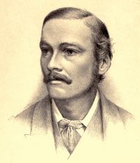