|
|
| (23 intermediate revisions by the same user not shown) |
| Line 1: |
Line 1: |
| {{Sabin1901 header}} | | {{Sabin1901 header}} |
| | | =An Atlas Of The Medulla And Midbrain= |
| | | [[File:Sabin1901 titlepage.jpg|right|400px]] |
| '''AN ATLAS OF THE MEDULLA AND MIDBRAIN'''
| | [[File:Florence Sabin 1938.jpg|thumb|alt=Florence Rena Sabin (1871 - 1953)|link=Embryology History - Florence Sabin|Florence Rena Sabin (1871-1953)]] |
| | |
| By [[Embryology History - Florence Sabin|Florence R. Sabin]] | | By [[Embryology History - Florence Sabin|Florence R. Sabin]] |
|
| |
|
|
| |
|
|
| |
|
| A LABORATORY MANUAL | | A Laboratory Manual |
|
| |
|
| ILLUSTRATED WITH SEVEN COLORED PLATES, ONE BLACK PLATE AND FIFTY-TWO FIGURES
| | Illustrated With Seven Colored Plates, One Black Plate And Fifty-Two Figures |
|
| |
|
|
| |
|
| EDITED BY
| | Edited By |
|
| |
|
|
| |
|
| Line 22: |
Line 21: |
|
| |
|
|
| |
|
| BALTIMORE, MD., U. S. A.
| | Baltimore, Md., U. S. A. |
|
| |
|
| THE FRIEDENWALD COMPANY
| | The Friedenwald Company |
|
| |
|
| PUBLISHERS
| | Publishers |
|
| |
|
| 1901 | | 1901 |
| Line 32: |
Line 31: |
|
| |
|
|
| |
|
| COPYRIGHT, 1901,
| | Copyright, 1901, |
| BY FLORENCE R. SABIN
| | By Florence R. Sabin |
|
| |
|
|
| |
|
| THE FRIEDENWALD COMPANY
| | The Friedenwald Company |
| BALTIMORE, MD M U. S. A.
| | Baltimore, Md M U. S. A. |
|
| |
|
| {{Historic Disclaimer}} | | {{Historic Disclaimer}} |
| Line 43: |
Line 42: |
| ==Editor's Preface== | | ==Editor's Preface== |
|
| |
|
| This Atlas is planned to meet the practical need of some quick and simple, yet full and reliable, means of aiding the student to | | This Atlas is planned to meet the practical need of some quick and simple, yet full and reliable, means of aiding the student to obtain, from a few sections (or from a series of sections), a reasonably clear idea of the important central relay-station of the brain |
| obtain, from a few sections (or from a series of sections), a reasonably clear idea of the important central relay-station of the brain | | here presented. (Though representing the human brain, the atlas can be applied to the study of the brains of lower mammals.) |
| here presented. (Though representing the human brain, the atlas | |
| can be applied to the study of the brains of lower mammals.) | |
|
| |
|
| The time allotted to a course in Neurology is generally so short; | | |
| the sections to be studied exhibit such great special complexity of | | The time allotted to a course in Neurology is generally so short; the sections to be studied exhibit such great special complexity of |
| structure, due to the presence and association of many different | | structure, due to the presence and association of many different |
| centres in the narrow limits of the region; and the descriptions in | | centres in the narrow limits of the region; and the descriptions in |
| Line 56: |
Line 53: |
| shown in their preparations, without spending more time in the | | shown in their preparations, without spending more time in the |
| effort than is reasonable. | | effort than is reasonable. |
| | |
|
| |
|
| We believe, and a number of well-known teachers in several of | | We believe, and a number of well-known teachers in several of |
| Line 62: |
Line 60: |
| stated above; and will save the student much time for real study, | | stated above; and will save the student much time for real study, |
| now often spent in getting started. | | now often spent in getting started. |
| | |
|
| |
|
| Supplied with these excellent drawings of the reconstruction, showing for the first time accurately and satisfactorily structures | | Supplied with these excellent drawings of the reconstruction, showing for the first time accurately and satisfactorily structures |
| to be studied, the student can quickly compare his own sections | | to be studied, the student can quickly compare his own sections with the figures of the Atlas and find the parts there clearly |
| with the figures of the Atlas and find the parts there clearly | |
| designated and explained. | | designated and explained. |
|
| |
|
| Again, if, as is usually the case, a student has only a few cross
| |
| -J-Tiio -narnrvn + Vi a A+laa with if.a 4-8 fiomrPS of
| |
|
| |
|
| | Again, if, as is usually the case, a student has only a few cross-sections through this region the Atlas with its 48 figures of sections. |
|
| |
|
|
| |
|
| At the urgent solicitation of Professor Ph. Stohr, | | |
| of Wiirzburg, Germany, Dr. F Ziegler, of Freiburg, | | At the urgent solicitation of Professor Ph. Stohr, of Wiirzburg, Germany, Dr. F Ziegler, of Freiburg, Germany, is considering the reduplication of the model on which this atlas is based. It is expected that such models, from his studio, will be available within |
| Germany, is considering the reduplication of the model | |
| on which this atlas is based. It is expected that | |
| such models, from his studio, will be available within | |
| the year . | | the year . |
|
| |
|
| Line 93: |
Line 87: |
| ==Editor's Preface== | | ==Editor's Preface== |
|
| |
|
| This Atlas is planned to meet the practical need of some quick The need of | | This Atlas is planned to meet the practical need of some quick and simple, yet full and reliable, means of aiding the student to obtain, from a few sections (or from a series of sections), a reasonably clear idea of the important central relay-station of the brain here presented. (Though representing the human brain, the atlas can be applied to the study of the brains of lower mammals.) |
| and simple, yet full and reliable, means of aiding the student to | |
| obtain, from a few sections (or from a series of sections), a reasonably clear idea of the important central relay-station of the brain | |
| here presented. (Though representing the human brain, the atlas | |
| can be applied to the study of the brains of lower mammals.) | |
|
| |
|
| The time allotted to a course in Neurology is generally so short;
| |
| the sections to be studied exhibit such great special complexity of
| |
| structure, due to the presence and association of many different
| |
| centres in the narrow limits of the region; and the descriptions in
| |
| text-books or lectures are commonly so detailed, or so general or
| |
| diagrammatic; that many students get but hazy ideas of what is
| |
| shown in their preparations, without spending more time in the
| |
| effort than is reasonable.
| |
|
| |
|
| We believe, and a number of well-known teachers in several of
| | The time allotted to a course in Neurology is generally so short; the sections to be studied exhibit such great special complexity of structure, due to the presence and association of many different centres in the narrow limits of the region; and the descriptions in text-books or lectures are commonly so detailed, or so general or diagrammatic; that many students get but hazy ideas of what is shown in their preparations, without spending more time in the effort than is reasonable. |
| our large universities have agreed in this opinion, that this little
| |
| Atlas will offer a valuable and new remedy for the difficulties
| |
| stated above; and will save the student much time for real study,
| |
| now often spent in getting started.
| |
|
| |
|
| Supplied with these excellent drawings of the reconstruction, its use with , sections.
| |
|
| |
|
| showing for the first time accurately and satisiactorily structures
| | We believe, and a number of well-known teachers in several of our large universities have agreed in this opinion, that this little Atlas will offer a valuable and new remedy for the difficulties stated above; and will save the student much time for real study, now often spent in getting started. |
| to be studied, the student can quickly compare his own sections
| |
| with the figures of the Atlas and find the parts there clearly
| |
| designated and explained.
| |
|
| |
|
| Again, if, as is usually the case, a student has only a few crosssections through this region, the Atlas, with its 48 figures of sections cut in two planes and drawn to resemble actual preparations,
| |
| furnishes a good supplementary series of sections for comparison.
| |
|
| |
|
| It is thus easy to understand the many sections which are not
| | Supplied with these excellent drawings of the reconstruction, showing for the first time accurately and satisiactorily structures to be studied, the student can quickly compare his own sections with the figures of the Atlas and find the parts there clearly designated and explained. |
| through particularly well-marked points usually figured in textbooks; and it is possible to get a very satisfactory idea of any structure, by turning to the two series figured, to the colored plates
| |
| and to the index, | |
| with sections of Tracts in the Spinal Cord may be more readily understood and Spinal Cord. 'traced forward into the brain with the aid of this manual.
| |
| The arrangement The text not only describes, in a convenient manner and fully,
| |
| ' everything figured in the reconstruction; but the paragraphs of
| |
| small print, and others referred to in the headings and index, explain just how to compare sections with the model, and how to trace
| |
| nerve-fibre tracts or masses of gray matter, from section to section through this region.
| |
|
| |
|
| The importance When it is realized that this model represents that part of the
| |
| f the Braku brain in which the nuclei of origin of all the true cranial nerves
| |
| are found; that association tracts between these centres are here
| |
| included; that the cells and fibre-tracts are brought into intimate
| |
| association, from their central position, with those of the Spinal
| |
| Cord, Cerebellum, and Forebrain; the usefulness of the Atlas to
| |
| the Anatomist, Physiologist, Pathologist, and Psychologist, whether
| |
| in the laboratory or in connection with lectures and demonstrations,
| |
| may be seen.
| |
|
| |
|
| supplementary A short list of text-books and journals has been included, to
| | Again, if, as is usually the case, a student has only a few crosssections through this region, the Atlas, with its 48 figures of sections cut in two planes and drawn to resemble actual preparations, furnishes a good supplementary series of sections for comparison. |
| " permit the tracing of certain tracts of nerve-fibres further up into
| |
| the higher brain centres or down into the cord, and to encourage the
| |
| student to seek information as to the many and varied sides of
| |
| Neurology from reliable sources where more extensive references
| |
| are to be found.
| |
|
| |
|
| The Editor wishes to explain that his participation in this Atlas
| |
| is confined to the suggestion of publishing the original research in
| |
| the present modified new form, and to assistance in a considerable
| |
| rearrangement of the text and index to facilitate ready reference.
| |
| He has urged this publication in order to furnish the student, in a
| |
| new and especially available form, a valuable guide to the ready
| |
|
| |
|
| interpretation of his preparations.
| | It is thus easy to understand the many sections which are not through particularly well-marked points usually figured in textbooks; and it is possible to get a very satisfactory idea of any structure, by turning to the two series figured, to the colored plates and to the index. |
|
| |
|
| HENRY Mo E. KNOWER.
| | Tracts in the Spinal Cord may be more readily understood and Spinal Cord traced forward into the brain with the aid of this manual. |
| ANATOMICAL LABORATORY,
| |
|
| |
|
| JOHNS HOPKINS UNIVERSITY.
| | The text not only describes, in a convenient manner and fully, everything figured in the reconstruction; but the paragraphs of small print, and others referred to in the headings and index, explain just how to compare sections with the model, and how to trace nerve-fibre tracts or masses of gray matter, from section to section through this region. |
|
| |
|
|
| |
|
| | When it is realized that this model represents that part of the brain in which the nuclei of origin of all the true cranial nerves are found; that association tracts between these centres are here included; that the cells and fibre-tracts are brought into intimate association, from their central position, with those of the Spinal Cord, Cerebellum, and Forebrain; the usefulness of the Atlas to the Anatomist, Physiologist, Pathologist, and Psychologist, whether in the laboratory or in connection with lectures and demonstrations, may be seen. |
|
| |
|
| AUTHOR'S PREFACE.
| |
|
| |
|
| A description and the plates of a reconstruction of the medulla | | A short list of text-books and journals has been included, to permit the tracing of certain tracts of nerve-fibres further up into the higher brain centres or down into the cord, and to encourage the student to seek information as to the many and varied sides of Neurology from reliable sources where more extensive references are to be found. |
| oblongata of the new-born babe was published in the " Contributions to the Science of Medicine," dedicated to William Henry
| |
| Welch. 1 The model was built in the Anatomical Laboratory of
| |
| the Johns Hopkins University at the suggestion of Dr. Franklin | |
| P. Mall and Dr. Lewellys F. Barker. It was the original thought
| |
| that such a reconstruction would not only show graphically for the
| |
| first time the form and relations of the tracts and nuclei, but that
| |
| it would simplify for the student of anatomy a region both complex and difficult. The shape of the tracts in the cord was well
| |
| known, the forms of the internal capsule in the brain could be
| |
| fairly well imagined, but the tracts between the cord and brain
| |
| were too complex to give mental pictures without the aid of a
| |
| model. The suggestion has been made by Dr. H. Me E. Knower,
| |
| of the Anatomical Laboratory of the Johns Hopkins Medical
| |
| School, that the description of the model be put into a more convenient form for the student; by means of fuller references to
| |
| the plates and sections; by a rearrangement of contents to make
| |
| the location in the model of any set of serial sections or any single | |
| section of the region an easy matter; by adding a full index; and
| |
| by a list of literature containing a few of the most important
| |
| references valuable to the student at the beginning of a study | |
| of the central nervous system of man or the mammals. I am indebted to him for the arrangements for this edition.
| |
|
| |
|
| I wish to thank Dr. John Hewetson for the material which
| |
| made the model possible. Both series were unbroken, and so admirably prepared that any omissions in the model are due not to the material, but to the nature of the structures in question.
| |
| I am greatly indebted to Mr. Max Broedel for the beautiful illustrations of the model. They are so accurate and clear as to be
| |
| equal in value to the model itself. It is through the kindness of
| |
| Dr. Henry M. Hurd that the plates of these drawings can be used
| |
| for the present edition. Dr. Franklin P. Mall controlled the construction of the model, Dr. Lewellys F. Barker its study. I acknowledge with thanks their unfailing help and interest.
| |
|
| |
|
| | The Editor wishes to explain that his participation in this Atlas is confined to the suggestion of publishing the original research in the present modified new form, and to assistance in a considerable rearrangement of the text and index to facilitate ready reference. He has urged this publication in order to furnish the student, in a new and especially available form, a valuable guide to the ready interpretation of his preparations. |
|
| |
|
|
| |
|
| 1 Model of the Medulla, Pons and Midbrain of a New-born Babe, by Florence R. Sabin. Contributions to the Science of Medicine, and vol. ix of the Johns Hopkins Hospital Reports.
| | Henry Mc E. Knower. Anatomical Laboratory, |
|
| |
|
| | Johns Hopkins University. |
|
| |
|
| | ==Author's Preface== |
|
| |
|
| ==Contents==
| | A description and the plates of a reconstruction of the medulla oblongata of the new-born babe was published in the " Contributions to the Science of Medicine," dedicated to William Henry Welch.<ref> Model of the Medulla, Pons and Midbrain of a New-born Babe, by Florence R. Sabin. Contributions to the Science of Medicine, and vol. ix of the Johns Hopkins Hospital Reports.</ref> The model was built in the Anatomical Laboratory of the Johns Hopkins University at the suggestion of Dr. Franklin P. Mall and Dr. Lewellys F. Barker. It was the original thought that such a reconstruction would not only show graphically for the first time the form and relations of the tracts and nuclei, but that it would simplify for the student of anatomy a region both complex and difficult. The shape of the tracts in the cord was well known, the forms of the internal capsule in the brain could be fairly well imagined, but the tracts between the cord and brain were too complex to give mental pictures without the aid of a model. The suggestion has been made by Dr. H. Me E. Knower, of the Anatomical Laboratory of the Johns Hopkins Medical School, that the description of the model be put into a more convenient form for the student; by means of fuller references to the plates and sections; by a rearrangement of contents to make the location in the model of any set of serial sections or any single section of the region an easy matter; by adding a full index; and by a list of literature containing a few of the most important references valuable to the student at the beginning of a study of the central nervous system of man or the mammals. I am indebted to him for the arrangements for this edition. |
|
| |
|
| Chapter I.
| |
|
| |
|
| Introductory
| | I wish to thank Dr. John Hewetson for the material which made the model possible. Both series were unbroken, and so admirably prepared that any omissions in the model are due not to the material, but to the nature of the structures in question. I am greatly indebted to Mr. Max Broedel for the beautiful illustrations of the model. They are so accurate and clear as to be equal in value to the model itself. It is through the kindness of Dr. Henry M. Hurd that the plates of these drawings can be used for the present edition. Dr. Franklin P. Mall controlled the construction of the model, Dr. Lewellys F. Barker its study. I acknowledge with thanks their unfailing help and interest. |
| | I wish to thank Dr. John Hewetson for the material which made the model possible. Both series were unbroken, and so admirably prepared that any omissions in the model are due not to the material, but to the nature of the structures in question. |
| | I am greatly indebted to Mr. Max Broedel for the beautiful illustrations of the model. They are so accurate and clear as to be |
| | equal in value to the model itself. It is through the kindness of |
| | Dr. Henry M. Hurd that the plates of these drawings can be used |
| | for the present edition. Dr. Franklin P. Mall controlled the construction of the model, Dr. Lewellys F. Barker its study. I acknowledge with thanks their unfailing help and interest. |
|
| |
|
| Method Of Using Atlas
| |
|
| |
|
| Chapter Ii. The Long Tracts.
| |
|
| |
| A. In The Medulla (Medulla Sheet)
| |
|
| |
| B. In The Pons And Midbrain (Lemnisci And Formatio Reticularis)
| |
|
| |
| Chapter Iii. The Columns Of The Spinal Cord.
| |
|
| |
| A. Ventrolateral Column
| |
|
| |
| (A) Ventral Part
| |
|
| |
| (&) Dorsal Part
| |
|
| |
| B. Dorsal Column
| |
|
| |
| Chapter Iv. Cerebellar Peduncles.
| |
|
| |
| Inferior Peduncle, Or Corpus Restiforme
| |
|
| |
| Superior Peduncle, Or Brachium Conjunctivum
| |
|
| |
| Chapter V. The Cerebral Nerves And Their Nuclei. Median Group (Red In Model).
| |
|
| |
| (A) N. Hypoglossus, XII
| |
|
| |
| Nucleus N., XII
| |
|
| |
| (&) N. Abducens, VI
| |
|
| |
| Nucleus N., VI
| |
|
| |
| (C) N. Trochlearis, IV
| |
|
| |
| Nucleus N., Iv 56
| |
|
| |
| (D) N. Oculomotorius, III
| |
|
| |
| Nucleus N., III
| |
|
| |
| Chapter Vi. The Cerebral Nerves And Their Nuclei (Continued). Lateral Group.
| |
|
| |
| A. Motor Nerves (Red In Model)
| |
|
| |
| (A) N. Accessprius, XI
| |
|
| |
| Nucleus N., XI.
| |
|
| |
|
| |
| (B) N. Glossopharyngeus Et N. Vagus, Ix And X
| |
|
| |
| Nucleus N., Ix And X
| |
|
| |
| (C) N. Facialis, VII
| |
|
| |
| Nucleus N., VII
| |
|
| |
| (D) N. Trigeminus, V
| |
|
| |
| Nucleus N., V
| |
|
| |
| B. Sensory Nerves (Blue In Model)
| |
|
| |
| (A) N. Glossopharyngeus Et N. Vagus, Ix And X
| |
|
| |
| Nucleus N. , Ix And X
| |
|
| |
| (&) N. Trigeminus, V
| |
|
| |
| Nucleus N., V
| |
|
| |
| (C) N. Vestibuli, Viii
| |
|
| |
| Nuclei N. Vestibuli
| |
|
| |
| (D) N. Cochleae, Viii
| |
|
| |
| Nuclei N. Cochlese
| |
|
| |
| Chapter Vii. The Inferior And Accessory Olives 86
| |
|
| |
| Chapter Viii. The Midbrain.
| |
|
| |
| 1. Relation Of Its Structures To The Central Fibre Mass
| |
|
| |
| 2. The Nucleus Ruber (Red Nucleus) And Its Capsule
| |
|
| |
| 3. The Fasciculus Retroflexus (Meynerti)
| |
|
| |
| 4. The Decussatio Tegmenti Dorsalis (Meynerti)
| |
|
| |
| 5. The Decussatio Tegmenti Ventralis Of Forel
| |
|
| |
| 6. Stratum Album Prof Undum (Deep White Layer)
| |
|
| |
| 7. Substantia Centralis Grisea (Central Gray Matter)
| |
|
| |
| 8. The Pyramidal Tract
| |
|
| |
| 9. Substantia Nigra
| |
|
| |
| Chapter Ix. The Formatio Reticularis Alba And Grisea
| |
|
| |
| General Summary of what Is shown In Reconstruction
| |
|
| |
| References To Literature
| |
|
| |
| ==List of Illustrations==
| |
|
| |
|
| |
| FIGURES : PAGE
| |
|
| |
| 1. Transverse Section of the Spinal Cord. Outline 36
| |
|
| |
| 2. Diagram of Medial Accessory Olive 91
| |
|
| |
| 3-24. Series of Horizontal (frontal) Sections, including Medulla and Midbrain 125-132
| |
|
| |
| 25-51. Series of Transverse Sections from the Cord to the Midbrain. .133-145
| |
|
| |
| 52. Diagram of the Model giving Levels of Sections here Figured 146
| |
|
| |
| PLATES following page 146
| |
|
| |
| I. The Inferior Olive.
| |
| II. View of the Lateral Surface of the Reconstruction.
| |
|
| |
| III. View of the Dorsal Surface of the Reconstruction.
| |
|
| |
| IV. First Dissection of the Reconstruction. Lateral view, showing
| |
|
| |
| Fibre Tracts, &c., and the Sensory Nuclei of Cerebral Nerves.
| |
| V. Second Dissection of the Reconstruction. Lateral view, showing
| |
|
| |
| Fibre Tracts, &c., and Motor Nuclei of Cerebral Nerves.
| |
| VI. Third Dissection of the Reconstruction. Lateral view, showing
| |
|
| |
| the Long Tracts of the Medulla.
| |
|
| |
| VII. Fourth Dissection of the Reconstruction. Dorsomedian view,
| |
| showing the Long Fibre Tracts as Related to Nuclei of Cerebral Nerves and to other Structures.
| |
|
| |
| VIII. View of the Midbraiu from Above, showing Relations of Fibre Tracts.
| |
|
| |
|
| |
| ==Chapter V. The Cerebral Nerves And Their Nuclei==
| |
|
| |
| An indication has already been given of the grouping of the cere- The grouping of
| |
|
| |
| , , it.fi ?i i j. i- i the cerebral nerves
| |
|
| |
| oral nerves, suggested by the model, namely, into a median and a
| |
| lateral group. This grouping has been used before by several
| |
| authors, notably by His, but it is rather more common to consider
| |
| the nerves in three groups according to the region they occupy,
| |
| namely, four in the medulla, four in the pons and two in the midbrain. Dr. L. F. Barker, in his recent book, has divided the nerves
| |
| into a sensory and a motor group on the basis of the direction followed by the fibres. The division herein used, on the basis of the
| |
| position of the nuclei, it seems to me has decided advantage, not
| |
| only for the purpose of describing the model, but in illustrating
| |
| the relation of these nuclei to the gray matter of the cord. 1
| |
|
| |
| The two groups possess characters in contrast. Each contains
| |
| four motor nuclei, while the lateral group has sensory nuclei as
| |
| well. The median group consists of the nuclei of the ~N. hypoglossus, N". abducens, N. trochlearis and !N". oculomotorius. The
| |
| four motor nuclei of the lateral group are the nuclei of the N.
| |
| accessorius, IN", vagus, "N. glossopharyngeus, !N". facialis and !N".
| |
| trigeminus. Its sensory nuclei are nuclei of termination associated
| |
| with the ~N. vestibuli, 2sT. cochleae and JST. trigeminus. The nuclei
| |
| of the median group all lie near the middle line just ventral to the
| |
| central canal and closely related to the fasciculus longitudinalis
| |
| medialis (Plate v). The nuclei of the "N. hypoglossus and IT.
| |
| abducens are very near the dorsal surface, while the nuclei of the
| |
| N. trochlearis and !N". oculomotorius are farther ventral inasmuch
| |
| as they lie on the midbrain curve. The nerve-fibres from all the
| |
| nuclei of the groups, except those of the root of the ET. trochlearis, pass directly ventralward to the surface near the middle line. The
| |
| root of the N. trochlearis, on the contrary, takes an anomalous
| |
| course, since it passes dorsalward, decussates in the velum and has
| |
| its superficial origin on the dorsal surface of the isthmus of the
| |
| rhombencephalon.
| |
|
| |
|
| |
|
| |
| 1 This division is in accord with the embryological work of His. Cf. His, W., Zur Geschichte des Gehirns sowie der centralen und peripherischen Nervenbahnen beim menschlichen Embryo. Abhandl. d. math, phys. Cl. d. K. sacfis. Gesellsch. d. Wissensch., Leipz., Bd. xiv (1887-88), S. 339-392.
| |
|
| |
|
| |
|
| |
| The motor nuclei of the lateral group lie in the f ormatlo reticularis at a considerable distance from the middle line, and at a level
| |
| ventral from the central canal. With the exception of the 1ST. trigeminus the root-fibres from these nuclei do not take a direct course
| |
| toward the surface of the central nervous system. The root-fibres
| |
| of all four of the nerves have their superficial origin in the lateral
| |
| sulcus. In the model the nucleus of the "N. accessorius does not
| |
| appear, for it could not be outlined with sufficient definiteness for
| |
| reconstruction. This is an interesting form relation, for the nucleus
| |
| of the "N. accessorius lies just at the point where the ventral horn of
| |
| the spinal cord divides into two parts to make the median and lateral motor groups of the region modeled. The nucleus of the N.
| |
| accessorius then marks the transition from the indefinitely outlined
| |
| nuclei of the cord to the definitely circumscribed nuclei of the cerebral motor-nerves.
| |
|
| |
| The sensory nuclei of the entire region belong to the lateral
| |
| group. The sensory nuclei are associated with the IsT. vagus, !N".
| |
| glossopharyngeus, 1ST. vestibuli, N". cochlese, E". intermedius and
| |
| N. trigeminus, and the superficial entrance of their root-fibres is
| |
| without exception on the lateral surface (Plates n and iv). These
| |
| nuclei tend to occupy a position farther dorsal than the motor nuclei of the lateral group, which is, as one might expect, inasmuch
| |
| as these nuclei correspond more or less closely to the gray matter of
| |
| the dorsal horn of the cord. In making this general statement, I
| |
| do not intend to enter into the discussion in regard to the development of the optic and cochlear nuclei. These sensory nuclei cover
| |
| a wide area, thus contrasting with the compact motor nuclei; in
| |
| some cases they spread even to the midline. In general, the
| |
| sensory nuclei lie nearer the surface and so show on Plates n and
| |
| iv, while the motor nuclei are deeper and show on Plate v.
| |
|
| |
| THE MEDIAN GROUP.
| |
|
| |
| Nucleus, N. in. (a) N. Hypoglossus (Fig. 31). The first nerve of the median
| |
| motor group is the N. hypoglossus (Plate v). In two instances,
| |
|
| |
|
| |
|
| |
| MEDIAN GROUP OF CEKEBEAL NEKVES, N. xii 53
| |
|
| |
| namely, in regard to the nucleus of the hypoglossus, and the nucleus
| |
| ambiguus, the model represents the position but not the exact form.
| |
| Inasmuch as the nucleus !N". hypoglossi lies on the medulla curve, it
| |
| will be seen that only a small portion of it would be cut in any one
| |
| longitudinal section. Moreover, the nucleus lies in the central
| |
| gray matter, and the scarcity of fibres tends toward the rapid washing out of the stain from the cells. However, the nucleus itself
| |
| has a few fibres which have a characteristic arrangement. They are
| |
| fine fibres that come up into the centre of the nucleus and spread
| |
| out like a fountain. 1 In longitudinal section these fibres are cut
| |
| across, and make a row of fine dots that represents the nucleus.
| |
| The position of the nucleus was determined (1) by the presence of
| |
| these fibres, (2) by well-known relations to neighboring structures,
| |
| (3) by measurements from the transverse series and (4) by the
| |
| position of the nuclear end of the root-bundle. Of the form of the
| |
| nucleus, I shall make but general statements, namely, that it is
| |
| long, that its dorsal border conforms with the cervical curve, and
| |
| that its breadth, judging from the transverse series, is not wholly
| |
| uniform.
| |
|
| |
| The nucleus extends more than one-half the length of the medulla and corresponds in length, generally speaking, to the nucleus olivaris inferior. The distal end is slightly farther spinalward than the olive, being opposite the distal end of the median
| |
| accessory olive. It lies lateral to the central canal before it opens
| |
| out into the fourth ventricle. Farther toward the cerebrum, the
| |
| nucleus is just beneath the floor of the fourth ventricle. Mention
| |
| has been made of the slight curve in the fasciculus longitudinalis
| |
| medialis which corresponds to this nucleus (Plate vi). The accessory nucleus of Roller lies within the fasciculus longitudinalis
| |
| medialis (Fig. 10) opposite the proximal end of the nucleus of the
| |
| hypoglossal nerve.
| |
|
| |
| The root-fibres of the nerve leave the nucleus from its ventral Root-fibres,
| |
| border (Plate v). When modelled, the root-bundle makes a N ' ra "
| |
| sheet which passes ventralward and slightly lateralward to a superficial origin between the olive and the pyramidal tract. As seen
| |
| from the side, this sheet has three borders a distal, a proximal and a ventral. The distal border is shorter than the proximal,
| |
| owing to its position on the cervical curve. It will be noticed that
| |
| near the ventral surface of the medulla some of the root-fibres leave
| |
| the surface of the sheet and turn lateralward. They enter the
| |
| hilus of the nucleus olivaris inferior and there the bundle ends
| |
| abruptly, for the sections were too decolorized to permit one to trace
| |
| it to a superficial origin (Fig. 21).
| |
|
| |
|
| |
|
| |
| 1 They have been well pictured by Koch and termed Fibrae afferentes
| |
| mi. xii. Cf. Edinger, L., Vorlesungen iiber den Bau der nervosen Centralorgane, Leipz. (1893), S. 180.
| |
|
| |
|
| |
|
| |
| The ventral border, namely, the line of the superficial origin of
| |
| the fibres, has a slightly greater length than the nucleus. It lies
| |
| in the groove between the pyramid and the olive. Its inferior limit
| |
| is just distal to the olive, while its proximal end is at the junction
| |
| of the medulla and pons. The surface of the sheet is smooth, except for the bundle of fibres that projects into the hilus of the
| |
| olive. The direction of the fibres is worthy of attention. Starting from the ventral border of the nucleus the fibres pass ventralward and slightly lateralward to the level of the dorsal border of
| |
| the olive. In passing the olive the fibres turn slightly more lateral,
| |
| while farther ventral still the lateral curve is marked. The reason
| |
| for these two curves will be evident. In the dorsal part only the
| |
| medulla sheet intervenes between the middle line and the hypoglossal nerve. In the middle part, the medial accessory olive, as
| |
| well as the medulla sheet, lies between the raphe and tne nerve,
| |
| while in the ventral region the pyramidal tract intervenes.
| |
|
| |
| N. in in sections. These points are shown in Fig. 31. The nerve can be traced through
| |
| the following sections (Figs. 7-23) : The first section shows a few cells
| |
| of the nucleus, while Fig. 9 shows the fine fibres that aided in its determination. In all of the succeeding sections it will be noted that the
| |
| root-fibres of the nerve pass out in small, definite bundles rather than
| |
| as single fibres. By tracing the series the course of the fibres outlined
| |
| above can be seen. In the dorsal part the fibres lie against the medulla
| |
| sheet; in the middle part they lie between the medial accessory olive
| |
| and in the inferior olivary nucleus, while in the extreme ventral portion
| |
| the fibres are bent markedly from the middle line, owing to the volume
| |
| of the pyramidal tract. The last two sections of the series give a good
| |
| idea of the surface-origin.
| |
|
| |
| Nucleus, N. vi. (&) N. Abduceus (Figs. 37 and 38). The N. abducens is the
| |
| second nerve of the median group. Its nucleus makes a landmark
| |
| in all of the views of the model, but shows best in Plates v and vi.
| |
| In Plate v it will be seen that a considerable distance (2.5 mm. in
| |
| the medulla) intervenes between the nucleus N. hypoglossi and the
| |
| nucleus N. abducentis.
| |
|
| |
|
| |
| The nucleus of the !N". abducens lies in the distal part of the g9^pons, just beneath the fourth ventricle and lateral to the fasciculus
| |
| longitudinalis medialis. In this relation to the fasciculus longitudinalis medialis it differs from the other nuclei of the group, all of
| |
| which lie dorsal to the tract and make a groove on its surface. The
| |
| model will show that the fibres of the N". f acialis in curving around
| |
| the nucleus of the "N. abducens appear to draw it from the middle
| |
| line. The root-fibres leave the nucleus at its ventromedial portion,
| |
| and pass ventralward and slightly spinalward to emerge between
| |
| the pons and medulla near the median line.
| |
|
| |
| The nucleus itself is nearly round, with a diameter of 1.7 mm.
| |
| It projects above, that is to say, dorsal to the fasciculus longitudinalis medialis (Plate vi). The relations of the root of the facial
| |
| nerve to the nucleus of the 1ST. abducens will be considered in detail in connection with the former nerve. The median boundary
| |
| of the nucleus is made by the root-fibres of the facial nerve and by
| |
| the fasciculus longitudinalis medialis (Figs. 6 and 7). The proximal boundary is made in part by the root of the facial nerve. The
| |
| root-fibres of the ~N. abducens leave the medio-ventral part of the
| |
| nucleus in small but dense bundles (Fig. 37). In passing to the
| |
| superficial origin at the junction of the pons and medulla, these
| |
| small bundles make a fairly compact bundle which passes ventralward and spinalward. In Plate vi it will be seen, however, that
| |
| the fibres describe a curve in their course, for in starting from the
| |
| nucleus they pass first toward the cerebrum, as well as ventralward,
| |
| before turning toward the cord. The bundle passes through the
| |
| trapezoid body, and leaves the central nervous system just proximal to the groove between the medulla and pons.
| |
|
| |
| The nucleus is prominent in both series: in the transverse series N. vi in sections,
| |
| because it projects so far into the central gray matter; in the longitudinal series because it is so definitely outlined by the root of the
| |
| facial nerve.
| |
|
| |
| Fig-. 36 and Fig-. 37. In tracing 1 the nerve in the transverse series, the
| |
| curve, seen in Plate vi, is slightly exaggerated by the obliquity of the
| |
| sections (Fig. 52). Three sections will illustrate this point; Fig. 36,
| |
| which shows simply the nucleus; Fig. 38, which shows the root-fibres
| |
| just as they are leaving the nucleus and again at the superficial origin,
| |
| and Fig. 39, which shows simply the middle of the curve with neither
| |
| the nucleus nor the superficial origin.
| |
|
| |
| The shape of the bundle in the dorsal portion is fairly rounded, as
| |
| seen in Figs. 16 and 19, but farther ventral, near the superficial origin,
| |
| the bundle becomes oval in shape, with the long axis perpendicular to the long axis of the ports. The fibres emerge from the lower part of
| |
| the pons just proximal to the groove between the pons and medulla.
| |
|
| |
| Nuclei, N. iv. (c) N. Trochlearis (Fig. 41). In the case of the remaining two
| |
| nerves of the group, the N. trochlearis and the N. oculomotorius,
| |
| the nuclei of both sides have been modelled (Plates n, in and iv).
| |
| These nuclei lie in the trough of the fasciculus longitudinalis
| |
| inedialis on its midbrain curve. The walls of the trough, as has
| |
| been seen, are hollowed out on either side of the deep central groove
| |
| to receive them (Plate vni). The nucleus of the trochlear nerve
| |
| lies farther distal, i. e., nearer the cord, and hence farther dorsal
| |
| on the curve. It lies in the proximal or cerebral part of the
| |
| colliculus inferior (Plate in).
| |
|
| |
| The nuclei of the two sides are not symmetrical, being single
| |
| on the left side and double on the right. However, the volumes
| |
| of the cell-masses of the two sides are about equal. In the case of
| |
| the double nucleus, the proximal part is evidently the main nucleus,
| |
| that is to say, it corresponds to the nucleus of the other side. The
| |
| left or single nucleus is approximately cubical in shape, with a projection at the distal medial angle. It measures about 1 mm. in
| |
| diameter. On the right side it is as if the small projecting portion
| |
| had been pulled dorsalward until it just separated from the main
| |
| mass.
| |
|
| |
| Nuclei of N. iv This lack of symmetry of the nuclei of the two sides is brought out
| |
| in sections. j n the sections. The accessory part of the double nucleus, which lies
| |
| farther dorsal and farther distal, shows on Fig. 10, while sections in
| |
| Figs. 11 and 12 show the main nuclei of both sides. The transverse
| |
| series does not show just the same irregularity, for in it the nuclei of
| |
| both sides are double. Moreover, the distal nucleus of either side lies
| |
| at least 1 mm. from the proximal. The section in Fig. 44 shows this
| |
| distal part, while Fig. 46 shows the main proximal part. In both sections the root-fibres of the nerve can be seen.
| |
|
| |
| Root-fibrea, N. IT. The nuclei of the nerve are compact and definitely outlined;
| |
| nevertheless small groups of cells may be traced all along the rootfibres on either side. When the root-fibres leave the lateral and
| |
| dorsal border they first pass laterally in scattered bundles and
| |
| then turn dorsalward and slightly spinalward. In their dorsal
| |
| course the fibres are collected into from two to four compact bundles. The fibres lie in the central gray matter just within the
| |
| stratum profundum album. Plate in shows the decussation dorsal
| |
| to the central canal. The superficial origin of the fibres is asymmetrical. On the side of the single nucleus the fibres have a larger
| |
| and more ventral superficial origin than those of the other side.
| |
|
| |
| The course of the fibres can be traced in the longitudinal series in the N. iv in sections,
| |
| following sections: Figs. 12-10 show the nucleus and its relation to the
| |
| fasciculus longitudinalis medialis. It will be noticed that the section in
| |
| Fig. 12 shows no root-fibres, since it passes through the ventral portion of the nucleus. The root-fibres can be followed through the rest
| |
| of the series from Fig. 10 to Fig. 3. They are to be distinguished from
| |
| the fibres of the fasciculus longitudinalis medialis by a difference in
| |
| direction. The last section, Fig. 3, shows the decussation.
| |
|
| |
| By looking at the nerve in the model from the side and from the
| |
| dorsal aspect, the appearances in the transverse series can be readily
| |
| predicted (Plates n and in). The most distal section w 7 ould show the
| |
| decussation, while each succeeding section would show two or three
| |
| small bundles cut across or slightly obliquely, and occurring a littlefarther ventral in each section until the level of the nucleus is reached,
| |
| and here the fibres would turn directly medialward. This course can
| |
| be followed in the following series, Figs. 41 to 46. 1
| |
|
| |
| (d) N. oculomotorius (Fig. 48). The E". oculomotorius is the Nuclei, N. m.
| |
| last of the median motor group to be considered. The position of
| |
| its nucleus is best seen from the dorsal surface (Plate m), but the
| |
| course of the root of the nerve and the relations must be followed
| |
| in a view from the side (Plate iv). The nucleus as seen in Plate
| |
| in is a long mass of cells lying in the midbrain trough of the fasciculus longitudinalis medialis. The root-bundle passes directly
| |
| ventralward near the middle line and emerges in the fossa interpedunculare. The position of the nucleus in the trough of the
| |
| fasciculus longitudinalis medialis determines two facts: (1) that
| |
| the nucleus as a whole lies farther ventral than the nucleus N.
| |
| trochlearis; (2) that the nucleus itself is placed obliquely to a
| |
| horizontal plane, so that the distal end is farther dorsal than the
| |
| proximal.
| |
|
| |
| 1 The root-fibres have been described as making a double bend, passing
| |
| at first dorsalward, then spinalward, and again dorsalward at a right
| |
| angle. Cf. von Kolliker, A., Handbuch der Gewebelehre, Bd. n, Leipz.
| |
| (1896); also Van Gehuchten, A., Anatomic du systeme nerveux de
| |
| 1'homme, 2 ed., Louvain (1897); and Barker, L. F., The Nervous System
| |
| and its Constituent Neurones, N. Y. (1899), p. 938, et seq. This course
| |
| does not appear on the model, which shows a gradual dorsal curve from
| |
| the very start. Such an angle was not suggested in either of my series
| |
| but there is no doubt that it might be missed in building the model.
| |
| The matter might fee settled by referring to sagittal sections. On the
| |
| other hand Forel (Arch. f. Psychiat., etc., Berl., Bd. vn [1897], S. 439)
| |
| describes the course as it is shown in the model.
| |
|
| |
| The oculomotor nerve is the only nerve in the central nervous
| |
| system of which the nuclei of the two sides lie near enough together
| |
| to be modelled as one. The nucleus as a whole consists of two
| |
| lateral parts which are fused together in the ventral portion so as
| |
| to make a gutter 3.1 mm. long for the median nucleus. The entire nucleus is 5.3 mm. long.
| |
|
| |
| The shape of the combined mass is that of a triangular prism
| |
| with the apex pointing ventralward and the base dorsalward. The
| |
| dorsal surface is a triangle with the apex cut off. The distal end is
| |
| the base of the triangle and the walls slant medialward, so that
| |
| the proximal edge is about one-half the length of the distal. These
| |
| two walls make the chief nucleus Hauptkern of the Germans.
| |
|
| |
| The distal fourth of the nucleus is bounded definitely by the
| |
| fasciculus longitudinalis medialis. In the next fourth of the
| |
| nucleus, however, the cells scatter out into the fasciculus longitudinalis medialis, and these cells have been sacrificed to the fibres
| |
| in the model (Fig. 48). These cells make the lateral part of the
| |
| main nucleus as described by v. Bechterew. 1
| |
|
| |
| The median nucleus is clearly shown in Plate in. The small
| |
| rounded proximal nuclei of v. Bechterew are shown as the proximal
| |
| end of the main nucleus, but the small paired accessory nuclei did
| |
| not come out clearly in the longitudinal series, though they were
| |
| seen in the transverse series.
| |
|
| |
| N. in in The following- series taken from the transverse sections will show
| |
| sections. mO re details of the nucleus than have been introduced into the model
| |
| (Figs. 47 to 50) :
| |
|
| |
| In the first place there are variations in the shape of the lateral nuclei: for example, Figs. 47 and 48 show an interesting contrast. The
| |
| former shows cells spreading out to the side, making the outlines of the
| |
| nucleus rounded, while the latter shows straight clean-cut sides narrowing to a sharp apex. Secondly, the sections show contrasts in the
| |
| arrangement of the fibres within the nuclei. For example, compare
| |
| the first two sections with the last two. Thirdly, the sections show
| |
| that the median nucleus is not uniform. (Compare Figs. 48 and 49.)
| |
| The longitudinal series shows practically the same points (Figs. 12 to
| |
| 19). The last section, however, gives a different view of the anterior
| |
| fused mass. All these details need a higher magnification.
| |
|
| |
| Root-fibres, Turning now to the course of the root-fibres after leaving the
| |
| nucleus, it will be found that the best view is given in Plate iv.
| |
|
| |
| *v. Bechterew, W., Ueber die Kerne des Oculomotorius, Abducens u.
| |
| Trochlearis. Arch. f. Anat. u. Phys., Anat. Abth., Leipz. (1897), S. 308.
| |
|
| |
|
| |
|
| |
| N. m.
| |
|
| |
|
| |
|
| |
| This view shows the brachium conjunctivum, the stratum profundum album of the superior colliculus and the posterior commissure. The shell of deep white layer is connected with the
| |
| fasciculus longitudinalis medialis. Through a space left in this
| |
| shell a portion of the nucleus of the oculomotor nerve can be
| |
| seen, while just ventral to this space the nerve-fibres emerge. The
| |
| root-bundle runs ventralward between the brachium conjunctivum
| |
| and the red nucleus to the superficial origin in the fossa interpedunculare.
| |
|
| |
| The bundle representing the fibres appears to be an irregular
| |
| one, but that is merely because it is encroached upon by other
| |
| structures. Were it not for these, the bundle would be nearly regular in outline. The root-bundle can be considered as divided
| |
| into two parts: (1) the part extending from the nucleus of the
| |
| nerve to the ventral surface of the red nucleus including about
| |
| three-quarters of the length of the bundle, and (2) the portion
| |
| ventral to the red nucleus. The reason for this division can be
| |
| made plain. In passing from the nucleus the fibres spread out to
| |
| the side and make a bundle which fits around the distal surface of
| |
| the red nucleus (Fig. 48). This makes the greatest breadth of the
| |
| bundle in a transverse diameter. On the other hand, the second
| |
| portion covers the exit zone of the nerve-fibres where the entire
| |
| bundle rotates so that its broad side faces the surface of the fossa
| |
| interpedunculare (Fig. 51). This is to be seen from the mesial
| |
| aspect, but the idea can be obtained from Plate iv. It is important to note that this relation of the shape of the bundle does not
| |
| involve any rotation of the fibres. The first portion of the bundle
| |
| has two curves: (1) a curve in a dorsoventral diameter which is
| |
| concave toward the spinal cord and enables the bundle to fit in
| |
| between the decussation of the brachium conjunctivum and the
| |
| nucleus ruber; (2) a curve in the transverse diameter with the convexity toward the cerebrum by which the bundle accommodates
| |
| itself to the ventral surface of the nucleus ruber. It must be
| |
| stated that some of the nerve-fibres pass directly through the nucleus
| |
| ruber, so this curve is only an arbitrary one. Opposite the extreme ventral part- of the nucleus ruber the bundle is separated by
| |
| a considerable space from the nucleus inasmuch as the surface of
| |
| the nucleus curves away from the plane of the nerve.
| |
|
| |
| Of the irregularities on the surface of the bundle only one is due to the nerve-fibres themselves, namely, the projection on the
| |
| lateral border near the dorsal surface. This is due to the fact that
| |
| just ventral to the lemniscus medialis the nerve-fibres spread out
| |
| laterally. The other irregularities are due to spaces left for other
| |
| structures: (1) a nucleus in the lateral capsule of the red nucleus,
| |
| (2) a little mass of cells I have referred to as nucleus columnaris,
| |
| and (3.) the fasciculus retroflexus (Meynerti) (Plates iv and vn).
| |
|
| |
| The shape of the bundle can best be seen in the longitudinal sections
| |
| (Figs. 16 to 24).
| |
|
| |
| In the transverse series the nerve-fibres show in the same sections as
| |
| the nucleus. The obliquity of the section must be taken into account
| |
| in comparing it with the model. The transverse sections show well
| |
| how some of the fibres pass through the nucleus ruber and the substantia nigra (Figs. 47 to 51).
| |
|
| |
| Summary of This completes the description of the median group of nerves,
| |
| "erebrafnerves! and the group characteristics will now be plain. First, all the
| |
| nerves are purely motor. Second, all the nuclei lie near the midline ventral from the central canal. Third, all are embedded in
| |
| the fasciculus longitudinalis medialis except the nucleus of the
| |
| !N". abducens, which lies farther lateral than the others, possibly due
| |
| to the knee of the facial nerve. Fourth, the fibres of each nerve
| |
| are collected into small definite bundles soon after leaving the
| |
| nucleus. Fifth, three of the nerves pass directly ventralward,
| |
| while the trochlear nerve passes dorsalward and decussates dorsal
| |
| to the central canal.
| |
|
| |
| The relation of this group of nuclei to the medial part of the
| |
| ventral horn of the cord is clearly illustrated by the model, and
| |
| motor cells can be traced in the sections from the ventral horn all
| |
| along the border of the medulla sheet to the nucleus of the hypoglossal nerve (red in Plates v and vi). On the other hand, no such
| |
| scattered cells exist between the hypoglossal nucleus and the
| |
| nucleus of the N. abducens, for it is characteristic of the cerebral
| |
| nerves, in contrast with the spinal, that their nuclei form definite
| |
| and isolated groups of cells rather than a part of a column of cells.
| |
| This distinction is being broken down by the recent work of Kaiser,
| |
| Sano, van Gehuchten and others on the groups of cells in the spinal
| |
| cord.
| |
|
| |
|
| |
|
| |
| ==Chapter VI.The Cerebral Nerves And Their Nuclei==
| |
|
| |
| THE LATERAL GROUP.
| |
|
| |
| The nerves of the lateral group are the 1ST. accessorius, JN". vagus,
| |
| N". glossopharyngeus, ~N. cochleae, "N. vestibuli, N. intermedius,
| |
| N". facialis and N". trigeminus. Instead of considering them serially, I propose to divide their nuclei into motor and sensory groups.
| |
| The motor group contains four nuclei, the nucleus 1ST. accessorii, the
| |
| nucleus ambiguus of the "N. vagus and !N". glossopharyngeus, and
| |
| the nucleus ~N. facialis and the nuclei "N. trigemini, while the sensory group has many nuclei belonging to the N. vagus, "N. glossopharyngeus, N. acusticus, 1ST. intermedius and N. trigeminus.
| |
| This grouping is of value in two ways: it brings out the contrast
| |
| (1) between the motor nuclei of the median group and those of the
| |
| lateral, and (2) between the motor and sensory nuclei of the lateral
| |
| group. Both of these distinctions are, in reality, distinctions of
| |
| development. In Plate v is shown the lateral group of motor
| |
| nuclei and the course of the root-fibres of the nerves. The lateral
| |
| funiculus of the cord has been removed in part in order to show
| |
| the floor of the trough in which lie the ventral-horn cells.
| |
|
| |
| A. MOTOR NERVES OF THE LATERAL GROUP.
| |
|
| |
| (a) N. accessorius (Fig. 25). The motor nuclei of the lateral Lateral motor
| |
| group, as well as of the median group, show in Plate v. The part nerves "
| |
| of the model representing the spinal cord has been added on from
| |
| another series in this plate. Into the cord no details have been
| |
| introduced. Thus it happens that only the extreme proximal part
| |
| of the spinal portion of N. accessorius is illustrated. With regard
| |
| to the study of the " vagus portion " of the nerve, it was not possible to find the fibres in the longitudinal sections and so it does not
| |
| appear in the model.
| |
|
| |
| Spinal portion. The nucleus of the spinal part of the N. Nucleus, N. .
| |
| accessorius has not been represented. At the beginning of the medulla oblongata the large motor cells of the ventral horn scatter
| |
| out into the formatio reticularis, so that it is quite impossible to
| |
| group them into a nucleus. As has been said, some of these motor
| |
| cells can be traced all the way from the definite ventral horn to
| |
| the nucleus of the hypoglossal nerve. On the other hand, the
| |
| cells lying farther to the side give rise apparently to the rootfibres of the "N. accessorius. In brief, here is the point at which
| |
| the motor cells of the cord divide into the two groups, the median
| |
| and the lateral groups of the medulla.
| |
|
| |
| Root-fibres, N. H. It will be seen in the model (Plate v) that at least some of the
| |
| root-fibres of the E". accessorius, instead of passing directly from
| |
| their cells of origin to the lateral groove, pass dorsalward and then
| |
| turn ventral ward and lateral ward toward the superficial origin. 1
| |
| The fibres, in their dorsal course, I shall refer to as the pars prima,
| |
| and, in the ventrolateral course, as the pars secunda. Inasmuch
| |
| as the bundle in the model represents but a few fibres of the nerve,
| |
| it can be taken to represent the nerve as a whole only in a general
| |
| way. The general direction of the loop of the fibres is of interest,
| |
| namely, that the loop is placed obliquely with reference to the
| |
| transverse plane and the pars prima lies slightly farther toward the
| |
| cord than the pars secunda. From this it will be seen that the pars
| |
| prima and pars secunda would not be cut in the same transverse
| |
| section. This is true, however, only in part, for certain of the
| |
| fibres do not pass so far dorsalward, and these fibres lie in approximately the same transverse plane throughout their course (Fig. 25).
| |
| On the other hand, Fig. 26 shows the longer fibres of the !N".
| |
| accessorius and includes only the pars secunda. The presence of
| |
| these longer and shorter fibres shown in a diagrammatic form, gives
| |
| an interesting suggestion of an easy transition from the condition
| |
| of the cord.
| |
|
| |
| N. M in sections. The two transverse sections given above represent the spinal portion
| |
| of the nerve. The same portion is shown in the longitudinal series in
| |
| Figs. 17 and 16. Fig. 17 shows a very definite pars secunda and a small
| |
| pars prima, while the second section shows these two portions of the
| |
| nerve nearer together, that is passing toward the loop.
| |
|
| |
| The question of a "vagus portion" of the nerve has been a
| |
| difficult one; first, on account of the indefiniteness of the nucleus ambiguus, and, secondly, on account of the difficulty of finding the
| |
| fibres in longitudinal sections. A definite nucleus of the vagus
| |
| part of the ~N. accessorius has not been made out in either series,
| |
| but there are motor cells scattered throughout the formatio reticularis of the region between the N. accessorius and the nucleus ambiguus shown in Plate v.
| |
|
| |
|
| |
| 1 Edinger, L. Bau der nervosen Centralorgane, Leipz. (1893), S. 168.
| |
|
| |
|
| |
| In tracing 1 the transverse series from the distal end of the medulla,
| |
| where the spinal part of the nerve is clearly made out (Figs. 25 and 26)
| |
| to a region, which is clearly that of the vagus nerve, it is certain that
| |
| there are fibres corresponding to the description of the N. accessorius
| |
| (Figs. 28 and 29). In the sections of this region certain points are of
| |
| interest. First, no section contains a definite pars prima, though certain sections, as Fig. 27, have small bundles of fibres within the central
| |
| gray matter suggesting a pars prima; a few sections show fibres of the
| |
| nerve making a loop as well as a pars secunda. From a complete series
| |
| of the N. accessorius it would be easy to construct a diagram showing
| |
| how the N. accessorius is a transition in form between the nerves of the
| |
| spinal cord and the N. vagus.
| |
|
| |
| In the longitudinal series the nerve-fibres of the vagus portion must be
| |
| cut across or nearly so and thus are exceedingly difficult to trace.
| |
|
| |
| Near the level of Fig. 14 a few delicate fibres are seen cutting across
| |
| the lateral funiculus of the cord. They were not definite enough to
| |
| introduce into the model.
| |
|
| |
| (b) N. glossopharyngeus and N. vagus (Fig. 30). The motor N . IX and x
| |
| fibres of the N. hypoglossus and N. vagus make the second member 2JbSuu8? uc
| |
| of the lateral motor group (Plate v). The nucleus ambiguus could
| |
| not be determined in the horizontal series, for the longitudinal
| |
| fibres of the formatio reticularis wholly obscure the cells. An
| |
| oblong block representing the position of the nucleus has been made
| |
| from a comparison with the transverse series and introduced into
| |
| the model. This block does not represent the shape of the nucleus.
| |
| In determining the length of the block, only those sections have
| |
| been included which show a definite group of cells related to the
| |
| fibres of the !N". glossopharyngeus and 1ST. vagus (Fig. 33). In this
| |
| way the proximal limit of the nucleus could be made out definitely,
| |
| but the distal limit was indefinite. I did not include in the nucleus
| |
| any of the scattered motor cells lying in the formatio reticularis
| |
| between the levels of the distal end of the dorsal accessory olive
| |
| and the lower end of the medulla, though these cells are commonly
| |
| called a part of the nucleus ambiguus. The block in the model
| |
| corresponds to the region of the root-fibres of the glossopharyngeal
| |
| and vagus nerves. It lies in the formatio reticularis, dorsolateral to the dorsal accessory olive and lateral from the tract from Belters'
| |
| nucleus to the spinal cord (Fig. 33). It lies at the same horizontal
| |
| level as the nucleus IN", facialis (Plate v).
| |
|
| |
| Root-fibres, The root-fibres, starting from the nucleus, pass medialward and
| |
| dorsalward to the floor of the fourth ventricle. In this part of
| |
| their course I have called the fibres a pars prima. At the floor of
| |
| the ventricle the fibres turn sharply to pass ventralward and lateralward, thereby making a pars secunda. The fibres of the pars
| |
| secunda take the same course as the entering sensory fibres passing
| |
| to the ala cinerea (Plate iv). In this plate it is well shown that
| |
| the fibres of the two nerves pass through the tractus spinalis E".
| |
| trigemini. The loop made by the root-fibres of the !N". glossopharyngeus and N. vagus lies transversely, so that the nucleiis, pars
| |
| prima and pars secunda all show in one section. In longitudinal
| |
| series the fibres are cut across, and the pars prima thus becomes too
| |
| delicate to be made out, while the fibres of the pars secunda are
| |
| plain (Fig. 12).
| |
|
| |
| Nucleus, N. vii. (c) N. facialis (Fig. 38). The N. facialis is the third nerve of
| |
| the group (Plate v). Its nucleus lies at the same horizontal level
| |
| as the nucleus ambiguus. It is situated in the distal part of the
| |
| pons just dorsal to the level of the corpus trapezoideum. Moreover, it lies in the slight angle which the tract from Deiters' nucleus
| |
| to the spinal cord makes with the lemniscus later alis. The nucleus
| |
| is a compact, oval mass of cells with its long axis parallel to the
| |
| long axis of the pons. Its length is about 2 mm.
| |
|
| |
| Root-fibres, The course of the fibres is well known. The pars prima leaves
| |
| L the dorsal surface of the nucleus in the form of a round bundle
| |
| 1 mm. in diameter. In this bundle the fibres are distinct and
| |
| somewhat scattered. It passes medialward and dorsalward to the
| |
| distal surface of the nucleus ~N. abducentis. Here the bundle
| |
| curves around the latter nucleus, making the knee, or genu internum
| |
| (Plates in, iv and v). In the knee the fibres form a small compact bundle which runs directly cerebralward along the medial
| |
| margin of the nucleus, then turns sharply lateralward along the
| |
| proximal border. This second part of the knee slopes very slightly
| |
| ventralward (Plate v), and ends abruptly at the lateral margin
| |
| of the nucleus, where the bundle turns ventralward. The pars
| |
| secunda is a small dense bundle more than twice as long as the pars prima. It plunges into the corpus trapezoideum just lateral to the
| |
| nucleus olivaris superior, and leaves, just ventral to the bundle
| |
| which enters the trapezoid body from the ventral nucleus of the
| |
| cochlear nerve. From here the bundle passes to its superficial
| |
| origin at the distal end of the pons. In Plate v it can be seen
| |
| that the pars prima and knee pass forward, that is, toward the cerebrum, but the pars secunda passes so far toward the cord that the
| |
| superficial origin is in the same transverse plane as the nucleus.
| |
|
| |
| The course of the fibres can be best followed by noting the lines of N. vn in
| |
| the sections on the diagram in Fig. 52 and comparing them with Plate sections.
| |
| v. For example, the section Fig. 36 passes through the nucleus and the
| |
| pars prima, Fig. 37 includes the longitudinal part of the knee and part
| |
| of the pars secunda, while Fig. 38 shows the transverse part of the knee
| |
| and the pars secunda.
| |
|
| |
| In tracing the nerve in the longitudinal series it will be best to start
| |
| with a dorsal section and thus pass toward both the nucleus and the
| |
| superficial origin (Figs. 6 to 19). Fig. 6 shows the longitudinal and Fig.
| |
| 7 the transverse fibres of the knee. From this it can be seen that transverse fibres are farther ventral than the longitudinal. In Fig. 9 the
| |
| fine fibres of the pars prima (N. vn, a.) are in contrast to the dense
| |
| black bundle of the pars secunda. The crescent shape of the pars
| |
| secunda is maintained almost the entire extent of the bundle. Fig. 12
| |
| shows the nucleus of the facial nerve with the pars secunda just lateral
| |
| from it. The nucleus is seen similarly in the next section (Fig. 13) , but
| |
| here the pars secunda is found plunging into the corpus trapezoideum.
| |
| Within the trapezoid body the fibres scatter but can be made out in
| |
| almost every section (Fig. 16). In emerging from the trapezoid body,
| |
| the bundle again becomes clear and distinct (Fig. 19).
| |
|
| |
| (d) N. trigeminus (Fig. 40). The last motor nerve of the
| |
| lateral group is the N. trigeminus. The 4 entire nerve is shown in
| |
| Plate iv, and the motor part can be seen in Plate v.
| |
|
| |
| The nucleus motorius princeps !N". trigemini, lies in the middle Nucleus, N. v
| |
| of the pons, just proximal to the transverse part of the knee of the (l
| |
| facial nerve. It is at a level ventral to the nucleus of the N. abducens and dorsal to the nucleus of the N". facialis. The distal
| |
| end lies opposite the proximal end of the nucleus olivaris superior.
| |
| The nucleus is oblong in shape, and its long diameter (1.6 mm.)
| |
| lies in a dorsoventral direction. From the distal dorsal angle a
| |
| small spur projects. This part of the nucleus shows on Figs. 9
| |
| and 38.
| |
|
| |
| In comparing the. motor and sensory roots of the N. trigeminus, R 00 t-nbres,
| |
| as seen on Plate n, the two bundles seem of equal size; nevertheless, N ' v < motor )the motor root has been called the portio minor and the sensory the portio major. The reason of this apparent discrepancy is plain
| |
| in Fig. 16, which shows that the motor fibres, which are few and
| |
| scattered, cover as great an area as the closely packed sensory fibres.
| |
| The fibres of the motor root border the entire lateral surface
| |
| of the motor nucleus (Plate v). Against the nucleus the fibre
| |
| bundle is curved so that in longitudinal section it forms a crescent
| |
| (Fig. 8), but ventral to the nucleus it becomes triangular in section
| |
| and passes directly ventralward, parallel to the entering sensory
| |
| bundle (Fig. 16).
| |
|
| |
| In longitudinal series the nucleus shows well on Figs. 8 and 9; the
| |
| root-fibres can be traced through Fig. 21. The distinction between the
| |
| motor and sensory roots shows especially clearly (Figs. 13 to 19). In
| |
| the transverse sections the motor nucleus shows on Figs. 38 and 39. The
| |
| fibres adjacent to the nucleus show in Fig. 39. Ventral to the nucleus
| |
| they cannot toe definitely separated from the sensory root-fibres.
| |
|
| |
| The question of the radix descendens !N~. trigemini is a difficult
| |
| v one. The bundle is easy to model throughout most of its course,
| |
| but at a most important point, namely, the distal end, where it
| |
| comes into relation with the main part of the nerve, it is difficult
| |
| to trace. The radix descendens (mesencephalica) ]^. trigemini,
| |
| starts at the distal border of the stratum album profundum
| |
| (Plate v). At first sight it looks as if the fibres of the deep white
| |
| layer might be continuous with the nerve, but closer inspection
| |
| shows that in reality the nerve bundle lies just medial to the deep
| |
| white layer (Plate in). The descending root traverses the pons
| |
| as a narrow bundle (.2 mm. in width) parallel to the raphe. The
| |
| dorsoventral diameter of the bundle measures on the average l.>2
| |
| mm., and throughout this part of its course the bundle lies eniLOCUS cajruieus. bedded in the locus caeruleus (Plate in). The locus cseruleus is a
| |
| long flat nucleus, triangular in shape (Fig. 6). (It is not labeled,
| |
| but it lies just proximal (above) the line marked ff). It extends
| |
| about half the length of the pons. 1 Within the locus caeruleus the
| |
| nerve fibres are scattered, but opposite the main motor nucleus of
| |
| the nerve the bundle becomes small and compact and then plunges
| |
| directly ventralward. 2
| |
|
| |
| 1 The locus cseruleus of the model probably includes the cells related
| |
| to radix descendens (m) N. trigemini.
| |
|
| |
| 2 This agrees with the description of Eamon y Cajal. Op. cit., S. 14.
| |
|
| |
|
| |
| These relations will be made clear in the longitudinal sections before N. v in sections,
| |
| passing on to the more difficult question of the distal end of the bundle.
| |
| The longitudinal sections are very valuable for the study of this part of
| |
| the nerve, for a single section shows the entire course of the fibres from
| |
| the proximal end of the bundle to the point at which the bundle turns
| |
| ventralward. Fig. 5 shows the break in the continuity of the stratum
| |
| album profundum just opposite the nucleus colliculi inferioris. The
| |
| nerve bundle in this section corresponds to the bundle in Plate n of the
| |
| model. Fig. 6 shows the locus caeruleus (not labeled) and the fibres of
| |
| the descending root within it.
| |
|
| |
| At the level of Fig. 7 there are many fibres apparently connecting the
| |
| gTay matter of the pons and midbrain, and these fibres are hard to
| |
| distinguish from those of the root of the N. trigeminus. After determining the proximal end of the nerve in the longitudinal series it is not
| |
| difficult to fix the point on the transverse series by noting the shape of
| |
| the stratum album profundum. This will be clear by tracing the series
| |
| (Figs. 51 to 45). On the last section will be noted the distinct bend of
| |
| the bundle toward the median line.
| |
|
| |
| The distal or spinal end of the nerve is difficult to trace in sections.
| |
| Starting with Fig. 5 the distal end of the nerve turns ventralward and
| |
| is seen in Fig. 6 just lateral to the bundle marked 0. In Fig. 7 the
| |
| fibres are near little groups of cells. In Fig. 8 the relation to the
| |
| motor root is evident. The black coarse fibres of the motor root make
| |
| a contrast with the delicate fibres of the mesencephalic root just lateral.
| |
| Below this level the fibres are difficult to trace, but, together with the
| |
| little groups of cells, they are seen as far as the level of the section
| |
| (Fig. 9) which passes through the sensory nucleus of the 1ST. trigeminus
| |
| (substantia gelatinosa Kolandi).
| |
|
| |
| In the transverse series the fibres are cut longitudinally in their ventral course and so can be followed to much better advantage. In Fig.
| |
| 39, it will be noted, that on the left side of the section the mesencephalic
| |
| root runs directly past the motor nucleus, through the sensory nucleus
| |
| to the root of the N. trigeminus. This section should be compared with
| |
| a Golgi section of a foetal mouse given by Eamon y Cajal 1 in which the
| |
| descending root definitely enters the main motor nucleus. In Fig. 39
| |
| the bundle on the left side just as definitely passes by the motor nucleus, but on the right side it runs directly to the motor root where it
| |
| lies adjacent to the motor nucleus, thus corresponding with Eamon y
| |
| Cajal's picture. Section Fig. 38 shows the mesencephalic root splitting
| |
| up as it enters the sensory nucleus. Thus it is clear that neither the
| |
| sections nor the model decide whether the mesencephalic root is related
| |
| to the motor nucleus, to the sensory nucleus, or to the root-bundle.
| |
| The locus cseruleus shows in Fig. 41 just external to the posterior
| |
| longitudinal bundle.
| |
|
| |
| In the view of the dorsal surface of the model (Plate in) can
| |
| be seen a small bundle of decussating fibres which lies just proximal
| |
| to the transverse part of the knee of the facial nerve. The bundle
| |
| has been cut off to show the motor nucleus of the ~N. trigeminus.
| |
|
| |
| 1 Ramon y Cajal. Op. cit., Fig. 4, S. 15.
| |
|
| |
|
| |
| In reality, it spreads over this motor nucleus (see Fig. 7, N. v. dec.).
| |
| This decussation has been called a motor decussation. Ramon y
| |
| Cajal 1 says, however, that in no case has he seen any evidence of
| |
| a motor decussation of the trigeminal nerve. The model adds
| |
| weight to the view that this decussation is sensory, for the bundle
| |
| passes directly over the motor nucleus and ends dorsal to the proximal end of the sensory nucleus.
| |
|
| |
| Having now described the separate nerves of the lateral motor
| |
|
| |
| group and their nuclei, it will be well to note the points which
| |
|
| |
| they have in common. In this consideration the radix descendens
| |
|
| |
| !N". trigemini, will be left out, since I am unable to place it.
| |
|
| |
| summary of the The group characteristics . are as follows: (1) The four motor
| |
|
| |
| lateral motor -..., . -n T 11 T i IT- i
| |
|
| |
| cerebral nerves, nuclei in the series all lie in the lateral region at a level distinctly
| |
| ventral from the central canal. (2) They all lie in the formatio
| |
| reticularis. (3) As regards the course of the fibres, it is easy to
| |
| group the root-bundles of the first three together, namely, those of
| |
| the "N. accessorius, "N. glossopharyngeus et vagus and !N". facialis,
| |
| inasmuch as each root-bundle has a pars prima and a pars secunda.
| |
| The first two form a sharp angle at the junction of these two parts,
| |
| while the N". facialis, in curving around the nucleus E". abducentis,
| |
| has its angle developed into a " knee." The pars prima of all these
| |
| nerves consists of fine scattered fibres, the pars secunda of dense
| |
| compact bundles. Such an agreement, of course, cannot but suggest development under similar circumstances. The motor root
| |
| of the E". trigeminus, however, forms no such loop. Its fibres
| |
| pass directly ventralward from the main nucleus. In considering
| |
| this point, it is to be noted that its nucleus lies farther dorsal than
| |
| any of the other nuclei of the group, and that the horizontal plane
| |
| occupied by the nuclei of the group is filled in this region, not by
| |
| formatio reticularis fibres, but by the definite and compact bundles
| |
| of the lateral and medial lemnisci. The motor part of the IS.
| |
| trigeminus then seems to lack a pars prima.
| |
|
| |
| B. SENSORY NERVES OF THE LATERAL GROUP.
| |
|
| |
| sensory nerves. The model shows the nuclei of four sensory cerebral nerves: (1)
| |
| the sensory part of the 1ST. vagus and 1ST. glossopharyngeus, (2) N.
| |
| intermedius, (3) the N. vestibuli and (4) the T. cochleae. The optic nerve, though it has an end station in the superior colliculus,
| |
| will not be included in the group, for it is probably not comparable
| |
| with the other cerebral nerves, but corresponds rather to a tract
| |
| inside the central nervous system. 1 All of the nuclei of this group
| |
| lie in the lateral region dorsal to the level of the lateral motor
| |
| nuclei; this is in accord with their correspondence morphologically
| |
| to the dorsal horn of the cord.
| |
|
| |
|
| |
|
| |
| 1 v. Ramon y Cajal. Op. cit., S. 16.
| |
|
| |
|
| |
|
| |
| IX-X
| |
|
| |
| sensory.
| |
|
| |
|
| |
|
| |
|
| |
| (a) N. glossopharyngeus and N. vagus (sensory) (Figs. 30 and N.
| |
| 33). In describing the sensory nerves, I shall start in each case
| |
| with the fibres as they enter the neural tube. The entering fibres
| |
| of the glossopharyngeus and vagus nerves can be divided readily
| |
| into two distinct groups : first, scattered fibres passing directly to the
| |
| ala cinerea, and, second, a compact bundle passing into the tractus
| |
| solitarius. Almost immediately after entrance, the fibres of both
| |
| sets plunge either through or over the tractus spinalis !N". trigemini,
| |
| and hence are shown in the plate that represents that nerve (Plate
| |
| iv). The ala cinerea and the tractus solitarius, however, are seen
| |
| best from the dorsal surface (Plate in).
| |
|
| |
| Fibres to the ala cinerea. The entry zone of the root-bundles Root-fibres to
| |
|
| |
| j* 11 i ala cinerea.
| |
|
| |
| 01 the glossopharyngeal and vagus nerves corresponds to about the
| |
| proximal third of the medulla oblongata (Plate iv). The fibres
| |
| passing into the ala cinerea cover fully two-thirds of this area.
| |
| Their entry zone begins just proximal to the nucleus funiculi
| |
| cuneati (Burdach), at about the point where the most proximal of
| |
| the direct cerebellar fibres pass dorsalward to enter, the corpus
| |
| restiforme (Plate n). It extends to the place of entry of the compact bundle making the tractus solitarius. The fibres to the ala
| |
| cinerea enter as small scattered bundles, not to be distinguished
| |
| from the motor fibres passing out. Since these bundles are too
| |
| small to be modelled accurately, they are represented diagrammatically in Plate iv. The sensory fibres pass dorsalward and
| |
| medialward through the fibre-bundle and nucleus of the tractus
| |
| spinalis N. trigemini. Beyond this nerve they continue in a direct
| |
| course to the floor of the fourth ventricle. Here the fibres end in
| |
| the nucleus alse cinerese. In entering the nucleus, the fibrebundle as a whole does not split into an ascending and descending
| |
| bundle, as do the trigeminal and vestibular root-bundles. 2
| |
|
| |
| 1 Barker, op. cit., pp. 541-543 and pp. 782-783.
| |
|
| |
| 2 Eamon y Cajal, op. cit., S. 44. Barker, op. cit., S. 479.
| |
|
| |
|
| |
| Nucleus ai The nucleus alse cinerese, as seen in Plate in, is a long narrow
| |
| nucleus placed obliquely across the floor of the fourth ventricle.
| |
| It lies parallel to the tractus solitarius, and is, at the same time,
| |
| both dorsal and medial to it. The extent of the nucleus is about
| |
| 6.2 mm., and its proximal end is 2.9 mm. from the median line.
| |
| The distal end is placed directly dorsal to the nucleus "N. hypoglossi
| |
| near the raphe. The nucleus is nearly uniform in width, averaging
| |
| about 1 mm. The model shows an interesting relation between the
| |
| nucleus and the root-bundle, namely, that the root-bundle runs
| |
| simply to the proximal end of the nucleus, and does not cover even
| |
| a half of its length. Moreover, the fibres from the entering bundle
| |
| cannot be traced running to the distal end of the nucleus, as in
| |
| the tractus solitarius. However, there are fibres running between
| |
| the nucleus alaa cineress and the tractus solitarius throughout the
| |
| region not covered by the root-bundle fibres to the nucleus alee
| |
| cinerese 1 (Plate in).
| |
|
| |
| Ala cinerea The full extent of the nucleus alas cinerese can be seen in Figs. 5 and 6.
| |
|
| |
| in sections, rp^e secon( j section shows the nucleus more clearly, for the cells have
| |
| taken the stain deeply. It is not possible to outline the nucleus accurately with low powers, for there are almost no fibres within the
| |
| nucleus. Both sections show the fibres crossing 1 between the nucleus
| |
| alas cinerese and the tractus solitarius. With a Leitz Obj. 3 these fibres
| |
| are much more abundant than they appear in the low-power drawings.
| |
| In transverse series the nucleus shows in Figs. 31 and 29. In the
| |
| second section it is made out only with high powers.
| |
|
| |
| Fibres to tractus Tractus solitarius (Fig. 34). The fibres that make up the
| |
| 1US ' tractus solitarius enter as a definite, compact bundle on the lateral
| |
| surface of the proximal part of the medulla (Plate iv). This
| |
| bundle enters at a level proximal to the entry zone of the fibres
| |
| which go to the nucleus alse cinerese. The bundle passes dorsalward along the lateral surface of the tractus spinalis N. trigemini,
| |
| rather than plunging obliquely through it, as do the fibres to the
| |
| nucleus alee cinerese (Plate iv). Thus it happens that the fibres
| |
| which go to the tractus solitarius do not pass through the substantia
| |
| gelatinosa (Fig. 34). At the 'dorsal surface of the tractus spinalis
| |
| !N". trigemini, the bundle arches medialward and dorsalward, curving over the substantia gelatinosa Rolandi, and then passes spinalward between the nucleus alee cinerese and the nucleus funiculi
| |
| cuneati (Plate in). The bundle in the model includes not only the
| |
| fibres of the tract, but also the cells which surround it, namely, its f^ e jf s J itaritls
| |
| nucleus. Cells can be traced all along the length of the tract, but
| |
| in two regions the nucleus is especially well developed, namely, at
| |
| the proximal and distal ends. The nucleus of the proximal end is
| |
| a small mass of cells which lies medial to the root-bundle at the
| |
| point, where it turns spinalward, just ventral to the level of the
| |
| medial vestibular nucleus (Plate in). It is not shown in the plate,
| |
| but it lies between the proximal ends of the tractus solitarius and
| |
| the nucleus alse cinerese. This nucleus extends farther cerebralward than the root-bundle fibres, and in this connection it should be
| |
| noted that the entire root-bundle turns spinalward. The nucleus
| |
| is readily made out in longitudinal sections by the basket-work-like
| |
| arrangement of its cells and fibres (Fig. 7, Nu. tr. Sol.). It has
| |
| been suggested that this part of the nucleus is related to the fibres
| |
| of the 1ST. intermedius.
| |
|
| |
|
| |
|
| |
| 1 According to Held certain of the root-fibres send one branch to the
| |
| tractus solitarius and the other to the ala cinerea. Held, H., Die Encligungsweise der sensiblen Nerven im Gehirn., Arch. f. Anat. und Physiol.,
| |
| Anat. Abth., Leipz. (1892), S. 33-39.
| |
|
| |
|
| |
|
| |
| The nucleus of the distal end of the tractus solitarius, possibly
| |
| the commissural nucleus of Ramon y Cajal 1 is seen in Plate in.
| |
| The fibres of the tractus solitarius lie parallel to the nucleus alse
| |
| cinerese; but beyond the spinal end of the nucleus alse cinereae a
| |
| bundle of the fibres from the tract turns ventralward and plunges
| |
| into a small nucleus which lies close to the raphe just medial to
| |
| Burdach's nucleus. The complete course of the tractus solitarius,
| |
| measuring from the proximal edge of its root-bundle to the distal
| |
| end of this commissural nucleus is 9.1 mm.
| |
|
| |
| The points illustrated by the model are readily confirmed in sections. Tractus solitarius
| |
| The compact entry bundle, and the fibres running to the distal nucleus, in sections,
| |
| or commissural nucleus of Barnon y Cajal, are shown in Fig. 9. The
| |
| entry bundle is more definite on the left side of the section.
| |
|
| |
| The series from Fig. 7 to Fig. 5 will show the relations of the tract
| |
| itself; its position, its oblique course, its relation to the nucleus alse
| |
| cinerese and to the nucleus funiculi cuneati. The sections in which the
| |
| fibres are fewest, show best the cells that accompany them, as well
| |
| as the fine fibres crossing to the nucleus alae cinereae. In Fig. 7 the
| |
| nucleus of the proximal end is seen, but the nucleus of the distal end
| |
| is farther ventral.
| |
|
| |
| In transverse section, the distal nucleus is well shown in Fig. 28. The
| |
| tract itself hardly needs illustration, Fig. 31. Fig. 34 shows how the definite compact bundle skirts along the border of the fibres of the
| |
| tractus spinalis N. trigemini, and passes medialward to the proximal
| |
| nucleus of the tract. This proximal nucleus is still to be seen in Fig. 35
| |
| (left side).
| |
|
| |
| 1 Ramon y Cajal, op. cit., S. 46. Barker, L. F., op. cit., S. 486.
| |
|
| |
|
| |
|
| |
|
| |
| (6) N. trigeminus (Fig. 39). The motor part of the N. trigeminus has already been described, but it will be of value to refer to
| |
| it again on account of its sensory part. Plate iv is designed to
| |
| show the entire nerve which, it will be noted, covers almost the
| |
| extent of the model, namely, the entire medulla oblongata, the pons,
| |
| and at least half of the midbrain.
| |
|
| |
| Sensory root of The sensory fibres of the X. trigeminus enter the lateral surface
| |
| ' spi r naii" s of the pons just distal to the motor bundle. The fibres form a
| |
| compact bundle, which passes nearly directly dorsalward past the
| |
| lemniscus lateralis into the region of the pars dorsalis pontis (Plates
| |
| ii and iv). After passing dorsalward a distance of 6.2 mm. from
| |
| the surface of the pons, the bundle bifurcates into a Y. Into the
| |
| notch of this Y fits the upper end of the nucleus. The lateral and
| |
| larger arm of the Y passes dorsalward a distance of 2.7 mm. and
| |
| then turns at an angle of 90 toward the spinal cord, making the
| |
| long tractus spinalis N". trigemini. The medial and smaller arm
| |
| of the Y passes dorsalward just medial to the nucleus. It does not
| |
| show in the plates, being hidden by the nucleus; but it grows
| |
| thinner in its dorsal course as if many of its fibres were ending.
| |
| The model suggests that the inner arm of the Y contains the ascending branches of the bifurcating root-fibres, and that they end in
| |
| the proximal part of the nucleus which makes a wedge in the fork
| |
| of the Y.
| |
|
| |
| The tractus spinalis N. trigemini is unfortunately shown in the
| |
| plate only from the lateral aspect in which it appears as a long band
| |
| of fibres (Plate iv, labeled Radix N. trigemini). It was not introduced into the view from the medial aspect (Plate vn), inasmuch
| |
| as it would entirely hide the cochlear nuclei and their relation to
| |
| the corpus trapezoideum. Its position can be imagined on the left
| |
| side of Plate vn. It extends from the middle of the pons to the
| |
| substantia gelatinosa Rolandi of the spinal cord, which is to be seen
| |
| just lateral to the nucleus funiculi gracilis. It lies medial to the
| |
| cochlear nuclei and radix N. facialis, pars secunda, and lateral to
| |
| the nuclei of the dorsal funiculi.
| |
|
| |
| The tractus spinalis N. trigemini is a long tract in the shape of a trough, the lateral wall of which is nearly complete, while the
| |
| floor and medial wall are complete only in the proximal or cerebral
| |
| half. However, the division line between the fibre-bundles and
| |
| nucleus is not a clear-cut one, for fine bundles of fibres pass spinalward between the cells, especially in the proximal part of the
| |
| nucleus.
| |
|
| |
| The lateral wall of the trough is derived from the expansion of
| |
| the lateral limb of the Y (Plate iv). As seen from the surface, it
| |
| is a broad band which gradually narrows in passing toward the
| |
| spinal cord to a point about 3 mm. from the distal end of the
| |
| medulla oblongata, where it expands into a sort of cap which fits
| |
| over the nucleus. This wall completely conceals the nucleus from
| |
| a lateral view except at the two ends. 1 At the proximal end the
| |
| large wedge of the nucleus, which fits into the fork of the Y, is
| |
| only in part covered by fibres, and at the distal or spinal end the
| |
| ventral part of the nucleus projects beneath the level of the tract.
| |
| This distal end is concealed in Plate iv by the lateral funiculus of
| |
| the spinal cord (cf. Fig. 25). The lateral wall of the trough is cut
| |
| by the root-fibres of the N. glossopharyngeus and -N". vagus in the
| |
| manner already described. In the proximal third of the tract the
| |
| lateral wall underlies the descending root of the vestibular nerve,
| |
| and it w r as impossible to determine the absolute line of separation
| |
| between the two (Plate iv). As seen from the medial aspect, the
| |
| lateral wall curves into the ventral wall or floor so that the two
| |
| make a crescent, in which the nucleus lies. From the medial surface of the lateral wall fibres enter the nucleus all along its course,
| |
| but at the distal end one large bundle cuts across the dorsal part of
| |
| the substantia gelatinosa. In consequence of the ending of the
| |
| fibres, the walls grow progressively thinner. In Plate iv is to be
| |
| seen the relation of this lateral wall of the tract to the spinal cord,
| |
| namely, that the tractus spinalis !N". trigemini passes directly into
| |
| Lissauer's zone. The ventral and medial walls of the trough
| |
| are not shown in the plates. The ventral wall appears to be simply
| |
| a curving of the lateral wall underneath the nucleus. It is clearly
| |
| derived from the lateral arm of the Y and extends a little more
| |
|
| |
| 1 For a resume of the different views which have been advanced concerning- the distal end of the tractus spinalis N. trigemini, see Barker,
| |
| L. F., op. cit., pp. 517-528.
| |
|
| |
|
| |
|
| |
| than half the length of the tract. The medial wall is shorter
| |
| than the ventral. Its fibres are derived from the medial arm of
| |
| the Y. It fades out at its distal end opposite the area of the motor
| |
| root-fibres of the glossopharyngeus and vagus nerves (Plate iv),
| |
| and the internal arcuate bundle from the nucleus funiculi cuneati
| |
| (Plate vn). These medial descending fibres of the 1ST. trigeminus
| |
| are most clearly shown in a section by Ramon y Cajal. 1
| |
|
| |
| sensory nucleus, The sensory nucleus of the trigeminal nerve has already been
| |
| continuation referred to in describing the fibres (Plate iv). The nucleus cong'eiatinosa forms in shape to the tract except at the two ends. The proximal
| |
| ' end is a wedge which fits into the fork of the Y, while the distal
| |
| end projects below the ventral border of the tract. The proximal
| |
| end of the nucleus shows best in the small view accompanying
| |
| Plate iv. The fact that the entering sensory bundle bifurcates
| |
| into a Y has already been mentioned and is indicated by the lines on
| |
| the sensory root-bundle. The medial arm of the Y runs just behind the nucleus, as seen from the side, and is parallel to the motor
| |
| root, the edge of which shows just dorsal to the sensory nucleus.
| |
| The proximal end of the nucleus is a forked wedge that fits into
| |
| this fork of the fibre-bundle. Internal to this wedge of the nucleus
| |
| and covering much of its mesial surface, are the ascending sensory
| |
| fibres side by side with the motor root, while external to the nucleus,
| |
| at least in its ventral part, are the descending sensory fibres. Inasmuch as the nucleus is forked, a little spur of cells must be imagined as projecting on the medial surface of the fibre-bundle to
| |
| correspond with the ventral tip of the nucleus seen from the side.
| |
|
| |
| The dorsoventral diameter of this wedge of the nucleus is 2.7
| |
| mm., fully twice the corresponding diameter of the central part of
| |
| the nucleus. It consists of cells so closely packed that it is evident
| |
| that here the nucleus is well developed. Reference has already
| |
| been made to the fact that the ascending branches of bifurcation of
| |
| the sensory nerve fibres appear to end in it.
| |
|
| |
| The nucleus of two-thirds of the spinal tract corresponds so
| |
| closely to the shape of the fibres as not to be seen from the surface view of the model. In the distal third, however, the nucleus
| |
| again shows an enlargement. This makes the nucleus project ventralward beneath the fibres. In the model the direct cerebellar
| |
| tract lies just lateral from this part of the nucleus and, therefore,
| |
| hides it (Plate iv). The point, however, is shown clearly in section (Fig. 25). The lateral view shows that the nucleus of the
| |
| tractus spinalis N. trigemini is a direct continuation of the substantia gelatinosa of the spinal cord. The dorsal horn of the spinal
| |
| cord will be seen to swell into a rounded mass in approaching the
| |
| medulla oblongata (Plate vn), which is continuous with the nucleus
| |
| of the spinal tract of the N. trigeminus (Plates n and iv).
| |
|
| |
|
| |
|
| |
| 1 Ramon y Cajal, op. cit., S. 4. The fibres of this tract are shown in
| |
| Bruce's Atlas, as the descending- fibres of the vestibular nerve.
| |
|
| |
|
| |
|
| |
|
| |
| The proximal wedge, or main nucleus, is continuous with the
| |
| substantia gelatinosa. The dorsal edge of the nucleus is somewhat
| |
| irregular. In the proximal third it shows an indentation where the
| |
| fibres of the lateral wall of the trigerninal trough almost meet the
| |
| medial wall. At the junction of the upper and middle thirds, the
| |
| edge slants ventralward as it passes toward the spinal cord. This
| |
| dorsal edge gradually broadens out into a surface toward the spinal
| |
| end; this surface is cut obliquely by a bundle of fibres from the
| |
| lateral wall. Over the end of the nucleus the lateral wall rounds
| |
| up and fits like a cap. The ventral edge of the nucleus has been
| |
| described; it is smooth except for the projecting forked proximal
| |
| end and the bulging, rounded distal end.
| |
|
| |
| The course of the nerve is seen far better in longitudinal sections N v ( Sensor y)
| |
| than in the transverse (Figs. 19 to 12). Starting with the root-bundle in sections.
| |
| in Figs. 19 and 17, it will be noticed how easy it is to separate the sensory from the motor root. Fig. 14 shows the ventral tip of the main
| |
| nucleus or wedge, where it projects even farther ventralward than the
| |
| tract itself. The next section (Fig. 13) passes through the ventral wall
| |
| of the tract and shows the main nucleus. Fig. 12 passes through the
| |
| entire nerve and shows many of its relations. Beginning at the proximal end, the main nucleus is well developed and lies just lateral from
| |
| the motor root and medial arm of the Y. The medial and lateral arms
| |
| of the Y are both plainly shown. The lateral arm of the Y becomes
| |
| the lateral wall of the tract. The medial wall shows best on the right
| |
| side and it ends opposite the bundle of the fibres arcuatse internse.
| |
| The nucleus likewise is seen in its full extent; its cells can be traced
| |
| all the way from the main nucleus to the large substantia gelatinosa
| |
| of the spinal cord. Between the lateral and medial bundles it is narrow, but toward the lower end of the section, it gradually broadens out,
| |
| until it projects markedly from the surface of the cord.
| |
|
| |
| The nerve can be traced through a long series of transverse sections
| |
| (Figs. 39 to 25). Fig. 39 makes an interesting study in connection with
| |
| Plate iv. It shows (1) the sensory root-bundle at the point of bifurcation into a Y, (2) the forked sensory nucleus which fits into the Y, the
| |
| medial tip of the fork being very clear, and (3) the motor nucleus with its root-bundle just external. In the next five sections can be seen the
| |
| nucleus, the thick lateral and ventral walls, and the thin medial tract.
| |
| In Fig. 31 only the lateral fibre Avail remains, but the nucleus is growing
| |
| larger. Fig. 29 shows the lateral wall becoming more massed against
| |
| the dorsal part of the nucleus, while in the last section, the dorsal fibre
| |
| cap is clear. In this section the nucleus is evidently the rounded substantia gelatinos'a.
| |
|
| |
| In describing the motor nucleus, I referred to a decussation lying in
| |
| the floor of the fourth ventricle just proximal to the transverse part of
| |
| the knee of the facial nerve. The model can not settle the question as
| |
| to whether this decussation is motor or sensory, but it certainly adds
| |
| weight to the idea that it is sensory, for the fibres pass beyond the
| |
| motor nucleus.
| |
|
| |
| N. vestibuii, vm. (c) N. vestibuli. Thus far, in considering the sensory nerves,
| |
| it has been found, first, that their nuclei are related to the dorsal
| |
| part of the medulla oblongata and pons, and, secondly, that the
| |
| intracerebral parts of the nerves consist in the main of long descending tracts with cells immediately adjacent. One part of the
| |
| acoustic nerve, namely, the vestibular, conforms in the main to this
| |
| type, while the other part, the cochlear nerve, is wholly different.
| |
| The cochlea* and vestibular nerve-roots enter together at the
| |
| lower end of the pons (Plate n). The vestibular bundle lies proximal to the cochlear and is easily distinguished from it. The two
| |
| nerve-roots pass dorsalward together, showing a slight spiral curve
| |
| around each other so that the cochlear root partially surrounds the
| |
| vestibular (Plate u). The cochlear nerve soon breaks up into the
| |
| nucleus ~N. cochleae ventralis, which, together with the fibres passing from it, completely surrounds the vestibular root-bundle (Plates
| |
| n and vn). Dorsal to the level of the ventral cochlear nucleus,
| |
| the vestibular root continues its course between the corpus restiforme and the tractus spinalis N. trigemini until it reaches a level
| |
| dorsal to the latter; here the bundle breaks up into ascending and
| |
| descending bundles which lie exactly parallel to the tractus spinalis
| |
| N". trigemini (Plates n and iv). The vestibular nerve does not
| |
| cover as extensive an area in the model as does the "N. trigeminus.
| |
| In reality, the areas of these two nerves cannot be compared, however, until the full course of the vestibular fibres to the cerebellum
| |
| is known.
| |
|
| |
| The view of the dorsal surface of the model shows the general
| |
| relation of the ascending and descending vestibular fibres (Plate in).
| |
| The point at which the root divides into its two parts is in the same transverse plane as the distal surface of the nucleus IN", abducentis. Ascending and
| |
| The descending fibres make a long, narrow tract which passes
| |
| toward the cord parallel to the corpus restiforme. The dorsoventral diameter of the tract is greater than the transverse (cf.
| |
| Plates in and iv). In Plate in can be seen that the descendingtract is tilted so that the dorsal surface is nearer the median line
| |
| than the ventral.
| |
|
| |
| The ascending bundle is broader than the descending. It passes
| |
| obliquely lateralward and cerebralward adjacent to the corpus restiforme (Plate in).
| |
|
| |
| In order to understand the ascending bundle in the model, it is
| |
| necessary to make certain explanations: (1) its relation to the cut
| |
| made in removing the cerebellum and (2) its relation to the superior
| |
| vestibular nucleus. In regard to the cerebellum, its relation to
| |
| the model is best seen in Plate 11, where the cut surfaces of two of
| |
| the cerebellar peduncles, namely, the corpus restiforme and
| |
| brachium conjunctivum are plain. In Plate in the cut surface
| |
| extends across (1) the edge of the corpus restiforme, (2) the surface
| |
| of the ascending vestibular bundle, (3) the edge of the adjacent
| |
| superior vestibular nucleus, and (4) the edge of the brachium
| |
| conjunctivum. Thus it is seen that in cutting off the cerebellum
| |
| a portion of the ascending vestibular bundle has been removed.
| |
| The relation of the ascending fibre-bundle of the model to the
| |
| nucleus N. vestibuli superior will be plain in Fig. 5. On the right
| |
| side of Fig. 5 the line labeled ISTu. n. vest. S. ends in the lateral
| |
| part of the nucleus. This part of the nucleus contains the ascending vestibular fibres and has been modelled as a fibre-bundle (Plate
| |
| in, R. n. vestibuli), dotted, however, with blue to show that it contains cells also. The superior vestibular nucleus in the model lies
| |
| just internal to this dotted fibre-bundle and shows in Fig. 5 as the
| |
| gray mass external to the lateral recess of the fourth ventricle.
| |
| The bundle of the ascending vestibular fibres is a complex one;
| |
| it contains at least three sets of fibres and many cells of the nucleus
| |
| N. vestibuli superior. These fibre-bundles are (1) a lateral set,
| |
| which lie adjacent to the corpus restiforme (Plate in) (Fig. 5.
| |
| F. v. c. o.), and run toward the nucleus N. vestibuli lateralis
| |
| (Plate iv), (2) short scattered fibres that enter the nucleus N.
| |
| vestibuli superior, (3) scattered fibres that enter the cerebellum
| |
| from the cut surface shown in Plate in. They constitute a medial bundle. These two bundles will be made plain later in a study of
| |
| the sections.
| |
|
| |
| vestibuiar Plate in gives the best idea of the form and relations of the
| |
| ' nuclei of the vestibuiar nerve. In general, the vestibuiar nuclei
| |
| might be divided into two parts. First, a great mass of cells lying
| |
| for the most part medial to the vestibuiar tract, including its
| |
| ascending and its descending roots. It includes the medial and
| |
| superior vestibuiar nuclei. Secondly, two small masses of cells
| |
| situated opposite the root-bundle, one lateral and the other medial
| |
| to the tract and main nucleus. These are the parts of the nucleus
| |
| N. vestibuli lateralis (cf. Plate in). The vestibuiar nuclei are
| |
| difficult to outline from the central gray matter both on the medial
| |
| and the lateral borders.
| |
|
| |
| Taking first the main mass of cells, its outlines are irregular, but
| |
| they can be related to surrounding structures (Plate in). The nucleus is divided by an hour-glass-like constriction into two unequal
| |
| parts. This constriction is opposite the root-bundle and its meaning
| |
| is evident if one notes that into the angle thus formed the lateral
| |
| recess of the fourth ventricle dips down. This is to be seen by
| |
| comparing the two sides of Plate in. The nucleus N. abducentis
| |
| lies opposite this angle of the vestibuiar nuclei. The part of this
| |
| nuclear mass lying proximal to the constriction corresponds to the
| |
| ascending fibres and has been called the nucleus N. vestibuli superior, while the distal and larger portion corresponds to the descending root and has been called the nucleus nervi vestibuli medialis.
| |
| Medial vestibuiar Nucleus N. vestibuli medialis. The nucleus 1ST. vestibuli me' dialis lies in the floor of the fourth ventricle (Plate in) between
| |
| the nuclei of the N. hypoglossus and "N. abducens. It is 6.8 mm.
| |
| long. In the proximal part it consists of a mass of cells reaching
| |
| nearly to the median line. Toward the distal end the nucleus
| |
| narrows rapidly as it runs along the border of the radix descendens
| |
| N". vestibuli. The curve made by this rapid narrowing is occupied
| |
| by the nucleus of the tractus solitarius and the ala cinerea. The
| |
| dorsal surface of the nucleus is practically level in the lateral part,
| |
| but the medial part slopes like a terrace toward the middle line.
| |
|
| |
| By this curve of the surface the nucleus conforms in shape to
| |
| that of the floor of the fourth ventricle. In the depth of the
| |
| proximal part of the nucleus the cells are closely crowded. This
| |
| area was referred to as nucleus Y in a previous paper, but the fact that it is an integral part of the nucleus IsT. vestibuli medialis is
| |
| clearly shown in the model. In among the fibres of the descending
| |
| vestibular tract can be traced cells which are not shown in the
| |
| model. These cells have been called the nucleus nervi vestibuli
| |
| spinalis (radix descendens).
| |
|
| |
| Nucleus N. vestibuli superior. As has been said, a portion of superior
| |
| the nucleus N. vestibuli superior has been removed in cutting oft' nucleus,
| |
| the cerebellum, but nevertheless the main features can be made
| |
| out. The relation of the superior nucleus to the ascending rootbundle has already been pointed out. The superior nucleus and
| |
| fibre-bundle together extend well into the region of the motor
| |
| nucleus of the N. trigemiiius. The medial border of the nucleus
| |
| makes an angle with the medial nucleus and extends forward parallel to the raphe. The lateral border is the line of the cut by
| |
| which the cerebellum was removed.
| |
|
| |
| Nucleus N. vestibuli lateralis (Deiters 7 nucleus). Thus far the Lateral vestivestibular nuclei, both superior and medial, have conformed to the (Delfe^s'Nu. 8
| |
| type of sensory nuclei already described; that is to say, they are
| |
| masses of cells scattered along, ascending and descending fibretracts. On the other hand, the two small masses previously mentioned as occurring opposite the root-bundle, are quite different in
| |
| character. Turning to the lateral view (Plate iv) there is a small
| |
| nucleus, trapezoid in shape, lying embedded in the root-bundle just
| |
| dorsal to the point at which it divides into ascending and descending branches. On the other hand, from the dorsal view (Plate in),
| |
| two small nuclei can be seen: (1) the mass just mentioned, lying
| |
| lateral to the vestibular tract and (2) a mass in the angle between
| |
| the superior and medial vestibular nuclei. These two masses lie
| |
| opposite one another at the same dorsoventral level. They are in
| |
| reality parts of a single nucleus, the nucleus 1ST. vestibuli lateralis,
| |
| for the motor cells of which they are made are scattered among the
| |
| fibres that separate the two parts in the model.
| |
|
| |
| The longitudinal sections give the best idea of the vestibular nuclei N. vestibuli
| |
| and roots. The main points, except the entering" root-bundle can be in sectionseen in two sections (Figs. 5 and 6). The ascending and descendingtracts can be distinguished, for the division line is marked, first, by the
| |
| nucleus IN", vestibuli lateralis; and second, by the distal border of the
| |
| nucleus N. abducentis (see Fig. 5). An interesting point in connection
| |
| with the shape of the descending tract is shown, namely, that its
| |
| proximal third curves lateralward and thereby gives room for the widest portion of the medial nucleus. The root-bundle can be traced through
| |
| Figs. 6 to 17. On Fig. 13 it is enclosed by the nucleus N. cochleae
| |
| dorsalis.
| |
|
| |
| In regard to the nuclei, Figs. 4, 5 and 6 show that there is a continuity
| |
| between the cells of the medial and superior nuclei as well as all the
| |
| cells scattered between the ascending and descending root-fibres. The
| |
| lateral portion of the nucleus N. vestibuli lateralis is plain in Figs. 5
| |
| and 6. The medial part is only made out with higher powers. In transverse section the radix descendens N. vestibuli can be traced through
| |
| Figs. 31 to 36. The last section shows the entering root-bundle. All of
| |
| these sections show the cells between the fibres and the medial nucleus. The medial nucleus is well developed from Figs. 33 to 36. Fig. 36
| |
| shows the root-bundle, the nucleus N. vestibuli lateralis, and the distal
| |
| part of the nucleus N. vestibuli superior. This nucleus can be traced in
| |
| Figs. 37 and 38.
| |
|
| |
| Thus far, in describing sensory nuclei, no tracts of the second
| |
| order from these nuclei, nor any special tracts, save the nerve-roots,
| |
| have been described. This is because such tracts have not been
| |
| definite enough to reconstruct. In connection with the vestibular
| |
| nuclei, however, several such tracts are to be described. (1) Intracts ternal arcuate fibres from the medial nucleus, (2) a commissure
| |
| co the e ves e tib^iar between Bechterew's nuclei, (3) a tract from Deiters' nucleus to
| |
| nerve> the spinal cord, and (4) two sets of fibres relating to the vestibular
| |
| area with the cerebellum.
| |
|
| |
| (1) Thus far only two sets of arcuate fibres have been mentioned,
| |
| namely, the bundles from the nucleus f uniculi gracilis and nucleus
| |
| funiculi cuneati. A glance at one longitudinal section (Fig. 9)
| |
| will show that the entire region proximal to the nucleus funiculi
| |
| cuneati is very rich in fine arcuates. These are to be related
| |
| to the sensory cerebral nuclei. Fig. 33 shows such fibres from the
| |
| trigeminal and vestibular nuclei. In the longitudinal series it
| |
| can be seen that a great bed of arcuate fibres underlies the medial
| |
| vestibular nucleus, as can be made out by tracing the nucleus
| |
| through the sections in Figs. 5, 6 and 7. These fibres were not
| |
| modelled, since they cannot be separated into a definite group.
| |
|
| |
| (2) In describing the superior cerebellar peduncle, reference was
| |
| made to the fact that the distal decussation was really related to
| |
| the superior vestibular, or Bechterew's nuclei (Plates n and viu).
| |
| This bundle underlies the proximal end of Bechterew's nucleus.
| |
| The decussating fibres are readily distinguished from the brachium
| |
| conjunctivum in the model, first, by the fact that they turn lateralward toward the superior vestibular nucleus, and, second, by the fact that they form a ridge on the medial surface of the brachium
| |
| conjunctivum, which can be traced to the decussation.
| |
|
| |
| The entrance of this bundle into Bechterew's nucleus is best seen in
| |
| Figs. 6 and 7. Fig. 6 is slightly torn. This decussation has been traced
| |
| in connection with the brachium conjunctivum through Figs. 7 to 16 and
| |
| Figs. 43 to 37. In Fig. 37 the brachium conjunctivum meets Bechterew's
| |
| nucleus.
| |
|
| |
| (3) The bundle from Deiters' nucleus to the spinal cord has
| |
| already been described and notice taken of the fact that it is not
| |
| possible in these sections to trace this bundle all the way from the
| |
| nucleus.
| |
|
| |
| (4) In a previous paper 1 mention was made of two bundles of
| |
| fibres connecting the vestibular nuclei with the cerebellum, one
| |
| lying in the medial border of the inferior peduncle or corpus restif orme, the other passing through the superior peduncle or brachium
| |
| conjunctivum. Fig. 6 (F. v. c. [o.]) shows the lateral bundle with
| |
| its coarse black fibres cut obliquely. The bundle appears to run
| |
| toward Deiters 7 nucleus. The medial bundle passing through the
| |
| brachium conjunctivum cannot be distinguished in the longitudinal
| |
| series from the fibres ending in the superior vestibular nucleus.
| |
| In the transverse series it can be traced in Figs. 34 and 35. The
| |
| contrast between these two bundles is brought out by following
| |
| their direction. The lateral bundle follows the course of the
| |
| corpus restiforme, and is seen entering the cerebellum on section
| |
| No. 182, Fig. 36 (cf. with the oblique lines on Fig. 52). The
| |
| mesial bundle is last seen in the roof of the cerebellum on section
| |
| ISTo. 146, Fig. 33. It passes spinalward, while the other bundle
| |
| passes cerebralward. It is important to relate these two bundles as
| |
| nearly as possible to the vestibular cerebellar bundles as described
| |
| by Ramon y Cajal. 2
| |
|
| |
| 1 On the Anatomical Relations of the Nuclei of Eeception of the Cochlear and Vestibular Nerves. Johns Hopkins Hosp. Bull., Bait., vol. viii
| |
| (1897), pp. 253-259.
| |
|
| |
| 2 Ramon y Cajal speaks of the vestibular cerebellar bundle as lying in
| |
| the border of the corpus restiforme and corresponding to the fibres described by Forel, Sala and v. Koelliker, namely, the lateral vestibular
| |
| fibres of v. Koelliker. He says that the bundle has end stations in
| |
| Deiters' nucleus, Bechterew's nucleus, Ramon y Cajal's cerebello-acustic
| |
| nucleus and the nucleus of the roof. It is the medial bundle of my
| |
| series that runs to the nucleus of the roof (Fig. 35) but this bundle
| |
| passes through the brachium conjunctivum, not the corpus restiforme.
| |
|
| |
|
| |
| N. cochiese vm. (c) N. Cochleae. The nerve enters the pons just distal to the N.
| |
| vestibuli (Plate n). The bundle passes dorsalward a distance of
| |
|
| |
| 1.6 mm., growing gradually wider until it spreads out into a
| |
| nucleus. This nucleus is rectangular in shape, placed vertically
| |
| against the corpus restiforme. From the surface it appears to be
| |
| one continuous nucleus, but it has received two names, nucleus N.
| |
| cochleae ventralis and the nucleus N". cochleae dorsalis. There is a
| |
| reason for this division, however, even from form alone, for the
| |
| ventral portion of the nucleus is thick and projects beneath, that is,
| |
| ventral to the corpus restiforme, while the dorsal part, on the other
| |
| hand, is a thin layer lying against the surface of the corpus restiforme (Plate n). The division is also justified on histological
| |
| grounds. I wish to lay especial emphasis on the relative position of
| |
| these two nuclei on account of an error in a previous paper. The
| |
|
| |
| ventral cochiear previous flat reconstruction was made from cross-sections taken
| |
| nu <2ntrai pith! slightly obliquely which threw the dorsal nucleus farther spinalward than is shown in the model. In point of fact, the dorsal
| |
| nucleus lies dorsal to the ventral. The relative thickness of the
| |
| two nuclei is best seen in the view from a dorsal aspect, which is
| |
| given to show the relations to the corpus trapezoideum (Plate vn).
| |
| From what has already been said, it is evident that the cochiear
| |
| nerve has no long descending tract similar to the other sensory
| |
| cerebral nerves. The central paths leading from the nuclei of
| |
| termination of the cochiear nerve pass cerebralward.
| |
|
| |
| Trapezoid body. In Plate vii the fibres from the ventral nucleus can be traced.
| |
| Two bundles leave the nucleus and pass medialward, forward and
| |
| ventralward. They enclose the vestibular root, then fuse and pass
| |
| medialward to become part of the trapezoid body (Plate iv). The
| |
| trapezoid body has already been mentioned in connection with its
| |
| relation to the medial lemniscus. It is a bar of fibres stretching
| |
| across the floor of the pars dorsalis pontis. The bar is thickest in
| |
| its dorso ventral diameter (2.7 mm. approximately). On either side,
| |
|
| |
| 2.7 mm. from the raphe the nucleus olivaris superior lies embedded
| |
| in the trapezoid body. Between the two nuclei of either side the
| |
| bar is straight; but lateral to the superior olive the bar curves dorsalward and spinalward owing to the position of the nucleus nervi
| |
| cochleae ventralis.
| |
|
| |
| superior olive. The superior olive is a mass of cells ovoid in shape. It does not
| |
| show the scroll shape of the corresponding nucleus of the cat. The other nuclei relating to the trapezoid body could not be outlined
| |
| with sufficient definiteness to be introduced into the model.
| |
|
| |
| The farther course of the auditory path beyond the trapezoid
| |
| body is shown in the views from the side (Plates n, v and vi).
| |
| These views show well the relation of the lateral lemniscus to the
| |
| corpus trapezoideum. The lateral lemniscus starts as a large Lateral
| |
| bundle of fibres directly dorsal to the superior olive, passes dorsalward through the pons and into the midbrain. It forms a narrow
| |
| band which lies at first adjacent to the medial lemniscus, but soon
| |
| becomes separated from the superior lemniscus by a narrow area.
| |
|
| |
| Though called lateral lemniscus, it does not lie as far lateralward
| |
| as the medial lemniscus in this region. In the bundle of the model
| |
| is a long groove which runs the full length of the bundle and opens
| |
| into the space occupied by the superior olive. This groove contains
| |
| the nucleus of the lateral lemniscus. The nucleus is, in reality,
| |
| surrounded by fibres, but the lateral fibre-wall has been removed
| |
| to show the area occupied by the cells. 1 At the beginning of the
| |
| midbrain, the band of the lateral lemniscus fuses with the superior inferior
| |
| lemniscus and becomes molded into the capsule of the nucleus c
| |
| colliculi inferioris. The shape of this capsule is best seen in
| |
| Plates vn and vm, which show that the fibres make a lateral,
| |
| distal and dorsal wall for the nucleus. This capsule is not clearly
| |
| defined in sections as is the capsule of the nucleus funiculi cuneati.
| |
| However, in the lateral border of the nucleus the fibres are more
| |
| densely crowded than elsewhere. Only the distal wall of the capsule is thick, for it receives the main bulk of the lateral lemniscus
| |
| fibres (Plate vn). The nucleus colliculi inferioris itself is oval in
| |
| shape, with the long diameter in the dorsoventral direction. It
| |
| fits accurately into the capsule made by the fibres.
| |
|
| |
| There are four small bundles of fibres to be considered in relation
| |
| to the lateral lemniscus: (1) a bundle which projects from the
| |
| proximal edge of the capsule and appears to pass toward the region
| |
| of the medial geniculate body (Plate in), not labeled. (2) a small
| |
| bundle which runs between the lemniscus lateralis and the brachium
| |
|
| |
| 1 That the nucleus is directly connected with the nucleus olivaris
| |
| superior is quite in accord with the views of Koller and Held. Kamon y
| |
| Cajal on the other hand does not ag*ree, inasmuch as the character of
| |
| the cells and the course of the fibres differ. Eamon y Cajal, op. cit.,
| |
| S. 100.
| |
|
| |
|
| |
| conjunctivum (Plates in, vn and vm a ). It is placed just
| |
| distal to the capsule of the nucleus colliculi inferioris. (3) Many
| |
| scattered fibres appear to leave the medial surface of the capsule to
| |
| enter the superior colliculus. (4) In Plate in there are two small
| |
| bundles that decussate dorsal to the central canal and connect the
| |
| nuclei of the inferior colliculi of the two sides.
| |
|
| |
| Dorsal cochiear Nucleus N. cochleae dorsdlis. Turning now to the dorsal cochstrKSSiaS lear nucleus (Plate n), the model does not trace the fibres from it as
| |
| completely as those from the ventral nucleus. In Plate in there is
| |
| a small bundle of fibres the striae acusticse running from the
| |
| dorsal edge of the nucleus obliquely forward and medialward across
| |
| the descending vestibular tract. 1 It becomes lost in the medial
| |
| vestibular nucleus, inasmuch as its fibres probably plunge ventralward to join the corpus trapezoideum, and in so doing pass through
| |
| a complex formatio reticularis region.
| |
|
| |
| As has been said, the model covers a considerable portion of the
| |
| cochiear path the dorsal and ventral nuclei, the fibres of the
| |
| trapezoid body, the superior olive, the lateral lemniscus, the nucleus
| |
| of the inferior colliculus and the arm of the medial geniculate body.
| |
|
| |
| N. cochleae in The form relations seen in the model are readily verified in the secsections, tions (Figs. 20 to 4). In the longitudinal series, the entering root-bundle
| |
| of the nerve is seen on Fig. 20. The same section shows a few fibres of
| |
| the extreme ventral portion of the trapezoid body. In passing dorsalward, the region of the trapezoid body is reached before the ventral
| |
| cochiear nucleus. In Fig. 16 the trapezoid body is well seen; its relations to the superior olive and the straightness of its fibres between the
| |
| two olivary nuclei are evident. On the right side of the section can be
| |
| seen the fibres passing from the ventral cochiear nucleus to the trapezoid body. Fig. 14 shows the micleiis N. cochleae ventralis, as well as
| |
| the origin of the lateral lemniscus. In passing still farther dorsalward
| |
| two structures can be traced: (1) the lateral lemniscus passing into the
| |
| nucleus inferioris colliculi, and (2) the dorsal cochiear nuclei and its
| |
| striae acusticae. In Fig. 12 the nucleus of the lateral lemniscus is well
| |
| marked, and on the right side, the beginning of the dorsal cochiear
| |
| nucleus. The longitudinal sections bring out the contrast between the
| |
| dorsal and ventral cochiear nuclei well. Compare Figs. 14 and 9. In
| |
| Figs. 7 to 5 the relations of the lateral lemniscus to the nucleus inferioris colliculi are well brought out as well as the bundle passing to
| |
| the medial geniculate body. In the last two sections, the bundle passing
| |
| between the lateral lemniscus and the superior peduncle is clearly
| |
| shown. Still farther dorsalward, the nucleus colliculi inferioris be comes more and more developed and the fibres of the striae acusticae
| |
| are seen (Fig. 4).
| |
|
| |
|
| |
|
| |
| 1 According to Held the striae acusticae are only sparingly medullated
| |
| in the cat at birth, v. Held, H., Die centralen Bahnen des N. acusticus
| |
| bei der Katze. Arch, f . Anat. u. Phys., Anat. Abth., Leipz. (1891) , S. 274.
| |
|
| |
|
| |
|
| |
| In the transverse series the nucleus N. cochleae dorsalis is seen in Fig.
| |
| 33, the nucleus N. cochleae ventralis, in Fig. 38. Fig. 36 shows the beginning of the corpus trapezoideum, which is seen best on Fig. 39.
| |
| From the superior olive the lateral lemniscus can be traced in its course
| |
| to the midbrain through Figs. 40, 41 and 42. Fig. 43 shows well the
| |
| relative positions of the lateral lemniscus and the superior lemniscus.
| |
| The gap between them, it will be noticed, is filled by very fine fibres,
| |
| apparently belonging to the lemniscus lateralis. The lemniscus lateralis
| |
| lies slightly farther medialward than does the lemniscus superior. 1
| |
|
| |
| x The preceding description corresponds, in the main points, with the
| |
| work of Held. It does not, of course, illustrate the details of the
| |
| direction and course of the fibres. Cf. Held, Arch. f. Anat. u. Phys.,
| |
| Anat. Abth., Leipz. (1893).
| |
|
| |
|
| |
|
| |
| ==Chapter VII. The Inferior and Accessoey Olives==
| |
|
| |
| 1. NUCLEUS OLIVARIS INFERIOE.
| |
|
| |
| inferior " Position. The surface form of the olive, as seen on the uncut
| |
| medulla, is shown in all text-books (Fig. 33). Its position in the
| |
| reconstruction is seen on Plates n, in and iv. The nucleus itself
| |
| extends from the proximal limit of the decussatio pyramidum to
| |
| the pons (Fig. 20). It lies in the ventral portion of the medulla
| |
| oblongata, lateral to the stratum interolivare lemnisci, from which
| |
| it is separated by the root-fibres of the N. hypoglossus and the
| |
| median accessory olive. Dorsal to the olive lies the large area of
| |
| formatio reticularis in the medulla oblongata, but the lateral and
| |
| ventral aspects of the olive are superficial. Opposite the distal half
| |
| of the lateral surface is a considerable mass of medullated fibres
| |
| belonging to the lateral funiculus of the spinal cord, but only one
| |
| small bundle of these fibres extends opposite the proximal half of
| |
| the olive (Plate iv).
| |
|
| |
| The dimensions of the olive are as follows: The dorsoventral
| |
| diameter 4.48 mm., the transverse 6.5 mm., and the anteroposterior
| |
| 7.5 mm.
| |
|
| |
| The gray matter of the olive forms a hollow shell with a wrinkled
| |
| wall (Fig. 35). In the model it is made nearly solid inasmuch as,
| |
| with the magnification used, its walls made strips of wax too thin
| |
| to handle. It presents for examination six surfaces dorsolateral,
| |
| lateral, ventral, proximal, distal and medial the last surface including the hilus. The dorsolateral surface, as its name indicates,
| |
| slopes toward the lateral surface and passes over into it by a gradual
| |
| curve. On the other hand, the ventral surface is practically level,
| |
| but likewise rounds onto the lateral surface. This corresponds to
| |
| the familiar form of the olive in cross-section (Fig. 35).
| |
|
| |
| In Plate i, Fig. 1, is shown a lateral view, including the dorsolateral surface. The general outline of this view is of interest.
| |
| The dorsal border is practically level. Starting from the distal
| |
|
| |
|
| |
|
| |
| THE INTEBIOB AND ACCESSORY OLIVES 87
| |
|
| |
| end of the nucleus, the outline passes ventralward and forward by
| |
| a gradual curve which reaches its most ventral point at the proximal
| |
| end of the ventral surface. This will be recognized as the curve
| |
| of the olive seen on the uncut medulla. The proximal border is
| |
| slightly curved and represents the edge that faces the pons.
| |
|
| |
| The surface of the olive is marked by deep sulci and more shallow grooves, so that the whole suggests the surface of a brain.
| |
| From the view of the lateral surface it will be noted that all of these
| |
| grooves run in an approximately dorsoventral direction.
| |
|
| |
| The dorsal or dorsolateral surface shows three sulci. They are
| |
| approximately parallel and divide the dorsal part of the olive into
| |
| four lobes. These sulci are distinguishable from the other grooves
| |
| on this surface in being deeper, for they reach down to the opening
| |
| of the hilus, as can be seen in Fig. 19. The sulci do not pass
| |
| over onto the curve of the lateral surface, which is marked by
| |
| comparatively shallow grooves that more or less alternate with the
| |
| deep sulci of the dorsolateral surface. From this fact it follows
| |
| that the lobes are well marked only in the dorsal portion. It is
| |
| evident that transverse sections of the olive would be all comparatively similar over the lateral curve, but would differ on the dorsolateral surface, according as the section passed through a sulcus or
| |
| a lobe. This will be clear by comparing Figs. 31 and 32.
| |
|
| |
| Of the four lobes, the first or proximal is the largest (Plate i,
| |
| Fig. 1, L. p.). It is subdivided in the dorsal portion by two or
| |
| three fairly deep grooves. The first sulcus runs a little obliquely,
| |
| the dorsal end being further spinalward than the ventral. The
| |
| first lobe has three surfaces (1) a dorsolateral, (2) a proximal and
| |
| (3) a mesial. In other words, the first lobe curves around the
| |
| proximal end of the nucleus to the mesial surface, so as to enclose
| |
| a portion of the hilus. This explains why a cross-section of the
| |
| proximal lobe shows the gray matter as a complete ring (Fig. 36).
| |
| The first lobe is made up of three convolutions, or gyri, two of
| |
| which are seen from the lateral view and the other from the mesial.
| |
|
| |
| The second and third lobes consist each of a single convolution,
| |
| or gyrus. They present but one surface, directed dorsolaterally.
| |
| At the dorsal border they are comparatively narrow, but broaden
| |
| out in joining the lateral surface.
| |
|
| |
| The fourth lobe forms the distal end of the olive and is the
| |
| smallest of all, consisting, like the second and third, of a single convolution or gyms. It does not curve around the hilus as does
| |
| the first lobe (Fig. 20). Nevertheless, the lobe is placed obliquely,
| |
| so that a cross-section of the extreme distal part is a small closed
| |
| ring, the same as the cross-section of the proximal lobe.
| |
|
| |
| The surface of the lobes and the sides of the sulci are marked by
| |
| small grooves which run in a dorsoventral direction. Indeed, in the
| |
| depth of the sulci are small folds in the nuclear wall. These can
| |
| be seen in longitudinal section (Fig. 19).
| |
|
| |
| The lateral surface forms the curve of the shell. Its dorsal
| |
| margin (Plate i, Fig. 1) is an indistinct line where it curves onto
| |
| the dorsolateral surface. Its ventral border makes the curve of
| |
| the surface form. This curve or ventral border is so placed that
| |
| the distal part of the lateral surface is narrow, that is, the part
| |
| opposite the fourth lobe. The greatest breadth of the lateral surface is at the proximal end. The entire surface is covered by
| |
| shallow grooves, of which the deepest alternates with the first and
| |
| second sulci. It is worthy of note that all of the grooves of this
| |
| surface run in a dorsolateral direction, as do those of the dorsolateral surface.
| |
|
| |
| The ventral surface is smaller than the dorsal (Plate i, Fig. 2).
| |
| It is for the most part horizontal, but at the spinal end it passes
| |
| by a gradual curve onto the distal surface (Fig. 33). In its surface
| |
| markings it contrasts with the dorsolateral and lateral surfaces, for
| |
| instead of taking a dorsoventral direction, its grooves all radiate out
| |
| from a point in the ventral border of the hilus (Plate i, Fig. 2).
| |
| This point is opposite the word mesial in the figure. Thus it
| |
| happens that while the distal groove runs transversely, as do those
| |
| of the dorsolateral surface, the proximal groove runs longitudinally
| |
| parallel to the raphe and at right angles to the grooves of the dorsolateral surface.
| |
|
| |
| The grooves of the ventral surface are not marked by secondary
| |
| furrows, as are the sulci of the dorsolateral surface. As has been
| |
| said, these grooves appear to radiate from a point about the middle
| |
| of the ventral border of the hilus. The first or most medial of these
| |
| grooves (S. p.) runs directly parallel to the long axis of the olive,
| |
| that is, in an anteroposterior direction. It is a deep groove and
| |
| extends far dorsalward on the median surface, separating off a
| |
| small gyrus from the main lobe. This is seen only on a view of the
| |
| mesial surface which, unfortunately, is not given. The second (S. s.) passes obliquely toward the cerebrum and becomes continuous with one of the grooves of the lateral surface. The third (S. t.)
| |
| lies still more obliquely and does not leave the ventral surface.
| |
| The distal part of the ventral surface is somewhat damaged, inasmuch as the sections were here a little too much decolorized in
| |
| preparation, but enough can be made out to say that the grooves
| |
| run transversely and are the continuation of the grooves of the
| |
| lateral surface.
| |
|
| |
| The medial surface is not given in the plates. It shows the hilus.
| |
| From this aspect it can be seen that the ventral part of the olive
| |
| makes a floor for the hilus and the dorsal part, a roof (Fig. 33).
| |
| The proximal end of the hilus is closed in by the first lobe (Fig. 20).
| |
| Opposite each lobe of the surface is a deep groove in the cavity to
| |
| which the hilus leads, and the sides of these main grooves are
| |
| marked again by grooves which correspond each to one convolution
| |
| of the external surface. In brief, the essential points of the form
| |
| of the olive are (1) its general shape as a hollow shell, (2) its hilus,
| |
| (3) its transverse grooves on the dorsolateral surface, and (4) its
| |
| radiating grooves on the ventral surface.
| |
|
| |
| The olives of both sides were modelled in the effort to see if the
| |
| fissures above described offered a basis of symmetry. The symmetry proved to be sufficiently apparent to lead one to an emphasis of the differences. The three deep fissures of the external
| |
| surface, as well as the three radiating grooves of the ventral aspect,
| |
| corresponded in the two lines. The differences consisted in the
| |
| relative size of two of the lobes. The first lobe of the right olive
| |
| was larger than the corresponding lobe of the other side, while the
| |
| fourth lobe of the right side was proportionately smaller. The
| |
| term proportionately is accurate, inasmuch as the two olives are of
| |
| the same length and the second and third lobes correspond in size
| |
| on the two sides. The symmetry of the two olives is shown in
| |
| Fig. 19. At this level the fourth lobe of the right side is but a
| |
| single fold in the nuclear wall.
| |
|
| |
| It is now necessary to relate the appearance of the sections to the Inferior olive
| |
| form of the olive in the model. A typical cross-section, so to speak, in sections,
| |
| shows the thin folded wall and the open hilus (Fig 1 . 33). Attention has
| |
| already been called to the fact that the form of the wall varies according as a section passes through a sulcus or a lobe (Figs. 32 and 33). It
| |
| will be noticed that, inasmuch as the sulci run slightly obliquely, no
| |
| one section passes through the entire length of one sulcus. The sulci can be used in determining the symmetry of an olive cut in crosssection. In tracing the olive of the cross series of the same stage, it
| |
| is found that the first lobe of one side is larger and the fourth smaller
| |
| than the corresponding lobes of the other side, the same peculiarity
| |
| that has been noted in the model. I have, however, no method of determining right and left side of either series as compared with a real
| |
| medulla.
| |
|
| |
| Sections in a longitudinal direction differ widely from those of the
| |
| transverse series; they illustrate the symmetry of the olive, that is they
| |
| show the relation of the lobes and sulci and the varying depth of the
| |
| sulci and grooves far better than the transverse series (Figs. 16 to 23).
| |
| The first section shows the dorsal border of the olive. In the next three
| |
| sections the symmetry can be traced, and note made of the relative size
| |
| of the first and fourth lobes. The relation of the wall of these two
| |
| lobes to cross-sections is evident, that is, the obliquity of the fourth
| |
| lobe and the curve of the wall of the first account for the closed circles
| |
| at either end in the transverse series. The last three sections pass
| |
| through the ventral wall and the direction of the sulci is the point of
| |
| special interest.
| |
|
| |
| In the various longitudinal sections it will be noted that there are
| |
| groups of cells either as rings or as irregular figures, that are wholly
| |
| separate from the main wall; some of these lie within and some without
| |
| the hilus (Fig. 19). These can be understood by reference to the transverse series. If, for example, in Fig. 33, a transverse line is drawn
| |
| across the dorsal wall of the olive, it will cut off rings of cells, and these
| |
| rings will be of two types: (1) a ring from a fold that projects on the
| |
| lateral surface, and (2) a ring from a fold that projects into the hilus.
| |
| The rings of the first type lie without the hilus and have their outer
| |
| margin corresponding to the surface of the olive; the rings of the
| |
| second type lie within the hilus and have their inner margin corresponding to the surface of the olive. This is a practical point in modeling
| |
| the olive.
| |
|
| |
| Fibres of From this study of the sections it is clear that the fibres of the olive
| |
| inferior olive. are non-medullated at birth and hence are not in the model.
| |
|
| |
| 3Pf
| |
|
| |
| Median Nucleus oUvaris accessorius medialis. The medial accessory
| |
| ' olive is an irregular nucleus, much broken by bands of fibres running through it. It lies opposite the main olive, between the interolivary layer of the lemniscus and the root-bundle of the !N".
| |
| hypoglossus (Plate v). Its inferior limit extends farther spinalward than the nucleus olivaris inferior, but its proximal does not
| |
| reach as far cerebralward. From the view of the lateral surface
| |
| (Fig. 2), it consists of. three columns of cells running in a dorsoventral direction. Each of the columns has small separate masses
| |
| of cells opposite its dorsal border. The first or distal column is
| |
| by far the largest. Its ventral portion is thick and a prominent
| |
| process from the distal ventral angle curves around the distal border
| |
| of the N". hypoglossus and the main olive (Plate vn). It makes an indentation in the ventral part of the medulla sheet (Plate YI).
| |
| At the proximal ventral angle the first column connects with the
| |
| second. The dorsal border of the first column shows a spur of
| |
| cells from the distal half, while corresponding to the proximal half
| |
| is a small separate group of cells. The spur corresponds to the
| |
| second indentation on the medulla sheet (Plate vi). The second
| |
| column is thin and flat; opposite its dorsal border are two small
| |
| masses of cells. The third column is about one-third as broad as
| |
| the others; it makes a narrow column of cells, opposite the dorsal
| |
| end of which is still another small nucleus.
| |
|
| |
| Dorsal
| |
|
| |
|
| |
|
| |
|
| |
| in sections.
| |
|
| |
|
| |
|
| |
| Ventral.
| |
|
| |
| FIG. 2. Diagram of Nucleus olivaris accessorius medialis.
| |
|
| |
| In longitudinal sections these relations are clear. Fig". 20 passes Median
| |
| through the three columns near the ventral border. The first, which is accessory olive
| |
| clearly the largest, appears curving around the inferior border of the
| |
| main olive. The root-fibres of the N. hypoglossus cut through it. The
| |
| second and third columns need only be mentioned. Passing dorsalward,
| |
| we have the three columns, making groups of cells between the fibres
| |
| of the N. hypoglossus and the stratum interolivare lemnisci (Fig. 19).
| |
| Fig. 17 shows the scattered groups of cells of the medial-accessory olive.
| |
| Cross-section. In Fig. 29, is seen the ventral part of the first column.
| |
| This is, however, much better developed in Figs. 30 and 31. In Fig. 32
| |
| the second column is present, while in Figs. 33 and 35 the third column
| |
| is seen.
| |
|
| |
| The nucleus olivaris accessorius dorsalis is not shown in the
| |
| model, but Fig. 15 skows its shape. It varies in shape on the two
| |
| sides. The distal portion lies dorsal to the third lobe of the nucleus
| |
| olivaris inferior, and the proximal portion corresponds to the second
| |
| lobe.
| |
|
| |
|
| |
|
| |
| ==Chapter VIII. The Midbrain==
| |
|
| |
| 1. THE RELATION OF ITS STRUCTURES TO THE CENTRAL FIBRE MASS.
| |
|
| |
| The midbrain has been difficult to model, inasmuch as it is not
| |
| easy to give definite outlines to all of its nuclei and fibre-bundles.
| |
| The key to the form relations of the region was given in the description of the medial lemniscus. The central fibre mass, including
| |
| the medial lemniscus, the superior lemniscus and the lateral part of
| |
| the capsule of the red nucleus, is placed obliquely in the midbrain
| |
| and divides it into two areas. The medial area contains the red
| |
| nucleus, a formatio reticularis area, the nuclei of the oculomotor
| |
| and trochlear nerves and the fasciculus longitudinalis medialis
| |
| (Plates ni, vn, vni), while the lateral area contains the substantia
| |
| nigra, and, later in course of development, the pyramidal tract
| |
| (Plate n). In the view from the lateral surface given in Plate n,
| |
| the midbrain sheet is shown, with the substantia nigra in place.
| |
| In Plate v the substantia nigra has been removed and the complete fibre-sheet is thus revealed. In a third view (Plate iv) the
| |
| fibre-sheet itself has been removed and the whole midbrain medial
| |
| to it is visible. In the ventral portion is seen the nucleus ruber
| |
| and its capsule; dorsal to it is the space for the formatio reticularis,
| |
| while toward the median line can be seen the fasciculus longitudinalis medialis, the stratum profundum album of the superior colliculus, together with the nucleus and root-fibres of the oculomotor
| |
| nerve. These three views give a general idea of the whole area.
| |
| Plate vni shows the midbrain seen from above. The form
| |
| relations of the region are (1) the great size of its nuclei, namely,
| |
| the nucleus ruber and the substantia nigra and (2) the deflection
| |
| of the medial lemniscus, apparently due to the development
| |
| of the nucleus ruber. In describing the midbrain, then, the relations of the fibre-sheet will first be considered; secondly, the structures that lie medial to it; and finally, those that lie lateral to it.
| |
|
| |
| The fibre-sheet as a whole has been described in connection with the medial lemniseus, and reference will be made here only to
| |
| those parts of it that belong essentially to the midbrain, namely,
| |
| the lemniscus superior and the lateral capsule of the red nucleus.
| |
| The latter will be considered in connection with that nucleus.
| |
|
| |
| The lemniscus superior is best seen in a lateral view (Plates n superior
| |
| and v). Its shape has already been described; it is a triangular
| |
| sheet of fibres placed upon the lemniscus medial is. In the model
| |
| the only means of distinguishing it from the medial lemniscus lies
| |
| (1) in the thinness of the sheet and (2) in the fact that, while the
| |
| medial lemniscus passes on beyond the region of the model, and is
| |
| therefore shown with a square-cut edge, the lemniscus superior
| |
| ends within the limits of the model and has a rounded edge.
| |
|
| |
| The lemniscus superior appears to begin opposite the proximal
| |
| part of the pons, where a few of the fibres of the medial lemniscus
| |
| seem to radiate from the main bundle, making a fairly thick sheet
| |
| at the start, which, however, grows thinner as it becomes wider. A
| |
| further point of interest is that the dorsal border of the superior
| |
| lemniscus comes to lie adjacent to the capsule of the nucleus
| |
| colliculi inferioris.
| |
|
| |
| The transverse series shows many interesting points in regard to the g uper i or
| |
| lemniscus superior. In Fig. 40 there is no distinction to be made be- lemniscus
| |
| tween the medial and the lateral lemnisci; but by passing farther cere- insectlons bralward to Fig. 42, it becomes evident that the two separate; between
| |
| them is an area of fine fibres, those of the lemniscus superior. In Fig.
| |
| 43 the fibres of the lemniscus superior are passing farther dorsalward.
| |
| This shows clearly on the model. The lemniscus superior lies even
| |
| farther lateral than the lemniscus lateralis itself. The next section
| |
| (Fig. 44) shows interesting relations. The nucleus colliculi inferioris
| |
| has almost disappeared. The lemniscus medialis is gradually curving
| |
| into its characteristic midbrain position and the superior lemniscus is
| |
| making its way to the region just lateral from the capsule of the nucleus of the inferior colliculus. Figs. 46, 47 and 48 take the superior
| |
| lemniscus well into the region of the superior colliculus, and here the
| |
| fibres are few, fine, and cut in cross-section. The transverse series is
| |
| not complete enough to show the proximal limit of the lemniscus
| |
| superior. In the longitudinal series, on the other hand, the lemniscus
| |
| superior is apparently complete and the sections show that it does not
| |
| pass beyond the midbrain (Fig. 12). Three sections will show the relative widths of the lemniscus medialis and the lemniscus superior: (1)
| |
| Fig. 16, which shows the lemniscus medialis; (2) Fig. 13, a transition,
| |
| and (3) Fig. 12, which shows the lemniscus superior. In this last section, as well as in Fig. 9, the superior lemniscus borders the large area
| |
| of gray matter in the superior colliculus and, indeed, appears to have
| |
| some relation with its cells. Indeed, there is a closely packed group of cells, almost a definite nucleus, opposite the end of the tract. 1 In
| |
| passing still farther dorsalward it becomes impossible to distinguish
| |
| the superior lemniscus from the capsule of the nucleus colliculi inf erioris (Fig. 7) ; but in the next section (Fig. 6) is seen the area of the
| |
| nuclear capsule without question.
| |
|
| |
| The model bears out closely the description of the relations of
| |
| the lemniscus superior given by Forel. 2 If any of its fibres pass
| |
| on, it must be those in the ventral part, where the bundle lies adjacent to the lemniscus medialis. On the other hand, the view of
| |
| Elechsig, that the superior lemniscus ends in the superior colliculus, is well sustained.
| |
|
| |
| The end of the tract, as seen in the model, is about opposite the
| |
| point at which the fasciculus retroflexus of Meynert plunges into
| |
| the nucleus ruber.
| |
|
| |
| 2. THE NUCLEUS RUBER AND ITS CAPSULE (PLATE iv).
| |
|
| |
| Red nucleus. The nucleus ruber has a capsule of cells and fibres on its dorsal,
| |
| lateral and superior surfaces. The spinal surface of the nucleus,
| |
| on the other hand, is related (1) to the fibres of the !N". oculomotorius, (2) to the brachium conjunctivum, (3) possibly to some
| |
| fibres of the lemniscus medialis. The ventral surface at this stage
| |
| lies in a mass of cells which underlies both the nucleus ruber and
| |
| the substantia nigra (Plates iv, v and vm). This mass I have
| |
| called the lectus or bed of the two nuclei.
| |
|
| |
| The red nucleus will be described first, inasmuch as it makes
| |
| so prominent a feature of the midbrain. It is seen from the side
| |
| in Plate iv, and from a mesial aspect, in Plate vn. In the latter
| |
| view a portion of the superior capsule of the red nucleus has been
| |
| removed, as can be seen by comparing with Plates v and vm. The
| |
| connection of the brachium conjunctivum with the red nucleus is seen in part on Plate iv, but far better in Plate vin. where other Red nucleus, structures have been sacrificed to show this relation on the right
| |
| side of the view. The position of the nucleus ruber in this view is
| |
| judged by the shape of its capsule.
| |
|
| |
|
| |
| 1 This is in accord with v. Monakow, C., Experimentelle und pathologisch-anatomische Untersuchungen ueber die Haubenregion, den Sehhuegel und die Regio subthalmica, nebst Beitragen zur Kenntniss friih
| |
| erworbener Gross- und Kleinhirn defecte. Arch. f. Psychiat., Berl., Bd.
| |
| 27 (1895), S. 1-128. On S. 452 in the same volume, he refers to what I
| |
| have termed the proximal limit of the lemniscus superior, as the place
| |
| where the superior lemniscus and the chief part of the lemniscus fuse.
| |
|
| |
| 2 Forel, A., Untersuchungen iiber die Haubenregion un ihre oberen
| |
| Verkniipfungen im Gehirne des Menschen und einiger Sangethiere mit
| |
| Beitragen zu den Methoden der Gehirnuntersuchung. Arch. f. Psychiat.,
| |
| Berl., Bd. VII (1877), S. 393-495.
| |
|
| |
|
| |
|
| |
|
| |
|
| |
|
| |
| The nucleus is roughly oval but not regular in shape. It is
| |
| placed in the ventral portion of the midbrain, surrounded on its
| |
| dorsal, lateral and superior surfaces by a capsule of cells and fibres.
| |
| The lemniscus medialis, in passing toward the thalamus, lies adjacent to the dorsolateral angle of the capsule, where the two fibre
| |
| masses are practically indistinguishable. Just distal to the red
| |
| nucleus, or at least to its dorsal portion, is situated the decussation
| |
| of the brachium conjunctivum. In order to study the relations of
| |
| the brachium conjunctivum to the red nucleus, it will be necessary
| |
| to recall the various decussations of the tegmeiitum. In the description of the brachium conjunctivum, note was made of three
| |
| decussations : (1) a commissure between Bechterew's nuclei, (2) the
| |
| dorsal bundle of the brachium conjunctivum and (3) the ventral or
| |
| main part of the brachium conjunctivum (Plate vin).
| |
|
| |
| In regard to the relations of the brachium conjunctivum to the
| |
| nucleus ruber, the model makes three points clear: first, that some
| |
| of the fibres of the brachium conjunctivum pass into the dorsal
| |
| capsule of the nucleus ; second, that some of the fibres pass through
| |
| the nucleus ; and third, that others end in the nucleus. The fibres
| |
| entering the dorsal capsule are distinctly visible on the left side
| |
| of Plate vin, where they appear to spread out over the nucleus.
| |
| The relation is evident in section (Fig. 16) which is taken just
| |
| dorsal to the level of the nucleus ruber. The fibres that pass
| |
| through the nucleus appear as a bundle cut in cross-section at the
| |
| lateral, proximal angle of the dorsal capsule (Plate vin, left side).
| |
| In regard to this bundle, a comparison of the two sides of the model
| |
| will show three points: (1) that the bundle passes obliquely through
| |
| the dorsal portion of the nucleus and leaves its dorsolateral portion ;
| |
| (2) that it enters the lateral region of the capsule, which is Forel's x
| |
| Feld BATh; (3) that it comes to lie immediately adjacent to the
| |
| lemniscus medialis. The fibres that end in the red nucleus enter
| |
| the dorsomedian portion and the space has been left vacant in
| |
|
| |
| 1 Forel says that BATh. consists mainly of an upward continuation of
| |
| the brachium conjunctivum. Forel, op. cit., S. 426.
| |
|
| |
|
| |
|
| |
| Plate vin to show their position. A single section will make these
| |
| relations plain (Fig. 19). In passing ventralward through the
| |
| nucleus, the fibres become much fewer and more scattered (Figs.
| |
| 20 to 23). In fact, there are no medullated fibres in the ventral
| |
| portion.
| |
|
| |
| Capsule of the The capsule of the nucleus ruber is peculiar in being a complex
| |
|
| |
| * of cells as well as fibres. The dorsal capsule is almost wholly made
| |
|
| |
| up of fibres; in the proximal capsule, however, cells predominate,
| |
|
| |
| while the ventral capsule at this stage of development is made up
| |
|
| |
| of cells with no medullated fibres.
| |
|
| |
| The spinal surface of the nucleus is related to three groups of
| |
| fibres; first, to the brachium conjunctivum ; second, to the fibres of
| |
| the jSL oculomotorius, and third, to a few fibres that enter the midbrain from the lemniscus medialis (Plate iv).
| |
|
| |
| The medial wall of the capsule is incomplete and consists of a
| |
| few fine fibres adjacent to the dorsal capsule. Besides these, the
| |
| fasciculus retroflexus of Meynert bounds a part of this surface
| |
| (Plate vn). The dorsal capsule consists mainly, as has been said,
| |
| of fibres from the brachium conjunctivum (Plates iv and vm). It
| |
| covers the dorsal surface of the nucleus, and its lateral border lies
| |
| adjacent to the medial lemniscus. Dorsal to this capsule is the
| |
| formatio reticularis region of the midbrain, and medial to it is the
| |
| fasciculus longitudinalis medialis. According to Forel, 1 the capsule receives fibres from each of these structures.
| |
|
| |
| The dorsal capsule passes immediately into the superior capsule.
| |
| Plate vm shows well the superior capsule with its relations to the
| |
| fasciculus longitudinalis medialis, the fasciculus retroflexus Meynerti and the lemniscus medialis.
| |
|
| |
| The area adjacent to the lemniscus medialis, including a portion
| |
| of the dorsal and superior walls of the capsule, corresponds to
| |
| Forel's 2 Feld BATh.
| |
|
| |
|
| |
|
| |
| 1 Forel, op. cit., S. 424.
| |
|
| |
| 2 Forel describes the area BATh as being dorsolateral to the nucleus
| |
| ruber. Op. cit., S. 415.
| |
|
| |
|
| |
|
| |
| The proximal capsule of the model will serve to illustrate in part
| |
| ForeFs description of the area between the nucleus ruber and the
| |
| thalamus, though the model includes but the lower border of the
| |
| region. The dorsal part of the proximal capsule is rich in fibres, which it receives from the dorsal capsule. The middle part consists of mixed fibres and cells, while the ventral part has more
| |
| cells than fibres; indeed, toward the lateral border there is a fairly
| |
| definite nucleus. 1
| |
|
| |
| In the centre of the dorsal edge of the proximal capsule the fasciculus retroflexus of Meynert plunges through the capsule and into
| |
| the nucleus ruber (Plates iv and vm). The capsule is thickest at
| |
| its lateral border. The relation of the fasciculus longitudinalis
| |
| medialis to the capsule is an interesting one. In Plate vm will be
| |
| seen the trough of the fasciculus longitudinalis medialis ' and the
| |
| groove for the nucleus of Darkschewitsch. This groove opens out
| |
| onto the surface of the superior capsule. "While the main bulk of
| |
| the fibres of the fasciculus longitudinalis medialis do not pass beyond the nucleus, yet a small bundle of fine fibres passes onward
| |
| into the medial border of the superior capsule and is gradually lost
| |
| among its cells and fibres. This relation has been demonstrated
| |
| already by Forel. 2
| |
|
| |
| Reference has already been made to the lateral capsule as a
| |
| part of the midbrain sheet. The lemniscus medialis itself forms
| |
| a part of the lateral wall of the nucleus. The part of the capsule
| |
| adjacent to the lemniscus medialis is by far its densest portion and
| |
| its fibres enter Forel's B Feld BATh, and Flechsig's Haubenstralilung. The fibres of the ventral part are few and scattered.
| |
|
| |
| The origin of the fibres of the lateral capsule deserves consideration. (1) The lemniscus medialis, as has been said, forms a part
| |
| of the capsule; (2) fibres of the medial and ventral portion of the
| |
| pontal sheet enter the midbrain and spread out over the lateral surface of the nucleus. In the sections it is hard to separate these
| |
| fibres from those of the brachium conjunctivum (Fig. 20).
| |
| (3) Fibres of the brachium conjunctivum appear in some sections
| |
| to enter the lateral capsule. (4) In Plate v of the model can be
| |
| seen a small nucleus lying in a lateral capsule and in the corresponding sections, certain fibres appear to be definitely related to
| |
| this nucleus (Fig. 21). According to Fore], the capsule receives
| |
| fibres from the nucleus ruber.
| |
|
| |
| 1 This agrees exactly with Forel, who has divided the area into three
| |
| zones: a dorsal or Forel's Feld H, a middle or the zona incerta, and a
| |
| ventral, or Liiy's body. Forel, op. cit., S. 415.
| |
|
| |
| 2 Forel, op. cit., S. 420.
| |
|
| |
| 3 Forel, op. cit., S. 425; v. Monakow, op. cit., S. 28. 7
| |
|
| |
|
| |
| The description of the lateral capsule as given in For el's article
| |
| appears to me to apply more to its dorsal portion. Their origin is
| |
| hard to trace. In passing farther ventralward (Fig. 20), there is a
| |
| great thinning out of the capsular fibres. In the first place, the
| |
| lateral wall has become reduced to a few scattered fibres. By
| |
| following carefully between the last two sections, it seems clear
| |
| that some of the fibres of the lateral wall come from the pontal
| |
| sheet and the brachium conjunctivum. At the level of Fig. 21,
| |
| however, its fibres appear to be directly related to a small mass of
| |
| cells lying distal to the nucleus ruber and surrounded by the fibres
| |
| of the root of the 1ST. oculomotorius (K\i. x. of 1. c. of N"u. r.,
| |
| Plate iv). Forel's Feld H shows clearly in Fig. 21. The fasciculus retroflexus on the medial border will be described later. In
| |
| passing through Figs. 22 and 23, it is evident that the fibres around
| |
| the nucleus are becoming fewer, while the cells become more and
| |
| more numerous, especially in the proximal and adjacent lateral
| |
| capsules. In Fig. 24 are the cell masses that underlie both the
| |
| nucleus ruber and the substantia nigra.
| |
|
| |
| The longitudinal series is better than the transverse for obtaining
| |
| a clear idea of the relations described above. Starting from, the dorsal
| |
| aspect, in Fig. 13, is seen the area of the formatio reticularis which
| |
| lies dorsal to the nucleus ruber; from this, one passes into the area of
| |
| the definite dorsal capsule in Fig. 16. The relation of the fasciculus
| |
| longitudinalis medialis to the nucleus of Darkschewitsch and the
| |
| superior capsule of the nucleus ruber may be followed at the same time
| |
| as far as Fig. 21. In Fig. 19 are seen, (1) the lemniscus medialis as a
| |
| part of the lateral capsule; (2) the brachium conjunctivum just distal
| |
| to the nucleus; (3) the relations of the N. oculomotorius; (4) Feld BATh
| |
| of Forel; and (5) the fibres of the fasciculus longitudinalis medialis
| |
| passing into the nucleus of Darkschewitsch (Nu. f. 1. m.). In this section it will be noted that there are a few fibres making a medial capsule.
| |
|
| |
| 3. FASCICULUS EETROFLEXUS OF MEYKEKT.
| |
|
| |
| Fasciculus The f asciculus retroflexus of Meynert is easy to follow in sections
| |
| t 8 (Fig. 19). Its position and relation to the nucleus ruber are clear
| |
| in the illustrations (Plates iv, vn and vm). Its nucleus of origin
| |
| is outside of the limits of the model. The bundle enters on the
| |
| proximal aspect of the model as a band 1 mm. wide, and passes
| |
| obliquely medialward and ventralward, and plunges through the
| |
| nucleus ruber and its capsule. It both enters and leaves the
| |
| medial surface of the nucleus entering near the proximal border just dorsal to the middle of the nucleus and leaving near the distal
| |
| border at about the middle of the dorsoventral diameter. On
| |
| emerging from the nucleus the bundle immediately spreads out
| |
| into a sheet more than twice as broad as the entering bundle (Plate
| |
| vn). The fibres end in the borders of the fossa interpedunculare
| |
| just distal to the nucleus ruber. Between the fibres are to be seen
| |
| a few scattered cells, the remains of the ganglion interpedunculare.
| |
| The root-fibres of the !N". oculomotorius pass through the area of
| |
| the bundle as it emerges from the nucleus ruber.
| |
|
| |
| In longitudinal sections the bundle is easily traced. In Fig. 16 are
| |
| seen a few fibres just entering* the edge of the section. It will be noted
| |
| that only the peripheral fibres of the bundle are medullated. Figs. 19
| |
| and 20 carry the bundle to the edge of the nucleus ruber, while Figs. 21
| |
| and 22 take it through the nucleus. The last trace of the bundle is seen
| |
| in Fig. 23, and here the cells of the ganglion are especially clear. The
| |
| transverse series does not go entirely through the nucleus ruber, so the
| |
| entrance of the bundle can not be seen; however, Fig. 50 shows its fibres
| |
| near the region of the ganglion interpedunculare.
| |
|
| |
| 4. DECUSSATIO TEGMENTI DORSALIS MEYNEKTI.
| |
|
| |
| The decussatio tegmenti dorsalis Meynerti shows in Plate vm Dorsal tegmentai
| |
| (Fig. 47). It lies proximal to the ventral part of the brachium
| |
| conjunctivum. The course of the fibres to the decussatio is indicated in Plate iv. If the bundle in Plate iv be imagined as continued to the level of the radix descendens (m.) 1ST. trigemini, the
| |
| tract can be well traced. The fibres are difficult to follow in the
| |
| longitudinal series in this part of their course, but easy to trace in
| |
| transverse sections. The bundle then starts just lateral to the radix
| |
| descendens (m.) E". trigemini, 1 and curves across the midbrain
| |
| between the brachium conjunctivum and the stratum album profundum.
| |
|
| |
| 1 Meynert believed that the bundle has a definite relation to this nerve.
| |
| Cf. Forel, op. cit., S. 442.
| |
|
| |
|
| |
| The decussation is just ventral to the fasciculus longitudinalis medialis. In Plate vm it can be made out that the
| |
| decussation curves so that the fibres on leaving it turn toward the
| |
| spinal cord. In the longitudinal sections these fibres can be seen
| |
| to pass through the brachium conjunctivum and to enter the longitudinal bundles of the formatio reticularis alba of the pars dorsalis
| |
| pontis. These fibres have not been differentiated in the model from the other longitudinal bundles of the middle region of the
| |
| tegmentum. 1
| |
|
| |
| In tracing the fibres of this bundle in the longitudinal series, it
| |
| will be easiest to begin with the decussation (Fig 1 . 19). By passing
| |
| dorsalward, one can trace at the same time the fibres coming- to the
| |
| decussation from the level of the radix descendens (m.) N. trigemini,
| |
| and the fibres leaving the decussation to form a longitudinal path in
| |
| the pons. In Figs. 16 and 18 are seen the fibres turning toward the
| |
| pons. The bundles passing to the decussation do not come out in these
| |
| drawings, since the fibres are delicate and are mixed with the bundles
| |
| of the root of the N. oculomotorius. In Fig. 13 (D. t.), however, these
| |
| fine fibres are plain.
| |
|
| |
| In the transverse series the decussation is seen just ventral to the
| |
| nucleus of the N. oculomotorius (Fig. 48), while the fibres passing to it
| |
| are plain on Fig. 47. In this section the fibres show a broad curve and
| |
| there is a space between them and the stratum profunduin album.
| |
|
| |
| 5. DECUSSATIO TEGMENTI VENTBALIS or FOEEL.
| |
|
| |
| ventral tegmentai The decussatio tegmenti ventralis of Forel is represented at this
| |
| VonL stage by a few delicate fibres ventral to the decussatio tegmenti
| |
| dorsalis Meynerti. They show in Plate vm, and in Fig. 48 and
| |
| in Fig. 20 (D. t).
| |
|
| |
| 6. STBATUM ALBUM PBOFTJNDTJM.
| |
|
| |
| Deep white layer The stratum album profundum of the superior colliculus is conn ' spicuous in the model of the midbrain (Plates in, iv, v and vm).
| |
| The deep white fibre layer is, in reality, a composite of fibre
| |
| systems, but its shape as a whole will be considered first. It is a
| |
| hollow shell that outlines the central gray matter of the midbrain
| |
| (Plate m). Through the centre of the cavity of the shell passes
| |
| the central canal describing the midbrain curve; the sides of the
| |
| shell rest upon the fasciculus longitudinalis medialis as a base
| |
| (Plate iv). As seen in Plate m, the shell is open on its distal and
| |
| dorsal aspects, while at the proximal end it is completed across the
| |
| midline by an arch. The arch is placed vertically (Plate vm).
| |
| The side view is of value in studying its relations (Plate iv). This
| |
| view shows the curve of the fasciculus longitudinalis medialis
| |
| through which passes the radix N. oculomotorii. Dorsal to the
| |
|
| |
|
| |
|
| |
| 1 The fibres from Meynert's decussation have been called the descending bundle of the tegmentum, and are pictured by Eamon y Cajal.
| |
| Ramon y Cajal, op. cit., S. 114.
| |
|
| |
|
| |
| fasciculus longitudinalis medialis stretches the lateral wall of the
| |
| deep white shell. In this wall is an oval space, the distal end of
| |
| which is just dorsal to the opening for the root of the oculomotor
| |
| nerve. The wall is divided into two parts by a curved ridge that
| |
| runs in a dorsoventral direction. This ridge lies at the junction
| |
| of the middle and proximal thirds of the wall and extends dorsalward from the proximal end of the space just mentioned. Distal
| |
| to the ridge is a depression, which is due simply to the prominence
| |
| of the ridge. This ridge appears to mark a difference in structure
| |
| between the two parts, for distal to it the wall consists of a thin
| |
| flat sheet in which the fibres run longitudinally, while proximal
| |
| to it is the arch of the shell in which the fibres curve across the midline. This arch, in contrast to the thin sheet below the ridge, is
| |
| thick and densely packed, at least in its ventral portion. The
| |
| details of the direction of the fibres are to be considered later.
| |
| The distal margin of the flat sheet is marked on the external surface
| |
| by a dorsoventral groove, below which pass the fibres of the radix
| |
| descendens (m.) !N". trigemini. This root of the trigeminal nerve
| |
| passes spinalward in about the same plane as that occupied by the
| |
| deep white sheet.
| |
|
| |
| The internal wall of the shell is practically the converse of the
| |
| external (Plate m), for opposite the external ridge is a concavity,
| |
| while just distal to it is an internal ridge. Passing spinalward
| |
| from this internal ridge is the flat sheet, which runs down to a
| |
| second ridge on the internal surface. This ridge lies in the lower
| |
| part of the midbrain and practically limits the deep white layer.
| |
| Distal to it runs the radix descendens 1ST. trigemini. The exact
| |
| point at which the deep white ends and the root of the ~N. trigeminus begins has never been determined, but the model at least
| |
| suggests that this ridge is the junction of two different fibre systems. The arch or commissure is to be seen in Figs. 5 and 12.
| |
| In the latter the inner wall of the base of the arch comes into sight.
| |
| Just at the junction of the pillars of the commissure with the fasciculus longitudinalis medialis and the proximal capsule of the
| |
| nucleus ruber is the groove for the nucleus of Darkschewitsch.
| |
| This groove has already been noted as opening out upon the superior capsule. The relative thickness of parts of the arch varies
| |
| markedly. The ventral part contains most of the fibres. Dorsal
| |
| to it is an area which shows no medullated fibres at all. The sections in this area, however, are decolorized more than the rest of
| |
| the series. Just dorsal to this space is a narrow band of a few, fine,
| |
| decussating fibres.
| |
|
| |
| Deep white layer These form relations, namely, the external and internal ridges and the
| |
| in sections. i ower limiting ridge, together with the arch over the canal, will make
| |
| clear the direction of the fibres, as seen in sections. For example, between the proximal, internal ridge and the lower limiting ridge, the
| |
| fibres run in a longitudinal direction and form a definitely circumscribed, though narrow bundle (Figs. 5 and 6). At the limiting groove
| |
| these fibres appear to break, and distal to the groove is situated the
| |
| radix descendens (m) N. trigemini. This can be traced in Fig. 5 directly
| |
| to the level of the main root of the N. trigeminus, a point which is seen
| |
| clearly in the model (Plate v). In section (Fig. 6) the descending root
| |
| of the nerve is more broken, inasmuch as this is the level of its nucleus,
| |
| of origin, which is situated in the locus cseruleus.
| |
|
| |
| To return to the midbrain shell, the same two sections (Figs. 5 and 6)
| |
| will show that the external ridge is made up of fibres running in a
| |
| wholly different direction. Indeed, they slant into the deep white from
| |
| the formatio reticularis, so that while the internal border of the deep
| |
| white is definite, the external border is extremely indefinite. These
| |
| fibres from (or to) the formatio reticularis slant into the ridge and
| |
| decussate in the commissure.
| |
|
| |
| Passing Tentralwa'rd we come to a level in which no decussating fibres
| |
| can be seen, but the external ridge continues to have the same structure (Fig. 9). The next few sections, however, enter into the area of
| |
| the decussation and the pillars of the arch (Figs. 11, 12 and 13). The
| |
| section in Fig. 16 passes through the nucleus of Darkschewitsch at
| |
| the base of the pillars of the arch. In the transverse series the stratum
| |
| album profundum can be traced through Figs. 43 to 51. Certain points
| |
| are brought out more clearly here than in the longitudinal series: (I)
| |
| the fibres that slant into the ridge (Figs. 47 and 48), (2) the posterior
| |
| commissure and its relation to the nucleus of Darkschewitsch (Figs.
| |
| 50 and 51) (Nu. c. p.).
| |
|
| |
| In regard to the course of these fibres the model confirms the
| |
| findings of Held rather than those of Ramon y Cajal, the former
| |
| stating that the fibres slant into the posterior commissure from the
| |
| superior colliculus. These fibres decussate in the arch and pass
| |
| down in the pillars to the nucleus of Darkschewitsch.
| |
|
| |
| The fibre relations of the deep white layer have now been completed, except the fact that the space left in the lateral wall really
| |
| contains fibres (Plate iv). A single section will show, however,
| |
| why this space was left in the model (Fig. 12). The contrast between this section and Fig. 6 is marked, for instead of having the
| |
| narrow band between the medial ridge and the N. trigeminus, as
| |
| in Fig. 6, the corresponding area in Fig. 12 is evidently a place where fibres of the fonnatio reticularis alba end (or begin) in the
| |
| central gray matter of the midbrain. This stratum profundum
| |
| album forms the boundary of the central gray matter, and the
| |
| area just described shows that at least some of its fibres are connected with the cells within. The contents of the midbrain shell
| |
| are: (1) the nuclei of the oculomotor and trochlear nerves and the
| |
| roots of these nerves, (2) the central gray matter, and (3) the central
| |
| canal.
| |
|
| |
| 7. SUBSTANTIA CENTRALIS GRISEA.
| |
|
| |
| The shape of the central gray matter, substantia centralis grisea, central gray
| |
| is worthy of mention. Its outline is given in the model only in the m
| |
| region of the midbrain, where the stratum album profundum
| |
| forms a definite border for it (Fig. 6). In the medulla oblongata,
| |
| however, it can be readily constructed from the shape of the floor
| |
| of the fourth ventricle. Certain structures in the central gray
| |
| matter have already been considered, namely, the various nuclei of
| |
| the cerebral nerves. These can be divided into two groups, first,
| |
| the median motor groups of nuclei, that is, of the Nn. hypoglossi,
| |
| abducentes, trochleares et oculomotorii, which lie embedded in the
| |
| fasciculus longitudinalis medialis on the border of the central gray
| |
| matter (Figs. 6 and 12); second, certain of the sensory nuclei,
| |
| namely, the superior and medial, vestibular nuclei, the nucleus
| |
| tractus solitarii and the nucleus alse cinerese (Figs. 6 and 7).
| |
|
| |
| Besides these nuclei connected with the cerebral nerves, there is
| |
| a definite and clearly defined nucleus in the pons region. It corresponds to the descriptions of the substantia ferruginea and lies
| |
| opposite the decussation of the brachium conjunctivum (Plate m,
| |
| Fig. 7). In both series, and in the model as well, this nucleus
| |
| is situated in the central gray matter, exactly dorsal to the fasciculus longitudinalis medialis, whereas, in ForeFs picture, it is
| |
| placed slightly dorsolateral (Figs. 7 and 41).
| |
|
| |
| The central gray matter of the midbrain is better developed than
| |
| that elsewhere. Its shape is outlined by the stratum album profundum and the fasciculus longitudinalis medialis; it contains the
| |
| nuclei of the oculomotor and trochlear nerves, as well as the nucleus
| |
| of Darkschewitsch. It has been mentioned that certain of the
| |
| fibres of the formatio reticularis appear to be connected with it.
| |
|
| |
| To complete the description of the area of the midbrain medial to the midbrain fibre-sheet, there is left the large area which lies
| |
| between the stratum album profundum and the lemniscus superior
| |
| and dorsal to the nucleus ruber (Plate vm). This area contains,
| |
| first, the nucleus colliculi inferioris, which has already been described, and second, the formatio reticularis of the inferior and
| |
| superior colliculi. The formatio reticularis of the entire model,
| |
| however, is to be considered later.
| |
|
| |
| These structures, lateral from the midbrain sheet, are the substantia nigra (together with a small nucleus possibly derived from
| |
| it) and the pyramidal tract.
| |
|
| |
| 8. THE PYRAMIDAL TRACT.
| |
|
| |
| The pyramidal The pyramidal tract is non-medullated at birth, but its position
| |
| ' can be related as follows: In Plate n it lies just external to the
| |
| substantia nigra. (This is best seen in Quain's Anatomy, op. cit.,
| |
| Vol. in, Pt. i, page 42, Fig. 33; also see other text-books). It
| |
| plunges through the pons (Fig. 22) and lies on the ventral surface
| |
| of the medulla oblongata. Its decussation at the junction of the
| |
| cord and medulla is on Fig. 21.
| |
|
| |
| 9. SUBSTANTIA NIGRA.
| |
|
| |
| substantia nigra. The substantia nigra lies in the ventral part of the midbrain
| |
| (Plate n). It is larger than the nucleus ruber. It is oblong in
| |
| shape and is placed against the lateral surface of the lemniscus
| |
| medialis and lateral capsule of the nucleus ruber. It lies at an
| |
| angle to the long axis of the model, so that its proximal edge is
| |
| farther from the midline than the distal. Just lateral to it is the
| |
| area in which the pyramidal tract develops at a later stage, and
| |
| though the model does not show the tract, it can readily be imagined
| |
| plunging into the pons in this region.
| |
|
| |
| The shape of the nucleus is fairly regular. The ventral border
| |
| a straight line in the model rests upon the cellular area which
| |
| the substantia nigra and the nucleus ruber have in common. The
| |
| nucleus ruber can be easily outlined from this cell-mass by its
| |
| color, but it is practically impossible to make a definite ventral limit
| |
| in these sections for the substantia nigra. The dorsal border of the
| |
| substantia nigra is curved, and, at the distal dorsal angle, the
| |
| nucleus is notched to receive a bundle of fibres from the lemniscus
| |
| medialis (Plate n). This bundle is shown in Plate v.
| |
|
| |
|
| |
| The substantia nigra is familiar in the sections ; it is to be seen in all Substantia nigrs
| |
| the cross-sections of the midbrain and in all the longitudinal sections in sections.
| |
| of the ventral part of the same. Its relations to the lemniscus medialis
| |
| and the nucleus ruber are brought out well in both series (Figs. 19 to
| |
| 24). The first two sections of the series show the bundle of fibres of
| |
| the lemniscus medialis, that enters the substantia nigra. The last section shows the cell-area common to the two large nuclei of the midbrain. (Lectus substantia nigrse et nuclei rubri.) The absence of fibres
| |
| except the small bundle from the lemniscus medialis is to be noted in
| |
| the sections. The transverse series (Fig. 49) may be taken as a type of
| |
| the nucleus. The angle, at which this section is cut, readily explains
| |
| itself on the model. A cross-section taken at the extreme spinal end
| |
| of the nucleus shows the bundle entering from the lemniscus medialis
| |
| (Fig. 46). The connection appears to be much more intimate from this
| |
| aspect than the longitudinal series showed.
| |
|
| |
|
| |
|
| |
| ==Chapter IX. Formation Reticulaeis Alba et Geisea==
| |
|
| |
| Formatio Though the formatio reticularis is represented in the model for
| |
| a ' the most part merely by a space, nevertheless certain of its relations
| |
| can be made quite clear. In considering the three regions of the
| |
| model, each is characterized, first, by a special form of the sensory
| |
| or central fibre-mass, and second, by nuclei limited to the region.
| |
| For example, the medulla oblongata has the vertical medial sheet
| |
| and the olive; the pons has the horizontal sheet and the pontal
| |
| nuclei, while the midbrain has the oblique-lateral sheet and the
| |
| nucleus ruber and substantia nigra, which have, as has been said,
| |
| a common bed of cells.
| |
|
| |
| The position of the formatio reticularis has a definite relation to
| |
| these main structures. It lies dorsal to the large nucleus of the
| |
| region in every case. In the medulla oblongata it lies dorsal to
| |
| the inferior olive and lateral to the vertical sheet; in the pons, it
| |
| lies dorsal both to the pontal nuclei and to the pontal sheet, that is,
| |
| the sheet forms a boundary between the pontal nuclei and the
| |
| formatio reticularis. In the midbrain, the formatio reticularis lies
| |
| dorsal to the nucleus ruber and the substantia nigra, but here the
| |
| fibre-sheet is reversed in position as compared with the medulla
| |
| oblongata, for it lies lateral rather than medial.
| |
|
| |
| The reticular area of the medulla oblongata is best seen from
| |
| the side (Plate v); the pontal and midbrain reticular areas from
| |
| the dorsal aspect in Plate vn, and the midbrain area in Plate vm.
| |
|
| |
| The intrinsic structures of the formatio reticularis are its long
| |
| and short fibre-tracts and its cells, both the diffuse areas and the
| |
| more or less definite nuclei.
| |
|
| |
| The longitudinal section in Fig. 9 gives a comprehensive view
| |
| of the entire formatio reticularis. It is bounded medially by the
| |
| fasciculus longitudinalis medialis and the stratum profundum
| |
| album; laterally by the nucleus funiculi gracilis and the nucleus
| |
| funiculi cuneati, the corpus restiforme, the sensory cerebral nuclei and the lemniscus lateralis. In this section several points are to be
| |
| noted: (1) the large number of longitudinal fibres, some of which
| |
| seem to run the entire length of the formatio reticularis; (2) the
| |
| comparatively even distribution of these fibres; (3) the large number of cells, and (4) the absence at this level of special groups or
| |
| nuclei, for the whole area seems to be one continuous nucleus.
| |
| This section may be taken as a type of the dorsal area of the formatio reticularis.
| |
|
| |
| This level of the formatio reticularis is in contrast to a level
| |
| farther ventral (Fig. 13). Here we have the longitudinal fibres
| |
| and the cells again ; but the fibres run in fairly definite bundles and
| |
| the cells form fairly definite nuclei. The first level was undifferentiated and showed fibres extending through the whole length of
| |
| the medulla oblongata, pons and midbrain, while this level is distinctly differentiated and shows shorter tracts breaking up into
| |
| nuclei. For example, distal to the radix 1ST. abducentis is the
| |
| middle part of the medulla sheet, namely, its formatio reticularis
| |
| Bundle; and proximal to the root of the E". abducens the fibrebundle turns lateral ward and splits into two parts, a medial and a
| |
| lateral.
| |
|
| |
| These two longitudinal fibre-bundles can be traced from the
| |
| region just proximal to the E". abducens through the pons and into
| |
| the midbrain; the fibres pass directly through the brachium conjunctivum. The more centrally placed of these longitudinal fibres
| |
| run to the border of the central gray masses and there end abruptly,
| |
| while the more lateral fibres end indefinitely in a great cell area
| |
| in the midbrain, namely, the nucleus lateralis superior of Flechsig.
| |
| The fibres from the decussatio tegmenti dorsalis of Meynert turn
| |
| spinalward and pass through the formatio reticularis of the pons.
| |
| They cannot be separated as a distinct bundle.
| |
|
| |
| The formatio reticularis area of the entire section is one large
| |
| nucleus; nevertheless five fairly distinct groups of cells can be
| |
| differentiated within it. The first of these is the nucleus centralis
| |
| inferior, which lies in the medulla sheet distal to the radix N". abducentis (Plate vi, Fig. 35). The second, the nucleus reticularis tegmenti, lies between the formatio reticularis fibres just proximal to
| |
| the E". abducens (Plate vm, Fig. 40). The third, the nucleus
| |
| centralis superior medialis, lies between the two medial fibrebundles in the proximal part of the pons (Plate vm, Fig. 42). In Plate vin the curve of the formatio reticularis bundle corresponds
| |
| to this nucleus. The fourth nucleus is the nucleus centralis superior
| |
| lateralis, which lies at the same level as the third but farther lateral.
| |
| It occupies the hollow of the brachium conjunctivum (Plate vm,
| |
| Fig. 42). The fifth is the nucleus lateralis superior, or formatio
| |
| reticularis grisea of the midbrain (Plate vm).
| |
|
| |
| As has been said, besides these fibre-bundles and nuclei, the section in Fig. 13 shows a diffuse formatio reticularis area extending
| |
| throughout the section and lying lateral from the tracts just considered. This lateral area is in contrast to the lateral area of the
| |
| more dorsal level. The longitudinal fibres are almost entirely
| |
| wanting, their place being taken by transverse fibres or internal
| |
| arcuates. These fibres are so delicate that they show better in
| |
| transverse section (cf. Fig. 30). Beside the definite arcuate bunbles from the dorsal funiculi of the cord and the decussating fibres
| |
| of the brachium conjunctivum, the entire area from the proximal
| |
| limit of the fasciculus cuneatus to the level of the motor root of
| |
| the ~N. trigeminus shows numbers of delicate arcuate fibres cut in
| |
| cross-section. This area corresponds in extent to that of the tractus
| |
| spinalis IN", trigemini, and doubtless many of these fibres come from
| |
| its nucleus.
| |
|
| |
| It will make the formatio reticularis more interesting to compare with two sections, one taken dorsal to the level of the formatio reticularis and the other ventral. In the first place Fig. 6
| |
| lies dorsal to the formatio reticularis. This might be called the
| |
| level of the dorsal cerebral nuclei or the level of the central gray
| |
| matter and its differentiated nuclei. The longitudinal fibres of
| |
| the formatio reticularis have disappeared, and the following nuclei
| |
| of the cerebral nerves are visible, the !N". glossopharyngeus, 1ST.
| |
| vagus, E". acusticus, "N. facialis and "N. trigeminus. Moreover, the
| |
| central area of the section is a mass of cells around the central canal.
| |
| The ventral level, on the other hand, as seen in Fig. 20, is the level
| |
| of the main regional nuclei, the olive, the pontal nuclei, the substantia nigra and nucleus ruber. At this level there are no nuclei
| |
| of cerebral nerves nor fibres of the formatio reticularis. It is,
| |
| in fact, a non-medullated area in which the motor fibres, that are
| |
| soon to characterize this level (i. e., pyramidal tract), can just be
| |
| seen, as lines of brown stain on the sections.
| |
|
| |
| In the reticular area of the medulla are two longitudinal tracts,
| |
| first, the descending bundle from Deiters' nucleus to the spinal
| |
| cord (Plate v), and second, the tract described as extending from
| |
| Burdach's nucleus up to the region of the nucleus ambiguus
| |
| (Plate vii, Fig. 12) (Tr. fr. Nu. D. and F. c. to F. r.).
| |
|
| |
| It will be noted in both of the sections (Figs. 9 and 13) that the
| |
| formatio reticularis region does not reach either the proximal or the
| |
| distal limit of the section; that is to say, the formatio reticularis
| |
| of the model region is not connected with the cord, nor yet with
| |
| the hypothalamic region at so dorsal a level. This is due to the
| |
| cervical and the midbrain curves. The model shows this point
| |
| well. A cross-section of the spinal cord, showing its reticular area
| |
| is to be seen in Plate v. The fibres of this area must curve over
| |
| the dorsal surface of the olive to enter the formatio reticularis area
| |
| of the medulla oblongata. On the other hand, the proximal connection shows best in Plate vm, where the transition is made just
| |
| over the dorsal capsule of the nucleus ruber. It is not necessary to
| |
| say that it is impossible to limit exactly the dorsal capsule from the
| |
| formatio reticularis; indeed, Forel says that the formatio reticularis enters into the formation of the capsule of the nucleus ruber.
| |
| Fig. 16 shows these relations clearly, for at either end of the section
| |
| is to be seen an area of formatio reticularis.
| |
|
| |
| Beside the large diffuse cell-masses of the formatio reticularis
| |
| and the more definite cell-groups connected with the longitudinal
| |
| tracts, there are scattered in the formatio reticularis certain definite
| |
| little masses of cells. They are situated on either side of the
| |
| brachium conjuiictivum sheet in its ventral course from the cerebellum to the decussation (Plates in and iv).
| |
|
| |
| CONCLUSION.
| |
|
| |
| GENERAL SUMMARY.
| |
|
| |
| It will now be possible, I think, to reduce the model to simple summary of
| |
| terms, even though it may seem to be complex. As viewed from
| |
| the side, the model consists, in general, of two levels a ventral J
| |
| and a dorsal. The ventral level is characterized, first, by a remarkable absence of medullated fibres at this stage of development, and
| |
| second, by the presence of large definite nuclei. These nuclei
| |
| characterize the regions they occupy; the olive of the medulla,
| |
|
| |
| Comparison the pontal nuclei, the nucleus ruber and substantia nigra in the
| |
| withcSS! midbrain. This is the level and these the structures by which the
| |
| medulla oblongata, pons and midbrain differ from the cord. The
| |
| absence of medullated fibres speaks for the later development of
| |
| these structures. This level is eventually occupied by the
| |
| brachium pontis and the pyramidal tract, the fibres of which have
| |
| long been laid down at this period. 1
| |
|
| |
| The pyramidal tract, which develops in this level, lies wholly on
| |
| the surface of this region, save where it plunges through the pontal
| |
| nuclei. It lies far from the dorsal area, which represents the more
| |
| developed part of the model and has no form relation to it save
| |
| that the decussation makes a slight impression on the trough for
| |
| the ventral horn; that is to say, the pyramidal tract has no influence in moulding the shape of other structures; it rather adjusts
| |
| itself to structures already formed. In this it is in contrast to the
| |
| sensory tract, which is so closely related in form to adjacent structures. This illustrates well the point which His has made, that
| |
| the difference in time development determines the relative positions of structure. 2
| |
|
| |
| Continuation The dorsal half of the model, on the other hand, corresponds
| |
| structures, rather to the spinal cord. The easy transition of the cord into the
| |
| dorsal part of the model is clear in the view of the lateral surface
| |
| of the model. This level, with all its complexity, is in reality
| |
| simple. It consists (1) of long tracts on the way to the cortex,
| |
| (2) of long tracts to the cerebellum, namely, the brachium conjunctivum and corpus restiforme; (3) of the cerebral nuclei, their
| |
| root-bundles and paths; (4) the association areas or formatio reticularis alba and grisea.
| |
|
| |
| The central fibre mass is a structural unit in the form of three
| |
| sheets, the medulla, pontal and midbrain, which contain the medial
| |
| and lateral lemniscus, or the main sensory path toward the cortex.
| |
| It contains also certain shorter tracts, the fasciculus longitudinalis
| |
| medialis, the lemniscus superior and unnamed formatio reticularis
| |
| fibres. Of the tracts to the cerebellum, the corpus restiforme lies on the lateral surface of the medulla oblongata, while the brachium
| |
| conjunctivum, being related to the nucleus ruber, lies within the
| |
| pons and midbrain.
| |
|
| |
|
| |
|
| |
| 1 Flechsig. Die Leitungsbahnen im Geblrn und Kiickenmark des Menschen, Leipzig, 1876, S. 192.
| |
|
| |
| 2 Die Neuroblasten und deren Entstehung im embryonalen Mark,
| |
| Abhandl. d. math.-phys. Cl. d. k. sachs. Gesellsch. d. Wissensch., Bd. xv,
| |
| Leipzig, 1889, S. 292.
| |
|
| |
|
| |
|
| |
| SUMMARY: GROUPING OF CEREBRAL NERVES, ETC. Ill
| |
|
| |
|
| |
|
| |
| The cerebral nerves are divided into two groups, a medial and a Grouping of
| |
| lateral. The motor nuclei are definite, compact masses, with the C(
| |
| exception of the nucleus ~N. accessorii, which represents the transition from the type of the nuclei to the spinal cord. The motor
| |
| nuclei correspond to the ventral-horn cells. They are related to
| |
| f ormatio reticularis areas ; the median group to the fasciculus longitudinalis medialis, the lateral to the formatio reticularis alba. The
| |
| root-fibres of the lateral group, with one exception, take an indirect
| |
| course to the surface, showing that they have developed in an area
| |
| of complex growth. The sensory nuclei all belong to the lateral
| |
| group. They correspond, in the main, to the dorsal horn of the
| |
| spinal cord, and thus occupy a dorsal level. They are diffuse and
| |
| cover a wide area. All but two of them, namely, the nuclei of the
| |
| !N". trigeminus and !N". cochleae, lie in the substantia centralis grisea.
| |
| With the exception of the nuclei N. cochleae, they border the
| |
| formatio reticularis. In general, they are characterized by long
| |
| descending tracts accompanied by nuclei. The nerves of the special
| |
| senses do not conform wholly to the general type, for the !N".
| |
| vestibuli is peculiar in its relations to the cerebellum, and the "N.
| |
| cochleae in its well-developed and complex central path.
| |
|
| |
| The formatio reticularis consists of mixed cells and fibres. The Formatio
| |
| dorsal level represents the longer association paths of the model, rt
| |
| and its cells are diffuse. In the ventral level the fibres are grouped
| |
| into shorter paths and then the cells form nuclei in connection with
| |
| these short bundles.
| |
|
| |
| The model brings out the fact that the region is divided into
| |
| four levels, as has been shown by His from a study of earlier
| |
| embryos. I wish to emphasize the large number of nerves represented by nuclei in the first layer, or the substantia centralis
| |
| grisea. They are (1) all of the spinal nerves through the nuclei
| |
| of the dorsal funiculi, (2) the nuclei of the four motor nerves of
| |
| the median group, and (3) all of the sensory cerebral nuclei of the
| |
| model except the nucleus of the !N". trigeminus and the N. cochleae.
| |
| The second layer, that of the formatio reticularis, includes the
| |
| remaining four motor nuclei of the cerebral nerves. That this
| |
| position is not primary for these nuclei, but is rather the result of development, is suggested by courses of their fibres. The third
| |
| layer includes the olive, pontal nuclei, substantia nigra and red
| |
| nucleus, the fourth the pyramidal tract.
| |
|
| |
| The question of the origin of these four layers is one of great
| |
| interest. According to His, the motor nuclei, both of the median
| |
| and lateral groups, come from the ground plate, while the sensory
| |
| nuclei come from the medial part of the wing plates. 1 The third
| |
| layer offers an attractive field for study.
| |
|
| |
| Three of its nuclei, namely, the olive, the pontal nuclei and
| |
| the red nucleus, are connected with the cerebellum. In the spinal
| |
| cord the cells related to the cerebellum are in the nucleus dorsalis
| |
| Clarkii which lies between the dorsal and ventral horns. The
| |
| origin of the olive, according to His, is from the cells of the
| |
| Rautenlippe, which forms the lateral part of the wing plate adjacent to the cells which form the sensory nuclei. This serves to
| |
| open up the question of the origin and relations of this ventral
| |
| nuclear layer. Have these nuclei a common origin, and what is
| |
| the relation of the substantia nigra to the other three?
| |
|
| |
| A part of this question is being studied in this laboratory.
| |
| Problems along this line have been opened up by the work of His,
| |
| and I can but believe that the application of the wax-plate method
| |
| to the study of the development of the central nervous system has
| |
| a promising future. With a series of models, the course of development may become a matter of sight and not a theory. Moreover, all those individual differences which may be misleading in
| |
| any one model can be easily eliminated when each model is but one
| |
| of a series.
| |
|
| |
| 1 His, W., Die Entwickehing des menschlichen EautenWms. Abhandl.
| |
| d. math.-phys. Cl. d. k. sachs. Gesell. d. Wissensch., Leipz., 1891, S. 1-74.
| |
|
| |
| ==Literature==
| |
|
| |
|
| |
| This list is designed to meet the needs of the student beginning the Study of the Central Nervous System. (It does not represent the full bibliography considered in writing the original article.)
| |
|
| |
| For the development of the Central Nervous System:
| |
|
| |
| His, W. Arch. f. Anat. u. Physiol., Anat. Abth., Leipz., 1893.
| |
|
| |
| For a comprehensive text-book on the Nervous System:
| |
|
| |
| VAN GEHUCHTEN, A. Anatome du Systeme Nerveux De L'Homme. 1897.
| |
|
| |
| For general text-books :
| |
|
| |
| Nervensystem. Prof. Dr. H. Ziehen (Jena), 1899; from Dr. Karl von Bardeleben's Anatomie des Menschen.
| |
| OBERSTEINER, H. Anleitung beim Studium der Nervosen Centralorgane. 1896.
| |
|
| |
| QUAIN'S Anatomy. Vol. in, Pt. i. The Spinal Cord and Brain. Edited by Schafer and Thane. 1895. Also Quain's Anatomy. Vol. m, Pt. n. The Peripheral Nerves.
| |
|
| |
| For the development and scope of the Neurone Conception, with a full review of modern investigation and with complete literature :
| |
|
| |
| BARKER, L. F. The Nervous System. 1899.
| |
|
| |
| For a detailed Anatomy of the Cord and Brain :
| |
|
| |
| v. KOELLIKER, A. Handbuch der Gewebelehre, Bd. n.
| |
|
| |
| Leipz., 1896.
| |
| For the Anatomy of the Spinal Cord :
| |
|
| |
| VON LENHOSSE'K, M.
| |
|
| |
| For the Tracts as studied by the Method of Successive Myelenization:
| |
|
| |
| FLECHSIG, P. Die Leitungsbahnen im Gehirn und Eiickenmark. Leipzig (1876).
| |
|
| |
| For the Midbrain :
| |
|
| |
| FOREL, A. Arch. f. Psychiat., Berl., Bd. vn (1877), S. 393495.
| |
|
| |
| For a study of the details of the Medulla Oblongata by the Golgi
| |
| Method:
| |
|
| |
| RAMO'N Y CAJAL. Beitrag zur Studium der Medulla Oblongata. Deutsche Uebersetz. von Bresler, Leipzig (1896).
| |
| For the Acustic Nerve and its central path :
| |
|
| |
| HELD. Arch. f. Anat. u. Phys., Anat. Abth., Leipzig (1891).
| |
| For the paths in the cord and brain, especially Medulla and Midbrain :
| |
|
| |
| v. BECHTEREW, W. Die Leitungsbahnen im Gehirn und
| |
|
| |
| TMickenmark. Leipzig (1894).
| |
| For Comparative Anatomy:
| |
|
| |
| EDINGER, L. Yorlesungen iiber den Bau der Nervb'sen Cen
| |
| tralorgane. Leipzig, 1893.
| |
| Journals of Current Contributions:
| |
|
| |
| Le Neuraxe, Van Gehuchten, Lou vain.
| |
|
| |
| Journal of Comparative Neurology, C. J. Herrick, Granville,
| |
|
| |
| Ohio, U. S. A.
| |
| Brain, London, England.
| |
|
| |
| Arch, fiir Psychiatric und Nervenkrankheiten, Berlin, Germany.
| |
|
| |
|
| |
| ==Abbreviations==
| |
|
| |
| o, Fibres running from region of lemniscus lateralis toward the dorsal border of brachium conjunctivum.
| |
|
| |
| A. c. (or Aq. c., or Aq. cer.), Aquaeductus cerebri. Silvian.
| |
| #, Decussating portion of root of N. trigeminus. N. V.
| |
|
| |
| B. c. (or Br. conj., or Brach. conj.), Brachium conjunctivum. Superior
| |
| cerebellar peduncle.
| |
|
| |
| B. c. (d.), Brachium conjunctivum (dorsal bundle).
| |
|
| |
| C. a., Columna anterior (Columna ventralis). Ventral column.
| |
| C. c., Canalis centralis.
| |
|
| |
| C. i. (or Coll. inf.), Colliculus inferior. Inferior Corpora Quadrigemina.
| |
|
| |
| C. p., Cornmissura posterior cerebri.
| |
|
| |
| C. r. (or Corp. rest.), Corpus restiforme. Inferior peduncle.
| |
|
| |
| C. s. (or Coll. s., or Coll. sup.), Colliculus superior. Superior Corpora
| |
| Quadrigemina.
| |
|
| |
| C. t., Corpus trapezoideum.
| |
|
| |
| D. b. c. (or Dec. B. c., or Dec. Br. Conj.), Decussatio brachii conjunctivi.
| |
| Decussation of superior cerebellar peduncle.
| |
|
| |
| D. betw. nu. c. i., Decussation between nuclei colliculi inferioris.
| |
|
| |
| D. c. n. r., Capsula dorsalis of the nucleus ruber. (Red nucleus.)
| |
|
| |
| Dec. Beet., Commissure between Bechterew's nuclei. (Superior vestibular nucleus.)
| |
|
| |
| Dec. 1., Decussatio lemniscorum.
| |
|
| |
| D. p., Decussatio pyramidum.
| |
|
| |
| D. rel. to N. V. (Same as /?.), Decussation related to N. trigeminus.
| |
|
| |
| D. t. v. F., Decussatio tegmenti ventralis, of Forel.
| |
|
| |
| D. t. Vide F. to d. M.
| |
|
| |
| D. t. d. M., Decussatio tegmenti dorsalis Meynerti.
| |
|
| |
| F. a. i., Fibrae areuatse internae.
| |
|
| |
| F. a. i. (d.) (or F. a. i. (Dec. 1.)), Fibrse arcuatae internae (distal
| |
| bundle) or Decussatio lemniscorum.
| |
|
| |
| F. a. i. (p.) (or F. a. i. (cun.)), Fibrae arcuatae internae (proximal
| |
| bundle).
| |
|
| |
| F. a. i. (vest.) , Fibrae arcuatae internae, pertaining to central vestibular
| |
| paths.
| |
|
| |
| F. a. e., Fibrae arcuatae externae.
| |
|
| |
| F. betw. B. c. & h. 1. Vide a.
| |
|
| |
| F. B. c. d. Vide B. c. (d.).
| |
|
| |
| F. c. (or F. cu., or Fa. cun.), Fasciculus cuneatus. Column of Burdach.
| |
|
| |
| F. c. s. (or F. do.), Fasciculus cerebellospinalis. Direct cerebellar
| |
| tract.
| |
|
| |
| F. c. to Fr., Fibres from fasciculus cuneatus to forma tio reticularis.
| |
| Column of Burdach.
| |
|
| |
| F. fr. d. M., Fibres from Decussatio tegmenti dorsalis Meynerti.
| |
|
| |
| F. fr. f. 1. (1.), Fibres from fasciculus lateralis (lateral group).
| |
| Lateral column.
| |
|
| |
| F. fr. f. 1. (m.), Fibres from fasciculus lateralis (medial group).
| |
|
| |
| F. fr. 1. m. to s. n., Fibres from lemniscus medialis to substantia nigra.
| |
|
| |
| F. g. (or Fa. gr.), Fasciculus gracilis. Column of Goll.
| |
|
| |
| F. L, Fossa interpedunculare.
| |
|
| |
| Fib. arc. int. Vide F. a. i. (p.).
| |
|
| |
| F. 1., Fasciculus lateralis.
| |
|
| |
| F. 1. m. (or Fasc. 1. med.), Fasciculus longitudinalis medialis. Posterior longitudinal bundle.
| |
|
| |
| F. 1. p., Fasciculus lateralis proprius. Lateral ground bundle.
| |
|
| |
| F. 1. p. (d.), (or F. 1. p. (4)), Bundle continuous with fasciculus lateralis of the cord.
| |
|
| |
| F. p. (or F. Py.), Fasciculi longitudinales pyramidales. (In Fig. 36
| |
| F. p. is Fibrse pontis.) Pyramidal tract.
| |
|
| |
| F. r. a., Formatio reticularis alba.
| |
|
| |
| F. r. M. (or Fasc. retrof.), Fasciculus retroflexus Meynerti.
| |
|
| |
| F. sc. dl., Fasciculus cerebellospinalis dorsolateralis. Direct cerebellar tract.
| |
|
| |
| F. to d. M. (or F. to d. t. d.), Fibres to decussatio tegmenti dorsalis
| |
| Meynerti.
| |
|
| |
| F. v. c. (i.), Fibres connecting vestibular area with cerebellum (inner
| |
| or medial group).
| |
|
| |
| F. v. c. (o.), Fibres relating the vestibular area with the cerebellum
| |
| (outer or lateral bundle).
| |
|
| |
| F. v. 1., Fasciculus ventrolateralis. Ventrolateral column.
| |
|
| |
| F. v. p., Fasciculus ventralis proprius. Ventral ground bundle.
| |
|
| |
| L. c. nu. r., Capsula lateralis nuclei rubri.
| |
|
| |
| L. 1. (or Lemn. lat.), Lemniscus lateralis.
| |
|
| |
| L. m., Lemniscus medialis.
| |
|
| |
| L. nu. r., Lectus nuclei rubri. Bed of red nucleus.
| |
|
| |
| L. s., Lemniscus superior.
| |
|
| |
| Med. obi., Medulla oblongata.
| |
|
| |
| Mesenc., Mesencephalon.
| |
|
| |
| Mot. V. Vide N. V (m.).
| |
|
| |
| N. c. Vide N. VIII (Coch.).
| |
|
| |
| N. 1. 1., Nucleus lemnisci lateralis.
| |
|
| |
| N. Ill, Eadix N. oculomotorii.
| |
|
| |
| N. IV (or N. troch.), Eadix N. trochlearis.
| |
|
| |
| N. V, Eadix N. trigemini.
| |
|
| |
| N. V (dec.). Vide/?.
| |
|
| |
| N. V (m.), or N. Mot. V, Eadix N. trigemini (motor).
| |
|
| |
| N. V (s.) (or N. Sen. V), Eadix N. trigemini (sensory).
| |
|
| |
| N. VI, Eadix N. abducentis.
| |
|
| |
| N. VII, Eadix N. facialis.
| |
|
| |
| N. VII p. p. (or VII (a.)), Eadix N. facialis, pars prima.
| |
|
| |
| N. VII p. s. (or N. VII (c.)), Eadix N. facialis, pars secunda.
| |
|
| |
| N. VII g. (i.) (or N. VII (b.)), Eadix N. facialis genu internuin.
| |
|
| |
| N. VIII (coch.) (or N. c.), Eadix N. cochleae.
| |
|
| |
| N. VIII (vest.) (or N. vest.), Eadix N. vestibuli.
| |
|
| |
| N. IX & X, Eadices N. glossopharyngei et vagi.
| |
|
| |
| N. XI, Eadix N. accessorii.
| |
|
| |
| N. XI p. p., Eadix N. accessorii, pars prima.
| |
|
| |
| N. XI p. s., Eadix N. accessorii, pars secunda.
| |
|
| |
| N. XII, Eadix N. hypoglossi.
| |
|
| |
| Nu. a., Nucleus arcuatus.
| |
|
| |
| Nu. a. c., Nucleus alae cinerese.
| |
|
| |
| Nu. c., Nucleus coluxnnaris.
| |
|
| |
| Nu. c. i., Nucleus colliculi inferioris, Figs. 43 and 44. In Fig. 13, Nu.
| |
| e. i. = Nucleus centralis inferior.
| |
|
| |
| Nu. com., Nucleus commissuralis.
| |
|
| |
| Nu. c. p., Nucleus commissurse posterior, or nucleus fasciculi longitudinalis medialis. Posterior longitudinal bundle.
| |
|
| |
| Nu. c. s. (1.), Nucleus centralis superior, pars lateralis.
| |
|
| |
| Nu. c. s. (m.), Nucleus centralis superior, pars medialis.
| |
|
| |
| Nu. d., Nucleus dentatus.
| |
|
| |
| Nu. f. c. (or Nu. f. cu., or Nuc. f. cun.), Nucleus funiculi cuneati.
| |
|
| |
| Nu. f. c. (1.), Nucleus funiculi cuneati lateralis (Blumenau).
| |
|
| |
| Nu. f. g. (or Nucl. f. gr.), Nucleus funiculi gracilis.
| |
|
| |
| Nu. f. 1. m., Nucleus fasciculi longitudinalis medialis or nucleus commissurae posterioris (oberer Oculomotoriuskern or Darkschewitsch) .
| |
|
| |
| Nu. g., Nucleus globosus.
| |
|
| |
| Nu. 1. s., Nucleus lateralis superior of Flechsig.
| |
|
| |
| Nu. N. c. d. (or Nu. N. cochl. cl.), Nucleus N. cochleae dorsalis.
| |
|
| |
| Nu. N. c. v., Nucleus N. cochleae ventralis.
| |
|
| |
| Nu. N. v. 1. (or Nu. N. vest. 1.), Nucleus N. vestibuli lateralis.
| |
|
| |
| Nu. N. v. m. (or Nu. N. vest, m.), Nucleus N. vestibuli medialis
| |
| (Schwalbe).
| |
|
| |
| Nu. N. v. s. (or Nu. N. vest, s.), Nucleus N. vestibuli superior (von
| |
| Bechterew).
| |
|
| |
| Nu. N. Ill, Nucleus N. oculomotorii.
| |
|
| |
| Nu. N. Ill, 1., (or Nu. N. Ill (a.)), Nucleus N. oculomotorii, pars lateralis.
| |
|
| |
| Nu. N. Ill, m. (or Nu. N. Ill (b.)), Nucleus oculomotorii (medial nucleus).
| |
|
| |
| Nu. N. IV, Nucleus N. trochlearis.
| |
|
| |
| Nu. N. V, Nucleus N. trigemini.
| |
|
| |
| Nu. m. m. N. V, Nuclei motorii minores N. trigemini.
| |
|
| |
| Nu. m. p. N. V, Nucleus motorius princeps N. trigemini.
| |
|
| |
| Nu. N. VI (or Nucl. N. abd.), Nucleus N. abducentis.
| |
|
| |
| Nu. N. VII, Nucleus N. facialis.
| |
|
| |
| Nu. N. XII (or Nucl. N. hyp.), Nucleus N. hypoglossi.
| |
|
| |
| Nu. N. XII, a. K., Nucleus of Holler.
| |
|
| |
| Nu. o. a. d., Nucleus olivaris accessorius dorsalis. Accessory olive.
| |
|
| |
| Nu. of r., Nucleus of the roof.
| |
|
| |
| Nu. o. a. m., Nucleus olivaris accessorius medialis.
| |
|
| |
| Nu. o. i., Nucleus olivaris inferior. Olive.
| |
|
| |
| Nu. o. s., Nucleus olivaris superior. Superior olive.
| |
|
| |
| Nu. o. s. 2, Nucleus olivaris superior, at its junction with the nucleus
| |
| lemnisci lateralis.
| |
|
| |
| Nu. r., Nucleus ruber. Eed nucleus.
| |
|
| |
| Nu. r. t., Nucleus reticularis tegmenti.
| |
|
| |
| Nu. t. s. (or Nu. tr. sol.), Nucleus tractus solitarii.
| |
|
| |
| Nu. x. of 1. c. of nu. r., Nucleus capsulse lateralis nuclei rubri.
| |
|
| |
| Nu. y., Anterolateral extremity of nucleus of N. vest, medialis.
| |
|
| |
| P. f., Pedunculus flocculi.
| |
|
| |
| Py., Pyramis. Pyramid.
| |
|
| |
| R. d. N. V, Radix descendens (mesencephalica) N. trigemini.
| |
|
| |
| R. d. N. vest, (or Rad. desc. N. vest.), Radix descendens N. vestibuli.
| |
|
| |
| R. 1., Recessus lateralis ventriculi quarti. Lateral recess of fourth
| |
| ventricle.
| |
|
| |
| S. 1., Transverse series.
| |
|
| |
| S. 2., Horizontal series.
| |
|
| |
| S. a., Striae acusticse.
| |
|
| |
| S. a. p. (or St. a. p., or Str. alb. p., or St. alb. p.), Stratum album profundum.
| |
|
| |
| Sen. V. Vide N. V. (s.).
| |
|
| |
| S. f., Substantia ferruginea.
| |
|
| |
| S. g. (or Sub. gel., or Sub. gel. Rolandi), Substantia gelatinosa Rolandi.
| |
|
| |
| S. i. 1. (or St. i. 1.), Stratum interolivare lemnisci. (In Fig. 20 extend
| |
| the line through the olive.)
| |
|
| |
| S. n., Substantia nigra.
| |
|
| |
| St. g. c. (or St. gr. c.), Stratum griseum centrale. Central gray
| |
| matter.
| |
|
| |
| T. fr. D. to c. (or Tr. fr. nu. D.), Tract from Deiters' nucleus to the
| |
| funiculus lateralis to lateral column.
| |
|
| |
| T. s. ( (or Tr. s., or Tr. sol.), Tractus solitarius.
| |
|
| |
| T. s. N. V (or T. s. n. t., or Tr. s. n. t., or Tr. s. N. V), Tractus spinalis
| |
| N. trigemini.
| |
|
| |
| V. q., Ventriculus quartus.
| |
|
| |
|
| |
|
| |
|
| |
| ==Description of Figures and Plates==
| |
|
| |
| Figs. 3-24. Series of horizontal sections passing through the medulla,
| |
| pons and midbrain of a new-born babe. The series is traced from the
| |
| dorsal to the ventral surface. The following sections, Figs. 6, 7, 9, 12, 13,
| |
| 16 and 19 are after Barker, L. F.: The Nervous System and its Constituent Neurones. D. Appleton & Co., 1899. (Preparations by Dr. John
| |
| Hewetson.)
| |
|
| |
| Figs. 25-51. Series of transverse sections passing through the medulla, pons and midbrain of a new-born babe. The series is traced from
| |
| the spinal cord toward the cerebrum. The following sections, Figs. 25,
| |
| 28, 31, 33, 35, 36, 39, 41, 42, 46 and 49 are after Barker, L. F.: Op. cit.
| |
| (Preparations by Dr. John Hewetson.)
| |
|
| |
| Fig. 52. KEY TO PLANES OF SECTIONS.
| |
|
| |
| PLATE I.
| |
|
| |
| Fig. 1. View of the dorsolateral and lateral surfaces of the nucleus
| |
| olivaris inferior.
| |
| F. dl. Facies dorsolateralis.
| |
| F. 1. Facies lateralis.
| |
| F. p. Fissura prima.
| |
| F. s. Fissura secunda.
| |
| F. t. Fissura tertia.
| |
| F. q. Fissura quarta.
| |
| L. p. Lobus primus.
| |
| L. s. Lobus secundus.
| |
| L. t. Lobus tertius.
| |
| L. q. Lobus quartus.
| |
|
| |
| Fig. 2. View of the ventral surface of the nucleus olivaris superior.
| |
| S. p. Sulcus primus.
| |
| S. s. Sulcus secundus.
| |
| S. t. Sulcus tertius.
| |
|
| |
| PLATE II.
| |
|
| |
| View of the model from the lateral surface. This view is designed to
| |
| relate the model to the cord, the cerebellum and the cerebrum. The
| |
| cut edge of the cord shows on the extreme right. The following points
| |
| will make the position of the model clear: the dorsal and lateral funiculi
| |
| and the dorsal horn of the spinal cord, the cerebellum, the fourth ventricle, the inferior and superior colliculi and the third ventricle.
| |
|
| |
| The color system is as follows: all fibres are in white and black, all
| |
| nuclei in colors. Red represents the nuclei of the motor cerebral
| |
| nerves, blue the nuclei of the sensory cerebral nerves and yellow all
| |
| other nuclei.
| |
|
| |
| Nu. et Radix N. vestibuli: The nucleus is distinguishable from the root
| |
| by its color. The ascending and descending parts of the root are to be
| |
| determined by their relation to the entering root-bundle of the nerve.
| |
| The part of the vestibular nucleus distal to the nucleus N. abducentis is
| |
| the nucleus N. vestibuli medialis; the part proximal, is the nucleus N.
| |
| vestibuli superior. The nucleus N. vestibuli lateralis (Deiters'), (pars
| |
| lateralis) lies in the vestibular tract just dorsal to the corpus restiforme.
| |
| 9
| |
|
| |
|
| |
|
| |
| 122 DESCRIPTION OF FIGURES AND PLATES
| |
|
| |
| PLATE III.
| |
|
| |
| View of the model from the dorsal surface. On the right side is
| |
| shown the floor of the fourth ventricle; on the left, the structures
| |
| beneath are exposed. The position of these structures can be related
| |
| to the dorsal funiculi of the spinal cord, the fourth ventricle, and the
| |
| inferior and superior colliculi.
| |
|
| |
| Nu. et Radix N. vestibuli: To be distinguished by the colors. The ascending root is marked by the most proximal of the three lines on the
| |
| figure; the descending by the most distal line, while the nucleus N.
| |
| vestibuli medialis is indicated by the middle of the three lines. The
| |
| nucleus N. vestibuli superior is continuous with the medial nucleus and
| |
| lies opposite the ascending root. The nucleus 1ST. vestibuli lateralis
| |
| consists of two parts, one between the corpus restiforme and the
| |
| ascending root, the other in the notch between the medial and superior
| |
| nuclei.
| |
|
| |
| Nucleus N. cochlew dor sails: The more proximal of the two lines points
| |
| to the striae acusticae.
| |
|
| |
| Traotus solitarius et Nu. alas cinerce: The former is in black and white,
| |
| the latter in blue.
| |
|
| |
| PLATE IV.
| |
|
| |
| View of the model from the lateral aspect. After removing from
| |
| Plate i, the following structures: the corpus restiforme, the substantia
| |
| nigra and the medial, lateral and superior lemnisci. The view is designed to show (1) the sensory nerves and their nuclei, and (2) the midbrain. The nuclei of the dorsal funiculi represent a way-station for the
| |
| sensory fibres from the spinal cord; the sensory cerebral nerves are
| |
| represented by the nuclei nervi glossopharyngei, vagi, vestibuli et trigemini. These include all of the sensory nerves of the region of the model
| |
| except the N. cochleae, which was removed with the corpus restiforme.
| |
|
| |
| Radix N. trigemini (Sens.) : The proximal line runs to the root bundle,
| |
| the distal to the tractus spinalis N. trigemini.
| |
|
| |
| Tract from Betters' nucleus to F. i. (3), and Fasciculus lateraMs (4): The
| |
| numbers are explained in the text.
| |
|
| |
| PLATE V.
| |
|
| |
| View of the model from the lateral aspect. The sensory nerves of
| |
| Plate iv have been removed and all of the motor cerebral nerves except
| |
| the N. trochlearis are now shown.
| |
|
| |
| Fasciculus lateralis (2), and Fasciculus lateralis (3): The numbers are
| |
| explained in the text.
| |
|
| |
| PLATE VI.
| |
|
| |
| View of the lateral surface of the medulla sheet. The view can be related to Plates n, iv and v, by the position of the nucleus N. abducentis.
| |
| Fasciculus ventrolateralis (1): The number is explained in the text.
| |
|
| |
| PLATE VII.
| |
|
| |
| View of the model from a dorsomedian aspect. This view is designed
| |
| to show the central fibre mass, that is, the medulla, pontal and midbrain sheets, together with the corpus trapezoideum.
| |
|
| |
| . Fibres running from Lemniscus lateralis to the brachium conjunctivum.
| |
|
| |
|
| |
|
| |
| DESCRIPTION OF FIGURES AND PLATES 123
| |
|
| |
| PLATE VIII.
| |
|
| |
| View of the midbrain from the superior or cerebral aspect. This
| |
| view can be understood by comparing 1 it with Plates n, iv and v, which
| |
| show the stratum profundum album, the lemniscus superior and the
| |
| capsula nuclei rubri from the lateral aspect.
| |
|
| |
| 7 is a space in the model, in the stratum profundum album where
| |
| fibres of the formatio reticularis alba are related to the substantia
| |
| centralis grisea.
| |
|
| |
| Fasciculus ventrolateralis (1) : The number is explained in the text.
| |
|
| |
|
| |
|
| |
| HORIZONTAL (Frontal) SECTIONS 38, 56 and 62.
| |
|
| |
|
| |
|
| |
|
| |
| (126)
| |
|
| |
|
| |
|
| |
| HORIZONTAL (Frontal) SECTIONS 66, 72 and 74.
| |
|
| |
|
| |
|
| |
|
| |
| Q26)
| |
|
| |
|
| |
|
| |
| HORIZONTAL (Frontal) SECTIONS 80, 86 and 94.
| |
|
| |
|
| |
|
| |
|
| |
| (127)
| |
|
| |
|
| |
|
| |
| HORIZONTAL (Frontal) SECTIONS 100, 108, 114 and 116.
| |
|
| |
|
| |
|
| |
|
| |
| (128)
| |
|
| |
|
| |
|
| |
| HORIZONTAL (Frontal) SECTIONS 122 and 126.
| |
|
| |
|
| |
|
| |
|
| |
|
| |
| (129)
| |
|
| |
|
| |
|
| |
| HOKIZONTAL (Frontal) SECTIONS 128 and 136.
| |
|
| |
|
| |
|
| |
|
| |
| J
| |
|
| |
| ~
| |
|
| |
|
| |
|
| |
|
| |
| (130)
| |
|
| |
|
| |
|
| |
| HORIZONTAL (Frontal) SECTIONS 146 and 162.
| |
|
| |
|
| |
|
| |
|
| |
|
| |
| (131)
| |
|
| |
|
| |
|
| |
| HOEIZONTAL (Frontal) SECTIONS 170, 180 and 202.
| |
|
| |
|
| |
|
| |
|
| |
|
| |
| be
| |
|
| |
|
| |
|
| |
| CROSS-SECTIONS 20-84.
| |
|
| |
|
| |
|
| |
|
| |
|
| |
|
| |
| be
| |
|
| |
|
| |
|
| |
|
| |
| CKOSS-SECTIONS 94-146.
| |
|
| |
|
| |
|
| |
|
| |
| f
| |
|
| |
|
| |
|
| |
|
| |
|
| |
| (134)
| |
|
| |
|
| |
|
| |
| CROSS-SECTION 158.
| |
|
| |
|
| |
|
| |
|
| |
| (135)
| |
|
| |
|
| |
|
| |
| CEOSS-SECTION 170.
| |
|
| |
|
| |
|
| |
|
| |
| N_^JJ
| |
|
| |
|
| |
|
| |
| (136)
| |
|
| |
|
| |
|
| |
| CROSS-SECTION 182.
| |
|
| |
|
| |
|
| |
|
| |
| (137)
| |
|
| |
|
| |
|
| |
| CROSS-SECTION 190.
| |
|
| |
|
| |
|
| |
|
| |
| p ml
| |
|
| |
|
| |
|
| |
| (138)
| |
|
| |
|
| |
|
| |
| CKOSS-SECTIONS 200 and 212.
| |
| Na.mp.n.T.
| |
|
| |
|
| |
|
| |
| B.c.
| |
|
| |
|
| |
|
| |
|
| |
| C.t.
| |
|
| |
|
| |
|
| |
| Urn.
| |
|
| |
|
| |
|
| |
| Fig-. 38, Series II, Section No. 200.
| |
|
| |
|
| |
|
| |
|
| |
| I(SPflS)
| |
|
| |
|
| |
|
| |
| Lm.,
| |
|
| |
|
| |
|
| |
| Fig. 39, Series II, Section No. 212.
| |
|
| |
| (139)
| |
|
| |
|
| |
|
| |
| CKOSS-SECTIONS 254 and 268.
| |
|
| |
|
| |
|
| |
|
| |
| Fig. 40, Series II, Section No. 254.
| |
|
| |
|
| |
|
| |
|
| |
| Fig-. 41, Series II, Section No. 268.
| |
|
| |
|
| |
|
| |
| (140)
| |
|
| |
|
| |
|
| |
| CROSS-SECTIONS 290 and 304.
| |
|
| |
|
| |
|
| |
| Nu.
| |
|
| |
|
| |
|
| |
| St.gr.
| |
|
| |
|
| |
|
| |
|
| |
| Fig-. 42, Series II, Section No. 290.
| |
|
| |
|
| |
|
| |
| D.betw.nax.L
| |
|
| |
| NiLcl.
| |
|
| |
|
| |
|
| |
|
| |
| 11
| |
|
| |
|
| |
|
| |
| Fig. 43, Series II, Section No. 304.
| |
| (141)
| |
|
| |
|
| |
|
| |
| CROSS-SECTIONS 316 and 330.
| |
|
| |
|
| |
|
| |
| S.CL.p,.
| |
|
| |
| NU.C.L
| |
|
| |
|
| |
|
| |
|
| |
| Fig-. 44, Series II, Section No. 316.
| |
|
| |
|
| |
|
| |
| a
| |
|
| |
|
| |
|
| |
|
| |
| JS.a.p
| |
| A.C-.
| |
|
| |
|
| |
|
| |
| N n
| |
|
| |
| Ill .J-V
| |
|
| |
|
| |
| Fig. 45, Series II, Section No. 330.
| |
|
| |
|
| |
|
| |
| (142)
| |
|
| |
|
| |
|
| |
| CEOSS-SECT1ONS 338 and 354.
| |
|
| |
|
| |
|
| |
|
| |
| Bec.Br.Con].
| |
| J
| |
|
| |
|
| |
|
| |
| Fig-. 46, Series II, Section No. 338.
| |
|
| |
|
| |
|
| |
|
| |
| n.m
| |
|
| |
|
| |
|
| |
| -S.n.
| |
|
| |
|
| |
|
| |
|
| |
| Fig-. 47, Series II, Section No. 354.
| |
|
| |
|
| |
|
| |
| (143)
| |
|
| |
|
| |
|
| |
| CKOSS-SECT1ONS 372 and 384.
| |
|
| |
|
| |
|
| |
| L.S,
| |
|
| |
|
| |
|
| |
|
| |
| NHL
| |
|
| |
| Fig. 48, Series II, Section No. 372.
| |
|
| |
|
| |
|
| |
| Aq.cer.
| |
|
| |
|
| |
|
| |
| St. gr. c
| |
|
| |
|
| |
|
| |
|
| |
| Fig. 49, Series II, Section No. 384.
| |
|
| |
|
| |
|
| |
| (144)
| |
|
| |
|
| |
|
| |
| CROSS-SECTIONS 396 and 420.
| |
|
| |
|
| |
|
| |
|
| |
| Fig-. 50, Series II, Section No. 396.
| |
|
| |
|
| |
|
| |
|
| |
| TtM.
| |
|
| |
| JUL
| |
|
| |
| Fig-. 51, Series II, Section No. 420.
| |
|
| |
|
| |
|
| |
| (145)
| |
|
| |
|
| |
|
| |
| GUIDE TO SECTIONS IN SERIES 1 and 2.
| |
|
| |
|
| |
|
| |
|
| |
| (146)
| |
|
| |
|
| |
|
| |
| INFERIOR OLIVE.
| |
|
| |
|
| |
|
| |
| PLATE I.
| |
|
| |
|
| |
|
| |
| Lateral Surface.
| |
|
| |
|
| |
|
| |
| T.i
| |
|
| |
|
| |
|
| |
| Dorsal
| |
|
| |
|
| |
|
| |
|
| |
| Veatrul
| |
|
| |
|
| |
|
| |
| FIG. 1.
| |
|
| |
|
| |
|
| |
| Ventral Surface.
| |
|
| |
|
| |
|
| |
| Lateral
| |
|
| |
|
| |
|
| |
|
| |
| Mesial
| |
|
| |
|
| |
|
| |
| FIG. 2.
| |
|
| |
|
| |
|
| |
| LATERAL SURFACE a
| |
| OF MODEL. |
| |
|
| |
| !~
| |
|
| |
|
| |
|
| |
| PLATE II.
| |
|
| |
|
| |
|
| |
|
| |
| s I
| |
|
| |
| 26
| |
|
| |
| III
| |
|
| |
|
| |
|
| |
| i o
| |
|
| |
| I!
| |
|
| |
|
| |
|
| |
| I
| |
|
| |
| 6"
| |
| 11
| |
|
| |
|
| |
|
| |
| DORSAL SURFACE OF MODEL.
| |
|
| |
|
| |
|
| |
| PLATE III.
| |
|
| |
|
| |
|
| |
| Stratum album
| |
| profundum
| |
|
| |
|
| |
|
| |
| Nucleus N. oculomotor!!
| |
|
| |
|
| |
|
| |
| Commissurae nuclei coll;
| |
|
| |
|
| |
|
| |
| Fasciculus retroflexus (Meynerti)
| |
|
| |
|
| |
|
| |
| Lemniscus medialls...
| |
|
| |
|
| |
|
| |
| Nucleus colliculi inferloris . .
| |
|
| |
|
| |
|
| |
| Radix N. trochlearls __
| |
| A
| |
|
| |
| Brachium conjunctivum
| |
|
| |
| Radix descendens
| |
|
| |
| N. trigemini
| |
|
| |
| Locus caeruleus.
| |
|
| |
| Radix N. trigemini (Mot.)
| |
| Radix N. trigemini (Sens.) ,
| |
|
| |
|
| |
|
| |
| Nucleus motorius
| |
| princeps N. trigemini
| |
|
| |
|
| |
|
| |
| Nucleus N. trigemini (Sens.)
| |
|
| |
| Nucleus N.
| |
|
| |
| vestibuli lateralis f|
| |
|
| |
| Corpus restiforme
| |
|
| |
|
| |
|
| |
| Nucleus et radix
| |
| N. vestibuli
| |
|
| |
|
| |
|
| |
| Nucleus N. cochleae dorsalis
| |
|
| |
|
| |
| Corpus restiforme
| |
|
| |
|
| |
| Tractus solitarius et
| |
| nucleus alae cinereae ""
| |
|
| |
|
| |
|
| |
| Nucleus funiculj cuneati .-"''
| |
|
| |
| Fasciculus cuneatus .
| |
| Nucleus funiculi gracills ....
| |
|
| |
| Fasciculus gracilis
| |
|
| |
|
| |
|
| |
|
| |
| Nucleus N.
| |
|
| |
|
| |
|
| |
| .Substantia fei
| |
| .Ventriculus qu
| |
|
| |
|
| |
|
| |
| Brachium con;
| |
|
| |
|
| |
|
| |
| Corpus restiforme
| |
|
| |
|
| |
|
| |
| -Radix N. facial!
| |
| --Nucleus N. abd
| |
|
| |
|
| |
|
| |
| - fasciculus long
| |
|
| |
|
| |
|
| |
| --Nucleus olivaris i
| |
|
| |
|
| |
|
| |
| --Nucleus N. hypog
| |
|
| |
|
| |
|
| |
| INTERIOR OF MODEL FROM SIDE (one layer removed).
| |
|
| |
|
| |
|
| |
| PLATE IV.
| |
|
| |
|
| |
|
| |
| <
| |
|
| |
|
| |
|
| |
|
| |
| FURTHER DISSECTION OF INTERIOR OF MODEL FROM SIDES.
| |
|
| |
|
| |
|
| |
| PLATE V.
| |
|
| |
|
| |
|
| |
|
| |
| LATERAL VIEW OF MEDULLA SHEET.
| |
|
| |
|
| |
|
| |
| PLATE VI.
| |
|
| |
|
| |
|
| |
|
| |
| RELATIONS OF LONG TRACTS.
| |
|
| |
|
| |
|
| |
| Capsula
| |
| superior
| |
| nuclei rubri
| |
|
| |
|
| |
|
| |
| PLATE VII.
| |
|
| |
|
| |
|
| |
| Radix N. oculomotor! i
| |
| Nucleus ruber
| |
|
| |
|
| |
|
| |
| Fasciculus retroflexus (Meynerti)
| |
|
| |
|
| |
|
| |
| Lemniscus lateralis*
| |
|
| |
|
| |
|
| |
| Fasciculus longitudinalis medialis-
| |
| Formatio reticularis alba -,Nucleus olivaris superior^
| |
|
| |
| Radix N. facialiSx \
| |
|
| |
| Nucleus N. abducentis -s^.-l
| |
|
| |
|
| |
|
| |
| Striae acusticseNucleus N. cochleae dorsalis
| |
|
| |
|
| |
| Radix N. vestibuli
| |
|
| |
| Corpus trapezoideum'' ,*'*'
| |
|
| |
| Radix N. cochleae- '
| |
| Nucleus olivaris inferior -;'''
| |
|
| |
| Nucleus funiculi cuneati
| |
|
| |
|
| |
|
| |
| Fasciculus cuneatus to
| |
| formatio reticularis
| |
|
| |
|
| |
|
| |
| Nucleus funiculi gracilis
| |
|
| |
|
| |
|
| |
|
| |
| Lemniscus
| |
|
| |
| medialis
| |
|
| |
|
| |
|
| |
| Corpus
| |
| ,- 'trapezoideu
| |
|
| |
|
| |
|
| |
| ^Nucleus oli'
| |
| ' superior
| |
|
| |
|
| |
|
| |
| Stratum interolivare lemnis
| |
| --- Nucleus N. hypoglossi
| |
|
| |
|
| |
|
| |
| Nucleus olivaris accessorius rr
| |
| "Decussatio lemniscorum
| |
|
| |
|
| |
|
| |
| Canalis centralis
| |
| Substantia gelatinosa (Rolandi)
| |
|
| |
|
| |
|
| |
| THE MIDBRAIN FROM ABOVE.
| |
|
| |
|
| |
|
| |
| PLATE VIII.
| |
|
| |
|
| |
|
| |
| Commissure between Bechterew's nuclei
| |
| Corpus trapezoideum and nucleus olivaris superior,
| |
|
| |
|
| |
|
| |
| Brachium conjunctivum (dorsal bundle)
| |
|
| |
|
| |
|
| |
| Brachium conjunctivum
| |
|
| |
|
| |
|
| |
| Decussatio
| |
| tegmenti dorsalis .
| |
|
| |
|
| |
|
| |
| Capsula dorsalis nuclei rubri
| |
|
| |
|
| |
|
| |
| Decussatio tegmenti dorsalis
| |
|
| |
|
| |
|
| |
| Decussatio tegmenti ventralis
| |
|
| |
|
| |
|
| |
|
| |
| Nucleus N. abducentis
| |
|
| |
|
| |
|
| |
| Fasciculus longitudinalis medialis
| |
|
| |
| Nucleus reticularis Kucleus colliculi inferioris
| |
|
| |
|
| |
|
| |
| Capsula nuclei
| |
| colliculi inferioris
| |
|
| |
|
| |
|
| |
| Position of nucleus
| |
| N. trochlearis
| |
| Lemniscus superior
| |
| V
| |
|
| |
| Position of nucleus
| |
| N. oculomotor!!
| |
| ^Stratum album profundui
| |
|
| |
| 7' Lemniscus medialis
| |
| Commissura posterior
| |
|
| |
|
| |
|
| |
| Fasciculus retroflexus
| |
| (Meynerti)
| |
|
| |
|
| | <references/> |
|
| |
|
| | ==Contents== |
|
| |
|
| tiapsula superior nuclei rubrl
| | [[Book - An Atlas of the Medulla and Midbrain_1|Chapter I. Introductory]] |
| | :Method Of Using Atlas |
| | [[Book - An Atlas of the Medulla and Midbrain_2|Chapter II. The Long Tracts]] |
| | :A. In The Medulla (Medulla Sheet) |
| | :B. In The Pons And Midbrain (Lemnisci And Formatio Reticularis) |
| | [[Book - An Atlas of the Medulla and Midbrain_3|Chapter III. The Columns Of The Spinal Cord]] |
| | :A. Ventrolateral Column |
| | ::(A) Ventral Part |
| | ::(B) Dorsal Part |
| | :B. Dorsal Column |
| | [[Book - An Atlas of the Medulla and Midbrain_4|Chapter IV. Cerebellar Peduncles]] |
| | :Inferior Peduncle, Or Corpus Restiforme |
| | :Superior Peduncle, Or Brachium Conjunctivum |
| | [[Book - An Atlas of the Medulla and Midbrain_5|Chapter V. The Cerebral Nerves And Their Nuclei]] |
| | :Median Group (Red In Model). |
| | ::(A) N. Hypoglossus, XII Nucleus N., XII |
| | ::(B) N. Abducens, VI Nucleus N., VI |
| | ::(C) N. Trochlearis, IV Nucleus N., Iv 56 |
| | ::(D) N. Oculomotorius, III Nucleus N., III |
| | [[Book - An Atlas of the Medulla and Midbrain_6|Chapter VI. The Cerebral Nerves And Their Nuclei (Continued). Lateral Group]] |
| | :A. Motor Nerves (Red In Model) |
| | ::(A) N. Accessprius, XI Nucleus N., XI. |
| | ::(B) N. Glossopharyngeus Et N. Vagus, Ix And X Nucleus N., Ix And X |
| | ::(C) N. Facialis, VII Nucleus N., VII |
| | ::(D) N. Trigeminus, V Nucleus N., V |
| | :B. Sensory Nerves (Blue In Model) |
| | ::(A) N. Glossopharyngeus Et N. Vagus, Ix And X Nucleus N. , Ix And X |
| | ::(B) N. Trigeminus, V Nucleus N., V |
| | ::(C) N. Vestibuli, Viii Nuclei N. Vestibuli |
| | ::(D) N. Cochleae, Viii Nuclei N. Cochlese |
| | [[Book - An Atlas of the Medulla and Midbrain_7|Chapter VII. The Inferior And Accessory Olives]] |
|
| |
|
| | [[Book - An Atlas of the Medulla and Midbrain_8|Chapter VIII. The Midbrain]] |
| | # Relation Of Its Structures To The Central Fibre Mass |
| | # The Nucleus Ruber (Red Nucleus) And Its Capsule |
| | # The Fasciculus Retroflexus (Meynerti) |
| | # The Decussatio Tegmenti Dorsalis (Meynerti) |
| | # The Decussatio Tegmenti Ventralis Of Forel |
| | # Stratum Album Prof Undum (Deep White Layer) |
| | # Substantia Centralis Grisea (Central Gray Matter) |
| | # The Pyramidal Tract |
| | # Substantia Nigra |
|
| |
|
| | [[Book - An Atlas of the Medulla and Midbrain_1|Chapter IX. The Formatio Reticularis Alba And Grisea]] |
|
| |
|
| / Nucleus
| | [[Book - An Atlas of the Medulla and Midbrain - Reconstruction General Summary|General Summary of what Is shown In Reconstruction]] |
| Fasciculus longitudinalis medialis fasciculi
| |
|
| |
|
| longitudinalis
| | [[Book - An Atlas of the Medulla and Midbrain_-_References|References To Literature]] |
| medialis
| |
|
| |
|
| | [[Book - An Atlas of the Medulla and Midbrain_-_Figures|Figures]] |
|
| |
|
|
| |
|
| Lectus nuclei rubri
| |
|
| |
|
| {{Sabin1901 footer}} | | {{Sabin1901 footer}} |
|
| |
|
| [[Category:Florence Sabin]] | | [[Category:Florence Sabin]] |


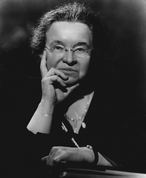