Book - A Laboratory Text-Book of Embryology Figures (1903)
| Embryology - 25 Apr 2024 |
|---|
| Google Translate - select your language from the list shown below (this will open a new external page) |
|
العربية | català | 中文 | 中國傳統的 | français | Deutsche | עִברִית | हिंदी | bahasa Indonesia | italiano | 日本語 | 한국어 | မြန်မာ | Pilipino | Polskie | português | ਪੰਜਾਬੀ ਦੇ | Română | русский | Español | Swahili | Svensk | ไทย | Türkçe | اردو | ייִדיש | Tiếng Việt These external translations are automated and may not be accurate. (More? About Translations) |
Minot CS. A Laboratory Text-Book Of Embryology. (1903) Philadelphia:P. Blakiston's Son & Co.
| Online Editor |
|---|

|
| Historic Disclaimer - information about historic embryology pages |
|---|
|
List of Illustrations

Chapter II. The Early Development of Mammals
- Chapter II. The Early Development Of Mammals: 1. Human Spermatozoa | 2. Human Ovum | 3. Worm Ovum | 4. Rabbit Ovum 17 Hours | 5. Mouse Ovum early Pro-nuclei | 6. Mouse Ovum Spindle | 7. Mouse Ovum late Pro-nuclei | 8. Rabbit Ovum 24 Hours | 9. Snail Ovum First Cleavage | Chapter 2 Figures | Figures
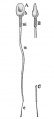
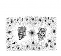
1. Human Spermatozoa, {after Retzius)
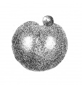
2. Full-grown Human Ovum before Maturation, (after W. Nagel)
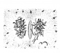
3. Ovum of a Worm (Sagitta) with Two Pro-nuclei. Around Each Pro-nucleus is shown the Aster (after O. Hertwig)
4. Ovum of a Rabbit, Seventeen Hours after Coitus, with the Pro-nuclei about to Conjugate {after Rein)
5. Ovum of White Mouse. Beginning of the Conjugation of the Pro-nuclei (after Sobotta)
6. Ovum of White Mouse. Conjugation of the Pro-nuclei, and Formation of the Segmentation Spindle (after Sobotta)
7. Ovum of White Mouse. First Segmentation Spindle with the Chromosomes of the Pro-nuclei still forming Two Distinct Groups (after Sobotta)
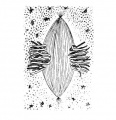
8. Ovum of a Rabbit of Twenty-four Hours {after Caste), 55
9. Ovum of a Snail (Limax campestris) during the First Cleavage. The Envelopes of the Ovum are not Drawn in (after E. L. Mark)
- Minot1897 fig010.jpg
10. Ovum of White Mouse. First Segmentation Spindle with Equatorial Plate of Chromosomes (after Sobotta)
- Minot1897 fig011.jpg
11. Ovum of White Mouse. First Segmentation Spindle {after Sobotta)
- Minot1897 fig012.jpg
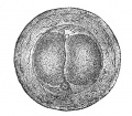
12. Ova of White Mouse with Two Segmentation Spheres or Cells (after Sobotta)
- Minot1897 fig013.jpg
13. Ovum of a Bat (Vespertilio murina) with Four Segmentation Spheres (after van Beneden and Julin)
- Minot1897 fig014.jpg
14. Ovum of a Virginian Opossum, with Four Segments (after Emit Selenka)
- Minot1897 fig015.jpg
15. Rabbit's Ovum of about Seventy Hours (after E. van Beneden)
- Minot1897 fig016.jpg
16. Young Blastodermic Vesicle of a Mole (after lt~. heafe)
- Minot1897 fig017.jpg
17. Sections through the Inner Mass of Blastodermic Vesicles of the Mole at Three Successive Stages (after IV. Heafe)
- Minot1897 fig018.jpg
18. Transverse Section through the Embryonic Shield of the Blastodermic Vesicle of a Dog of Eleven or Fifteen Days (Precise Age Unknown) (after Bonnet)
- Minot1897 fig019.jpg
19. Surface View of the Embryonic Shield of the Blastodermic Vesicle of a Dog of Thirteen to Fifteen Days (Precise Age Unknown) (after Bonnet)
- Minot1897 fig020.jpg
20. Central Portion of a Sheep's Blastodermic Vesicle of Twelve to Thirteen Days (after Bonnet)
- Minot1897 fig021.jpg
21. Blastodermic Vesicle of a Rabbit of Seven Days. Portion of the Mescderm of the Area opaca (after Kolliker)
- Minot1897 fig022.jpg
22. Germinal Area of a Guinea-pig at Thirteen Days and Twenty Hours, seen from the Under (Entodermal) Side
- Minot1897 fig023.jpg
23. Transverse Section of a Mole Embryo (Heape's Stage H) (after IV. Heafe)
- Minot1897 fig024.jpg
24. Degenerating Tissue of the Notochord from the Central Portion of the Intervertebral Disc of a Cow's Embryo (after Leboucg)
- Minot1897 fig025.jpg
25. Surface View of the Embryonic Shield of a Dog Embryo, with Medullary Plate
- Minot1897 fig026.jpg
26. Cross-section of a Human Embryo of 1.54 mm. (after Count Spee)
- Minot1897 fig027.jpg
27. Section of a Young Cat Embryo and of the Uterine Wall to Which it is Attached. (Embryo No. 398, Section 76)
- Minot1897 fig028.jpg
28. Transverse Section of a Rabbit Embryo of Eight Days and Two Hours
- Minot1897 fig029.jpg
29. Three Diagrams of Embryonic Areas of Chicks to show the Growth of the Mesoderm (after Dnvai)
- Minot1897 fig030.jpg
30. Transverse Section of an Early Stage of an Axolotl (after Bellonci)
- Minot1897 fig031.jpg
31. Generalized Diagram of an Amniote Vertebrate Embryo
- Minot1897 fig032.jpg
32. Transverse Section from a Chick Embryo with about Eighteen Segments
- Minot1897 fig033.jpg
33. Section of a Very Young Cat Embryo. iTransverse Series 413, section 181)
- Minot1897 fig034.jpg
34. Diagrammatic Section of the Yolk of a Hen's Egg at an Early Stage to show the Relation of the Primitive Entodermal Cavity
- Minot1897 fig035.jpg
35. Wall of the Yolk-sac in the Region of the Area opaca of a Chick of the Second Day
- Minot1897 fig036.jpg
36. Wall of the Yolk-sac in the Region of the Area opaca of a Rabbit Embryo of Thirteen Days
- Minot1897 fig037.jpg
37. Section of the Yolk-sac of a Young Human Embryo (alter Keibel)
- Minot1897 fig038.jpg
38. Human Embryo, 2.15 mm. Long (after W.His)
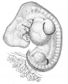
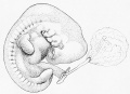
39. Human Embryo of 2. 6 mm. (after IV. His)
40. Human Embryo of 9.8 mm. Probable Age Thirty Days
41. Section of the Area vasculosa of a Chick Embryo of the Second Day
- Minot1897 fig042.jpg
42. Development of Blood-corpuscles
- Minot1897 fig043.jpg
43. Diagrams to Illustrate the Separation of the Embryo from the Yolk
- Minot1897 fig044.jpg
44. Diagram of the Circulation in a Chick at the End of the Third Day, as Seen from the Under (Entodermal) Side
- Minot1897 fig045.jpg
45. Area Vasculosa of a Rabbit, Presumably of about Twelve Days (after Van Beneden and Juliti)
- Minot1897 fig046.jpg
46. Longitudinal Section of the Posterior End of a Sheep Embryo of Sixteen Days (after P. Bonnet)
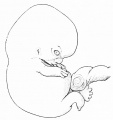
- Minot1897 fig047.jpg
47. Frog (Rana temporaria) Tadpole of 12.0 mm. Cross-section of the Pronephric Region (after M. Fiirbrin^cr)
- Minot1897 fig048.jpg
48. Diagrams illustrating the Relations of the Allantois in Unguiculate Mammals
- Minot1897 fig049.jpg
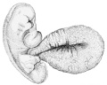
49. Pig, 15.0 mm Series 135, Section 58, to Show the Relations of the Chorion to the Uterus
- Minot1897 fig050.jpg
50. Transverse Section of an Embryo Catfish (Amiurus) ; Series 25, Section 43
- Minot1897 fig051.jpg
51. Sections of Two Human Umbilical Cords


Chapter III. The Human Embryo
- Chapter II. The Early Development Of Mammals: 52. Human Embryo Week 3 | 53. Early Primate Development | 54. Early Stage Primate Embryo | 55. Antero-posterior Section Uterus and Embryo 5 Weeks | 56. Human Uterus 40 Days Pregnancy | 57. Monkey Vesicle in Uterus | 58. Monkey Vesicle in Uterus detail | 59. Peters's Ovum | 60. Gibbon Embryo 3rd Stage | 61. Gibbon Embryo lateral | 62. Gibbon Embryo Transverse Section | 63. Embryonic Area of Ovum | 64. Human Embryo 1.54 mm | 65. Human Embryo 1.54 mm Median Section | 66. Human Embryo of 1.54 mm | 67. Human Embryo Open Medullary Groove | 68. Human Embryo 13-14 Days | 69. Human Ovum 15-18 Days | 70. Embryo of Fig. 69 | 71. Human Embryo, 2.15 mm | 72. Embryo Shown in Fig. 71 | 73. Human Embryo of 2.6 mm | 74. Embryo 2.6 mm in Fig. 72 | 75. Human Embryo 42 mm | 76. Entodermal Canal Embryo 4.2 mm | 77. Human Embryo, 3.2 mm | 78. Human Embryo 4.2 mm Shown in Fig. 75 | 79. Human Embryo 23 Days | 80. Human Embryo 7 mm | 81. Human Embryo 7.5 mm | 82. Pharyngeal Region Human Embryo 11.5 mm | 83. Human Embryo 9.8 mm | 84. Human Embryo 9.8 mm | 85. Human Embryo 11 mm | 86. Human Embryo 14 mm | 87. Human Embryo 35 Days | 88. Human Embryo 16 mm | 89. Human Embryo 22 mm | 90. Human Embryo 28 mm | 91. Human Embryo 32 mm | 92. Human Embryo 34 mm Face | 93. Human Embryo 55 mm 75 Days | 94. Human Embryo of 78 mm 3 Months | 95. Embryo face in Fig. 94 | 96. Human Embryo 120 mm 110 Days | 97. Human Embryo 118 mm 106 Days | 98. Human Embryo155 mm 123 Days | Chapter 3 Figures | Figures
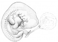
52. Human Embryo at the Beginning of the Third Week
53. Two Diagrams to Illustrate the Hypothetical Early Development of Primates
54. Diagram of an Early Stage of a Primate Embryo
- Minot1897 fig055.jpg
55. Semi-diagrammatic Outline of an Antero-posterior Section of a Human Uterus Containing an Embryo of about Five Weeks (after Allen Thompson)
- Minot1897 fig056.pg
56. Human Uterus, about Forty Days Advanced in Pregnancy (after Coste)
- Minot1897 fig057.jpg
57. Blastodermic Vesicle of a Monkey (Semnopithecus nasicus) Attached to the Uterus ; Vertical Section (after E. Selenka)
- Minot1897 fig058.jpg
58. Embryo of the Preceding Figure More Highly Magnified (after E. Selenka)
- Minot1897 fig059.jpg
59. Section of Peters's Ovum in Situ (after H. Peters)
- Minot1897 fig060.jpg
60. Embryo of a Gibbon (Hylobates concolor) in the Third Stage (after E. Selenka)
- Minot1897 fig061.jpg
61. Embryo of a Gibbon, Side View of the Embryo of Fig. 60 (after E. Selenka)
- Minot1897 fig062.jpg
62. Transverse Section of the Embryo of the Preceding Figure (after E. Selenka)
- Minot1897 fig063.jpg
63. Surface View of the Embryonic Area of the Ovum Shown in Fig. 61
- Minot1897 fig064.jpg
64. Reconstruction of a Human Embryo 1.54 mm Long (after Count Spee)
- Minot1897 fig065.jpg
65. Human Embryo of 1. 54 mm Median Section from a Wax Model Reconstructed from Sections (after Count Spee)
- Minot1897 fig066.jpg
66. Human Embryo of 1.54 mm (after Count Spee)
- Minot1897 fig067.jpg
67. Human Embryo with Open Medullary Groove (after Wais)
- Minot1897 fig068.jpg
68. Human Embryo of from Thirteen to Fourteen Days (after J. Kallmann)
- Minot1897 fig069.jpg
69. Human Ovum, said to be from Fifteen to Eighteen Days Old
- Minot1897 fig070.jpg
70. Embryo of Fig. 69, Separated from the Yolk-sac and Viewed from the Under Side
- Minot1897 fig071.jpg
71. Human Embryo, 2.15 mm. Long {after IV. His)
- Minot1897 fig072.jpg
72. Reconstruction of the Anatomy of the Embryo Shown in Fig. 71 (after W. His)
- Minot1897 fig073.jpg
73. Human Embryo of 2.6 mm. Length (after IV. His)
- Minot1897 fig074.jpg
74. Reconstruction of the Anatomy of the Embryo of 2.6 mm. in Fig. 72 (after W. His)
- Minot1897 fig075.jpg
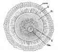
75. Human Embryo 42 mm (after IV. His)
- Minot1897 fig076.jpg
76. Outline of the Entodermal Canal of a Human Embryo of 4.2 mm (after IV. His)
- Minot1897 fig077.jpg
77. Reconstruction of the Anatomy of a Human Embryo, 3.2 mm Long, showing the Anterior End Viewed from the Ventral Side
- Minot1897 fig078.jpg
78. Reconstruction of the Anatomy of the Human Embryo of 4.2 mm Shown in Fig. 75 (after W His)
- Minot1897 fig079.jpg
79. Human Embryo of About Twenty-three Days (after W. His)
80. Human Embryo of 7 mm (after F. P. Mall)
81. Human Embryo of 7.5 mm in Maximum Length (after W. His),
82. Reconstruction of the Pharyngeal Region of a Human Embryo of 11.5 mm (after W. His)
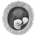
83. Human Ovum with Embryo of 9.8 mm The Chorion Has Been partly Removed to Show the Embryo (Minot Collection, 275)
84. Embryo of the Preceding Figure
85. Human Embryo of 11 mm (after W. His)
86. Human Embryo of about 14 mm
87. Human Embryo of about Thirty-five Days (after Coste)
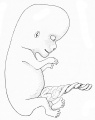
88. Human Embryo of about 16 mm (after W. His)
89. Human Embryo of 22 mm
90. Human Embryo of 28 mm
91. Human Embryo of 32 mm
- Minot1897 fig092.jpg
92. Human Embryo of 34 mm Front View of Face
- Minot1897 fig093.jpg
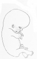
93. Human Embryo of 55 mm Seventy-live Days
- Minot1897 fig094.jpg
94. Human Embryo of 78 mm Three Months
- Minot1897 fig095.jpg
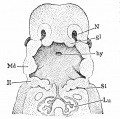
95. Front View of the Face of the Embryo Shown in Fig. 94
- Minot1897 fig096.jpg
96. Human Embryo of 120 mm (One Hundred and Ten Days)
- Minot1897 fig097.jpg
97. Human Embryo of 118 mm One Hundred and Six Days
- Minot1897 fig098.jpg
98. Human Embryo of 155 mm One Hundred and Twenty-three Days


Chapter IV. Study Of Pig Embryos
- Chapter IV. Study Of Pig Embryos: 99. Pig Embryo 10 mm | 100. Pig Embryo 12 mm | 101. Pig Embryo 12.0 mm Transverse Section | 102. Pharynx Human Embryo 3.2 mm | 103. Aortic System His's Embryo Bl 4.25 mm | 104. Pig Embryo 12 mm Transverse Section | 105. Pig Embryo 12.0 mm Transverse Section Series 5 | 106. Transverse Section Pig Embryo 12 mm | 107. Pig Embryo 12.0 mm Series 5 | 108. Pig Embryo of 12 mm | 109. Pig Embryo 12.0 mm Series 5 | 110. Pig Embryo of 15 mm | 111. Pig Embryo 20 mm | 112. Pig Embryo 12.0 mm with Planes of Sections Figured | 113. Pig, 12.0 mm Transverse Series 5, Section 185 | 114. Portion of Fig. 113 More Highly Magnified | 115. Pig, 12.0 mm - Transverse Series 5, Section 19S | 116. Pig 12.0 mm Transverse Series 5, Section 249 | 117. Pig, 12.0 mm Transverse Series 5, Section 292 | 115. Pig, 12.0 mm Transverse Series 5, Section 321 | 119. Pig, 12.0 m Transverse Series 5, Section 353 | 120. Pig, 12.0 mm Transverse Series 5, Section 470 | 121. Pig 12.0 mm Transverse Series 5, Section 513 | 122. Pig, 12.0 mm. Transverse Series 5, Section 633 | 123. Pig, 12.0 mm. Sagittal Series 7, Section 70 | 124. Pig, 12.0 mm. No. 7. Sagittal Section 25 | 125. Pig, 12.0 mm. Frontal Series 6, Section 284 | 126. Pig, 12.0 mm. Frontal Series 6, Section 340 | 127. Pig, 12.0 mm Frontal Series 6, Section 350 | 128. Pig, 12.0 mm. Frontal Series 6, Section 572 | 129. Pig, 9.0 mm. Transverse Series 9, Section 171 | 130. Pig, 9.0 mm. Sagittal Series 53, Section 213 | 131. Pig, 9.0 mm. Frontal Series 54, Section 194 | 132. Pig, 9.0 mm. Frontal Series 54, Section 194 | 133. Pig, 9.0 mm. Frontal Series, 54, Section 459 | 134. Pig, 6.0 mm. Transverse Series 9, Section 90 | 135. Pig, 6.0 mm. Transverse Series 9, Section 519 | 136. Pig, 17.0 mm. Transverse Series 51, Section 464 | 137. Pig, l7.0 mm. Transverse Series 51, Section 651 | 138. Pig, 17.0 mm Transverse Series 51, Section 759 | 139. Pig. 170 mm Frontal Series 39, Section 63 | 140. Pig 20.0 mm Transverse Series 59, Section 522 | 141. Pig, 20.0 mm. Transverse Series 59, Section 522 | 142. Pig 20.0 mm Transverse Series 59, Section 701 | 143. Pig, 20.0 mm. Transverse Series 59, Section 1253 | 144. Pig, 20.0 mm. Transverse Series 59, Section 1043 | 145. Pig, 20.0 mm. Sagittal Series 60, Section 213 | 146. Pig, 20.0 mm. Frontal Section of Head. Series 40, Section 65 | 147. Pig 20.0 mm Frontal Head section Series 40, Section 84 | 148. Pig, 20.0 mm. Frontal Section of Head. Series 40, Section 123 | 149. Rabbit Embryo Eye 13 Days | 150. Pig 24.0 mm Transverse Series 62, Section 428 | 151. Pig, 24.0 mm Sagittal Series 63, Section 30 | Chapter 4 Figures | Figures
- Minot1897 fig099.jpg
99. Pig Embryo of 10 mm
- Minot1897 fig100.jpg
100. Transverse Section of Pig Embryo of 12 mm
- Minot1897 fig101.jpg
101. Pig Embryo of 12.0 mm. Reconstruction from the Transverse Sections, Series 5 (drawn by Dr. F. T. Leivis)
- Minot1897 fig102.jpg
102. Anterior Wall of the Pharynx of a Human Embryo of 3.2 mm (after IV. //is)
- Minot1897 fig103.jpg
103. Aonic System of His's Embryo Bl, 4.25 mm (after W. His)
- Minot1897 fig104.jpg
104. Transverse Section of Pig Embryo of 12 mm facing
- Minot1897 fig105.jpg
105. Pig Embryo of 12.0 mm Reconstruction from the Transverse Sections, Series 5 (drawn by Dr. F. T. Lewis)
- Minot1897 fig106.jpg
106. Transverse Section of Pig Embryo of 12 mm facing
- Minot1897 fig107.jpg
107. Pig Embryo of 12.0 mm Reconstruction from the Transverse Sections, Series 5 (drawn by Dr. F. T. Lewis)
- Minot1897 fig108.jpg
108. Transverse Section of Pig Embryo of 12 mm Jacim
- Minot1897 fig109.jpg
109. Pig Embryo of 1 2.0 mm Reconstruction from the Transverse Sections, Series 5 (drawn by Dr. F. T. Lewis)
- Minot1897 fig110.jpg
110. Pig Embryo of 15 mm
- Minot1897 fig111.jpg
111. Pig Embryo of 20 mm
- Minot1897 fig112.jpg
112. Reconstruction of a Pig Embryo of 12.0 mm with Indications of the Planes of Sections Figured
- Minot1897 fig113.jpg
113. Pig, 12.0 mm Transverse Series 5, Section 185
- Minot1897 fig114.jpg
114. Portion of Fig. 113 More Highly Magnified
- Minot1897 fig115.jpg
115. Pig, 12.0 mm - Transverse Series 5, Section 19S
- Minot1897 fig116.jpg
116. Pig, 12.0 mm Transverse Series 5, Section 249
- Minot1897 fig117.jpg
117. Pig, 12.0 mm Transverse Series 5, Section 292
- Minot1897 fig118.jpg
115. Pig, 12.0 mm Transverse Series 5, Section 321
- Minot1897 fig119.jpg
119. Pig, 12.0 m Transverse Series 5, Section 353
- Minot1897 fig120.jpg
120. Pig, 12.0 mm Transverse Series 5, Section 470
- Minot1897 fig121.jpg
121. Pig, 12.0 mm. Transverse Series 5, Section 513
- Minot1897 fig122.jpg
122. Pig, 12.0 mm. Transverse Series 5, Section 633
- Minot1897 fig123.jpg
123. Pig, 12.0 mm. Sagittal Series 7, Section 70
- Minot1897 fig124.jpg
124. Pig, 12.0 mm. No. 7. Sagittal Section 25
- Minot1897 fig125.jpg
125. Pig, 12.0 mm. Frontal Series 6, Section 284
- Minot1897 fig126.jpg
126. Pig, 12.0 mm. Frontal Series 6, Section 340
- Minot1897 fig127.jpg
127. Pig, 12.0 mm. Frontal Series 6, Section 3S0
- Minot1897 fig128.jpg
128. Pig, 12.0 mm. Frontal Series 6, Section 572
- Minot1897 fig129.jpg
129. Pig, 9.0 mm. Transverse Series 9, Section 171
- Minot1897 fig130.jpg
130. Pig, 9.0 mm. Sagittal Series 53, Section 213
- Minot1897 fig131.jpg
131. Pig, 9.0 mm. Frontal Series 54, Section 194
- Minot1897 fig132.jpg
132. Pig, 9.0 mm. Frontal Series 54, Section 194
- Minot1897 fig133.jpg
133. Pig, 9.0 mm. Frontal Series, 54, Section 459
- Minot1897 fig134.jpg
134. Pig, 6.0 mm. Transverse Series 9, Section 90
- Minot1897 fig135.jpg
135. Pig, 6.0 mm. Transverse Series 9, Section 519
- Minot1897 fig136.jpg
136. Pig, 17.0 mm. Transverse Series 51, Section 464
- Minot1897 fig137.jpg
137. Pig, l7.0 mm. Transverse Series 51, Section 651
- Minot1897 fig138.jpg
138. Pig, 17.0 mm. Transverse Series 51, Section 759
- Minot1897 fig139.jpg
139. Pig. 170 mm. Frontal Series 39, Section 63
- Minot1897 fig140.jpg
140. Pig, 20.0 mm. Transverse Series 59, Section 522
- Minot1897 fig141.jpg
141. Pig, 20.0 mm. Transverse Series 59, Section 522
- Minot1897 fig142.jpg
142. Pig, 20.0 mm. Transverse Series 59, Section 701
- Minot1897 fig143.jpg
143. Pig, 20.0 mm. Transverse Series 59, Section 1253
- Minot1897 fig144.jpg
144. Pig, 20.0 mm. Transverse Series 59, Section 1043
- Minot1897 fig145.jpg
145. Pig, 20.0 mm. Sagittal Series 60, Section 213
- Minot1897 fig146.jpg
146. Pig, 20.0 mm. Frontal Section of Head. Series 40, Section 65
- Minot1897 fig147.jpg
147. Pig, 20.0 mm. Frontal Section of Head. Series 40, Section 84
- Minot1897 fig148.jpg
148. Pig, 20.0 mm. Frontal Section of Head. Series 40, Section 123
- Minot1897 fig149.jpg
149. Rabbit Embryo of Thirteen Days ; Section of the Eye
- Minot1897 fig150.jpg
150. Pig, 24.0 mm.Transverse Series 62, Section 428
- Minot1897 fig151.jpg
151. Pig, 24.0 mm Sagittal Series 63, Section 30
- Minot1897 fig152.jpg
152. Embryo Chick with about Twenty-four Segments. Surface View from the Dorsal Side
Chapter V. Study Of Young Chick Embryos
- Chapter V. Study Of Young Chick Embryos: 153. Chick Embryo 28 Segments Transverse Series 92 Section 73 | 154. Chick Embryo 28 Segments Transverse Series 92, Section 83 | 155. Chick Embryo 28 Segments Transverse Series 92, Section 96 | 156. Chick Embryo 28 Segments Transverse Series 92, Section 104 | 157. Chick Embryo 28 Segments Transverse Series 92, Section 114 | 158. Chick Embryo 28 Segments Transverse Series 92, Section 144 | 159. Chick Embryo 28 Segments Transverse Series 92, Section 165 | 160. Chick Embryo 28 Segments Transverse Series 92, Section 179 | 161. Chick Embryo 28 Segments Transverse Series 92, Section 220 | 162. Chick Embryo 28 Segments Transverse Series 92, Section 356 | 163. Chick Embryo 28 Segments Transverse Series 92, Section 419 | 164. Chick Embryo 28 Segments Transverse Series 92, Section 424 | 165. Chick Embryo 28 Segments Transverse Series 92, Section 427 | 166. Horizontal Section of a Chick Embryo with about Twenty-eight Segments | 167. Chicken Embryo 27 Hours 8 Primitive Segments | 168. Rabbit 9 Days | 169. Chick Embryo Longitudinal Section | 170. Chick Head Embryo 7 Segments | 171. Chick Heart Embryo 7 Segments | 172. Chicken Embryo Heart Section more Advanced than Fig. 171 | 173. Chicken Embryo 7 Segments | 174. Chicken Primitive Groove Embryo 7 Segments | Chapter 5 Figures | Figures
- Minot1897 fig150.jpg
153. Section of Chick Embryo with about Twenty-eight Segments. Transverse Series 92, Section 73
- Minot1897 fig150.jpg
154. Section of Chick Embryo with about Twenty-eight Segments. Transverse Series 92, Section 83
- Minot1897 fig150.jpg
155. Section of Chick Embryo with about Twenty-eight Segments. Transverse Series 92, Section 96
- Minot1897 fig150.jpg
156. Section of Chick Embryo with about Twenty-eight Segments. Transverse Series 92, Section 104
- Minot1897 fig150.jpg
157. Section of Chick Embryo with about Twenty-eight Segments. Transverse Series 92, Section 114
- Minot1897 fig150.jpg
158. Section of Chick Embryo with about Twenty-eight Segments. Transverse Series 92, Section 144
- Minot1897 fig150.jpg
159. Section of Chick Embryo with about Twenty-eight Segments. Transverse Series 92, Section 165
- Minot1897 fig160.jpg
160. Section of Chick Embryo with about Twenty-eight Segments. Transverse Series 92, Section 179
- Minot1897 fig160.jpg
161. Section of a Chicken Embryo with about Twenty-eight Segments. Transverse Series 92, Section 220
- Minot1897 fig160.jpg
162. Section of a Chicken Embryo with about Twenty-eight Segments. Transverse Series 92, Section 356
- Minot1897 fig160.jpg
163. Section of a Chicken Embryo with Twenty-eight Segments. Transverse Series 92, Section 419
- Minot1897 fig160.jpg
164. Chick Embryo with about Twenty-eight Segments. Transverse Series 92, Section 424
- Minot1897 fig160.jpg
165. Chick Embryo with about Twenty-eight Segments. Transverse Series 92, Section 427
- Minot1897 fig160.jpg
166. Horizontal Section of a Chick Embryo with about Twenty-eight Segments
- Minot1897 fig160.jpg
167. Chicken Embryo, after Twenty-seven Hours' Incubation, with Eight Primitive Segments (after Duval)
- Minot1897 fig160.jpg
168. Embryonic Area of a Rabbit of (? Nine) Days, with the Placental Area Partly Torn oft" [after van Bene Jin and Juiin)
- Minot1897 fig160.jpg
169. Longitudinal Section of a Young Chick Embryo
- Minot1897 fig170.jpg
170. Chick Embryo with Seven Segments. Transverse Section of the Head
- Minot1897 fig170.jpg
171. Chicken Embryo with Seven Segments. Transverse Section through the Anlage of the Heart
- Minot1897 fig170.jpg
172. Chicken Embryo, Transverse Section across the Anlage of the Heart in a Stage slightly more Advanced than Fig. 171
- Minot1897 fig170.jpg
173. Chicken Embryo with Seven Segments
- Minot1897 fig170.jpg
174. Chicken Embryo with Seven Segments. Transverse Section across the Primitive Groove
Chapter VI. Study Of The Blastodermic Vesicle And The Segmentation Of The Ovum
- Chapter VI. Study Of The Blastodermic Vesicle And The Segmentation Of The Ovum: 175. Rabbit 7 Days | 176. Rabbit 7 Days Anterior Embryonic Shield | 177. Rabbit Embryo 7 Days Posterior Embryonic Shield | 178. Mouse Ovum First Polar Spindle | 179. Ovum of While Mouse, dividing to Produce the Polar Globule {after J. Sobotta) | 180. Mouse Ovum Metaphase Producing First Polar Globule | 181. Mouse Ovum Both Pro-nuclei | 182. Mouse Ovum Two Pro-nuclei | Chapter 2 Figures | Figures
- Minot1897 fig175.jpg
175. Blastodermic Vesicle of a Rabbit at Seven Days
- Minot1897 fig176.jpg
176. Rabbit Embryo of Seven Days. Transverse Series 12, Section 216, through the Anterior Portion of the Embryonic Shield
- Minot1897 fig177.jpg
177. Rabbit Embryo of Seven Days. Transverse Series 15, Section 39, through the Posterior Part of the Embryonic Shield
- Minot1897 fig178.jpg
178. Ovum of White Mouse, with the First Polar Spindle in Tangential Position (after J. Sobotta)
- Minot1897 fig179.jpg
179. Ovum of While Mouse, dividing to Produce the Polar Globule {after J. Sobotta)
- Minot1897 fig180.jpg
180. Ovum of White Mouse, Showing the Metaphase of the Division Producing the First Polar Globule (after J. Sobotta)
- Minot1897 fig181.jpg
181. Ovum of While Mouse, after the Formation of the Polar Globule. Both Pro-nuclei are Present (after J. Sobotta)
- Minot1897 fig182.jpg
182. Ovum of White Mouse, with Two Well-developed Pro-nuclei (after J. Sobotta)
Chapter VII. Study. Of The Uterus And The Foetal Appendages Of Man
- Chapter VI. Study Of The Blastodermic Vesicle And The Segmentation Of The Ovum: 183. Section Human Uterus Day 1 Menstruation | 184. Section Human Uterus 1 Month Pregnant | 185. Human Uterus 1 Month Pregnant Cavernous Layer | 186. Human Uterus 1 Month Pregnant Epithelium | 187. Uterus 1 Month Pregnant Compact Decidua | 188. Human Decidua Reflexa 2 Months | 189. Human Uterus 7 Months Decidua Vera | 190. Human Placenta 7 Months | 191. Human Placental Chorion and Amnion 5 Month | 192. Human Chorion 7 Months Placenta | 193. Villus Human Placenta 4 Months | 194. Human Decidua Serotina 7 Months | 195. Decidual Cells Section in Fig. 194 | 196. Human Placenta Term, Doubly Injected | 197. Human Placenta after Term Delivery | 198. Section Young Human Chorion | 199. Portion 298 Figure | 200. Villus Human Chorion Month 2 | 201. Chorion of Fig. 69 | 202. Terminal Branch of a Villus 12 Weeks | 203. Villous Stem Human Placenta 5 Month | 204. Terminal Branches Villus Term Placenta | 205. Injected Villus Placenta 5 Months | 206. Injected Villous Stem Placenta 5 Months | 207. Transverse Section Amnion 2 Months | 208. Two Sections of the Human Amnion | 209. Surface View of the Human Amniotic Epithelium of the Fourth Month | 210. Natural Mesoderm Nuclei Amnion 5 Month | 211. Umbilical Cord at Term | 212. Allantois Umbilical Cord 3 Months | 213. Umbilical Cord Human Embryo 21 mm | 214. Umbilical Cord Human Embryo 3 Months | 215. Ectoderm Umbilical Cord 3 Months | 216. Yolk-sac section Young Human Embryo | Chapter 7 Figures | Figures
- Minot1897 fig183.jpg
183. Vertical Section of a Human Uterus of the First Day of Menstruation
- Minot1897 fig184.jpg
184. Vertical Section of a Human Uterus (Decidua vera) , One Month Pregnant
- Minot1897 fig185.jpg
185. Human Uterus, One Month Pregnant. Section of Gland from the Cavernous Layer, with the Epithelium Partly Adherent to the Walls
- Minot1897 fig186.jpg
186. Human Uterus, ( lue Month Pregnant. Section of a Gland from the Cavernous Layer with the Epithelium Loosened from the Walls. The Epithelial Cells are Swollen
- Minot1897 fig187.jpg
187. Uterus One Monlh Pregnant; Portion of the Compact Layer of the Decidua Seen in Vertical Section
- Minot1897 fig188.jpg
188. Section of Human Decidua Reflexa at Two Months
- Minot1897 fig189.jpg
189. Human Uterus about Seven Months Pregnant. Vertical Section of the Decidua Vera with the Foetal Membranes in situ
- Minot1897 fig190.jpg
190. Human Placenta in situ, about Seven Months. Vertical Section
- Minot1897 fig191.jpg
191. Human Placental Chorion and Amnion of the Fifth Month
- Minot1897 fig192.jpg
192. Human Chorion of Seven Months' Placenta
- Minot1897 fig193.jpg
193. Adenoid Tissue from a Villus of a Human Placenta of Four Months
- Minot1897 fig194.jpg
194. The Human Decidua Serotina at Seven Months. The Section is Taken from near the Margin of the Placenta
- Minot1897 fig195.jpg
195. Decidual Cells from the Section Represented in Fig. 194
- Minot1897 fig196.jpg
196. Human Placenta at Full Term, Doubly Injected to show the Superficial Distribution of the Blood-vessels 33%
- Minot1897 fig197.jpg
197. Human Placenta after Delivery at Full Term
- Minot1897 fig198.jpg
198. Section of a Very Young Human Chorion
- Minot1897 fig199.jpg
199. Portion of the Preceding Figure More Highly Magnified
- Minot1897 fig200.jpg
200. Aborting Villus from the Human Chorion Lceve of the Second Month
- Minot1897 fig201.jpg
201. Fragment of the Chorion of Fig. 69, Highly Magnified
- Minot1897 fig202.jpg
202. Isolated Terminal Branch of a Villus from a Human Chorion of Twelve Weeks
- Minot1897 fig203.jpg
203. Villous Stem from a Human Placenta of the Fifth Month
- Minot1897 fig204.jpg
204. Terminal Branches of a Villus from a Human Placenta at Full Term
- Minot1897 fig205.jpg
205. Portion of an Injected Villus from a Placenta of about Five Months
- Minot1897 fig206.jpg
206. Portion of a Small Injected Villous Stem from a Placenta of about Five Months
- Minot1897 fig207.jpg
207. Transverse Section of a Human Amnion of Two Months
- Minot1897 fig208.jpg
208. Two Sections of the Human Amnion
- Minot1897 fig209.jpg
209. Surface View of the Human Amniotic Epithelium of the Fourth Month
- Minot1897 fig210.jpg
210. Natural Group of Nuclei from the Mesoderm of the Human Amnion of the Fifth Month
- Minot1897 fig211.jpg
211. Cross-section of a Human Umbilical Cord at Term
- Minot1897 fig212.jpg
212. Section of the Allantois from a Human Umbilical Cord of Three Months
- Minot1897 fig213.jpg
213. Connective Tissue from the Umbilical Cord of a Human Embryo of 21 mm. Stained with Alum Cochineal and Eosin
- Minot1897 fig214.jpg
214. Connective Tissue from the Umbilical Cord of a Human Embryo of Three Months, Stained with Alum Cochineal and Eosin
- Minot1897 fig215.jpg
215. Ectoderm of an Umbilical Cord of a Human Embryo of Three Months
- Minot1897 fig216.jpg
216. Section of the Yolk-sac of a Very Young Human Embryo (after Fr. A'eibel)
Chapter VIII. Methods
- Chapter VIII. Methods: 217. The Precision Microtome | 218. The Automatic Rotary Microtome | Chapter 8 Figures | Figures
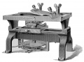
217. The Precision Microtome
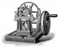
218. The Automatic Rotary Microtome


| Historic Disclaimer - information about historic embryology pages |
|---|
|
Cite this page: Hill, M.A. (2024, April 25) Embryology Book - A Laboratory Text-Book of Embryology Figures (1903). Retrieved from https://embryology.med.unsw.edu.au/embryology/index.php/Book_-_A_Laboratory_Text-Book_of_Embryology_Figures_(1903)
- © Dr Mark Hill 2024, UNSW Embryology ISBN: 978 0 7334 2609 4 - UNSW CRICOS Provider Code No. 00098G