Book - A Laboratory Text-Book of Embryology 3 (1903)
| Embryology - 19 Apr 2024 |
|---|
| Google Translate - select your language from the list shown below (this will open a new external page) |
|
العربية | català | 中文 | 中國傳統的 | français | Deutsche | עִברִית | हिंदी | bahasa Indonesia | italiano | 日本語 | 한국어 | မြန်မာ | Pilipino | Polskie | português | ਪੰਜਾਬੀ ਦੇ | Română | русский | Español | Swahili | Svensk | ไทย | Türkçe | اردو | ייִדיש | Tiếng Việt These external translations are automated and may not be accurate. (More? About Translations) |
Minot CS. A Laboratory Text-Book Of Embryology. (1903) Philadelphia:P. Blakiston's Son & Co.
| Online Editor |
|---|
| This historic 1903 embryology textbook by Minot describes human development.
|
| Historic Disclaimer - information about historic embryology pages |
|---|
| Pages where the terms "Historic" (textbooks, papers, people, recommendations) appear on this site, and sections within pages where this disclaimer appears, indicate that the content and scientific understanding are specific to the time of publication. This means that while some scientific descriptions are still accurate, the terminology and interpretation of the developmental mechanisms reflect the understanding at the time of original publication and those of the preceding periods, these terms, interpretations and recommendations may not reflect our current scientific understanding. (More? Embryology History | Historic Embryology Papers) |
Chapter III. The Human Embryo
Our knowledge of the early stages of human development is very imperfect. Upon the fertilization and segmentation of the ovum in man there are no observations whatever at present. It is not even known exactly how long the ovum requires for its passage through the Fallopian tubes. The earliest stage of which we have any comparatively adequate account is that represented by the ovum described by H. Peters in 1899. A number of human embryos in various early stages after the formation of the medullary canal and up to the stage with four aortic arches have now been reported and studied ; some few of them thoroughly and carefully.
Calculation of the Age of the Human Embryo
The age of the embryo must be reckoned from the date of the fertilization of the ovum, which presumably occurs in man in the upper third of the Fallopian tube. It may be that ova become fertilized at various epochs, but fail to continue their development except when the fertilization occurs at the beginning of a menstrual period. Ovulation occurs at all periods, but most frequently about the time of menstruation, which is the expression of structural changes in the uterus which enable the ovum to implant itself in the uterine wall. Hence only when fertilization coincides with the beginning of menstruation can conception follow with the result that the menstrual flow is stopped. Accordingly, the age of the embryo is usuallv to be reckoned from the date of the beginning of the first menstrual period which has lapsed.
Experience, however, shows that sometimes conception occurs without stopping the menstrual change at the time, but eliminating only the subsequent periods, and in such cases the age must be estimated from the beginning of the last menstruation. In the two cases the age of the embryo would differ by a month (twenty-eight days), and this difference is so great that it obviates errors of estimate.
Up to the end of the ninth week the form and size of the embryo exhibit a correlated development, so that generally an embryo at a given stage of development in form will agree with its fellows in size; but to this rule there are not infrequently exceptions, and sometimes an embryo is found much larger than others at the same stage. Moreover, the variability of embryos is very great, for in specimens otherwise alike we find this or that organ advanced or retarded in its development, as compared with the embryo as a whole. Nevertheless it is possible with the information at command to determine with tolerable certainty the age of an embryo within two days plus or minus, up to the end of the ninth week. For the course of development during the third week we possess as yet no satisfactory data, but embryos of full three months are quite frequently obtained, and are very characteristic in size and configuration ("see page 89).
The Classification of the Early Stages
Any attempt to divide embryos into stages must necessarily establish artificial groups, for in nature there is no demarcation. Division into stages is for convenience, and ought, therefore, to be made by natural and obvious characteristics. It seems to me that eleven stages may be conveniently discriminated, as follows:
First Stage. — Segmentation of the Ovum: The general process is described on pages 54 to 59. There are no observations upon this stage in man.
Second Stage. — Blastodermic Vesicle: The general development of the blastodermic vesicle in mammals is described on page 60. Its development in man is unknown. During this stage the embryonic shield is differentiated. An ovum of a monkey in this stage is described on page 121; the single human ovum known is described on page 123.
Third Stage. — Primitive Streak: No human ovum with a primitive streak before the formation of the medullary plate has been observed.
Fourth Stage. — The Medullary Plate: In this stage there are several embryos known. In all of them the amnion and chorion are already differentiated. There is a large extra-embryonic coelom. The chorionic vesicle is rounded and somewhat flattened. In its greatest diameter it measures from 8 to 10 mm. It is beset with short branching villi which were found present over the entire surface, except in one case described by Reichert. The general relations are indicated in the accompanying diagram (Fig. 54). The chorion has a distinct epidermal and mesodermal layer and bears villi. To its inner surface is attached the body-stalk which unites the embryo and chorion. From it springs the amnion covering the embryo, which measures only 1.0 to 1.5 mm., and from the ventral surface of the embryo arises the yolk-sac, which is of rounded form and about equal in diameter to the length of the embryo.
Fifth Stage. — The Medullary Groove: The general relations of the embryo and its appendages are the same as in the previous stage (compare Fig. 66). In the cases recorded the chorionic vesicle varied greatly in size. It bore villi over its entire surface, and the villi were considerably branched. The embryos varied in length, but measured about 2.2 mm. The medullary ridges are very characteristic, rising high above the yolk-sac and enclosing a deep medullary groove between them. Of this stage our knowledge is very imperfect.
Sixth Stage. — Medullary Tube: In this stage the medullary groove is partly or wholly closed and the heart is clearly differentiated. The embryo measures from 2.2 to 2.5 mm. in length. The head projects in front of the yolk. The primitive segments are partly developed. In one case seven, in another thirteen, were found to have been formed. The caudal end of the embryo also projects beyond the yolk, but less than does the head (compare Fig. 68). The auditory invagination is probably not yet formed. There are no ^ill clefts showing externally.
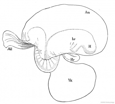
Fig. 52. Human Embryo At The Beginning Of The Third Week. All, Allantois. Am, Amnion, br, Branchial region. H, Head, Hr. Heart. Yk, Yolk-sac.
Seventh Stage. — One Gill Cleft Silencing Externally: Not known by observation.
Eighth Stage. — Two Gill Clefts Showing Externally: Several embryos in this stage have been found and some of them accurately studied. They usually have a remarkable bend in the back (Fig. 52), which imparts to the embryo a very singular appearance. Nothing similar to this bend or dorsal flexure has been observed in any other embryos. It has been held by His and others to be a normal condition, and not the accidental result of a mechanical strain exerted by the yolk-sac. If the condition is normal, it must exist for only a very brief period, as it is not encountered in older or younger stages. We may suppose if it is normal that the change from the concave to the convex position of the embrvo, as found in the next stage, is very abrupt. The head of the embryo (Fig. 52) shows the characteristic head bend, and the tail end of the embryo is also bent over ventral wards. The heart is large and very protuberant. It is bent so that we can clearly distinguish the auricular, ventricular, and aortic limbs. It shows distinctly its inner endothelial portion and outer mesoderm. The yolk-sac extends from the heart backward to where the body of the embryo turns to make the dorsal flexure. Between the heart and the head the oral invagination has been formed, but is still separated by the oral plate from the entodermic canal. Above the heart on either side is an open invagination of the ectoderm, the anlage of the so-called otocyst, which in its turn is the anlage of the epithelial labyrinth of the adult ear. In one embryo of this stage there were shape found twenty-nine primitive segments.
Ninth Stage. — Three Gill Clefts Showing Externally: This is, on the whole, the best known of the early stages of human development. The embryos described as belonging to it vary from 2.6 to 4.2 mm. in length. In one of them, in which the embryo measured 3.2 mm., the chorionic vesicle measured 11 by 14 mm., and its supposed age was from twenty to twenty-one days. The general of these embryos is indicated by figure 74.
The head is bent down and the back is very convex. In figure 74 the tail is rolled up and turned to the left. Usually, however, the tail turns to the right and the head is twisted to the left, so that the long axis of the body describes a large segment of a spiral revolution; the spiral form is marked in embryos a little older.
Tenth Stage. — Four Gill Clefts Showing Externally: There are no satisfactory observations on this stage in man.
Eleventh Stage. — Appearance of the Limb-buds: The embryo is much rolled up, so that the head and tail overlap; four slight protuberances appear as the beginnings of the limbs; the cervical sinus is commencing by the invagination of the posterior gill arches.
Hypothetical Development of the Blastodermic Vesicle in Primates
As there exist no direct observations on the earliest stages of man, we can only surmise what those stages may be. It is evident that there is a very precocious development of the mesoderm, of the extra-embryonic ecelom, of the amnion, and of the trophoblast, because these four features are found very marked in the earliest known stages alike of man, apes, and monkeys. There are certain rodents and insectivora in which these same peculiarities occur more or less emphasized, in the earliest stages of which we possess knowledge. If we utilize these data as a basis, we can reconstruct the following hypothetical scheme of the earliest stages in man.
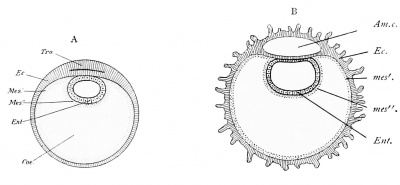
The accompanying diagrams (Figs. 53 and 54) represent three successive
Fig. 53. Two Diagrams to Illustrate the Hypothetical Early Development of Primates Aiii.c, Amniotic cavity. Coe, Coelom. Ec, Ectoderm, in B, bearing the anlages of villi. Ent, Entoderm.
Mes', Somatic mesoderm. Mes" ', Splanchnic mesoderm. Tro, Trophoblast.
purely hypothetical stages of the human ovum. They are all conceived to represent longitudinal sections. In the first stage the ectoderm, Ec, forms a moderate sized vesicle and is already thickened. It should probably be conceived as consisting of an inner distinctly cellular layer and an outer much thicker trophoblastic layer which is thickest over what corresponds to the embryonic region. This special thickening is marked Tro in diagram A. The entoderm, Ent, forms a small vesicle underlying the thickened portion of the trophoblast. The mesoderm, Mes, is well advanced in its development and already contains the large extra-embryonic ctelom, Or, and is therefore divided into one layer which surrounds the entoderm, and a second layer which underlies the ectoderm. In other words, the splanchnopleure and somatopleure are already differentiated. In the next stage (Fig. 53, B) there has been a growth, the ovum has become larger, the trophoblast has increased in thickness, and in the mass of thickened ectoderm overlying the yolk-sac there has appeared a cavity, — the future amniotic cavity, — which is, of course, entirely surrounded by ectoderm. The portion of the ectoderm on the under side of this cavity consists of a single layer of cells which by assuming a cylindrical form constitute the thickened area which we can identify as the embryonic shield (compare Fig. 18 and Fig. 53, B). The solid mass of ectoderm above the amniotic cavity is later to form a part of the amnion and part of the chorion. At the posterior end of the embryo there appears a considerable accumulation of mesoderm (Fig. 54, b. s), which is the anlage of the body-stalk. Into this the entoderm has grown in the form of a cylindrical tubular prolongation, the anlage of the allantois. As a consequence of the growth of the trophoblast and of the formation of
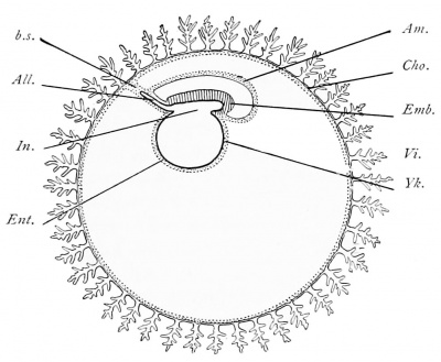
the amniotic cavity, the embryo or embryonic shield, Emb, together with the yolk-sac, Yk, attached to it, has been forced down into the interior of the chorionic vesicle. This phenomenon is very marked in certain rodents and leads to the so-called inversion of the germ-layers. In the next stage the amnion is formed. This is accomplished by the penetration of the mesoderm with accompanying extension of the extra-embryonic coelom into the mass of the ectoderm overlying the amniotic cavity (compare Figs. 53, B, and 54) until the condition shown in figure 54 is brought about. This is the stage known by observation.
Fig. 54. Diagram of an Early Stage of a Primate Embryo. All, Allantois. Am, Amnion, b.s, Body-stalk. Cho, Chorion. Emb, Embryo. Ent, Entoderm. Entodermal cavity of embryo. Vi, Villi of chorion. Yk, Yolk-sac.
The amnion, A m, is now completely separated from the chorion, Cho, whichforms a relatively large vesicle and consists of a thin layer of mesoderm, and a very thick layer of ectoderm, which has an inner cellular stratum and an outer very much thicker trophoblastic stratum. The trophoblast is now very much altered by the appearance of numerous spaces or channels in it which develop so that each of these spaces ends blindly toward the interior of the chorion, but many of them are open upon the surface of the trophoblast. As the ovum at this stage is already embedded in the uterine mucosa, the channels in the trophoblast can receive maternal blood, and such is their original function. The embryo and yolk-sac, as compared with the chorionic vesicle, are very small in size. The body-stalk, b. s., is well developed and contains a well-marked allantoic anlage, All, formed by the entoderm. The embryo contains as yet very little, if any, mesoderm. Probably no neurenteric canal exists at this stage. During the transition of stage B (Fig. 53) to stage C (Fig. 54), the blood-vessels appear in the mesoderm of the yolk-sac.
Relations of the Embryo to the Uterus
The study of Peters's ovum and of early stages of various primates leads us to conceive that the ovum first implants itself in the mucous membrane of the uterus. The conception, "implantation," is the outcome of very recent researches. The essential idea we have formed of implantation is that the trophoblast of the ovum corrodes or digests the uterine tissues with which it comes in contact, and thus produces a cavity in which it is lodged and where it attaches itself intimately to the maternal tissues. Owing to this process the ovum is at first partly uncovered, and this condition seems to be permanent in monkeys. In man and the apes, however, the uterine mucosa grows over the exposed portion of the ovum, forming a layer of maternal tissue which separates the ovum from the cavity of the uterus. This layer is the anlage of the decidua refiexa. As the ovum grows, the decidua refiexa must also expand, and we soon reach a condition in which the primitive relations of the parts can be easily followed.
When the uterus becomes pregnant, the mucous membrane of the organ undergoes changes in structure, and it is then commonly no longer termed the mucosa, but the decidua or caduca. The decidual membrane is histologically characterized by modifications in the glands, the epithelium of which in large part degenerates, by the transformation of a large number of the connectivetissue cells into cells of large size, which, on account of their being so extremely characteristic, are called the decidual cells, and, finally, it is characterized by a growth of its blood-vessels.
The decidual membrane of the uterus is divided into three regions : first, the decidua serotina, the area (Fig. 55, s,s) to which the ovum is attached ; second, the decidua vera, comprising all the remaining portions of the mucosa forming part of the walls of the body of the uterus; third, the decidua reflexa, the arching dome of maternal tissue, r,r, which rises from the walls of the uterus and completely encapsules the ovum. The arrangement of the parts is illustrated in figure 55, which represents a median section of a uterus about five weeks pregnant. The whole uterus is considerably enlarged. The mucous lining of the uterus is very greatly thickened. The ovum is attached on the dorsal side of the uterus. This is the normal position. The diagrams so commonly met with which represent the insertion of the ovum at other points should not be accepted by the student. The reflexa rises around the ovum, completely covering it in so as to make a closed bag. The ovum itself is a sac known as the chorionic vesicle. The trophoblast has now quite disappeared, except so far as it persists to cover the villi. The villi themselves are shaggy, more or less branched, and their tips are united either with the surface of the decidua serotina or with that of the decidua reflexa. In the interior of the chorion is lodged the embryo with its yolk-sac and surrounded by the amnion.
If the walls of the uterus are cut through and simply reflected, leaving the bag of the decidua reflexa intact, the appearances will be found essentially as in figure 56. The mucosa is enormously hypertrophied and contains a great many
dilated irregular blood-sinuses. From the dorsal side of the organ is suspended a large closed bag or sac, the decidua reflexa, D. re), nearly filling the cavity of the uterus. The reflexa presents in the stage figured the same general appearance as the surface of the uterus. If the reflexa be open, we come, of course, upon the villous chorion of the ovum, and find, as above stated, that only the tips of the villi are united with the surface of the reflexa. In the finished stage the decidua is reddish-gray, spongy or pulpy, soft, and moist. After the fourth month it acquires, especially in the superficial layers, a duller brownish color, which subsequently becomes more marked. This coloration is due to the decidual cells. During the first two or three months the scattered openings of the uterine glands can still be distinguished over the surface of the serotina and vera.
Fig. 55. Semi-diagrammatic outline of an Anteroposterior Section of a Human Uterus Containing an Embryo of about Five Weeks.
a, Anterior, p, posterior surface. g, Outer limit of the decidua s,s, Limits of the decidua serotina. ch, Chorion, within which is the embryo enclosed by the amnion, and attached to the chorion by the umbilical cord ; from the cord hangs the pedunculate yolk-sac. r,r, Decidua reflexa. — (After Allen Thompson.}
Fig. 56. Human Uterus, about Forty Hays ahvam-ed in Pkkcnancy.
Muse, Muscularis. Dv, Decidua vera. D.ref, Decidua reflexa. Ov, Ovary. Or J, Oviduct (Fallopian tube). Lig, Round ligament. Vg, Vagina. The uterus has been opened by cutting through the anterior walls and reflecting the sides. — (After Costt.)
The surfaces themselves of the vera and reflexa, though somewhat irregular, remain more or less smooth. The inner surface of the reflexa is more irregular and has protuberant parts united with the tips of the future chorionic villi. The surface of the decidua serotina, on the contrary, becomes very irregular during the progress of pregnancy, forming little mounds which may become so high as to resemble columns, or so broad as to constitute septa. In later stages the septa become very well developed, attaining a height of from 5 to 15 mm. They are irregularly disposed, but subdivide the placenta of later stages into the so-called cotyledons (compare page 337).
The body-stalk becomes converted into the umbilical cord. This cord runs from the body of the embryo to the chorion (Figs. 55 and 72). It is always connected with that portion of the chorion which is adjacent to the decidua serotina. It carries the arteries and veins from the body of the embryo to the chorion. From the end of the umbilical cord the blood-vessels branch out over the chorion and into the chorionic villi. Thus the chorionic circulation of the embryo centers about the chorionic end of the umbilical cord, and as this end is in the part of the chorion overlying the decidua serotina, we have here established from the very start an important factor in the further differentiation. From what has been said it is evident that the portion of the chorion underlying the decidua reflexa is more remote from the center of the embryonic circulation. In the same way we find that the decidua reflexa is remote from the blood-supply in the uterus, and, as a matter of fact, we may observe that during the second month of pregnancy the blood-vessels, both in the decidua reflexa and in the portion of the chorion near it, begin to disappear and ultimately are completely atrophied. After this atrophy has been accomplished the circulation of the chorion is restricted to that portion overlying the decidua serotina. When the blood-vessels of the chorion under the decidua reflexa abort, the villi of that region also abort, so that that part of the chorion becomes smooth, and is, therefore, called the chorion Iceve. Over the serotina the villi continue to grow, hence that region of the chorion becomes known as the chorion frondosum. The chorion frondosum constitutes the foetal portion, the decidua serotina the maternal portion, of the permanent placenta. The maternal blood circulates in the intervillous spaces, which are bounded by fcetal ectoderm. The foetal blood circulates in the foetal bloodvessels of the chorionic villi. The circulatory channels of mother and foetus are always distinct, and no mingling of the maternal and fcetal blood is possible under normal conditions.
Ovum of a Monkey in the Second Stage
This embryo was obtained from a Semnopithecus nasicus in Borneo by Selenka. The ovum represents the earliest stage of any primate yet known. It rested against the wall of the uterus and was uncovered, there being no decidua reflexa developed in monkeys. It measured about 2 mm. in its greatest diameter. Figure 57 represents a section through the ovum and adjacent tissues of the uterus. The chorionic vesicle is very large, but the embryo, Sh, and yolksac, Yk, are relatively very small. The chorion on one side is quite smooth ; on the opposite side it has developed numerous outgrowths, most of which are formed exclusively of the ectoderm, but a few contain an ingrowth of mesoderm in their interior. The ectoderm on the side toward the uterus has two layers, an inner cellular layer with relatively small nuclei, and an outer syncytial or trophoblastic layer with larger nuclei of variable size. The ovum occupies a depression on the surface of the uterus from which the uterine tissues have disappeared, with the result of breaking through the walls of some of the blood-vessels, bl.lac, so that now the maternal blood may escape from these vessels into the spaces left between the irregular outgrowths and the embryonic chorion. We must assume that the trophoblast of the embryo has actually dissolved away or digested the tissues of the uterus, thus providing an attachment for the ovum, securing its embedding in the wall of the uterus, and establishing an opportunity for the maternal blood to flow into the intervillous spaces. In later stages of the primates the trophoblast is very much reduced, and therefore fulfils its functions in the very earliest stages by establishing these primitive conditions of bloodsupply.
Fig. 57. Blastodermic Vesicle of a Monkey (Semnopithecus nasicus) Attached to thk Uterus ; Vertical Section.
Am.e, Amniotic cavity, bl.lac, Blood lacuna. Ca, Extraembryonic coelom. Conn, Connective tissue of the uterus. Ec, Ectoderm. Ug, Ug. Uterine glands. Mes, Mesoderm of embryonic chorion. Sh, Embryonic shield. Tro, Trophoblast. M, Mesodermic core of a chorionic villus. Yk, Yolk-sac. — {After E. Selenka.)
A section of the embryo on a larger scale is shown in figure 58. There appears only the embryonic shield, Sh, which is remarkable for its small area and great thickness. The yolk-sac is also very small and is lined by a distinct layer of entoderm, Ent. Above the embryonic shield is the amniotic cavity, which is, of course, bounded by ectoderm which is continuous with the ectoderm of the embryonic shield. The amniotic cavity has a curious extension into the body-stalk, b. s, by which the embryo is connected with the chorion. The mesoderm is chiefly developed over the chorion, as shown in figure 57. It is very slightly developed in the embryo (Fig. 58, mes), but forms a layer over the yolksac and over the amnion, and forms a considerable mass of tissue to constitute the body-stalk, b. s.
Fig. 58. Embryo of the Preceding Figure More Highly Magnified. Am., c, Amniotic cavity. A. ec, Amniotic ectoderm. A. mes, Amniotic mesoderm, i.s, Body-stalk. Cce, Extraembryonic coelom. Ent, Entoderm, mes', Somatic, mes" , splanchnic, mesoderm. Sh, Embryonic shield. — [After E. Selenka.)
Human Embryo in the Second Stage
The embryo to be described was investigated by H. Peters. It was found attached to the dorsal wall of a uterus almost completely embedded in the mucosa, but it was not wholly covered thereby, so that there was no decidua reflexa vet present. A blood-clot was found overlying what would have been otherwise the exposed portion of the ovum. The trophoblast formed an enormously thick layer of very irregular outline and contained many large spaces filled with maternal blood (Fig. 59). The exact external diameter of the ovum could not, therefore, be determined. It measured, however, approximately 2.4 mm. by 1.2 mm. The internal diameter of the chorionic vesicle was about 1.6 by 0.8 mm. The trophoblast was everywhere intimately united with the uterine tissue. The embryo, Sk, was represented by an embryonic shield consisting of cylinder cells. It is small and lies on the side of the ovum away from the cavity of the uterus. It rests upon the small yolk-sac, Yk, and is overlain by the amniotic cavity, Am. c, which is bounded everywhere by ectoderm — on one side, of course, that of the embryonic shield ; on the other the thin amniotic ectoderm proper. The mesoderm extends around the ovum, forming a layer underneath the chorionic ectoderm over the yolk-sac and above the amnion. At one point, close to the embryo and yolk-sac, it encloses a triangular space the meaning of which is not known. As indicated in the figure, the mesoderm was found to have shrunken somewhat, and the appearance of the embryo and yolk-sac also suggests a somewhat imperfect preservation, histologically speaking, of the tissues. As regards the condition of the uterus, the following points may be noted. In the neighborhood of the ovum the decidua vera had acquired a thickness of about 8 mm., while on the opposite or anterior side it was only from 5 to 6 mm. in diameter. Only in the immediate neighborhood of the ovum could there be seen any differentiation of the mucous membrane into an upper, more compact layer, and a deeper, looser cavernous layer. The epithelium of the glands and the tissues of the uterus were well preserved, except in the immediate neighborhood of the ovum. The picture produces the impression that the ovum, in order to secure a place for itself, has completely destroyed the uterine tissues with which it has been in contact, thus implanting itself in the maternal tissue. And as a consequence of the destruction of the maternal tissues the walls of some of the bloodvessels have been broken through, and this has allowed the blood to escape from those vessels into the lacunas of the trophoblast.
The trophoblast of the ovum offers a very complex picture, owing chiefly to the changes which it is undergoing. The changes seem here due apparently to hypertrophic degeneration. The layer of the chorionic ectoderm next to the mesoderm retains more or less evidently a cellular character. The remaining portions tend to form a syncytium in which the nuclei become enlarged and the cell-boundaries obliterated, while the protoplasm of the cells also changes in character and becomes more homogeneous in texture and much denser. The syncytium disappears by resorption, and its disappearance causes the formation of spaces in the trophoblast. Many different pictures occur in connection with these processes, for in some places the nuclei tend to gather in groups, in others they disappear, in some instances strands of degenerative material are left, while nearby some of the trophoblast may retain its more primitive appearance and be but slightly altered. Finally, it should be noted that at various points the chorionic mesoderm is growing out into the trophoblast. Each of these mesodermic outgrowths is to be interpreted as the anlage of the central portion of a chorionic villus, and out of the neighboring chorionic ectoderm will be differentiated the ectodermal covering of the villus. It seems, from a comparison of later stages, that the trophoblastic degeneration never goes so far as to leave any of the chorionic villi without an ectodermal covering. But this covering varies extremely in its exact character as we find it in later stages, even in adjacent parts of the same villus, for it may be either a single layer of cells or a layer of cells covered by a thin coat of syncytium or merely a syncytial layer (compare page 342). The disappearance of all of the trophoblast, except so much as remains to share in forming the ectodermal covering of the villi, produces the socalled intervillous spaces of later stages, in which, as above stated, maternal blood circulates.
Fig. 60. Embryo of a Gibbon (Hylobates concolor) in the Third Stage. Am, Amnion. Yk, Yolk-sac. Cho, Chorion. Vi, Villi. — [After E. Seltnka.)
The Embryo of a Gibbon in the Third Stage
The embryo to be described was obtained from a female Hylobates concolor in Borneo by Selenka. It still had traces of the primitive streak, the anterior end of which was an open neurenteric canal. The medullary plate was partially differentiated from the embryonic shield. It was thoroughly studied by Selenka, and is the best known very early stage of any primate. It is more advanced than the human embryo described by Peters (page 123). The entire ovum is represented in figure 60. The figure was reconstructed from a study of the sections. It shows the chorionic membrane studded with villi. The diameter of the chorion was about 8.5 mm. The number of villi was about one hundred, of which some seventy are clustered about the region where the embryo was found. The others are scattered over the surface of the membrane. They are considerably branched. Each one is covered by ectoderm which consists of two layers, an inner distinctly cellular, and an outer one in which the cell-boundaries are indistinct and which is known, therefore, as a syncytium and represents the remains of the original trophoblast. Each villus contains a core of mesodermic tissue. The chorionic membrane is represented as open in order to show the size and position of the yolk-sac, Yk, and of the amnion, Am, which encloses the embryo as it rests upon the yolk-sac The embryo itself is not shown in the illustration. Both the yolk-sac and the amnion are, of course, covered by a layer of mesoderm. The entire space between these two inner structures and the chorion corresponds to the extra-embryonic coelom, the very precocious and enormous development of which is a special characteristic of primates, including man, and is not at present known to be paralleled by the conditions in the early stages in any other mammals.
Fig. 61. Embryo of a Gibbon, the Embryo of Fig Emb, Embryo. Am, Amnion.
All.
Side View of 60. neu, Neurenteric canal, b.s, Body-stalk. Yk, Yolk-sac. Ve, Blood-vessels. All, Allantois. — (After E. Selenka. )
Fig. 61. Transverse Section of the Embryo of the Preceding Figure.
Am, Amnion. Ec, Ectoderm. F, Dorsal furrow. mes, Mesoderm. Ent, Entoderm. Ve, Bloodvessel. — {After E. Selenka.)
A side view of the embryo on a larger scale is represented in figure 61 . The embryo is connected with the chorion by a well-marked body-stalk, b. s, is covered by the arching amnion, .4 m, and rests upon the yolk-sac, which in comparison to the chorionic sac seems very small. The yolk-sac, Yk, already has developed from it a network of blood-vessels, Ve, which contain blood-corpuscles, but have not yet developed into an embryo itself. The disposition of these vessels is best illustrated by the section (Fig. 62). The yolk-sac is, of course, lined in its interior by entoderm. It has formed already a prolongation, All, into the body-stalk. This prolongation is the anlage of the future allantois. Figure 63 represents a surface view of the same embryo, or perhaps one should say, rather, of the embryonic shield. At the posterior end there is the short primitive streak, the anterior limit of which is marked by the opening of the neurenteric canal, neu, which passes obliquely downward and forward, as shown also in figure 61. From the end of the neurenteric canal there extends forward a slight thickening of the entoderm which can be recognized as the anlage of the notochord, nek. Figure 62 represents a transverse section through the region of the notochord. It shows the amnion. .4 m, arching over the embryo, the thickened ectoderm of the embryonic shield, and the anlage of the notochord, Ch. The mesoderm, vies, of the embryo no longer extends across the median line, and is without any coelom. At the edge of the embryo the mesoderm splits and one layer passes over on to the amnion, the other on to the yolk-sac. In the wall of the yolk-sac, D, one can easily distinguish a layer of the entoderm, Ent, and also in the mesodermic portion the young blood-vessels, Ve. Comparison with a section of a somewhat older embryo of another gibbon, Ilylobates Rafflesi, also described by Selenka, will be found instructive. The relations are here similar to those shown in the section just described, although the stage is somewhat more advanced, for we see that the amniotic cavity is larger, that the formation of the medullary groove has begun, that the coelom is beginning to appear in the embryonic mesoderm, and that the blood-vessels of the yolk-sac have increased greatly in size. In this embryo there were traces of the formation of three segments a little in front of the neurenteric canal which was still present and open. This embryo was found to be attached to the wall of the uterus and to be enclosed in a decidua reflexa. In later stages the decidua reflexa of the gibbon unites with the decidua vera, and is then lost completely by resorption. The general character of the ovum and its relations to the uterus justify us in the belief that it is extremely similar to the human embryo at the same stage.
Fig. 63. Surface View of the Embryonic Area of the ovum shown in Fig. 61.
pr.a, Primitive axis, ntu, Neurenteric canal, nch, Notochord. pr.s, Primitive streak, i.s, Borty-stalk.
Human Embryo in the Fourth Stage with the Medullary Plate
The general relations in this stage have been indicated by the diagram (Fig. 54). A more exact idea of the embryonic structures may be gathered from figure 65, which represents a median section of the embryo taken from a wax model reconstructed from the sections. The general disposition of the parts agrees very closely with the previous stage as described for primates. The embryo and yolk-sac are very small in comparison with the entire ovum, and they are connected by means of the body-stalk, b.s, with the chorion, Clio. The bodystalk contains the entodermal anlage, All, of the allantois. The embryo is covered by the amnion, Am, which arises in front of the head of the embryo, now becoming marked off, and runs above the embryo to join the distal end of the bodystalk. The opening of the yolk-sac, Yk, is about equal to the length of the embryo. The yolk-sac is, of course, lined by entoderm and has a thick layer of mesoderm supplied already with relatively large blood-vessels containing blood-corpuscles; the vessels are developed chiefly upon the inferior hemisphere of the yolk-sac. The embryo (Fig. 65) measured 1.54 mm. in length. Its dorsal surface is represented in figure 64. This surface is occupied by the very broad medullary plate of thickened ectoderm. Toward the middle of its length the medullary plate is somewhat narrower than elsewhere. Along its median line runs the deep, narrow, dorsal groove which at its caudal end widens out and disappears. Just behind it is the opening of the relatively large neurenteric canal, behind which again follows a remnant of the primitive groove. A transverse section a little in front of the middle of the embryo is shown in figure 26. The ectoderm, ek, is very much thickened to constitute the medullary plate; the narrow central longitudinal furrow, mentioned above is very noticeable. Outside of the embryo the ectoderm is reflected on to the amnion, Ct, over the back of the embryo. The entoderm is a thin layer of cells in the center of which the notochordal band can be distinguished, ch. In sections near the neurenteric canal the notochord is better marked, being there much thicker than the remaining entoderm. The mesoderm, me, is a distinct layer, although, as other sections show, it is fused in the median line of the primitive streak behind the neurenteric canal with both ectoderm and entoderm. Although the extra-embryonic coelom is fully developed, that of the embryo is present as a small fissure, p, only. Figure 66 is a section passing through the neurenteric canal, and shows, therefore, the amnion, am, the thickened medullary plate, e, of the embryo, and the large yolk-sac, d. The yolk-sac is formed, of course, of splanchnopleure. The thickening of the mesodermic layer in the lower part of the yolk-sac in order to allow space for the developing blood-vessels, b, b, b, is well shown in the figure. Eternod has studied an embryo in this stage. He finds that the heart is already present underneath the slightly projecting head. From its anterior end it sends out two aortic branches which run on either side near the notochord, pass in a gentle curve around the neurenteric canal, come nearer together in the region of the primitive groove, and enter the body-stalk, through which they run parallel to the allantois and form ramifications in the chorion. He finds also two veins in the body-stalk which, when they reach the embryo, unite to a single median trunk, which quickly divides into two vessels which run in the mesoderm of the yolk-sac near the embryo proper until they reach the venous end of the heart, into which they open. They each receive a venous branch from the caudal side of the yolk-sac.
Fig. 64. Reconstruction of a Human Embryo 1.54 mm. Long. The amnion has been opened to show the dorsal surface of the embryo.
Yolk-sac. Am, Amnion. MJ.gr, Medullary groove. Neu.c, Neurenteric canal. Pr.gr, Primitive groove. b.s, Body-stalk. Cho, Chorion. — (After Count Spee.)
Fig. 65. Human Embryo of 1.54 mm. Median Section from a Wax Model Reconstructed from Sections.
All, AUaotois. A m. Amnion. b.s, Body-stalk. Clio, Chorion. Ee, Ectoderm. Ent, Entoderm, mes, Mesoderm. Vi, Chorionic villus. Yk, Cavity of yolk-sac. — [After Count Spec.)
Fig. 66. Human Embryo of 1.54 mm. Transverse section passing the neurenteric canal and yolk-sac. am. Amnion, ek. Ectoderm. ct, Amniotic mesoderm, g, Meeting-point of somatopleure and splanchnopleure. d/\ Mesoderm of yolk-sac. b, b, b. Blood-vessels, en, Entoderm, n, Neurenteric canal. d. Cavity of yolk-sac. e, Medullary plate. — {After Count S/*ee.)
Fig. 67. Human Embryo with Open Medullary Groove. Am, Amnion, b.s, Body-stalk. Clio, Chorion. Md, Medullary folds. Yh, Yolk-sac. — {After I!'. His.)
Human Embryo in the Fifth Stage with Open Medullary Groove
Although several embryos in this stage have been studied, none of them has furnished very thorough information. The two best studied were recorded by His; one he designates as " E" and the other as " SR" (Fig. 67). The chorionic vesicle of "E" measured 8.5 X 5.5 mm.; of "SR," 9X8 mm. The embryo in "E" measured (?) 2.1 mm.; in " SR," 2.2 mm. (Fig. 67). It will be noticed "at once that the condition is very similar to that shown in figure 65, but the embryo is somewhat more advanced. The most important changes in the embryo at this stage are its general growth, so that it rises above the yolk and has both projecting head and projecting tail. The medullary groove is very deep and extends the entire length of the embryo. Toward its caudal end it probably has an open neurenteric canal. The dorsal outline of the embryo is somewhat concave. On the under side of the projecting head, between it and the anterior limit of the yolk-sac, the anlage of the heart has appeared, and its cavity may be supposed to be in connection with the blood-vessels of the yolk-sac. The development of segments has begun ; how many were present in either of these embryos is uncertain. From the under side of the projecting tail end springs the body-stalk, to the distal end of which the chorion is attached. The chorion is completely covered by short branching villi. The yolk-sac has still a very broad connection with the embryo, and contains blood-vessels throughout its entire extent. The space between it and the chorion, the extra-embryonic coelom, is very large.
Fig. 68. Human Embryo of from Thirteen to Fourteen Days. Am, Amnion. S.y, Seventh segment. Md, Medullary groove. A?, Heart. Yk.s, Yolk-sac. .41, Body-stalk. (After J. Kallmann.)
Human Embryo in the Sixth Stage with Medullary Canal
This stage, if we define it to include the whole period from the beginning to the completion of the closure of the medullary groove to form the medullary canal, covers a considerable epoch of development. The best-known specimen of this stage was described by Kollmann. It measured 2.2 mm. in length and had the medullary groove open through the anterior two-thirds of its length, but closed along the caudal third. The embryo had thirteen segments (Fig. 68). The yolk-sac was attached to the embryo for a distance of 1.5 mm., leaving the head to project 0.58 mm. and the tail to project 0.3 mm. The head is already somewhat enlarged and slightly bent over toward the ventral side. It forms at least one-third of the whole embryo. The dorsal outline of the embryo is concave in the region where the segments have developed. The caudal end is slightly curved over and is connected on its under side with the body-stalk, Al, by which the embryo is attached to the chorion. Between the yolk-sac, Yk. s, and the head, the heart, Ht, is prominent. By analogy with other vertebrates we assume that the heart tube, when it first appears in man, is straight and occupies a longitudinal median position. In this embryo it has already become a relatively large organ and the tube itself is strongly bent. No anlage of the eye or ear was distinguished. The amnion was a thin, transparent membrane enveloping the embryo quite closely. The closeness of the amnion to the embryo was probably accidental (compare Figs. 69 and 71). The chorion was covered externally by branching villi ; its diameter, including the villi, was 18 mm.
Fig. 69. Human Ovum, said to be from Fifteen to Eighteen Days Old. (Compare footnote, Page 134-) The chorion has been opened and spread out to show the embryo and its adnexa. A/, Body-stalk containing the allantoic diverticulum. Am, Amnion surrounding the embryo. Vi, Yolk-sac.
Another embryo, the position of which in the series of known stages has long been a matter of dispute, I feel, after renewed study, must be assigned to a place very close to Kollmann's embryo just described. The specimen in question was figured by Coste in his monumental "Atlas of Embryology."* The embryo was enclosed in a villous chorion (Fig. 69) and was provided with a large vitelline sac, Vi, having a very broad connection with the embryo and covered with a network of vessels, in which was a fluid not yet red. A thick body-stalk, Al, can be seen running from the under side of the embryo's tail to the chorion; from the anterior side of the stalk springs the amnion, Am, completely inclosing the embryo. It is important to notice that in this, as in still older embryos, the disposition of the amnion is essentially the same as in the earliest stages ; the line of attachment of the amnion is down the sides of the allantois and around the embryo about on a line with the top of the yolk. As regards the embryo, it is drawn slightly canted on to its left side; its back is concave; the head end is thickest ; behind and below it can be seen the heart, already a bent tube, shining through; and on the dorsal side, the light-looking oesophagus is distinguishable ; in the figure a wedge-shaped shadow intervenes between the straight oesophagus and the bent heart ; the heart causes a conspicu
Fig. 70. Embryo of Fig. 69, Separated from the Yolk-sac and Viewed from the Under Side.
Am, Amnion. H, Heart. Spl, Splanchnopleure extending beyond the embryo to form the yolk-sac. S, Notochord with a row of primitive segments on each side. Al, Body-stalk.
- The greatest difficulty comes from Coste's statement as to the magnification of his drawings, according to which the embryo must have been about 4.4 mm. long, or nearly double the length which we now know to be normal for embryos in the stage in which this one seems to be. Other difficulties arise because Coste has given no further description of this embryo than that which appears in the explanation of his plate. Neither that explanation nor the figures themselves afford any information concerning the dorsal side of the embryo or as to whether it had a partially open medullary groove or not. Coste's figures indicate that thirteen or fourteen seg
ous bulging of the body between the head and the yolk-sac ; the caudal extremity is thick and rounded and curves upward. Figure 70 is a ventral view of the same embryo after most of the yolk-sac has been cut off; its walls, Spl (splanchnopleure), are seen to pass over without any break into those of the intestinal cavity. In the central line the chorda dorsalis, s , can be perceived through the translucent dorsal wall of the intestinal cavity ; it is flanked on each side by the row of square segments. Behind, we see the large bodystalk, Al, and in front the tubular heart, ■ ... Ht, with a decided flexure to the right of the embryo; the anterior end of the heart makes an opposite bend, separating off a limb which becomes the bulbus aorta. The . .^ chorion consists of two membranes, one of which forms the uninterrupted inner surface of the chorion, while the outer membrane alone forms the hollow villi (Figs. 69 and 201); hence, in looking at the inside of the chorion, we see numerous
Fig. 71. Human Embryo, 2.15 mm. Long. round openings which do not penetrate the {After W. His.)
inner membrane. Fortunately we learn from Kolliker, who had an opportunity in 1861 to examine the chorion, that the outer membrane was epithelial, with cells of the same character as in the epithelium of older vascularized villi, and that the inner layer consisted of developing connective tissue, and carried fine blood-vessels. It thus appears that Coste was the first to observe the role of the epithelium in the growth of the villi.
Human Embryo in the Seventh Stage with One Gill Cleft Showing Externally
No human embryo with only one gill cleft showing externally is known.
Human Embryo in the Eighth Stage with Two Gill Clefts Showing Externally.
Several embryos in this stage have been described and some of them studied anatomically. Those which are best preserved and which we have best reason to think are normal present a very singular appearance, owing to
ments were visible externally. The shape of the head, the size and curvature of the heart, the form of the tail, and the concavity of the dorsal outline in the segmented region of the embryo all indicate an extremely close resemblance to Kollmann's embryo. As Coste's figures were all made from fresh specimens freehand, we shall probably commit no error if we assume that the magnification was not correctly given. By making this assumption I think the difficulties as to placing Coste's embryo vanish.
Coste's private collection was said to be at the College of France, but upon search this specimen could not be found, so that attempts to ascertain its actual length were without result.
the deep bend in the segmented region of the body so as to constitute at the dorsal outline of the embryo at that point a U-shaped curve (Fig. 71). This bend is known as the dorsal flexure. Embryos of earlier stages have an indication of this flexure, as shown in figure 69. Until we have intermediate stages we cannot be sure that the assumption which seems natural is also correct; namely, that the deep dorsal flexure of figure 71 is merely an accentuation of the cavity on the dorsal side of the embryo in earlier stages. In older embryos the dorsal flexure is normally absent (compare Fig. 73 and the following figures). It is possible that the change from the concave to the convex position is very abrupt, and it is not improbable that the time of the occurrence of this change is variable. The head of the embryo and the tail both project far beyond the yolk-sac, which, however, still shows a broad attachment to the embryo. The right-angled head bend is well marked and the region of the fore-brain projects downward so as to leave a depressed area between the head and the heart. This depression corresponds to the position of the oral cavity. The heart is large, protuberant, and considerably bent, so that we can distinguish its three primary limbs. From the under side of the caudal end of the embryo springs the stout bodystalk by which the embryo is united with the villous chorion. In another figure of that embryo there were twenty-nine segments present. Above the heart on the side of the pharyngeal region two external depressions are visible corresponding to the first two gill clefts. They are elongated in a dorsoventral direction and are narrow. This position of the amnion is well shown in figure 71. It arises from the body-stalk at the side of the embryo along the yolk-sac and cardiac region, and extends around the embryo, but is not
yet fitted closely.
The anatomy of this stage is known to us chiefly through the observations of His upon two embryos designated by him as Lg and Sch. 1 . Lg measured 2.15 mm.;Sch. 1,2.20 mm. The two embryos resemble one another closely. The following description applies especially to Lg. The anatomy can be understood from
Fig. 72. Reconstruction of the Anatomy of the Embryo Shown in Fig. 71.
Op, Optic vesicle, o.pl, Oral plate. Ht, Endothelial heart. Li, Liver. Om, Omphalo-mesaraic vein. Yk, Yolk-sac. All, Allantoic diverticulum formed by the entoderm, u.v, Umbilical vein. Ao, Aorta. Ot, Otocyst.— {After W. His.)
the accompanying figure 72. The medullary tube extends the entire length of the embryo and is the principal component of the head. From the region of the fore-brain has been formed an outgrowth to constitute the optic vesicle, Op. At the side of the hind-brain and on the dorsal side of the pharynx is situated the anlage of the ear, Ot, which at this stage is merely an open invagination of the ectoderm. The region of the mid-brain is marked by the head bend, so that the axis of the fore-brain is approximately at right angles to the axis of the hindbrain. Another consequence of the head bend is that the lower process of the head is brought very close to the pericardial chamber enclosing the heart, Hi. Between the head and the pericardial sac is situated the oral invagination or future mouth-cavity, which is still separated from the entodermal canal by the oral plate, 0. pi, which consists merely of a thin layer of cells belonging to the ectoderm and entoderm (compare page 100). The pericardial chamber is large; in the figure only the endothelial portion of the heart, Ht, is represented. Around this endothelial tube is a second and more bulky one from which arises the muscular wall of the heart. The volume of the heart is, therefore, much greater than indicated by the figure, hence the large size of the pericardial chamber. On the dorsal side of the heart, between it and the hind-brain, lies the entodermal canal, which is here the anlage of the pharynx. It has two diverticula or gill pouches which are not indicated in the figure. On the side toward the mouth the endothelial part is continued beyond the pericardial chamber and gives off two vessels on each side, the first and second aortic arches, which pass around the pharynx and unite again upon its dorsal side, and then, as the aortae, Ao, descend along the ventral side of the nervous system, soon uniting in the median line to form the single dorsal aorta which runs along nearly to the tail of the embryo, where it forks; and its branches, passing one on each side of the intestinal canal, enter the body-stalk and run to the chorion, where they branch out. Behind the pharynx the entodermal canal merges into the cavity of the yolk-sac, Yk, and then beyond the yolk-sac extends again into the tail of the embryo, forming an expansion there which is known as the bursa. From the under side of the bursa runs out the allantoic diverticulum, ,4//, which extends as a narrow tube of entoderm through the allantoic stalk to the level of the chorion, where it ends blindly. The pericardial chamber on its caudal side is bounded by the septum transversum, in which we find the anlage of the liver, Li, already present, and through which, on either side, the great vein from the yolk-sac, the omphalo-mesaraic or vitelline vein, passes to the heart. Of the veins of the embryo only the umbilical, uv, is shown in the figure. This vein gathers the vessels from the chorion, passes through the body-stalk, then runs in the somatopleure of the embryo to join the omphalo-mesaraic vein and enter the heart. In the figure only the general course of the vein is indicated. The fact that it is situated in the somatopleure could not well be shown.
Fig. 73. Human Embryo of 2.6 mm. Length. — (After W. His.)
Fig. 74. Reconstruction of the Anatomy of THE EMBRYO of 2.6 mm. in Fig. 72.
op, Optic vesicle. A, Ventral aorta. On/, Omphalo-mesaraic vein. Au, Umbilical artery. All, Allantois. Car, Cardinal vein. Vli, Right umbilical vein. Ao, Dorsal aorta. Jg, Anterior cardinal vein, ot, Otocyst. — {After W. His.)
Human Embryo in the Ninth Stage with Three Gill Clefts Showing Externally
Our knowledge of this stage is quite good. The described embryos vary in length from 2.6 to 4.2 mm. The chorionic vesicles are about 10 mm. in diameter, varying according to the size of the embryo. Figure 73 and figure 75 represent two embryos of this stage, the latter being probably somewhat more advanced. The back of the embryo is normally (or at least usually) convex. The head is bent to one side, usually to the right, and the tail to the other, the whole embryo having a slight spiral twist. The connection of the yolk-sac with the embryo has diminished in size, so that it may be said to be connected by a narrower process or neck with the body of the embryo. Although the head and tail ends of the embryo have become further differentiated, it should be noticed particularly that there is now a rounded mass which begins between the mouth-cavity and first gill cleft and extends ventral wards between the mouth and the heart, forming a rounded protuberance. This mass of tissue between the mouth and first gill cleft is known as the mandibular process, because it is the anlage of the mandibular region. The heart has grown and something of its more complicated form is indicated in the external modeling of the embryo. The anlage of the future ear is now a closed vesicle or otocyst. From the region over the heart, almost the caudal extremity, the segments of the body are distinctly marked externally.
The general anatomy of this stage will be understood by the aid of the accompanying figures. Figure 76 is a reconstruction from sections. The position of the notochord, Ch, is indicated by a line. The pharynx is large and wide. It has three lateral outgrowths on each side, 1,2,3, the gill pouches. In front and near the cephalic end of the notochord there is a small median outgrowth, the anlage of the hypophysis. Toward the neck bend the pharynx becomes narrower and passes over into the small entodermal tube, from which we can detect the outgrowth, Lu, which represents the commencing formation of the lungs. This narrow tube leads to the space above the yolk-sac, Yk. s. Just where it passes into the yolk-sac the entoderm has formed the rudiment of
the liver, Li. Figure 102 gives a view of the anterior wall of the pharynx of another embryo. In front is the large opening of the mouth, M , the oral plate between the mouth-cavity and the entodermal canal having disappeared. This embryo being a little older, the traces of the four gill clefts can already be seen, and there are four entodermal gill pouches. The aortic vessels are indicated by dotted lines. The cardiac aorta reaches the pharynx between the bases of the second and third gill arches, and divides into two branches on each side. The anterior branch forks and runs through the first and second arches. The posterior branch forks, one fork going to the third, and the other, after again forking, supplies the fourth and fifth branchial arches. This arrangement of the aortic branches is typical. Between the bases of the first and second arches is a small protuberance which is the anlage of the tongue and is named by His the tuberculum impar. Studies of the sections demonstrate that the cavity of the abdominal region (splanchnocele) has on each side of its dorsal surface a longitudinal ridge, the commencement of the Wolffian body. The ridge already contains traces of the canals of the Wolffian body.
Fig. 75. Human Embryo 4.2 mm.
Yks, Yolk-sac. Am, Amnion. All, Body-stalk.— {After IV. His. )
Fig. 76. Outline of the Entodermal Canal of a Human Embryo of 4.2 mm.
Hy, Hypophysis. /, 2, 3, Lines marking the position of the pharyngeal gill pouches. Lu, Lungs. Li, Liver. Yks, Yolk-sac. Al, Allantois. IV, Wolffian duct. C/i, notochord. — {After IV. His.)
Fig. 77. Reconstruction of the Anatomy of a Human Embryo, 3.2 MM. Long, showing the Anterior End Viewed from the Ventral Side.
Op, Optic vesicle. Ht, Heart. Li, Liver. /', Allantoic vein. An, Auricle of the heart. 1, 2, 3, 4, Aortic arches.
Of especial interest is the arrangement of the circulatory apparatus (Figs. 74 and 77). In the first figure the arteries are shaded dark; the heart is an S-shaped tube which is really double, consisting of an inner endothelial tube continuous with the arteries and veins at either end of the heart, and which is confined to the heart itself and has nothing to do with the blood-vessels proper. The venous end of the heart lies near the yolk-sac. It is convexed toward the head. The arterial end of the heart is convexed toward the tail. When viewed from the ventral side, the venous process of the heart (Fig. 77, Au) is seen on the left and the arterial process, Ht, is seen on the right. The heart is continued forward by the large aorta (Fig. 74, A ), which gives off five branches on each side of the neck. These branches again unite on the dorsal side and run backward to unite with the fellow-stem, and so form the single median dorsal aorta, Ao, which runs way back and terminates in two branches ,Au, which , curving round, pass out through the body-stalk and supply the circulation of the chorion. The five branches in the neck are known as the aortic arches. The column around each branch constitutes the so-called branchial arch. Each branchial arch is further marked out by the gill cleft in front of it and behind it, as shown in figure 74. The reconstruction of the third embryo in the side view (Fig. 78) affords further information concerning the disposition of the heart and the large blood-vessels. The veins, as is there shown, are (1) the anterior cardinals,^ J, which are of ten referred to as the jugular veins, although they are not identical with the jugulars of the adult; (2) the cardinals (compare Fig. 74, Car), or posterior cardinals as they are already called; the posterior and anterior cardinals, coming from the caudal and cephalic regions respectively, unite to form a single transverse stem, the duct of Cuvier, D. C. (the posterior cardinals receive their blood chiefly from the Wolffian bodies, and later undergo complicated metamorphoses); (3) the large umbilical or allantoic veins, Al.v, which pass up from the chorion through the body-stalk into the somatopleure until at the level of the septum transversum, above the liver, Li, they empty into the duct of Cuvier; (4) the omphalo-mesaraic or vitelline veins, om, which come up from the yolk-sac on either side and meet the ducts of Cuvier at the venous end of the heart. This figure also shows the disposition of the aortic arches and an early stage of the primitive internal carotid artery, cay. The muscular, but not the endothelial, heart is represented in the reconstruction.
Fig. 78. Reconstruction of the Anatomy of the Human Embryo of 4.2 mm. Shown in Fig. 74. Otocyst. J, Jugular vein. car, Carotid artery. /, First aortic arch. Au, Auricle. I'fii, Ventricle. Li, Liver. 0111, Omphalo-mesaraic vein. Al, Allantoic diverticulum. Art, Allantoic artery. Al. v, Allantoic vein. Am, Origin of the amnion. D.C, Duct of Cuvier. — {After W. His.)
Human Embryo in the Tenth Stage with Four Gill Clefts Showing Externally
Although a few embryos belonging to this stage have been obtained, they have yielded almost no satisfactory information. However, concerning embryos slightly older, in which the posterior gill arches have begun to disappear owing to the formation of the cervical sinus, we have quite accurate information; hence, as we know the next younger and next older stages, we can form fairly accurate conceptions of what must be the condition during the tenth stage concerning which we lack direct observations.
Embryo of about TwentyX about io diams. — (After
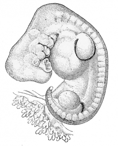
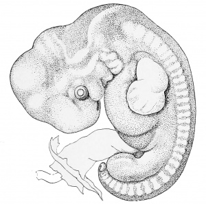
Fig. 79. Human three Days. W. His.) at, Anterior limb-bud. B. S, Body stalk. Op, Optic vesicle. //, Posterior limb bud. iv, Fourth ventricle. /, Mandibular process. 2, II void arch, 3, 4, Third and fourth gill arches.
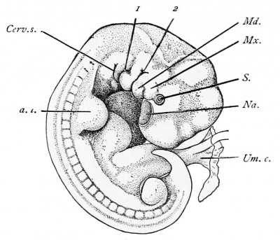
Fig. 80. Human Embryo of 7 mm. X 8 diams. — (After F. P. Mall.) a.l, Anterior limb bud. Cerv.s, Cervical sinus. MJ, Mandibular process. Mx. Maxillary process.
Na, Nasal pit. S, Eye. Uin.c, Umbilical cord.
1, 2, Kirst and second "ill clefts.
Human Embryo in the Eleventh Stage. Appearance of Limb=buds. Twenty three Days
In this stage the embryo is found very much rolled up, so that the head and tail come very close together (Fig. 79). It is further characterized by the appearance of four protuberances, two upon each side of the body, in which we can recognize the so-called limb-buds or anlages of the extremities. As shown in the figure, the dorsal outline of the embryo describes more than a complete circle. The embryo has a marked spiral twist, the head being bent to the right, the tail to the left. The bending of the body is especially marked at the region of the mid-brain or head bend, and at the posterior limit of the hind-brain. The anlages of both pairs of limbs have appeared, but that of the leg is smaller than that of the arm. The position of the optic vesicles can be clearly recognized by a small external protuberance. The four external gill clefts still show clearly. Between the first gill cleft and the mouth lies the mandibular process, i ; between the first cleft and the second lies the hyoid or second branchial arch. The third and fourth branchial arches are also distinct, but the fifth branchial arch, or that behind the fourth gill cleft, no longer appears externally, but has been turned inward, this turning-in marking the first step toward the development of the cervical sinus. The tail overlaps the pericardial chamber. The body-stalk, B. S., comes off on the left side of the embryo, so that when the specimen was obtained it lay with its left side against the chorion, the body-stalk being quite short. The specimen was derived from a chorionic vesicle measuring 25 X 30 mm. The greatest length of the embryo was 4 mm.
Human Embryo of Twenty six Days
The embryo figured (Fig. 80) was described by Mall. Another specimen almost identical has been studied by H. Piper. At this stage (Fig. 80) the embryo is flexed upon itself, forming almost a complete circle. The limb-buds have increased in size. The posterior branchial arches are in process of disappearance by invagination on the ectodermal side to form the cervical sinus. The three branchial arches are visible on the right side, as shown in the figure, but four can still be seen on the left. The body-stalk is now merged in the formation of the true umbilical cord (compare page 109). The head is bent down so as partially to overlap the pericardial region. On its ventral side it shows a large, shallow depression, Na, which is the anlage of the future nasal cavity. The eye is marked by the commencing formation of the lens, S. The mandibular process, Md, represents the lower boundary of the mouth, but it has an extension toward the high nasal pit. This extension, Mx, is known as the maxillary process. The first and second gill clefts, 1 and 2, can be easily seen, and that which might at first be mistaken for the third, Cerv. s, is really the opening of the cervical sinus, in which the gill clefts are partly buried. Between the first andsecond gill cleftsappearsthesecondorhyoidbranchialarch. Twentyfour segments are indicated externally on the right-hand side of the embryo shown in the figure. Between the head and the anterior limb runs the large pericardial region. Between the anterior limb, a. I, and the umbilical cord, Urn. c, another protuberance shows externally, due to the development of the liver.
Fig. 81. Human Embryo 7.5 mm In Maximum Length. — {After W. His.)
Human Embryos of Twenty-seven to Twenty-eight Days
Embryos of this age are characterized by the extreme development of the neck bend (compare Fig. 83). As is well illustrated in figure 81, the apex of the neck bend forms, as it were, the summit of the embryo, the greatest length of which is from 7 to 8 mm. As it changes from the twenty-sixth day we note especially the increased distinctness of the nasal pit and of the still open invagination of the ectoderm to form the lens of the eye, and, still more, the great deepening of the cervical sinus. Although the cervical sinus is an ectodermal structure, yet its formation is due to modifications of the gill arches, the third, fourth, and fifth of which become pushed inward. At the stage we are considering, the process of invagination, though far advanced, is not completed. It results in the formation of a deep fissure on each side of the neck, the general nature of which may be understood from the accompanying figure 82. The fifth arch is turned in first, the fourth second, and the third last. The sinus when fully formed is very narrow, so that the surfaces of the branchial arches come in contact with the opposite wall of the sinus. The ectoderm on two sides unites and the sinus thus becomes closed, and is for a time represented by an epithelial cord connected with the epidermis or superficial ectoderm. So far as known, the cervical sinus entirely disappears, but its abnormal persistence may account for certain cysts occurring pathologically in the neck. In all parts of the embryo of twenty-eight days there is an obvious advance upon the conditions found at twenty-six days. Comparison with one another of embryos in this stage shows that there is considerable variation, especially as to the nature and degree of curvature of the back, in consequence of which specimens differ in general form and in maximum length, though agreeing closely in structure.
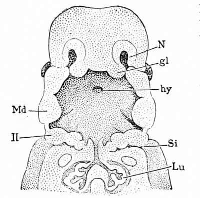
Fig. 82. Reconstruction of the Pharyngeal Region of a Human Embryo of 11.5 mm. N, Nasal pit. gl, Processus globularis. hy. Hypophysis. Si, Sinus cervicalis. Lit, Lung. Md, Mandible. II, Second branchial arch. X I0 diams. — (After W. His.)
Embryos of the Second, Third, and Fourth Months
The following descriptions and figures illustrate the external form, no attempt being made to describe the anatomical structures.
Twenty-eight to Thirty Days
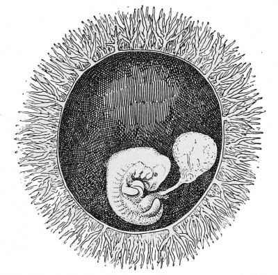
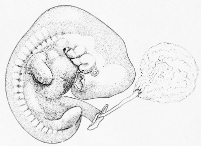
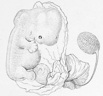
Embryos of 8 to 10 mm. Figs. 83 and 84 show an embryo of 9.0 mm. There is a general resemblance to the stage last described, but the following changes may be especially noted, the first and most conspicuous being the modification of the limb-buds, which have increased greatly. The end of each bud has become somewhat flattened, rounded in outline, and marked off by a slight constriction from the portion of the limb-bud near the body. The expanded, flattened distal portion of the limb is the anlage of the hand in the upper limb, of the foot in the lower. The position of the limbs is also characteristic, the arm projecting in a line nearly parallel with the axis of the body, while the leg stands out at a wide angle. The cervical sinus has entirely disappeared externally by closure of its orifice. The nasal pit has contracted and deepened so as to have lost its original saucer-like form and more truly to deserve its name of pit. The large cavity of the fourth ventricle of the brain has become more conspicuous. The umbilical cord has increased in length. From its distal end springs the amnion, beyond which there passes out from the cord the narrow stalk of the pear-shaped yolk-sac. The cavity in the interior of the umbilical cord is a prolongation of the coelom of the embryo, and through this coelom the yolk-stalk takes its course. It bears blood-vessels which ramifv over the surface of the yolk-sac proper. Unfortunately in the figure the amnion is not shown.
Fig. 83. Human Ovum with Embryo of 9.8 mm. The Chorion Has Been Partly Removed to Show the Embryo. X 2 diams. — {Minot Collection, 275.)
Fig. 84. Embryo of hie Preceding Figure. • S diams.
Thirty-one to Thirty-two Days
Embryos of 10 to 12 mm. As a typical specimen of this stage we may take the embryo designated by His as Br. i , which measured 1 1 mm. As compared with the previous stage (Fig. 85), the back has straightened out somewhat, though the lower end of the body is still rolled over. The head has risen and increased considerably in size. Between the end of the region of the hind-brain and the level of the arm the dorsal outline has become slightly concave. This concavity His designated the " Nackengrtibe." The first gill cleft, owing to the completed closure of the cervical sinus, is the only one visible externally. It is the anlage of the external auditory meatus. It is separated from the mouth by a prominent mandibular arch. On the cephalic side of the mouth the maxillary process has become more prominent, but the two portions of the maxilla do not yet meet in the median line. The primitive segments are still marked externally. The limbs show indications of their tripartite division, the fore-limb being more advanced than the hind-limb. The division of the digits of the hand is just indicated. The abdomen bulges out, showing to the growth of the liver. There is a true tail, which is now near its maximum development. The umbilical cord has lengthened and shows the commencement of its spiral twisting. The amnion springs from the end of the cord, leaving only a short stretch of the body-stalk between the cord proper and the chorion. The amnion envelops the embryo closely. In embryos slightly older than these the changes in form above mentioned have progressed further. The body is straighter, the head is larger, and has risen so as to be about at right angles to the body. The concavity (Nackengrube) below the hind-brain in the outline of the neck is more marked. The limbs are longer, the fingers more distinct. Where the mandibles meet in the median line, the separation of lip and chin has begun.
Thirty-six Days
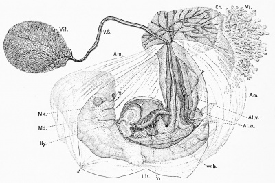
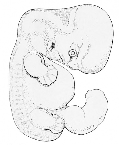
Embryos of 14 mm. The correlation of age and size for this stage cannot be recorded as absolute, but we may say that embryos of this length are about five weeks old. The body is now nearly straight (Figs. 86 and 87). The limbs project beyond the outline of the body in profile views. The ventral outline, owing to the large size of the heart and liver, is very protuberant, and at this stage we find that the portion of the umbilical cord adjoining the embryo is greatly enlarged, owing to the distention of its coelom, so that a large cavity is furnished in which there are always found, as indicated in figure 84, several coils of intestine. This protrusion of a portion of the intestinal canal, and sometimes even of a small portion of the liver, into the extra-embryonic coelom of the umbilical cord is a constant phenomenon. It begins at a somewhat earlier stage and continues for a considerable period. This curious condition has been observed in many different kinds of mammals in the corresponding stage. Later on, the viscera are entirely withdrawn from the umbilical cord and the cavity itself is wholly obliterated. Figure 87 is inserted to illustrate the relation of the foetal appendages to the embryo at this stage and in slightly younger embryos. The figure shows clearly that the umbilical cord is a hollow prolongation of the body-wall or somatopleure of the embryo, and that the amnion springs from its distal end. The yolk-stalk, V.S, is very long and narrow. Its entodermal cavity is obliterated. It is the representative of the original broad connection between the yolk-sac and the entodermal cavity of the embryo, although it is now only a small appendage of a loop of the intestine. It bears the blood-vessels which run from the embryo and ramify upon the yolk-sac. On the caudal side of the umbilical cord we find the tissue of the original body-stalk in which runs the allantoic vein, Al. v, and the two allantoic arteries, Al.a, which ramify upon the chorion, Ch.
Fig. 86. Human Embryo of about 14 mm. 5 diams.
Fig. 87. Human Embryo of about Thirty-five Days.
Ys, Yolk-sac. V.S, Vitelline stalk. Am, Amnion. C/i, Chorion. Vi, Cliorionic villi vein. Al.n, Allantoic artery. W.b, Wolffian body. n, Main vein through liver. Hyoid arch. Md, Mandibular arch. Mx, Maxillary process. 0p, Olfactory pit, AI., Allantoic Lu, Lung, ( After Cost,: )
Thirty-eight Days
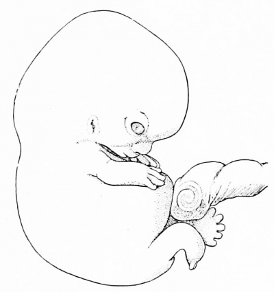
Embryo of 16 mm. in a chorionic vesicle of 45 by 40 mm. The age of this specimen (Fig. 88) is known by estimate only. This stage represents the transition of the embryo to the foetus, because after the fortieth day the form is distinctly human. The head has risen considerably, and the back has straightened still more, the rectangular neck bend thus becoming emphasized. The body has become still more protuberant on the ventral side, and in side views the arms no longer reach to the outline of the body.
Fig. 88. Human Embryo of about 16 mm. X 5 diatm.— {After W. His.)
Forty Days
Embryos of 19 mm. The head has risen far toward its definite position, with the result of a very rapid apparent increase in the length of the embryo. The change of position of the head results in bringing the mid-brain finally directly above the hind-brain, the embryo being conceived as having the body vertical. During the elevation of the head the concavity (N ackengrube) at the back of the neck is gradually obliterated. In both head and rump the external modeling, which in earlier stages indicated more or less the position of the internal organs, has become blurred, and in the next stage is found to have nearly or quite disappeared. The maxillary processes have met and united in the median line. The anlages of the eyelids have developed. The concha of the ear is indicated. The arm reaches beyond the heart; the fingers appear as separate outgrowths.
Fifty Days
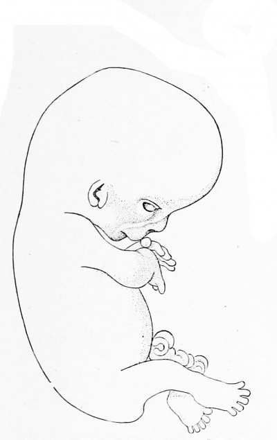
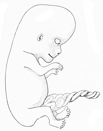
Embryo of 21 mm. The author has a fair specimen which came into his possession with no history whatever, but it agrees very closely with His's embryo Ltz, of which he fixes the probable age as just over seven weeks. The head is nearer its final position than in figure 88, and relatively larger in proportion to the body. In the eye, cornea and conjunctiva are clearly separated; the face has the foetal form, the nose, mouth, and chin being fully marked off. The arms are clearly divided into upper and lower segments; the five digits are well developed ; the hands rest over the heart and nearly touch one another. In the specimen figured the outline of the abdomen is abnormal. The leg ' shows the tripartite division ; the toes are just beginning to be free, but the hind-limb is much less advanced than the fore-limb. The tail is still a freely projecting appendage. Fifty-three Days. — Embryo of 22 mm. The specimen 1 Fig. 89) is probably not quite normal, but, except for the extreme and unusual curvature of the back, it agrees closely with His's embryo Zw, which is figured by him as a normal embryo of presumably about seven and one-half weeks. The specimen was received in 1884 with the following history: " Menstruation began January 26th. February and March slight show ever}- few days. Abortion March 30th," which is insufficient to determine the age. As compared with the hist stage, there are comparatively few changes of external form; the most noteworthy are perhaps the increased development of the legs and feet and the commencing disappearance of the free tail. At this time the protrusion of the coils of the intestine into the coelom of the umbilical cord is about at its maximum.
Fig. 89. Human Kmhryo of 22 mm. diams.
Sixty Days
Embryo of 28 mm. The specimen figured resembles closely in form, though it is larger, His's embryo Wt, which he has determined as a normal embryo of about eight and one-half weeks. The present specimen (Fig. 90) came with no data. The head is still larger in proportion to the body than in figure 89. The face shows the two lines, which, as seen in profile, mark the two ridges which run over the cheek, one alongside the nose to the corner of the mouth, the other from the eye ; these ridges are highly characteristic of the ninth week, and traces of them not rarely persist in the adult physiognomy. The limbs have grown considerably, the hands being lifted toward the face ; at the elbow there is a considerable bend ; the toes are all free and the soles of the feet are turned toward one another. The tail has disappeared as a free appendage. The external genitalia are considerably developed ; the clitoris-penis projects some distance.
Fig. 90. Human Embryo of 2S mm. 3 (Hams.)
Fig. 91. Human Embryo of 32 mm. X 3 diams.
Sixty-four Days
Embryo of 32 mm. The specimen (Fig. 91) came with the following history: "January 4, 1886, last flow began; March 13, 1886, abortion"; between these two dates are sixty-eight days; but as the flow took place, conception probably occurred after menstruation, therefore if we deduct four days, making the age sixty-four days, we shall probably be not far wrong. It will be noticed that the head has not yet assumed its final angle with the body. On the other hand, the protuberance of the abdomen is much reduced, so that the body as a whole has begun to have a more slender form than in earlier stages. In this specimen the eyelids have not even begun to meet ; in another they have met (Fig. 92), except just in the center, where is still a loophole. This specimen was brought with the statement that it was just sixty days. The endeavor to get the exact data was unsuccessful. The large size, 34 mm., and advanced development of the embryo lead us to consider the age given as erroneous, and to believe the true age to be perhaps sixty-seven days.
Fig. 92. Human Embryo of 34 mm. View of Face. 3 diams.
Fig. 93. Human Embryo of 55 mm. Seventy five Days.
Seventy-five Days
Embryo of 55 mm. We figure next (Fig. 93) a foetus concerning which I possess no data. Comparison with embryos of two and three months leads me to place it a little under half-way between them. The specimen has essentially the configuration of the young child ; but the head is very large and the body slender ; the position of the limbs is typical ; the upper arm is bent down, the forearm extends toward the chin; the knee is bent so as to throw the foot toward the median line ; the soles of the feet are placed obliquely facing one another; the anlages of the nails can be recognized on both the fingers and toes.
Embryos of the eleventh and twelfth weeks are very rarely obtained. I have never had a normal one of this period with data to determine the age.
Three Months
Embryos of 78 to 80 mm. In my experience there is no other age at which abortion of normal embryos occurs so frequently as at three months, and I possess a number of specimens of this age, which agree very closely with one another in size and form. The foetus drawn in figure 94 may be taken to represent accurately the appearance of the human embryo at three months. The position of the limbs is typical for this age, but there are slight variations, in that the hands, one or both, may project more and be higher or lower; usually the right foot lies in front of the left, but not always. Figure 95 gives the front view of face of the same embryo to show the closed eyelids, the broad triangular nose, the thick lips, and the pointed chin.
Three and One-half Months
Embryos of 108 to no mm. I have several specimens which represent this age. I figure two of them, one to show the natural attitude (Fig. 96) in utero, the other to show the natural attitude assumed by the embryo when released from its membranes. The first specimen came to me with no history, but as it is certainly a little larger than several other foetuses of about one hundred and six days, it is probably a little older. The head is bent forward (Fig. 96); the back is curved; the arms and legs are both raised toward the face; the right leg is nearly straight, so that the toes are brought against the forehead, while the left leg is bent at the knee, bringing the left foot against the right thigh. In this attitude the embryo fills out as perfectly as possible an oval space, and fits, therefore, the cavity of the uterus. The second specimen (Fig. 97) shows the attitude assumed by the embryo when free, and proves that the position in utero (Fig. 96) is a constrained one. This foetus was received November 30, 1883. The delivery took place on the morning of that day, and the last menstruation had ceased one hundred and six days previously; the remarkably fresh condition of the foetus indicated that it had been dead only a very short time, so that we cannot be far wrong in putting its exact age at one hundred and six days.
Fig. 94. Human Embryo of 78 mm. Three Months.
Fig. 95. Front View of the Face of the Embryo Shown in Fig. 94.
Fig. 96. Human Embryo or 120 mm (One Hundred and Ten Days)
Fig. 97. Human Embryo oi 118 mm Hundred and Six Days.
Four Months
Embryo 155 mm. The foetus shown in figure 98 came in a very fresh condition, January 2,1887, with the statement: "Conception said to have taken place September 1, 1886; foetus came away January 2, about noon." The embryo is typical in size and development for four months, except that the crown is higher than usual, and the antero-posterior diameter of the head somewhat below the average.
The natural attitude in utero is similar to that of figure 96 ; the attitude shown is that assumed by the foetus when released from the membranes.
Fig. 98. Human Embryo of 155 mm. One Hundred and Twenty-three Days.
| Historic Disclaimer - information about historic embryology pages |
|---|
| Pages where the terms "Historic" (textbooks, papers, people, recommendations) appear on this site, and sections within pages where this disclaimer appears, indicate that the content and scientific understanding are specific to the time of publication. This means that while some scientific descriptions are still accurate, the terminology and interpretation of the developmental mechanisms reflect the understanding at the time of original publication and those of the preceding periods, these terms, interpretations and recommendations may not reflect our current scientific understanding. (More? Embryology History | Historic Embryology Papers) |
Cite this page: Hill, M.A. (2024, April 19) Embryology Book - A Laboratory Text-Book of Embryology 3 (1903). Retrieved from https://embryology.med.unsw.edu.au/embryology/index.php/Book_-_A_Laboratory_Text-Book_of_Embryology_3_(1903)
- © Dr Mark Hill 2024, UNSW Embryology ISBN: 978 0 7334 2609 4 - UNSW CRICOS Provider Code No. 00098G


