Book - A Laboratory Text-Book of Embryology (1903)
| Embryology - 20 Apr 2024 |
|---|
| Google Translate - select your language from the list shown below (this will open a new external page) |
|
العربية | català | 中文 | 中國傳統的 | français | Deutsche | עִברִית | हिंदी | bahasa Indonesia | italiano | 日本語 | 한국어 | မြန်မာ | Pilipino | Polskie | português | ਪੰਜਾਬੀ ਦੇ | Română | русский | Español | Swahili | Svensk | ไทย | Türkçe | اردو | ייִדיש | Tiếng Việt These external translations are automated and may not be accurate. (More? About Translations) |
Minot CS. A Laboratory Text-Book Of Embryology. (1903) Philadelphia:P. Blakiston's Son & Co.
| Online Editor |
|---|

|
| Historic Disclaimer - information about historic embryology pages |
|---|
|

A Laboratory Text-Book Of Embryology
Dedication
|
|

Preface
The accompanying volume is intended primarily for the use of students, taking a practical laboratory course in Embryology. The author's experience has led him to believe that the study of carefully selected sections of embryos, accompanied by directions and explanations of the significant structures in each section, offers many advantages. This conviction has determined the arrangement of the book. Attention is given chiefly to such points as serve to explain adult anatomical relations, to illustrate general biological principles, and to afford insight into pathological processes.
Portions of the text and many of the figures have been borrowed from the author's "Human Embryology." The woodcuts in Chapter IV were made by C. L. Albert Probst, of Braunschweig, after drawings by Dr. E. A. Locke. To both of these artists the author is indebted for their beautiful work. Much assistance has been rendered by Dr. F. T. Lewis, of the Harvard Medical School, to whom special acknowledgments are due for the reconstructions of the anatomy of the pig embryo of twelve millimeters and for invaluable help in the correction of the proofs.
Many of the illustrations are from the Harvard Embryological Collection, without which this work could not have been undertaken. The number of the embryo and of the section is given for all such illustrations.
The title was suggested by Dr. W. T. Porter's "A Laboratory Text-book of Physiology," and is adopted with his approval.
The author requests those who use this book to communicate to him any suggestions, which their experience may lead to, for improving it, in case it meets with sufficient favor to call for a new edition.
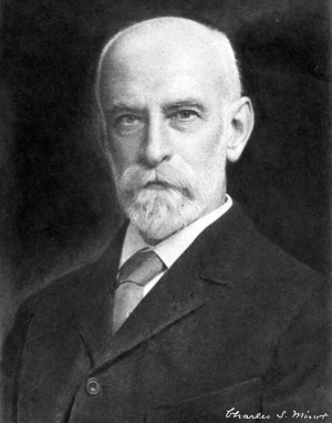
Charles Sedgwick Minot.
Cortina d'Ampezzo, August, 1902.
| 1903 Book Review by Frank R. Lillie |
|---|
| Review - A Laboratory Text-Book of Embryology by Charles Sedgwick Minot Review by Frank R. Lillie |
| Science, New Series, Vol. 17, No. 438 (May 22, 1903), pp. 817-819
The first furnishes the student with the only complete summary of the embryology of ver tebrates published since Balfour’s ‘Comparative Embryology’ appeared in 1881; in it the enormous mass of literature since that date is fully digested, and the results are presented in connected form, so that it may serve as a new starting point for the student of vertebrate embryology. In the general part of their text-book Korschelt and Heider furnish the long-promised completion of the special parts by a full treatment of the structure, origin, maturation. and fertilization of the germcells, and the experimental embryology of invertebrates. McMurrich’s book is an excellent brief treatise for the medical student of the main facts of human embryology. Minot’s new book is a laboratory guide, mainly in the embryology of mampnals. Thus the teacher of embryology is furnished with a fairly complete ‘ up-to-date ’ equipment of the literature in his subject for the use of his students. Minot’s laboratory text—book is written from the standpoint of the anatomist rather than of the biologist. In this point of view lie both its limitations and its excellencies. It is the outgrowth of the actual experience of one of the best known of the teachers of embryology, and hence is strongly individualized. Too much praise can not be‘ given to’ ‘the large number of new and beautifully executed illustrations; a number of fine figures are transferred from the original sources to a text-book for the first time, and only the best of the stock illustrations of other textbooks are retained. The book is, indeed, built up around the illustrations, and the text often suifers by comparison. The figures‘ of reconstructions of the pig embryo of twelve millimeters neck-length are especially fine, as are also the figures of sections of this embryo and of other sizes. In all of these figures there is the most painstaking reproduction of details, and the accuracy of the work is equaled only by its beauty. The illustrations of the two stages of the chick embryo studied are also noteworthy for accuracy and finish.
It is not, however, incumbent on the user to follow the order of the book, for the description of each stage is complete in itself. Those who use it, therefore, will probably follow their own ideas of order; and the practical parts can be unhesitatingly recommended as excellent in themselves. The chapter on methods is rather brief, but good as far as it goes; a larger number of formulae of killing fluids and stains and the methods of using them would undoubtedly be an improvement. The three general introductory chapters are best, as is to be anticipated, in the parts allied to the author’s own province. of work; thus the ‘law of genetic restriction’ is well expressed and discussed; and- the third chapter on thehuman embryo is by far the best brief outline of human development known to me. On the other hand, the unfortunate student who might have to derive his ideas on karyokinesis and on the maturation of the ovum from the vague accounts of this book, would probably conclude, for his own peace of mind, that these subjects are not of much importance after all. As regards the germ-layers in mammals we read on p.59 that ‘it is probable that
Such sweeping statements as the two following are at least regrettable: ‘It is fortunate for our comprehension of embryological processes that we are already able to say that Roux’s hypothesis is erroneous,’ referring to the mosaic theory of the segmented ovum; we know, as a matter of fact, that certain ova (e. g., of Otenophoras) are true mosaics; and the general bearing of recent embryological results is that all ova are more or less mosaic, in an unstable fashion. On page 41 we are told that Weismann’s hypotheses are ‘complicated’ and ‘useless’; not to mention the stimulus they have given to research, this sounds strange on the eve of a general rehabilitation of such hypotheses in connection with Mendel’s laws of inheritance.
FRANK R. LILLIE. |
Contents
- General Conceptions - The Vertebrate Type of Structure, The Principal Modifications of the Vertebrate Type, Definition of Anlage, A Summary of Embryological Development, Cytomorphosis, Comparison of Larval and Embryonic Types of Development, Germ-layers, The Relations of Surface to Mass, The Law of Unequal Growth, Germ-cells, The Theory of Heredity, The Law of Recapitulation.
- The Early Development of Mammals - The Spermatozoon, The Fully Grown Ovum before Maturation, Ovulation, The Maturation of the Ovum, Impregnation of the Ovum, Segmentation of the Ovum, The Blastodermic Vesicle, The Embryonic Shield, Origin of the Mesoderm, The Primitive Axis, The Notochordal Canal, The Notochord, The Ultimate Fate of the Notochord, The Origin of the Nervous System, The Structure of the Medullary Canal, The Early History of the Mesoderm, Somatopleure and Splanchnopleure, The Embryonic coelom, The Mesenchyma, The Germ-cells, The Yolk-sac, The Origin of the Blood-vessels and Blood, The Blood-corpuscles, The Origin of the Heart, The Germinal Area, The Main Vessels of the Area Vasculosa, The Liver, The Oral and Anal Plates, The Excretory Organs, The Allantois, The Trophoblast, The Growth of the Embryo, The Umbilical Cord.
- The Human Embryo - Calculation of the Age of the Human Embryo, The Classification of the Early Stages, Hypothetical Development of the Blastodermic Vesicle in Primates, Relations of the Embryo to the Uterus, Ovum of a Monkey in the Second Stage, Human Embryo in the Second Stage, Embryo of a Gibbon in the Third Stage, Human Embryo in the Fourth Stage with Medullary Plate, Human Embryo in the Fifth Stage with Open Medullary Groove, Human Embryo in the Sixth Stage with Medullary Canal, Human Embryo in the Seventli Stage with One Gill-cleft, Human Embryo in the Eighth Stage with Two Gill-clefts, Human Embryo in the Ninth Stage with Three Gill-clefts, Human Embryo in the Tenth Stage with Four Gill-clefts, Human Embryo in the Eleventh Stage with Limb-buds, Human Embryo of Twenty-six Days, Human Embryo of Twenty-eight Days, Embryos of the Second, Third, and Fourth Months.
- Study of Pig Embryos - Methods of Obtaining Embryos, The Making of Serial Sections, Selection of the Planes of Section and the Stages for Practical Study, The Study of the External Form, Pig Embryo of 10 mm, Pig Embryo of 12 mm General Anatomy, Pig Embryo of 15 mm (External Form), Pig Embryo of 20 mm (External Form), Pig Embryo of 12 mm (Studied in Sections), The Study of Transverse Sections, Section through the Upper Part of the Otocyst, Section through the Lower Part of the Otocyst, Section through the First Gill-cleft, Section through the Second Gill-cleft, Section through the Third Gill-cleft, Section through the Fourth Gill-cleft, Section through the Anterior Limbs and Heart, Section to show the Brachial Plexus, Section through the Stomach and Liver, The Study of Sagittal Sections, Median Section of the Head, Section of the Head through the Principal Ganglia, The Study of Frontal Sections, Section through the Trigeminal Roots, Section through the Acusttco-facial Ganglion, Section through the Otocyst, Section through the Dorsal Vertebrae, Pig Embryo of 9 mm. (Studied in Sections), Transverse Section through the Branchial Arches, Sagittal Section to the Right of the Median Plane, Frontal Section through the Mid-brain and Fore-brain, Frontal Section through the Umbilical Opening, Frontal Section through the Second and Third Gill-clefts, Pig Embryo of 6 mm. (Studied in Sections), Pig Embryo of 17 mm (Studied in Sections), Transverse Section through the Lungs, Section through the Wolffian Body and Genital Gland, Section through the Kidney, Frontal Section of the Umbilical Cord, Pig Embryo of 20 mm (Studied in Sections), Transverse Section through the Snout, Transverse Section through the Lower Part of the Neck, Transverse Section through the Lungs, Transverse Section through the Posterior Limbs, Transverse Section through the Mammary Anlage, Sagittal Section through the Right Lung and Kidney, Frontal Sections of the Head, Pig Embryo of 24 mm (Studied in Sections), Frontal Section through the Eye, Median Sagittal Section.
- Study of Young Chick Embryos - Method of Obtaining Embryos, Embryo Chick with Twenty-four Segments, Embryo Chick with Twenty-eight Segments, The Study of Transverse Sections, Horizontal Section, Histological Differentiation of Chick Embryo with Three Gill-clefts, Embryo Chick with Seven Segments, Examination in the Fresh State, Examination after Hardening, Comparison with a Rabbit Embryo, Longitudinal Section, Study of Transverse Sections.
- Study of the Blastodermic Vesicle and the Segmentation of the Ovum - Method of Obtaining Blastodermic Vesicles from the Rabbit, Study of Rabbit Blastodermic Vesicles in Alcohol, The Maturation, Fertilization, and Segmentation of the Ovum in White Mice, The First Polar Globule, The Second Polar Globule, The Single Polar Globule, Fertilization.
- Study of the Uterus and the Foetal Appendages in Man - Histology of the Uterus, Menstruation, Decidua Menstrualis, The Pregnant Uterus: the Two Stages, The Human Uterus Three Months Pregnant, Human Uterus Seven Months Pregnant, Decidua Vera of the First Stage in Section, Decidua Reflexa of the First Stage, Decidua Vera and Chorion Leeve of the Second Stage, The Placenta in Situ, Decidua Serotina at Seven Months, The Human Placenta, Histology of the Human Chorion, The Chorion with Trophoblast, The Chorionic Villi The Structure of the Amnion, The Umbilical Cord, The Structure of the Human Yolk-sac.
- Methods - Measuring the Length of Embryos, Methods of Reconstruction, Reconstruction of Drawings, Reconstruction with Wax-plates, Directions for Orienting Serial Sections, Microtomes, Methods of Hardening and Preserving, Preservation in Alcohol, Directions for Imbedding Specimens to be Microtomed, Method of Mounting Paraffin Sections, Methods of Staining.
| Historic Disclaimer - information about historic embryology pages |
|---|
|
Cite this page: Hill, M.A. (2024, April 20) Embryology Book - A Laboratory Text-Book of Embryology (1903). Retrieved from https://embryology.med.unsw.edu.au/embryology/index.php/Book_-_A_Laboratory_Text-Book_of_Embryology_(1903)
- © Dr Mark Hill 2024, UNSW Embryology ISBN: 978 0 7334 2609 4 - UNSW CRICOS Provider Code No. 00098G