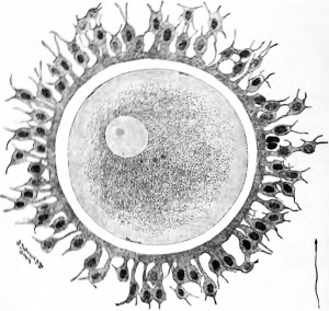
Book - A Laboratory Manual and Text-book of Embryology
of
EMBRYOLOGY
By
CHARLES WILLIAM PRENTISS, A. M., Ph. D.
LATE PROFESSOR OF MICROSCOPIC ANATOMY, NORTHWESTERN UNIVERSITY MEDICAL SCHOOL, CHICAGO
Revised and Extensively Rewritten by
LESLIE BRAINERD AREY, Ph. D.
ASSOCIATE PROFESSOR OF ANATOMY IN THE NORTHWESTERN UNIVERSITY MEDICAL SCHOOL, CHICAGO
SECOND EDITION, ENLARGED
WITH 388 ILLUSTRATIONS
MANY IN COLOR
PHILADELPHIA AND LONDON
W. B. SAUNDERS COMPANY
1918
Copyright, I915, by W. B. Saunden Company. Reprinted August. 1915. Revised, entirdy
reset, reprinted, and recopyrighted October. 1917
Copyright. 1917. by W. B. Saunders Company
Reprinted July. 1918
Charles William Prentiss (1874 - 1915)
| Historic Disclaimer - information about historic embryology pages |
|---|
| Pages where the terms "Historic" (textbooks, papers, people, recommendations) appear on this site, and sections within pages where this disclaimer appears, indicate that the content and scientific understanding are specific to the time of publication. This means that while some scientific descriptions are still accurate, the terminology and interpretation of the developmental mechanisms reflect the understanding at the time of original publication and those of the preceding periods, these terms, interpretations and recommendations may not reflect our current scientific understanding. (More? Embryology History | Historic Embryology Papers) |
Preface to the Second Edition
The untimely death of Professor Prentiss has made necessary the transfer of his Embryology into other hands. In this second edition, however, the general plan and scope of the book remain unchanged although the actual descriptions have been extensively recast, rewritten, and rearranged. A new chapter on the Morphogenesis of the Skeleton and Muscles covers briefly a subject not included hitherto. Forty illustrations replace or supplement certain of those in the former edition.
In preparing the present manuscript a definite attempt has been made to render the descriptions as clear and consistent as is compatible with brevity and accuracy. It has likewise been essayed to properly evaluate the embryological contributions of recent years, and, by incorporating the fundamental advances, to indicate the trend of modern tendencies. Since no page remains in its entirety as originally penned by Professor Prentiss, the reviser must assume full responsibility for the subject-matter as it now stands.
It is hoped that those who read this text will co-operate with the writer by freely offering criticisms and suggestions.
L. B. A.
Chicago
Preface
This book represents an attempt to combine brief descriptions of the vertebrate embryos which are studied in the laboratory with an account of human embryology adapted especially to the medical student. Professor Charles Sedgwick Minot, in his laboratory textbook of embryology, has called attention to the value of dissections in studying mammalian embryos and asserts that "dissection should be more extensively practised than is at present usual in embryological work" The writer has for several years experimented with methods of dissecting pig embryos, and his results form a part of this book. The value of pig embryos for laboratory study was first emphasized by Professor Minot, and the development of my dissecting methods was made possible through the reconstructions of his former students. Dr. F. T. Lewis and Dr. F. W. Thyng.
The chapters on hiunan organogenesis were partly based on Keibel and Mall's Human Embryology. We wish to acknowledge the courtesy of the publishers of Kollmann's Handatlas, Marshall's Embryology, Lewis-Stohr's Histology and McMurrich's Development of the Human Body, by whom permission was granted us to use cuts and figures from these texts. We are also indebted to Professor J. C. Heisler for permission to use cuts from his Embryology, and to Dr. J. B. De Lee for several figures taken from his *' Principles and Practice of Obstetrics." The original figures of chick, pig and hiunan embryos are from preparations in the collection of the anatomical laboratory of the Northwestern University Medical School. My thanks are due to Dr. H. C. Tracy for the loan of valuable human material, and also to Mr. K. L. Vehe for several reconstructions and drawings.
C. W. Prentiss.
Northwestern University Medical School.
Contents
- Chapter I. The Germ Cells
- The Ovum
- Ovulation and Menstruation
- The Spermatozoon
- Mitosis and Amitosis
- Maturation
- Fertilization
- Heredity and the Determination of Sex
- Chapter II. Cleavage and Formation of the Germ Layers
- Cleavage in Amphioxus, Amphibia, Birds, and Reptiles
- Cleavage in Mammals
- Origin of the Ectoderm and Entoderm
- Origin of the Mesoderm, Notochord and Neural Tube
- The Notochord
- Chapter III. The Study of Chick Embryos
- Chick Embryo of Twenty Hours 36
- Chick Embryo of Twenty-five Hours (7 Segments)
- Transverse Sections
- Chick Embryo of Thirty-eight Hours (17 segments)
- General Anatomy
- Transverse Sections
- Derivatives of the Germ Layers
- Chick Embryo of Fifty Hours (27 segments)
- General Anatomy
- Transverse Sections
- Chapter IV. The Fetal Membranes and Early Human Embryos
- Fetal Membranes of the Pig Embryo
- Umbilical Cord
- Early Human Embryos and Their Membranes
- Anatomy of a 4.2 mm. Human Embryo
- ge of Human Embryos
- Chapter V. The Study of Pig Embryos
- The Anatomy of a 6 mm. Pig Embryo
- External Form and Internal Anatomy
- Transverse Sections
- The Anatomy of 10-12 mm. Pig Embryos
- External Form and Internal Anatomy
- Transverse Sections
- Chapter VI. Methods of Dissecting Pig Embryos: Development of the Face, Palate, Tongue, Teeth and Sauvary Glands
- Directions for Dissecting Pig Embryos
- Dissections of 18-35 mm. Embryos
- Development of the Face
- Development of the Hard Palate
- Development of the Tongue
- Development of the Sialivary Glands
- Development of the Teeth
- Chapter VII. Entodermal Canal and its Derivatives
- Pharyngeal Pouches and their Derivatives
- Thyreoid Gland
- Larynx, Trachea and Lungs
- Digestive Canal
- Liver
- Pancreas
- Body Cavities, Diaphragm and Mesenteries
- Chapter VIII. Urogenital System
- Pronephros
- Mesonephros
- Metanephros
- Cloaca, Bladder, Urethra and Urogenital Sinus
- Genital Glands and Ducts
- External Genitalia
- The Uterus during Menstruation and Pregnancy
- The Decidual Membranes
- The Placenta
- The Relation of Fetus to Placenta
- Chapter IX. Vascular System
- The Primitive Blood Vessels and Blood Cells
- Development of the Heart
- Primitive Blood Vascular System
- Development of the Arteries
- Development of the Veins
- The Fetal Circulation
- The Lymphatic System
- Lymph and Hemolymph Glands
- Spleen
- Chapter X. Histogenesis
- The Entodermal Derivatives
- The Mesodermal Tissues
- The Ectodermal Derivatives
- The Nervous Tissues
- Chapter XI. Morphogenesis of the Skeleton and Muscles
- The Skeletol System
- Axial Skeleton
- Appendicular System
- The Muscular System
- Chapter XII. Morphogenesis of the Central Nervous System
- The Spinal Cord
- The Brain
- The Differentiation of the Subdivisions of the Brain
- Chapter XIII. The Peripheral Nervous System
- The Spinal Nerves
- The Cerebral Nerves
- The Sympathetic Nervous System
- Chromaffin Bodies: Suprarenal Gland
- The Sense Organs
- Index
| Historic Disclaimer - information about historic embryology pages |
|---|
| Pages where the terms "Historic" (textbooks, papers, people, recommendations) appear on this site, and sections within pages where this disclaimer appears, indicate that the content and scientific understanding are specific to the time of publication. This means that while some scientific descriptions are still accurate, the terminology and interpretation of the developmental mechanisms reflect the understanding at the time of original publication and those of the preceding periods, these terms, interpretations and recommendations may not reflect our current scientific understanding. (More? Embryology History | Historic Embryology Papers) |
Glossary Links
- Glossary: A | B | C | D | E | F | G | H | I | J | K | L | M | N | O | P | Q | R | S | T | U | V | W | X | Y | Z | Numbers | Symbols | Term Link
Cite this page: Hill, M.A. (2024, April 16) Embryology Book - A Laboratory Manual and Text-book of Embryology. Retrieved from https://embryology.med.unsw.edu.au/embryology/index.php/Book_-_A_Laboratory_Manual_and_Text-book_of_Embryology
- © Dr Mark Hill 2024, UNSW Embryology ISBN: 978 0 7334 2609 4 - UNSW CRICOS Provider Code No. 00098G

