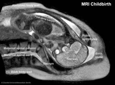
Birth MRI Movie: Difference between revisions
From Embryology
(Created page with "{| border='0px' |- | <qt>file=Birth MRI.mov|width=720px|height=550px|controller=true|autoplay=false</qt> | valign="top" | |- |} {| | [[File:Birth-_Magnetic_Resonance_Imag...") |
No edit summary |
||
| Line 1: | Line 1: | ||
{| border='0px' | {| border='0px' | ||
|- | |- | ||
| < | | <mediaplayer width='720' height='550' image="http://embryology.med.unsw.edu.au/embryology/images/3/34/Embryo_stages_002_icon.jpg">file:Birth MRI.mp4</mediaplayer> | ||
|- | |- | ||
|} | |} | ||
Revision as of 17:06, 26 February 2013
| <mediaplayer width='720' height='550' image="http://embryology.med.unsw.edu.au/embryology/images/3/34/Embryo_stages_002_icon.jpg">file:Birth MRI.mp4</mediaplayer> |
Reference
<pubmed>22425409</pubmed>| Am J Obstet Gynecol.
