Basic - Embryonic Heart Divisions: Difference between revisions
No edit summary |
No edit summary |
||
| Line 9: | Line 9: | ||
[[Image:HeartILP-draft003.jpg|thumb|right|upright=1.5|22 day embryo showing segments of heart tube]] | [[Image:HeartILP-draft003.jpg|thumb|right|upright=1.5|22 day embryo showing segments of heart tube]] | ||
From here we can see the primitive heart from a ventral view as it consists of the two tubes. These tubes fuse together (as seen in the diagram on the right) to form a single, primordial heart tube situated in the midline of the embryo, ventral to the pharynx. | From here we can see the primitive heart from a ventral view as it consists of the two tubes. These tubes fuse together (as seen in the diagram on the right) to form a single, primordial heart tube situated in the midline of the embryo, ventral to the '''pharynx'''. | ||
==Segments of the Heart Tube== | ==Segments of the Heart Tube== | ||
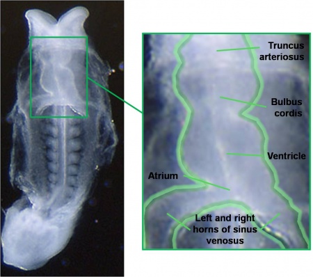
At this stage the tube already has minor constrictions within it, indicating sections of the heart tube that will form parts of the adult heart. The most caudal (tail end) segment of the heart tube is the sinus venosus which will later become the ends of the major veins carrying blood to the heart as well as parts of the atria. The next segments are the primitive atrium and primitive ventricle which will become the atria and ventricles of the adult heart. Cranial to these segments are the bulbus cordis, most of which will become the right ventricle, and the truncus arteriosus which forms the pulmonary and aortic trunks carrying blood away from the heart. | At this stage the tube already has minor constrictions within it, indicating sections of the heart tube that will form parts of the adult heart. The most '''caudal''' (tail end) segment of the heart tube is the '''sinus venosus''' which will later become the ends of the major veins carrying blood to the heart as well as parts of the atria. The next segments are the '''primitive atrium''' and '''primitive ventricle''' which will become the atria and ventricles of the adult heart. Cranial to these segments are the '''bulbus cordis''', most of which will become the right ventricle, and the '''truncus arteriosus''' which forms the '''pulmonary''' and '''aortic''' trunks carrying blood away from the heart. | ||
===Heart Tube Looping=== | ===Heart Tube Looping=== | ||
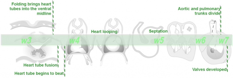
This tubular heart undergoes a process of looping during week four of development to form a shape that resembles that of the adult heart. It initially forms a C-shape (with the convex portion of the C situated on the right side of the embryo) and then an S-shape. Eventually the atria are brought backwards and upwards so that they lie cranially and behind the ventricles. The following animation outlines this process. | This tubular heart undergoes a process of looping during week four of development to form a shape that resembles that of the adult heart. It initially forms a ''C-shape'' (with the convex portion of the C situated on the right side of the embryo) and then an ''S-shape''. Eventually the atria are brought backwards and upwards so that they lie cranially and behind the ventricles. The following animation outlines this process. | ||
| Line 29: | Line 29: | ||
{| align=center | {| align=center | ||
|- style="background:lightsteelblue" | |- style="background:lightsteelblue" | ||
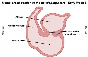
|'''1.''' Cells from the dorsal and ventral (back and front) walls of the heart grow and form two protrusions called the endocardial cushions. These grow towards each other and fuse to form the left and right atrioventricular canals. | |'''1.''' Cells from the dorsal and ventral (back and front) walls of the heart grow and form two protrusions called the '''endocardial cushions'''. These grow towards each other and fuse to form the left and right '''atrioventricular canals'''. | ||
|[[Image:HeartILP_draft_AVcanalsept.jpg|thumb|Division of the atrioventricular canal]] | |[[Image:HeartILP_draft_AVcanalsept.jpg|thumb|Division of the atrioventricular canal]] | ||
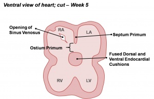
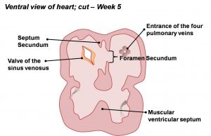
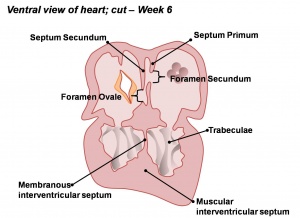
|'''2.''' Within the primordial atrium a septum (the septum primum) grows towards the endocardial cushions. The space between the cushions and septum is known as the foramen primum. As the foramen primum decreases in size a second opening forms in the septum: the foramen secundum. | |'''2.''' Within the primordial atrium a septum (the '''septum primum''') grows towards the endocardial cushions. The space between the cushions and septum is known as the '''foramen primum'''. As the foramen primum decreases in size a second opening forms in the septum: the '''foramen secundum'''. | ||
|[[Image:HeartILP_draft_atrialsept1.jpg|thumb|center|Early atrial septation]] | |[[Image:HeartILP_draft_atrialsept1.jpg|thumb|center|Early atrial septation]] | ||
|- | |- | ||
|'''3.''' A second septum (septum secundum) develops on the right of the septum primum. | |'''3.''' A second septum ('''septum secundum''') develops on the right of the septum primum. | ||
|[[Image:HeartILP_draft_atrialsept2.jpg|thumb|center|Atrial and ventricular septation]] | |[[Image:HeartILP_draft_atrialsept2.jpg|thumb|center|Atrial and ventricular septation]] | ||
|'''4.''' A primordial muscular ridge exists in the floor of the ventricle. As the left and right ventricles grow, their medial (midline) walls fuse to form the interventricular septum. | |'''4.''' A primordial muscular ridge exists in the floor of the ventricle. As the left and right ventricles grow, their medial (midline) walls fuse to form the '''interventricular septum'''. | ||
|[[Image:HeartILP_draft_aandvsept.jpg|thumb|center|Atrial and ventricular septation]] | |[[Image:HeartILP_draft_aandvsept.jpg|thumb|center|Atrial and ventricular septation]] | ||
|-style="background:lightsteelblue" | |-style="background:lightsteelblue" | ||
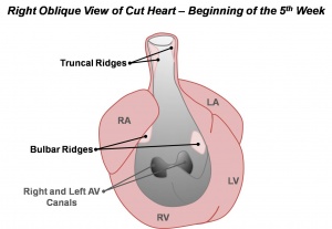
|'''5.''' Within the bulbus cordis and truncus arteriosus, which form the outflow tract, small ridges develop. They are continuous throughout the outflow tract and form a spiral shape. | |'''5.''' Within the '''bulbus cordis''' and '''truncus arteriosus''', which form the '''outflow tract''', small ridges develop. They are continuous throughout the outflow tract and form a spiral shape. | ||
|[[Image:HeartILP_draft_outflowtract1.jpg|thumb|center|Development of the conotruncal ridges]] | |[[Image:HeartILP_draft_outflowtract1.jpg|thumb|center|Development of the conotruncal ridges]] | ||
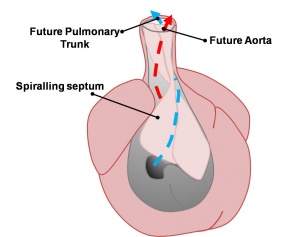
|'''6.''' As these ridges fuse they create a spiral shaped septum throughout the outflow tract. The original outflow tract is therefore separated into both the aorta and pulmonary trunk. | |'''6.''' As these ridges fuse they create a spiral shaped septum throughout the outflow tract. The original outflow tract is therefore separated into both the '''aorta''' and '''pulmonary trunk'''. | ||
|[[Image:HeartILP_draft_outflowtract2.jpg|thumb|center|Division of the outflow tract forms the aorta and pulmonary trunk]] | |[[Image:HeartILP_draft_outflowtract2.jpg|thumb|center|Division of the outflow tract forms the aorta and pulmonary trunk]] | ||
|} | |} | ||
Revision as of 10:11, 9 November 2009
| Begin Basic | Primitive Heart Tube | Embryonic Heart Divisions | Vascular Heart Connections |
| Cardiac Embryology | Begin Basic | Begin Intermediate | Begin Advanced |
From here we can see the primitive heart from a ventral view as it consists of the two tubes. These tubes fuse together (as seen in the diagram on the right) to form a single, primordial heart tube situated in the midline of the embryo, ventral to the pharynx.
Segments of the Heart Tube
At this stage the tube already has minor constrictions within it, indicating sections of the heart tube that will form parts of the adult heart. The most caudal (tail end) segment of the heart tube is the sinus venosus which will later become the ends of the major veins carrying blood to the heart as well as parts of the atria. The next segments are the primitive atrium and primitive ventricle which will become the atria and ventricles of the adult heart. Cranial to these segments are the bulbus cordis, most of which will become the right ventricle, and the truncus arteriosus which forms the pulmonary and aortic trunks carrying blood away from the heart.
Heart Tube Looping
This tubular heart undergoes a process of looping during week four of development to form a shape that resembles that of the adult heart. It initially forms a C-shape (with the convex portion of the C situated on the right side of the embryo) and then an S-shape. Eventually the atria are brought backwards and upwards so that they lie cranially and behind the ventricles. The following animation outlines this process.
<Flowplayer height="564" width="720" autoplay="false">Heart looping 005.flv</Flowplayer>
Septation of the Heart
The internal heart then undergoes significant changes in order to form the atria and ventricles of the adult heart. These can be summarised as follows:
| 1. Cells from the dorsal and ventral (back and front) walls of the heart grow and form two protrusions called the endocardial cushions. These grow towards each other and fuse to form the left and right atrioventricular canals. | 2. Within the primordial atrium a septum (the septum primum) grows towards the endocardial cushions. The space between the cushions and septum is known as the foramen primum. As the foramen primum decreases in size a second opening forms in the septum: the foramen secundum. | ||
| 3. A second septum (septum secundum) develops on the right of the septum primum. | 4. A primordial muscular ridge exists in the floor of the ventricle. As the left and right ventricles grow, their medial (midline) walls fuse to form the interventricular septum. | ||
| 5. Within the bulbus cordis and truncus arteriosus, which form the outflow tract, small ridges develop. They are continuous throughout the outflow tract and form a spiral shape. | 6. As these ridges fuse they create a spiral shaped septum throughout the outflow tract. The original outflow tract is therefore separated into both the aorta and pulmonary trunk. |
Thus the heart begins to resemble the adult heart in that it has two atria, two ventricles and the aorta forming a connection with the left ventricle while the pulmonary trunk forms a connection with the right ventricle.
| Back to the Primitive Heart Tube | Next: Vascular Connections | |
| Go to this section in the intermediate level |
