BGDB Sexual Differentiation - Fetal: Difference between revisions
mNo edit summary |
|||
| Line 28: | Line 28: | ||
| '''Plane D (midline)''' | | '''Plane D (midline)''' | ||
|} | |} | ||
==Uterus and Vagina== | ==Uterus and Vagina== | ||
| Line 44: | Line 42: | ||
{| | {| | ||
| | | width=500px|<mediaplayer width='490' height='500' image="http://embryology.med.unsw.edu.au/embryology/images/2/2d/Uterus_001_icon.jpg">File:Uterus_001.mp4</mediaplayer> | ||
| '''Female Uterus and Vagina (between week 9 and 20)''' | | '''Female Uterus and Vagina (between week 9 and 20)''' | ||
| Line 56: | Line 54: | ||
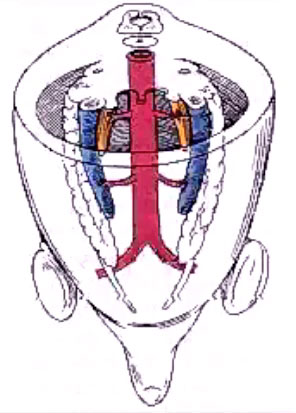
* posteriorly - uterorectal pouch (pouch of Douglas) | * posteriorly - uterorectal pouch (pouch of Douglas) | ||
* anteriorly - uterovesical pouch | * anteriorly - uterovesical pouch | ||
{{Gonad_movie_links}} | |||
|} | |} | ||
| Line 73: | Line 73: | ||
===Female External Genitalia=== | ===Female External Genitalia=== | ||
{| | {| | ||
| | | width=300px|<mediaplayer width='270' height='380' image="http://embryology.med.unsw.edu.au/embryology/images/3/37/Male_external_001_icon.jpg">File:Female_external_001.mp4</mediaplayer> | ||
| | [[External Genital Female Development Movie]] | ||
| Animation showing the development of external female genitalia from the indifferent external structure (week 9 to 12 approximately). | |||
* original cloacal membrane becomes separated into the urogenital membrane and anal membrane | * original cloacal membrane becomes separated into the urogenital membrane and anal membrane | ||
| Line 87: | Line 88: | ||
===Male External Genitalia=== | ===Male External Genitalia=== | ||
{| | {| | ||
| | | width=300px|<mediaplayer width='270' height='380' image="http://embryology.med.unsw.edu.au/embryology/images/3/37/Male_external_001_icon.jpg">File:Male_external_001.mp4</mediaplayer> | ||
| | |||
[[External Genital Male Development Movie]] | |||
| Animation showing the development of external male genitalia from the indifferent external structure (week 9 to 12 approximately). | |||
* original cloacal membrane becomes separated into the urogenital membrane and anal membrane (identical to female). | * original cloacal membrane becomes separated into the urogenital membrane and anal membrane (identical to female). | ||
| Line 102: | Line 105: | ||
==Gonad Descent== | ==Gonad Descent== | ||
{| | {| | ||
| | | width=320px|<mediaplayer width='296' height='430' image="http://embryology.med.unsw.edu.au/embryology/images/7/75/Gonad_blood_01_icon.jpg">File:Gonad blood 01.mp4</mediaplayer> | ||
| | |||
[[Gonad Blood Supply Development]] | |||
| Animation shows the descent of the gonads and their blood supply. | |||
* both male and female gonads undergo this relative descent within the peritoneal cavity | * both male and female gonads undergo this relative descent within the peritoneal cavity | ||
** this descent carries the gonads away from the kidneys which are ascending | ** this descent carries the gonads away from the kidneys which are ascending | ||
Revision as of 11:31, 3 June 2013
Introduction
In the previous section we observed late embryonic male genital development and now in fetal development we will observe early fetal female development. Then we will explore fetal development of the external genitalia and gonadal descent.
Week 10 Female
|

|

|
| Plane A (most lateral) | Plane B (lateral) |

|

|
| Plane C (medial) | Plane D (midline) |
Uterus and Vagina

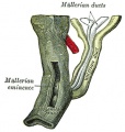
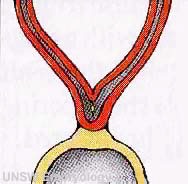
Mouse paramesonephric duct (Müllerian duct)[1] |
This mouse image shows the relationship between the mesonephric and paramesonephric ducts opening into the urogenital sinus.
|
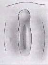
| <mediaplayer width='490' height='500' image="http://embryology.med.unsw.edu.au/embryology/images/2/2d/Uterus_001_icon.jpg">File:Uterus_001.mp4</mediaplayer> | Female Uterus and Vagina (between week 9 and 20)
The uterus and broad ligament will eventulaly divide the pelvic cavity into two separate pouches.
|

|
This graph shows the growth during the fetal period of the uterus between week 19 and 38.[2]
|
External Genitalia

This next section will look at the development of the external genitalia using a series of animations and online resources.
Female External Genitalia
| <mediaplayer width='270' height='380' image="http://embryology.med.unsw.edu.au/embryology/images/3/37/Male_external_001_icon.jpg">File:Female_external_001.mp4</mediaplayer> | Animation showing the development of external female genitalia from the indifferent external structure (week 9 to 12 approximately).
|
Male External Genitalia
| <mediaplayer width='270' height='380' image="http://embryology.med.unsw.edu.au/embryology/images/3/37/Male_external_001_icon.jpg">File:Male_external_001.mp4</mediaplayer> | Animation showing the development of external male genitalia from the indifferent external structure (week 9 to 12 approximately).
|
External Genitalia Comparison
Gonad Descent
| <mediaplayer width='296' height='430' image="http://embryology.med.unsw.edu.au/embryology/images/7/75/Gonad_blood_01_icon.jpg">File:Gonad blood 01.mp4</mediaplayer> | Animation shows the descent of the gonads and their blood supply.
|
Internal Gonad Descent
Testes Descent
| The linked animation shows the descent of the testes (between week 7 to 38, birth).
Descent of the testes into the scrotal sac begins generally during week 26 and may take several days.
Incomplete or failed descent can occur unilaterally or bilaterally, is more common in premature births, and can be completed postnatally. (see also cryptorchidism). |

|

|
| Start of testis descent | End of testis descent |
Additional Information
Testes Descent Timeline
Data from a study of male human fetal (between 10 and 35 weeks) gonad position.[3]
- 10 to 23 weeks - (9.45%) had migrated from the abdomen and were situated in the inguinal canal
- 24 to 26 weeks - (57.9%) had migrated from the abdomen
- 27 to 29 weeks - (16.7%) had not descended to the scrotum
A second study looked at the position of the testes[4]
- 33 weeks fetal testes had descended to the scrotum
- between 33 to 40 weeks (term) both testes have normally descended to the scrotum
Failure of descent (cryptorchidism) either unilateral or bilateral testicular descent, occurring in up to 30% premature and 3-4% term males.
Cryptorchidism in common eutherian mammals.[5]- Species comparison of descent timeline
Historic Genital Images
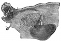
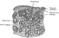
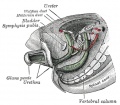
Broad ligament of adult showing Epoöphoron
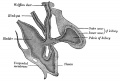
Urogenital Sinus of Female Human Embryo of 8.5 to 9 weeks old
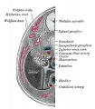
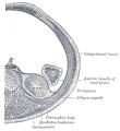
Transverse section of Human Embryo 8.5 to 9 Weeks Old
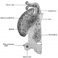
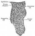
Longitudinal Section of Ovary of Cat Embryo of 9.4 cm long
Section of the Ovary of a Newly Born Child
Human Embryo (3.5 cm long) Testis Section of a Genital Cord
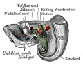
Tail end of Human Embryo 25 to 29 Days Old
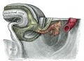
Tail end of human embryo eight and a half to nine weeks old
Primitive Kidney and Bladder
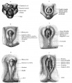
Stages in the development of the external sexual organs in the male and female
Retroperitoneal structures


References
BGDB: Lecture - Gastrointestinal System | Practical - Gastrointestinal System | Lecture - Face and Ear | Practical - Face and Ear | Lecture - Endocrine | Lecture - Sexual Differentiation | Practical - Sexual Differentiation | Tutorial
Glossary Links
- Glossary: A | B | C | D | E | F | G | H | I | J | K | L | M | N | O | P | Q | R | S | T | U | V | W | X | Y | Z | Numbers | Symbols | Term Link
Cite this page: Hill, M.A. (2024, April 23) Embryology BGDB Sexual Differentiation - Fetal. Retrieved from https://embryology.med.unsw.edu.au/embryology/index.php/BGDB_Sexual_Differentiation_-_Fetal
- © Dr Mark Hill 2024, UNSW Embryology ISBN: 978 0 7334 2609 4 - UNSW CRICOS Provider Code No. 00098G