BGDB Sexual Differentiation - Early Embryo: Difference between revisions
mNo edit summary |
mNo edit summary |
||
| Line 129: | Line 129: | ||
[[Testis Development Movie]] | [[Testis Development Movie]] | ||
| | | | ||
====Development of the male gonad showing medullary sex cords==== | |||
* <font color=red>'''Paramesonephric duct'''</font> (red left, Müllerian duct) degenerates under the influence of Mullerian duct inhibitory factor (MDIF) secreted by sertoli cells (differentiated by SRY expression). | |||
* <font color=darkmagenta>'''Mesonephric duct'''</font> (purple, Wolffian duct) differentiates under the influence of testosterone secreted by Leydig cells. Within the testes these mesonephric tubules grow towards the medullary sex cords and will form the rete teste. The mesonephric duct extending out of the gonad forms the ductus deferens. | |||
* <font color=orange>'''Medullary sex cords'''</font> (orange) form testis cords. | |||
** these later differentiate into solid seminiferous tubules that during puberty become hollow and actively produce spermatozoa. | |||
* The surrounding connective tissue (pink) differentiates to form stromal cells. | |||
* The '''tunica albuginea''' (white) covers the testis and bands extend inward to form connective tissue septa. | |||
|} | |} | ||
| Line 143: | Line 149: | ||
[[Ovary Development Movie]] | [[Ovary Development Movie]] | ||
| | | | ||
====Development of the female gonad showing medullary sex cords==== | |||
* <font color=darkmagenta>'''Mesonephric duct'''</font> (purple, Wolffian duct) degenerates, small remnants may remain as epoophoron and paroophoron (in the mesentry of the ovary) and Gartner's cycts (near vagina). | * <font color=darkmagenta>'''Mesonephric duct'''</font> (purple, Wolffian duct) degenerates, small remnants may remain as epoophoron and paroophoron (in the mesentry of the ovary) and Gartner's cycts (near vagina). | ||
| Line 156: | Line 165: | ||
{| | {| | ||
| < | ! Ovary | ||
| < | ! Testis | ||
|- | |||
| width=380px|<mediaplayer width='350' height='650' image="http://embryology.med.unsw.edu.au/embryology/images/f/fa/Gonad-icon.jpg">File:Ovary_001.mp4</mediaplayer> | |||
| width=380px|<mediaplayer width='350' height='650' image="http://embryology.med.unsw.edu.au/embryology/images/f/fa/Gonad-icon.jpg">File:Testis 001.mp4</mediaplayer> | |||
|- | |- | ||
| Ovary | | [[Ovary Development Movie]] | ||
| Testis | | [[Testis Development Movie]] | ||
|} | |} | ||
{{BGDB SexDiffn}} | {{BGDB SexDiffn}} | ||
| Line 191: | Line 203: | ||
Nussey S, Whitehead S. '''Endocrinology: An Integrated Approach.''' Oxford: BIOS Scientific Publishers; 2001. [http://www.ncbi.nlm.nih.gov/books/NBK29/ Chapter 6 The gonad] | Nussey S, Whitehead S. '''Endocrinology: An Integrated Approach.''' Oxford: BIOS Scientific Publishers; 2001. [http://www.ncbi.nlm.nih.gov/books/NBK29/ Chapter 6 The gonad] | ||
{{ | |||
{{BGDBFooter}} | |||
Revision as of 11:49, 3 June 2013
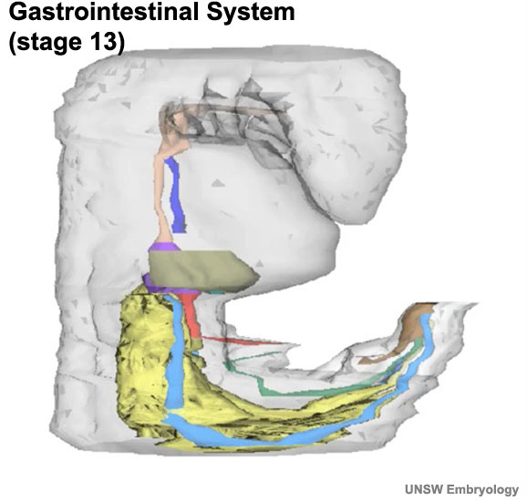
Week 4 to 5
| <mediaplayer width='550' height='580' image="http://embryology.med.unsw.edu.au/embryology/images/4/46/Stage13-GIT-icon.jpg">File:Stage13 GIT3d.mp4</mediaplayer> | Begin by observing the internal structure of the embryo at the end of week 4 and the beginning of week 5.
Colour code:
|


Mesonephros and Mesonephric Duct
Section 29-46: (E1-G4) Section passes dorsally to the mesonephros. Return to G4 and then follow the caudal route of the mesonephric duct into the sacral region.

|

|

|

|

|

| |
| 29 (E1L) | 30 (E2L) | 31 (E3L) | 32 (E4L) | 33 (E5L) | 34 (E6L) | 35 (E7L) |

|

|

|

|

|

|

|
| 36 (F1L) | 37 (F2L) | 38 (F3L) | 39 (F4L) | 40 (F5L) | 41 (F6L) | 42 (F7L) |

|

|

|

| |||
| 43 (G1L) | 44 (G2L) | 45 (G3L) | 46 (G4L) |
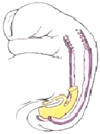
Kidney and Mesonephric Duct
| <mediaplayer width='360' height='500' image="http://embryology.med.unsw.edu.au/embryology/images/f/fe/Urogenital_sinus_001_icon.jpg">File:Urogenital_sinus_001.mp4</mediaplayer> | Now go back and observe the development of the intermediate mesoderm.
|
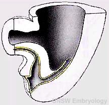
Primitive Urogenital Sinus
| <mediaplayer width='420' height='415' image="http://embryology.med.unsw.edu.au/embryology/images/8/88/Urogenital_septum_001_icon.jpg">File:Urogenital_septum_001.mp4</mediaplayer> | Stages in septation of the urogenital sinus between Week 4 and 6.
Several different defects in both sexes can occur if this septation process is not correctly aligned (rectourethral fistula, rectovaginal fistula).
Cloacal Membrane (intact) (week 4 Carnegie stage 12) |

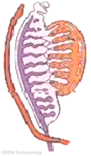
Gonad Development
Male Testis
| <mediaplayer width='350' height='650' image="http://embryology.med.unsw.edu.au/embryology/images/f/fa/Gonad-icon.jpg">File:Testis 001.mp4</mediaplayer> |
Development of the male gonad showing medullary sex cords
|
Female Ovary
| <mediaplayer width='350' height='650' image="http://embryology.med.unsw.edu.au/embryology/images/f/fa/Gonad-icon.jpg">File:Ovary_001.mp4</mediaplayer> |
Development of the female gonad showing medullary sex cords
|
Gonadal Development Comparison
Now directly compare the development of the male and female gonad (click image to start, these may not be synchronised).
| Ovary | Testis |
|---|---|
| <mediaplayer width='350' height='650' image="http://embryology.med.unsw.edu.au/embryology/images/f/fa/Gonad-icon.jpg">File:Ovary_001.mp4</mediaplayer> | <mediaplayer width='350' height='650' image="http://embryology.med.unsw.edu.au/embryology/images/f/fa/Gonad-icon.jpg">File:Testis 001.mp4</mediaplayer> |
| Ovary Development Movie | Testis Development Movie |
Additional Information
The following figures are from a recent review of Sex determination and gonadal development in mammals.[1]
- Signaling in genital development
- Sex reversal in humans caused by abnormal X-Y exchange
- Structure of the early fetal testis
- Mesonephric tubules in the 11.5 dpc mouse urogenital ridge
- Development and differentiation of the genital duct system
- The migratory pathway of primordial germ cells
- Structure of the early fetal testis
- Differentiation of pre-Sertoli cells into Sertoli cells
- Model for cell-autonomous and prostaglandin-mediated upregulation of Sox9 in pre-Sertoli cells
- Visualization of testicular cell types
- Postulated molecular pathway leading to the formation of the bipotential genital ridge
- Structure of mouse and human SRY protein
- Postulated interaction of molecular players involved in early testicular development
References
- ↑ <pubmed>17237341</pubmed>| Physiol. Rev.
Nussey S, Whitehead S. Endocrinology: An Integrated Approach. Oxford: BIOS Scientific Publishers; 2001. Chapter 6 The gonad
BGDB: Lecture - Gastrointestinal System | Practical - Gastrointestinal System | Lecture - Face and Ear | Practical - Face and Ear | Lecture - Endocrine | Lecture - Sexual Differentiation | Practical - Sexual Differentiation | Tutorial
Glossary Links
- Glossary: A | B | C | D | E | F | G | H | I | J | K | L | M | N | O | P | Q | R | S | T | U | V | W | X | Y | Z | Numbers | Symbols | Term Link
Cite this page: Hill, M.A. (2024, April 24) Embryology BGDB Sexual Differentiation - Early Embryo. Retrieved from https://embryology.med.unsw.edu.au/embryology/index.php/BGDB_Sexual_Differentiation_-_Early_Embryo
- © Dr Mark Hill 2024, UNSW Embryology ISBN: 978 0 7334 2609 4 - UNSW CRICOS Provider Code No. 00098G