|
|
| (One intermediate revision by the same user not shown) |
| Line 21: |
Line 21: |
| [[File:Amniocentesis.jpg|thumb|{{Amniocentesis}} is a prenatal diagnostic test, carried out by removing a small fluid sample.]] | | [[File:Amniocentesis.jpg|thumb|{{Amniocentesis}} is a prenatal diagnostic test, carried out by removing a small fluid sample.]] |
|
| |
|
| | {| |
| | | |
| # required for lung development. | | # required for lung development. |
| # enables movement and symmetrical musculoskeletal development. | | # enables movement and symmetrical musculoskeletal development. |
| # maintains relatively constant temperature. | | # maintains relatively constant temperature. |
| # protects by cushioning sudden blows or movements. | | # protects by cushioning sudden blows or movements. |
| | | | Volume - increases as the fetus grows. |
| Volume - increases as the fetus grows. | |
| * 34 weeks (GA) - peaks at about 800 mL. | | * 34 weeks (GA) - peaks at about 800 mL. |
| * 40 weeks (GA) - about 600 mL at term. | | * 40 weeks (GA) - about 600 mL at term. |
| Line 32: |
Line 33: |
| * replacing by fetal exhalation and urination. | | * replacing by fetal exhalation and urination. |
| * low magnesium levels associated with preeclampsia and diabetes. | | * low magnesium levels associated with preeclampsia and diabetes. |
| | | |} |
|
| |
|
| In early embryonic development both the buccopharyngeal and cloacal membranes degenerated, allowing the GIT to be filled with amniotic fluid. Towards the end of the fetal period the fetus is now swallowing approximately 500 ml of amniotic fluid / day. | | In early embryonic development both the buccopharyngeal and cloacal membranes degenerated, allowing the GIT to be filled with amniotic fluid. Towards the end of the fetal period the fetus is now swallowing approximately 500 ml of amniotic fluid / day. |
| Line 47: |
Line 48: |
| ** If no discharge (bowel motion) is observed in this early period it can be indicative of an abnormality of the GIT. | | ** If no discharge (bowel motion) is observed in this early period it can be indicative of an abnormality of the GIT. |
| * The first stool (meconium) is passed within 24 hours in most healthy term infants. | | * The first stool (meconium) is passed within 24 hours in most healthy term infants. |
| | |
| | |
| | ==Nutrition - Milk== |
| | [[File:Mammary_anatomy.jpg|thumb|300px|Adult female mammary anatomy cartoon]] |
| | Breast {{milk}} makes us mammals! A review article by Goldman{{#pmid:10721920|PMID10721920}} may provide a way of thinking about GIT and human milk. |
| | |
| | :"Human milk contains agents that affect the growth, development and functions of the epithelium, immune system or nervous system of the gastrointestinal tract. Some human and animal studies indicate that human milk affects the growth of intestinal villi, the development of intestinal disaccharidases, the permeability of the gastrointestinal tract and resistance to certain inflammatory/immune-mediated diseases. Moreover, one cytokine in human milk, interleukin (IL)-10, protects infant mice genetically deficient in IL-10 against an enterocolitis that resembles necrotizing enterocolitis (NEC) in human premature infants. |
| | |
| | There are seven overlapping evolutionary strategies regarding the relationships between the functions of the mammary gland and the infant’s gastrointestinal tract as follows: |
| | |
| | # certain immunologic agents in human milk compensate directly for developmental delays in those same agents in the recipient infant |
| | # other agents in human milk do not compensate directly for developmental delays in the production of those same agents, but nevertheless protect the recipient |
| | # agents in human milk enhance functions that are poorly expressed in the recipient |
| | # agents in human milk change the physiologic state of the intestines from one adapted to intrauterine life to one suited to extrauterine life |
| | # some agents in human milk prevent inflammation in the recipient’s gastrointestinal tract |
| | # survival of human milk agents in the gastrointestinal tract is enhanced because of delayed production of pancreatic proteases and gastric acid by newborn infants, antiproteases and inhibitors of gastric acid production in human milk, inherent resistance of some human milk agents to proteolysis, and protective binding of other factors in human milk |
| | # growth factors in human milk aid in establishing a commensal enteric microflora" |
| | |
| | (Text from: Goldman ref{{#pmid:10721920|PMID10721920}}) |
| | |
| | |
| | :'''Links:''' {{milk}} |
|
| |
|
| ===Gut Microorganism Population=== | | ===Gut Microorganism Population=== |
| Line 60: |
Line 83: |
| * Vibrio cholerae | | * Vibrio cholerae |
| * Listeria | | * Listeria |
| | |
| | ===Antibiotics=== |
| | |
| | Treatment of other neonatal infections systemically with antibiotics can alter the bacterial population. |
|
| |
|
|
| |
|
| Line 76: |
Line 103: |
| # What is the most common gastrointestinal motility abnormality of the newborn? | | # What is the most common gastrointestinal motility abnormality of the newborn? |
| # Which is more common omphalocele or gastroschisis? | | # Which is more common omphalocele or gastroschisis? |
| | |
|
| |
|
| ==3. Gut Diagnostics== | | ==3. Gut Diagnostics== |
Learning Activity 4
- Describe the fetal and early post-natal changes of the gut.
- Identify the common abnormalities of the gut.
- Identify the diagnostic techniques associated in testing gut function.
|
Gastroschisis
|
1. Fetal and Postnatal Changes
Intestinal Length Growth
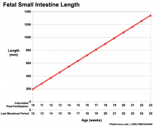
Small Intestine length (mm)
Amniotic Fluid Swallowing
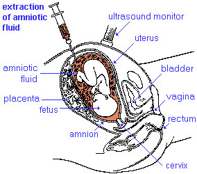
amniocentesis is a prenatal diagnostic test, carried out by removing a small fluid sample.
- required for lung development.
- enables movement and symmetrical musculoskeletal development.
- maintains relatively constant temperature.
- protects by cushioning sudden blows or movements.
|
Volume - increases as the fetus grows.
- 34 weeks (GA) - peaks at about 800 mL.
- 40 weeks (GA) - about 600 mL at term.
- circulated by fetal inhaling and swallowing.
- replacing by fetal exhalation and urination.
- low magnesium levels associated with preeclampsia and diabetes.
|
In early embryonic development both the buccopharyngeal and cloacal membranes degenerated, allowing the GIT to be filled with amniotic fluid. Towards the end of the fetal period the fetus is now swallowing approximately 500 ml of amniotic fluid / day.
<html5media width="600" height="400">https://www.youtube.com/embed/r9D7aiFG7N8</html5media>
This swallowed amniotic fluid moves through the GIT from esophagus, to stomach, to small intestine, stopping at the large bowel. In the large bowel the majority of fluid (water) is absorbed, along with electrolytes, glucose, urea and hormones. This process may contribute to fetal nutrition and prepare the GIT for its postnatal function. The process of swallowing amniotic fluid has been suggested to also help regulate fluid volume.
Fetal Meconium
- Mixture of substances (debris, glandular secretions, fatty material and bile pigments) that accumulate in the large bowel (green fecal material).
- Will form the neonatal meconium which is the first (usually within 24h to 48h) postnatal excretion from the GIT.
- If no discharge (bowel motion) is observed in this early period it can be indicative of an abnormality of the GIT.
- The first stool (meconium) is passed within 24 hours in most healthy term infants.
Nutrition - Milk
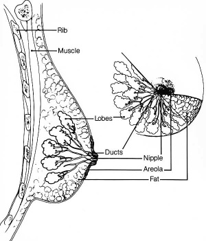
Adult female mammary anatomy cartoon
Breast milk makes us mammals! A review article by Goldman[1] may provide a way of thinking about GIT and human milk.
- "Human milk contains agents that affect the growth, development and functions of the epithelium, immune system or nervous system of the gastrointestinal tract. Some human and animal studies indicate that human milk affects the growth of intestinal villi, the development of intestinal disaccharidases, the permeability of the gastrointestinal tract and resistance to certain inflammatory/immune-mediated diseases. Moreover, one cytokine in human milk, interleukin (IL)-10, protects infant mice genetically deficient in IL-10 against an enterocolitis that resembles necrotizing enterocolitis (NEC) in human premature infants.
There are seven overlapping evolutionary strategies regarding the relationships between the functions of the mammary gland and the infant’s gastrointestinal tract as follows:
- certain immunologic agents in human milk compensate directly for developmental delays in those same agents in the recipient infant
- other agents in human milk do not compensate directly for developmental delays in the production of those same agents, but nevertheless protect the recipient
- agents in human milk enhance functions that are poorly expressed in the recipient
- agents in human milk change the physiologic state of the intestines from one adapted to intrauterine life to one suited to extrauterine life
- some agents in human milk prevent inflammation in the recipient’s gastrointestinal tract
- survival of human milk agents in the gastrointestinal tract is enhanced because of delayed production of pancreatic proteases and gastric acid by newborn infants, antiproteases and inhibitors of gastric acid production in human milk, inherent resistance of some human milk agents to proteolysis, and protective binding of other factors in human milk
- growth factors in human milk aid in establishing a commensal enteric microflora"
(Text from: Goldman ref[1])
- Links: milk
Gut Microorganism Population
The normal newborn gastrointestinal tract contains little if any microorganisms (commensal intestinal microbiota, microbiota, flora, microflora). Postnatally, the tract has to be populated by microorganisms, which are mainly anaerobic bacteria and then aerobic bacteria, but may also include yeast and fungi. The foregut comparatively has few microorganisms when compared to the midgut and hindgut.
Infections
There are several infectious pathogens that can also populate the postnatal gut leading to a number of different diseases:
- Gastroenteritis - (acute infectious enteritis) Occurs in children and is generally viral (rotavirus) rather than bacterial. By 5 years of age, nearly every child worldwide will have had at least one episode of rotavirus gastroenteritis. Note that maternal gastroenteritis during pregnancy can also affect birthweight.
- Escherichia coli (enterotoxigenic)
- Shigella a gram-negative, non-spore forming rod-shaped bacteria infectious through poor hygeine and ingestion, fecal–oral contamination. (More? Dysentery)
- Vibrio cholerae
- Listeria
Antibiotics
Treatment of other neonatal infections systemically with antibiotics can alter the bacterial population.
2. Common Abnormalities
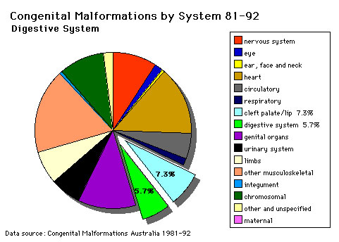
Gastrointestinal Tract Defects of all notifiable birth defects in Australia (1981-1992)
The table below shows the most recent ICD-11 coding for congenital gastrointestinal abnormalities.
- What is oesophageal atresia (EA) and what are the possible developmental causes?
- What is the most common gastrointestinal motility abnormality of the newborn?
- Which is more common omphalocele or gastroschisis?
3. Gut Diagnostics
Heelprick Test
 Guthrie test is a neonatal blood test to detect aa number of different congenital abnormalities, including metabolic disorders.
Additional Information
| Additional Information - Content shown under this heading is not part of the material covered in this class. It is provided for those students who would like to know about some concepts or current research in topics related to the current class page.
|
- For more detailed descriptions see gastrointestinal abnormalities.
- Meconium Aspiration - can occur near term or at delivery, if meconiumis discharged into the amiotic fluid (meconium stained amniotic fluid) and then injested by the fetus as it swallows amiotic fluid. Can then lead to meconium aspiration syndrome (MAS), meconium is drawn into the fetal/newborn lungs, causing inflammation, cell death and potentially perinatal death. Meconium can also damage the placenta and associated blood vessels.
- Polyhydramnios - (hydramnios, amniotic fluid disorder) refers to abnormally high amniotic fluid levels.
- Elliott EJ & Dalby-Payne JR. (2004). 2. Acute infectious diarrhoea and dehydration in children. Med. J. Aust. , 181, 565-70. PMID: 15540971
| USA Selected Statistics (2006)
|
| USA Selected Abnormalities (CDC National estimates for 21 selected major birth defects 2004–2006)
|
| Birth Defects
|
Cases per Births (1 in ...)
|
Estimated Annual Number of Cases
|
| anencephaly
|
4,859
|
859
|
| spina bifida without anencephaly
|
2,858
|
1,460
|
| encephalocele
|
12,235
|
341
|
| Anophthalmia/microphthalmia
|
5,349
|
780
|
| patent ductus arteriosus/common truncus
|
13,876
|
301
|
| transposition of the great vessels
|
3,333
|
1,252
|
| Tetralogy of Fallot
|
2,518
|
1,657
|
| atrial septal defects/ventricular septal defects
|
2,122
|
1,966
|
| hypoplastic left heart
|
4,344
|
960
|
| cleft palate without cleft lip
|
1,574
|
2,651
|
| cleft lip with and without cleft palate
|
940
|
4,437
|
| Esophageal atresia/tracheoesophageal fistula
|
4,608
|
905
|
| Rectal and large intestinal atresia/stenosis
|
2,138
|
1,952
|
| Reduction deformity, upper limbs
|
2,869
|
1,454
|
| Reduction deformity, lower limbs
|
5,949
|
701
|
| gastroschisis
|
2,229
|
1,871
|
| omphalocele
|
5,386
|
775
|
| Diaphragmatic hernia
|
3,836
|
1,088
|
| Trisomy 13
|
7,906
|
528
|
| Trisomy 21 (Down syndrome)
|
691
|
6,037
|
| Trisomy 18
|
3,762
|
1,109
|
- Links: Human Abnormal Development | CDC Birth Defects - Data & Statistics | USA Statistics | Victoria 2004 | USA 2006 | Europe 2010
|
|
| Gastrointestinal Tract Terms
|
- allantois - An extraembryonic membrane, endoderm in origin extension from the early hindgut, then cloaca into the connecting stalk of placental animals, connected to the superior end of developing bladder. In reptiles and birds, acts as a reservoir for wastes and mediates gas exchange. In mammals is associated/incorporated with connecting stalk/placental cord fetal-maternal interface.
- amnion - An extra-embryonic membrane, ectoderm and extraembryonic mesoderm in origin, also forms the innermost fetal membrane, that produces amniotic fluid. This fluid-filled sac initially lies above the trilaminar embryonic disc and with embryoic disc folding this sac is drawn ventrally to enclose (cover) the entire embryo, then fetus. The presence of this membrane led to the description of reptiles, bird, and mammals as amniotes.
- amniotic fluid - The fluid that fills amniotic cavity totally encloses and cushions the embryo. Amniotic fluid enters both the gastrointestinal and respiratory tract following rupture of the buccopharyngeal membrane. The late fetus swallows amniotic fluid.
- atresia - is an abnormal interruption of the tube lumen, the abnormality naming is based upon the anatomical location.
- buccal - (Latin, bucca = cheek) A term used to relate to the mouth (oral cavity).
- bile salts - Liver synthesized compounds derived from cholesterol that function postnatally in the small intestine to solubilize and absorb lipids, vitamins, and proteins. These compounds act as water-soluble amphipathic detergents. liver
- buccopharyngeal membrane - (oral membrane) (Latin, bucca = cheek) A membrane which forms the external upper membrane limit (cranial end) of the early gastrointestinal tract. This membrane develops during gastrulation by ectoderm and endoderm without a middle (intervening) layer of mesoderm. The membrane lies at the floor of the ventral depression (stomodeum) where the oral cavity will open and will breakdown to form the initial "oral opening" of the gastrointestinal tract. The equivilent membrane at the lower end of the gastrointestinal tract is the cloacal membrane.
- celiac artery - (celiac trunk) main blood supply to the foregut, excluding the pharynx, lower respiratory tract, and most of the oesophagus.
- cholangiocytes - epithelial cells that line the intra- and extrahepatic ducts of the biliary tree. These cells modify the hepatocyte-derived bile, and are regulated by hormones, peptides, nucleotides, neurotransmitters, and other molecules. liver
- cloaca - (cloacal cavity) The term describing the common cavity into which the intestinal, genital, and urinary tracts open in vertebrates. Located at the caudal end of the embryo it is located on the surface by the cloacal membrane. In many species this common cavity is later divided into a ventral urogenital region (urogenital sinus) and a dorsal gastrointestinal (rectal) region.
- cloacal membrane - Forms the external lower membrane limit (caudal end) of the early gastrointestinal tract (GIT). This membrane is formed during gastrulation by ectoderm and endoderm without a middle (intervening) layer of mesoderm. The membrane breaks down to form the initial "anal opening" of the gastrointestinal tract.
- coelomic cavity - (coelom) Term used to describe a space. There are extra-embryonic and intra-embryonic coeloms that form during vertebrate development. The single intra-embryonic coelom forms the 3 major body cavities: pleural cavity, pericardial cavity and peritoneal cavity.
- crypt of Lieberkühn - (intestinal gland, intestinal crypt) intestinal villi epithelia extend down into the lamina propria where they form crypts that are the source of epithelial stem cells and immune function.
- duplication - is an abnormal incomplete tube recanalization resulting in parallel lumens, this is really a specialized form of stenosis. (More? Image - small intestine duplication)
- esophageal - (oesophageal)
- foregut - first embryonic division of gastrointestinal tract extending from the oral (buccopharyngeal) membrane and contributing oesophagus, stomach, duodenum (to bile duct opening), liver, biliary apparatus (hepatic ducts, gallbladder, and bile duct), and pancreas. The forgut blood supply is the celiac artery (trunk) excluding the pharynx, lower respiratory tract, and most of the oesophagus.
- galactosemia - Metabolic abnormality where the simple sugar galactose (half of lactose, the sugar in milk) cannot be metabolised. People with galactosemia cannot tolerate any form of milk (human or animal). Detected by the Guthrie test.
- gastric transposition - clinical term for postnatal surgery treatment for esophageal atresia involving esophageal replacement. Typically performed on neonates between day 1 to 4. (More? gastrointestinal abnormalities | PMID 28658159
- gastrointestinal divisions - refers to the 3 embryonic divisions contributing the gastrointestinal tract: foregut, Midgut and hindgut.
- gastrula - (Greek, gastrula = little stomach) A stage of an animal embryo in which the three germ layers (endoderm/mesoderm/ectoderm) have just formed. All of these germ layers have contributions to the gastrointestinal tract.
- gastrulation - The process of differentiation forming a gastrula. Term means literally means "to form a gut" but is more in development, as this process converts the bilaminar embryo (epiblast/hypoblast) into the trilaminar embryo (endoderm/mesoderm/ectoderm) establishing the 3 germ layers that will form all the future tissues of the entire embryo. This process also establishes the the initial body axes. (More? gastrulation)
- Guthrie test - (heel prick) A neonatal blood screening test developed by Dr Robert Guthrie (1916-95) for determining a range of metabolic disorders and infections in the neonate. (More? Guthrie test)
- heterotaxia - (Greek heteros = different; taxis = arrangement) is the right/left transposition of thoracic and/or abdominal organs.
- hindgut - final embryonic division of gastrointestinal tract extending to the cloacal membrane and contributing part of the transverse colon (left half to one third), descending colon, sigmoid colon, rectum, part of anal canal (superior), urinary epithelium (bladder and most urethra). The hindgut blood supply is the inferior mesenteric artery.
- inferior mesenteric artery - main blood supply to the hindgut
- intestine - (bowel) part of the gastrointestinal tract (GIT) lying between the stomach and anus where absorption of nutrients and water occur. This region is further divided anatomically and functionally into the small intestine or bowel (duodenum, jejunum and ileum) and large intestine or bowel (cecum and colon).
- intestinal perforation - gastrointestinal abnormality identified in neonates can be due to necrotizing enterocolitis, Hirschsprung’s disease or meconium ileus.
- intraembryonic coelom - The "horseshoe-shaped" space (cavity) that forms initially in the third week of development in the lateral plate mesoderm that will eventually form the 3 main body cavities: pericardial, pleural, peritoneal. The intraembryonic coelom communicates transiently with the extraembryonic coelom.
- meconium ileus intestine obstruction within the ileum due to abnormal meconium properties.
- mesentery - connects gastrointestinal tract to the posterior body wall and is a double layer of visceral peritoneum.
- mesothelium - The mesoderm derived epithelial covering of coelomic organs and also line their cavities.
- Midgut - middle embryonic division of gastrointestinal tract contributing the small intestine (including duodenum distal bile duct opening), cecum, appendix, ascending colon, and part of the transverse colon (right half to two thirds). The midgut blood supply is the superior mesenteric artery.
- neuralation - The general term used to describe the early formation of the nervous system. It is often used to describe the early events of differentiation of the central ectoderm region to form the neural plate, then neural groove, then neural tube. The nervous system includes the central nervous system (brain and spinal cord) from the neural tube and the peripheral nervous system (peripheral sensory and sympathetic ganglia) from neural crest. In humans, early neuralation begins in week 3 and continues through week 4.
- neural crest - region of cells at the edge of the neural plate that migrates throughout the embryo and contributes to many different tissues. In the gastrointestinal tract it contributes mainly the enteric nervous system within the wall of the gut responsible for peristalsis and secretion.
- peritoneal stomata - the main openings forming the pathways for drainage of intra-peritoneal fluid from the peritoneal cavity into the lymphatic system.
- pharynx - uppermost end of gastrointestinal and respiratory tract, in the embryo beginning at the buccopharyngeal membrane and forms a major arched cavity within the phrayngeal arches.
- recanalization - describes the process of a hollow structure becoming solid, then becoming hollow again. For example, this process occurs during GIT, auditory and renal system development.
- retroperitoneal - (retroperitoneum) is the anatomical space (sometimes a potential space) in the abdominal cavity behind (retro) the peritoneum. Developmentally parts of the GIT become secondarily retroperitoneal (part of duodenum, ascending and descending colon, pancreas)
- somitogenesis The process of segmentation of the paraxial mesoderm within the trilaminar embryo body to form pairs of somites, or balls of mesoderm. A somite is added either side of the notochord (axial mesoderm) to form a somite pair. The segmentation does not occur in the head region, and begins cranially (head end) and extends caudally (tailward) adding a somite pair at regular time intervals. The process is sequential and therefore used to stage the age of many different species embryos based upon the number visible somite pairs. In humans, the first somite pair appears at day 20 and adds caudally at 1 somite pair/4 hours (mouse 1 pair/90 min) until on average 44 pairs eventually form.
- splanchnic mesoderm - Gastrointestinal tract (endoderm) associated mesoderm formed by the separation of the lateral plate mesoderm into two separate components by a cavity, the intraembryonic coelom. Splanchnic mesoderm is the embryonic origin of the gastrointestinal tract connective tissue, smooth muscle, blood vessels and contribute to organ development (pancreas, spleen, liver). The intraembryonic coelom will form the three major body cavities including the space surrounding the gut, the peritoneal cavity. The other half of the lateral plate mesoderm (somatic mesoderm) is associated with the ectoderm of the body wall.
- stomodeum - (stomadeum, stomatodeum) A ventral surface depression on the early embryo head surrounding the buccopharyngeal membrane, which lies at the floor of this depression. This surface depression lies between the maxillary and mandibular components of the first pharyngeal arch.
- stenosis - abnormal a narrowing of the tube lumen, the abnormality naming is based upon the anatomical location.
- superior mesenteric artery - main blood supply to the Midgut.
- viscera - the internal organs in the main cavities of the body, especially those in the abdomen, for example the Template:Intestines.
- visceral peritoneum - covers the external surfaces of the intestinal tract and organs within the peritoneum. The other component (parietal peritoneum) lines the abdominal and pelvic cavity walls.
- yolk sac - An extraembryonic membrane which is endoderm origin and covered with extraembryonic mesoderm. Yolk sac lies outside the embryo connected initially by a yolk stalk to the midgut with which it is continuous with. The endodermal lining is continuous with the endoderm of the gastrointestinal tract. The extra-embryonic mesoderm differentiates to form both blood and blood vessels of the vitelline system. In reptiles and birds, the yolk sac has a function associated with nutrition. In mammals the yolk sac acts as a source of primordial germ cells and blood cells. Note that in early development (week 2) a structure called the "primitive yolk sac" forms from hypoblast, this is an entirely different structure.
- yolk stalk - (vitelline duct, omphalomesenteric duct, Latin, vitellus = yolk of an egg) The endodermal connection between the midgut and the yolk sac. See vitelline duct.
|
|
|

BGDB: Lecture - Gastrointestinal System | Practical - Gastrointestinal System | Lecture - Face and Ear | Practical - Face and Ear | Lecture - Endocrine | Lecture - Sexual Differentiation | Practical - Sexual Differentiation | Tutorial
Glossary Links
- Glossary: A | B | C | D | E | F | G | H | I | J | K | L | M | N | O | P | Q | R | S | T | U | V | W | X | Y | Z | Numbers | Symbols | Term Link
Cite this page: Hill, M.A. (2024, April 18) Embryology BGDB Gastrointestinal - Activity 4. Retrieved from https://embryology.med.unsw.edu.au/embryology/index.php/BGDB_Gastrointestinal_-_Activity_4
- What Links Here?
- © Dr Mark Hill 2024, UNSW Embryology ISBN: 978 0 7334 2609 4 - UNSW CRICOS Provider Code No. 00098G
- ↑ 1.0 1.1 Goldman AS. (2000). Modulation of the gastrointestinal tract of infants by human milk. Interfaces and interactions. An evolutionary perspective. J. Nutr. , 130, 426S-431S. PMID: 10721920


