BGDB Gastrointestinal - Activity 2: Difference between revisions
mNo edit summary |
mNo edit summary |
||
| Line 31: | Line 31: | ||
|} | |} | ||
{| | |||
| [[File:Pancreas rotation.jpg]] | |||
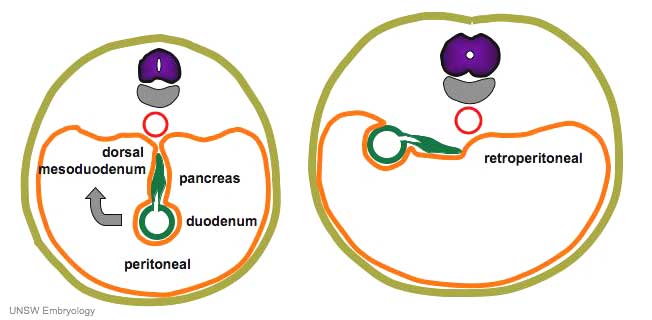
| After the stomach the initial portion of the gastrointestinal tract tube is the duodenum which initially lies in the midline within the peritoneal cavity. | |||
This region, along with the attached pancreas, undergoes rotation to become a retroperitoneal structure. | |||
This diagram shows the rotation with spinal cord at the top, vertebral body then dorsal aorta then pertioneal wall and cavity. | |||
|} | |||
==3. Abnormality Origins== | ==3. Abnormality Origins== | ||
Revision as of 13:51, 16 April 2019
| Practical 1: Activity 1 | Activity 2 | Activity 3 | Activity 4 |
Learning Activity 2
- Describe how elongation and rotation of the simple primitive tube establish the gut anatomy.
- Explain the developmental processes of elongation, herniation and rotation of the mid-gut.
- Describe the structure of the attachment of the gut to the body wall.
- Identify the developmental origins of abnormalities of the gut lumen, innervation, herniation and malrotation.
1. Mechanical Changes
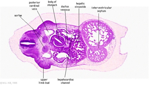
Transverse section of the developing stomach mid-embryonic period (Week 5 Carnegie stage 13)

|

|
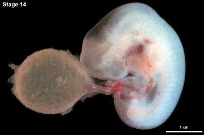
| <html5media height="820" width="580">File:Stage14 EFIC C01.mp4</html5media> | 
|
|
|
2. Mesentery
|

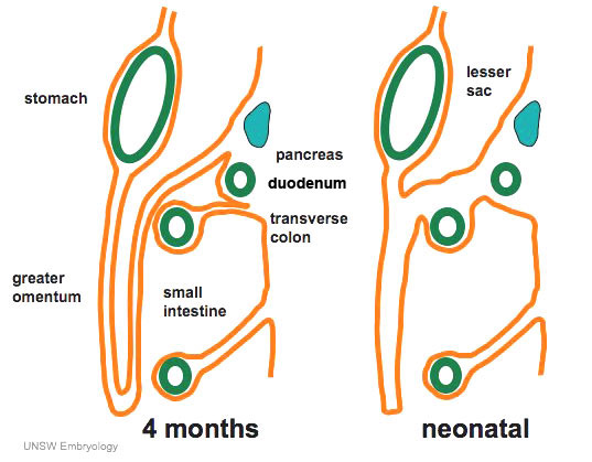
In the second trimester, the ventral and dorsal mesenteries associated with the stomach are still anatomically different from the newborn. The figure shows a lateral view of this process comparing the early second trimester arrangement with the newborn structure. |
3. Abnormality Origins
Interactive Component
To be added before class.
| Practical 1: Activity 1 | Activity 2 | Activity 3 | Activity 4 |
Additional Information
| Additional Information - Content shown under this heading is not part of the material covered in this class. It is provided for those students who would like to know about some concepts or current research in topics related to the current class page. |
BGDB: Lecture - Gastrointestinal System | Practical - Gastrointestinal System | Lecture - Face and Ear | Practical - Face and Ear | Lecture - Endocrine | Lecture - Sexual Differentiation | Practical - Sexual Differentiation | Tutorial
Glossary Links
- Glossary: A | B | C | D | E | F | G | H | I | J | K | L | M | N | O | P | Q | R | S | T | U | V | W | X | Y | Z | Numbers | Symbols | Term Link
Cite this page: Hill, M.A. (2024, April 16) Embryology BGDB Gastrointestinal - Activity 2. Retrieved from https://embryology.med.unsw.edu.au/embryology/index.php/BGDB_Gastrointestinal_-_Activity_2
- © Dr Mark Hill 2024, UNSW Embryology ISBN: 978 0 7334 2609 4 - UNSW CRICOS Provider Code No. 00098G
