BGDB Face and Ear - Abnormalities
Pharyngeal Abnormalities
There are terms for the different types of pharyngeal abnormalities, all of these except clefting are relatively rare.
- Sinuses - a pharyngeal groove defect, when a portion of the groove persists and opens to the skin surface, located laterally on the neck.
- Fistula - a pharyngeal membrane defect, a tract extends from pharynx (tonsillar fossa) beween the carotid arteries (internal and external) to open on side of neck.
- Cysts -a cervical sinus defect, remants of the cervical sinus remains as a fluid-filled cyst lined by an epithelium.
- Vestiges - a cartilaginous or bony developmental remnants that lie under the skin on side of neck.
- Clefting - the way in which the upper jaw forms from fusion of the smaller upper prominence of the first pharyngeal arch leads to a common congenital defect in this region called "clefting", which may involve either the upper lip, the palate or both structures.
Embryo Cleft Lip and Palate

Cleft lip and palate develop between the 4th and 8th week of gestation and is dominated by changes resulting in the formation of the nose. Palatal development occurs between the 7th and 12th week of gestation and is divided into the formation of the primary palate (prolabium), premaxilla and cartilaginous septum) and formation of the secondary palate (hard and soft palate).
300+ different abnormalities different cleft forms and extent upper lip and ant. maxilla hard and soft palate
Embryonic
Primary palate, fusion in the human embryo between week 6-7 (GA Week 8-9, stage 17 and 18), maxillary component of the first pharyngeal arch and the frontonasal prominence (philtrum) fuse from an epithelial seam to the mesenchymal bridge.
Fetal
Secondary palate, fusion in the human embryo in week 9-10 (GA week 11-12). Maxillary component of the first pharyngeal arch form lateral palatal shelves in the oral cavity that grow, elevation and fuse during this early fetal period. The fusion event is to both each other and the primary palate.
Fetal Cleft Ultrasound Movies

|
| ||||||
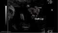
| Movie shows a cleft lip in an 13 week (GA 15 weeks) fetus. | Movie shows a cleft lip in an 18 week fetus. Fetal facial clefting can be detected by ultrasound scans. A common form of facial abnormality is that of cleft lip and palate. This is associated with the way in which the maxillary processes of the first pharyngeal arch must grow and fuse.
A measurement of the facial cleft of 4.8 mm (between the 2 plus marks + +) |

Clefting
The way in which the upper jaw forms from fusion of the smaller upper prominence of the first pharyngeal arch leads to a common congenital defect in this region called "clefting", which may involve either the upper lip, the palate or both structures.
Cleft Lip and Palate Classification
unilateral incomplete - cleft on one side of the lip that does not extend into the nose. unilateral complete - cleft on one side of the lip that extends into the nose. bilateral complete - cleft that involves both sides of the lip and extends into and involves the nose.
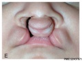
Cleft Lip

|
International Classification of Diseases code 749.1 for isolated cleft lip and 749.2 for cleft lip with cleft palate
|
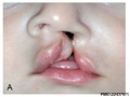
Uniateral Cleft Lip
Uniateral Cleft Lip
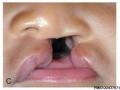
Complete Unilateral cleft lip and palate
Complete Unilateral cleft lip and palate
Complete bilateral cleft lip and palate
Complete bilateral cleft lip and palate


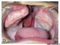
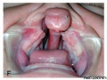
Cleft Palate

|
International Classification of Diseases code 749.0
|
(Data: Congenital Malformations Australia 1981-1992 P. Lancaster and E. Pedisich ISSN 1321-8352)
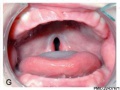
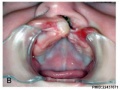
Incomplete Cleft Palate (Involving only the soft palate and uvula)
- Cleft palate 002.jpgComplete Cleft Palate (Completely involving the secondary palate)

| Palate Links: palate | cleft lip and palate | cleft palate | head | Category:Palate |
Fetal Alcohol Syndrome (FAS)

|
The following facial features may be associated with FAS
|
| Fetal Alcohol Syndrome face appearance |
First Arch Syndrome

There are 2 major types of genetic first arch syndrome, Treacher Collins and Pierre Robin, both result in extensive facial abnormalites.
Treacher Collins syndrome (TCS)
- a rare autosomal dominant craniofacial disorder (1:50.000)
- caused by frameshift deletions or duplications in the TCOF1 gene.
- hypoplasia of the mandible and zygomatic complex
- down-slanting palpebral fissures
- coloboma of the lower eyelid
- absence of eyelashes medial to the defect
- external and middle ear malformation
- conductive hearing loss
Auricular Abnormalities

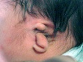
Microtia
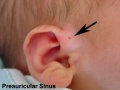
Preauricular sinus
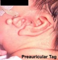
Preauricular tag
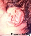
Preauricular tag


Additional Information
| Additional Information - Content shown under this heading is not part of the material covered in this class. It is provided for those students who would like to know about some concepts or current research in topics related to the current class page. |
Cephalic Disorders
Cephalic (Greek, kephale = head) are a group of abnormalities that relate to a wide range of skeletal (skull) and neural (brain) associated defects. Listed below are some selected skull defects.
- Acephaly (absence of head)
- Exencephaly (brain outside skull)
- Macrocephaly (large head)
- Micrencephaly (small brain)
- Otocephaly (absence of lower jaw)
- Brachycephaly (premature fusion of coronal suture)
- Oxycephaly, also known as turricephaly (premature fusion of coronal suture + other)
- Plagiocephaly (premature unilateral fusion of coronal or lambdoid sutures)
- Scaphocephaly, also known as dolichocephaly (premature fusion of sagittal suture)
- Trigonocephaly (premature fusion of metopic suture)
| Abnormal Neonatal Skull (CT) | ||
|---|---|---|

Scaphocephaly (Dolichocephaly) |

Coronal Synostosis |

Anterior Plagiocephaly |

Oxycephaly (Turricephaly) |

Posterior Plagiocephaly |

Deformational Plagiocepahly |

Trigonocephaly |

Oxycephaly |
- Skull CT Images: Normal overview | Normal vertex and lateral | Normal endocranial and vertex | Normal Vertex - Fontanels | Dolichocephaly and Scaphocephaly | Coronal Synostosis | Anterior Plagiocephaly | Turricephaly | Posterior Plagiocephaly | Deformational Plagiocepahly | Trigonocephaly | Oxycephaly | Computed Tomography
- Links: Head Abnormalities | Skull Abnormal Synostosis | Hearing Abnormalities | Vision Abnormalities
Surgical Cleft Repairs
| Palatoplasty | Cheiloplasty |
|---|---|
| Surgical repair of the palate | Surgical repair of the lip |

|

|
| Cleft Palate | Cleft Lip and Palate |
BGDB: Lecture - Gastrointestinal System | Practical - Gastrointestinal System | Lecture - Face and Ear | Practical - Face and Ear | Lecture - Endocrine | Lecture - Sexual Differentiation | Practical - Sexual Differentiation | Tutorial
Glossary Links
- Glossary: A | B | C | D | E | F | G | H | I | J | K | L | M | N | O | P | Q | R | S | T | U | V | W | X | Y | Z | Numbers | Symbols | Term Link
Cite this page: Hill, M.A. (2024, April 16) Embryology BGDB Face and Ear - Abnormalities. Retrieved from https://embryology.med.unsw.edu.au/embryology/index.php/BGDB_Face_and_Ear_-_Abnormalities
- © Dr Mark Hill 2024, UNSW Embryology ISBN: 978 0 7334 2609 4 - UNSW CRICOS Provider Code No. 00098G