BGDA Practical Placenta - Implantation and Early Placentation: Difference between revisions
| Line 50: | Line 50: | ||
===Decidual Immune Cells=== | ===Decidual Immune Cells=== | ||
:''Specialised immune cells.'' | :''Specialised immune cells.'' | ||
{| | |||
Decidual Macrophages | ! Decidual Macrophages | ||
! Decidual T cells | |||
! Uterine Natural Killer cells | |||
|- | |||
| | |||
* regulatory role at the fetal-maternal interface. | * regulatory role at the fetal-maternal interface. | ||
* M2 macrophage phenotype involved in tissue remodeling and inhibit inflammation | * M2 macrophage phenotype involved in tissue remodeling and inhibit inflammation | ||
* maintenance of tolerance to the non-self semi-allogeneic fetus | * maintenance of tolerance to the non-self semi-allogeneic fetus | ||
| | |||
* activated by fetal HLA-C (expressed on extravillous trophoblast cells) | * activated by fetal HLA-C (expressed on extravillous trophoblast cells) | ||
* specific immune tolerance to fetal alloantigens | * specific immune tolerance to fetal alloantigens | ||
| | |||
* Killer Inhibitory Receptor (KIR) activation by fetal HLA-C (expressed on extravillous trophoblast cells) | |||
|} | |||
===Chemokine Gene Silencing=== | ===Chemokine Gene Silencing=== | ||
Revision as of 10:36, 12 June 2013
| Practical 14: Implantation and Early Placentation | Villi Development | Maternal Decidua | Cord Development | Placental Functions | Diagnostic Techniques | Abnormalities |
Early Placenta

|

|
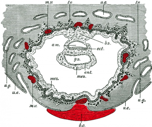
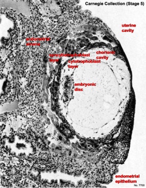
| Human Embryo Day 8 to 9 | Human Embryo - early implantation (stage 5) |
|
|
|
|
| Practical 14: Implantation and Early Placentation | Villi Development | Maternal Decidua | Cord Development | Placental Functions | Diagnostic Techniques | Abnormalities |
Additional Information
| Additional Information - Content shown under this heading is not part of the material covered in this class. It is provided for those students who would like to know about some concepts or current research in topics related to the current class page. |
Maternal Immune
How does the implanting conceptus avoid immune rejection by the maternal immune system? There are a number of maternal and embryonic mechanisms that are thought to act to prevent immune rejection of the implanting conceptus, though the complete mechanism(s) are unknown. This is particularly relevant to Assisted Reproductive Technologies involving donor eggs.
Below are some examples of research on this topic.
Decidual Immune Cells
- Specialised immune cells.
| Decidual Macrophages | Decidual T cells | Uterine Natural Killer cells |
|---|---|---|
|
|
|
Chemokine Gene Silencing
- Remove the attraction of maternal immune cells.
A mouse study[1] has shown that the normal immune response to inflammation, accumulation of effector T cells in response to chemokine secretion does not occur during implantation. This is prevented locally by epigenetic silencing of chemokine expression in the decidual stromal cells.
Corticotropin-Releasing Hormone
- Kill the maternal immune cells.
Both maternal and implanting conceptus release CRH at the embryo implantation site. This hormone then binds to receptors on the surface of trophoblast (extravillous trophoblast) cells leading to expression of a protein (Fas ligand, FasL) that activates the extrinsic cell death pathway on any local maternal immune cells ( T and B lymphocytes, natural killer cells, monocytes and macrophages).[2] (Note - This cannot be the only mechanism, as mice with dysfunctional FasL proteins are still fertile).


