BGDA Practical 7 - Week 5: Difference between revisions
mNo edit summary |
mNo edit summary |
||
| (32 intermediate revisions by the same user not shown) | |||
| Line 1: | Line 1: | ||
==Introduction== | ==Introduction== | ||
{{ | {{BGDALab7}} | ||
Key Events of Human Development during the fifth week (week 5) following fertilization or | Key Events of Human Development during the fifth week (week 5) following fertilization or clinical {{GA}} week 7. | ||
By this week there are many different organs and tissues differentiating throughout the embryo, only a few select systems will be discussed. | By this week there are many different organs and tissues differentiating throughout the embryo, only a few select systems will be discussed. | ||
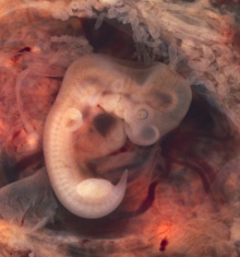
[[File:Stage15 sagittal section upper half 01.jpg|thumb|Later week 5 development ([[Carnegie_stage_15|Stage 15]]) showing a sagittal section upper half of human embryo.]] | |||
{{SlideStage15bf2}} | |||
===Cardiovascular=== | ===Cardiovascular=== | ||
* Heart - [[S#septation|septation]] starts, [[A#atrial septation|atrial septation]] and [[V#ventricular septation|ventricular septation]] | * Heart - [[S#septation|septation]] starts, [[A#atrial septation|atrial septation]] and [[V#ventricular septation|ventricular septation]] | ||
* Vascular - 3 vascular systems (systemic, placental, [[V#vitelline|vitelline]]) extensively remodelled | * Vascular - 3 vascular systems (systemic, placental, [[V#vitelline|vitelline]]) extensively remodelled | ||
See also Heart Development Timeline - [[Basic Cardiac Embryology]] | See also Heart Development Timeline - [[Basic Cardiac Embryology]] | ||
===Respiratory=== | ===Respiratory=== | ||
| Line 43: | Line 30: | ||
Heart Development Timeline (see [[Basic Cardiac Embryology]]) | Heart Development Timeline (see [[Basic Cardiac Embryology]]) | ||
{| | |||
| valign=bottom|{{Cardiovascular stage 13 movie}} | |||
| | |||
{{CVS cartoons}} | |||
|} | |||
[[File: | ===Atrial Septation=== | ||
[[File:Stage_13_image_067.jpg|600px]] | |||
[[File:Stage_13_image_068.jpg|600px]] | |||
==Respiratory== | ==Respiratory== | ||
{| | {| | ||
|- | |- | ||
| | | {{Gastrointestinal stage 13 movie}} | ||
| '''Week 4''' - [[L#laryngotracheal groove|laryngotracheal groove]] forms on floor foregut. | | '''Week 4''' - [[L#laryngotracheal groove|laryngotracheal groove]] forms on floor foregut. | ||
| Line 65: | Line 59: | ||
|} | |} | ||
The trachea can be see making its initial branching into the lung bud mesenchyme. | |||
{| | {| | ||
| [[File:Stage 13 image 070.jpg|600px]] | |||
| [[File:Gray0961.jpg|300px]] | |||
|- | |- | ||
| | | Bifurcation of the trachea into the main or primary bronchi, the left and right bronchus. | ||
| | | Adult bronchial tree. | ||
|} | |} | ||
==Neural== | |||
During week 5 the five secondary brain vesicles form from the original primary vesicles formed in week 4 and the cranial end of the neural tube now has 3 flexures (bends). | |||
(see also [[BGDA_Practical_7_-_Week_5#Neural_Development|Additional Information]]). | |||
[[File:CNS secondary vesicles.jpg|600px]] | |||
{| | |||
{| | |||
|- | |- | ||
| | | {{Neural stage 13 movie}} | ||
| | | Rapid growth folds the neural tube forming '''3 brain flexures''': | ||
| [ | # [[C#cranial flexure|cranial flexure]] (cephalic)- pushes mesencephalon upwards | ||
# [[P#pontine flexure|pontine flexure]] - generates 4th ventricle | |||
# [[C#cervical flexure|cervical flexure]] - between brain stem and spinal cord | |||
|} | |||
[ | [[File:Embryo_stage13-_brain_flexures.jpg|600px]] | ||
Neural tube - brain flexures (stage 13) | |||
{{BGDA Practical 6 - Week 5 Interactive}} | |||
{{BGDALab7}} | |||
==Additional Information== | |||
{{Med Prac additional Information}} | |||
===Timeline=== | |||
{{Week 5 Embryo Stages and Events table}} | |||
===Neural Development=== | |||
{{Carnegie stages CNS images table}} | |||
{{Neural Table}} | |||
====References==== | |||
<references/> | |||
{{ | {{BGDALab7}} | ||
[[Category:Week 5]] | [[Category:Week 5]] | ||
Latest revision as of 20:51, 12 May 2019
Introduction
Key Events of Human Development during the fifth week (week 5) following fertilization or clinical GA week 7.
By this week there are many different organs and tissues differentiating throughout the embryo, only a few select systems will be discussed.

| Stage 15 - Ventral View
|
| Stage 15 | Embryo Slides |
Cardiovascular
- Heart - septation starts, atrial septation and ventricular septation
- Vascular - 3 vascular systems (systemic, placental, vitelline) extensively remodelled
See also Heart Development Timeline - Basic Cardiac Embryology
Respiratory
- left and right lung buds push into the pericardioperitoneal canals (primordia of pleural cavity)
Neural
- Central nervous system - secondary brain vesicles
- Sensory - Hearing cochlear part of otic vesicle elongates (humans 2.5 turns)
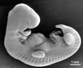
Stage 14
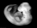
Stage 15


Cardiovascular

Heart Development Timeline (see Basic Cardiac Embryology)
|
|
Atrial Septation


Respiratory
|
Week 4 - laryngotracheal groove forms on floor foregut.
Week 5 - left and right lung buds push into the pericardioperitoneal canals (primordia of pleural cavity) Week 6 - descent of heart and lungs into thorax. Pleuroperitoneal foramen closes. Week 7 - enlargement of liver stops descent of heart and lungs. Month 3-6 - lungs appear glandular, end month 6 alveolar cells type 2 appear and begin to secrete surfactant. Month 7 - respiratory bronchioles proliferate and end in alveolar ducts and alveolar sacs. |
The trachea can be see making its initial branching into the lung bud mesenchyme.

|

|
| Bifurcation of the trachea into the main or primary bronchi, the left and right bronchus. | Adult bronchial tree. |
Neural
During week 5 the five secondary brain vesicles form from the original primary vesicles formed in week 4 and the cranial end of the neural tube now has 3 flexures (bends).
(see also Additional Information).

|
Rapid growth folds the neural tube forming 3 brain flexures:
|

Neural tube - brain flexures (stage 13)
Week 5 Interactive Component
| Attempt the Quiz - Week 5 | |
|---|---|

Here are a few simple Quiz questions that relate to Week 5 (GA week 7) from the lecture and practical. See your Quiz Result - Answer all the questions, then click "submit" to complete. The page will reload and you can then reopen this table to see your result and feedback.
|
Additional Information
| Additional Information - Content shown under this heading is not part of the material covered in this class. It is provided for those students who would like to know about some concepts or current research in topics related to the current class page. |
Timeline
| Week 5 - Human Embryo Stages and Events (GA week 7) | ||
|---|---|---|
| Embryo Week: Week 1 | Week 2 | Week 3 | Week 4 | Week 5 | Week 6 | Week 7 | Week 8 | Week 9 | ||
| Event | ||
| pituitary - Week 5 elongation, contacts infundibulum, diverticulum of diencephalon
heart - Week 5 septation starts, atrial and ventricular respiratory - Week 5 left and right lung buds push into the pericardioperitoneal canals (primordia of pleural cavity) respiratory - Week 5 to 17 lung histology - pseudoglandular hearing - Week 5 cochlear part of otic vesicle elongates (humans 2.5 turns) | ||
| Stage 14 | 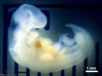 mesoderm - continued segmentation of paraxial mesoderm (somite pairs), heart prominence Head - 1st, 2nd and 3rd pharyngeal arch, forebrain, site of lens placode, site of otic placode, stomodeum Body - heart, liver, umbilical cord, mesonephric ridge visible externally as bulges. Limb - upper and lower limb buds growing neural - first appearance of the future cerebral hemispheres. Cerebellar plate differentiated to an intermediate layer, and future rhombic lip identifiable[1] liver hepatic gland and its vascular channels enlarge, hematopoietic function appears[2] | |
| Stage 15 |  neural - cranial nerves (except olfactory and optic) are identifiable in more advanced embryos[3] | |
| vision - 35 to 37 days retinal pigment present | ||
| Note - the day timing of stages is only approximate, system names link to first page of that specific system, and events are based upon the literature cited below. | ||
References
| ||
Neural Development
| Embryonic Central Nervous System | |||
|---|---|---|---|
| Stage 13 | Stage 14 | Stage 16 | Stage 21 |

scale bar = 1 mm |

|

|

|
| Week 4 | Week 5 | Week 6 | Week 8 |
| Neural Tube | Primary Vesicles | Secondary Vesicles | Adult Structures |
|---|---|---|---|
| week 3 | week 4 | week 5 | adult |
| prosencephalon (forebrain) | telencephalon | Rhinencephalon, Amygdala, hippocampus, cerebrum (cortex), hypothalamus, pituitary | Basal Ganglia, lateral ventricles | |
| diencephalon | epithalamus, thalamus, Subthalamus, pineal, posterior commissure, pretectum, third ventricle | ||
| mesencephalon (midbrain) | mesencephalon | tectum, Cerebral peduncle, cerebral aqueduct, pons | |
| rhombencephalon (hindbrain) | metencephalon | cerebellum | |
| myelencephalon | medulla oblongata, isthmus | ||
| spinal cord, pyramidal decussation, central canal | |||
References