BGDA Practical 3 - Week 3 Summary: Difference between revisions
mNo edit summary |
mNo edit summary |
||
| (5 intermediate revisions by the same user not shown) | |||
| Line 1: | Line 1: | ||
{{ | {{BGDALab3}} | ||
==Introduction== | ==Introduction== | ||
| Line 24: | Line 24: | ||
[[File:Stage7-sem2.jpg|240px|left]] | [[File:Stage7-sem2.jpg|240px|left]] | ||
===Facts=== | ===Facts=== | ||
Human embryonic stage 7 occurs during week 3 between 15 to 17 days. | [[Carnegie stage 7|Human embryonic stage 7]] occurs during week 3 between 15 to 17 days. | ||
The embryo is now 0.4 mm diameter in size. | The embryo is now 0.4 mm diameter in size. | ||
| Line 31: | Line 31: | ||
===Events=== | ===Events=== | ||
{| | |||
Gastrulation is continuing as cells migrate from the epiblast, continuing to form mesoderm. | | Gastrulation is continuing as cells migrate from the epiblast, continuing to form mesoderm. | ||
Mesoderm lies between the ectoderm and endoderm as a continuous sheet except at the buccopharyngeal and cloacal membranes. These membranes have ectoderm and endoderm only and will lie at the rostral (head) and caudal (tail) of the gastrointestinal tract. | Mesoderm lies between the ectoderm and endoderm as a continuous sheet except at the buccopharyngeal and cloacal membranes. These membranes have ectoderm and endoderm only and will lie at the rostral (head) and caudal (tail) of the gastrointestinal tract. | ||
| Line 39: | Line 39: | ||
The notochord is a key to embryonic folding and regulation of ectoderm and mesoderm differentiation. It lies in the rostrocordal axis and the embryonic disc will fold either side ventrally, pinching off a portion of the yolk sac to form the lining of the gastrointestinal tract. | The notochord is a key to embryonic folding and regulation of ectoderm and mesoderm differentiation. It lies in the rostrocordal axis and the embryonic disc will fold either side ventrally, pinching off a portion of the yolk sac to form the lining of the gastrointestinal tract. | ||
| {{SlideStage7bf5}} | |||
|} | |||
== Carnegie Stage 8 == | == Carnegie Stage 8 == | ||
[[Image:Stage8 SEM1.jpg|240px|left]] | [[Image:Stage8 SEM1.jpg|240px|left]] | ||
===Facts=== | ===Facts=== | ||
Human embryonic stage 8 occurs during week 3 between 17 to 19 days. | [[Carnegie stage 8|Human embryonic stage 8]] occurs during week 3 between 17 to 19 days. | ||
The embryo is now 1.0 - 1.5 mm in size. | The embryo is now 1.0 - 1.5 mm in size. | ||
| Line 71: | Line 72: | ||
[[File:Stage9_sem4c.jpg|left]] | [[File:Stage9_sem4c.jpg|left]] | ||
===Facts=== | ===Facts=== | ||
Human embryonic stage 9 occurs during week 3 between 19 to 21 days. | [[Carnegie stage 9|Human embryonic stage 9]] occurs during week 3 between 19 to 21 days. | ||
The embryo is now 1.5 to 2.5 mm in size and somites have begun to form and number between 1 to 3 somite pairs during this stage. | The embryo is now 1.5 to 2.5 mm in size and somites have begun to form and number between 1 to 3 somite pairs during this stage. | ||
The initial images are displayed unlabeled to allow you to explore the embryo for yourself, linked labeled versions are also available for some images. | The initial images are displayed unlabeled to allow you to explore the embryo for yourself, linked labeled versions are also available for some images. | ||
[[File:Stage9 sem1.jpg|600px]] | |||
===Events=== | ===Events=== | ||
| Line 100: | Line 103: | ||
[[Carnegie stage 9]] | [[Carnegie stage 9]] | ||
== Carnegie Stage 10== | |||
[[Carnegie stage 10|Human embryonic stage 10]] occurs at the beginning of week 4 development ({{GA}} week 6). This week will be covered in detail in the next practical class. The virtual slide image below shows a dorsal view of the embryo at the beginning of week 4. | |||
{{SlideStage10bf10}} | |||
| Line 112: | Line 122: | ||
The timing assumes fertilization the day after ovulation and the "weeks" refer to embryonic development and differ from Gestational Age ({{GA}}, clinical weeks shown in brackets, from last menstrual period) and "stages" refer to Carnegie stages of development. | The timing assumes fertilization the day after ovulation and the "weeks" refer to embryonic development and differ from Gestational Age ({{GA}}, clinical weeks shown in brackets, from last menstrual period) and "stages" refer to Carnegie stages of development. | ||
<br> | |||
{{First Trimester Timeline}} | |||
<br> | |||
{{Carnegie_stage_table_1}} | |||
== Next == | == Next == | ||
| Line 373: | Line 144: | ||
The next Practical will continue on through embryonic development ([[BGDA Practical - Implantation to 8 Weeks|Implantation to 8 Weeks]]). | The next Practical will continue on through embryonic development ([[BGDA Practical - Implantation to 8 Weeks|Implantation to 8 Weeks]]). | ||
===References=== | |||
<references/> | |||
{{BGDAFooter}} | {{BGDAFooter}} | ||
[[Category:Carnegie Stage 8]] [[Category:Carnegie Stage 9]] [[Category:Week 3]] | [[Category:Carnegie Stage 8]] [[Category:Carnegie Stage 9]] [[Category:Week 3]] | ||
Latest revision as of 15:50, 5 May 2019
Introduction
This page is a overview of events that occur in human development up to week 3 post-fertilization. From this Practical understand concepts of: fertilization, blastocyst development, implantation, bilaminar and trilaminar embryo formation, development of embryonic cavities and brief understanding of early placenta development.
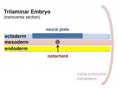
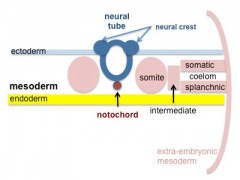
By the end of week 3, segmentation of the trilaminar embryo 3 germ layers has begun:

- Ectoderm - central neural plate and lateral parts form epidermis
- Mesoderm - midline notochord, adjacent somites, formation of the internal embryonic space (intraembryonic ceolom)
- Endoderm - epidermal lining of gastrointestinal tract and yolk sac lining
Note
Use the links to Carnegie stage 7, Carnegie stage 8 and Carnegie stage 9 to see a number of different views of the human embryo in the third week of development.
The timeline at the bottom of this page should give you a better perspective of the sequence of early developmental events. You would not be expected to know exact days (as they are only approximate anyway) it is more important to get the weeks and sequence right.
| Week: | 1 | 2 | 3 | 4 | 5 | 6 | 7 | 8 |
| Carnegie stage: | 1 2 3 4 | 5 6 | 7 8 9 | 10 11 12 13 | 14 15 | 16 17 | 18 19 | 20 21 22 23 |
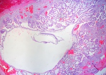
Stage 7

Facts
Human embryonic stage 7 occurs during week 3 between 15 to 17 days.
The embryo is now 0.4 mm diameter in size.
The initial images are displayed unlabeled to allow you to explore the embryo for yourself, linked labeled versions are also available for some images.
Events
| Gastrulation is continuing as cells migrate from the epiblast, continuing to form mesoderm.
Mesoderm lies between the ectoderm and endoderm as a continuous sheet except at the buccopharyngeal and cloacal membranes. These membranes have ectoderm and endoderm only and will lie at the rostral (head) and caudal (tail) of the gastrointestinal tract. From the primitive node a tube extends under the ectoderm in the opposite direction to the primitive streak. This tube forms first the axial process then notochordal process, then finally the notochord. The notochord is a key to embryonic folding and regulation of ectoderm and mesoderm differentiation. It lies in the rostrocordal axis and the embryonic disc will fold either side ventrally, pinching off a portion of the yolk sac to form the lining of the gastrointestinal tract. |
|
Carnegie Stage 8

Facts
Human embryonic stage 8 occurs during week 3 between 17 to 19 days.
The embryo is now 1.0 - 1.5 mm in size.
Events
Gastrulation is continuing as cells migrate from the epiblast, continuing to form mesoderm.
Mesoderm lies between the ectoderm and endoderm as a continuous sheet except at the buccopharyngeal and cloacal membranes. These membranes have ectoderm and endoderm only and will lie at the rostral (head) and caudal (tail) of the gastrointestinal tract.
From the primitive node a tube extends under the ectoderm in the opposite direction to the primitive streak. This tube forms first the axial process then notochordal process, then finally the notochord.
The notochord is a key to embryonic folding and regulation of ectoderm and mesoderm differentiation. It lies in the rostrocordal axis and the embryonic disc will fold either side ventrally, pinching off a portion of the yolk sac to form the lining of the gastrointestinal tract.
Identify
- embryonic disc
- primitive node, primative streak, primative groove
- connecting stalk
- cut amniotic membrane
Carnegie Stage 9

Facts
Human embryonic stage 9 occurs during week 3 between 19 to 21 days.
The embryo is now 1.5 to 2.5 mm in size and somites have begun to form and number between 1 to 3 somite pairs during this stage.
The initial images are displayed unlabeled to allow you to explore the embryo for yourself, linked labeled versions are also available for some images.

Events
Ectoderm - Neural plate brain region continues to expand, neural plate begins folding over the notochord. Gastrulation continues through the primitive streak region.
Mesoderm - Paraxial mesoderm segmentation into somites begins (1 - 3 somite pairs). Lateral plate mesoderm begins to vacuolate, dividing it into somatic and splanchnic mesoderm and to later form the intra-embryonic coelom. Prechordal splanchnic mesoderm begins to form the cardiogenic region, from which the primordial heart will develop.
Endoderm - Notochordal plate still visible which will form the notochord. Endoderm is still widely open to the yolk sac and germ cells form part of this layer. Extra-embryonic mesoderm on the yolk sac surface begins to form "blood islands".
- Mesoderm and Ectoderm Cartoons
Trilaminar Embryo
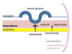
Paraxial and Lateral Plate
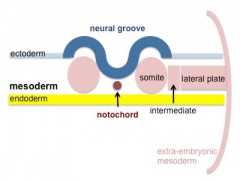
Somites
Somatic and Splanchnic


Identify
- Neural groove and neural folds, the mesoderm showing first somite bulges, that segments beside the neural groove to form somites but extends laterally to margin of embryonic disc lateral plate mesoderm, where it merges with the covering extraembryonic mesoderm.
- The intra-embryonic coelom develops in the middle of the lateral plate mesoderm.
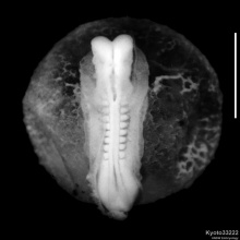
Carnegie Stage 10
Human embryonic stage 10 occurs at the beginning of week 4 development (GA week 6). This week will be covered in detail in the next practical class. The virtual slide image below shows a dorsal view of the embryo at the beginning of week 4.
| Stage 10 - Dorsal View
|
| Stage 10 | Embryo Slides |
- Carnegie Stages: 1 | 2 | 3 | 4 | 5 | 6 | 7 | 8 | 9 | 10 | 11 | 12 | 13 | 14 | 15 | 16 | 17 | 18 | 19 | 20 | 21 | 22 | 23 | About Stages | Timeline
Additional Information
| Additional Information - Content shown under this heading is not part of the material covered in this class. It is provided for those students who would like to know about some concepts or current research in topics related to the current class page. |
Human Development Timeline
The table below shows human development features and approximate timing during the menstrual cycle to fertilization and the first 3 weeks of development.
The timing assumes fertilization the day after ovulation and the "weeks" refer to embryonic development and differ from Gestational Age (GA, clinical weeks shown in brackets, from last menstrual period) and "stages" refer to Carnegie stages of development.
| Links: human timeline | first trimester timeline | second trimester timeline | third trimester timeline | ||||
| Event | ||||
|---|---|---|---|---|
| Menstrual Phase |  menstrual cycle changes: uterus endometrium (loss), ovary (follicle development) | |||

| ||||
| Proliferative Phase |   menstrual cycle changes: uterus endometrium (proliferation), ovary (Follicle Development) menstrual cycle changes: uterus endometrium (proliferation), ovary (Follicle Development)
| |||
| Proliferative Phase | ||||
    menstrual cycle - Mid proliferative menstrual cycle - Mid proliferative
| ||||
  menstrual cycle - Late Proliferative menstrual cycle - Late Proliferative
| ||||
| ovulation
Capacitation |
  | |||
| Event | ||||
| Secretory Phase |    fertilization, zygote, Secretory Phase fertilization, zygote, Secretory Phase
| |||
| Stage 2 |  | |||
| Stage 3 |  blastocyst Hatching (zona pellucida lost) blastocyst Hatching (zona pellucida lost)
| |||
  Late Secretory, blastocyst (free floating) Late Secretory, blastocyst (free floating)
| ||||
| Stage 4 | Adplantation | |||
| Stage 5 | 
| |||
| Stage 6 |  | |||
| Event | ||||
| Stage 7 | 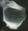 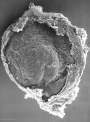 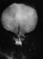 gastrulation, ectoderm, mesoderm, endoderm gastrulation, ectoderm, mesoderm, endoderm
| |||
| Stage 8 | 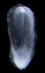 | |||
| ||||
| Stage 9 | 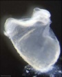  Musculoskeletal somitogenesis, first somites form and continue to be added in sequence caudally (1 - 3 somite pairs). Musculoskeletal somitogenesis, first somites form and continue to be added in sequence caudally (1 - 3 somite pairs).
neural the three main divisions of the brain, which are not cerebral vesicles, can be distinguished while the neural groove is still completely open Neural Crest mesencephalic neural crest is visible[1] | |||
| heart cardiogenesis, week 3 begins as paired heart tubes. | ||||
| Event | ||||
| Stage 10 |   Neural Crest differentiation at spinal cord level from day 22 until day 26 neural folds begin to fuse near the junction between brain and spinal cord, when Neural Crest cells are arising mainly from the neural ectoderm Neural Crest trigeminal, facial, and postotic ganglia components visible[1] Neural Crest migration of vagal level neural crest cells begins (7-10 somite stage) neural rostral neural tube forms 3 primary brain vesicles (week 4) respiratory Week 4 - laryngotracheal groove forms on floor foregut. | |||
| heart begins to beat in Humans by day 22-23, first functioning embryonic organ formed. | ||||
| Stage 11 | 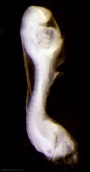
thyroid - thyroid median endodermal thickening in the floor of pharynx neural rostral (or cephalic) neuropore closes within a few hours; closure is bidirectional, it takes place from the dorsal and terminal lips and may occur in two areas simultaneously. The two lips, however, behave differently. ventricular Optic ventricle appears and the neural groove/tube space is initially filled with amniotic fluid.[2] | |||
| Stage 12 | 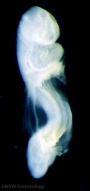 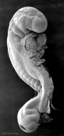
pituitary Week 4 hypophysial pouch, Rathke's pouch, diverticulum from roof liver septum transversum forming liver stroma and hepatic diverticulum forming hepatic trabeculae[3] neural caudal neuropore takes a day to close (closure is approximately at future somitic pair 31/sacral vertebra 2) neural secondary neurulation begins ventricular onset of the ventricular system and separates the ependymal from the amniotic fluid.[2] neural crest cardiac crest, neural crest from rhombomeres 6 and 7 that migrates to pharyngeal arch 3 and from there the truncus arteriosus[1] neural crest vagal neural crest enter the foregut (20-25 somite stage) | |||
| Stage 13 |   neural the neural tube is normally completely closed, ventricular system now separated from amniotic fluid. Neural crest at spinal level is segregating, and spinal ganglia are in series with the somites. Spinal cord ventral roots beginning to develop.[4] neural the neural tube is normally completely closed, ventricular system now separated from amniotic fluid. Neural crest at spinal level is segregating, and spinal ganglia are in series with the somites. Spinal cord ventral roots beginning to develop.[4]
telencephalon cavity appears Neural - Vascular Development - hindbrain is supplied by two parallel neural arteries (or channels) that obtain their blood supply from carotid-vertebrobasilar anastomoses given by the pharyngeal arch arteries; trigeminal artery, the otic artery, hypoglossal artery, and the proatlantal artery.[5] liver epithelial cord proliferation enmeshing stromal capillaries[3] smell Crest comes from the nasal plates[6] integumentary 4 weeks - simple ectoderm epithelium over mesenchyme integumentary 1-3 months ectoderm- germinative (basal) cell repeated division of generates stratified epithelium; mesoderm- differentiates into connective tissue and blood vessels vision Optic vesicle lies close to the surface ectoderm. The surface ectoderm overlying the optic vesicle, in response to this contact, has thickened to form the lens placode.[7] Diaphragm - pleuroperitoneal fold (PPF) first discernible in human embryos (CRL 6mm).[8] | |||
| pituitary Week 5 elongation, contacts infundibulum, diverticulum of diencephalon
heart Week 5 septation starts, atrial and ventricular respiratory Week 5 left and right lung buds push into the pericardioperitoneal canals (primordia of pleural cavity) Respiratory Week 5 to 17 lung histology - pseudoglandular hearing Week 5 cochlear part of otic vesicle elongates (humans 2.5 turns) | ||||
| Stage 14 |   Placodes sensory placodes, lens pit, otocyst, nasal placode, primary/secondary vesicles, fourth ventricle of brain Placodes sensory placodes, lens pit, otocyst, nasal placode, primary/secondary vesicles, fourth ventricle of brain
somite continued segmentation of paraxial mesoderm (somite pairs), heart prominence head 1st, 2nd and 3rd pharyngeal arch, forebrain, site of lens placode, site of otic placode, stomodeum Body - heart, liver, umbilical cord, mesonephric ridge visible externally as bulges. limb upper and lower limb buds growing. Abdominal Wall mesoderm of the primary body wall coalesced in the ventral midline to create the abdominal cavity.[9] neural first appearance of the future cerebral hemispheres. Cerebellar plate differentiated to an intermediate layer, and future rhombic lip identifiable[10] Neural - Vascular Development - basilar artery forms from the consolidation of the neural arteries.[5] ventricular Subarachnoid space initially as irregular spaces on the ventral surface of the spinal cord.[11] liver hepatic gland and its vascular channels enlarge, hematopoietic function appears[3] | |||
| Stage 15 | 
neural cranial nerves (except olfactory and optic) are identifiable in more advanced embryos[12] Neural - Vascular Development - vertebral arteries form from transverse anastomoses between cervical intersegmental arteries, beginning with the proatlantal artery and proceeding downward to the 6th intersegmental artery,[5] vision lens pit is closed. The lens vesicle and optic cup lie close to the surface ectoderm and appear to press against the surface.[7] | |||
| vision 35 to 37 days retinal pigment present | ||||
| pituitary Week 6 - connecting stalk between pouch and oral cavity degenerates
parathyroid Week 6 - diverticulum elongate, hollow then solid, dorsal cell proliferation thymus Week 6 - diverticulum elongate, hollow then solid, ventral cell proliferation adrenal Week 6 - fetal cortex forms from mesothelium adjacent to dorsal mesentery, medulla neural crest cells from adjacent sympathetic ganglia respiratory Week 6 - descent of heart and lungs into thorax. Pleuroperitoneal foramen closes tongue Week 6 - gustatory papilla, caudal midline near the foramen caecum (week 6 to 7 - nerve fibers approach the lingual epithelium) | ||||
| Stage 16 |  Neural first parasympathetic ganglia, submandibular and ciliary, are identifiable[13] Neural first parasympathetic ganglia, submandibular and ciliary, are identifiable[13]
Neural - Vascular Development - development of the middle cerebral artery is first identified as small buds originating proximal to the anterior cerebral artery on the anterior division of the primitive internal carotid artery.[5] limb upper limb bud nerves median nerve, radial nerve and ulnar nerve entered into hand plate, myoblasts spindle shaped and oriented parallel to limb bud axis. Abdominal Wall muscle cell migration about 25% of the hemicircumference of the abdominal cavity, the lateral plate mesoderm has become more condensed and thicker in the area around the myoblasts.[9] heart outflow tract elliptical configuration with four cushions, the two larger fusing at this stage. Semilunar valve leaflets form at the downstream end of the cushions head lip and palate components of the upper lip, medial nasal prominence and maxillary process present, median palatine process appears. Eyelid prior to the development of the eyelids, one small sulcus or groove forms above the eye (eyelid groove) and another below it.[7] | |||
| Stage 17 | 
| |||
| heart separation of common cardiac outflow (aortic arch and pulmonary aorta) | ||||
| Event | ||||
| pancreas Week 7 to 20 pancreatic hormones secretion increases, small amount maternal insulin
respiratory Week 7 - enlargement of liver stops descent of heart and lungs | ||||
| Stage 18 | 
limb bone forms by endochondrial ossification and throughout embryo replacement of cartilage with bone (week 5-12). neural smell vomeronasal fibres and nervus terminalis[6] liverobturation due to epithelial proliferation, bile ducts became reorganized, continuity between liver cells and gut[3] ventricular duramater appears and spaces surround the circumference of the spinal cord, which coalesce and contain many blood vessels.[15] Female uterus opening of the paramesonephric (Müllerian) duct to the coelomic cavity formed as an invagination of the coelomic epithelium[16] Abdominal Wall separation of the myoblasts into distinct inner and outer layers, with unidirectional orientation. Abdominal wall thicker in the region where secondary structures were forming compared with the primary body wall region, dorsally outermost layer of connective tissue approximately half of this thickness.[9] | |||
|
liver (stage 18 to 23) biliary ductules developed in periportal connective tissue produces ductal plates that receive biliary capillaries[3] | ||||
| Stage 19 | 
| |||
| Stage 20 | 
Head scalp vascular plexus visible limb upper limbs begin to rotate ventrally neural amygdaloid body has at least four individual nuclei[24] oculomotor nerve shows a dorsolateral and a ventromedial portion rhombic lip (rhombencephalon) formation of the cerebellum (intermediate layer) and of the cochlear nuclei cerebellum cell layer (future Purkinje cells) develops choroid plexuses of the fourth and lateral ventricles | |||
| gastrointestinal tract anal membrane perforates | ||||
| Stage 21 | 
neural cortical plate appears in the area of future insula[25] Neural - Vascular Development - formation of the anterior communicating artery.[5] limb upper and lower limbs rotate Intraembryonic Coelom pericardioperitoneal canals close Abdominal Wall Myoblasts have reached the ventral midline and myotubes were present and oriented uniformly within all muscle groups. The rectus abdominis formed distinct bundles of muscle. Connective tissue layers comprised the majority of the thickness of the abdominal wall, outermost layer of connective tissue accounted for the majority of this thickness.[9] | |||
| Stage 22 |  neural neocortical fibres project to epithalamus, to dorsal thalamus, and to mesencephalon[25] neural neocortical fibres project to epithalamus, to dorsal thalamus, and to mesencephalon[25]
limb fingers and toes lengthen smell Stage 22 to early fetal period - migratory streams of neurons from the subventricular zone of the olfactory bulb towards the future claustrum[6] Uterus Vagina fused duct (uterovaginal canal) bifurcated at the caudal portion at Carnegie stages 22 and 23[16] | |||
| Genital 8 Weeks Testis - mesenchyme, interstitial cells (of Leydig) secrete testosterone, androstenedione
Genital 8 to 12 Weeks - hCG stimulates testosterone production Tongue Week 8 - nerves penetrate epitheilai basal lamina and synapse with undifferentiated, elongated, epithelial cells (taste bud progenitor cell)[26] | ||||
| Stage 23 |  Stage 23 defines the end of the embryonic (organogenesis) period Stage 23 defines the end of the embryonic (organogenesis) period
Mesoderm heart prominence, ossification continues Head nose, eye, external acoustic meatus, eyelids, external ears, rounded head Body - straightening of trunk, umbilical cord, intestines herniated at umbilicus limb upper limbs longer and bent at elbow, hands and feet turned inward, foot with separated digits, wrist, hand with separated digits Extraembryonic Coelom chorionic cavity is now lost by fusion with the expanding amniotic cavity neural rhombencephalon, pyramidal decussation present, nuclei and tracts similar to those present in the newborn cerebellum present as only a plate connected to midbrain and hindbrain through fibre bundles[27] Axial Skeleton vertebral column 33 or 34 cartilaginous vertebrae (20-33 mm in total length), vertebral pedicles, articular and transverse processes identifiable (no spinous processes)[28] Abdominal Wall Rectus muscle forms 2 or 3 distinct layers with myotube orientation uniform in all muscles. The external oblique and internal oblique started to expand in thickness, transversus a thin layer of muscle.[9] | |||
| Week 8 | Stomach Week 8 - Gastrin containing cells in stomach antrum. Somatostatin cells in both the antrum and the fundus.
Genital - Female Development paired paramesonephric (Müllerian) ducts contact each other and are fused into a single tube that separates again and returns to the mesonephric (Wolffian) ducts. The paramesonephric ducts have not yet reached the urogenital sinus.[16] | |||
| Week 9 | Beginning of Fetal Development
CRL 43 mm, femur length 6 mm 9 weeks CRL 50 mm - genital genitalia in both sexes look identical[30] uterus - paramesonephric ducts come into apposition with the urorectal septum and begin to fuse | |||
| Event | ||||

Gastrointestinal Tract Week 10 intestines in abdomen Pituitary growth hormone and ACTH detectable Pancreas Week 10 glucagon (alpha) differentiate first, somatostatin (delta), insulin (beta) cells differentiate, insulin secretion begins Tongue Week 10 shallow grooves above the taste bud primordium Stomach Week 10 - Glucagon containing cells in stomach fundus. Nail Development fingernails appear outer ear Week 10 - Meatal plug extends in a disc-like fashion, the meatus is boot-shaped with a narrow neck and the sole of the meatal plug spreading widely to form the future tympanic membrane medially. Proximal portion of the neck starts to be resorbed. inner ear Week 10 - neural-crest-derived melanocytes migrate into the cochlea. They penetrate the basement membrane of the lateral wall epithelium and develop into the intermediate cells of the stria vascularis.[31] | ||||
| Week 10 - CRL 55 mm, femur length 9 mm, biparietal diameter 17 mm | ||||
| Event
neural - Cerebrum appearance of the first sulcus (week 11-15, GA 13-17 weeks)[32] | ||||
|
Thyroid colloid appearance in thyroid follicles, iodine and thyroid hormone (TH) synthesis Stomach Week 11 - Serotonin containing cells in both the antrum and the fundus. | ||||
| Week 11 - CRL 68 mm, femur length 12 mm, biparietal diameter 20 mm | ||||

| Week: | 1 | 2 | 3 | 4 | 5 | 6 | 7 | 8 |
| Carnegie stage: | 1 2 3 4 | 5 6 | 7 8 9 | 10 11 12 13 | 14 15 | 16 17 | 18 19 | 20 21 22 23 |
Next
Finished Lab 3 !
If you have finished and would like to apply your knowledge, I have also included some Clinical Questions based around this period of development.
If you have finished and need some more help understanding this period of development, I have included some links to Online References.
If you have finished and are interested in looking at tissues involved in this period of development, I have included some links to Histology Images.
Note that this Practical has discussed mainly development of the embryo as placental development will be covered in detail in another practical (Placenta and Fetal Membranes).
The next Practical will continue on through embryonic development (Implantation to 8 Weeks).
References
- ↑ 1.0 1.1 1.2 O'Rahilly R & Müller F. (2007). The development of the neural crest in the human. J. Anat. , 211, 335-51. PMID: 17848161 DOI.
- ↑ 2.0 2.1 O'Rahilly R & Müller F. (1990). Ventricular system and choroid plexuses of the human brain during the embryonic period proper. Am. J. Anat. , 189, 285-302. PMID: 2285038 DOI.
- ↑ 3.0 3.1 3.2 3.3 3.4 Godlewski G, Gaubert-Cristol R, Rouy S & Prudhomme M. (1997). Liver development in the rat and in man during the embryonic period (Carnegie stages 11-23). Microsc. Res. Tech. , 39, 314-27. PMID: 9407542 <314::AID-JEMT2>3.0.CO;2-H DOI.
- ↑ Müller F & O'Rahilly R. (1988). The development of the human brain from a closed neural tube at stage 13. Anat. Embryol. , 177, 203-24. PMID: 3354839
- ↑ 5.0 5.1 5.2 5.3 5.4 5.5 Menshawi K, Mohr JP & Gutierrez J. (2015). A Functional Perspective on the Embryology and Anatomy of the Cerebral Blood Supply. J Stroke , 17, 144-58. PMID: 26060802 DOI.
- ↑ 6.0 6.1 6.2 6.3 Müller F & O'Rahilly R. (2004). Olfactory structures in staged human embryos. Cells Tissues Organs (Print) , 178, 93-116. PMID: 15604533 DOI.
- ↑ 7.0 7.1 7.2 7.3 7.4 7.5 7.6 Pearson AA. (1980). The development of the eyelids. Part I. External features. J. Anat. , 130, 33-42. PMID: 7364662
- ↑ 8.0 8.1 Clugston RD, Zhang W & Greer JJ. (2010). Early development of the primordial mammalian diaphragm and cellular mechanisms of nitrofen-induced congenital diaphragmatic hernia. Birth Defects Res. Part A Clin. Mol. Teratol. , 88, 15-24. PMID: 19711422 DOI.
- ↑ 9.0 9.1 9.2 9.3 9.4 9.5 9.6 Nichol PF, Corliss RF, Yamada S, Shiota K & Saijoh Y. (2012). Muscle patterning in mouse and human abdominal wall development and omphalocele specimens of humans. Anat Rec (Hoboken) , 295, 2129-40. PMID: 22976993 DOI.
- ↑ Müller F & O'Rahilly R. (1988). The first appearance of the future cerebral hemispheres in the human embryo at stage 14. Anat. Embryol. , 177, 495-511. PMID: 3377191
- ↑ Patelska-Banaszewska M & Woźniak W. (2005). The subarachnoid space develops early in the human embryonic period. Folia Morphol. (Warsz) , 64, 212-6. PMID: 16228957
- ↑ Müller F & O'Rahilly R. (1988). The development of the human brain, including the longitudinal zoning in the diencephalon at stage 15. Anat. Embryol. , 179, 55-71. PMID: 3213956
- ↑ Müller F & O'Rahilly R. (1989). The human brain at stage 16, including the initial evagination of the neurohypophysis. Anat. Embryol. , 179, 551-69. PMID: 2751117
- ↑ Müller F & O'Rahilly R. (1989). The human brain at stage 17, including the appearance of the future olfactory bulb and the first amygdaloid nuclei. Anat. Embryol. , 180, 353-69. PMID: 2802187
- ↑ 15.0 15.1 Patelska-Banaszewska M & Woźniak W. (2004). The development of the epidural space in human embryos. Folia Morphol. (Warsz) , 63, 273-9. PMID: 15478101
- ↑ 16.0 16.1 16.2 16.3 Hashimoto R. (2003). Development of the human Müllerian duct in the sexually undifferentiated stage. Anat Rec A Discov Mol Cell Evol Biol , 272, 514-9. PMID: 12740945 DOI.
- ↑ Keibel F. and Mall FP. Manual of Human Embryology II. (1912) J. B. Lippincott Company, Philadelphia.
- ↑ Congdon ED. Transformation of the aortic-arch system during the development of the human embryo. (1922) Contrib. Embryol., Carnegie Inst. Wash. Publ 277, 14:47-110.
- ↑ Teal SI., Moore GW. and Hutchins GM. Development of aortic and mitral valve continuity in the human embryonic heart. (1986) Amer. J. Anat., 176:447-460.
- ↑ Wells LJ. Development of the human diaphragm and pleural sacs. (1954) Contrib. Embryol., Carnegie Inst. Wash. Publ. 603, 35: 107-134.
- ↑ Jirásek JE. Development of the Genital System and Male Pseudohermaphroditism. (1971) Johns Hopkins Press, Baltimore.
- ↑ Wilson KM. Origin and development of the rete ovarii and the rete testis in the human embryo. (1926) Carnegie Instn. Wash. Publ. 362, Contrib. Embryol., Carnegie Inst. Wash., 17:69-88.
- ↑ Gasser RL. Atlas of Human Embryos. (1975) Harper & Row, Hagerstown, Maryland.
- ↑ 24.0 24.1 Müller F & O'Rahilly R. (1990). The human brain at stages 18-20, including the choroid plexuses and the amygdaloid and septal nuclei. Anat. Embryol. , 182, 285-306. PMID: 2268071
- ↑ 25.0 25.1 Müller F & O'Rahilly R. (1990). The human brain at stages 21-23, with particular reference to the cerebral cortical plate and to the development of the cerebellum. Anat. Embryol. , 182, 375-400. PMID: 2252222
- ↑ Witt M & Reutter K. (1996). Embryonic and early fetal development of human taste buds: a transmission electron microscopical study. Anat. Rec. , 246, 507-23. PMID: 8955790 <507::AID-AR10>3.0.CO;2-S DOI.
- ↑ Müller F & O'Rahilly R. (1990). The human rhombencephalon at the end of the embryonic period proper. Am. J. Anat. , 189, 127-45. PMID: 2244584 DOI.
- ↑ O'Rahilly R, Muller F & Meyer DB. (1980). The human vertebral column at the end of the embryonic period proper. 1. The column as a whole. J. Anat. , 131, 565-75. PMID: 7216919
- ↑ Streeter GL. Developmental horizons in human embryos (fourth issue). A review of the histogenesis of cartilage and bone. (1949) Carnegie Instn. Wash. Publ. 583, Contrib. Embryol., 33: 149-169. PMID: 18144445
- ↑ Wünsch L & Schober JM. (2007). Imaging and examination strategies of normal male and female sex development and anatomy. Best Pract. Res. Clin. Endocrinol. Metab. , 21, 367-79. PMID: 17875485 DOI.
- ↑ Locher H, de Groot JC, van Iperen L, Huisman MA, Frijns JH & Chuva de Sousa Lopes SM. (2015). Development of the stria vascularis and potassium regulation in the human fetal cochlea: Insights into hereditary sensorineural hearing loss. Dev Neurobiol , 75, 1219-40. PMID: 25663387 DOI.
- ↑ Afif A, Bouvier R, Buenerd A, Trouillas J & Mertens P. (2007). Development of the human fetal insular cortex: study of the gyration from 13 to 28 gestational weeks. Brain Struct Funct , 212, 335-46. PMID: 17962979 DOI.
BGDA: Lecture 1 | Lecture 2 | Practical 3 | Practical 6 | Practical 12 | Lecture Neural | Practical 14 | Histology Support - Female | Male | Tutorial
Glossary Links
- Glossary: A | B | C | D | E | F | G | H | I | J | K | L | M | N | O | P | Q | R | S | T | U | V | W | X | Y | Z | Numbers | Symbols | Term Link
Cite this page: Hill, M.A. (2024, April 25) Embryology BGDA Practical 3 - Week 3 Summary. Retrieved from https://embryology.med.unsw.edu.au/embryology/index.php/BGDA_Practical_3_-_Week_3_Summary
- © Dr Mark Hill 2024, UNSW Embryology ISBN: 978 0 7334 2609 4 - UNSW CRICOS Provider Code No. 00098G