UNSW Embryology
The Developing Human: Clinically Oriented Embryology
Moore, K.L., Persaud, T.V.N. & Torchia, M.G. (2015). The developing human: clinically oriented embryology (10th ed.). Philadelphia: Saunders. (links only function with UNSW connection)
Larsen's Human Embryology
Schoenwolf, G.C., Bleyl, S.B., Brauer, P.R., Francis-West, P.H. & Philippa H. (2015). Larsen's human embryology (5th ed.). New York; Edinburgh: Churchill Livingstone.(links only function with UNSW connection)
More Textbooks?
Week 3
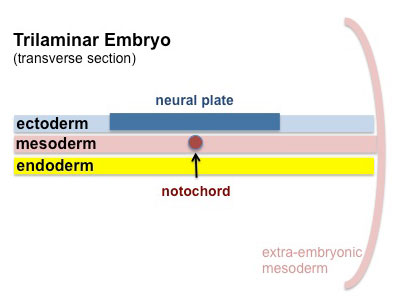
Ectoderm
- neural plate - midline (columnar cells)
- neural crest - outside lateral edges of neural plate
- surface ectoderm - lateral (cuboidal cells)
- head - sensory and anterior pituitary (placodes)
- integument - epidermis of skin, hair, glands, teeth enamel
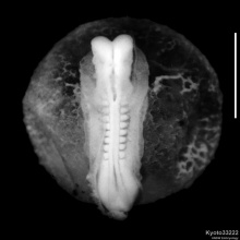
Neural Plate
- extends from buccopharyngeal membrane (oral membrane) to primitive node (Hensen's node)
- forms above notochord and paraxial mesoderm
- neuroectodermal cells - neural plate, neural crest
- rostrocaudal width
- brain plate (broad)
- spinal cord (narrow)
|
<html5media height="520" width="320">File:Neuralplate_001.mp4</html5media>
|
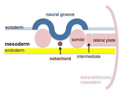
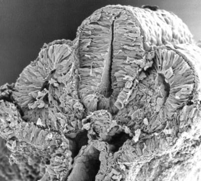
Week 4
Neural Tube
| neural groove
|
neural tube and neural crest
|
|

|
| <html5media height="480" width="480">File:Neuraltube_001.mp4</html5media>
|
<html5media height="440" width="380">File:Mouse neural tube 01.mp4</html5media>
|
Neural Crest
Neural Crest Origin
| System
|
Cell Type
|
| Peripheral Nervous System (PNS)
|
Neurons - sensory ganglia, sympathetic and parasympathetic ganglia, enteric nervous system, and plexuses
Glia (neuroglial cells) - Schwann cells[1], satellite cells, olfactory ensheathing cells[2]
|
| endocrine
|
Adrenal medulla
Calcitonin-secreting cells
Carotid body type I cells
|
| integumentary
|
Epidermal pigment cells melanocyte
|
| Facial cartilage and bone
|
Facial and anterior ventral skull cartilage and bones
|
| Sensory
|
inner ear, cornea endothelium and stroma
|
| Connective tissue
|
tooth odontoblast
smooth muscle, and adipose tissue of skin in head and neck
Connective tissue of meninges, salivary, lachrymal, thymus, thyroid, and pituitary glands
Connective tissue and smooth muscle in arteries of aortic arch origin
|
| Links: neural crest | Category:Neural Crest | Neural Crest collapsible table
|
Neural Crest Development
Primary Brain Vesicles
Traditional vesicle description (simplified name and alternate neuromere description in brackets)
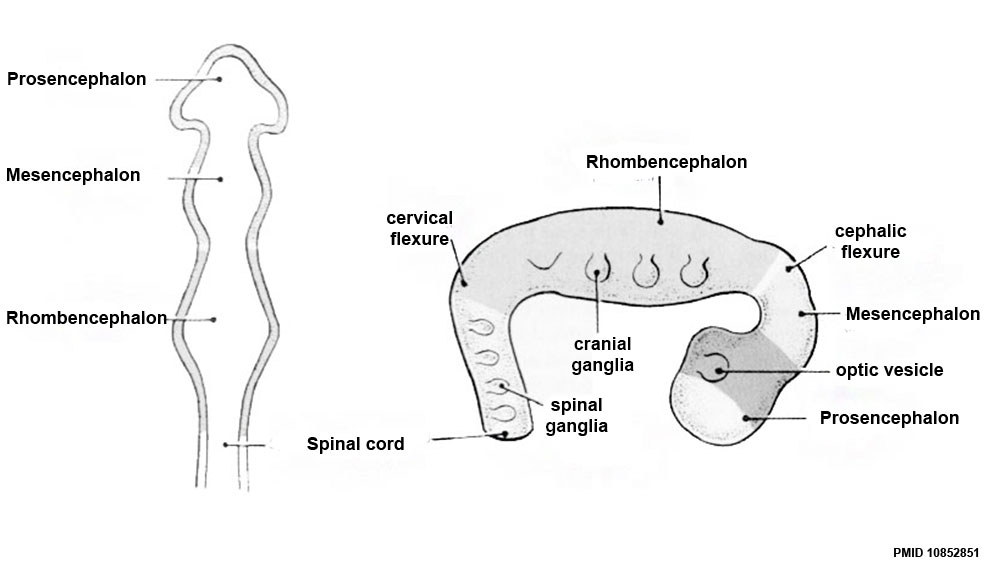
Brain
- Prosencephalon (forebrain, prosomeres)
- Mesencephalon (midbrain, mesomeres)
- Rhombencephalon (hindbrain, rhombomeres)
Spinal Cord
| Neural Tube Regions
|
| Neural Tube Early Structures
|
| Neural Tube (stage 11)
|
Regions
|
Anatomical location
|
Patterning region
|
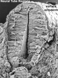
|
roof plate
|
dorsal
|
surface ectoderm
|
| alar plate
|
dorsal lateral
|
surface ectoderm
|
| basal plate
|
ventral lateral
|
notochord and floor plate
|
| floor plate
|
ventral
|
notochord
|
Table above shows the future transient regions that develop from the early neural tube.
|
Links: Spinal Cord
Week 5
Secondary Brain Vesicles
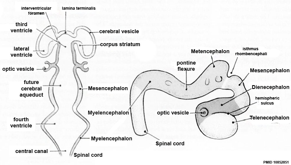
- Telencephalon
- Diencephalon
- Mesencephalon
- Metencephalon
- Myelencephalon
Brain Flexures
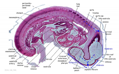
|
Rapid growth folds the neural tube forming 3 brain flexures (cranial to caudal)
- cephalic flexure - (mesencephalic) pushes mesencephalon upwards
- pontine flexure - generates 4th ventricle (cerebellum will grow into this space)
- cervical flexure - between brain stem and spinal cord
|
Ventricles
- cavity within neural tube will form the contiguious space of the ventricules of the brain and central canal of spinal cord
- space is filled initially with amniotic fluid, later with CerebroSpinal Fluid (CSF)
- CSF is secreted by
- chorioid plexus modified vascular structures lying within the ventricles
- floor of lateral ventricle and roof of the third and fourth ventricles
- ventricular ependymal cells and cells lining the subarachnoid space
- CSF also fills the subarachnoid space (between arachnoid mater and pia mater).
| Adult Ventricular Structures
|
Brain four ventricles and several foramina (openings that connect ventricular spaces)
- 2 lateral ventricles (right and left)
- interventricular foramina (foramina of Monro)
- third ventricle
- cerebral aqueduct (Sylvius)
- fourth ventricle
- median aperture (Magendie) subarachnoid space via the cisterna magna
- right and left lateral aperture (Luschka) subarachnoid space via the cistern of great cerebral vein
Spinal cord
|
|
| Adult Meninges Layers
|
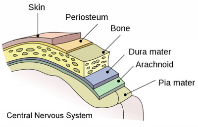
|
|
- Links: Neural - Ventricular System Development
|
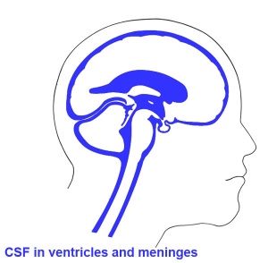
CSF-filled spaces in adult brain.
|
Week 6
| <html5media height="600" width="520">File:Human embryo tomography Carnegie stage 17.mp4</html5media>
Movie
|
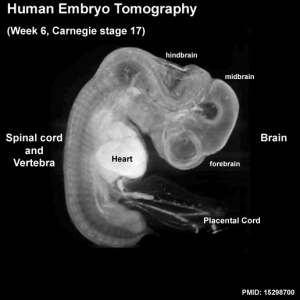
Note the shape and size of the different regions of the brain and spinal cord.
- Telencephalon (cerebrum) has begun to expand and will eventually cover the midbrain region.
- Dorsal root ganglia are visible outside the spinal cord.
|
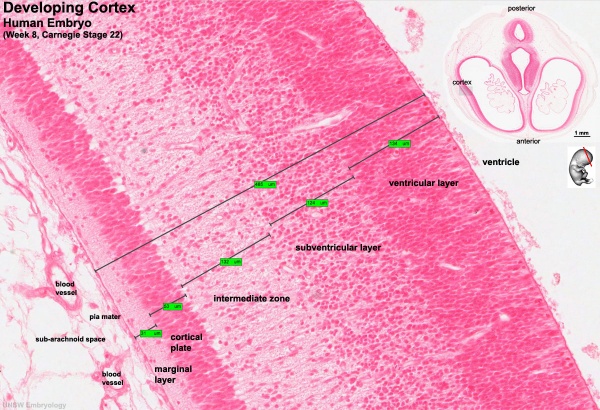
Week 8
Cortex
Spinal Cord

Fetal
Second Trimester
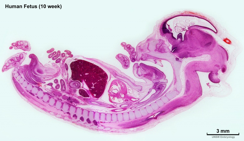
Human week 10 fetus
|

|
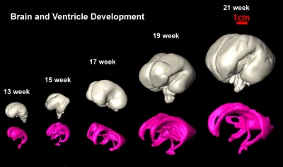
| Brain and Ventricular Development[3]
|
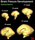
Brain Fissure Development[3]
|
Sylvian Fissure Development
<html5media height="480" width="400">File:Neural_-_Sylvian_fissure.mp4</html5media>
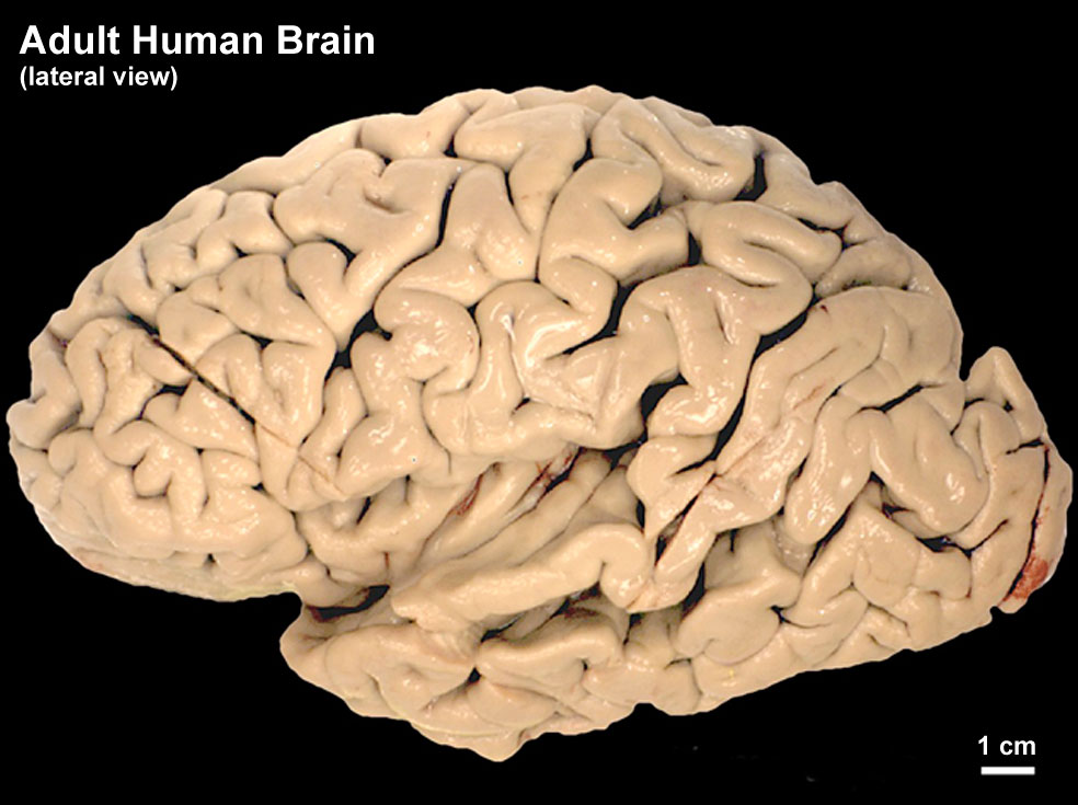
Third Trimester
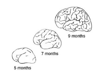
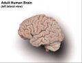
The brain goes from a smooth surface to begin to fold.
- Folds occur as millions of cells push into the cortex, increasing the surface area.
- groove - fissure (plural, fissures).
- fold - gyrus (plural, gyri).
|
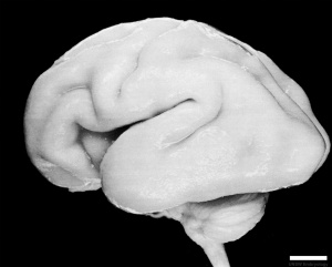
Human Fetus (CRL 240mm) Brain
|
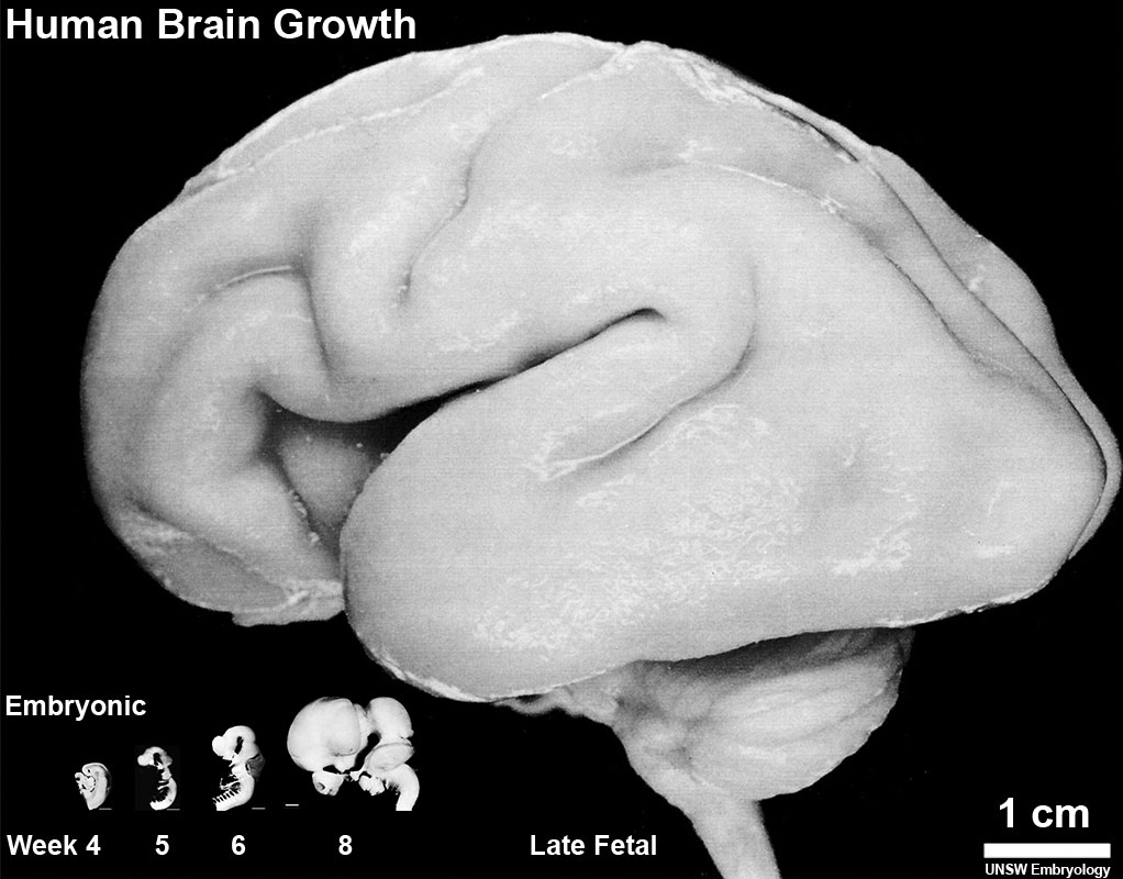
| Human Brain Growth
|
| Embryonic
|
| Table below shows a direct comparison of brain growth in size between week 4 to 8 (GA 6-10)
|
| Fetal
|
|
| Adult
|
|
| Adult CNS Structures
|
Neural Tube Development
| Neural Tube
|
Primary Vesicles
|
Secondary Vesicles
|
Adult Structures
|
| week 3
|
week 4
|
week 5
|
adult
|
neural plate
neural groove
neural tube
Brain
|
prosencephalon (forebrain)
|
telencephalon
|
Rhinencephalon, Amygdala, hippocampus, cerebrum (cortex), hypothalamus, pituitary | Basal Ganglia, lateral ventricles
|
| diencephalon
|
epithalamus, thalamus, Subthalamus, pineal, posterior commissure, pretectum, third ventricle
|
| mesencephalon (midbrain)
|
mesencephalon
|
tectum, Cerebral peduncle, cerebral aqueduct, pons
|
| rhombencephalon (hindbrain)
|
metencephalon
|
cerebellum
|
| myelencephalon
|
medulla oblongata, isthmus
|
| spinal cord, pyramidal decussation, central canal
|
|
Fetal Timeline
Postnatal
Movies
Abnormalities
BGDA: Lecture 1 | Lecture 2 | Practical 3 | Practical 6 | Practical 12 | Lecture Neural | Practical 14 | Histology Support - Female | Male | Tutorial
Glossary Links
- Glossary: A | B | C | D | E | F | G | H | I | J | K | L | M | N | O | P | Q | R | S | T | U | V | W | X | Y | Z | Numbers | Symbols | Term Link
Cite this page: Hill, M.A. (2024, April 19) Embryology BGDA Lecture - Development of the Nervous System. Retrieved from https://embryology.med.unsw.edu.au/embryology/index.php/BGDA_Lecture_-_Development_of_the_Nervous_System
- What Links Here?
- © Dr Mark Hill 2024, UNSW Embryology ISBN: 978 0 7334 2609 4 - UNSW CRICOS Provider Code No. 00098G
|


