BGDA Lecture - Development of the Nervous System
| Embryology - 24 Apr 2024 |
|---|
| Google Translate - select your language from the list shown below (this will open a new external page) |
|
العربية | català | 中文 | 中國傳統的 | français | Deutsche | עִברִית | हिंदी | bahasa Indonesia | italiano | 日本語 | 한국어 | မြန်မာ | Pilipino | Polskie | português | ਪੰਜਾਬੀ ਦੇ | Română | русский | Español | Swahili | Svensk | ไทย | Türkçe | اردو | ייִדיש | Tiếng Việt These external translations are automated and may not be accurate. (More? About Translations) |
Introduction
Mark Hill (talk) 11:37, 15 May 2017 (AEST) New lecture under development - this notice removed when completed. Begin your background reading by looking at the textbook links.
Neural development is a complex and ongoing process that commences in week 3 and continues through into the postnatal period. This lecture will introduce concepts about the timing, origin and abnormalities of the nervous system. Lecture content will be added to this current page, the linked online textbook chapters are available as pre-reading for this lecture.
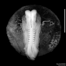
The human MRI movie below (head, sagittal plane, left to right) shows the central nervous system (CNS) development at the end of the embryonic period (week 8; GA week 10).
<html5media height="500" width="550">File:Stage23 MRI S01.mp4</html5media>
|
|
|
|
|
Aim
To develop an understanding of the development of the nervous system and the consequences of abnormal development.
Draft Lecture Timetable - Monday 29 May 2017 09:00 AM - 10:00 AM Development of the nervous system Kensington - Rex Vowels Theatre
Textbooks
UNSW Embryology
The Developing Human: Clinically Oriented EmbryologyMoore, K.L., Persaud, T.V.N. & Torchia, M.G. (2015). The developing human: clinically oriented embryology (10th ed.). Philadelphia: Saunders. (links only function with UNSW connection)
Larsen's Human EmbryologySchoenwolf, G.C., Bleyl, S.B., Brauer, P.R., Francis-West, P.H. & Philippa H. (2015). Larsen's human embryology (5th ed.). New York; Edinburgh: Churchill Livingstone.(links only function with UNSW connection) Week 3Ectoderm
Neural Plate
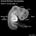
Week 4Neural Tube
Neural CrestPrimary Brain VesiclesTraditional vesicle description (simplified name and alternate neuromere description in brackets)
Brain
Spinal Cord
Links: Spinal Cord Week 5Secondary Brain Vesicles
Brain Flexures
Ventricles
Week 6
Week 8Cortex
Spinal Cord
FetalSecond Trimester
Human week 10 fetus
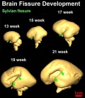
Sylvian Fissure Development <html5media height="480" width="400">File:Neural_-_Sylvian_fissure.mp4</html5media> Third Trimester
Fetal Timeline
Postnatal
Movies
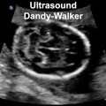
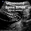
Abnormalities
Glossary Links
Cite this page: Hill, M.A. (2024, April 24) Embryology BGDA Lecture - Development of the Nervous System. Retrieved from https://embryology.med.unsw.edu.au/embryology/index.php/BGDA_Lecture_-_Development_of_the_Nervous_System
|
|||||||||||||||||||||||||||||||||||||||||||||||||||||||||||||||||||||||||||||||||||||||||||||||||||||||||||||||||||||||||||||||||||||||||||||||||||||||||||||||||||||||||||||||||||||||||||||||||||||||||


- ↑ 1.0 1.1 <pubmed>19339620</pubmed>| PMC2721010 | J Neurosci.