Atlas of the Development of Man 1 - Part 2: Difference between revisions
No edit summary |
No edit summary |
||
| (11 intermediate revisions by the same user not shown) | |||
| Line 4: | Line 4: | ||
{{KollmannAtlas1}} | {{KollmannAtlas1}} | ||
==I. Cleavage, Segmentation== | ==I. Cleavage, Segmentation== | ||
<gallery> | |||
File:Kollmann015.jpg | |||
File:Kollmann016.jpg | |||
File:Kollmann017.jpg | |||
File:Kollmann018.jpg | |||
File:Kollmann019.jpg | |||
File:Kollmann020.jpg | |||
</gallery> | |||
a) External appearances of cleavage | a) External appearances of cleavage | ||
<gallery> | |||
File:Kollmann021.jpg | |||
File:Kollmann022.jpg | |||
</gallery> | |||
b) Internal appearances of cleavage, mitosis | b) Internal appearances of cleavage, mitosis | ||
<gallery> | |||
File:Kollmann023.jpg | |||
File:Kollmann024.jpg | |||
File:Kollmann025.jpg | |||
File:Kollmann026.jpg | |||
File:Kollmann027.jpg | |||
File:Kollmann028.jpg | |||
File:Kollmann029.jpg | |||
File:Kollmann030.jpg | |||
</gallery> | |||
# Occurrence and transformation of the dark chromatic nuclear division figure | # Occurrence and transformation of the dark chromatic nuclear division figure | ||
# Occurrence and transformation of the achromatic (colorless) core figure, called the mitotic spindle | # Occurrence and transformation of the achromatic (colorless) core figure, called the mitotic spindle | ||
| Line 18: | Line 39: | ||
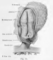
==II. Germinal vesicle (vesicula blastodermica) with the germinative (area embryonalis), viewed from outside== | ==II. Germinal vesicle (vesicula blastodermica) with the germinative (area embryonalis), viewed from outside== | ||
<gallery> | |||
File:Kollmann033.jpg | |||
File:Kollmann034.jpg | |||
File:Kollmann035.jpg | |||
File:Kollmann036.jpg | |||
File:Kollmann037.jpg | |||
File:Kollmann038.jpg | |||
File:Kollmann039.jpg | |||
</gallery> | |||
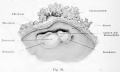
==III. Germinal vesicle internal structure== | ==III. Germinal vesicle internal structure== | ||
| Line 26: | Line 57: | ||
The Fundamental organs | The Fundamental organs | ||
<gallery> | |||
File:Kollmann040.jpg | |||
File:Kollmann041.jpg | |||
File:Kollmann042.jpg | |||
File:Kollmann043.jpg | |||
File:Kollmann044.jpg | |||
File:Kollmann045.jpg | |||
File:Kollmann046.jpg | |||
</gallery> | |||
==IV. Primitive streak and neurenteric canal== | ==IV. Primitive streak and neurenteric canal== | ||
<gallery> | |||
File:Kollmann046.jpg | |||
File:Kollmann047.jpg | |||
File:Kollmann048.jpg | |||
File:Kollmann049.jpg | |||
File:Kollmann050.jpg | |||
</gallery> | |||
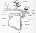
==V. Notochord== | ==V. Notochord== | ||
| Line 34: | Line 82: | ||
# Sub-chordal line | # Sub-chordal line | ||
# Origin of the notochord | # Origin of the notochord | ||
<gallery> | |||
File:Kollmann052.jpg | |||
File:Kollmann053.jpg | |||
File:Kollmann054.jpg | |||
File:Kollmann055.jpg | |||
File:Kollmann056.jpg | |||
File:Kollmann057.jpg | |||
</gallery> | |||
==VI. Middle germ layer, mesoderm== | ==VI. Middle germ layer, mesoderm== | ||
a) root zone and parietal zone | a) root zone and parietal zone | ||
<gallery> | |||
File:Kollmann058.jpg | |||
File:Kollmann059.jpg | |||
File:Kollmann060.jpg | |||
File:Kollmann061.jpg | |||
File:Kollmann062.jpg | |||
File:Kollmann063.jpg | |||
</gallery> | |||
b) the origin of the middle germ layer | b) the origin of the middle germ layer | ||
<gallery> | |||
File:Kollmann064.jpg | |||
File:Kollmann065.jpg | |||
File:Kollmann066.jpg | |||
File:Kollmann067.jpg | |||
</gallery> | |||
c) Histogenetic significance of germ layers | c) Histogenetic significance of germ layers | ||
| Line 46: | Line 119: | ||
e) The term germ layers | e) The term germ layers | ||
<gallery> | |||
File:Kollmann068.jpg | |||
File:Kollmann069.jpg | |||
</gallery> | |||
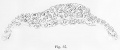
==VII. Somites, Protovertebrae and their derivatives: myotome, sclerotome; head cavities== | ==VII. Somites, Protovertebrae and their derivatives: myotome, sclerotome; head cavities== | ||
a) The somites, protovertebrae (somite) | a) The somites, protovertebrae (somite) | ||
<gallery> | |||
File:Kollmann070.jpg | |||
File:Kollmann071.jpg | |||
File:Kollmann072.jpg | |||
File:Kollmann073.jpg | |||
File:Kollmann074.jpg | |||
</gallery> | |||
* The first unit of the vertebra and the emergence of its derivatives, the myotome and the sclerotome | * The first unit of the vertebra and the emergence of its derivatives, the myotome and the sclerotome | ||
| Line 56: | Line 142: | ||
b) head cavities and somites of the head, somites | b) head cavities and somites of the head, somites | ||
<gallery> | |||
File:Kollmann075.jpg | |||
File:Kollmann076.jpg | |||
File:Kollmann077.jpg | |||
</gallery> | |||
e) mesenchyme | |||
==VIII. The boundaries of the bead== | ==VIII. The boundaries of the bead== | ||
<gallery> | |||
File:Kollmann078.jpg | |||
File:Kollmann079.jpg | |||
File:Kollmann080.jpg | |||
File:Kollmann081.jpg | |||
File:Kollmann082.jpg | |||
File:Kollmann083.jpg | |||
File:Kollmann084.jpg | |||
</gallery> | |||
{{Glossary}} | {{Glossary}} | ||
{{Footer}} | {{Footer}} | ||
[[Category:Historic Embryology]] | |||
Latest revision as of 13:41, 3 January 2012

Historic Textbook - Atlas of the Development of Man Volume 1 (1907)
- (Handatlas der entwicklungsgeschichte des menschen: Volume 1)
- Kollmann Atlas 1: Predevelopment | Ontogeny | Fetal membranes | Body shape | Systems and organs | Kollmann Atlas 1 | Kollmann Atlas 2 | Julius Kollmann
| Historic Disclaimer - information about historic embryology pages |
|---|
 |
- This text is a Google translate computer generated translation and may contain many errors.
Ontogeny, Blastogenesis
- Kollmann Atlas 1: Predevelopment | Ontogeny | Fetal membranes | Body shape | Systems and organs | Kollmann Atlas 1 | Kollmann Atlas 2 | Julius Kollmann
I. Cleavage, Segmentation
- Kollmann015.jpg
- Kollmann016.jpg
- Kollmann017.jpg
- Kollmann018.jpg
- Kollmann019.jpg
- Kollmann020.jpg
a) External appearances of cleavage
- Kollmann021.jpg
- Kollmann022.jpg
b) Internal appearances of cleavage, mitosis
- Kollmann023.jpg
- Kollmann024.jpg
- Kollmann025.jpg
- Kollmann026.jpg
- Kollmann027.jpg
- Kollmann028.jpg
- Kollmann029.jpg
- Kollmann030.jpg
- Occurrence and transformation of the dark chromatic nuclear division figure
- Occurrence and transformation of the achromatic (colorless) core figure, called the mitotic spindle
- Radiations in the yolk
c) Amitosis or division of cells without the appearance of cell division
d) Growth and regeneration in conjunction with the process of cell division
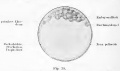
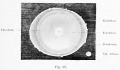
II. Germinal vesicle (vesicula blastodermica) with the germinative (area embryonalis), viewed from outside
- Kollmann035.jpg


III. Germinal vesicle internal structure
a) Theory of endoderm origin and the gastrulation
b) Germ layer, blastosphere and blastula. Homology of the primary germ layers
The Fundamental organs


IV. Primitive streak and neurenteric canal


V. Notochord
- Front and rear end of the notochord
- Sub-chordal line
- Origin of the notochord


VI. Middle germ layer, mesoderm
a) root zone and parietal zone


b) the origin of the middle germ layer


c) Histogenetic significance of germ layers
d) Homology of the middle germ
e) The term germ layers


VII. Somites, Protovertebrae and their derivatives: myotome, sclerotome; head cavities
a) The somites, protovertebrae (somite)


- The first unit of the vertebra and the emergence of its derivatives, the myotome and the sclerotome
- Special features of the trunk myotomes
b) head cavities and somites of the head, somites


e) mesenchyme
VIII. The boundaries of the bead


Glossary Links
- Glossary: A | B | C | D | E | F | G | H | I | J | K | L | M | N | O | P | Q | R | S | T | U | V | W | X | Y | Z | Numbers | Symbols | Term Link
Cite this page: Hill, M.A. (2024, April 18) Embryology Atlas of the Development of Man 1 - Part 2. Retrieved from https://embryology.med.unsw.edu.au/embryology/index.php/Atlas_of_the_Development_of_Man_1_-_Part_2
- © Dr Mark Hill 2024, UNSW Embryology ISBN: 978 0 7334 2609 4 - UNSW CRICOS Provider Code No. 00098G