Amniotic Cavity Development Movie: Difference between revisions
mNo edit summary |
mNo edit summary |
||
| Line 11: | Line 11: | ||
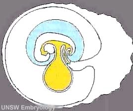
* '''yellow''' - outside embryo [[ | * '''yellow''' - outside embryo [[#yolk sac|yolk sac]] (circular balloon) and inside embryo gastrointestinal tract regions (foregut, midgut and hindgut) | ||
* '''blue''' - [[A#amniotic cavity|amniotic cavity]] | * '''blue''' - [[A#amniotic cavity|amniotic cavity]] | ||
* '''white''' - [[C#chorionic cavity|chorionic cavity]] surrounding amnion and yolk sac | * '''white''' - [[C#chorionic cavity|chorionic cavity]] surrounding amnion and yolk sac | ||
| Line 21: | Line 21: | ||
|- | |- | ||
|} | |} | ||
==Terms== | |||
===yolk sac=== | |||
An extraembryonic membrane which is endoderm origin and covered with extraembryonic mesoderm. Yolk sac lies outside the embryo connected initially by a yolk stalk to the midgut with which it is continuous with. The endodermal lining is continuous with the endoderm of the gastrointestinal tract. The extra-embryonic mesoderm differentiates to form both blood and blood vessels of the vitelline system. In reptiles and birds, the yolk sac has a function associated with nutrition. In mammals the yolk sac acts as a source of primordial germ cells and blood cells. Note that in early development (week 2) a structure called the "primitive yolk sac" forms from hypoblast, this is an entirely different structure. | |||
[[Category:Coelomic Cavity]] | [[Category:Coelomic Cavity]] | ||
{{Movie footer}} | {{Movie footer}} | ||
Revision as of 08:25, 27 April 2013
| Embryology - 24 Apr 2024 |
|---|
| Google Translate - select your language from the list shown below (this will open a new external page) |
|
العربية | català | 中文 | 中國傳統的 | français | Deutsche | עִברִית | हिंदी | bahasa Indonesia | italiano | 日本語 | 한국어 | မြန်မာ | Pilipino | Polskie | português | ਪੰਜਾਬੀ ਦੇ | Română | русский | Español | Swahili | Svensk | ไทย | Türkçe | اردو | ייִדיש | Tiếng Việt These external translations are automated and may not be accurate. (More? About Translations) |
| <mediaplayer width='268' height='260' image="http://embryology.med.unsw.edu.au/embryology/images/e/e1/Amnion_001_icon.jpg">File:Amnion 001.mp4</mediaplayer> | Note that as the yolk sac (yellow) is continuous with the midgut you can also follow development of the gastrointestinal tract regions of foregut, midgut and hindgut. Embryo and placental membranes are shown to the left and the developing placenta is shown to the right.
|
Terms
yolk sac
An extraembryonic membrane which is endoderm origin and covered with extraembryonic mesoderm. Yolk sac lies outside the embryo connected initially by a yolk stalk to the midgut with which it is continuous with. The endodermal lining is continuous with the endoderm of the gastrointestinal tract. The extra-embryonic mesoderm differentiates to form both blood and blood vessels of the vitelline system. In reptiles and birds, the yolk sac has a function associated with nutrition. In mammals the yolk sac acts as a source of primordial germ cells and blood cells. Note that in early development (week 2) a structure called the "primitive yolk sac" forms from hypoblast, this is an entirely different structure.
Glossary Links: A | B | C | D | E | F | G | H | I | J | K | L | M | N | O | P | Q | R | S | T | U | V | W | X | Y | Z | Numbers | Symbols | Movies
Cite this page: Hill, M.A. (2024, April 24) Embryology Amniotic Cavity Development Movie. Retrieved from https://embryology.med.unsw.edu.au/embryology/index.php/Amniotic_Cavity_Development_Movie
- © Dr Mark Hill 2024, UNSW Embryology ISBN: 978 0 7334 2609 4 - UNSW CRICOS Provider Code No. 00098G