Advanced - Valve Development: Difference between revisions
No edit summary |
No edit summary |
||
| Line 2: | Line 2: | ||
{{Advanced Cardiac menu}} | {{Advanced Cardiac menu}} | ||
{{Template:Cardiac_modules}} | {{Template:Cardiac_modules}} | ||
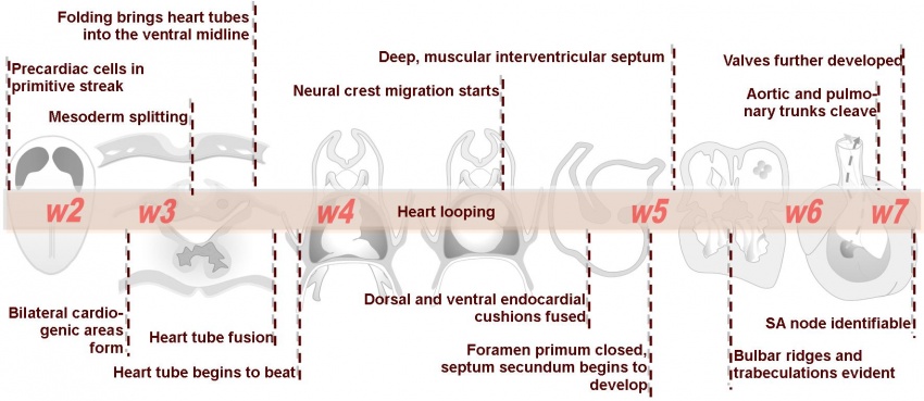
[[Image:HeartILP_draft_atimeline.jpg|center|850px]] | |||
Revision as of 11:49, 5 November 2009
| Begin Advanced | Heart Fields | Heart Tubes | Cardiac Looping | Cardiac Septation | Outflow Tract | Valve Development | Cardiac Conduction | Cardiac Abnormalities | Molecular Development |
| Cardiac Embryology | Begin Basic | Begin Intermediate | Begin Advanced |
Many of the mechanisms involved in the development of cardiac valves, particularly the process of epithelial to mesenchymal transformation (EMT), are poorly understood. Much of valve formation revolves around expansion of endocardial cushion tissue, yet limitation of cell proliferation is vital in ensuring the cushion swellings can be remodelled to form thin sheets. EMT which allows for cushion proliferation is limited by neurofibromin acting through the inhibition of Ras signalling as well as Smad6 which interferes with TGFβ signalling.
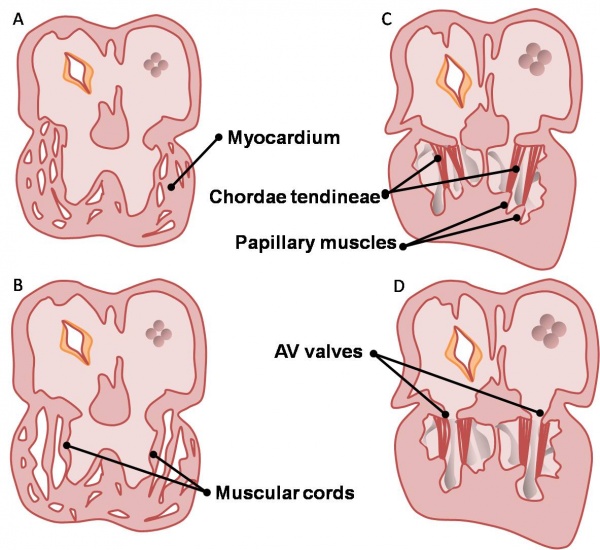
The AV valves begin to form between the fifth and eighth weeks of development. The valve leaflets are attached to the ventricular walls by thin fibrous chords, the chordae tendineae, which insert into small muscles attached to the ventricle wall, the papillary muscles. These structures are sculpted from the ventricular wall. The left AV valve has anterior and posterior leaflets and is termed the bicuspid or mitral valve. The right AV valve has a third, small, septal cusp and thus is called the tricuspid valve. These concepts are depicted on the right.
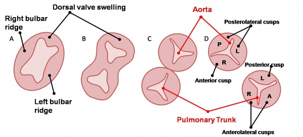
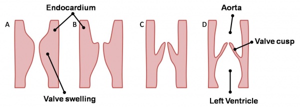
The aortic and pulmonary valves, termed the semilunar valves, are formed from the bulbar ridges and subendocardial valve tissue. The primordial semilunar valve consists of a mesenchymal core covered by endocardium. Excavation occurs, thinning the valve tissue thus creating its final shape (see left). These valves form the four valves of the adult heart, depicted below. The mechanisms of valve remodelling in these final steps in both the AV and semilunar valves are not fully understood yet are thought to involve apoptotic pathways.
| Back to Outflow Tract | Next: Conduction System | |
| Go to this section in the intermediate level |


