Adrenal Medulla Development Movie: Difference between revisions
mNo edit summary |
mNo edit summary |
||
| Line 1: | Line 1: | ||
{{Movie header}} | {{Movie header}} | ||
{| | {| | ||
| <mediaplayer width='720' height='560' image="http://embryology.med.unsw.edu.au/embryology/images/ | | <mediaplayer width='720' height='560' image="http://embryology.med.unsw.edu.au/embryology/images/b/b5/Adrenal_medulla.jpg">File:Adrenal medulla.mp4</mediaplayer> | ||
|- | |- | ||
| [[File:Adrenal medulla.jpg|thumb]] | | [[File:Adrenal medulla.jpg|thumb]] | ||
Revision as of 15:17, 16 March 2013
| Embryology - 16 Apr 2024 |
|---|
| Google Translate - select your language from the list shown below (this will open a new external page) |
|
العربية | català | 中文 | 中國傳統的 | français | Deutsche | עִברִית | हिंदी | bahasa Indonesia | italiano | 日本語 | 한국어 | မြန်မာ | Pilipino | Polskie | português | ਪੰਜਾਬੀ ਦੇ | Română | русский | Español | Swahili | Svensk | ไทย | Türkçe | اردو | ייִדיש | Tiếng Việt These external translations are automated and may not be accurate. (More? About Translations) |
| <mediaplayer width='720' height='560' image="http://embryology.med.unsw.edu.au/embryology/images/b/b5/Adrenal_medulla.jpg">File:Adrenal medulla.mp4</mediaplayer> |
|
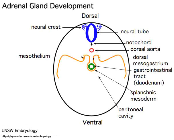
Adrenal Medulla Development This animation shows the dual embryonic origins of the adrenal development. Adrenal Medulla
|

Medulla
Neural crest cells migrate toward the coelomic cavity wall and form the adrenal medulla. These chromaffin (chromaphil) cells originally named because of their staining (yellow) with chromium salts.
Cortex
Week 4 celomic epithelium (mesothelium) cells proliferate initially forming small buds that separate from the epithelium.
Week 6 these now mesenchymal cells first form the fetal adrenal cortex which will be later replaced by the adult cortex.
Glossary Links: A | B | C | D | E | F | G | H | I | J | K | L | M | N | O | P | Q | R | S | T | U | V | W | X | Y | Z | Numbers | Symbols | Movies
Cite this page: Hill, M.A. (2024, April 16) Embryology Adrenal Medulla Development Movie. Retrieved from https://embryology.med.unsw.edu.au/embryology/index.php/Adrenal_Medulla_Development_Movie
- © Dr Mark Hill 2024, UNSW Embryology ISBN: 978 0 7334 2609 4 - UNSW CRICOS Provider Code No. 00098G