Adipose Tissue Development

From Embryology
Revision as of 21:51, 21 June 2016 by Z8600021 (talk | contribs)
Embryology - 18 Apr 2024    Facebook link Pinterest link Twitter link  Expand to Translate  
Google Translate - select your language from the list shown below (this will open a new external page)

العربية | català | 中文 | 中國傳統的 | français | Deutsche | עִברִית | हिंदी | bahasa Indonesia | italiano | 日本語 | 한국어 | မြန်မာ | Pilipino | Polskie | português | ਪੰਜਾਬੀ ਦੇ | Română | русский | Español | Swahili | Svensk | ไทย | Türkçe | اردو | ייִדיש | Tiếng Việt    These external translations are automated and may not be accurate. (More? About Translations)

Introduction

Brown adipose tissue

Draft Page- notice removed when completed.

Connective tissues in the body have a mesoderm origin, while in the head neural crest also contributes to these tissues.

This topic is also covered in musculoskeletal (Tendon Development), integumentary (Integumentary Development) and endocrine development (Adipose Tissue).

Blood is a liquid connective tissue (More? Blood Development).


  • Loose and dense connective tissue
  • Reticular connective tissue
  • Adipose Tissue
  • Mesenchymal connective tissue


Connective Tissue: adipose | tendon | integumentary

Some Recent Findings

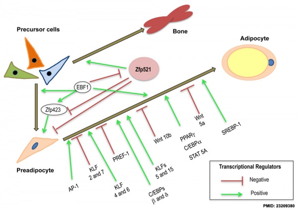
  • The fat controller: adipocyte development[1] "Obesity is a condition characterized by excess adipose tissue that results from positive energy balance and is the most common metabolic disorder in the industrialized world. ... Adipocytes are not created from other adipocytes, but they arise from precursor cells. In the last two decades, scientists have discovered the function of many proteins that influence the ability of precursor cells to become adipocytes. If the expansion of the adipose tissue is the problem, it seems logical that adipocyte development inhibitors could be a viable anti-obesity therapeutic. However, factors that block adipocyte development and limit adipocyte expansion also impair metabolic health. This notion may be counterintuitive, but several lines of evidence support the idea that blocking adipocyte development is unhealthy. For this reason it is clear that we need a better understanding of adipocyte development."
  • Brown adipose tissue: function and physiological significance[2] "The function of brown adipose tissue is to transfer energy from food into heat; physiologically, both the heat produced and the resulting decrease in metabolic efficiency can be of significance. ... The development of brown adipose tissue with its characteristic protein, uncoupling protein-1 (UCP1), was probably determinative for the evolutionary success of mammals, as its thermogenesis enhances neonatal survival and allows for active life even in cold surroundings."


Molecular Development

Adipocyte differentiation regulation 01.jpg

Adipocyte differentiation regulation.[1]

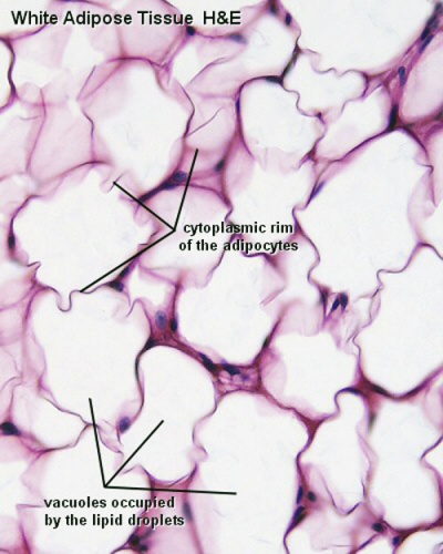
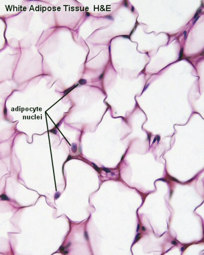
White Adipose

White adipose 01.jpg White adipose 02.jpg

Brown Adipose

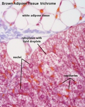
Brown adipose tissue
  • Brown Adipose Tissue (BAT) arises from progenitor cells that also give rise to skeletal muscle,
  • Brown adipocytes have numerous small lipid droplets rather than a single large one as in white adipocytes
  • Elevated numbers of mitochondria
    • mitochondrial expression of the nuclear gene UCP1, the uncoupler of oxidative phosphorylation responsible for non-shivering thermogenesis.

Development Overview

Mesoderm Development

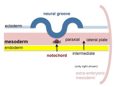
Mesoderm cartoon 01.jpg Cells migrate through the primitive streak to form mesodermal layer. Extraembryonic mesoderm lies adjacent to the trilaminar embryo totally enclosing the amnion, yolk sac and forming the connecting stalk.
Mesoderm cartoon 02.jpg Paraxial mesoderm accumulates under the neural plate with thinner mesoderm laterally. This forms 2 thickened streaks running the length of the embryonic disc along the rostrocaudal axis. In humans, during the 3rd week, this mesoderm begins to segment. The neural plate folds to form a neural groove and folds.
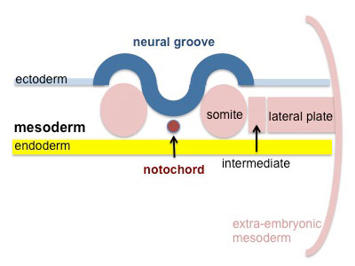
Mesoderm cartoon 03.jpg Segmentation of the paraxial mesoderm into somites continues caudally at 1 somite/90minutes and a cavity (intraembryonic coelom) forms in the lateral plate mesoderm separating somatic and splanchnic mesoderm.

Note intraembryonic coelomic cavity communicates with extraembryonic coelom through portals (holes) initially on lateral margin of embryonic disc.

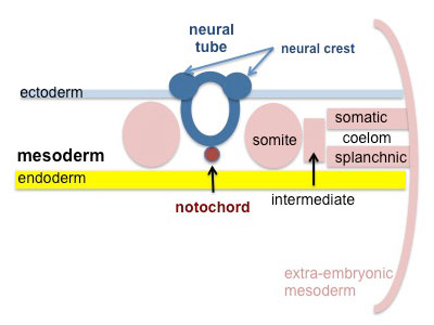
Mesoderm cartoon 04.jpg Somites continue to form. The neural groove fuses dorsally to form a tube at the level of the 4th somite and "zips up cranially and caudally and the neural crest migrates into the mesoderm.


Somite - Dermatome

The dermis and hypodermis layers of the skin.

Somatic Mesoderm

The body wall connective tissue.

Splanchnic Mesoderm

The lamina propria and submucosa layers of the gastrointestinal tract wall.

References

  1. 1.0 1.1 <pubmed>23209380</pubmed>| PMC3507952 | PLoS Biol.
  2. <pubmed>14715917</pubmed>


Reviews

<pubmed>21372557</pubmed> <pubmed>19896888</pubmed> <pubmed>19188249</pubmed> <pubmed>18793119</pubmed> <pubmed>14715917</pubmed>

Articles

<pubmed>20678241</pubmed> <pubmed>17507398</pubmed>

Search PubMed

Search Pubmed: adipose Development

Additional Images

Terms

Glossary Links

Glossary: A | B | C | D | E | F | G | H | I | J | K | L | M | N | O | P | Q | R | S | T | U | V | W | X | Y | Z | Numbers | Symbols | Term Link

Cite this page: Hill, M.A. (2024, April 18) Embryology Adipose Tissue Development. Retrieved from https://embryology.med.unsw.edu.au/embryology/index.php/Adipose_Tissue_Development

What Links Here?
© Dr Mark Hill 2024, UNSW Embryology ISBN: 978 0 7334 2609 4 - UNSW CRICOS Provider Code No. 00098G