ANAT3411 Neuroanatomy
From Embryology
Introduction
Course convenor: Dr. Elizabeth Tancred
The aim of this course is to provide students in the BSc and BMedSc programs with a basic understanding of the structural organisation of the human central nervous system in sufficient depth to form the basis for further clinical or research studies of the nervous system.
The following images are prepared for Dr Tancred's Neurodevelopment class from UNSW Embryology. The listed cross-sections are recommended to be viewed in the order in which they are shown below.
Embryo Stage 22
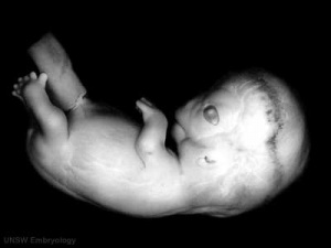
Carnegie stage 22 Week 8 (27 mm Embryo)
| Series | Unlabeled | Labeled |
| D6 spinal cord | 
|

|
| A1 head and brain | 
|

|

Embryo Stage 13
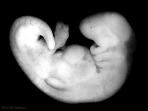
Carnegie stage 13 Week 4 (6 mm Embryo)
| Series | Unlabeled | Labeled |
| G6L Midline longitudinal | 
|

|
| G7L Lateral longitudinal | 
|

|
| A3L Rhombomeres and otic vesicle | 
|

|
| B4L Spinal cord and optic vesicle | 
|

|
| B5L Spinal cord and diencephalon | 
|

|
Embryo Stage 22
Carnegie stage 22 Week 8 (27 mm Embryo)
| Series | Unlabeled | Labeled |
| A1L | 
|

|
| A3L | 
|

|
| A4L | 
|

|
| A6L | 
|

|
| B1L | 
|

|
| B2L | 
|

|
| B3L | 
|

|
| B4L | 
|

|
| B5L | 
|

|
| B6L | 
|

|
| B7L | 
|

|
| C1L | 
|

|
| C2L | 
|

|
| C3L | 
|

|
Additional Information
| Additional Information - Content shown under this heading is not part of the material covered in this class. It is provided for those students who would like to know about some concepts or current research in topics related to the current class page. |
Scanning Electron Microscopy
Stage 10 - Neural Groove


Stage 11 - Cut through the neural tube.
Neural Movies
|
|
|
| ||||||||||||
|
|
|
Glossary Links
- Glossary: A | B | C | D | E | F | G | H | I | J | K | L | M | N | O | P | Q | R | S | T | U | V | W | X | Y | Z | Numbers | Symbols | Term Link
Cite this page: Hill, M.A. (2024, April 16) Embryology ANAT3411 Neuroanatomy. Retrieved from https://embryology.med.unsw.edu.au/embryology/index.php/ANAT3411_Neuroanatomy
- © Dr Mark Hill 2024, UNSW Embryology ISBN: 978 0 7334 2609 4 - UNSW CRICOS Provider Code No. 00098G