ANAT2511 Introduction to Histology: Difference between revisions
| Line 19: | Line 19: | ||
==Blood Smear== | ==Blood Smear== | ||
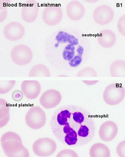
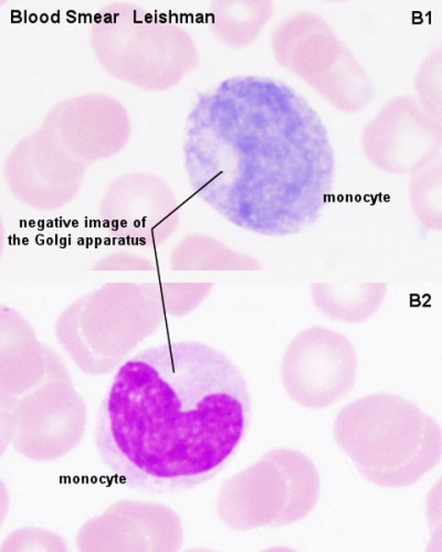
[[File:Neutrophil 01.jpg|400px]] | [[File:Neutrophil 01.jpg|400px]] [[File:Monocyte_01.jpg|400px]] | ||
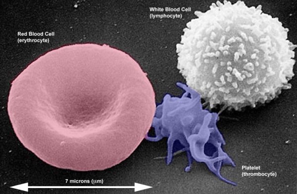
The following image is a scanning electron microscope image of a red blood cell (erythrocyte), a platelet (thrombocyte) and a white blood cell (lymphocyte). | The following image is a scanning electron microscope image of a red blood cell (erythrocyte), a platelet (thrombocyte) and a white blood cell (lymphocyte). | ||
Revision as of 09:35, 20 February 2013

| ANAT2511 Fundamentals of Anatomy This histology practical support page content is not part of the histology practical class and provides only background information for student self-directed learning purposes. Histology lecturer notice. |
Objectives
- To learn how to use virtual microscopy for studies in histology and to recognise parts of a cell and common features of cells and tissues.
- To recognise common features and differences between cells (e.g. size, shape, nuclear appearance, secretory components, microvilli and cilia).
Reading: Human Anatomy, Marieb et al., 6th ed., pages 23‐46
Learning Activities
- Examine the following virtual microscope slides and find examples of cells of different sizes and shapes.
- Virtual Microscopy - Cells and Types of Tissue (6 slides)
- The images are located in the ANAT2511 folder on the computer desktop.
- Identify examples of the following cellular components: nucleus, nucleolus, densely packed rough endoplasmic reticulum (rough ER), microvilli, and cilia.
Blood Smear
The following image is a scanning electron microscope image of a red blood cell (erythrocyte), a platelet (thrombocyte) and a white blood cell (lymphocyte).
- Electron microscopes have a higher resolution than a light microscope (virtual slides).
- Image shows the relative sizes and shapes (morphologies).
- Remember a platelet (thrombocyte) is not a cell, but circulating part or a fragment of a cell.
Course Links
- Histology Glossary: A | B | C | D | E | F | G | H | I | J | K | L | M | N | O | P | Q | R | S | T | U | V | W | X | Y | Z | ANAT2241 Support | Histology | Histology Stains | Embryology Glossary
Virtual slides
- Pages require student zpass to access.
ANAT2511 | Virtual Microscopy, Cells and Types of Tissue | Selected Basic Tissues | Histology of Bone and Joints | Histology of Muscle | Histology of Nervous Tissue | Integumentary System | Circulatory System Histology | Respiratory System Histology | GIT Histology | Urinary System Histology
Practical support
- Pages can be accessed from any internet connected computer.
ANAT2511: Practical 1 Introduction to Histology | Practical 3 Basic Tissues | Practical 5 Bones and Joints | Practical 7 Muscle Tissue | Practical 9 Nervous Tissue | Practical 11 Integumentary (Skin) System | Practical 13 Circulatory System | Practical 15 Respiratory System | Practical 17 Gastro‐intestinal Tract, Liver and Gallbladder | Practical 19 Urinary System | Histology Drawings


