ANAT2511 Bones and Joints: Difference between revisions
From Embryology
| Line 18: | Line 18: | ||
{| | {| | ||
| valign=top|[[File:Bone histology 101.jpg| | | valign=top|[[File:Bone histology 101.jpg|350px]] | ||
| valign=top|[[File:Bone histology 201.jpg| | | valign=top|[[File:Bone histology 201.jpg|350px]] | ||
|} | |} | ||
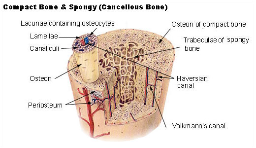
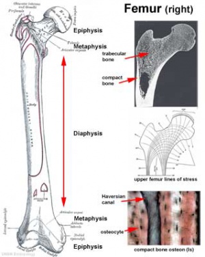
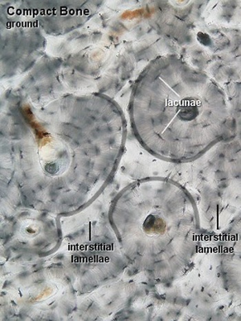
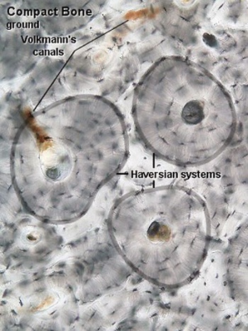
Individual bone forming cells ('''osteocytes''') live in the spaces ('''lacunae''') with the bone matrix surrounding a central blood vessel and nerve filled space ('''Haversian canal''') that provides nutrition to these cells. | Individual bone forming cells ('''osteocytes''') live in the spaces ('''lacunae''') with the bone matrix surrounding a central blood vessel and nerve filled space ('''Haversian canal''') that provides nutrition to these cells. | ||
Revision as of 11:33, 24 February 2013
General Objective
To recognise bone in histological sections and understand its microscopic structure. To know the histology of a synovial joint and a symphysis joint (intervertebral disc) and the function of different tissues within the joints.
|
|
|
Haversian System
|
|
Individual bone forming cells (osteocytes) live in the spaces (lacunae) with the bone matrix surrounding a central blood vessel and nerve filled space (Haversian canal) that provides nutrition to these cells.
